The signal peptidase II (Isp) gene of Bacillus subtilis
7
Printed in Great Britain The signal peptidase II (Isp) gene of Bacillus subtilis Zoltán Prágai, t Harold Tjalsrna, Albert Bolhuis, Jan Maarten van Dijl, Gerard Venerna and Sierd Bron Author for correspondence: Sierd Bron. Tel: +31503632105. Fax: +31503632348. e-mail: [email protected] Department of Genetics, University of Groningen, Groningen Biomolecular Sciences and Biotechnology Institute, Kerklaan 30, 9751 NN Haren, The Netherlands The gene encoding the type II signal peptidase (SPase 11) of Bacillus subti/is was isolated by screening a genomic DNA library of this bacterium for the ability to increase the levels of globomycin resistance in Escherichia co/i, and to complement the growth deficiency at the non-permissive temperature of E. co/i strain Y81 S carrying a temperature-sensitive mutation in its /sp gene for SPase II. The deduced amino acid sequence of the B. subti/is SPase II showed significant similarity with those of other known SPase II enzymes. Activity of the B. subti/is SPase II was demonstrated bya pulse-labelling experiment in E. co/i. In B. subti/is, the /sp gene is flanked by the isoleucyl-tRNA synthetase (i/eS) gene and the pyrimidine biosynthetic (pyr) gene cluster, which is known to map at 139° of the chromosome. In the Gram-positive bacteria studied thus far, /sp appears to be the first gene in an operon. The promoter-distal gene (' orf4 ') of this operon specifies a hypothetical protein in bacteria and yeast. INTRODUCTION Finally, the apolipoproteins are further modified by N- fatty acylation of the diacylglyceryl-cysteine amino group to yield mature lipoproteins (Tokunaga et al., 1982). Signal peptidases are integral membrane proteins which remove signal peptides from precursors of secretory proteins. In bacteria, three types of signal peptidases are known (for a review see von Heijne, 1994). The type II signal peptidases (SPaseII ; EC 3.4 .23 .36), or prolipo- protein signal peptidases (Lsp) , specifically remove signal peptides from glyceride-modified prolipoproteins during their translocation across the membrane (Sankaran & Wu, 1994). Prior to the processing of lipoprotein precursors by SPase II, the Cys residue at the + 1 position (relative to the processing site) of these proteins is modified by a diacylglyceryl moiety (Giam et al., 1984). The lipomodified precursor proteins are cleaved by SPase II on the amino-terminal side of the Cys residue, resulting in apolipoproteins and signal peptides. This step can be inhibited by the peptide antibiotic globomycin (Glm), which is a potent, reversible and noncompetitive inhibitor of SPase II (Inukai et al., 1978). lsp genes encoding SPase II have been identified and sequenced from Escherichia coli (Yamagata et al., 1983 ; Tokunaga et al., 1983), Pseudomonas fluorescens (Isaki et al., 1990a), Enterobacter aerogenes (Isaki et al., 1990b), Haemophilus influenzae (Fleischmann et al., 1995), Staphylococcus aureus (Zhao & Wu, 1992), Staphylococcus carnosus (Witke & Gotz, 1995), and Mycoplasma genitalium (Fraser et al., 1995). In Gram- negative bacteria, the lsp gene is part of the ileS-lsp operon, containing four other genes with unrelated functions, such as the isoleucyl-tRNA synthetase (ileS) gene (MilIer et al., 1987). In S. aureus, S. carnosus and M. genitalium, the lsp and ileS genes are not organized in one operon (Zhao & Wu, 1992; Fraser et al., 1995 ; Witke & Gotz, 1995). In Gram-positive bacteria, lipomodified proteins play important roles in: (i) the uptake of nutrients; (ii) sporulation and germination ; (iii) protein secreti6n; (iv) conferring resistance to certain antibiotics; and (v) targeting bacteria to different substrates, host tissues and bacteria via adhesins (Sutcliffe & Russell, 1995). To analyse the pathway for the export of lipoproteins in B. 1~:>7 t Present address : Department of Biotechnology and Molecular Genetics, Godol16 University of Agricultural Sciences, Godol16, H-2103, Hungary. Abbreviations: Ap, ampicillin; G lm, globomycin; SD, Shine-Dalgarno; Tc, tetracycline. The GenBank accession number for the sequence reported in this paper is U48870. 0002-1180 @ 1997 SGM
Transcript of The signal peptidase II (Isp) gene of Bacillus subtilis

Printed in Great Britain
The signal peptidase II (Isp) gene of Bacillus
subtilis
Zoltán Prágai, t Harold Tjalsrna, Albert Bolhuis, Jan Maarten van Dijl,
Gerard Venerna and Sierd Bron
Author for correspondence: Sierd Bron. Tel: +31503632105. Fax: +31503632348.e-mail: [email protected]
Department of Genetics,University of Groningen,Groningen BiomolecularSciences and BiotechnologyInstitute, Kerklaan 30,9751 NN Haren, TheNetherlands
The gene encoding the type II signal peptidase (SPase 11) of Bacillus subti/is wasisolated by screening a genomic DNA library of this bacterium for the ability toincrease the levels of globomycin resistance in Escherichia co/i, and tocomplement the growth deficiency at the non-permissive temperature of E.co/i strain Y81 S carrying a temperature-sensitive mutation in its /sp gene forSPase II. The deduced amino acid sequence of the B. subti/is SPase II showedsignificant similarity with those of other known SPase II enzymes. Activity ofthe B. subti/is SPase II was demonstrated bya pulse-labelling experiment in E.co/i. In B. subti/is, the /sp gene is flanked by the isoleucyl-tRNA synthetase(i/eS) gene and the pyrimidine biosynthetic (pyr) gene cluster, which is knownto map at 139° of the chromosome. In the Gram-positive bacteria studied thusfar, /sp appears to be the first gene in an operon. The promoter-distal gene(' orf4 ') of this operon specifies a hypothetical protein in bacteria and yeast.
INTRODUCTION Finally, the apolipoproteins are further modified by N-fatty acylation of the diacylglyceryl-cysteine aminogroup to yield mature lipoproteins (Tokunaga et al.,1982).
Signal peptidases are integral membrane proteins whichremove signal peptides from precursors of secretoryproteins. In bacteria, three types of signal peptidases areknown (for a review see von Heijne, 1994). The type IIsignal peptidases (SPase II ; EC 3.4 .23 .36), or prolipo-protein signal peptidases (Lsp) , specifically removesignal peptides from glyceride-modified prolipoproteinsduring their translocation across the membrane(Sankaran & Wu, 1994). Prior to the processing oflipoprotein precursors by SPase II, the Cys residue at the+ 1 position (relative to the processing site) of theseproteins is modified by a diacylglyceryl moiety (Giam etal., 1984). The lipomodified precursor proteins arecleaved by SPase II on the amino-terminal side of the Cysresidue, resulting in apolipoproteins and signal peptides.This step can be inhibited by the peptide antibioticglobomycin (Glm), which is a potent, reversible andnoncompetitive inhibitor of SPase II (Inukai et al., 1978) .
lsp genes encoding SPase II have been identified andsequenced from Escherichia coli (Yamagata et al., 1983 ;Tokunaga et al., 1983), Pseudomonas fluorescens (Isakiet al., 1990a), Enterobacter aerogenes (Isaki et al.,1990b), Haemophilus influenzae (Fleischmann et al.,1995), Staphylococcus aureus (Zhao & Wu, 1992),Staphylococcus carnosus (Witke & Gotz, 1995), andMycoplasma genitalium (Fraser et al., 1995). In Gram-negative bacteria, the lsp gene is part of the ileS-lspoperon, containing four other genes with unrelatedfunctions, such as the isoleucyl-tRNA synthetase (ileS)gene (MilIer et al., 1987). In S. aureus, S. carnosus andM. genitalium, the lsp and ileS genes are not organizedin one operon (Zhao & Wu, 1992; Fraser et al., 1995 ;Witke & Gotz, 1995).
In Gram-positive bacteria, lipomodified proteins playimportant roles in: (i) the uptake of nutrients; (ii)sporulation and germination ; (iii) protein secreti6n; (iv)conferring resistance to certain antibiotics; and (v)targeting bacteria to different substrates, host tissuesand bacteria via adhesins (Sutcliffe & Russell, 1995). Toanalyse the pathway for the export of lipoproteins in B.
1~:>7
t Present address : Department of Biotechnology and Molecular Genetics,
Godol16 University of Agricultural Sciences, Godol16, H-2103, Hungary.
Abbreviations: Ap, ampicillin; G lm, globomycin; SD, Shine-Dalgarno; Tc,
tetracycline.
The GenBank accession number for the sequence reported in this paper is
U48870.
0002-1180 @ 1997 SGM
subtilis, the cloning and characterization of the B.subtilis lsp gene [lsp(Bsu)] is desirable. SPase II is alsolikely to be important for the secretion of non-lipo-proteins, since the lipomodified protein PrsA is requiredfor the correct folding of secreted proteins after theirtranslocation across the membrane (Kontinen & Sarvas,1993).
In this article, we report the isolation and characteriza-tion of the Isp(Bsu) gene of B. subtilis 168. The deducedamino acid sequence of the corresponding SPase II wascompared with that of known type II SPases and thegenomic location of Isp(Bsu) was determined.
Ap and Tc. The overnight culture was diluted tenfold in M9medium-1, and grown for 3 h at 30 °C. Next, the cells werewashed and resuspended in M9 medium-2 (methionine- andcysteine-free medium) containing 0.6 mM IPTG, to induce theE. coli lpp gene on the resident plasmid pHYO01, andincubated for 30 min at 42 °C to inactivate the SPase II of E.coli Y815. Pulse-chase labelling of proteins with[35S]methionine (5 min, 42 °C), immunoprecipitation withserum against Braun's lipoprotein, tricine SDS-P AGE(Schagger & von Jagow, 1987) and f1uorography were carriedout as described by van Dijl et al. (1992).
Sequencing, data handling and sequence analysis. The DNAsequence was determined on both strands using the dideoxychain-termination procedure (Sanger et al., 1977). Cloneswere sequenced with universal, reverse and synthetic primersusing [35S]dATP-aS (Amersham) and the T7 polymerasesequencing kit (Pharmacia). For sequence assembly andanalysis of the sequences, the programs COMPARE, BESTFIT,PILEUP, PRETTY and GAP of the University of WisconsinGenetics Computer Group (GCG) software package(Devereux et al., 1984) were used. Database searches weredone using the BLAST program of the Kyoto-Centre, Japan(Altschul et al., 1990).
METHODS
RESULTS
Construction of a B. subtilis 168 gene library
B. subtilis 168 genomic DNA was partially digested withSau3AI and after size fractionation by agarose gelelectrophoresis, DNA fragments ranging from 1.5 to4 kb were isolated. The 5' sticky ends generated bySau3AI treatment were partially filled-in using KlenowDNA polymerase and dA TP plus dGTP. SalI-digestedpBluescript II KS( + ) vector DNA was similarly partiallyfi"ed-in with dTTP and dCTP. Chromosomal andvector DNA were ligated in a 1: 1 ratio. To prevent theloss of plasmids containing inserts which are difficult toclone in E. coli, the library was not amplified in E. coli.For the initial analysis of the library, E. coli DH5(X waselectrotransformed with 1 J.11 of the ligation mixture,containing 0.04 J.1g vector and 0.04 J.1g genomic DNA.Transformants were selected on TY agar mediumsupplemented with Ap, IPTG and X-Gal. Of the1.7 x 105 Ap-resistant (Apr) colonies obtained, 90.5 %were white, indicating that they carried potential re-combinant plasmids. The average size of the insertDNAs was 2.5 kb, as determined by EcoRI and XhoIdouble digestion of the plasmids purified from 30randomly chosen white colonies. Based on the averageinsert size and the known size of the B. subtilis genome(4. 2 Mb) , about 7700 white ( or 8500 Apr) colonies wouldbe necessary for a representative gene library (99 %probability of the presence of the desired fragment).
Screening of the library for the presence of theIsp(Bsu} gene
Since the (over-)expression of cloned lsp genes fromGram-negative and Gram-positive bacteria caused Glmresistance (Glmr) in E. coli (Isaki et al., 1990a, b; Zhao& Wu, 1992; Witke & G6tz, 1995), the B. subtilis 168
Bacterial strains. plasmids and media. E. coli DH5cx[F- /endAl hsdR17(r- rn+) supE44 thi-l recAl gyrA (Nalr)relAl ~(laclZY A-argF)U169 deoR (4>80dlac~(lacZ)M15)] andE. coli JMl10 [F' traD36 laclq ~(lacZ)M15 proA+B+ /rpsL(Strr) thr leu thi lacY galK galT ara fhuA dam dcm supE44~(lac-proAB)] were the hosts for cloning experiments. For thecomplementation analysis, E. coli strain Y815 was used,which has a temperature-sensitive SPase II, and which carriesan IPTG-inducible lpp gene for the major outer-membranelipoprotein, also known as Braun's lipoprotein, on the low-copy-number plasmid pHYO01. Plasmid pHYO01 also containsa tetracycline (Tc) resistance determinant (Yamagata et al.,1982). B. subtilis strain 168 (trpC2) from our laboratorycollection was used as a source of chromosomal DNA for theconstruction of a gene library in the phagemid pBluescript IIKS( + ) (Stratagene) vector. Plasmid pKSA11 and its deletionderivatives (pG1-pG5), constructed in pBluescript II KS( + ),contain various inserts from the B. subtilis lsp region (Fig. la).Strains were grown in TY medium, M9 medium-lor M9medium-2 (van Dijl et al., 1988) .If required, 100 f.lg ampicillin(Ap) mI-l, 100 f.lg Glm mI-l, 10 f.lg Tc mI-l, 0.6 mM IPTG and40 f.lg X-Gal mI-l were added.
DNA manipulations. Plasmid DNA preparation, restrictionendonuclease digestion, ligation, filling-in of cohesive ends,agarose gel electrophoresis and transformation of electro-competent E. coli cells were carried out as described bySambrook et al. (1989). Total DNA of B. subtilis 168 wasextracted according to Bron (1990). The genomic library of B.subtilis 168 was constructed by the method of partial filling-inof cohesive ends (Zabarovsky & Allikmets, 1986). Enzymesand deoxynucleotides were purchased from Boehringer andPharmacia.
Complementation test for SPase II activity in E. co/i. Thegrowth of E. coli strain Y815 is sensitive to IPTG addition athigh temperature due to the accumulation of lipid-modifiedprolipoprotein (Yamagata et al., 1982). This strain waselectrotransformed with plasmid DNAs. Dilutions were platedin two series on AB3 agar medium (Difco) supplemented with100 f.lg Ap mi-l, 10 f.lg Tc mI-l and 0.6 mM IPTG, and theplates were incubated for 4 d at either the permissive (30 °C),or the non-permissive (42 °C) temperature for growth. Trans-formants carrying the lsp-containing plasmid DNA were ableto grow at 42 °C in the presence of IPTG. Cloning vectorpBluescript II KS( + ) was the negative control.
Assay for SPase II activity in E. co/i. E. coli Y815 wastransformed with pBluescript II KS( + ), pKSA11 or pG3, andtransformants were grown at 30 °C in AB3 medium containing
Signa! peptidase II of Bacillus subtilis
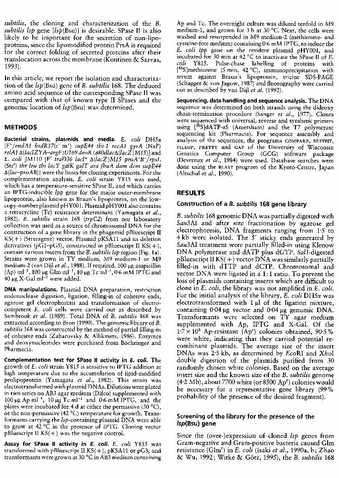
{a}Fig. ,. (a) Restriction map and deletionanalysis of the 3.3 kb insert in pKSA 11.Deletion derivatives of pKSA 11, designatedpG1-pG5, were generated with restrictionenzymes. The fragments still present inpG1-pG5 are shown below the map. For thelocalization of the Isp gene, the growth ofE. co/i DH5(X transformants with thedifferent deletion derivatives was tested onTV agar medium containing 100 l-Ig G lm mi-l(+, growth; -, no growth). The test forcomplementation of the temperature-sensitive (ts) mutation in the Isp gene of E.co/i Y815 was carried out in the presence ofIPTG at 42 0( after transformation with thevarious deletion derivatives of pKSA 11 ( + ,complementation; -, no complementation).Vector pBluescript II KS( + ) served as anegative col"!trol, and pKSA 11 as a positivecontrol. (b) ORFs (shown as heavy arrows)on the 3.3 kb insert in pKSA11. T, putativetranscription terminator.
Glm ts Isp
assay ~Plasmids
Hind1Il,61, Sac I,322
: Hind!II,417: BglII,472
, , , BclI,720
, : , EcoRV,IO57, BclI,1293, Cl I 49, , , , , , a ,1 2
B II 1831, , , , c ,, "' , , " 'SmaI1967: " : , , : :Sm~I,2047""' ""; ClaI2066; ; ,1111111I 1111pKSAll
pGl
pG2
pG3
pG4
pG5
(b)T
ileS oif2 Isp oif4 pyrR
orj7 01115
gene library was screened for this property. The minimalinhibitory concentration of Glm for E. coli DH5cx is30 !!g mI-l. For the screening of the library, 100 !!g GlmmI-l was used. E. coli DH5cx was electrotransformedwith an amount of DNA from the gene library, sufficientto obtain about 7 x 10⁵ Apr transformants, and thetransformation mixture was plated on TY agar mediumcontaining Glm and Ap. Seventeen Glmr transformantswere obtained. Plasmids purified from these trans-formants were used for electrotransformation of E. colistrain Y815, which has a temperature-sensitive Spase IIand an IPTG-inducible lpp gene. The resulting trans-formants were tested for complementation of the growthdeficiency at 42 °C {non-permissive temperature) in thepresence of IPTG for 4 d. Under these conditions, all E.coli Y815 transformants containing plasmids from the17 Glmr transformants were able to grow, indicatingthat each of these plasmids carried an lsp gene for afunctional SPase II protein. As shown by restrictionanalysis and PCR, the 17 recombinant plasmids con-tained the same insert on overlapping Sau3AI fragments,the sizes of which ranged from H to 3.3 kb {data notshown). For further analysis, one of these plasmids,pKSA11, was chosen which carried the largest {3.3 kb)insert. Also, pKSA11 gave the largest colonies in thecomplementation tests suggesting that, compared to theother plasmids, pKSA11 resulted in the best expressionof the cloned lsp gene.
taining the 1.4 kb BgIIl-BcII fragment of pKSA11. Thisindicates that the Isp(Bsu) gene is located on thisfragment. Direct evidence for this conclusion will beprovided in the next section (see Fig. 4) in which an invivo assay for SPase II activity is described.
The nucleotide sequence of the entire 3.3 kb insert wasdetermined on both strands using subclones and syn-thetic primers. ORFs are shown in Fig. l(b). Theputative Isp(Bsu) gene was identified in the middle partof the insert. This gene comprises 462 nucleotides andcan encode a protein of 154 amino acid residues with amolecular mass of 17395 Da. A Shine-Dalgarno (SD)sequence, AcuGGAGGaacg, was identified three nucleo-tides upstream of the putative GUG start codon. Thededuced amino acid sequence of the B. subtilis SPase IIwas compared with that of the corresponding enzymesfrom several other bacteria. A multiple alignment isshown in Fig. 2. The levels of the amino acid identity(and similarity) were: s. carnosus, 50.0% (73.3%); S.aureus, 44.8% (72.1% ) ; P. fluorescens, 36.8% (63.2% ) ;H. influenzae, 36.2% (66.5%) ;E. coli, 34.2% (64.4%) ;E. aerogenes, 34.0% (65.3%); and M. genitalium,22.3% (58.1%).
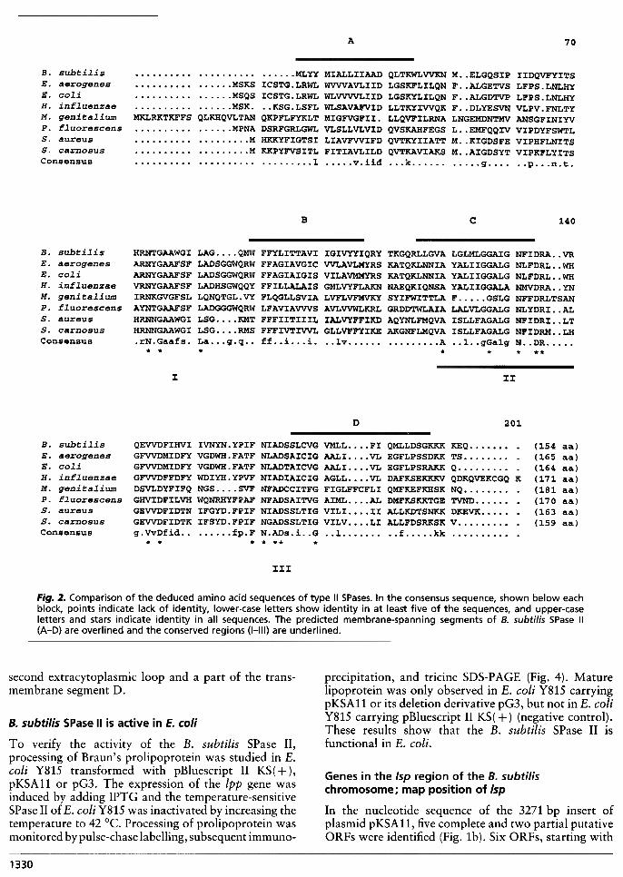
The hydropathy profile of the B. subtilis SPase II wasdetermined as described by Kyte & Doolittle (1982) andfour membrane-spanning segments (A-D) could bedistinguished (Fig. 2). Following the positive-inside rule(von Heijne, 1992), a model for the membrane topologyof the B. subtilis SPase II can be proposed which isshown in Fig. 3. Three conserved regions (I, II and III)were revealed by the amino acid sequence alignmentshown in Fig. 2. According to the model for themembrane topology (Fig. 3), region I would be locatedin the carboxy-terminal part of the first extracytoplasmicloop. Region II would correspond to the membrane-spanning segment C and the amino-terminal part of thesecond extracytoplasmic loop, while region III wouldcorrespond to the carboxy-terminal portion of the
Nucleotide sequence determination and analysis ofthe B. subtilis Isp gene
Various restriction endonucleases were used to constructa map of the 3-3 kb insert in plasmid pKSAll (Fig. la).Based on this map, deletion derivatives (plasmidspGl-pG5) were constructed to delineate the part of theinsert required for complementing the growth defect ofE. coli Y815 at 42 °C in the presence of IPTG.Complementation was still observed with pG3 con-
A 70
B. subtilisE. aerogenesE. coliH. inrluenzaeM. genitaliwnP. rluorescenss. aureuss. carnosusConsensus
MLYY MIALLIIAAD QLTKWLVVKN M..ELGQSIP IIDQVFYITSMSKS ICSTG.LRWL WVVVAVLIID LGSKFLILQN F. .ALGETVS LFPS.LNLHYMSQS ICSTG.LRWL WLVVVVLIID LGSKYLILQN F. .ALGDTVP LFPS.LNLHYMSK. ..KSG.LSFL WLSAVAFVID LLTKYIVVQK F..DLYESVN VLPV.FNLTY
MKLRKTKFFS QLKHQVLTAN QKPFLFYKLT MIGFVGFII. LLQVFILRNA LNGEMDNTMV ANSGFINIYVMPNA DSRFGRLGWL VLSLLVLVID QVSKAHFEGS L..EMFQQIV VIPDYFSWTL
M HKKYFIGTSI LIAVFVVIFD QVTKYIIATT M..KIGDSFE VIPHFLNITSM KKPYFVSITL FITIAVLILD QVTKAVIAKS M..AIGDSYT VIPKFLYITS
I v.iid ...k g ..p...n.t.
B c 140
B. subtilis HRNTGAAWGI LAG. ...QMW FFYLITTAVI IGIVYYIQRY TKGQRLLGVA LGLMLGGAIG NFIDRA. .VR
E. aerogenes ARNYGAAFSF LADSGGWQRW FFAGIAVGIC VVLAVLMYRS KATQKLNNIA YALIIGGALG NLFDRL. .WH
E. coli ARNYGAAFSF LADSGGWQRW FFAGIAIGIS VILAVMMYRS KATQKLNNIA YALIIGGALG NLFDRL. .WH
H. influenzae VRNYGAAFSF LADHSGWQQY FFILLALAIS GMLVYFLAKN NAEQKIQNSA YALIIGGALA NMVDRA. .YN
M. genitalium IRNKGVGFSL LQNQTGL.VY FLQGLLSVIA LVFLVFMVKY SYIFWITTLA F. GSLG NFFDRLTSAN
P. fluorescens AYNTGAAFSF LADGGGWQRW LFAVIAVVVS AVLVVWLKRL GRDDTWLAIA LALVLGGALG NLYDRI. .AL
S- aureus HRNNGAAWGI LSG. ...KMT FFFIITIIIL IALVYFFIKD AQYNLFMQVA ISLLFAGALG NFIDRI. .LT
S. carnosus HRNNGAAWGI LSG. ...RMS FFFIVTIVVL GLLVFFYIKE AKGNFLMQVA ISLLFAGALG NFIDRM. .LH
Consensus .rN.Gaafs. La...g.q.. ff..i...i. ..lv A ..1..gGalg N..DR * * * * * * **
3: II
D 201
B. subtilis QEVVDFIHVI IVNYN. YPIF NIADSSLCVG VMLL. ...FI QMLLDSGKKK KEQ. E. aerogenes GFVVDMIDFY VGDWH.FATF NLADSAICIG AALI VL EGFLPSSDKK TS.. E. coli GFVVDMIDFY VGDWH.FATF NLADTAICVG AALI. ...VL EGFLPSRAKK Q. H. influenzae GFVVDFFDFY WDIYH. YPVF NIADIAICIG AGLL. ...VL DAFKSEKKKV QDKQVEKCGQ K
M. genitalium DSVLDYFIFQ NGS SVF NFADCCITFG FIGLFFCFLI QMFKEFKHSK NQ .
P. fluorescens GHVIDFILVH WQNRHYFPAF NFADSAITVG AIML. ...AL DMFKSKKTGE TVND. s. aureus GEVVDFIDTN IFGYD.FPIF NIADSSLTIG VILI II ALLKDTSNKK DKEVK .
S. carnosus GEVVDFIDTK IFSYD.FPIF NGADSSLTIG VILV LI ALLFDSRKSK V .
Consensus g.VvDfid.. fp.F N.ADs.i..G ..1 ..f kk* * * * ** *
(154 aa)(165 aa)(164 aa)(171 aa)(181 aa)(170 aa)(163 aa)(159 aa)
xxx
Fig. 2. Comparison of the deduced amino acid sequences of type II SPases. In the consensus sequence, shown below eachblock, points indicate lack of identity, lower-case letters show identity in at least five of the sequences, and upper-caseletters and stars indicate identity in all sequences. The predicted membrane-spanning segments of B. subtilis SPase II(A-D) are overlined and the conserved regions (1-111) are underlined.
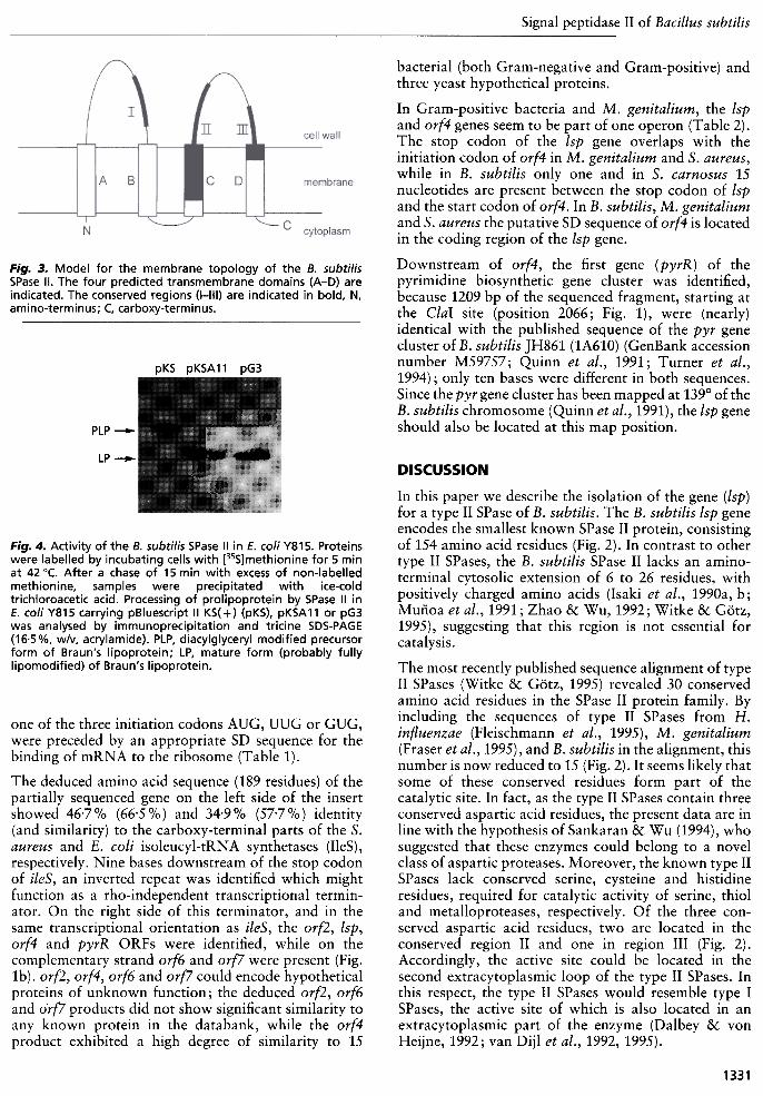
precipitation, and tricine SDS-PAGE (Fig. 4). Maturelipoprotein was only observed in E. coli Y815 carryingpKSAll or its deletion derivative pG3, but not in E. coliY815 carrying pBluescript II KS( + ) (negative control).These results show that the B. subtilis SPase II isfunctional in E. coli.
second extracytoplasmic loop and a part of the trans-membrane segment D.
B. subtilis SPase II is active in E. co/i
To verify the activity of the B. subtilis SPase II,processing of Braun's prolipoprotein was studied in E.coli Y815 transformed with pBluescript II KS( + ),pKSAll or pG3. The expression of the lpp gene wasinduced by adding IPTG and the temperature-sensitiveSPase1I of E. coli Y815 was inactivated by increasing thetemperature to 42 °C. Processing of prolipoprotein wasmonitored by pulse-chase labelling, subsequent immuno-
1330
Genes in the Isp region of the B. subtilischromosome; map position of Isp
In the nucleotide sequence of the 3271 bp insert ofplasmid pKSA11, five complete and two partial putativeORFs were identified (Fig. 1b). Six ORFs, starting with
Signal peptidase II of Bacillus subtilis
bacterial {both Gram-negative and Gram-positive) andthree yeast hypothetical proteins,
In Gram-positive bacteria and M. genitalium, the tspand orf4 genes seem to be part of one operon {Table 2).The stop codon of the tsp gene overlaps with theinitiation codon of orf4 in M. genitalium and s. aureus,while in B. subtilis only one and in S. carnosus 15nucleotides are present between the stop codon of tspand the start codon of orf4. In B. subtilis, M. genitaliumand s. aureus the putative SD sequence of orf4 is locatedin the coding region of the tsp gene.
Downstream of orf4, the first gene {pyrR) of thepyrimidine biosynthetic gene cluster was identified,because 1209 bp of the sequenced fragment, starting atthe Clal site {position 2066; Fig. 1), were {nearly)identical with the published sequence of the pyr genecluster of B. subtilis JH861 {IA610) {GenBank accessionnumber M59757; Quinn et at., 1991; Turner et at.,1994) ; only ten bases were different in both sequences.Since the pyr gene cluster has been mapped at 139° of theB. subtilis chromosome {Quinn et at., 1991), the tsp geneshould also be located at this map position.
Fig. 3. Model for the membrane topology of the 8. 5ubtili5SPase II. The four predicted transmembrane domains (A-D) areindicated. The conserved regions (1-111) are indicated in bold, N,amino-terminus; C, carboxy-terminus.
pKS pKSA 11 pG3
P LP --
LP-
F;g. 4. Activity of the B. subtilis SPase II in E. co/i Y815. Proteinswere labelled by incubating cells with [35S]methionine for 5 minat 42 °C. After a chase of 15 min with excess of non-labelledmethionine, samples were precipitated with ice-coldtrichloroacetic acid. Processing of prolipoprotein by SPase II inE. co/i Y815 carrying pBluescript II KS( + ) (pKS), pKSA 11 or pG3was analysed by immunoprecipitation and tricine SDS-PAGE(16.5 %, w/v, acrylamide). PLP, diacylglyceryl modified precursorform of Braun's lipoprotein; LP, mature form (probably fullylipomodified) of Braun's lipoprotein.
DISCUSSION
In this paper we describe the isolation of the gene (lsp)for a type II SPase of B. subtilis. The B. subtilis lsp geneencodes the smallest known SPase II protein, consistingof 154 amino acid residues (Fig. 2). In contrast to othertype II SPases, the B. subtilis SPase II lacks an amino-terminal cytosolic extension of 6 to 26 residues, withpositively charged amino acids (Isaki et al., 1990a, b ;Mufioa et al., 1991; Zhao & Wu, 1992; Witke & G6tz,1995), suggesting that this region is not essential forcatalysis.
The most recently published sequence alignment of typeII SPases (Witke & G6tz, 1995) revealed 30 conservedamino acid residues in the SPase II protein family. Byincluding the sequences of type II SPases from H.influenzae (Fleischmann et al., 1995), M. genitalium(Fraser et al., 1995), and B. subtilis in the alignment, thisnumber is now reduced to 15 (Fig. 2). It seems likely thatsome of these conserved residues form part of thecatalytic site. In fact, as the type II SPases contain threeconserved aspartic acid residues, the present data are inline with the hypothesis of Sankaran & Wu (1994), whosuggested that these enzymes could belong to a novelclass of aspartic proteases. Moreover, the known type IISPases lack conserved serine, cysteine and histidineresidues, required for catalytic activity of serine, thioland metalloproteases, respectively. Of the three con-served aspartic acid residues, two are located in theconserved region II and one in region III (Fig. 2).Accordingly, the active site could be located in thesecond extracytoplasmic loop of the type II SPases. Inthis respect, the type II SPases would resembIe type ISPases, the active site of which is also located in anextracytoplasmic part of the enzyme (Dalbey & vonHeijne, 1992; van Dijl et al., 1992, 1995).
one of the three initiation codons AUG, UUG or GUG,were preceded by an appropriate SD sequence for thebinding of mRNA to the ribosome (Table 1).
The deduced amino acid sequence (189 residues) of thepartially sequenced gene on the left side of the insertshowed 46.7% (66.5%) and 34.9% (57.7%) identity(and similarity) to the carboxy-terminal parts of the S.aureus and E. coli isoleucyl-tRNA synthetases (l1eS),respectively. Nine bases downstream of the stop codonof ileS, an inverted repeat was identified which mightfunction as a rho-independent transcriptional termin-ator. On the right side of this terminator, and in thesame transcriptional orientation as ileS, the orf2, lsp,orf4 and pyrR ORFs were identified, while on thecomplementary strand orf6 and orf7 were present (Fig.1b). orf2, orf4, orf6 and orf7 could encode hypotheticalproteins of unknown function; the deduced orf2, orf6and orf7 products did not show significant similarity toany known protein in the databank, while the orf4product exhibited a high degree of similarity to 15
1331

Table ,. Putative ORFs in the lsp region of the B. subtilis chromosome
" Coordinates are according to the U48870 nucleotide sequence in GenBank.
t Nucleotides identical to the consensus Shine-Dalgarno sequence (5' AAAGGAGGUGAU 3') areshown in upper case.
:!:Partially sequenced ORF.
Table 2. Comparison of the intercistronic regions ofthe Isp-orf4 operon in Gram-positive bacteria andM. genitalium
the SPase II protein, and the effects of changing the levelsof its production on the export of lipoproteins and non-lipoproteins by B. subtilis. In future work, the questionwill be addressed whether the expression of Isp(Bsu) istemporally regulated, and whether SPase II can be alimiting factor in the export of (lipo)proteins in B.subtilis.
ACKNOWLEDGEMENTS
We would like to thank Henk Mulder for preparing thefigures, Dr M. Inukai from Sankyo Co. Ltd, Tokyo, Japan, forgenerously providing globomycin, Dr H. Yamada and Pro-fessor S. Mizushima for generously providing E. coli Y815,and serum against Braun's lipoprotein. Z. P. was supported bya Hungarian State E6tv6s Fellowship from the NationalScholarships Board (Hungary). Z. P. is grateful to Professor L.Orosz and Dr F. Fábián for their help in obtaining thisfellowship. H. T. was supported by Genencor International ;A.B., J.M.v.D. and S.B. were supported by Biotech Grants(Bio2-CT93-0254 and PL950097) from the Commission of theEuropean Union (CEU). In addition, J.M. v.D. was supportedby a long-term fellowship (ALTF 63-1994) from EMBO.
* The stop codon (uaa or uag) ofthe lsp gene is indicated in lower
case, while the SD sequence of the orf4 gene is underlined and thetranslation start codon (aug) is indicated in bold lower case.
t The 3' end of the available sequence is too short to conclude thatthe Orf4 protein of s. aureus consists of 94 amino acid residues
only.
:I: Partially sequenced ORF.
In Gram-negative bacteria, the lsp gene is present in anoperon consisting of the genes x, ileS, lsp, orf149 andorf315 (MilIer et al., 1987; Isaki et al., 1990a, b). Theorganization of genes in the lsp regions of Gram-positivebacteria and M. genitalium seems to be different, as allknown lsp genes from these organisms are likely to forman operon with orf4. Furthermore, none of the genes inthe lsp regions of Gram-positive bacteria and M.genitalium, such as orf2, orf4 and pyrR of B. subtilis,showed significant similarities with gene x, orf149 andorf315 of Gram-negative bacteria. Although ileS and lspare located close together on the B. subtilis chromosome,it seems unlikely that these genes are transcriptionallylinked, since a putative terminator sequence was iden-tified between ileS and orf2 (Fig. 1b). Transcriptionallinkage of orf2, lsp, orf4 and pyrR cannot, however, beexcluded.
With the availability of the Isp(Bsu) gene and itsnucleotide sequence, we can now analyse the function of
REFERENCES
Altschul, S. F., Gish, W., MilIer, W., Myers, E. W. & Lipman, D. J.(1990). Basic local alignment search tool. J Mol Biol215, 403-410.
Bron, S. (1990). Plasmids. 1n Molecular Biological Methods forBaci[[us, pp. 75-174. Edited by C. R. Harwood & S. M. Cutting.Chichester: Wiley.
Dalbey, R. E. & von Heijne, G. (1992). Signal peptidases inprokaryotes and eukaryotes -a new protease family. TrendsBiochem Sci 17,474-478.
Devereux, J., Haeberli, P. & Smithies, 0. (1984). A comprehensiveset of sequence analysis programs for the V AX. Nucleic Acids Res
l2,387-395.
Fleischmann, R. D., Adams, M. D., White, 0. & 37 other authors
(1995). Whole-genome random sequencing and assembly ofHaemophilus influenzae Rd. Science 269, 496-512.
van Dijl, J. M., Smith, H., Bron, S. & Venema, G. {1988). Synthesisand processing of Escherichia coli TEM-p-lactamase and Baci[[us

Signal peptidase II of Bacillus subtili;
organization and nucleotide sequence of the Bacjllus subtjljs
pyrimidine biosynthetic operon. J Bjol Chem 266,9113-9127.
Sambrook. J.. Fritsch. E. F. & Maniatis. T. (1989). MolecularClonjng: a Laboratory Manual, 2nd edn. Cold Spring Harbor,NY: Cold Spring Harbor Laboratory.
Sanger. F.. Nicklen. s. & Coulson. A. R. (1977). DNA sequencingwith chain-terminating inhibitors. Proc Natl Acad Scj USA 74,
5463-5467.
Sankaran. K. & Wu. H. C. (1994). Signal peptidase II -specificsignal peptidase for bacteriallipoproteins. In Sjgnal Peptjdases,pp.17-29. Edited by G. von Heijne. Austin: R. G. Landes
Company.
Schagger. H. & von Jagow. G. (1987). Tricine-sodium dodecyl
sulfate-polyacrylamide gel electrophoresis for the separation ofproteins in the range from 1 to 100 kDa. Anal Bjochem 166,368-379.
Sutcliffe. I. C. & RusselI. R. R. B. (1995). Lipoproteins of Gram-
positive bacteria. J Bacterjol177 , 1123-1128.
Tokunaga. M.. Tokunaga. H. & Wu. H. C. (1982). Posttranslationalmodification and processing of Escherjchja colj prolipoprotein invitro. Proc Natl Acad Scj USA 79, 2253-2259.
Tokunaga. M.. Loranger. J. M. & Wu. H. C. (1983). Isolation andcharacterization of an Escherjchja colj clone overproducing
prolipoprotein signal peptidase. J Bjol Chem 258, 12102-12105.
Turner. R. J.. Lu. Y. & Switzer. R. L. (1994). Regulation of theBacjllus subtjljs pyrimidine biosynthetic ( pyr) gene cluster by an
autogenous transcriptional attenuation mechanism. J Bacterjol
176,3708-3722.
Witke. C. & GOtz. F. (1995). Cloning and nucleotide sequence ofthe signal peptidase II (Isp)-gene from Staphylococcus carnosus.FEMS Mjcrobjol Lett 126, 233-240.
Yamagata. H.. Ippolito. C.. Inukai. M. & Inouye. M. (1982).Temperature sensitive processing of outer membrane lipoproteinin an Escherjchja colj mutant. J Bacterjol152, 1163-1168.
Yamagata. H.. Daishima. K. & Mizushima. S. (1983). Cloning andexpression of a gene coding for the prolipoprotein signal peptidaseof Escherjchja colj. FEBS Lett 158,301-304.
Zabarovsky. E. R. &Allikmets. R. L. (1986). An improved techniquefor the efficient construction of gene libraries by partial filling-inof cohesive ends. Gene 42, 119-123.
Zhao. X.-J. & Wu. H. C. (1992). Nucleotide sequence of the
Staphylococcus aureus signal peptidase II (Isp) gene. FEBS Lett299, 80-84.
Received 29 Ju I y 1996; revised 25 October 1996; accepted 6 November
1996.
licheniformis (X-amylase in Escherichia coli: the role of signalpeptidase I. Mol Gen Genet 214, 55-61.
van Dijl, J. M., de Jong, A., Vehmaanpera, J., Venema, G. & Bron,
S. {1992). Signal peptidase laf Bacillus subtilis: patterns of
conserved amino acids in prokaryotic and eukaryotic type I signal
peptidases. EMBO 1 11,2819-2828.
van Dijl, J. M., de Jong, A., Venema, G. & Bron, S. {1995).
Identification of the potential active site of the signal peptidase
SipS of Bacillus subtilis: structural and functional similaritieswith LexA-Iike proteases. 1 Biol Chem 270,3611-3618.
Fra5er, C. M., Gocayne, J. D., White, 0. & 26 other authors {1995).The minimal gene complement of Mycoplasma genitalium.
Science 270, 397-403.
Giam, C. Z., Chai, T., Haya5hi, S. & Wu, H. C. {1984). Prolipoprotein
modification and processing in Escherichia coli. A unique
secondary structure in prolipoprotein signal sequence for the
recognition by glyceryl transferase. Eur 1 Biochem 141,331-337.
von Heijne, G. {1992). Membrane protein structure prediction.
Hydrophobicity analysis and the positive-inside rule. 1 Mol Biol225, 487-494.
von Heijne, G. {1994). Signal peptidases and protein trafficking. In
Signa[ Peptidases, pp. 1-3. Edited by G. von Heijne. Austin: R.G. Landes Company.
Inukai, M., Takeuchi, M., Shimizu, K. & Arai, M. {1978).Mechanism of action of globomycin. 1 Antibiot 31, 1203-1205.
15aki, L., Beer5, R. & Wu, H. C. {1990a). Nucleotide sequence of the
Pseudomonas fluorescens signal peptidase II gene (lsp) and
flanking genes. 1 Bacteriol172, 6512-6517.
15aki, L., Kawakami, M., Beer5, R., Hom, R. & Wu, H. C. {1990b).Cloning and nucleotide sequence of the Enterobacter aerogenes
signal peptidase II (lsp) gene. 1 Bacteriol172, 469-472.
Kontinen, V. P. & Sarva5, M. {1993). The PrsA lipoprotein is
essential for protein secretion in Bacillus subtilis and sets a limitfor high-level secretion. Mol Microbiol8, 727-737.
Kyte, J. & Doolittle, R. F. {1982). A simple method for displayingthe hydrophobic character of a protein. 1 Mol Biol157, 105-132.
MilIer, K. W., Bouvier, J., Stragier, P. & Wu, H. C. {1987).
Identification of the genes in Escherichia coli ileS-lsp operon.
Analysis of multiple polycistronic mRNAs made in vivo. 1 BiolChem 262,7391-7397.
MulÏoa, F. J., MilIer, K. W., Beer5, R., Graham, M. & Wu, H. C.
{1991). Membrane topology of Escherichia coli prolipoprotein
signal peptidase (signal peptidase II). 1 Biol Chem 266,17667-17672.
Quinn, C. L., Stephen5On, B. T. & Switzer, R. L. {1991). Functional