Structure of Human Tripeptidyl Peptidase II determined by a Hybrid ...
ORIGINAL RESEARCH ARTICLEpublished: 06 February 2015
doi: 10.3389/fpls.2015.00037
Extracellular peptidase hunting for improvement of proteinproduction in plant cells and rootsJérôme Lallemand 1,2 , Frédéric Bouché1,2 , Carole Desiron1†, Jennifer Stautemas1,
Frédéric de Lemos Esteves1†, Claire Périlleux1,2 and Pierre Tocquin1,2*
1 Laboratory of Plant Physiology, Department of Life Sciences, University of Liège, Liège, Belgium2 PhytoSYSTEMS, University of Liège, Liège, Belgium
Edited by:
Giuseppe Dionisio, Aarhus University,Denmark
Reviewed by:
Stefan Schillberg, Fraunhofer Institutefor Molecular Biology and AppliedEcology, GermanyKarl Kunert, University of Pretoria,South Africa
*Correspondence:
Pierre Tocquin, PhytoSYSTEMS,University of Liège, Boulevard duRectorat 27, Liège, Belgiume-mail: [email protected]†Present address:
Carole Desiron, Business & DecisionLife Sciences, 141 Rue Saint-Lambert,1200 Woluwe-Saint-Lambert,Belgium;Frédéric de Lemos Esteves, LifeSciences Library, University of Liège,Liège, Belgium
Plant-based recombinant protein production systems have gained an extensive interestover the past few years, because of their reduced cost and relative safety. Althoughthe first products are now reaching the market, progress are still needed to improveplant hosts and strategies for biopharming. Targeting recombinant proteins toward theextracellular space offers several advantages in terms of protein folding and purification, butdegradation events are observed, due to endogenous peptidases.This paper focuses on theanalysis of extracellular proteolytic activities in two production systems: cell cultures androot-secretion (rhizosecretion), in Arabidopsis thaliana and Nicotiana tabacum. Proteolyticactivities of extracellular proteomes (secretomes) were evaluated in vitro against twosubstrate proteins: bovine serum albumin (BSA) and human serum immunoglobulinsG (hIgGs). Both targets were found to be degraded by the secretomes, BSA beingmore prone to proteolysis than hIgGs. The analysis of the proteolysis pH-dependenceshowed that target degradation was mainly dependent upon the production system:rhizosecretomes contained more peptidase activity than extracellular medium of cellsuspensions, whereas variations due to plant species were smaller. Using class-specificpeptidase inhibitors, serine, and metallopeptidases were found to be responsible fordegradation of both substrates. An in-depth in silico analysis of genomic and transcriptomicdata from Arabidopsis was then performed and led to the identification of a limitednumber of serine and metallo-peptidases that are consistently expressed in both productionsystems. These peptidases should be prime candidates for further improvement of planthosts by targeted silencing.
Keywords: molecular pharming, peptidases, Arabidopsis thaliana, Nicotiana tabacum, root-secretion, suspension
cells, in silico analysis
INTRODUCTIONSince 25 years and the demonstration by Hiatt et al. (1989) thatthe plant secretory pathway was able to carry out the foldingand the assembling of complex eukaryotic proteins such as anti-bodies, plants have emerged as potential alternative hosts forthe production of biopharmaceuticals. The amazing versatilityof plant-based systems that have been developed (about a 100platforms Schillberg et al., 2013), together with the economicand safety advantages they offer, aroused great expectations forthis technology known as “molecular pharming.” However, itis only recently (2012) that the first plant-produced biophar-maceutical, a glucocerebrosidase produced in carrot cells as atreatment for the Gaucher’s disease (Shaaltiel et al., 2007), hasbeen approved by the US Food and Drug Administration. Sev-eral reasons explain this slow industrial and market uptake: therelatively low and variable yields compared to the gold stan-dard Chinese hamster ovary (CHO) cells for the production ofcomplex human proteins (Twyman et al., 2013), the negativeperception and restrictions on genetically modified organisms(GMOs; Schillberg et al., 2013), and the absence of a compre-hensive regulatory framework (Fischer et al., 2013). High yieldsand regulatory compliance are key prerequisites to transform
molecular pharming into an industrial success. Thus, while tech-nologies were initially designed for transgenic plants grown inopen fields, recent researches are rather focused on systems witha higher containment, which not only reduces the risk of GMOsrelease in the environment but also leads to a better control of thegrowing and production conditions (Paul and Ma, 2011; Schillberget al., 2013).
In this context, systems based on plant cell- or tissue-cultureshave emerged. They are either cell suspension cultures, mainly butnot limited to tobacco Bright Yellow-2 cells (BY-2), or hairy rootcultures induced by Agrobacterium rhizogenes (Schillberg et al.,2013). Both strategies share the advantage of producing biomassfaster than whole plant cultures. Moreover, the product is oftensecreted into the culture medium, making its recovery easier andcheaper than extraction from the biomass (Twyman et al., 2013).Somehow intermediate between suspension and whole plant cul-tures, ‘floating’ systems based on the use of whole organisms thatare fully or partly in contact with a culture medium (micro algae,moss, or aquatic plants) also have the advantages of being fullycontained and allowing the secretion-based recovery of the prod-uct (Cox et al., 2006; Decker et al., 2014; Mathieu-Rivet et al.,2014). It is also the case of the rhizosecretion strategy where
www.frontiersin.org February 2015 | Volume 6 | Article 37 | 1

Lallemand et al. Hunting for secreted plant peptidases
roots of hydroponically growing plants produce and secrete therecombinant protein into the nutrient solution. Such a system wasinitially proposed by Borisjuk et al. (1999) and later developed byMa and colleagues (Drake et al., 2002, 2003, 2009).
One major limitation of secretion-based systems comes fromproteolytic events frequently observed on the products (Pillayet al., 2014), a problem that is well documented for antibody pro-duction (e.g., Sharp and Doran, 2001; Drake et al., 2003; Niemeret al., 2014). Extracellular peptidases were demonstrated to beresponsible for these degradations in various production systems:cell suspensions (Sharp and Doran, 2001), leaves (Hehle et al.,2011), or (hairy-)roots (Sharp and Doran, 2001; Drake et al.,2009). The extent of degradation depends on organs or pro-duction systems (Drake et al., 2009), developmental stages (DeMuynck et al., 2009), culture medium (Häkkinen et al., 2014),or plant species (Magy et al., 2014). Cell wall proteome analyses,mainly performed in Arabidopsis, revealed that peptidases repre-sent more than 10% of the extracellular proteins (Albenne et al.,2013). Moreover, Goulet et al. (2012) showed by a genomic analy-sis of Arabidopsis, rice, and Nicotiana spp. that, consistently acrossthese species, a large proportion of peptidases are predicted tobe targeted to the extracellular space. Considering that little isknown about the function or the substrate of apoplastic pepti-dases (van der Hoorn, 2008; Tsiatsiani et al., 2012), their amountand diversity represent a major obstacle to the use of secretion inmolecular pharming. However, counteracting strategies such asco-secretion of a single peptidase inhibitor (Komarnytsky et al.,2006; Robert et al., 2013) or the silencing of a single peptidasegene (Kim et al., 2008a; Mandal et al., 2014) have already provenefficient. To be successful, these strategies rely on a prior knowl-edge of the proteolytic activities likely to lead, in the operatedproduction system, to the degradation of the target recombinantprotein.
In the present paper, we aimed at cross-comparing the extracel-lular proteolytic activities of two production systems, cell cultureand rhizosecretion, set-up from two species, Arabidopsis thalianaand Nicotiana tabacum. We specifically addressed this questionin the case of human immunoglobulin (hIgGs) production. Wehypothesized that in silico analyses of genomic and transcriptomicdata obtained from a model species such as Arabidopsis couldbe merged with experimental results obtained from biochemicalassays with existing production systems to provide robust insightsabout the major peptidases that limit hIgGs yields.
MATERIALS AND METHODSPLANT CULTURESPlants of A. thaliana cv. Columbia-0 (Col-0) and N. tabacum cv.Petit Havana SR1 were grown in hydroponics (Araponics, Bel-gium), at 20◦C, with a relative humidity of 70%, a photoperiod of10 h and a light intensity of 100 μmol m−2 s−1. Plant culture wasadapted from Tocquin et al. (2003): seeds were sown on the top ofseed-holders filled with a gel [0.5% Phytagel™ (Sigma-Aldrich, St.Louis, MO, USA), 5 mM CaCl2, 0.15% polyvinylpyrrolidone (MW10,000)] and the nutrient solution was prepared with Flora Seriesfertilizers (FloraBloom, FloraMicro and FloraGro; GHE, France),0.5 mL per liter each. The solution was renewed after 5 weeks ofcultivation.
RHIZOSECRETOME HARVESTINGThe direct analysis of the extracellular medium (EM) of hydro-ponically growing plants is hindered by the high dilution levelof endogenous secreted proteins. Moreover, many extracellularproteins, including peptidases, are known to be weakly boundto the cell wall. In order to get a comprehensive overview of thepeptidases that could be involved in the degradation of secretedrecombinant proteins, we used a harvesting protocol adaptedfrom cell wall proteomics studies (Boudart et al., 2005). 7 weeksafter sowing, roots were briefly drained, harvested and weighed.The total fresh weight (FW) measured is comprised of the rootbiomass, the intercellular fluid and the adsorbed nutrient solution.In preliminary experiments, we estimated the drained FW aftercentrifugation (10 min, 2700 g) to be ∼30 and ∼80% of total FWfor A. thaliana and N. tabacum, respectively. The water content wastaken into account to calculate the volume of a NaCl stock solutionthat must be added to the fresh root to obtain a final concentra-tion of 1 M. The root samples were then incubated for 1 h at 4◦Cin 1 M NaCl under strong agitation in order to recover extracel-lular compounds. Roots were thereafter centrifuged in a Pierce™Centrifuge Column (Thermo Scientific, Rockford, IL, USA) dur-ing 10 min at 2700 g and at 4◦C. The collected samples, hereaftercalled rhizosecretomes, were either used freshly prepared or storedat –80◦C until use.
CELL CULTURESCells of A. thaliana cv. plant system biology-dark (PSB-D) werecultivated in a 250-mL Erlenmeyer flask containing 50 mL of aliquid medium [0.44% Linsmaier and Skoog Medium (DuchefaBiochemie, Haarlem, the Netherlands), 3% sucrose, 5.10−5%1-naphthaleneacetic acid, 5.10−6% kinetin, pH 5.7 (KOH)] inthe dark, at 25◦C and under agitation at 90 rpm on a rotaryshaker. Each week, fresh liquid medium was inoculated with a10% inoculum of the former cell culture. Cells of N. tabacum cv.BY-2 were grown in the same conditions as A. thaliana cells, in anadapted culture medium [0.44% Murashige and Skoog salts (MPBIOMEDICALS, Solon, OH, USA), 3% sucrose, 0.02% KH2PO4,2.5.10−4% thiamine, 5.10−3% myo-inositol, 2.10−5% 2,4-D, pH5.8 (KOH)]. A 5% inoculum was used to inoculate fresh liquidmedium each week. Both types of cells were kindly provided byProf. Marc Boutry and his colleagues (Physiological Biochem-istry Unit, Catholic University of Louvain, Louvain-la-Neuve,Belgium).
EXTRACELLULAR MEDIUM HARVESTINGThe EM of the 7-days-old Arabidopsis cell cultures was vacuum-filtered through a superposition of a glass fiber prefilter and acellulose acetate filter, with a pore size of 0.2 μm (Sartorius StedimBiotech GmbH, Goettingen, Germany); tobacco cell cultures werevacuum-filtered through three layers of Miracloth (Calbiochem;Merck KGaA, Darmstadt, Germany). The EM was either directlyused or stored at –80◦C.
GELATIN ZYMOGRAPHYTwenty micro liter of rhizosecretome or EM were incubated during20 min at room temperature in a non-reducing loading buffer (1%SDS,10% glycerol, 0.02% bromophenol blue, 60 mM Tris-HCl, pH
Frontiers in Plant Science | Plant Biotechnology February 2015 | Volume 6 | Article 37 | 2
Lallemand et al. Hunting for secreted plant peptidases
6.8, final concentrations) and loaded on a 10% polyacrylamide gelcontaining 0.05% gelatin. After an electrophoresis of 45 min at180 V, the gel was washed 3 × 20 min in 2.5% Triton X-100 andincubated for 16 h in 10 mM MES, 0.1 mM ZnCl2, 5 mM CaCl2,1% Triton X-100, pH 5.75 (KOH). The gel was then stained bycolloidal Coomassie blue as described further.
TARGET PROTEINS IN VITRO DEGRADATIONFive micro liter of rhizosecretome or EM were incubated dur-ing 6 h at 25◦C in a buffer at a fixed value of pH [100 mMglycine (for pH values between 2 and 4.5) or 100 mM Tris (forpH values between 5 and 9), 100 mM MES, 0.1 mM ZnCl2, 5 mMCaCl2, 0.1 M dithiothreitol], with 1 μg bovine serum albumin(BSA; A7906, ≥98% purity, Sigma-Aldrich, St. Louis, MO, USA)or 1 μg immunoglobulin G from human serum (hIgGs; I4506,≥95% purity, Sigma-Aldrich, St. Louis, MO, USA), in a finalvolume of 20 μL. Inactivation of peptidase classes was obtainedby pre-incubating the rhizosecretome or EM in a buffer dur-ing 30 min at room temperature, with class-specific inhibitors:5 mM PMSF (specific inhibitor of serine peptidases), 40 μM E-64(specific inhibitor of cysteine peptidases), 16 μM Pepstatin A (spe-cific inhibitor of aspartic peptidases) and 20 mM EDTA (specificinhibitor of metalloproteases). Mixes of all but one inhibitors wereused, leaving only one peptidase class active per sample. 1 μg ofBSA or human IgG was then added and incubated during 3 h(rhizosecretome) or 6 h (EM) at 25◦C. After incubation, the tar-get protein degradation profile was analyzed by SDS-PAGE andcolloidal Coomassie blue staining.
SDS-PAGE AND COLLOIDAL COOMASSIE BLUE STAININGProtein samples were incubated during 10 min at 95◦C in areducing loading buffer (0.1 M dithiothreitol, 1% SDS, 10%glycerol, 0.02% bromophenol blue, 60 mM Tris-HCl, pH 6.8,final concentrations) before being separated by electrophoresisin a 10% polyacrylamide gel during 45 min at 180 V. The gelwas then washed briefly in water, fixed during 1 h at roomtemperature in a fixation solution (30% ethanol, 10% aceticacid), washed 2 × 10 min in water, incubated overnight in acolloidal Coomassie blue staining solution [20% methanol, 8%ammonium sulfate, 1.6% phosphoric acid, 0.08% CoomassieBrilliant blue G-250 (Merck KGaA, Darmstadt, Germany)] andfinally destained in a destaining solution (5% methanol, 7%acetic acid).
IN SILICO ANALYSESFasta sequences of Arabidopsis peptidases were first retrieved fromthe FTP server of the MEROPS database1. We then searched forcorresponding sequences in the genome of Arabidopsis (TAIR102)by BlastP. The best hit of each sequence was then further ana-lyzed to remove non-perfectly matching sequences (>10% lengthdifference, >2% mismatches).
In silico transcriptomic analyses were performed on Arabidop-sis Affymetrix ATH1 raw data retrieved from the ArrayExpress
1ftp://ftp.sanger.ac.uk/pub/MEROPS/current_release/protease.lib2ftp://ftp.arabidopsis.org/Sequences/blast_datasets/TAIR10_blastsets/TAIR10_pep_20110103_representative_gene_model_updated
database3 using “roots” and “suspension cells” as queries. Theresulting list of microarrays was manually sorted to removeexperiments lacking comprehensive methodological informa-tions. The list of experiments included in the survey is availableas a supplemental data file. Within each experiment, we man-ually removed the microarrays performed on leaves and wholeseedlings. The subsequent data analysis was performed usingthe R programming language (R Core Team, 2014). The “sim-pleaffy” Bioconductor package (Gentleman et al., 2004; Wilsonand Miller, 2005) was used to read the raw data and perform thepresent/absent call on individual arrays using the detection.p.val()function. Genes were considered as being expressed when p-value<0.01. Within each experiment, we computed the proportion ofarrays in which the gene of interest could be detected. Data weresorted using the list of peptidases defined above.
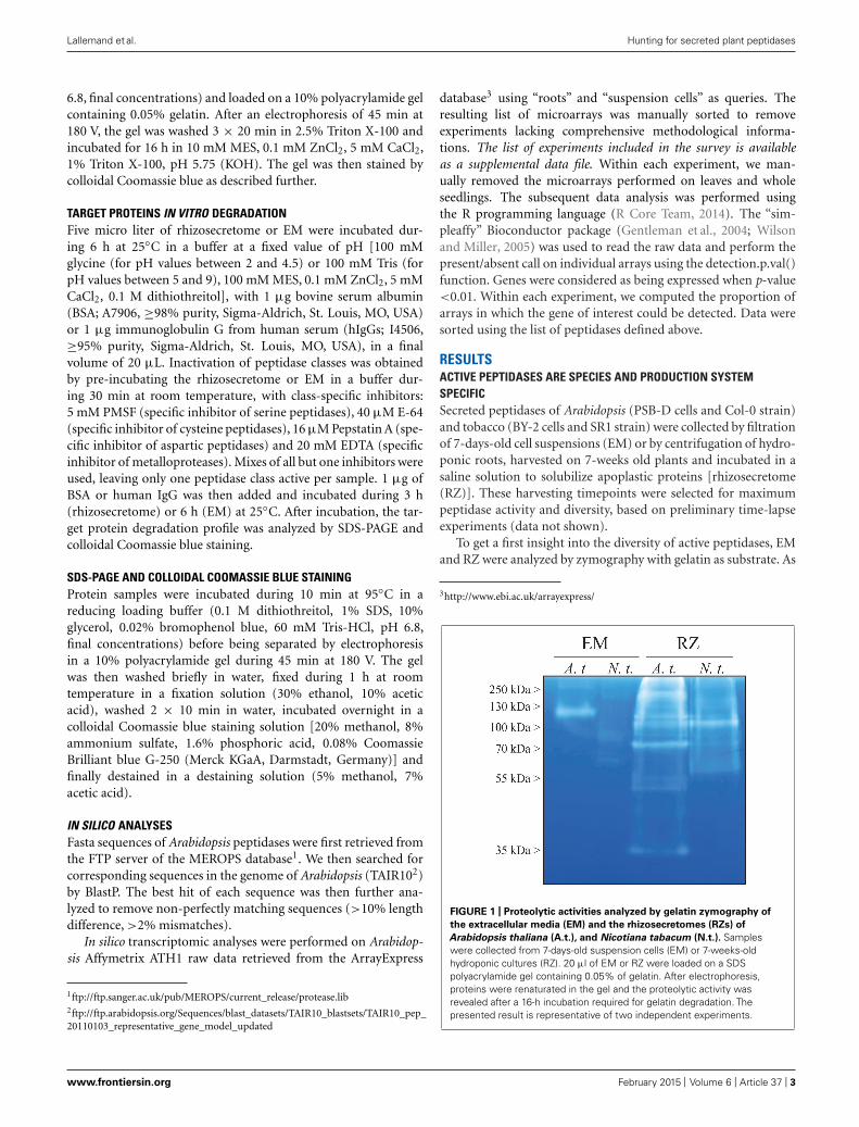
RESULTSACTIVE PEPTIDASES ARE SPECIES AND PRODUCTION SYSTEMSPECIFICSecreted peptidases of Arabidopsis (PSB-D cells and Col-0 strain)and tobacco (BY-2 cells and SR1 strain) were collected by filtrationof 7-days-old cell suspensions (EM) or by centrifugation of hydro-ponic roots, harvested on 7-weeks old plants and incubated in asaline solution to solubilize apoplastic proteins [rhizosecretome(RZ)]. These harvesting timepoints were selected for maximumpeptidase activity and diversity, based on preliminary time-lapseexperiments (data not shown).
To get a first insight into the diversity of active peptidases, EMand RZ were analyzed by zymography with gelatin as substrate. As
3http://www.ebi.ac.uk/arrayexpress/
FIGURE 1 | Proteolytic activities analyzed by gelatin zymography of
the extracellular media (EM) and the rhizosecretomes (RZs) of
Arabidopsis thaliana (A.t.), and Nicotiana tabacum (N.t.). Sampleswere collected from 7-days-old suspension cells (EM) or 7-weeks-oldhydroponic cultures (RZ). 20 μl of EM or RZ were loaded on a SDSpolyacrylamide gel containing 0.05% of gelatin. After electrophoresis,proteins were renaturated in the gel and the proteolytic activity wasrevealed after a 16-h incubation required for gelatin degradation. Thepresented result is representative of two independent experiments.
www.frontiersin.org February 2015 | Volume 6 | Article 37 | 3
Lallemand et al. Hunting for secreted plant peptidases
shown in Figure 1, the degradation patterns were highly differentas regards with species and production systems.
The diversity of peptidase activities detected on zymogram waslarger in RZ: more degradation bands were observed, over a widerrange of molecular weights, than in EM (Figure 1). Moreover,in both RZ and EM setups, Arabidopsis samples displayed moredegradation bands and bands with stronger activities than thosefrom tobacco.
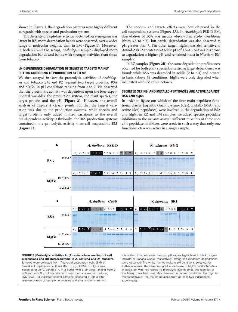
pH-DEPENDENCE DEGRADATION OF SELECTED TARGETS MAINLYDIFFERS ACCORDING TO PRODUCTION SYSTEMSWe then assayed in vitro the proteolytic activities of Arabidop-sis and tobacco EM and RZ, against two target proteins: BSAand hIgGs, in pH conditions ranging from 2 to 9. We observedthat the proteolytic activity was dependent upon the four exper-imental variables: the production system, the plant species, thetarget protein and the pH (Figure 2). However, the overallanalysis of Figure 2 clearly points out that the largest vari-ation was due to the production systems, while species andtarget proteins only added limited variations to the overallpH-dependent activity. Obviously, the RZ production systemscontained more proteolytic activity than cell suspensions EM(Figure 1).
The species- and target- effects were best observed in thecell suspensions systems (Figure 2A). In Arabidopsis PSB-D EM,degradation of BSA was mainly observed in acidic conditions(from 3.5 to ∼5), but partial degradation was also observed atpH greater than 7. The other target, hIgGs, was also sensitive toArabidopsis EM proteases at acidic pH of 3.5–4.5 but was less proneto degradation at higher pH, and remained intact in Nicotiana EMsamples.
In RZ samples (Figure 2B), the same degradation profiles wereobtained for both plant species but a strong target dependency wasfound: while BSA was degraded in acidic (2 to ∼4) and neutralto basic (above 6) conditions, hIgGs were only degraded whenincubated with RZ at pH below 5.
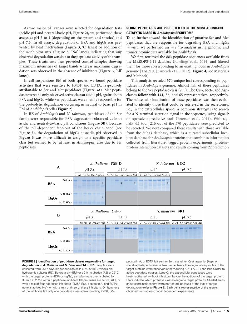
SECRETED SERINE- AND METALLO-PEPTIDASES ARE ACTIVE AGAINSTBSA AND hIgGsIn order to figure out which of the four main peptidase func-tional classes [aspartic (Asp), cysteine (Cys), metallo (Met), andserine (Ser) peptidases] were involved in the degradation of BSAand hIgGs in RZ and EM samples, we added specific peptidaseinhibitors to the in vitro assays. Different mixtures of three spe-cific peptidase inhibitors were used, in such a way that only onefunctional class was active in a single sample.
FIGURE 2 | Proteolytic activities in (A) extracellular medium of cell
suspensions and (B) rhizosecretome in A. thaliana and N. tabacum.
Samples were collected from 7-days-old suspension cells (EM) or7-weeks-old hydroponic cultures (RZ). 1 μg of BSA or hIgGs wasincubated at 25◦C during 6 h, in a buffer with a pH-value ranging from 2to 9 and with 5 μl of secretome. It was then analyzed on reducingSDS-PAGE. C3 indicates control samples incubated at pH 3 afterheat-inactivation of secretome proteins and thus shows maximum
intensities of target-protein band(s). pH values highlighted in black or grayindicate pH ranges where, respectively, strong and moderate degradationswere observed. The white frames indicate pH conditions selected forfurther analyses. The observed gradual decrease in hIgGs band intensitiesat acidic pH was not related to proteolytic events since this fade-out ofthe heavy chain band was also observed in control conditions. Each gel isrepresentative of the results obtained from at least two independentexperiments.
Frontiers in Plant Science | Plant Biotechnology February 2015 | Volume 6 | Article 37 | 4
Lallemand et al. Hunting for secreted plant peptidases
As two major pH ranges were selected for degradation tests(acidic pH and neutral-basic pH, Figure 2), we performed theseassays at pH 3 to 4 (depending on the system and species) andpH 7.5. In all assays, degradation of BSA and hIgGs was pre-vented by heat inactivation (Figure 3, ‘C’ lanes) or addition ofthe 4-inhibitor mix (Figure 3, ‘No’ lanes) indicating that anyobserved degradation was due to the peptidase activity of the sam-ples. These treatments thus provided control samples showingmaximum intensities of target bands whereas maximum degra-dation was observed in the absence of inhibitors (Figure 3, ‘All’lanes).
In cell suspensions EM of both species, we found peptidaseactivities that were sensitive to PMSF and EDTA, respectivelyattributable to Ser and Met peptidases (Figure 3A). Met pepti-dases were the only observed active class at acidic pH, against bothBSA and hIgGs, while Ser peptidases were mainly responsible forthe proteolytic degradation occurring in neutral to basic pH inEM of Arabidopsis cells (Figure 2).
In RZ of Arabidopsis and N. tabacum, peptidases of the Serfamily were responsible for BSA degradation observed at bothacidic and neutral-to-basic pH conditions (Figure 3B). Becauseof the pH-dependent fade-out of the heavy chain band (seeFigure 2), the degradation of hIgGs at acidic pH observed inFigure 3 was more difficult to assign to a specific peptidaseclass but seemed to be, at least in Arabidopsis, also due to Serpeptidases.
SERINE PEPTIDASES ARE PREDICTED TO BE THE MOST ABUNDANTCATALYTIC CLASS IN Arabidopsis SECRETOMETo go further toward the identification of putative Ser and Metpeptidases that are responsible for degrading BSA and hIgGsin vitro, we performed an in silico analysis using genomic andtranscriptomic data available for Arabidopsis.
We first retrieved the 883 peptidase sequences available fromthe MEROPS 9.11 database (Rawlings et al., 2014) and filteredthem for those corresponding to an existing locus in Arabidopsisgenome [TAIR10, (Lamesch et al., 2012); Figure 4, see Materialsand Methods].
This analysis revealed 570 unique loci corresponding to pep-tidases in Arabidopsis genome. Almost half of these peptidasesbelong to the Ser peptidase class (255). The Cys-, Met-, and Asp-classes follow with 144, 86, and 65 representatives, respectively.The subcellular localization of these peptidases was then evalu-ated to identify those that could be retrieved in the secretomes,i.e., in the extracellular space. A common strategy is to searchfor a N-terminal secretion signal in the sequence, using signalPor equivalent predictive tools (Petersen et al., 2011). With sig-nalP 4.1 tool, 216 out of the 570 peptidases were predicted tobe secreted. We next compared these results with those availablefrom the Suba3 database, which is a curated subcellular loca-tion database for Arabidopsis proteins that combines informationcollected from literature, tagged protein experiments, protein–protein interaction datasets and results coming from 22 prediction
FIGURE 3 | Identification of peptidase classes responsible for target
degradation in A. thaliana and N. tabacum EM or RZ. Samples werecollected from (A) 7-days-old suspension cells (EM) or (B) 7-weeks-oldhydroponic cultures (RZ). Before a six- (EM) or a 3-h incubation (RZ) at 25◦Cwith the target proteins (BSA or hIgGs), samples were pre-incubated for30 min at 25◦C without peptidase inhibitors (all proteases are active; ‘All’), orwith a mix of four peptidase inhibitors (PMSF, E64, pepstatin A, and EDTA;none is active; ‘No’), or with a mix of three of these inhibitors. Omitting oneof the inhibitors left only one peptidase class active: omitting PMSF, E64,
pepstatin A, or EDTA left serine-(Ser), cysteine- (Cys), aspartic- (Asp), ormetallo-(Met) peptidases active, respectively. The degradation profiles of thetarget proteins were observed after reducing SDS-PAGE. Lane labels refer toactive peptidase classes. Lane C: the extracellular peptidases wereheat-inactivated, without inhibitors, before the addition of the target protein.Stars indicate which protease classes degrade target proteins. Shaded areasshow combinations that were not tested, because of the lack of targetdegradation (refer to Figure 2). Each gel is representative of the resultsobtained from at least two independent experiments.
www.frontiersin.org February 2015 | Volume 6 | Article 37 | 5

Lallemand et al. Hunting for secreted plant peptidases
FIGURE 4 | Identification and distribution of Arabidopsis peptidases as
inferred from MEROPS 9.11 database andTAIR10 genomic data. (A)
BlastP analysis (see Materials and Methods) of the 883 MEROPS sequencesrevealed 570 actual peptidases in Arabidopsis genome; the other MEROPSsequences did not have a perfect match in the genome (misaligned andnon-matched), were duplicated (redundant), or were non-peptidase homologs
(nph). (B) The 570 peptidases were classified according to their catalytic class(A, Asp peptidases; C, Cys peptidases; M, Met Peptidases; S, Ser peptidases;and T, Threonine peptidases) and family (from A1 to S33). Secreted peptidaseswere predicted either by the presence of a signal peptide (SignalP) or by theircomputed subcellular localization available in the SUBA3 database. Onlyfamilies having at least two representatives are displayed as colored bars.
programs (Tanz et al., 2013). As shown in Figure 4, a significantsmaller amount of peptidases (147) were predicted to be extra-cellular by the Suba3 analysis compared with signalP 4.1, exceptfor two families (S8 and S33) for which the opposite result wasobtained. Among the 147 predicted secreted peptidases, 85 belongto the Ser-class and 6 to the Met-class and are thus putative can-didates to explain proteolytic degradation detected in RZ and EMof Arabidopsis.
MORE SECRETED PEPTIDASES ARE EXPRESSED IN HYDROPONICS vs.SUSPENSION CELLSTo evaluate which of those putative 147 secreted peptidases aremost likely expressed in rhizosecretion and cell suspension pro-duction systems, we analyzed transcriptomic data available fromthe EBI Array express repository (Rustici et al., 2013). Experimentsretrieved from a search with the ‘root’ and ‘suspension cells’ key-words were manually curated to select the most similar to ours,in terms of growing conditions and tissue sampling. Data gath-ered for hydroponics encompassed 16 experiments containing 310arrays while we found 11 cell suspensions experiments with a totalof 126 arrays. We then applied a ‘present/absent call’ functionfor each of the 147 peptidases, using the simpleaffy Bioconductorpackage (Wilson and Miller, 2005). A peptidase was considered asbeing expressed in an experiment if it was seen as ‘present’ in atleast 90% of the arrays of this experiment (Figure 5). We chose thishigh cut-off to foster the selection of peptidases that were consti-tutively expressed, i.e., whose transcripts were present in almost allarrays of an experiment and thus independent of the experimentalset-up. We then calculated, for both hydroponics and suspensioncells, the proportion of experiments in which a peptidase wasexpressed. As shown in Figure 5, only about 50% of the putativesecreted peptidases were found to be expressed in at least 10% ofthe experiments. We identified 50 peptidases expressed in a leasthalf of the experiments in hydroponics and 26 in at least half ofthe experiments in suspension cells (Figure 5, threshold 50%). It is
noteworthy that whatever the threshold, the number of expressedpeptidases was always greater in hydroponics experiments than insuspensions cells.
With the threshold ratio set to 50% (i.e., expression detected ina least 50% of the experiments), only four peptidases were revealedas specifically expressed in suspension cells: one Asp and three Serpeptidases (Figures 5B,C). Two matrix metallopeptidases werefound to be expressed in hydroponics but none in suspension cellcultures (AT1G59970, AT1G70170). With no threshold ratio set(i.e., expression detected in a least one experiment), two addi-tional Met peptidases were detected in hydroponics (AT1G24140,AT1G71696). Two of these four Met peptidases were also expressedin suspension cells experiments (AT1G59970, AT1G71696). Serpeptidases were always the most represented catalytic class in tran-scriptomes (data not shown). At the 50% threshold, a total of 28Ser peptidases were found to be expressed in hydroponics and/orsuspension cells. Five of them were expressed in all experiments(threshold 100%) and are listed in Table 1; the complete list ofsecreted peptidases is available as Supplemental Data file.
DISCUSSIONUnwanted degradation of recombinant proteins by endogenouspeptidases is one of the major problems of plant-based heterolo-gous production systems (Pillay et al., 2014). Peptidases are knownto be involved in a multitude of processes from the cellular to wholeorganism level, but the exact functions and targets of most of themare still unknown (van der Hoorn, 2008). The strategies to pre-vent proteolysis thus generally rely on broad range inhibition ofone or several catalytic classes, either by coexpression of peptidaseinhibitors or direct gene silencing (Komarnytsky et al., 2006; Kimet al., 2008b; Goulet et al., 2010, 2012; Redkiewicz et al., 2012).
The increasing amount of ‘omics’ data provides an attractivestarting point toward the identification of peptidases poten-tially at work in a given production system. However, thisapproach is rapidly limited by (1) the number of peptidases
Frontiers in Plant Science | Plant Biotechnology February 2015 | Volume 6 | Article 37 | 6

Lallemand et al. Hunting for secreted plant peptidases
FIGURE 5 |Transcriptomic analysis of Arabidopsis secreted peptidases
by a meta-analysis of publicly available microarrays data of hydroponic
and suspension cell experiments. A present/absent call (p < 0.01) wasapplied to each peptidase in each arrays of each experiment available. Foreach experiment, a peptidase was considered as ‘present’ if detected in at
least 90% of its arrays. We then plotted in (A) the number of peptidases thatwere considered as present in at least a given proportion of the experiments.For peptidases that were found to be expressed in at least half of theexperiments, we evaluated their specificity toward the expression context (B)
and their distribution among the main represented peptidase families (C).
Table 1 |TAIR10 functional annotation of the five Ser peptidases that were found to be expressed in all hydroponics and/or suspension cells
transcriptomic experiments analyzed in this study.
TAIR locus MEROPS identifier Peptidase family Short description Hydroponics Suspension cells
AT1G17430 MER036056 S33 NA 16/16 5/11
AT1G71950 MER039047 S08 NA 16/16 11/11
AT2G05920 MER015427 S08 Subtilase family protein 16/16 11/11
AT4G12910 MER005597 S10 Serine carboxypeptidase-like 20 14/16 11/11
AT5G67360 MER001368 S08 Subtilisin-like serine protease (ARA12) 15/16 11/11
The ‘Hydroponics’ and ‘Suspension cells’ columns indicate the number of experiments where the protease is expressed compared to the total number of transcriptomicexperiments analyzed.
identified in genomes, (2) the limited knowledge about their func-tions, regulation and targets, and (3) the potential redundancybetween peptidases of the same class, clan or family. Becausepeptidase activity is often controlled at the post-translational
level, genomic and transcriptomic information are not suffi-cient. Moreover, functional redundancy hinders the identifi-cation of peptidases by simple activity-based assays. In thispaper, we showed that meta-analyses of available genomic and
www.frontiersin.org February 2015 | Volume 6 | Article 37 | 7

Lallemand et al. Hunting for secreted plant peptidases
transcriptomic data allow to reduce the huge list of pepti-dase candidates that results from activity-based assays, whichessentially gives information about the catalytic class or thefamily.
We first focused on identifying active peptidase classes andaddressing the question whether substantial differences existbetween plant materials, production systems or target proteins,when assayed simultaneously. By using class-specific inhibitors,we showed that only Met and Ser peptidases were active againstBSA and/or hIgGs, depending on the production system (RZ orEM) and the target protein.
Extracellular Met peptidases count only a few members, most ofthem belonging to the matrix metalloproteinases (MMPs) family(Marino and Funk, 2012). In our in silico analysis, only two MMPswere detected in a majority of hydroponic experiments, one ofthem being observed only in a few suspension cells experiments,together with a Zn2+ carboxypeptidase. Despite the scarcity of Metpeptidases in secretomes, our results clearly showed that Met activ-ity was present in EM of both Arabidopsis and BY-2 suspensioncells, in agreement with previous reports for BY-2 (Delannoy et al.,2008; Schiermeyer et al., 2009; Mandal et al., 2010). The produc-tion system thus appears to be a strong determinant of proteolyticactivities. The similarity between PSB-D and BY-2 cells overridestheir species and tissue origins, which are Arabidopsis stem andtobacco root explants, respectively. By contrast, the two tobacco-based systems are very different in terms of pH dependence andactive peptidases, despite their common species and tissue origins.
In contrast with Met peptidases, Ser peptidases are the mostabundant class in plant cells and were therefore primarily targetedfor plant-based production system improvement. Co-expressionof Ser inhibitor, either as a second transgene or a fusion pro-tein, improved, yet not totally, stability of recombinant proteins(Komarnytsky et al., 2006; Kim et al., 2008b; Goulet et al., 2010,2012; Redkiewicz et al., 2012). Prior assessment of active pep-tidases is rarely performed and, if any, is usually done withnon-specific targets (Rivard et al., 2006; Goulet et al., 2012).However, even closely related proteins, for example recombinantimmunoglobulins, exhibit variable sensitivity to peptidases (Magyet al., 2014; Niemer et al., 2014), and hence end-product evalua-tion of peptidase activities is needed. The actual risk of proteolyticdegradation is even more difficult to predict if several produc-tion systems or hosts are available, since each of them has its ownpeptidase assortment. This diversity was clearly illustrated here bycomparing suspension cells and rhizosecretion in Arabidopsis andtobacco and was reported before (Magy et al., 2014; Pillay et al.,2014).
Merging activity assays with geno-transcriptomic data allowedus to narrow down the list of Ser peptidases potentially respon-sible for target degradations in Arabidopsis RZ and EM: out of255 Ser peptidases identified in the genome, 85 were predictedto be extracellular and 25 were expressed in conditions similar tothe production systems. Five of them were consistently expressedin suspension cells or hydroponics experiments included in themeta-analysis (Table 1), among which the serine carboxypepti-dase SCP20 and the subtilisin-like ARA12 (Table 1). SCP20 wasdetected in the extracellular space of seedlings and leaves, andwas reported to be strongly induced following fungal infection
(Charmont et al., 2005; Floerl et al., 2012). ARA12 was frequentlyidentified in cell wall proteomics studies either in EM of suspen-sion cell cultures (Hamilton et al., 2003; Shen et al., 2013) or inseedlings and leaves (Boudart et al., 2005; Charmont et al., 2005;Fraser et al., 2005; Floerl et al., 2012). If the exact function ofARA12 remains unknown, its purification and biochemical char-acterization showed that it is a heat-stable peptidase functioningin a wide range of pH, from 3 to 7 with an optimum around5 (Hamilton et al., 2003). These properties fit very well with theconditions in which we performed the activity assays presentedhere. Interestingly, a close N. tabacum homolog of ARA12 wasidentified in BY-2 EM (Navarre et al., 2012), indicating that pro-duction systems are similar, whatever the plant species, as alsoinferred from our cross-comparison.
Our cross-comparison of production systems and plant hosts,together with our in silico analysis of peptidases, consistentlyshow that suspension cell cultures provide a less proteolytic envi-ronment for the production of recombinant proteins, especiallyantibodies. Even if less degradation was observed in BY-2 tobaccocells compared with Arabidopsis PSB-D cells, the relatively weakinfluence of the plant species permits to use model species suchas Arabidopsis as a starting point for optimization of other hostspecies. The top listed peptidases identified by our combinedbiochemical/bioinformatic analysis are thus prime candidates fornew technology development, e.g., amiRNA- or MIGS multigenesilencing, or nuclease assisted genome engineering (Schwab et al.,2006; Felippes et al., 2012; Voytas, 2013). These methods shouldsoon be applicable to various plant species as the acquisition ofgenomic information increases quickly. Nevertheless, Arabidopsisis a competitive production system, even when compared withthe well established N. tabacum BY-2 cells (Magy et al., 2014), andmay even take a stronger position if one takes advantage of itstechnological advance for genetic engineering.
ACKNOWLEDGMENTSThe research was funded by the Service Public de Wallonie, DG06(Waléo3 program, 08/1/6861). The authors warmly thank theircollaborators Marc Boutry, Jacques Dommes, Christine Dupont,Catherine Navarre, and Marie-France Versali for their critical com-ments on the experiments and results. Jérôme Lallemand andFrédéric Bouché are grateful to the F.R.S.-FNRS for their PhDfellowships (FC95800; FC87200), as well as Carole Desiron for herF.R.I.A. grant. While working on this paper, we were truly shockedby the terrible terrorist attack against the French weekly “Char-lie Hebdo.” The freedom of making and publishing science is soclosely related to the freedom of expression that the authors wantsymbolically to express their complete solidarity with the victimsof this attack by these simple words: “We are Charlie.”
SUPPLEMENTARY MATERIALThe Supplementary Material for this article can be found onlineat: http://www.frontiersin.org/journal/10.3389/fpls.2015.00037/abstract
REFERENCESAlbenne, C., Canut, H., and Jamet, E. (2013). Plant cell wall proteomics: the leader-
ship of Arabidopsis thaliana. Front. Plant Sci. 4:111. doi: 10.3389/fpls.2013.00111
Frontiers in Plant Science | Plant Biotechnology February 2015 | Volume 6 | Article 37 | 8
Lallemand et al. Hunting for secreted plant peptidases
Borisjuk, N. V., Borisjuk, L. G., Logendra, S., Petersen, F., Gleba, Y., and Raskin,I. (1999). Production of recombinant proteins in plant root exudates. Nat.Biotechnol. 17, 466–469. doi: 10.1038/8643
Boudart, G., Jamet, E., Rossignol, M., Lafitte, C., Borderies, G., Jauneau, A., et al.(2005). Cell wall proteins in apoplastic fluids of Arabidopsis thaliana rosettes:identification by mass spectrometry and bioinformatics. Proteomics 5, 212–221.doi: 10.1002/pmic.200400882
Charmont, S., Jamet, E., Pont-Lezica, R., and Canut, H. (2005). Proteomic analy-sis of secreted proteins from Arabidopsis thaliana seedlings: improved recoveryfollowing removal of phenolic compounds. Phytochemistry 66, 453–461. doi:10.1016/j.phytochem.2004.12.013
Cox, K. M., Sterling, J. D., Regan, J. T., Gasdaska, J. R., Frantz, K. K., Peele,C. G., et al. (2006). Glycan optimization of a human monoclonal antibody inthe aquatic plant Lemna minor. Nat. Biotechnol. 24, 1591–1597. doi: 10.1038/nbt1260
De Muynck, B., Navarre, C., Nizet,Y., Stadlmann, J., and Boutry, M. (2009). Differentsubcellular localization and glycosylation for a functional antibody expressed inNicotiana tabacum plants and suspension cells. Transgenic Res. 18, 467–482. doi:10.1007/s11248-008-9240-1
Decker, E. L., Parsons, J., and Reski, R. (2014). Glyco-engineering for bio-pharmaceutical production in moss bioreactors. Front. Plant Sci. 5:346. doi:10.3389/fpls.2014.00346
Delannoy, M., Alves, G., Vertommen, D., Ma, J., Boutry, M., and Navarre, C.(2008). Identification of peptidases in Nicotiana tabacum leaf intercellular fluid.Proteomics 8, 2285–2298. doi: 10.1002/pmic.200700507
Drake, P. M. W., Barbi, T., Sexton, A., McGowan, E., Stadlmann, J., Navarre, C.,et al. (2009). Development of rhizosecretion as a production system for recombi-nant proteins from hydroponic cultivated tobacco. FASEB J. 23, 3581–3589. doi:10.1096/fj.09131771
Drake, P. M. W., Chargelegue, D., Vine, N. D., Dolleweerd, C. J. V., Obregon, P.,and Ma, J. K.-C. (2002). Transgenic plants expressing antibodies: a model forphytoremediation. FASEB J. 16, 1855–1860. doi: 10.1096/fj.02-0148com
Drake, P. M. W., Chargelegue, D. M., Vine, N. D., van Dolleweerd, C. J., Obre-gon, P., and Ma, J. K.-C. (2003). Rhizosecretion of a monoclonal antibodyprotein complex from transgenic tobacco roots. Plant Mol. Biol. 52, 233–241.doi: 10.1023/A:1023909331482
Felippes, F. F., Wang, J.-W., and Weigel, D. (2012). MIGS: miRNA-induced genesilencing. Plant J. 70, 541–547. doi: 10.1111/j.1365-313X.2011.04896.x
Fischer, R., Schillberg, S., Buyel, J., and Twyman, R. (2013). Commercial aspects ofpharmaceutical protein production in plants. Curr. Pharm. Des. 19, 5471–5477.doi: 10.2174/1381612811319310002
Floerl, S., Majcherczyk, A., Possienke, M., Feussner, K., Tappe, H., Gatz, C.,et al. (2012). Verticillium longisporum infection affects the leaf apoplastic pro-teome, metabolome, and cell wall properties in Arabidopsis thaliana. PLoS ONE7:e31435. doi: 10.1371/journal.pone.0031435
Fraser, C. M., Rider, L. W., and Chapple, C. (2005). An expression and bioinformaticsanalysis of the Arabidopsis serine carboxypeptidase-like gene family. Plant Physiol.138, 1136–1148. doi: 10.1104/pp.104.057950
Gentleman, R. C., Carey, V. J., Bates, D. M., Bolstad, B., Dettling, M., Dudoit,S., et al. (2004). Bioconductor: open software development for computationalbiology and bioinformatics. Genome Biol. 5:R80. doi: 10.1186/gb-2004-5-10-r80
Goulet, C., Benchabane, M., Anguenot, R., Brunelle, F., Khalf, M., and Michaud,D. (2010). A companion protease inhibitor for the protection of cytosol-targeted recombinant proteins in plants. Plant Biotechnol. J. 8, 142–154. doi:10.1111/j.1467-7652.2009.00470.x
Goulet, C., Khalf, M., Sainsbury, F., D’Aoust, M.-A., and Michaud, D. (2012).A protease activity-depleted environment for heterologous proteins migratingtowards the leaf cell apoplast. Plant Biotechnol. J. 10, 83–94. doi: 10.1111/j.1467-7652.2011.00643.x
Häkkinen, S. T., Raven, N., Henquet, M., Laukkanen, M.-L., Anderlei, T., Pitkänen,J.-P., et al. (2014). Molecular farming in tobacco hairy roots by triggering thesecretion of a pharmaceutical antibody. Biotechnol. Bioeng. 111, 336–346. doi:10.1002/bit.25113
Hamilton, J. M. U., Simpson, D. J., Hyman, S. C., Ndimba, B. K., and Slabas,A. R. (2003). Ara12 subtilisin-like protease from Arabidopsis thaliana: purifica-tion, substrate specificity and tissue localization. Biochem. J. 370, 57–67. doi:10.1042/BJ20021125
Hehle, V. K., Paul, M. J., Drake, P. M., Ma, J. K., and van Dolleweerd, C. J. (2011).Antibody degradation in tobacco plants: a predominantly apoplastic process.BMC Biotechnol. 11:128. doi: 10.1186/1472-6750-11-128
Hiatt, A., Caffferkey, R., and Bowdish, K. (1989). Production of antibodies intransgenic plants. Nature 342, 76–78. doi: 10.1038/342076a0
Kim, N.-S., Kim, T.-G., Kim, O.-H., Ko, E.-M., Jang, Y.-S., Jung, E.-S., et al. (2008a).Improvement of recombinant hGM-CSF production by suppression of cysteineproteinase gene expression using RNA interference in a transgenic rice culture.Plant Mol. Biol. 68, 263–275. doi: 10.1007/s11103-008-9367-8
Kim, T.-G., Lee, H.-J., Jang, Y.-S., Shin, Y.-J., Kwon, T.-H., and Yang,M.-S. (2008b). Co-expression of proteinase inhibitor enhances recombinanthuman granulocyte–macrophage colony stimulating factor production in trans-genic rice cell suspension culture. Protein Expr. Purif. 61, 117–121. doi:10.1016/j.pep.2008.06.005
Komarnytsky, S., Borisjuk, N., Yakoby, N., Garvey, A., and Raskin, I. (2006). Cose-cretion of protease inhibitor stabilizes antibodies produced by plant roots. PlantPhysiol. 141, 1185–1193. doi: 10.1104/pp.105.074419
Lamesch, P., Berardini, T. Z., Li, D., Swarbreck, D., Wilks, C., Sasidharan, R., et al.(2012). The Arabidopsis information resource (TAIR): improved gene annotationand new tools. Nucleic Acids Res. 40, D1202–D1210. doi: 10.1093/nar/gkr1090
Magy, B., Tollet, J., Laterre, R., Boutry, M., and Navarre, C. (2014). Accu-mulation of secreted antibodies in plant cell cultures varies according to theisotype, host species and culture conditions. Plant Biotechnol. J. 12, 457–467. doi:10.1111/pbi.12152
Mandal, M. K., Fischer, R., Schillberg, S., and Schiermeyer, A. (2010). Biochemicalproperties of the matrix metalloproteinase NtMMP1 from Nicotiana tabacumcv. BY-2 suspension cells. Planta 232, 899–910. doi: 10.1007/s00425-010-1221-y
Mandal, M. K., Fischer, R., Schillberg, S., and Schiermeyer, A. (2014). Inhibition ofprotease activity by antisense RNA improves recombinant protein production inNicotiana tabacum cv. Bright yellow 2 (BY-2) suspension cells. Biotechnol. J. 9,1065–1073. doi: 10.1002/biot.201300424
Marino, G., and Funk, C. (2012). Matrix metalloproteinases in plants: a briefoverview. Physiol. Plant. 145, 196–202. doi: 10.1111/j.1399-3054.2011.01544.x
Mathieu-Rivet, E., Kiefer-Meyer, M.-C., Vanier, G., Ovide, C., Burel, C., Lerouge, P.,et al. (2014). Protein N-glycosylation in eukaryotic microalgae and its impact onthe production of nuclear expressed biopharmaceuticals. Front. Plant Sci. 5:359.doi: 10.3389/fpls.2014.00359
Navarre, C., De Muynck, B., Alves, G., Vertommen, D., Magy, B., and Boutry, M.(2012). Identification, gene cloning and expression of serine proteases in theextracellular medium of Nicotiana tabacum cells. Plant Cell Rep. 31, 1959–1968.doi: 10.1007/s00299-012-1308-y
Niemer, M., Mehofer, U., Torres Acosta, J. A., Verdianz, M., Henkel, T., Loos, A., et al.(2014). The human anti-HIV antibodies 2F5, 2G12, and PG9 differ in their sus-ceptibility to proteolytic degradation: down-regulation of endogenous serine andcysteine proteinase activities could improve antibody production in plant-basedexpression platforms. Biotechnol. J. 9, 493–500. doi: 10.1002/biot.201300207
Paul, M., and Ma, J. K.-C. (2011). Plant-made pharmaceuticals: leading prod-ucts and production platforms. Biotechnol. Appl. Biochem. 58, 58–67. doi:10.1002/bab.6
Petersen, T. N., Brunak, S., von Heijne, G., and Nielsen, H. (2011). SignalP 4.0:discriminating signal peptides from transmembrane regions. Nat. Methods 8,785–786. doi: 10.1038/nmeth.1701
Pillay, P., Schlüter, U., van Wyk, S., Kunert, K. J., and Vorster, B. J. (2014). Proteolysisof recombinant proteins in bioengineered plant cells. Bioengineered 5, 15–20. doi:10.4161/bioe.25158
Rawlings, N. D., Waller, M., Barrett, A. J., and Bateman, A. (2014). MEROPS: thedatabase of proteolytic enzymes, their substrates and inhibitors. Nucleic Acids Res.42, D503–D509. doi: 10.1093/nar/gkt953
R Core Team. (2014). R: A Language and Environment for Statistical Com-puting. Vienna: R Foundation for Statistical Computing. Available at:http://www.R-project.org/.
Redkiewicz, P., Wiesyk, A., Góra-Sochacka, A., and Sirko, A. (2012). Transgenictobacco plants as production platform for biologically active human interleukin2 and its fusion with proteinase inhibitors. Plant Biotechnol. J. 10, 806–814. doi:10.1111/j.1467-7652.2012.00698.x
Rivard, D., Anguenot, R., Brunelle, F., Le, V. Q., Vézina, L.-P., Trépanier, S., et al.(2006). An in-built proteinase inhibitor system for the protection of recombinant
www.frontiersin.org February 2015 | Volume 6 | Article 37 | 9

Lallemand et al. Hunting for secreted plant peptidases
proteins recovered from transgenic plants. Plant Biotechnol. J. 4, 359–368. doi:10.1111/j.1467-7652.2006.00187.x
Robert, S., Khalf, M., Goulet, M.-C., D’Aoust, M.-A., Sainsbury, F., and Michaud,D. (2013). Protection of recombinant mammalian antibodies from development-dependent proteolysis in leaves of Nicotiana benthamiana. PLoS ONE 8:e70203.doi: 10.1371/journal.pone.0070203
Rustici, G., Kolesnikov, N., Brandizi, M., Burdett, T., Dylag, M., Emam, I.,et al. (2013). ArrayExpress update–trends in database growth and links todata analysis tools. Nucleic Acids Res. 41, D987–D990. doi: 10.1093/nar/gks1174
Schiermeyer, A., Hartenstein, H., Mandal, M. K., Otte, B., Wahner, V., and Schill-berg, S. (2009). A membrane-bound matrix-metalloproteinase from Nicotianatabacum cv. BY-2 is induced by bacterial pathogens. BMC Plant Biol. 9:83. doi:10.1186/1471-2229-9-83
Schillberg, S., Raven, N., Fischer, R., Twyman, R., and Schiermeyer, A. (2013).Molecular farming of pharmaceutical proteins using plant suspension cell andtissue cultures. Curr. Pharm. Des. 19, 5531–5542. doi: 10.2174/1381612811319310008
Schwab, R., Ossowski, S., Riester, M., Warthmann, N., and Weigel, D. (2006).Highly specific gene silencing by artificial microRNAs in Arabidopsis. Plant Cell18, 1121–1133. doi: 10.1105/tpc.105.039834
Shaaltiel, Y., Bartfeld, D., Hashmueli, S., Baum, G., Brill-Almon, E., Galili,G., et al. (2007). Production of glucocerebrosidase with terminal mannoseglycans for enzyme replacement therapy of Gaucher’s disease using a plantcell system. Plant Biotechnol. J. 5, 579–590. doi: 10.1111/j.1467-7652.2007.00263.x
Sharp, J. M., and Doran, P. M. (2001). Characterization of monoclonal anti-body fragments produced by plant cells. Biotechnol. Bioeng. 73, 338–346. doi:10.1002/bit.1067
Shen, J., Suen, P. K., Wang, X., Lin, Y., Lo, S. W., Rojo, E., et al. (2013). Anin vivo expression system for the identification of cargo proteins of vacuo-lar sorting receptors in Arabidopsis culture cells. Plant J. 75, 1003–1017. doi:10.1111/tpj.12257
Tanz, S. K., Castleden, I., Hooper, C. M., Vacher, M., Small, I., and Millar, H. A.(2013). SUBA3: a database for integrating experimentation and prediction todefine the SUBcellular location of proteins in Arabidopsis. Nucleic Acids Res. 41,D1185–D1191. doi: 10.1093/nar/gks1151
Tocquin, P., Corbesier, L., Havelange, A., Pieltain, A., Kurtem, E., Bernier, G.,et al. (2003). A novel high efficiency, low maintenance, hydroponic system forsynchronous growth and flowering of Arabidopsis thaliana. BMC Plant Biol. 3:2.doi: 10.1186/1471-2229-3-2
Tsiatsiani, L., Gevaert, K., and Van Breusegem, F. (2012). Natural substrates of plantproteases: how can protease degradomics extend our knowledge? Physiol. Plant.145, 28–40. doi: 10.1111/j.1399-3054.2011.01534.x
Twyman, R., Schillberg, S., and Fischer, R. (2013). Optimizing the yield of recom-binant pharmaceutical proteins in plants. Curr. Pharm. Des. 19, 5486–5494. doi:10.2174/1381612811319310004
van der Hoorn, R. A. (2008). Plant proteases: from phenotypes tomolecular mechanisms. Annu. Rev. Plant Biol. 59, 191–223. doi:10.1146/annurev.arplant.59.032607.092835
Voytas, D. F. (2013). Plant genome engineering with sequence-specific nucleases.Annu. Rev. Plant Biol. 64, 327–350. doi: 10.1146/annurev-arplant-042811–105552
Wilson, C. L., and Miller, C. J. (2005). Simpleaffy: a bioConductor package foraffymetrix quality control and data analysis. Bioinformatics 21, 3683–3685. doi:10.1093/bioinformatics/bti605
Conflict of Interest Statement: The authors declare that the research was conductedin the absence of any commercial or financial relationships that could be construedas a potential conflict of interest.
Received: 28 November 2014; accepted: 14 January 2015; published online: 06 February2015.Citation: Lallemand J, Bouché F, Desiron C, Stautemas J, de Lemos Esteves F, Périlleux Cand Tocquin P (2015) Extracellular peptidase hunting for improvement of proteinproduction in plant cells and roots. Front. Plant Sci. 6:37. doi: 10.3389/fpls.2015.00037This article was submitted to Plant Biotechnology, a section of the journal Frontiers inPlant Science.Copyright © 2015 Lallemand, Bouché, Desiron, Stautemas, de Lemos Esteves, Périlleuxand Tocquin. This is an open-access article distributed under the terms of the CreativeCommons Attribution License (CC BY). The use, distribution or reproduction in otherforums is permitted, provided the original author(s) or licensor are credited and thatthe original publication in this journal is cited, in accordance with accepted academicpractice. No use, distribution or reproduction is permitted which does not comply withthese terms.
Frontiers in Plant Science | Plant Biotechnology February 2015 | Volume 6 | Article 37 | 10