Implications for Glenohumeral Stability - University...
11
Rotator Cuff Muscle Architecture Implications for Glenohumeral Stability Samuel R. Ward, PT, PhD*; Eric R. Hentzen, MD, PhD*; Laura H. Smallwood, BS*; Robert K. Eastlack, MD*; Katherine A. Burns, MD†; Donald C. Fithian, MD†; Jan Friden, MD, PhD‡; and Richard L. Lieber, PhD* We examined the architectural properties of the rotator cuff muscles in 10 cadaveric specimens to understand their func- tional design. Based on our data and previously published joint angle-muscle excursion data, sarcomere length operat- ing ranges were modeled through all permutations in 75º medial and lateral rotation and 75º abduction at the gleno- humeral joint. Based on physiologic cross-sectional area, the subscapularis would have the greatest force-producing ca- pacity, followed by the infraspinatus, supraspinatus, and teres minor. Based on fiber length, the supraspinatus would operate over the widest range of sarcomere lengths. The su- praspinatus and infraspinatus had relatively long sarcomere lengths in the anatomic position, and were under relatively high passive tensions at rest, indicating they are responsible for glenohumeral resting stability. However, the subscapu- laris contributed passive tension at maximum abduction and lateral rotation, indicating it plays a critical role in glenohu- meral stability in the position of apprehension. These data illustrate the exquisite coupling of muscle architecture and joint mechanics, which allows the rotator cuff to produce near maximal active tensions in the midrange and produce passive tensions in the various end-range positions. During surgery relatively small changes to rotator cuff muscle length may result in relatively large changes in shoulder function. Rotator cuff muscles function dynamically to stabilize the spherical humeral head in the shallow glenoid fossa. 5 Ro- tator cuff tears are common, affecting approximately 7% of elderly patients. 10 These tears frequently lead to debili- tating instability and pain. The supraspinatus tendon is the most commonly damaged structure in rotator cuff tears. 5,13 In small or medium-sized tears, tendon mobilization alone may permit direct repair without imposing undue tension on the reconstructed tendon. 24 However, after larger or chronic tears, the tendon and muscle belly often retract, leading to difficulty in repairing the tendon stump to its original anatomic insertion. The degree of lengthening during rotator cuff mobilization is unknown. The impact of changing the resting length of these muscles on force- generating capacity is also unknown. This is because the detailed architectural properties of the rotator cuff muscles have not been well defined. Skeletal muscle architecture has been defined as the arrangement of muscle fibers relative to the axis of force generation. 11,16 Investigations performed across muscle groups in varied species have consistently upheld the tenet that muscle architecture provides the only anatomic basis for successful prediction of muscle force and excur- sion. 4,25 Gross physical parameters, such as muscle mass and volume, and metabolic parameters, such as fiber type distribution, substantially influence contractile properties. However, none predict muscle function as well as muscle architecture. 4,16,25 Force generation, contraction velocity, and excursion may be predicted accurately by analyzing the arrangement, length, and number of muscle fibers. Implementation of surgical reconstructive procedures has evolved based on the increased understanding of mus- cle function derived from studies of muscle architec- ture. 8,9,15,17 Detailed study of the architectural design of the rotator cuff muscles can provide insight into how the glenohu- meral joint is stabilized. Although the architectural char- acteristics of the rotator cuff have been reported, 2,3,26 these studies did not measure the length of the sarcomeres in the Received: May 26, 2005 Revised: November 18, 2005; January 4, 2006 Accepted: February 7, 2006 From the *Department of Orthopaedics and Bioengineering, University of California and Veterans Administration Medical Centers, San Diego, CA; the †Department of Orthopaedic Surgery, Kaiser Permanente Medical Group, San Diego, CA; and the ‡Department of Hand Surgery, University of Göteborg and Sahlgrenska University Hospital, Göteborg, Sweden. One or more of the authors (SRW, DCF, JF, RLL) has received funding from the Department of Veterans Affairs Rehabilitation Research and Develop- ment, the Swedish Research Foundation, the Kaiser Permanente Foundation, and NIH grants AR40050 and HD048501. Each author certifies that his or her institution has waived approval for the human protocol for this investigation and that all investigations were con- ducted in conformity with ethical principles of research. Correspondence to: Richard L. Lieber, PhD, Department of Orthopaedics (9151), V.A. Medical Center and U.C. San Diego, 3350 La Jolla Village Drive, San Diego, CA 92161. Phone: 858-552-8585, ext. 7016; Fax: 858- 552-4381; E-mail: [email protected]. DOI: 10.1097/01.blo.0000194680.94882.d3 CLINICAL ORTHOPAEDICS AND RELATED RESEARCH Number 448, pp. 157–163 © 2006 Lippincott Williams & Wilkins 157
Transcript of Implications for Glenohumeral Stability - University...

Rotator Cuff Muscle ArchitectureImplications for Glenohumeral Stability
Samuel R. Ward, PT, PhD*; Eric R. Hentzen, MD, PhD*; Laura H. Smallwood, BS*;Robert K. Eastlack, MD*; Katherine A. Burns, MD†; Donald C. Fithian, MD†;
Jan Friden, MD, PhD‡; and Richard L. Lieber, PhD*
We examined the architectural properties of the rotator cuffmuscles in 10 cadaveric specimens to understand their func-tional design. Based on our data and previously publishedjoint angle-muscle excursion data, sarcomere length operat-ing ranges were modeled through all permutations in 75ºmedial and lateral rotation and 75º abduction at the gleno-humeral joint. Based on physiologic cross-sectional area, thesubscapularis would have the greatest force-producing ca-pacity, followed by the infraspinatus, supraspinatus, andteres minor. Based on fiber length, the supraspinatus wouldoperate over the widest range of sarcomere lengths. The su-praspinatus and infraspinatus had relatively long sarcomerelengths in the anatomic position, and were under relativelyhigh passive tensions at rest, indicating they are responsiblefor glenohumeral resting stability. However, the subscapu-laris contributed passive tension at maximum abduction andlateral rotation, indicating it plays a critical role in glenohu-meral stability in the position of apprehension. These dataillustrate the exquisite coupling of muscle architecture andjoint mechanics, which allows the rotator cuff to producenear maximal active tensions in the midrange and producepassive tensions in the various end-range positions. Duringsurgery relatively small changes to rotator cuff muscle lengthmay result in relatively large changes in shoulder function.
Rotator cuff muscles function dynamically to stabilize thespherical humeral head in the shallow glenoid fossa.5 Ro-tator cuff tears are common, affecting approximately 7%of elderly patients.10 These tears frequently lead to debili-tating instability and pain. The supraspinatus tendon is themost commonly damaged structure in rotator cuff tears.5,13
In small or medium-sized tears, tendon mobilization alonemay permit direct repair without imposing undue tensionon the reconstructed tendon.24 However, after larger orchronic tears, the tendon and muscle belly often retract,leading to difficulty in repairing the tendon stump to itsoriginal anatomic insertion. The degree of lengtheningduring rotator cuff mobilization is unknown. The impactof changing the resting length of these muscles on force-generating capacity is also unknown. This is because thedetailed architectural properties of the rotator cuff muscleshave not been well defined.
Skeletal muscle architecture has been defined as thearrangement of muscle fibers relative to the axis of forcegeneration.11,16 Investigations performed across musclegroups in varied species have consistently upheld the tenetthat muscle architecture provides the only anatomic basisfor successful prediction of muscle force and excur-sion.4,25 Gross physical parameters, such as muscle massand volume, and metabolic parameters, such as fiber typedistribution, substantially influence contractile properties.However, none predict muscle function as well as musclearchitecture.4,16,25 Force generation, contraction velocity,and excursion may be predicted accurately by analyzingthe arrangement, length, and number of muscle fibers.Implementation of surgical reconstructive procedures hasevolved based on the increased understanding of mus-cle function derived from studies of muscle architec-ture.8,9,15,17
Detailed study of the architectural design of the rotatorcuff muscles can provide insight into how the glenohu-meral joint is stabilized. Although the architectural char-acteristics of the rotator cuff have been reported,2,3,26 thesestudies did not measure the length of the sarcomeres in the
Received: May 26, 2005Revised: November 18, 2005; January 4, 2006Accepted: February 7, 2006From the *Department of Orthopaedics and Bioengineering, University ofCalifornia and Veterans Administration Medical Centers, San Diego, CA; the†Department of Orthopaedic Surgery, Kaiser Permanente Medical Group,San Diego, CA; and the ‡Department of Hand Surgery, University ofGöteborg and Sahlgrenska University Hospital, Göteborg, Sweden.One or more of the authors (SRW, DCF, JF, RLL) has received funding fromthe Department of Veterans Affairs Rehabilitation Research and Develop-ment, the Swedish Research Foundation, the Kaiser Permanente Foundation,and NIH grants AR40050 and HD048501.Each author certifies that his or her institution has waived approval for thehuman protocol for this investigation and that all investigations were con-ducted in conformity with ethical principles of research.Correspondence to: Richard L. Lieber, PhD, Department of Orthopaedics(9151), V.A. Medical Center and U.C. San Diego, 3350 La Jolla VillageDrive, San Diego, CA 92161. Phone: 858-552-8585, ext. 7016; Fax: 858-552-4381; E-mail: [email protected]: 10.1097/01.blo.0000194680.94882.d3
CLINICAL ORTHOPAEDICS AND RELATED RESEARCHNumber 448, pp. 157–163© 2006 Lippincott Williams & Wilkins
157

muscles. This measurement is critical because the sarco-mere is the basic unit of force generation, determiningpassive and active muscle contractile properties.12 Previ-ously published data are unreliable because of limited in-formation regarding the sarcomere length at which musclefiber length and physiologic cross-sectional area (PCSA)were calculated. The sarcomere length-joint angle rela-tionship (operating range) is the only method for predict-ing relative muscle force as a function of joint angle. Thismeasurement can predict the effects of reparative tech-niques and provide insight into normal muscle function.
We asked whether muscle fiber length and PCSA dif-fered between muscles of the rotator cuff. Using that in-formation and a model of the glenohumeral joint, we es-timated the sarcomere length-joint angle relationships ofeach muscle to predict the contribution of each muscle toshoulder function as a function.
MATERIALS AND METHODS
Ten shoulders free of arthritis, tendon disruption, or other visibledisorders were obtained from 10 fresh frozen cadavers (age, 89± 12 years; male to female ratio, 5:5; humeral head diameter,4.42 cm ± 0.37 cm). Muscle architecture was determined ac-cording to the methods of Sacks and Roy27 as previously de-scribed by Lieber et al15 for muscles of the upper extremity. Thehumerus was transected just distal to the deltoid insertion and theskin and overlying muscles including the deltoid were removed.The scapula and proximal humerus were freed from the chestwall, leaving the rotator cuff muscles intact (supraspinatus, in-fraspinatus, subscapularis, and teres minor). Specimens were po-sitioned in 0° abduction, 0° flexion, and 0° lateral rotation andimmersed in 10% buffered formaldehyde for 72 hours. The del-toid tuberosity and humeral shaft were placed parallel and in thesame plane as the vertebral border of the scapula. After fixation,the coracoid process and scapular spine were cut to expose thesupraspinatus and subscapularis (Fig 1). After mapping the lo-cations for muscle fascicle harvesting, the proximal and distaltendinous attachments of each muscle were dissected free oftheir bony attachments. Because the muscles had been fixed,detachment of the fascicles did not change their resting length orthe resting length of the sarcomeres in them. The muscles wereplaced in phosphate buffered saline (PBS) for 24 to 48 hours toremove residual fixative. Specimens were removed from thebuffer, gently blotted dry, and weighed.
Muscle length (Lm) was defined as the distance from theorigin of the most proximal fibers to the insertion of the mostdistal fibers in each tendon. Surface pennation angle was mea-sured with a standard goniometer as the angle between the fibersin each region and the distal tendon of each muscle. In subre-gions with well-defined tendons (eg, those in the subscapularis),pennation angles were defined as the angle between fibers in thatsubregion and the distal tendon for that subregion.
We carefully dissected muscle fiber bundles (fascicles) fromthe proximal tendon to the distal tendon of each muscle region.The bundle length was measured using a digital caliper (accu-
racy, 0.01 mm). The bundles were placed in mild sulfuric acidsolution (15% v/v) for 30 minutes to partially digest surroundingconnective tissue and then rinsed in PBS. Rotator cuff musclefibers were sampled from predefined regions to objectively de-fine the architectural properties and to identify region-specificarchitectural differences. Regions SS–A1, A2, and A3 of thesupraspinatus (Fig 1A) were located on the anterior head,whereas regions SS–P1 and P2 were located on the posteriorhead. These two regions of supraspinatus have been de-scribed.26,28,29 Regions 1, 2, and 3 of the infraspinatus, teresminor, and subscapularis corresponded to the superior, middle,and inferior portions (Fig 1B–C). Under magnification, threesmall muscle fiber bundles (consisting of approximately 20single fibers) were isolated from each muscle region andmounted on slides. The sarcomere length (Ls) for the dissectedbundles was determined by laser diffraction using the zeroth-to-first order diffraction angle.15 Values for sarcomere number (Sn)and normalized fiber length (Lf) were calculated for the isolatedbundles according to the following equations18:
Sn =Lf �
Ls�
and
Lf = Lf ��2.7 �m
Ls��
where Lf’ is the measured muscle fiber length, Ls’ is the mea-sured sarcomere length in each fiber bundle, and Lf is normalizedmuscle fiber length. This normalization value was used becauseit represents the optimum sarcomere length in human muscle.
We also calculated the Lf/Lm ratio and PCSA according to thefollowing equation25:
PCSA �mm2� =M �g� gcos�
��g�m3� gLf �mm�
where � is pennation angle and � is muscle density (1.112g/cm3).31 The Lf/Lm ratio is an index of the excursion design. Forexample, if muscles contain fibers spanning the length of themuscle (Lf/Lm ratio � 1), they are designed more for excursioncompared with muscle fibers spanning 1⁄2 the muscle length(Lf/Lm ratio � 0.5). This ratio is useful because it is independentof the absolute magnitude of muscle-fiber length. The PCSA isproportional to a muscle’s maximum force-producing capacity.25
We examined how muscle fiber lengths scale with skeletaldimensions by using simple linear regression to compute therelationship between normalized muscle fiber length and theanteroposterior (AP) diameter of the humeral head. It also wouldhave been useful to understand how the PCSA scales with bodysize; however, body mass was not available for any of the speci-mens.
We created a simple lumped parameter model to define thesarcomere length-joint angle and to determine relative tension-joint angle relationships between each muscle and the glenohu-meral kinematics. The muscle architecture (resting sarcomerelength, average normalized fiber length, and PCSA) was deter-mined using our primary data. Muscle excursion-joint angle
Clinical Orthopaedicsand Related Research158 Ward et al

functions were adapted from published data21 to define musclelength change as a function of joint angle. Muscle fiber lengthsand serial sarcomere numbers were scaled to match a 51-mmhumeral head.21 The length changes were distributed evenlyamong the sarcomeres in each muscle fiber. Active and passivetension values were computed using length-tension curves de-rived from human muscle.18,19 Using the computed tension val-
ues, sarcomere lengths were iteratively adjusted for tendonstrain.18 To understand the behavior of the operating range-jointangle and relative tension-joint angle profiles, the glenohumeraljoint was modeled in all permutations of 75º medial and lateralrotation (internal and external rotation) to 75º abduction in thescapular plane. This yielded the sarcomere length-joint angle andactive and passive-tension surfaces.
Rotator cuff muscle comparisons were made with one-wayrepeated analyses of variance (ANOVA) after confirming theassumptions of normality and homogeneity of variances. Posthoc t-tests with Bonferroni corrections were used to identifydifferences when main effects were identified. After the initialwhole-muscle comparisons, within-muscle comparisons weremade using one-way repeated ANOVA measures and post hoc ttests to identify regional fiber length differences. These com-parisons determined the relative homogeneity of muscle fiberlength in a muscle, and also determined the shape of the musclelength tension curve. The scaling functions (and confidence in-tervals) between muscle fiber length and humeral head diameterwere computed using simple linear regression. Because the mod-eled operating range and relative tension-joint angle data repre-sented mean values, between-muscle comparisons were not il-lustrated with statistical comparisons. All values are reported asmean ± standard error (SE) unless otherwise noted. Statisticaltests were performed using SPSS software (SPSS, Inc, Version11.5, Chicago, IL). Significance was set at the p < 0.05 levelexcept for post hoc tests where p < 0.05 was adjusted accordingto the Bonferroni correction.
RESULTS
Muscle mass, muscle length, fiber length, and PCSA var-ied considerably. There were differences (p < 0.05) inmuscle mass, with the subscapularis and infraspinatus asthe heaviest and the longest (Table 1). Muscle fiberlengths were shortest (p < 0.05) in the supraspinatus. TheLf/Lm ratios were relatively small, but the subscapularishad the smallest ratio (Table 1). The supraspinatus andinfraspinatus had longer (p < 0.05) resting sarcomerelengths than the teres minor and subscapularis (Table 1).The ratio of fiber length to sarcomere length was the serialsarcomere number (Sn), which was smallest (p < 0.05)in the supraspinatus (Table 1). The PCSA was different(p < 0.05) in all muscles, with the subscapularis havingthe largest and teres minor having the smallest PCSA(Table 1).
Muscle fiber length variability indicated regional dif-ferences were within 20%, except for the superior andinferior regions of subscapularis where they were within27% (Fig 2). The superior and middle regions of infraspi-natus also differed (p < 0.05). In general, across allmuscles, inferior fibers tended to be longer than the supe-rior fibers, and anterior fibers tended to be longer com-pared with posterior fibers.
Muscle fiber lengths scaled with humeral head diameteronly in the supraspinatus and teres minor muscles (Table
Fig 1A–C. The photographs show a right shoulder with thecoracoid process and scapular spine cut to expose the (A)supraspinatus (SS), (B) infraspinatus (IS) and teres minor(TM), and (C) subscapularis (Sub). The supraspinatus wasdivided into five regions: A1–3 in the anterior belly and P1–2 inthe posterior belly. The infraspinatus, teres minor, and sub-scapularis were divided into three regions corresponding tosuperior, middle, and inferior regions.
Number 448July 2006 Rotator Cuff Muscle Architecture 159

2). However, none of the scaling functions had a slopenear one, suggesting fiber length and skeletal dimensionsmay not increase proportionately.
The modeled operating ranges revealed the supraspina-tus sarcomere lengths were most sensitive to abductionmotions, whereas the infraspinatus, teres minor, and sub-scapularis were most sensitive to medial-lateral rotationmotions (Supplemental material [Supplemental Fig 1] isavailable via the Article Plus feature at www.corronline.com.). The subscapularis operated over the widest rangeof sarcomere lengths (1.23–3.75 �m), followed by theinfraspinatus (1.95–4.17 �m), teres minor (1.86–3.85
�m), and supraspinatus (1.95–3.39 �m). Despite these dif-ferences, muscles had sarcomere lengths between 1.5 �mand 3.25 �m (> 75% of maximum force generating ca-pacity) throughout 72% (range, 68–80%) of the range ofmotion (ROM), and operated on the plateau of the length-tension curve throughout 10% (range, 9.8–11%) of theROM (Supplemental material [Supplemental Fig 2] isavailable via the Article Plus feature at www.corronline-.com). The passive tension distributions were more local-ized than the broad, active tension surfaces (Supplementalmaterial [Supplemental Fig 2] is available via the ArticlePlus feature at www.corronline.com). Each muscle had adistinct region of passive tension production. For example,the subscapularis produced maximum passive tension inlateral rotation, whereas the infraspinatus produced maxi-mum passive tension in internal rotation.
The subscapularis contributed 43% (range, 23–60%) ofthe total active tension, the infraspinatus contributed 28%(range, 0–44%), the supraspinatus contributed 21% (range,14–44%), and the teres minor contributed 8% (range,5–11%) (Supplemental materials [Supplemental Fig 3] isavailable via the Article Plus feature at www.corronline.com). The peak of the active tension surface was at 25ºabduction and 20º lateral rotation. The supraspinatus andinfraspinatus contributed 39% and 57%, respectively, ofthe total resting passive tension in the anatomic position,whereas the subscapularis contributed 100% of the total
TABLE 2. Scaling Relations for NormalizedMuscle Fiber Length (cm) as a Function ofAnatomic Neck Diameter (cm)
Muscle Slope Y Intercept r2
Supraspinatus 1.94 ± 0.67 −4.06 ± 2.96 0.513*Infraspinatus 1.42 ± 0.84 0.32 ± 3.67 0.265Teres minor 2.27 ± 0.68 −3.93 ± 3.01 0.583*Subscapularis 1.75 ± 1.26 −1.78 ± 5.58 0.195
*Significant regression model fit; values are mean ± standard deviation of 10specimens
Fig 2. A graph shows muscle fiber length in different regionsof the rotator cuff. Regions 1–3 refer to SS–A1 through SS–A3and Regions 1–3 in all other muscles, whereas Regions 4 and5 refer to SS–P1 and SS–P2. Muscle fiber lengths in the su-perior regions (Region 1) of the infraspinatus and subscapu-laris were shorter than more inferior regions (Region 3) of thesame muscles. The bars indicate mean ± standard deviationfrom 10 independent specimens (†the superior region of theinfraspinatus had shorter (p = 0.006) muscle fiber lengths thanthe middle region; ‡the superior region of subscapularismuscle had shorter (p = 0.029) muscle fiber lengths than theinferior region).
TABLE 1. Muscle Architectural Properties*
Muscle Mass (g)
MuscleLength
(cm) Lf (cm) Lf/Lm Ls (µm) Sn
PennationAngle
(degrees) PCSA (cm2)
Supraspinatus 34.0 ± 4.3¶ 8.5 ± 0.4¶ 4.50 ± 0.32¶ 0.53 ± 0.03� 3.23 ± 0.05§,� 16,655 ± 1182¶ 5.1 ± 0.8¶ 6.65 ± 0.56¶Infraspinatus 78 ± 7.5¶ 12.1 ± 0.5†,§ 6.57 ± 0.33† 0.55 ± 0.02� 3.18 ± 0.06§,� 24,332 ± 1203† 1.4 ± 0.4†,� 10.71 ± 0.95¶Teres minor 21.2 ± 2¶ 10.8 ± 0.6†,‡ 6.09 ± 0.35† 0.57 ± 0.03� 2.80 ± 0.07†,‡ 22,569 ± 1299† 0.6 ± 0.3† 3.18 ± 0.30¶Subscapularis 101.8 ± 11.5¶ 13 ± 0.6† 6 ± 0.47† 0.45 ± 0.02¶ 2.52 ± 0.09†,‡ 22,069 ± 1735† 0 ± 0†,‡ 15.53 ± 1.41¶
*Values are mean ± standard error of 10 specimens; †significantly different than supraspinatus; ‡significantly different than infraspinatus; §significantly different thanteres minor; �significantly different than subscapularis; ¶significantly different than all other muscles; Lf = normalized fiber length; Lf/Lm = normalized fiber length/musclelength ratio; Ls = resting sarcomere length; Sn = sarcomere number; PCSA = physiologic cross-sectional area
Clinical Orthopaedicsand Related Research160 Ward et al

passive tension at maximum abduction and lateral rotation(Supplemental material [Supplemental Fig 4] is availablevia the Article Plus feature at www.corronline.com). Pas-sive tension was relatively low in abduction and lateralrotation.
DISCUSSION
We defined the architectural properties of the rotator cuffmuscles and predicted their operating ranges by perform-ing detailed architectural measurements and calculationson fixed human tissue. We then created a simple lumpedparameter model to estimate the sarcomere length-jointangle and relative tension-joint angle relationships foreach muscle and the rotator cuff.
Our study has several limitations. First, formalin fixa-tion shrinks tissue approximately 10%,6 which means ournormalized fiber-length data and the initial sarcomerelengths may have been underestimated. However, normal-ized fiber length would be unaffected. Second, the muscleexcursion functions were regression-based, meaning thereis some inherent uncertainty in how well the predictedexcursions match individual data. Regional sarcomerelength differences were not considered, such as those pos-sibly occurring when only a portion of a muscle isstretched over a bony prominence. Third, there were no invivo data available to calibrate our sarcomere length-jointangle results. Finally, fixation position was an estimate ofthe neutral joint configuration and may not reflect theclinically accepted definition of 0º abduction, flexion, andlateral rotation. However, the purpose of this model was togain an understanding of the relative changes in the sar-comere length and relative tension as a function of jointangle. The modeled data were within the physiologicrange of sarcomere lengths, which provides confidence inour scaling and modeling algorithms. The most importantfuture experiment to calibrate the model would be to mea-sure in situ passive length-tension and sarcomere length-joint angle relationships. To make the model most useful,accurate muscle excursion-joint angle relationships areneeded for all three planes of motion.
Our supraspinatus and infraspinatus muscle fiberlengths were approximately 20% shorter, and our PCSAswere approximately 20% larger than reported by Alusio etal (Tables 1, 3).2 These differences are explained primarilyby our sarcomere length normalization procedures. Supra-spinatus and infraspinatus-measured sarcomere lengthswere 3.23 �m and 3.18 �m, respectively, longer than theoptimal sarcomere length of 2.7 �m. Therefore, normal-izing these fiber lengths would shorten the overall musclefiber lengths, increasing PCSA by approximately 15%(Table 3).
The rotator cuff muscles are capable of producing rela-tively large forces compared with the biceps, brachialis,brachioradialis, and posterior deltoid, yet they havesmaller excursions compared with many of the digital andelbow flexors and extensors (Fig 3).7,15 The architecturalarrangement of the rotator cuff muscle fibers indicatesthey are designed for force production rather than excur-
TABLE 3. Previous Architectural Studies*
Muscle
Roh et al26 (2000) (n = 25) Bassett et al3 (1990) (n = 4; 3 bilateral) Aluisio et al2 (2003) (n = 5)
Fiber length (cm) PCSA (cm2) Fiber length (cm) PCSA (cm2) Fiber length (cm) PCSA (cm2)
Supraspinatus 7.44 ± 1.04 2.02 ± 0.35 6.95 ± 0.07 5.7 ± 1.2 6.06 ± 0.32 5.26 ± 0.71Infraspinatus — — 8.9 ± 0.57 13.7 ± 3.9 7.98 ± 0.64 9.94 ± 2.01Teres minor — — — — 6.78 ± 0.61 2.36 ± 0.56Subscapularis — — 8.05 ± 0.92 16.3 ± 7 6.80 ± 0.54 15.6 ± 2.31
*Values are mean ± standard error; no parameters are normalized to sarcomere length; PSCA = physiologic cross-sectional area
Fig 3. A scatter plot shows muscle fiber length (abscissa)versus physiologic cross-sectional area (ordinate). The sub-scapularis produced the largest forces, and the supraspinatusmuscle had the shortest excursion range. Values are mean ±standard error for 10 specimens. The short fiber lengths andlarge PCSAs of the rotator cuff muscles compared with othermuscles in the upper extremity (posterior deltoid and brachio-radialis muscles) indicate their stabilizing function. 1Data fromFriden et al7; 2Data from Lieber et al17
Number 448July 2006 Rotator Cuff Muscle Architecture 161

sion, which is consistent with their proposed role of sta-bilizing the humeral head in the glenoid. Because of thelimited stabilization afforded by the shallow glenoid andthe variety of shoulder positions, it seems intuitive thejoint would require robust yet adaptable soft tissue stabi-lization over a range of joint positions. The modeled sar-comere length-joint angle and relative tension-joint anglerelationships support this proposed functional role, as allmuscles generated relatively large tensions over most jointangles.
Based on architecture alone, the short and relativelyhomogeneous fiber lengths of these muscles imply theywould function efficiently over a relatively narrow rangeof sarcomere lengths. However, the modeled sarcomerelength operating ranges indicate resting sarcomere lengths,fiber lengths, and moment arms are all tightly regulated,allowing the rotator cuff muscles to produce near-maximalactive tensions in midrange and produce passive tensionsat rest and in extreme joint positions. However, the com-bination of short fibers and long resting sarcomere lengthsmake this muscle relatively sensitive to stretch so smallperturbations would result in relatively high restoringforces.22,23,30 Therefore, for muscles with similar penna-tion angles but with differing fiber lengths, imposing agiven stretch across the muscle will lengthen each sarco-mere in a short fiber to a greater extent compared withthose in a long fiber. Contractile function may be compro-mised if the muscle is moved to the descending limb of itslength-tension curve (eg, if sarcomeres are stretched somyofilament overlap is critically decreased).12
Our findings have important implications for currentstrategies of rotator cuff repair. During traditional repairs,the retracted muscle and tendon often are mobilized andstretched to permit reattachment as close to the originalinsertion site as possible. This is based on the assumptionthat stretching the musculotendinous unit to its originallength restores normal anatomy and native function. In theacute setting, this may restore optimal gross and ultrastruc-tural muscle length if the musculotendinous length ismaintained and extensive débridement is not necessary.However, this technique may be detrimental to musclefunction in the common condition of retraction and reor-ganization as observed in chronic tears. Chronic rotatorcuff tears are commonly associated with changes includingfatty infiltration, net loss of muscle volume, and extensiveretraction.14,32 These changes may accompany remodelingin the muscle by subtraction of serial sarcomeres, as hasbeen reported after tenotomy in other systems.1 Hypotheti-cally, the sensitivity of the supraspinatus to stretch wouldbe compounded in a chronically retracted muscle withsarcomere subtraction. If the repair required muscle ad-vancement, then one reasonably could expect the sarco-mere length-joint angle and relative tension-joint angle
curves to shift to very long lengths, resulting in profoundmuscle weakness.
Our data suggest the rotator cuff muscles collectivelyreach their optimum force-producing capacity at approxi-mately 25º abduction and 20º lateral rotation. Our data alsoindicate passive joint stability in the anatomic position wasdue to the relatively long supraspinatus and infraspinatusresting sarcomere lengths, whereas stability in the positionof instability (abduction and lateral rotation) was due tothe subscapularis.
The sarcomere length operating-range data could beused to optimize rotator cuff strengthening programs.Newham et al20 suggested eccentric-induced muscle injuryand strengthening may be more pronounced when musclesare loaded at longer lengths. This implies muscle strength-ening may be more efficacious at longer muscle lengths,assuming the patient is not placed in a position of impinge-ment or instability.
In summary, the rotator cuff architecture suggests thesemuscles are designed primarily for force production, con-gruent with their role as shoulder stabilizers. The consis-tently short muscle fiber lengths suggest rotator cuffmuscles (particularly the supraspinatus) are highly sensi-tive to length changes, and function after repair dependson the site of tendon anastomosis. Clinically, the implica-tion is that relatively small changes to rotator cuff musclelength occurring during surgery may result in relativelylarge changes in shoulder function.
AcknowledgmentsWe thank James Esch, MD, for assistance in obtaining shoulderspecimens and Michael Ryan, BS, for technical assistance.
References1. Abrams RA, Tsai AM, Watson B, Jamali A, Lieber RL. Skeletal
muscle recovery after tenotomy and 7-day delayed muscle lengthrestoration. Muscle Nerve. 2000;23:707–714.
2. Aluisio FV, Osbahr DC, Speer KP. Analysis of rotator cuff musclesin adult human cadaveric specimens. Am J Orthop. 2003;32:124–129.
3. Bassett RW, Browne AO, Morrey BF, An KN. Glenohumeralmuscle force and moment mechanics in a position of shoulder in-stability. J Biomech. 1990;23:405–415.
4. Bodine SC, Roy RR, Meadows DA, Zernicke RF, Sacks RD,Fournier M, Edgerton VR. Architectural, histochemical, and con-tractile characteristics of a unique biarticular muscle: the cat semi-tendinosus. J Neurophysiol. 1982;48:192–201.
5. Codman EA. The Classic: Rupture of the supraspinatus tendon.1911. Clin Orthop Relat Res. 1990;254:3–26.
6. Eisenberg BR. Quantitative ultrastructure of mammalian skeletalmuscle. In: Peachey LD, Adrian RH, eds. Handbook of Physiology:Section 10: Skeletal Muscle. Baltimore, MD: American Physiologi-cal Society; 1983:73–112.
7. Fridén J, Lieber RL. Quantitative evaluation of the posterior del-toid-to-triceps tendon transfer based on muscle architectural prop-erties. J Hand Surg Am. 2001;26:147–155.
8. Fridén J, Lieber RL. Mechanical considerations in the design ofsurgical reconstructive procedures. J Biomech. 2002;35:1039–1045.
Clinical Orthopaedicsand Related Research162 Ward et al

9. Fridén J, Lieber RL. Tendon transfer surgery: clinical implicationsof experimental studies. Clin Orthop Relat Res. 2002;403(Suppl):S163–S170.
10. Fuchs S, Chylarecki C, Langenbrinck A. Incidence and symptomsof clinically manifest rotator cuff lesions. Int J Sports Med. 1999;20:201–205.
11. Gans C. Fiber architecture and muscle function. In: Teijung RT, ed.Exercise and Sport Science Reviews. Philadelphia, PA: FranklinUniversity Press; 1982:160–207.
12. Gordon AM, Huxley AF, Julian FJ. The variation in isometric ten-sion with sarcomere length in vertebrate muscle fibres. J Physiol.1966;184:143–169.
13. Harryman DT II, Mack LA, Wang KY, Jackins SE, Richardson ML,Matsen FA III. Repairs of the rotator cuff: correlation of functionalresults with integrity of the cuff. J Bone Joint Surg Am. 1991;73:982–989.
14. Kjeldsen SR, Tordrup PJ, Hvidt EP. Return to sport after a Bankartoperation of the shoulder using the Mitek anchor system. Scand JMed Sci Sports. 1996;6:346–351.
15. Lieber RL, Fazeli BM, Botte MJ. Architecture of selected wristflexor and extensor muscles. J Hand Surg Am. 1990;15:244–250.
16. Lieber RL, Fridén J. Functional and clinical significance of skeletalmuscle architecture. Muscle Nerve. 2000;23:1647–1666.
17. Lieber RL, Jacobson MD, Fazeli BM, Abrams RA, Botte MJ. Ar-chitecture of selected muscles of the arm and forearm: anatomy andimplications for tendon transfer. J Hand Surg Am. 1992;17:787–798.
18. Lieber RL, Loren GJ, Fridén J. In vivo measurement of human wristextensor muscle sarcomere length changes. J Neurophysiol. 1994;71:874–881.
19. Lieber RL, Murray W, Clark DL, Hentz VR, Fridén J. Biomechan-ical properties of the brachioradialis muscle: implications for sur-gical tendon transfer. J Hand Surg Am. 2005;30:273–282.
20. Newham DJ, Jones DA, Ghosh G, Aurora P. Muscle fatigue andpain after eccentric contractions at long and short length. Clin Sci.1988;74:553–557. [Lond]
21. Otis JC, Jiang CC, Wickiewicz TL, Peterson MG, Warren RF, Sant-
ner TJ. Changes in the moment arms of the rotator cuff and deltoidmuscles with abduction and rotation. J Bone Joint Surg Am. 1994;76:667–676.
22. Petit J, Filippi GM, Emonet-Dénand F, Hunt CC, Laporte Y.Changes in muscle stiffness produced by motor units of differenttypes in peroneus longus muscles of cat. J Neurophysiol. 1990;63:190–197.
23. Petit J, Filippi GM, Gioux M, Hunt CC, Laporte Y. Effects oftetanic contraction of motor units of similar type on the initialstiffness to ramp stretch of the cat peroneus longus muscle. J Neu-rophysiol. 1990;64:1724–1732.
24. Post M, Silver R, Singh M. Rotator cuff tear: diagnosis and treat-ment. Clin Orthop Relat Res. 1983;173:78–91.
25. Powell PL, Roy RR, Kanim P, Bello M, Edgerton VR. Predictabilityof skeletal muscle tension from architectural determinations inguinea pig hindlimbs. J Appl Physiol. 1984;57:1715–1721.
26. Roh MS, Wang VM, April EW, Pollock RG, Bigliani LU, FlatowEL. Anterior and posterior musculotendinous anatomy of the supra-spinatus. J Shoulder Elbow Surg. 2000;9:436–440.
27. Sacks RD, Roy RR. Architecture of the hindlimb muscles of cats:functional significance. J Morphol. 1982;173:185–195.
28. Vahlensieck M. an Haack K, Schmidt HM. Two portions of thesupraspinatus muscle: a new finding about the muscles macroscopyby dissection and magnetic resonance imaging. Surg Radiol Anat.1994;16:101–104.
29. Vahlensieck M, Pollack M, Lang P, Grampp S, Genant HK. Twosegments of the supraspinous muscle: cause of high signal intensityat MR imaging? Radiology. 1993;186:449–454.
30. Walmsley B, Proske U. Comparison of stiffness of soleus and me-dial gastrocnemius muscles in cats. J Neurophysiol. 1981;46:250–259.
31. Ward SR, Lieber RL. Density and hydration of fresh and fixedhuman skeletal muscle. J Biomech. 2005;38:2317–2320.
32. Williams GR Jr, Iannotti JP, Rosenthal A, Kneeland JB, Dalinka M,Schwaam H. Anatomic, histologic, and magnetic resonance imagingabnormalities of the shoulder. Clin Orthop Relat Res. 1996;330:66–74.
Number 448July 2006 Rotator Cuff Muscle Architecture 163
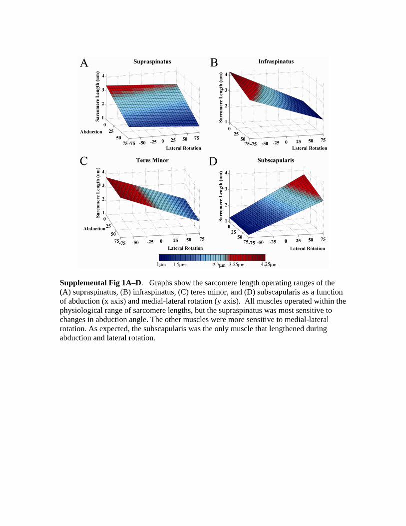
Supplemental Fig 1A–D. Graphs show the sarcomere length operating ranges of the (A) supraspinatus, (B) infraspinatus, (C) teres minor, and (D) subscapularis as a function of abduction (x axis) and medial-lateral rotation (y axis). All muscles operated within the physiological range of sarcomere lengths, but the supraspinatus was most sensitive to changes in abduction angle. The other muscles were more sensitive to medial-lateral rotation. As expected, the subscapularis was the only muscle that lengthened during abduction and lateral rotation.

Supplemental Fig 2A–D. Graphs show the active (upper surface) and passive (lower surface) relative tension profiles of the (A) supraspinatus, (B) infraspinatus, (C) teres minor, and (D) subscapularis muscles as a function of abduction (x axis) and medial-lateral rotation (y axis). Muscles were able to generate at least 75% of their maximum active tension through the majority of ranges of motion, while passive tension peaked in specific joint configurations for each muscle.

Supplemental Fig 3A–D. Graphs show the percent contributions to the total active tension profile for (A) supraspinatus, (B) infraspinatus, (C) teres minor, and (D) subscapularis muscles as a function of abduction (x axis) and medial-lateral rotation (y axis). The total active tension surface peaks at 25º of abduction and 20º of lateral rotation. The subscapularis makes the largest contribution to total active tension followed by the infraspinatus, supraspinatus, and teres minor.
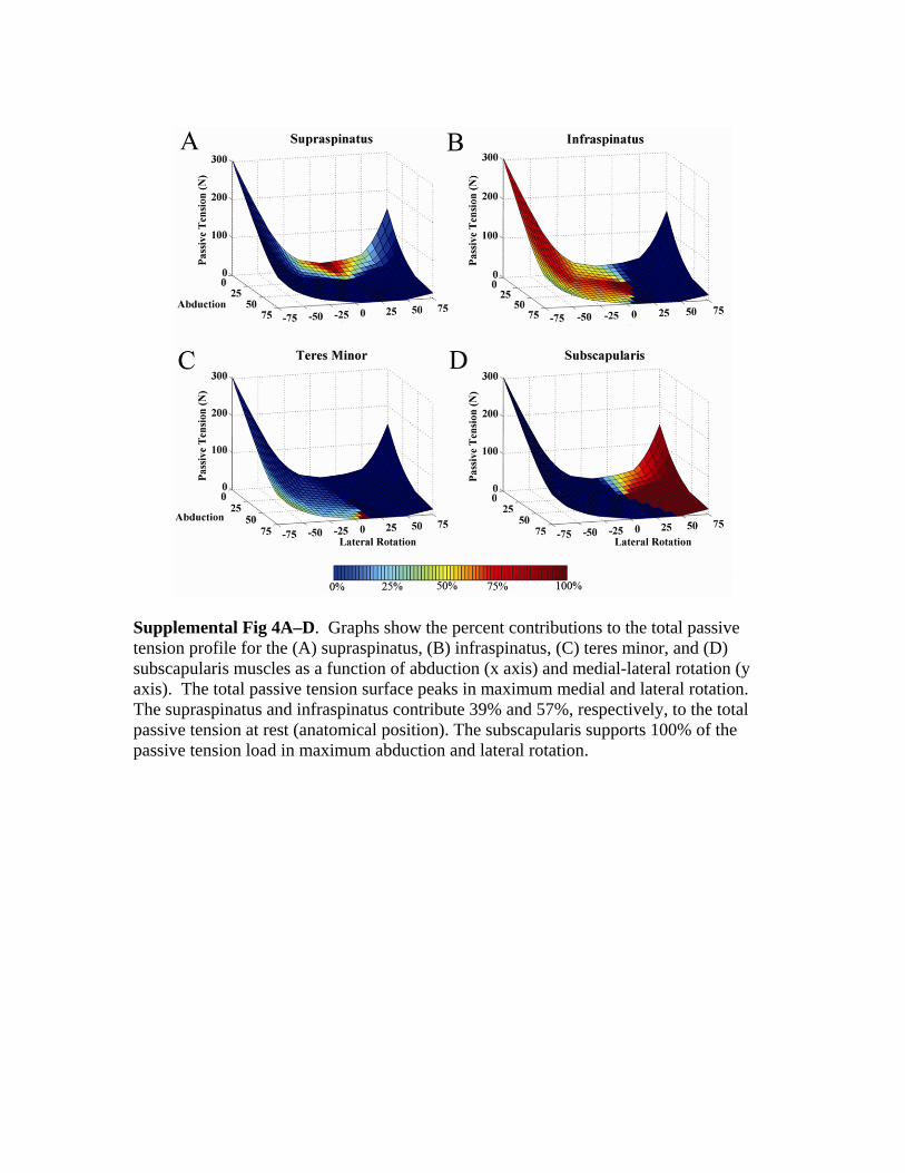
Supplemental Fig 4A–D. Graphs show the percent contributions to the total passive tension profile for the (A) supraspinatus, (B) infraspinatus, (C) teres minor, and (D) subscapularis muscles as a function of abduction (x axis) and medial-lateral rotation (y axis). The total passive tension surface peaks in maximum medial and lateral rotation. The supraspinatus and infraspinatus contribute 39% and 57%, respectively, to the total passive tension at rest (anatomical position). The subscapularis supports 100% of the passive tension load in maximum abduction and lateral rotation.