Immunogenic Subviral Particles Displaying Domain III of ... · Immunogenic Subviral Particles...
97
Immunogenic Subviral Particles Displaying Domain III of Dengue 2 Envelope Protein Vectored by Measles Virus by Indira Harahap A Thesis Presented in Partial Fulfillment of the Requirements for the Degree Master of Science Approved June 2015 by the Graduate Supervisory Committee: Jorge Reyes del Valle Brenda G. Hogue, Chair Douglas Lake Hugh Mason ARIZONA STATE UNIVERSITY August 2015
-
Upload
trinhtuyen -
Category
Documents
-
view
218 -
download
2
Transcript of Immunogenic Subviral Particles Displaying Domain III of ... · Immunogenic Subviral Particles...

Immunogenic Subviral Particles Displaying Domain III of Dengue 2 Envelope Protein
Vectored by Measles Virus
by
Indira Harahap
A Thesis Presented in Partial Fulfillment
of the Requirements for the Degree
Master of Science
Approved June 2015 by the
Graduate Supervisory Committee:
Jorge Reyes del Valle
Brenda G. Hogue, Chair
Douglas Lake
Hugh Mason
ARIZONA STATE UNIVERSITY
August 2015

i
ABSTRACT
Vaccines against the arthropod-borne dengue virus (DENV) are still
commercially nonexistent. A subunit immunization strategy may be of value, especially
if a safe viral vector acts as a biologically active adjuvant. The DENV envelope protein
(E), the main target for neutralizing immune responses, has three conformational
domains. The immunoglobulin-like and independently folding domain III (DIII) contains
epitopes that elicit highly specific neutralizing antibodies. The hepatitis B small surface
antigen (HBsAg, S) was used as a scaffold to display DENV 2 DIII on a virus-like
particle (VLP). A measles virus (MV) was engineered to vector HBsAg and the hybrid
glycoprotein DIII-HBsAg in two different loci (DIII-S). Despite the relatively deleterious
effect on replication caused by the insertion of two transcription cassettes, the
recombinant virus MVvac2(DIII-S,S)P induced the secretion of DIII-S hybrid VLP with
a similar sucrose density as HBsAg particles (1.10-1.12g/ml) and peaked at 48 h post-
infection producing 1.3x10⁶ TCID50/ml infectious MV units in vitro. A second
recombinant virus, MVvac2(DIII-S)N, was engineered to vector only the hybrid DIII-S.
However, it did not induce the secretion of hybrid HBsAg particles in the supernatant of
infected cells. The immunogenicity of the recombinant viruses was tested in a MV-
susceptible small animal model, the experimental group which received two 105 TCID50
I.P. doses of MVvac2(DIII-S,S)P in a 28 day interval developed a robust immune
response against MV (1:1280), HBsAg (787 mIU/ml) and DENV2 (Log10 neutralization
index of 1.2) on average. In summary, it is possible to display DENV E DIII on hybrid
HBsAg particles vectored by MV that elicit an immune response. This forms the basis for
a potential vaccine platform against DENV.

ii
TABLE OF CONTENTS
Page
LIST OF TABLES ...............................................................................................................v
LIST OF FIGURES ........................................................................................................... vi
ABBREVIATIONS ......................................................................................................... viii
CHAPTER
1. INTRODUCTION ...............................................................................................1
1.1 Dengue Epidemiology ...........................................................................1
1.2 Transmission of Dengue Virus. .............................................................4
1.3 Dengue Virus Structure and Replication Cycle .....................................6
1.4 Manifestation of Symptoms in Dengue Virus Infected Patients ..........10
1.5 Enhancement of Pathogenesis ..............................................................11
1.6 Various Approaches to Creating a Vaccine and its Challenges ...........14
1.7 Domain III as a Vaccine Target ...........................................................20
1.8 Hepatitis B Virus-like Particles ...........................................................21
1.9 Measles Virus as Viral Vector .............................................................24
1.10 Research Project Objectives and Hypothesis .....................................26
1.11 Specific Aims .....................................................................................28
2. MATERIALS AND METHODS .......................................................................30
2.1 Cells and Viruses .................................................................................30
2.2 Plasmid Design and Construction ........................................................31
2.3 MV Reverse Genetics System .............................................................34

iii
CHAPTER Page
2.4 Multi-step Growth Kinetic Analysis of Recombinant MVs ................35
2.5 Titration of Viruses ..............................................................................35
2.6 Expression of HBsAg ..........................................................................36
2.7 Preparation of Protein Extracts ............................................................37
2.8 Analysis of Protein Expression ............................................................37
2.9 Particle Isolation and Determination of Density ..................................39
2.10 Animal Experiments ..........................................................................40
2.11 Analysis of the Immune Response of Vaccinated Animals ...............43
3. RESULTS ..........................................................................................................46
3.1 Plasmid Construction and MV Rescue ................................................46
3.2 Reverse Genetics System in MV Rescue .............................................49
3.3 Multi-step Growth Kinetics .................................................................51
3.4 Production of HBsAg Detected by ELISA ..........................................53
3.5 Characterization of the Expression of Hybrid DIII-HBsAg Antigens
from MVvac2(DIII-S,S)P ..........................................................................54
3.6 Characterization of the Expression of Hybrid DIII-HBsAg Antigens
from MVvac2(DIII-S)N and the Correct Display of DIII .........................56
3.7 Particle Isolation and Characterization ................................................58
3.8 Immunogenicity Study .........................................................................61
4. DISCUSSION ....................................................................................................70
5. CONCLUSION ..................................................................................................83

iv
CHAPTER Page
6. FUTURE PERSPECTIVE .................................................................................83
7. REFERENCES ..................................................................................................84
v
LIST OF TABLES
Table Page
1. Dengue Vaccine Candidates in Various Stages of Development ..................................19
2. Outline of the Specific Steps Completed to Reach the Research Aims .........................29
3. Primer Sequences for Overlapping PCR to Construct the Hybrid DIII-HBsAg Coding
Sequence ............................................................................................................................32
4. Primer Sequences Used in DNA Sequencing of Full-length Recombinant Plasmid .....34

vi
LIST OF FIGURES
Figure Page
1. Diagram Showing a World Map Highlighting Countries where Dengue Virus is
Prevalent ..............................................................................................................................1
2. Two World Maps Presenting the Increased International Distribution of the Four
Different Serotypes of DENV, Over 34 Years ....................................................................3
3. Timeline Representing the Transmission of DENV, Following the Feeding Schedule
and Life Cycle of the Mosquito Vector ...............................................................................5
4. Diagram Representing the Replication Cycle of DENV in Infected Host Cell ...............9
5. Diagram Representing the Stages Leading to Antibody-dependent Enhancement .......12
6. Illustration of the Three Main DENV Proteins Targeted for an Antibody Response and
the Function of those Specific Antibodies .........................................................................14
7. Diagram Representing the Different Approaches that has been done to Create a Viable
Tetravalent Vaccine against DENV ...................................................................................17
8. Structure of the Dengue E Protein with its Three Domains...........................................20
9. Illustration of the Chimeric VLPs Engineered to Express DIII and HBsAg .................23
10. Schematic of the Transcription and Replication Mechanism of the Measles Virus
Genome ..............................................................................................................................24
11. Diagram of the Ultracentrifugation of the Discontinuous Sucrose Gradient to Isolate
VLPs ..................................................................................................................................39
12. Diagram Representing the Vaccination Schedule Given to the HDD-SLAM-IFNarKO
Transgenic Mice in the First Animal Experiment ..............................................................41

vii
Figure Page
13. Diagram Representing the Vaccination Schedule Given to the huCD46Ge-IFNarKO
Transgenic Mice in the Second Animal Experiment .........................................................42
14. The Genomic Map of the Recombinant MV Generated ..............................................46
15. Diagram of the Confirmation Digest for the Recombinant Plasmid
pB(+)MVvac2(DIII-S)N ....................................................................................................47
16. Diagram of the Confirmation Digest for the Recombinant Plasmid
pB(+)MVvac2(DIII-S,S)P .................................................................................................48
17. Generation of a Recombinant MV Using the Reverse Genetics System .....................50
18. Multi-step Growth Kinetics of Recombinant MVs ......................................................52
19. Identification of Secreted HBsAg by Recombinant MVs............................................53
20. Western Blots Analyzing the Expression of Hybrid DIII-S ........................................55
21. Analysis of Successful Expression of the Hybrid DIII-S Antigen in Cell Lysates from
Controls and Recombinant MV Infected Cells ..................................................................57
22. Particles Isolation by Discontinuous Sucrose Gradient and Analysis of Each Fractions
Collected ............................................................................................................................60
23. Immunogenicity Study against MV, Hepatitis B and DENV-2 ...................................63
24. Immunogenicity Study against MV and DENV-2 .......................................................66

viii
ABBREVIATION
Term Definition
ATU Additional Transcription Unit
ADE Antibody-dependent Enhancement
CDC Center for Disease Control and Prevention
DC Dendritic Cell
DENV Dengue Virus
DF Dengue Fever
DHF Dengue Hemorrhagic Fever
dsRNA Double-stranded RNA
DSS Dengue Shock Syndrome
DI Domain I of the DENV E Protein
DII Domain II of the DENV E protein
DIII Domain III of the DENV E protein
DIII-S Hybrid DIII-HBsAg Antigen
EC Epithelial Cell
E Protein Envelope Protein
ER Endoplasmic Reticulum
GTase Guanylytransferase
HBsAg Hepatitis B small surface antigen
HBV Hepatitis B Virus
HPV Human Papilloma Virus
IFN Interferon

ix
I.P. Intraperitoneal
IRES Internal Ribosome Entry Site
LAV Live Attenuated Vaccine
LN Lymph Node
MOI Multiplicity of Infection
MTase Methyltransferase
MV Measles Virus
NIH National Institute of Health
NS Non-structural Protein
NTPase Nucleoside Triphosphatase
PDK Primary Dog Kidney Cells
PIV Purified Inactivated Vaccine
Pre-M/M Protein Pre-Membrane/Membrane Protein
RdRp RNA-dependent RNA Polymerase
RTPase RNA Triphosphatase
TGN Trans-Golgi Network
TMB Tetramethylbenzemidine
UTR Untranslated region
VLP Virus-like Particles
YFV Yellow Fever Virus

1
Chapter 1. Introduction
1.1 Dengue Epidemiology. Dengue is the most important arthropod borne viral
disease in the world. It is caused through infection by dengue virus (DENV). In 2013, it
was estimated that 390 million people has been infected with DENV, causing 96 million
clinical cases annually (1). Dengue has become endemic in various countries in the
Americas, the Middle East, Africa, the Western Pacific and Southeast Asia (2). About
40% of the world’s populations live in locations that are highly at risk for the
transmission of DENV (Fig. 1). Areas at risk are mainly tropical or sub-tropical bearing
the optimal breeding ground for the arthropod vectors that transmits DENV, which are
the mosquitoes: Aedes aegypti and Aedes albopictus (3).
Figure 1. Diagram Showing a World Map Highlighting Countries where Dengue Virus is
Prevalent. Dengue is endemic in over 100 countries. The three different shades of red indicate the
level of prevalence of the virus in each country. Dengue and dengue hemorrhagic fever are prevalent
in urban and suburban areas in the Americas, South-East Asia, Eastern Mediterranean, and Western
Pacific; it is mainly prevalent in rural areas in Africa (3).

2
Modernization and societal changes have aided in the geographic expansion of the
mosquito vectors. These changes include globalization, increased international travel, as
well as unplanned urbanization (2). Unplanned urbanization has pushed human dwellings
to invade the rainfall forest, placing humans in mosquitoes’ home environment, which
increases exposure rate (4). In addition, inadequate waste and sewer management, along
with lack of a vector control program that is usually present in unplanned urbanizations
exacerbate epidemic activity of DENV (2). These types of scenarios can be seen in
developing countries where DENV infections run rampant, like the Philippines, Thailand,
and Vietnam, which are the Asian countries that has the highest dengue prevalence (2). In
these countries, DENV infection is one of the top 10 causes of death in hospitalized
children (5). In addition to the burden that DENV infection place on the health care
system of each country, it also imposes a high economic burden on the country’s
government and on each individual (3). In Southeast Asia, clinical DENV infection cause
an annual economic burden of ~$950 million. Therefore a reduced transmission of
DENV would greatly lessen the burden placed on both the healthcare system and the
economic stability of developing countries where DENV infection is endemic.
Many other factors have been associated to the 30-fold increases in DENV
prevalence in the past 50 years. The main factor being the expansion of the habitat for A.
aegypti and A. albopictus; due to globalization, international travel, climate changes,
arthropod vector’s adaptation, etc. In the U.S. recent outbreaks have happened in Hawaii
in 2001, Texas in 2005, and Florida in 2009 to 2011 (6). In 2010, the 1st cases of DENV
infections due to indigenous transmission in Europe was reported (2). This was
acknowledged to be caused by A. albopictus whose eggs have adapted to the subfreezing

3
temperature in Europe. All of these factors only magnify the dire need of a viable vaccine
against DENV as the habitats for the vector transmitting DENV broaden (6). Most severe
cases of DENV infection reported annually originate from the Asia-Pacific region, the
Americas, and Africa; where there is a high concentration of all four heterologous
serotypes circulating (7). In addition, the spread of the different DENV serotypes have
steadily increased all over the world in recent years (Fig. 2). The increase in international
travel have aided in the spreading of the mosquito vectors that transmits DENV; this
includes the movement of the virus in infected individuals who travels internationally as
well as cases where the mosquito vector’s eggs were unknowingly transported in tires
Figure 2. Two World Maps Presenting the Increased International Distribution of the Four
Different Serotypes of DENV, Over 34 Years. The top world map shows that only one area of the
world, the Asia-Pacific region, have all four DENV serotypes circulating. 34 years later, the bottom
world map shows that now the majority of the world harbors all four DENV serotypes (8).

4
being shipped overseas (8). The broadening of the habitat for A. aegypti and A. albopictus
have also been encouraged by international travel, where they become adapted to their
new environment. The expansion and development of human housings into the natural
habitat for these vectors have also increase the rate of exposure between humans and
mosquitoes (8). Overall, many factors have encouraged the increased distribution of
DENV, which have promoted more frequent and larger dengue epidemics that are
associated with more severe symptoms of the illness.
1.2 Transmission of Dengue Virus. DENV is an arbovirus, transmitted by the
bite of female mosquitoes, specifically: A. aegypti and A. albopictus (2). A. aegypti
originated from Africa and has now spread throughout most of the world, whereas A.
albopictus originated from Southeast Asia, with a cold-resistant strain in Europe (5).
Transmission of DENV happens during the feeding activity for these female mosquitoes,
which peaks in the morning time and late afternoon. The virus is transported from person
to person while these mosquitoes have their blood meal, at which point the virus is
subcutaneously injected into the person (2). The transmission is carried exclusively by
female mosquitoes due to their dependence on blood for oviposition. It has been
demonstrated that there is vertical transmission from infected females to their ova, this is
the main mechanism that explain the persistence of infected mosquitoes during winter in
some non-tropical regions. Male mosquitoes feed from fruit.

5
Viremia, the presence of a high titered virus in the bloodstream, can usually be
observed 24-48 hours before the appearance of clinical symptoms. Viremia may last up to
10 days in patients infected with DENV (2). If the mosquito feed on an individual with
viremia, the mosquitoes can become infected by DENV which will target the epithelial
cells of the mosquito’s mid-gut (2). These mosquitoes will then become infectious, after
an incubation period of 8-10 days post-feeding. After spreading systemically through the
haemocele, DENV reaches the salivary glands where it can be easily transmitted to
humans (2). In summary, the mosquito can transmit DENV by directly changing host
during a blood meal or after 8-10 days when DENV has multiplied in the salivary glands
(Fig. 3). The mosquito will then be able to transmit DENV for the rest of its life in
addition to the possibility of transmitting DENV to its eggs, as mentioned (5).
Figure 3. Timeline Representing the Transmission of DENV, Following the Feeding Schedule
and Life Cycle of the Mosquito Vector. A mosquito may obtain DENV when it feeds from a
viremic host, who has a high titer of DENV in their bloodstream, enough to be taken up by the
mosquito. Mosquito may infect a second host if it directly takes another blood meal after it obtained
DENV. As well as after 8-10 days post-blood meal from a viremic first host, because the DENV
would have had time to replicate and reach the salivary gland of the mosquito [CDC].
6
Initially in humans, DENV targets macrophages and dendritic cells (DC) because
they are present in the epidermis and subcutaneous tissue where the virus would be
injected by the mosquito (2). The infected macrophages and DCs will then migrate to the
lymph node (LN) where the virus will spread to other macrophages and ultimately
peripheral monocytes. This will result in the first viremia, as viruses are moved in
draining and remote LN. The major sites of viral replication for DENV in humans are
posited to be DCs, macrophages, and monocytes. In addition, DENV may also be found
in other tissues throughout the body, such as the lungs, the liver, the spleen and the
kidneys (2). Cells that are infected with DENV usually die through apoptosis or necrosis.
Necrosis causes the increased production of toxins which in turn will activates the
fibrinolytic and coagulation systems. Viremia, platelet dysfunction, and severe
thrombocytopenia, will result in the fragility of the capillaries, presented as bruising,
petechiae, and gastrointestinal mucosal bleeding; these are the characteristics of DHF (9).
In addition, increased vascular permeability and coagulopathy is amplified when IgM
antibodies reacting with the epithelial cells (EC), platelets, and plasmin are produced.
During secondary infection, enhancement of infection by IgG (discussed below) will
contribute to the high viral load that will lead to the secondary viremia (9).
1.3 Dengue Virus Structure and Replication Cycle. DENV is a member of the
family of Flaviviridae; It is an enveloped virus that has an icosahedral nucleocapsid
containing ~10.7kb of positive-sense, single-stranded RNA as genome (1). This encodes
a polyprotein that will be cleaved into seven nonstructural proteins and three structural
proteins that will be processed co- and post-translation by cellular and viral encoded
proteases. The three structural proteins are C, pre-M/M, and E proteins. The seven

7
nonstructural proteins are NS1, NS2A, NS2B, NS3, NS4A, NS4B, and NS5 (2). The
capsid protein (C protein) serve as RNA chaperone and binds at a high affinity with the
viral RNA; this protein is confined to the nucleus and nucleoli when independently
expressed on its own in infected cells. The pre-membrane or membrane protein (pre-M/M
protein) plays a vital role in viral fusion and entry; it has been shown that mutations in
the M protein resulted in lower rates of virus assembly and entry (2). The envelope
protein (E protein) in DENV lies parallel to the surface of the virion. Since it has been
shown that anti-E antibodies will neutralize viral infectivity through the inhibition of viral
binding to cells, the E protein has been concluded to play a vital part in virus attachment
and virus-specific membrane fusion (10). In addition, it has been observed that the E
protein’s stem region plays a vital role in heterodimerization of pre-M, hence affecting
membrane binding and virus assembly (2). The non-structural protein 1 (NS1) is 46 kDa,
it is a glycoprotein that co-localizes with double-stranded RNA (dsRNA) in mammalian
and insect cells, which makes it vital for viral replication. The non-structural protein 2-A
(NS2A) is 22 kDa, it is important in the replication of the genome, RNA. The non-
structural protein 2-B (NS2B) is 14 kDa, it is a membrane associated protein that is a
required cofactor for the NS3 protein, as it recruits NS3 to the endoplasmic reticulum
(ER) membrane. The non-structural protein 3 (NS3) is 69 kDa, it serves as a
multifunctional viral protease; it has the protease domain at the N-terminal and a domain
that includes nucleoside triphosphatase (NTPase), RNA triphosphate (RTPase), and
helicase activities at the C-terminal. The non-structural protein 4-A (NS4A) is 16 kDa, it
co-localizes with NS3 and the viral dsRNA in cytoplasmic foci, which contains viral
replication complexes. Constitutes a cofactor for the viral protease. It may be important

8
in formation of replication vesicles, making it a viral membrane protein. The non-
structural 4B (NS4B) protein weighs 28 kDa, it also co-localizes with NS3 and the viral
dsRNA at the site of RNA replication. The non-structural protein 5 (NS5) is the largest
protein (105 kDa). NS5 is also the most conserved protein because it encodes the RNA-
dependent RNA polymerase (RdRp) on its C-terminal domain, which makes it vital for
viral RNA synthesis; methyltransferase (MTase) and guanylytransferase (GTPase) are
also encoded on its N-terminal domain. In addition, this protein is also involved in
counteracting the interferon (IFN) system of the innate immune response (2, 5, 6).
During host cell infection, DENV enters the cell via clathrin-dependent receptor-
mediated endocytosis (3). After endocytosis, the environment inside the endosomes
becomes acidified, which causes a pH-dependent conformational change that rearranges
the E and M proteins prompting a class II membrane fusion machinery; exposing the
fusion loop into the endosomal membrane (Fig. 4). This allows the fusion of the viral
envelope with the endosomal membrane; releasing the viral nucleocapsid into the
cytoplasm (2). The nucleocapsid will then dissociate in the cytoplasm, releasing the viral
RNA to be translated by the ribosomes on the rough ER. The polyprotein is then
translated and co-translationally processed and cleaved by host and viral proteases into
the respective structural and non-structural viral proteins. The RdRp generates the
negative complementary strand to the viral genome, which will be used as a template to

9
Figure 4. Diagram Representing the Replication Cycle of DENV in Infected Host Cell. DENV
binds to the host cell’s surface receptor and enter the cell via receptor-mediated endocytosis. The
acidification of the internal environment of the endosome allows for the rearrangement of the DENV
E and M protein, resulting in the fusion of the viral envelope with the endosomal membrane. Hence,
the viral RNA is released into the cell cytoplasm. The viral genome is then translated creating a
polyprotein, which is cleaved into the respective 3 structural and 7 non-structural viral proteins. The
RdRp encoded by NS5 generates the complementary strand to the viral genome for viral replication.
The viral RNA will then associate with capsid proteins to form the nucleocapsid. The nucleocapsid
will associate with the E and pre-M rich ER membranes and buds into the ER lumen to create
immature virion. This will exit the cell via exocytosis through a secretory pathway (2).

10
replicate more copies of the viral RNA. The nucleocapsid is then formed by the
association of the viral RNA with the capsid protein. The nucleocapsid buds into the
lumen of the ER and obtain the lipid membrane proteins pre-M/M and E (2, 3). After the
assembly of the immature virions, it is transported out of the host cell through the
classical secretory pathway. The viral particles will be contained in a vesicle, transported
through the Golgi stacks and secreted by exocytosis. During its transport through the
trans-Golgi network (TGN), the protease furin cleaves the pre-M protein to its mature
form, M protein that remains on the mature and infectious virion.
1.4 Manifestation of Symptoms in Dengue Virus Infected Patients. There are 4
different serotypes of DENV: DENV-1, DENV-2, DENV-3, and DENV-4 (1). The
presence of multiple serotype represents a challenge in controlling illnesses and
symptoms in patients infected with DENV as well as in creating a viable commercial
vaccine with complete protection against DENV. Clinically, DENV infection has been
divided in two main syndromes: Classic Dengue and severe Dengue infection. The
patient can evolve clinically from non-significant, dengue fever (DF), to the extremes of
dengue hemorrhagic fever (DHF) and dengue shock syndrome (DSS) (4). After infection
with a mosquito bite, an individual may be asymptomatic, or may develop initially a flu-
like syndrome. In most cases, the infection will be resolved without any complications
(1). The majority of severe DENV infection cases will result in DF, where the symptoms
include: sudden onset of fever reaching 102.2ºF, headache, myalgia, macular or
maculopapular rashes, joint pains, body aches, and retro-orbital pain (5, 6). Usually, this
acute phase is limited to last only 1 week, followed by recovery. However, 1% to 7% of
DENV patients may develop DF with hemorrhage, where symptoms include: petechial

11
purpura, ecchymosis, and epistaxis. Furthermore, up to 2% of DENV patients may
develop the more severe and life-threatening DHF, where complications include:
increased vascular permeability, liver damage, thrombocytopenia, and hemorrhaging
from the skin, gum, nose, and gastrointestinal tract. These cases may also worsen and
progress to DSS, where a redistribution of the effective central vascular volume towards
the periphery is caused by vasodilatation and diapedesis, resulting in a sudden drop in
effective perfusion pressure (2). The main problem in DHF or DSS is not blood loss but
intravascular fluid loss. Therapy at this stage of symptoms is the management of blood
volume and blood pressure. The current therapy for patients infected with DENV is
focused on support, mainly with bed rest, fluid intake, and the control of fever or pain
with antipyretics and analgesics. In mild cases, patients with DENV infection will
respond in one or two days after plasma expansion with isotonic saline solution (5).
1.5 Enhancement of Pathogenesis. Pathogenesis in an individual infected with
DENV may be enhanced due to the existence of the four different serotypes. Two
different pathways of pathogenesis enhancement non-mutually exclusive have been
considered: antibody dependent enhancement (ADE) and the original antigenic sin. Upon
DENV infection, serotype-specific life-long immunity is acquired; however it will only
confer a transient immunity against another serotype (11). Studies have shown that
antibodies elicited from a primary infection are a major factor in the rapid development
of the more severe DHF and DSS after a secondary infection with a heterologous
serotype (12, 13).

12
The explanation for ADE is that the non-neutralizing, cross-reactive antibodies
gained from the primary infection will bind to the heterotypic virus (1, 9, 13). The
antibody-virus complex will increase the uptake of the viral particle by Fc-receptor
expressing cells such as monocytes, macrophages and DCs (Fig. 5). This will in turn
increase the viral replication and increase the severity of infection (14). In addition, ADE
may also occur in situations when there is a low antibody concentration or when there is
low antibody avidity for the current DENV infecting serotype. Specifically, when the
amount of antibodies bound the virion is below the amount needed for neutralization,
which is a minimum of ~30 for each virion (1, 2). The occurrence of ADE has been
supported through its observation in vitro; where non-neutralizing antibodies that were
specific to DENV were able to increase viral replication in peripheral blood leukocytes
(6). In addition, ADE incidences in vivo have also been observed in monkey. Scientists
observed a higher viral load in monkeys that received a passive transfer of humanized
anti-DENV monoclonal antibody or human sera that are immune against DENV (6).
Figure 5. Diagram Representing the Stages Leading to Antibody-dependent Enhancement.
Cross-reactive heterotypic antibodies from the primary DENV infection bind to the viral particles
from the current DENV infection. These antibodies are non-neutralizing that creates an Ab-virus
complex which enhances its uptake by Fc-receptor expressing cells such as monocytes,
macrophages, and DCs. Resulting in an increased viral load (14).
13
Original antigenic sin occurs when pathogenesis enhancement is mediated by the
cellular immune response (2). Scientists have found that the memory T cells that are
specific for DENV serotype responsible of the primary infection may be reactivated due
to cross-reactive characteristics. These T cells show suboptimal degranulation, but it will
induce an increased secretion of cytokines, resulting in apoptosis of infected and non-
infected bystander cells (6). This excessive activation of the cellular immune system
causes a cytokine storm, which increases vascular permeability. This reaction has been
concluded to be the main reason behind life-threatening DSS cases (1, 2, 6).
Since all of the DENV vaccines that are most developed target the humoral
immune system, ADE is the major cause of difficulties in the generation of a vaccine
against DENV. This created a fear in the production of DENV vaccines because a
vaccine that could not generate an equilibrated neutralizing immunity against all four
serotypes could increase the risk of vaccinees developing the more severe and life-
threatening DHF and DSS. Therefore it has been concluded that a tetravalent vaccine is
mandatory (15). A successful vaccine against DENV must induce a balanced and long-
lasting immunity against all four of the heterologous DENV serotypes. In addition, it
must target an antigenic epitope on DENV, which is conserved in all four DENV
serotypes, yet also be highly specific to each serotype to avoid cross-reactivity. This
challenge in creating a safe vaccine for DENV has resulted in a lack of commercially
available vaccines against DENV, which has been approved.

14
1.6 Various Approaches to Creating a Vaccine and its Challenges. An ideal
dengue vaccine must have a couple of vital characteristics. First, it must be a tetravalent
vaccine formulation and induce a long-lasting balanced immune response to each
serotype. The vaccine should also be safe in 9-12 months old children, so it can be used
as a pediatric vaccine, since this is the population most at risk of complicated Dengue
infections. A vaccine that can be produced at a low cost with high efficiency for the ease
of scaling up production would also be desirable (10). The main targets that have been
used in vaccines against DENV infection in humans are the pre-M-E, and NS1 proteins
(Fig. 6). The pre-M protein creates a heterodimer with the E proteins and is cleaved when
the virion matured. The M protein is completely inaccessible for antibody binding
because it is hidden beneath the E protein dimer. However, in some cases where
incomplete cleavage of pre-M by furin results in partially mature/immature virion, this
Figure 6. Illustration of the Three Main DENV Proteins Targeted for an Antibody Response
and the Function of those Specific Antibodies. Antibodies against the envelope protein of the
mature virion is found to be neutralizing, but may induce antibody-dependent enhancement.
Antibodies against the pre-M protein on the immature virion are found to be serotype cross-reactive
and may induce antibody-dependent enhancement. Antibodies against NS1 will cause complement-
dependent lysis of the infected host cell (1).

15
can be recognized by M protein-specific antibodies. Yet, antibodies against the pre-M
protein are very cross-reactive across the four different serotypes (1). The NS1 protein
has a multimeric structure and it can be found displayed on the surface of the infected
host cells or released as a soluble particulates. Although antibodies for NS1 are very
serotype specific, this glycoprotein is not incorporated into the virion; leading scientists
to conclude that ADE can be avoided by utilizing NS1 as the target epitope. In addition,
antibodies against NS1 also cause complement-dependent lysis of the DENV infected
host cell (1). The E glycoprotein is the main membrane surface component of DENV. In
its tightly packed mature form the cross reactivity of the E protein is controlled by the
dimeric conformation on the virion surface. Each virion is composed of ninety dimers
tightly packed with an icosahedral symmetry, which limits the accessibility of epitopes to
bind antibodies. It has been found that antibodies against the E protein after the first
DENV infection are very serotype cross-reactive. These antibodies are also short-lived,
since most of them are not represented in the memory immune response that protects the
individual against the serotype that caused the prime infection. Structurally, it has been
described three domains in each monomer of E. The fist two domains are not sequential
and represent antibody epitopes with a high cross-reactive potential. However, domain III
(DIII) of the E protein has the highest variable in amino acid sequence between the four
different serotypes. Therefore, it has been observed that antibodies specifically against
DIII of the E protein are highly serotype-specific.
In addition to ADE, there are multiple challenges that are faced in the production
of a DENV vaccine. The mechanism of protective immune response against DENV is not
completely understood (16). In addition, vaccine development is further halted due to the

16
lack of animal model for DENV. Normal mice infected with human DENV do not show
measurable viremia or disease (17, 18). Therefore researchers have to use a mouse
adapted strain of the virus or infect an immunocompromised mouse model (17). Some
have argued the relevance of the results from these studies lacking a reliable animal
model when applied to human infection.
Despite these challenges, there are multiple DENV vaccine platforms that have
been pursued and some of which have been approved for clinical testing. These include
live attenuated virus (LAV), purified inactivated virus (PIV), recombinant subunits,
virus-like particles (VLPs), and plasmid/viral vectors. LAV vaccines make up the
majority of vaccine candidates in clinical trials (Table 1). It has been shown to produce a
large, long-lasting, and broad humoral and cellular immune response. DENV strain for
LAV were generated by some research groups through serial passaging in tissue culture,
and these were found to induce T cell and antibody responses that are similar to those
caused by a natural infection (1). However, it has been difficult for LAV vaccines to
achieve a balance between an optimal level of attenuation and a low level of
reactogenicity (16, 17). The top 3 most developed DENV vaccines are the recombinant
LAV from Sanofi Pasteur, the US National Institute of Health (NIH), and the US Centers
for Disease Control and Prevention (CDC) – partnered with Inviragen (Fig. 7). The
vaccine developed by Sanofi Pasteur utilized the attenuated vaccine strain 17D of the
yellow fever virus (YFV) as the backbone of their recombinant virus. They replace the
gene encoding pre-M and E proteins with those from each DENV serotypes (1). The
vaccine developed by the NIH utilizes the attenuated version of DENV-1, 3, and 4
genome, the attenuating mutation can be found in the 3’ untranslated region (UTR).
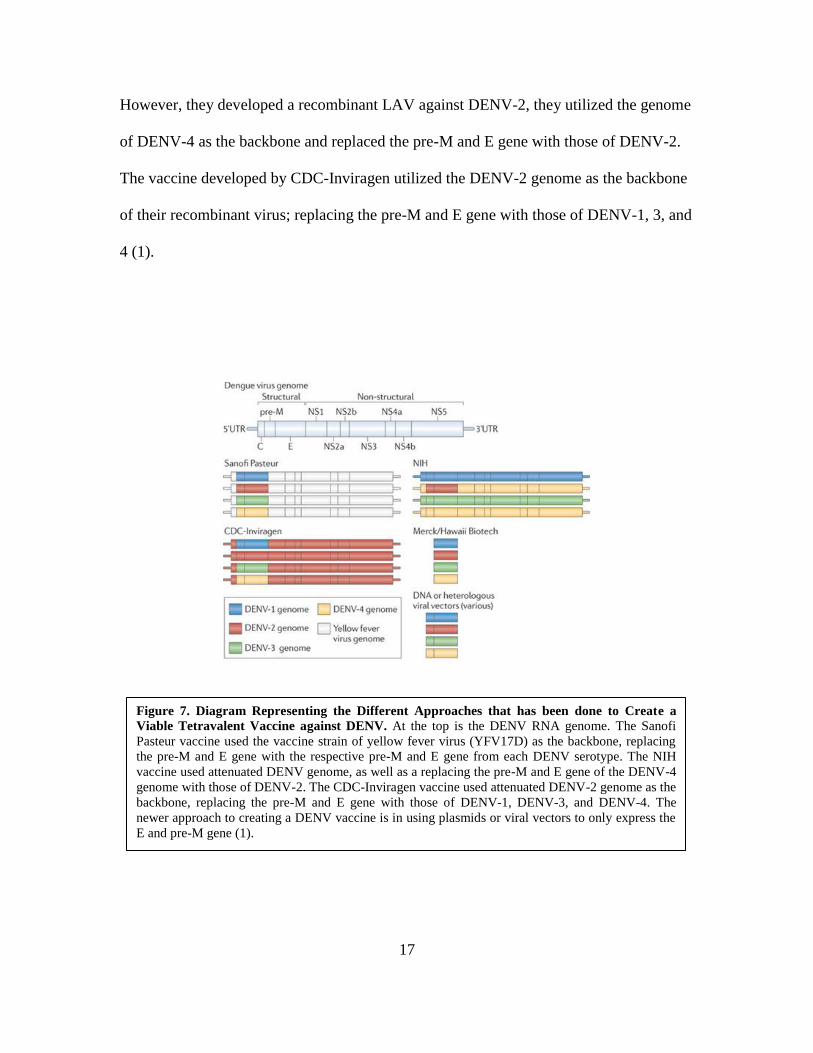
17
However, they developed a recombinant LAV against DENV-2, they utilized the genome
of DENV-4 as the backbone and replaced the pre-M and E gene with those of DENV-2.
The vaccine developed by CDC-Inviragen utilized the DENV-2 genome as the backbone
of their recombinant virus; replacing the pre-M and E gene with those of DENV-1, 3, and
4 (1).
Figure 7. Diagram Representing the Different Approaches that has been done to Create a
Viable Tetravalent Vaccine against DENV. At the top is the DENV RNA genome. The Sanofi
Pasteur vaccine used the vaccine strain of yellow fever virus (YFV17D) as the backbone, replacing
the pre-M and E gene with the respective pre-M and E gene from each DENV serotype. The NIH
vaccine used attenuated DENV genome, as well as a replacing the pre-M and E gene of the DENV-4
genome with those of DENV-2. The CDC-Inviragen vaccine used attenuated DENV-2 genome as the
backbone, replacing the pre-M and E gene with those of DENV-1, DENV-3, and DENV-4. The
newer approach to creating a DENV vaccine is in using plasmids or viral vectors to only express the
E and pre-M gene (1).
18
The results from the clinical trial for some of these vaccines found a problem seen
in tetravalent vaccines, where the simultaneous vaccination with all 4 different serotypes
caused an inter-serotype interference. The consequence was observed in viremic patients
caused by one or two dominant serotype with higher viral replication fitness compared to
the other serotypes. The newer approaches followed by some research groups in creating
a vaccine against DENV are utilizing protein subunit vaccines, heterologous viral
vectors, inactivated virion containing only structural proteins, or nucleic acid vaccines
encoding pre-M and E proteins (1).
A different approach, the PIV vaccine, contains all of the viral structural protein
and RNA, since it is an inactivated wild-type virus. The inclusion of structural proteins
allows the induction of an immune response against them. Recombinant subunit vaccines
allow for the targeting of a specific viral antigen to induce an immune response. Most
subunit vaccines express truncated parts of the envelope protein. Various protein
expression systems has been used such as bacterial, yeast, insect, mammalian cells, etc.
(17). VLP vaccines are advantageous because it does not contain any replicative gene
material, but it is able to express a high and repetitive amount of antigens on its
membrane. Studies have shown that this increases the immunogenicity of the vaccine.
DNA and virus-vectored vaccines is a great design for in vivo expression of antigens;
most are in the form of VLPs. DNA vaccines are also advantageous in their thermo-
stability, which causes it to be cost-efficient for vaccine storage and transport. Virus-
vectored vaccine may induce immune response against both the expressed DENV antigen
as well as the viral vector itself. The utilization of a well-known and well-characterized
licensed vaccine has been established as a vaccine vector. Some concerns have arisen,

19
however, that pre-existing immunity against the viral vector itself may halt the
immunogenicity of the virus-vectored DENV vaccines (17, 19). All of the vaccine
designs against DENV have its advantages and disadvantages.
Type Vaccine approach Developer Stage of
Development
LAV,
Tetravalent,
Chimeric
Chimeric viruses YFV17D-DENV Sanofi Pasteur Phase III
LAV.
Tetravalent,
Chimeric
Chimeric DENV with 3’ UTR deletion
mutations (Δ30) NIH/ NIAID Phase I
LAV,
Tetravalent,
Chimeric
Empirically attenuated (passaged in
tissue culture) Recombination using
DENV-2 backbone and pre-M/E from
DENV-1 to DENV-4
CDC-Inviragen Phase II
LAV,
Tetravalent
Empirically attenuated by passage in
primary dog kidney cells (PDK) WRAIR/GSK Phase II
PIV, Tetravalent Purified and inactivated DENV-1 to
DENV-4 WRAIR Phase I
PIV, Monovalent Purified and inactivated DENV-1 WRAIR Phase I
Recombinant
Subunit
E protein of DENV, affinity purified,
produced in insect cells
Hawaii
Biotec/Merck Phase I
Recombinant
Subunit
DIII of the E protein fused to a carrier
protein Preclinical
DNA Targeting NS1 Multiple Preclinical
DNA Tetravalent DENV-1 to DENV-4, pre-M and E
proteins NMRC Phase I
DNA Tetravalent
DIII of the E protein from DENV-1 to
DENV-4, synthetic consensus
(SynConTM
) Human codon optimized
Inovio Precilinical
DNA
Monovalent DENV-1 pre-M and E proteins NMRC Phase I
DNA Shuffle
E protein from DENV-1 to DENV-4,
codon optimized DNA shuffling,
generating single chimeric antigen
NMRC/ Maxygen Preclinical
Adenoviral
Vector
Expression of the pre-M and E proteins
from DENV-1 to DENV-4 in
recombinant adenoviral vector
NMRC/GenPhar Preclinical
Alphavirus
replicon particles
Expression of the pre-M and E proteins
or soluble E dimers from DENV-1 to
DENV-4, in VRP
Global Vaccines Preclinical
Table 1. Dengue Vaccine Candidates in Various Stages of Development (6).

20
1.7 Domain III as a Vaccine Target. The E protein of DENV forms a dimeric
assembly of ninety subunits displayed in icosahedral symmetry. Each monomer contains
three structural domains (Fig. 8): Domain I (DI), Domain II (DII) and Domain (DIII). DI
of the E protein has an 8-stranded central β-barrel structure and two large loops that form
the elongated DII, which has the highly conserved hydrophobic fusion peptide (20, 21).
The C-terminal of DIII can fold independently from the other two domains in an
immunoglobulin-like manner as a core protein, stabilized by a single conserved disulfide
bridge (10). During the fusion transition, DIII undergoes the most significant
displacement (20).
DIII of the envelope protein has been known to contain a cell surface receptor
recognition site, and in turn it is important in host cell binding. Previous studies have
observed that recombinant DIII from the E protein of DENV have inhibited viral
infectivity through binding competition with the host receptor (20). This suggests that
DIII contains a neutralizing type-specific and sub complex-specific epitopes. Anti-DIII
antibodies that have been found to be potently neutralizing and type-specific, hence less
cross-reactive with a lower risk of causing ADE (17, 19). Although DIII displays an
Figure 8. Structure of the Dengue E Protein with its Three Domains. A, The three domains of
dengue E protein: DI (red), DII (yellow), and DIII (blue). B, Conformation of the E protein in the
mature virus particle. C, Arrangement of each domain of the E protein on the surface of the mature
virus particles; 90 dimers in an icosahedral lattice (21).

21
identical fold across all four different serotypes of DENV, it also has the most variable in
amino acid sequences between the four serotypes (1, 20). Moreover, studies have shown
that neutralizing serotype-specific antibodies against DENV focuses on DIII (22).
Mutations to DIII residues have also lead to the decrease in the binding of neutralizing
monoclonal antibodies (23). Therefore, for our project, we have decided to focus on
DENV 2 E DIII as the antigenic epitope to target in creating our recombinant vaccine.
1.8 Hepatitis B Virus-like Particles. Virus-like particles (VLPs) are multi-
protein complexes that are similar in structure to complete viruses, but it does not contain
a viral genome, making them non-infectious, thus safer (1). VLPs formation is completed
through budding from the usual cellular mechanism, which allows it to be similar to the
infective virus structure (24). VLPs have been used in production of vaccines in the
pharmaceutical industry due to its ability to cause a good immune system response; VLPs
has been shown to elicit both humoral and cellular immune response (25). This is partly
due to their highly repetitive surface proteins that form its structure, which will induce
the activation of the immune system. Two of the well-known vaccines that have been
developed through the use of VLPs are human papilloma virus (HPV) and hepatitis B
virus (HBV) (24). This was done using the hepatitis B virus small surface antigen
(HBsAg) and the VLP produced by the recombinant L1 protein of HPV. These VLPs
induce both neutralizing and protective immune responses and has been shown to be
stable and safe. Hepatitis B virus creates surface antigens that make up the viral envelope
protein. There are three different types of surface antigens that are produced: the large
surface antigen (L), made up of preS1, preS2 and the S domain, the middle surface
antigen (M), made up of the preS2 and the S domain, and the small surface antigen

22
(HBsAg), made up of only the S domain (26). The small surface antigen of hepatitis B
virus (HBsAg) has been shown to be produced in the highest quantity. In addition,
HBsAg have been observed to spontaneously assemble to create empty VLPs in the
absence of any other viral. The VLPs created by HBsAg is made up of 100 to 150
subunits of the 226 amino acid of the HBsAg protein (26). Due to the small size of DIII
of the E protein (~100 amino acids), expressing it by itself will not elicit a strong immune
response; hence we will use chimeric VLPs as our vaccine platform (Fig. 9). Chimeric
VLPs are an adaptation to the VLP design and structure, where the immunogenic
antigens are incorporated onto their surface (25, 27). This will be done through the fusion
of the target epitope to the envelope protein of a virus used to form the VLP. This process
allows the expression of any specific epitope that is targeted by neutralizing antibodies to
be expressed on a highly immunogenic VLP, which may act as an adjuvant and boosts
the immune response (27, 28). As previously mentioned, purified HBsAg VLPs are used
in the currently licensed vaccine against HBV. Antibody protection induced by this
vaccine is mainly specific for the “a”-determinant of the viral envelope. Previous studies
have shown the successful insertion of foreign sequences into the N-terminus of HBsAg
without affecting the conformational epitopes of the “a”-determinant (26, 29, 30).
Therefore, it is possible to create a chimeric HBsAg VLP to function as a bivalent
immunogens that will induce immune response against HBV and the foreign epitope.
23
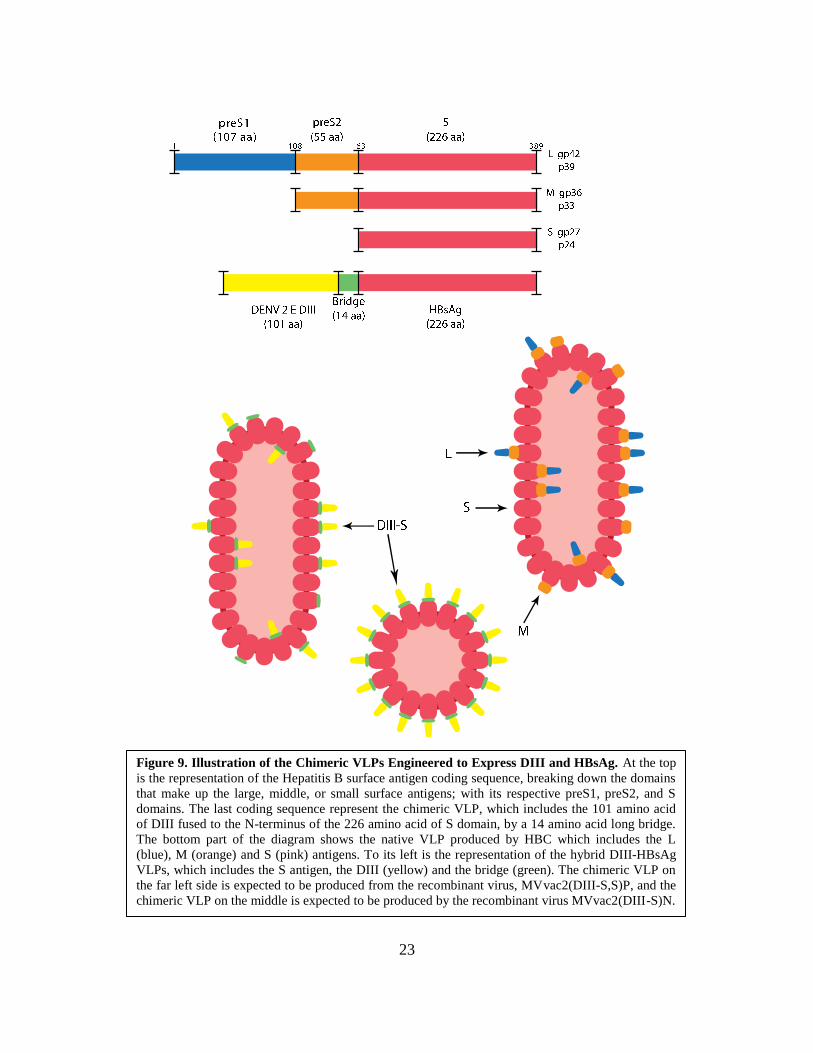
Figure 9. Illustration of the Chimeric VLPs Engineered to Express DIII and HBsAg. At the top
is the representation of the Hepatitis B surface antigen coding sequence, breaking down the domains
that make up the large, middle, or small surface antigens; with its respective preS1, preS2, and S
domains. The last coding sequence represent the chimeric VLP, which includes the 101 amino acid
of DIII fused to the N-terminus of the 226 amino acid of S domain, by a 14 amino acid long bridge.
The bottom part of the diagram shows the native VLP produced by HBC which includes the L
(blue), M (orange) and S (pink) antigens. To its left is the representation of the hybrid DIII-HBsAg
VLPs, which includes the S antigen, the DIII (yellow) and the bridge (green). The chimeric VLP on
the far left side is expected to be produced from the recombinant virus, MVvac2(DIII-S,S)P, and the
chimeric VLP on the middle is expected to be produced by the recombinant virus MVvac2(DIII-S)N.

24
1.9 Measles Virus as Viral Vector. Measles virus (MV), a member of the
Paramyxoviridae family, is an enveloped, negative-sense single-stranded RNA virus. The
negative sense genome functions as template to generate sub-genomic mRNAs though a
conserved mechanism (31). There are six cistrons coding for eight proteins in MV, which
includes coding for: N, P/C/V, M, F, H, and L proteins (32, 33). The transcription
mechanism of mononegavirales posits that boundary signals between cistrons determine
a gradient transcription from 3’ to 5’ extremes (Fig. 10). The gene closest to the 3’ end
(coding for N protein) is to be transcribed in higher amount and the gene towards the 5’
end (coding for L protein) is to be transcribed in the least amount (31, 32).
The recognition of these sequences allows us to insert additional transcription
units (ATUs) between native cistrons. Thus recombinant strain of MV can be created by
various ATUs that may be placed strategically within the viral genome, generating a viral
Figure 10. Schematic of the Transcription and Replication Mechanism of the Measles Virus
Genome. The top image of the diagram depicts the genomic map of the measles virus genome, a
negative sense, single-stranded RNA. The bottom image of the diagram shows the viral transcripts,
5’-3’ mRNAs, which represent the amount of each mRNA produced due to the viral transcription
gradient (32). This in turn will cause a gradient in the amount of proteins translated based on the
location of the gene encoding that specific protein.
25
vector that expresses foreign proteins (34). The locations of these ATUs determine the
amount of protein that will be produced from that inserted gene (32). Theoretically, ATU
sites cloned after the coding region of each structural protein can be generated.
Practically, we have available in the laboratory ATUs cloned in N-P, P-M, H-L and L-
trailer gene boundaries and some combinations; the presence of convenient restriction
sites allows the straightforward cloning strategy.
There are several features that make MV an attractive option as viral vector. Its
genome only replicates in the cytoplasm, which confirms that it will not integrate into the
host’s cell DNA, and the attenuated strain of MV has never reversed back to
pathogenicity, pointing at the genetic stability of MV (33). On this regard, this project
will use MVvac2 as the viral vector, which is the equivalent to the Moraten vaccine
strain. The attenuated MV strain (Moraten) has been in use as a vaccine for almost fifty
years with an excellent record of safety and efficacy. The Moraten vaccine strain was
empirically attenuated from the Edmonston wild-type MV strain. It was passaged: 24
times in human kidney cells at 37ºC, 28 times in human amnion cells at 37ºC, 12 times in
the intra-amniotic cavity of chick embryo at 37ºC, 16 times in chick embryo fibroblast at
37ºC, 8 times in chick embryo fibroblast at 36ºC, and then 40 times in chick embryo
fibroblast in 32ºC (35, 36). The summation of these passaging resulted in many
nucleotide substitutions in the MV genome which contributes to its attenuation; the
specific substitution that caused the attenuation is still unknown (35).
26
After a single dose (Moraten strain) the vaccine confers life-long immunity,
showing a robust immune response, both cell mediated and neutralizing antibodies (33,
37). In addition, a previous study on MV-vector vaccine expressing HIV envelope protein
observed high-tittered antibodies against the expressed proteins in macaques previously
immunized with MV. Thus, disputing the claim that revaccination with a MV-vectored
vaccine in previously immunized individuals will reduce the immunogenicity of the
recombinant vaccine. On the other hand, the long-term use of the vaccine strain makes a
recombinant MV based on this vaccine vector to have an advantage economically; in
which it can be produced on a large scale for a low cost using the production and
distribution line for the current MV vaccine (33, 34). Various successful vaccines have
been made through attenuated viruses, which include measles, mumps, polio, rubella, and
yellow fever (37). It follows then that a recombinant MV as a viral vector will also be
useful in presenting multiple viral antigens to the immune system. Therefore, we will use
MV as a viral vector expressing our engineered hybrid DIII-HBsAg, to create the
respective chimeric VLPs.
1.10 Research Project Objectives and Hypothesis. Since DIII correlates with
the serotype-specific neutralizing epitopes, this research project centers on the
development of a vaccine platform that will display this antigenic determinant on the
surface of subviral HBsAg particles vectored by a recombinant MV. Thus, this chimeric
VLP engineered from the hybrid DIII-HBsAg will be expressed as an additional
transcription unit (ATU) inserted into the MV genome.
In this study we used HBsAg as a display scaffolding the DIII of DENV-2 that is
only about 100 amino acids long. A previous study successfully expressed a repetitive

27
epitope from Plasmodium falciparum CS protein using an HBsAg as a carrier (38). Sixty-
four amino acids of the CS protein were fused to the N-terminus of HBsAg, followed by
226 amino acids of the HBsAg itself. Our design followed this successful model by
fusing the 101 amino acid of DIII of DENV-2 to the N-terminus of the 226 amino acid of
HBsAg, using a 14 amino acid bridge (Fig. 9). Therefore, we only used the S domain of
the HBV surface antigen fused with the DIII by the utilization of a disulfide bridge to
generate DIII-HBsAg. This disulfide bridge will ensure the correct formation and
independent folding for both DIII and HBsAg (Fig. 9). The generated artificial gene
encoding for this protein was inserted into the MV genome so that the viral mechanism
will drive the expression of this chimeric antigen. Following previous observation of
HBsAg protein, the hybrid DIII-HBsAg is expected to self-assemble into a chimeric
VLP. We hypothesize that the recombinant MVs will induce an immune response against
MV, HBV, and DENV.

28
1.11 Specific Aims.
Aim 1: Generate a recombinant, infectious cDNA containing full-length measles
virus genome with the co-linear HBsAg and hybrid DIII-HBsAg coding sequences. An
overlapping PCR will be done in order to create the hybrid DIII-HBsAg coding sequence.
The ATU containing this coding sequence will be inserted after the P cistron in
MVvac2(DIII-S,S)P and after the N cistron in MVvac2(DIII-S)N. MVvac2(DIII-S,S)P
will also have an ATU encoding HBsAg inserted after the N cistron. We expect that the
insertion of the ATU will allow for the successful assembly of the hybrid VLP, with
different quantity of DIII-S transcribed due to the location of the ATU in the MV
genome; with or without the presence of native HBsAg.
Aim 2: Generation of recombinant viruses and analysis of their replication
fitness. The infectious cDNA previously generated will be used to rescue the recombinant
viruses following an established reverse genetic system. We expect to successfully rescue
the recombinant MVs, which will follow a similar replication pattern as the parental
strain, MVvac2.
Aim 3: Biochemical characterization of MVvac2(DIII-S,S)P and MVvac2(DIII-
S)N. Biochemical tests done to characterize the recombinant MVs will analyze the
expression of the hybrid DIII-S antigen and the correct formation of the hybrid VLP. We
expect the DIII antigen to be displayed on the surface of the hybrid VLP, which will be
successfully self-assembled, following the same characteristics as the native HBsAg
VLP.

29
Aim 4: Immunogenicity study of recombinant viruses in measles-susceptible small
animal model. Animal experiments will be done to analyze the immunogenicity of the
recombinant MVs compared to the reference and parental strains. We hypothesize that
the animal sera collected will contain neutralizing antibodies against measles, hepatitis B
and dengue virus.
Table 2. Outline of the Specific Steps Completed to Reach the Research Aims.
30
Chapter 2. Materials and Methods
2.1 Cells and Viruses. Vero/hSLAM cell line (African green monkey cells which
expresses the MV receptor, SLAM, on its surface) was used for experiments done with
MV and recombinant MVs. These cells, courtesy of Yusuke Yanagi, were sustained with
DMEM 5% FBS P/S, containing: Dulbecco’s Modified Eagles’s Medium with high
glucose concentration (DMEM, Sigma-Aldrich, St. Louis, MO), 5% Fetal Bovine Serum
(FBS, Atlanta Biologicals, Flowery Branch, GA), and 1% Penicillin-Streptomycin (P/S,
Sigma-Aldrich, St. Louis, MO). To promote selection for the expression of SLAM, these
cells were also supplemented with 0.5 mg/ml of G418 (Enzo Life Sciences, Farmingdale,
NY). Helper 293-3-46 cell line, a derivative of HEK 293 cells, was used to rescue
recombinant MVs. These cells, courtesy of Roberto Cattaneo, were sustained with
DMEM 10% FBS P/S, and supplemented with 1.2 mg/ml of G418. BHK-21 cell line
(Baby Hamster Kidney Fibroblast Cells) was used for experiments done with DENV-2.
These cells were sustained with DMEM 10% FBS P/S when passaged for stock, but
maintained in 50/50 DMEM 5% and DMEM 10% when seeded for experimental
purposes.
Viral stocks for MV and recombinant MVs were prepared by infecting
Vero/hSLAM cell monolayer with a multiplicity of infection (MOI) of 0.03 and
incubated at 37oC while being monitored for cytopathic effect. Infected cells were
collected in Opti-MEM (Gibco by Life Technologies, Grand Island, NY) when it reached
~80% cytopathic effect. Cells collected were lysed by two cycles of freeze and thaw, and
then centrifuged to clear the supernatants, of which small aliquots will be prepared from.
Viral aliquots are titrated and stored at -80ºC. Viral stocks for DENV-2 (ATCC, Strain
31
16681) were prepared by infecting BHK-21 cell monolayer with a MOI of 1, and
incubated at 37ºC for a specific amount of time, 5 days. Supernatant was then collected
and cleared when necessary, small aliquots are made, which are then titrated and stored at
-80ºC. Opti-MEM is used to replace the media during infection for MV as wells as
DENV-2. Two viruses were used as controls: the parental strain, MVvac2 and the
reference strain, MVvac2(HBsAg)N (39). The (HBsAg)N control strain contains an
HBsAg ATU after the N cistron. In one animal experiment, DENV-2 was also used as a
control.
2.2 Plasmid Design and Construction. Primers were designed for construction
of an artificial gene expressing a fusion glycoprotein composed of the Domain III
(Dengue 2) fused to HBsAg (Table 3). Primer 1 is designed as the forward primer to
amplify DIII, it includes the coding sequence for the signal peptide of the light
immunoglobulin chain. Primer 2 is the reverse primer to amplify DIII; it includes the
partial formation of the coding sequence of an amino acid bridge composed of (SG3)3.
Primer 3 is the forward primer to amplify HBsAg; it also includes a part of the
aforementioned bridge. Primer 2 and primer 3 complementary sequence of the bridge aid
in the overlapping PCR of the two gene sequences. Primer 4 is the reverse primer to
amplify HBsAg and was also used as the reverse primer to amplify the final product DIII
and HBsAg, in a similar way primer 5 is the forward primer that was used for the final
amplification of the fused DIII-HBsAg coding sequence.
32
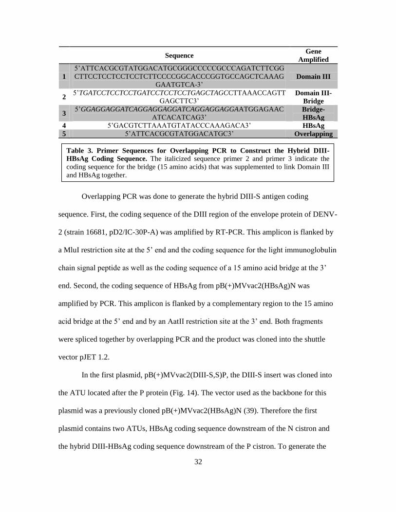
Sequence Gene
Amplified
1
5’ATTCACGCGTATGGACATGCGGGCCCCCGCCCAGATCTTCGG
CTTCCTCCTCCTCCTCTTCCCCGGCACCCGGTGCCAGCTCAAAG
GAATGTCA-3’ Domain III
2 5’TGATCCTCCTCCTGATCCTCCTCCTGAGCTAGCCTTAAACCAGTT
GAGCTTC3’ Domain III-
Bridge
3 5’GGAGGAGGATCAGGAGGAGGATCAGGAGGAGGAATGGAGAAC
ATCACATCAG3’ Bridge-
HBsAg
4 5’GACGTCTTAAATGTATACCCAAAGACA3’ HBsAg
5 5’ATTCACGCGTATGGACATGC3’ Overlapping
Overlapping PCR was done to generate the hybrid DIII-S antigen coding
sequence. First, the coding sequence of the DIII region of the envelope protein of DENV-
2 (strain 16681, pD2/IC-30P-A) was amplified by RT-PCR. This amplicon is flanked by
a MluI restriction site at the 5’ end and the coding sequence for the light immunoglobulin
chain signal peptide as well as the coding sequence of a 15 amino acid bridge at the 3’
end. Second, the coding sequence of HBsAg from pB(+)MVvac2(HBsAg)N was
amplified by PCR. This amplicon is flanked by a complementary region to the 15 amino
acid bridge at the 5’ end and by an AatII restriction site at the 3’ end. Both fragments
were spliced together by overlapping PCR and the product was cloned into the shuttle
vector pJET 1.2.
In the first plasmid, pB(+)MVvac2(DIII-S,S)P, the DIII-S insert was cloned into
the ATU located after the P protein (Fig. 14). The vector used as the backbone for this
plasmid was a previously cloned pB(+)MVvac2(HBsAg)N (39). Therefore the first
plasmid contains two ATUs, HBsAg coding sequence downstream of the N cistron and
the hybrid DIII-HBsAg coding sequence downstream of the P cistron. To generate the
Table 3. Primer Sequences for Overlapping PCR to Construct the Hybrid DIII-
HBsAg Coding Sequence. The italicized sequence primer 2 and primer 3 indicate the
coding sequence for the bridge (15 amino acids) that was supplemented to link Domain III
and HBsAg together.

33
first plasmid, a shuttle vector pUC19 was digested at SbfI and KasI restriction site;
producing a 2.4 kb vector. The insert was also digested with SbfI and KasI restriction
enzymes, coming from pB(+)MVvac2(HBsAg)P, producing a 4.8 kb insert. The ligation
of these products resulted in pUC(HBsAg)P, which was confirmed by a restriction digest
with DraI. The HBsAg coding sequence cloned in the shuttle plasmid was then swapped
by the hybrid DIII-HBsAg coding sequence using MluI and AatII digestion; producing a
~7 kb vector and ~600 bp insert. This ligation of this vector and insert resulted in
pUC(DIII-S)P, which was confirmed by a restriction digest with PvuI. Then, the SbfI and
KasI restriction fragment from pUC(DIII-S)P containing the hybrid DIII-S coding
sequence downstream of the MV P cistron substituted the corresponding fragment in
pB(+)MVvac2(HBsAg)N. This restriction digest produced a 17 kb vector and 4 kb insert
(Fig. 16. A). The ligations of these digest products then resulted in the final full-length
plasmid pB(+)MVvac2(DIII-S,S)P, which was confirmed by a restriction digest with
HindIII (Fig. 16. B). In addition, the full-length plasmid was also sent for DNA
sequencing, using the appropriate forward and reverse primers for MV sequencing the
ligation boundaries, shown in table 4.
The second plasmid, pB(+)MVvac2(DIII-S)N was constructed using a vector that
is pB(+)MVvac2(HBsAg)N. The insert of the HBsAg gene sequence at the N position is
then swapped with the DIII-S insert. Therefore this plasmid only has one ATU where the
HBsAg gene sequence is fused to DIII. The vector, pB(+)MVvac2(HBsAg)N and the
insert from pUC(DIII-S)P were both digested with MluI and AatII, producing a 20 kb
vector and a 1 kb insert. The ligations of these digest products then resulted in the final
full-length plasmid pB(+)MVvac2(DIII-S)N, which was confirmed by a restriction digest

34
with HindIII (Fig. 15. A). This full-length plasmid was also sent for DNA sequencing,
using the appropriate forward and reverse primers for MV, shown in table 4.
Plasmid Sequenced DNA Sequencing Primers Name
pB(+)MVvac2(DIII-S,S)P 5’-AGCTGCTGAAGGAATTTC-3’ 3101F
pB(+)MVvac2(DIII-S,S)P 5’-AGCCTGCCATCACTGTA-3’ 3548R
pB(+)MVvac2(DIII-S)N 5’-GGCAAGAGATGGTAAGG-3’ 1201F
pB(+)MVvac2(DIII-S)N 5’-GTTGTCTGATATTTCTGAC-3’ 1939R
2.3 MV Reverse Genetics System. A Measles Virus reverse genetics technique
was used to rescue the recombinant MV (Fig. 17) following the method established by
Radecke et al. modified by Parks et al. (36, 40). Day 1: Cell culture (293-3-46) was
seeded in a T75. After the T75 flask has become confluent, cells were split into 2 plates
(6-wells), using PBS and trypsin (Mediatech) in a 3:1 dilution. Done using 13 mL of
DMEM-10%FBS and plating 1 mL of cell per well plus 1 mL of fresh DMEM-10%FBS.
Incubated for 3-5 hours (until cells are attached). Transfection of cell line was done
through calcium phosphate precipitation using a ProFection kit (Promega, Madison, WI)
with two plasmids, the specific MV full length genome (Recombinant MV) and the
pEMC-La (coding for the MV polymerase protein, courtesy of R. Cattaneo). Day 2: The
transfected cells were heat-shocked for 3 hours at 42ºC. Day 3 and 4: Vero/hSLAM cells
were seeded in P100 plates. After cell attachments, the rescue cells were overlaid on the
Vero/hSLAM cells to recover MV infectivity on them. The appearance of cytopathic
effects was monitored in the following days. Once syncytia are observed, single isolated
syncytia can be picked and propagated on Vero/hSLAM cell line.
Table 4. Primer Sequences Used in DNA Sequencing of Full-length Recombinant
Plasmid. To sequence pB(+)MVvac2(DIII-S,S)P, we used 3101F forward primer and
3548R reverse primer, which means that the 3101-3578 nucleotides of the recombinant
plasmid was sequenced. To sequence pB(+)MVvac2(DIII-S)N, we used 1201F forward
primer and 1939R reverse primer, which means that the 1201-1939 nucleotides of the
recombinant plasmid was sequenced.

35
2.4 Multi-step Growth Kinetic Analysis of Recombinant MVs. Multiple-step
growth kinetic was performed to document the replication fitness of recombinant MVs,
MVvac2(DIII-S,S)P and MVvac2(DIII-S)N. Vero/hSLAM cells (5x10⁵ cells/well) were
infected with a multiplicity of infection (MOI) of 0.03, in a 6-wells plate incubated at
37ºC. Cells were scraped in 1 mL of Opti-MEM for the intracellular sample and
supernatant for the extracellular sample was collected at 12, 24, 36, 48, 72, and 96 hours
post-infection. Intracellular sample was lysed by a single freeze-thaw cycle at the 50%
tissue culture infectious dose (TCID50) was assessed in Vero/hSLAM cells using the
Spearman-Kärber end-point dilution method to verify the efficiency of the production of
MV recombinant particles (41). The controls used were the parental strain, MVvac2, and
the reference strain, MVvac2(HBsAg)N.
2.5 Titration of Viruses. Titration of MV and recombinant MVs was done by
assessing its TCID50. Vero/hSLAM cells were seeded into 96-well plates with 2x10⁴
cells
and 200 µl of DMEM 5% FBS P/S, per well. Plates were incubated at 37ºC for 1 hour. A
ten-fold serial dilution is then done in a separate 96-well plate, by using 270 µl of Opti-
MEM and 30 µl of the viral prep; the serial dilution is done 12 times. The dilution is then
transferred by adding 30 µl of each dilution in 8 repeats (8 rows of the 96-well plate).
The plate is then incubated at 37ºC and read 3 days later. The TCID50 was calculated
using the Spearman-Kärber end-point dilution method (41).
Titration of DENV-2 was done by a plaque assay using BHK-21 cells. A 24-well
plate was seeded with 1x10⁵ cells and 500 µl of 250 µl DMEM 5% and 250 µl DMEM
10% FBS P/S. Plate was incubated at 37ºC for at least 4 hours to let the cells attach.
Next, 8 series of ten-fold serial dilution is done by using 900 µl of Opti-MEM and 100 µl

36
of the viral prep. The dilutions is incubated at 37ºC for 1 hour and then 100 µl of each
dilutions is added to the 24-well plate seeded with BHK-21 cells; this is done in
duplicate. The 24-well plate is incubated at 37ºC for 4 hours, to allow for viral infection.
Then 500 µl of overlay media is added to each well, which encourages the formation of
isolate plaques. The overlay media contains: 3% carboxymethyl cellulose (CMC) made in
100 ml of Nanopure H2o with the addition of 3 g of CMC (Sigma-Aldrich, St. Louis,
MO), 20 ml of FBS, 2 ml of 200mM L-glutamine (Gibco by Life Technologies, Grand
Island, NY), 200 ml of Modified Eagle’s Medium 2x (MEM, Gibco by Life
Technologies, Grand Island, NY), and P/S (Mediatech). Plates were then incubated at
37ºC for 5 days. Plates are developed by adding 500 µl of Naphtol Blue Black (NBB) to
each well. NBB contains: 1 g of NBB (Sigma-Aldrich), 13.6 g of Sodium Acetate
(Sigma-Aldrich), 60 ml of Glacial Acetic Acid (Sigma-Aldrich), and Nanopure H2o up to
1000 ml. NBB is allowed to stain the cell for about 1 hour, then washed off by immersion
of the plates in water.
2.6 Expression of HBsAg. A test using the Abnova HBsAg enzyme-linked
immunosorbent assay (ELISA) kit (Novus Biologicals, Littleton, CO) was performed to
observe the production of HBsAg by the recombinant MV. Using the supernatant
collected from, 6-well plates seeded with 5x10⁵ Vero/hSLAM cells, infected with a MOI
of 0.03 of the recombinant strain, MVvac2(DIII-S,S)P or the reference strain as a control,
MVvac2(HBsAg)N. The supernatant was collected at 24, 48, 72 and 96 hours post-
infection. These supernatants were used in the HBsAg ELISA compared to a commercial
HBsAg standards and the included negative and positive control from the ELISA kit.

37
2.7 Preparation of Protein Extracts. To analyze the protein produced by the
recombinant MVs, protein extracts must be prepared to analyze through a western blot.
1x10⁶ Vero/hSLAM cells were seeded in 100-mm-diameter (p100) dishes and incubated
at 37ºC for 2-3 hours to attach. The cells were infected with a MOI of 0.3 with one of the
viruses of mock-infected (Non-infected sample). During infection, media was aspirated,
and virus was added mixed in 6 ml of Opti-MEM, then incubated at 37ºC. Infection was
stopped 2 hours later by aspirating the Opti-MEM/virus and replacing the DMEM 5%
FBS P/S. Cell monolayer was monitored and collected when about 80% cytopathic effect
has been reached. To collect the protein extract, the media from the plate was aspirated
and the plate was washed with phosphate-buffered saline (PBS) 3 times. Then cells are
lysed by adding 500µL of RSB-NP40 buffer (1.5 mM of MgCl2, 10 mM of Tris-HCl [pH
7.5], 10 mM of NaCl, and 1% Nonidet p-40, Sigma-Aldrich, St. Louis, MO) plus
protease inhibitors (cOmplete Mini Protease Inhibitor Tablets, Roche Diagnostics,
Manheim, Germany). The sample was then centrifuged at 4ºC for 20 minutes at 14,000
RPM. The supernatant containing the protein was then prepared by the addition of 6x
SDS Page loading buffer (Laemmli buffer, Bio-Rad Laboratories, Hercules, CA) and
denatured at 96ºC for 10 minutes. Samples stored in -30ºC if not directly used.
2.8 Analysis of Protein Expression. The samples of protein extracts were
separate by SDS polyacrylamide gel electrophoresis (PAGE) in a 10% acrylamide gel.
After the gel has been run, the protein were then transferred to nitrocellulose for
immunoblotting using a 1:1000 dilution of a rabbit polyclonal anti-HBsAg antibody
coupled to horseradish peroxidase (HRP, OBT0990P, AbD Serotec, Raleigh, North
Carolina, USA), and a 1:500 dilution of a mouse monoclonal anti-DIII DENV-2

38
(GTX29202, GeneTex, Irvine, California, USA). The blot was incubated in the antibody,
anti-HBsAg-HRP, for 2 hours; a secondary is not necessary because the primary antibody
is already conjugated to HRP. This blot was developed using a chemiluminescence kit
(SuperSignal West Pico Chemiluminescent Substrate, Pierce Biotechnology, Rockford,
IL). When incubating with anti-DIII DENV-2, the blot was incubated for 3 hours with the
primary antibody. Then incubated with the anti-mouse HRP-conjugated secondary
antibody (GE Healthcare, Little Chalfont, United Kingdom) for 1 hour. Similarly, this
blot was developed using the chemiluminescence kit. Membranes incubated with MV
Anti-N were incubated with a primary antibody of rabbit polyclonal Anti-N 1:10,000
(provided by R. Cattaneo) for 1 hour and a secondary antibody of goat Anti-Rabbit-
Alkaline Phosphatase conjugated (Anti-Rabbit-AP, Thermo Fisher Scientific, Anthem,
AZ) 1:1000 for another 1 hour. The membrane was developed by visualizing the AP
reactivity using Western Blue Stabilized Substrate for Alkaline Phosphatase Reagent
(Promega, Madison, WI).

39
2.9 Particle Isolation and Determination of Density. HBsAg particles isolation
was done using a discontinuous sucrose gradient of various concentrations: 60%, 50%,
40%, 30%, and 20% (Fig. 11). To purify the released particles, supernatants from 5x10⁶
Vero/hSLAM cells infected with either MVvac2, MVvac2(HBsAg)N, MVvac2(DIII-
S,S)P or MVvac2(DIII-S)N were collected and clarified by centrifugation in 4ºC at 5,000
RPM for 15 min in an SX4750 rotor. 35 ml of the clarified supernatants were pelleted by
ultracentrifugation in 4ºC at 45,000 RPM for 18 hours in an SW28 rotor. The supernatant
was aspirated and the pellets were re-suspended in 1 ml of TNE buffer (10 mM Tris [pH
Figure 11. Diagram of the Ultracentrifugation of the Discontinuous Sucrose Gradient
to Isolate VLPs. Monolayers of Vero/hSLAM cells were infected with recombinant MVs
or controls. Supernatants from infected cells were collected and clarified in 4ºC at 5,000
RPM for 15 min. Clarified supernatant was collected and 35 ml was placed in an
ultracentrifugation tubes. This was ultracentrifuged in 4ºC at 45,000 RPM for 18 hrs.
Supernatant was aspirated and the pellet containing the VLPs was re-suspended with 1 ml
of TNE buffer containing 20% of sucrose. 5ml ultracentrifugation tubes were prepared with
the discontinuous sucrose gradient, including: 1 ml of each TNE buffer containing the
sucrose concentration of 60%, 50%, 40%, and 30%, from the bottom to the top of the tube
respectively. The 1 ml of re-suspended pellet is added on top of the 30% layer. This was
ultracentrifugated in 4ºC at 37,000 RPM for 24 hrs. Then nine fractions of 500 µl, were
collected from the top to bottom of the equilibrated tube.

40
7.8], 100 mM NaCl, 1mM EDTA) containing 20% sucrose. The TNE Buffer was used to
create a 60% sucrose solution that was further diluted to produce 50%, 40%, 30% and the
20% sucrose solutions. Polyallomer centrifuge tubes (5 ml Centrifuge Tubes, Beckman
Coulter, Brea, CA) were prepared with the discontinuous sucrose gradient, containing
60%, 50%, 40%, and 30% sucrose solution (1 ml each from the bottom respectively). The
1 ml of 20% sucrose solution containing the re-suspended pellet is added as the top layer.
The tubes of fractions were ultracentrifugated in 4ºC at 37,000 RPM for 24 hours in a
MLS50 rotor. To determine particle density and content, 500 µL fractions were taken
from the top to bottom layers. Fractions were weighed and their density calculated. 25 µl
aliquots of these fractions were taken and mixed 1:1 with SDS PAGE loading buffer.
Samples were prepared to be run in a SDS PAGE, denatured at 96ºC for 10 min. Western
blots containing these fractions were developed by various antibodies (Fig. 22).
2.10 Animal Experiments. Two animal experiments were performed for this
project. The Arizona State University Institutional Animal Care and Use Committee
sanctioned all experimental procedure. The first animal experiment was performed on
HDD-SLAM-IFNarKO
transgenic mice. These mice express one of the human MV
receptor, hSLAM, in a type I interferon deficient environment, which will allow for MV
replication. These mice are also H2Kb and D
b knock out, which inhibits the mice MHC
class I allele presentation restriction. In addition, these transgenic mice also presents
MHC class I antigenic peptides with a HLA A2.1 restriction, the most common human
MHC class I allele, using an artificial allele termed HHD, which contains the human 2
microglobulin fused to the HLA-A2.1 molecule where the 3 domain was substituted by
the homologous region of murine origin. With this arrangement, the HHD molecule is

41
functionally independent of murine macroglobulin for expression and display and
recognized by murine CD8+ T lymphocytes.
Groups of 2 to 5 transgenic mice were vaccinated by the intraperitoneal (i.p) route
with three doses of 5x10⁵ TCID50, including: 4 animals vaccinated with the parental
strain, MVvac2, 5 animals vaccinated with the reference strain, MVvac2(HBsAg)N, and
5 animals vaccinated with the recombinant strain, MVvac2(DIII-S,S)P. Two animals had
a vaccination schedule of three doses of 1x10⁵
PFU of the control, DENV-2. However,
these 2 animals in actuality received only 5x10² PFU doses. All of the animals were
vaccinated at three different times, at day 0, 1 and 14 (Fig. 12). Then all of the animals
were euthanized and exsanguinated 28 days after the last vaccination. Sera collected from
immunized animals were separated and heat-inactivated at 56ºC for 1 hour. Aliquots of
heat-inactivated sera were stored in -30ºC until analyzed.
The second animal experiment was performed on huCD46Ge-IFNarKO
transgenic
mice. These mice express a different MV receptor, CD46, with human-like distribution.
Figure 12. Diagram Representing the Vaccination Schedule Given to the HDD-SLAM-
IFNarKO
Transgenic Mice in the First Animal Experiment. In the first animal
experiment, the animals received three doses at day 0, 1, and 14. Day 0 was given to induce
the expression of the MV receptor, SLAM, since it is only expressed in activated
lymphocytes. Day 1 is the first dose given to the animals, followed by the booster second
dose 14 days later. All animals were exsanguinated 28 days after their last vaccination.
Animal sera were isolated and decomplemented. (Syringe – dose given, Blood – Mice
exsanguinated).

42
They also lacked of type I interferon pathway, which will allow for MV replication.
These transgenic mice represent the gold standard for small animal models testing
measles vaccine immunogenicity. These groups of mice were also vaccinated through the
i.p. route, with one or two doses of 1x10⁵ TCID50, including: 5 animals vaccinated with
one dose of the parental strain, MVvac2 x1, 3 animals vaccinated with two doses of the
reference strain, MVvac2(HBsAg)N x2, 4 animals vaccinated with one dose of the
recombinant strain, MVvac2(DIII-S,S)P x1, 7 animals vaccinated with two doses of the
same recombinant strain, MVvac2(DIII-S,S)P x2, and 3 animals vaccinated with two
doses of the second recombinant strain, MVvac2(DIII-S)N x1. The first dose for all of the
animals was given on day 0. Then animals scheduled to only receive one dose of their
vaccine were exsanguinated 28 days after the first done, whereas the animals schedule to
receive two doses gets their booster vaccine (Fig. 13). The rest of the animals were
exsanguinated 14 days after the second dose was given. Sera collected from immunized
Figure 13. Diagram Representing the Vaccination Schedule Given to the huCD46Ge-
IFNarKO
Transgenic Mice in the Second Animal Experiment. In the second animal
experiment, the animals received either one or two doses of their respective vaccine. The
first dose was given on day 0. Then, 28 days after the first dose, those animals with a
vaccination schedule of only 1 dose were exsanguinated: MVvac2 x1 and MVvac2(DIII-
S,S)P x1; whereas those animals with a vaccination schedule of two doses, received their
second dose: MVvac2(HBsAg)N x2, MVvac2(DIII-S,S)P x2, and MVvac2(DIII-S)N x2.
Then, 14 days after the second dose, the rest of the animals were exsanguinated. Animal
sera were isolated and decomplemented. (Syringe – dose given, Blood – Mice
Exsanguinated.
43
animals were separated and heat-inactivated at 56ºC for 1 hour. Aliquots of heat-
inactivated sera were stored in -30ºC until analyzed.
2.11 Analysis of the Immune Response of Vaccinated Animals. The sera
collected from each immunized mice were analyzed for its immunogenicity against all
three viruses: measles, hepatitis B, and dengue 2. To determine MV neutralization titer,
we performed the same experiment for sera collected from the first and second animal
experiments. We performed serial two-fold dilutions of the sera in Opti-MEM and
incubated with the addition of 100 TCID50 of MVvac2 at 37ºC for 1 hour. To the sera-
virus mixtures, 1x10⁶
Vero/hSLAM cells were added for every 96-well plate. These
plates were then incubated at 37ºC for three days. Cytopathic effect of MV was
monitored, and the neutralization titer was determined to be the highest dilution of serum
that is still capable to cause complete neutralization of MVvac2 infectivity.
Neutralization titer was recorded from an average of this experiment done in triplicates.
To determine the anti-HBsAg titer in both animal experiments, we used a
commercial species-independent quantitative ELISA kit (Alpha Diagnostic Intl. Inc., San
Antonio, Texas, USA). The anti-HBsAg titer was recorded in milli-international units per
milliliter by comparison with the standards from the World Health Organization that was
supplied by the manufacturer; these standards were included in the concentrations of: 20,
40, 80, and 160 mIU/ml. The anti-HBsAg antibody titer was recorded from an average of
two readings for each sample done in this experiment.
The immunogenicity studies done against DENV-2 were done different in the first
animal experiment compared to the second animal experiment. In the first animal
experiment, a homemade ELISA was performed to test the reactivity of the collected sera

44
against DENV-2. This ELISA, however, was not quantitative. On a 96-well plate, whole
DENV-2 was adsorbed to the bottom of the well overnight at 4ºC; each well contained
103 PFU of DENV-2 in 100 µl of PBS TWEEN 20 0.05%. Next, all mixture in each well
is aspirated and 150 µl of the blocking solution, PBS TWEEN 20 0.05% containing 5%
milk (Biorad, Bio-Rad Laboratories, Hercules, CA); then incubated at 37ºC for 1 hour.
Wells were washed three times with PBS TWEEN 20 0.05%. Sera samples diluted 1:50
in 100 µl were added, and plates were again incubated at 37ºC for 1 hour; then wells were
washed three times. An anti-mouse antibody added (1:2500), 100 µl to each well, then
plates were incubated for at 37ºC for 30 min. Wells were again washed three times, and
developed with HRP and 3M TMB-H2SO4 (tetramethylbenzemidine, Sigma-Aldrich, St.
Louis, MO). The plates were then read at OD450.
In the second animal experiment, the immunogenicity study against DENV-2 was
done in two different approaches. In the first experiment, 105 PFU of DENV-2 were
mixed with heat-inactivated sera that are dilution 1:5 in 200 µl of Opti-MEM, and
incubated at 37ºC for 1 hour. Infectivity of DENV-2 remaining after sera neutralization
was determined through a DENV-2 plaque assay in BHK-21 cells. In 96-well plate, ten-
fold serial dilutions of the virus/sera mixtures were done using Opti-MEM. A 24-well
plate seeded with 1x10⁵ cells per well in 500 µl of 50/50 DMEM 5% and DMEM 10%
FBS P/S was incubated at 37ºC for 2-4 hours to allow the cells to attach. Dilutions done
in Opti-MEM was then added to the 24-well plate, 100 µl of each dilution, and incubated
at 37ºC for 4 hours. Next, 500 µl of the overlay media, 3% CMC, was added to each well,
and these plates were incubated at 37ºC for 5 days. The plates were developed by
aspirating each well and adding 500 µl of NBB, which fixed and stained the monolayer
45
of cells with acetic acid/napthol blue-black. The neutralization index was recorded as the
logarithmic difference between the average DENV-2 titers when incubated with the
control sera: sera collected from animals vaccinated with MVvac2, MVvac2(HBsAg)N,
and previously obtained pre-immune sera; and the average DENV-2 titers when
incubated with the experimental sera: sera collected from animals vaccinated with
MVvac2(DIII-S,S)P and MVvac2(DIII-S)N. The determined anti-DENV-2 neutralization
index is recorded on as log10NI. The second experiment performed to analyze the sera’s
immunogenicity against DENV-2 was the classis 50% plaque reduction neutralization
assay (PRNT50) using pooled sera from each experimental group of mice. 200 PFU of
DENV-2 were mixed with a series of two-fold serial dilutions (1:5 to 1:320) of each
pooled sera. This virus/sera mixture was incubated at 37ºC for 1 hour. Infectivity of
DENV-2 remaining after sera neutralization was determined through a DENV-2 plaque
assay in BHK-21 cells as described for the previous experiment. The neutralization titer
was determined at the dilution that had a 50% reduction in the amount of plaques formed,
compared to the control samples.

46
Chapter 3. Results
3.1 Plasmid Construction and MV Rescue. For this project, I aimed to generate
two types of recombinant plasmids, the full length infectious cDNA plasmids are termed
pB(+)MVvac2(DIII-S,S)P and pB(+)Mvvac2(DIII-S)N. The basic chimeric gene that all
of these recombinants have in common is composed by the fusion of four elements (Fig.
14). At the amino terminus is the light immunoglobulin chain signal peptide, which is a
stretch of 22 amino acids peptide (MDMRAPAQIFGFLLLLFPGTRC), to direct the
synthesis of our chimeric protein to the endoplasmic reticulum. Then followed by 101
amino acids of the DIII of the DENV-2 envelope protein, a 14 amino acid bridge that is
composed of the sequence (SG3)3 with a predicted α-helical structure. This bridge is
important in allowing the independent folding of the DIII and HBsAg coding sequence
that is located at the carboxyl terminus of this construction. The hybrid glycoprotein is
Figure 14. The Genomic Map of the Recombinant MV Generated. The top diagram
represents pB(+)MVvac2(DIII-S,S)P containing the HBsAg ATU after the N cistron and
the hybrid DIII-S ATU after the P cistron. The bottom diagram represents
pB(+)MVvac2(DIII-S)N containing only the DIII-S ATU after the N cistron.

47
expressed from either of two different loci in MV, downstream the N gene and
downstream the P gene, in addition to this insertion, pB(+)MVvac2(DIII-S,S)P contains a
separate unmodified HBsAg coding sequence cloned downstream the N gene (Fig. 14).
In figure 16 A, the insert and vector is obtained from the SbfI and KasI restriction
fragments of pUC(DIII-S)P containing the hybrid DIII-S insert (4 kb), and of
pB(+)MVvac2(HBsAg)N containing the vector (17 kb), which has the additional HBsAg
ATU independently inserted after the N cistron. In Figure 15 and 16, we confirmed the
identity of our candidate plasmids through a restriction digest using HindIII. We were
able to observe the expected DNA band size in our experimental samples for both
plasmids. In addition, we further corroborated the accuracy of our insertion by DNA
sequencing of the gene boundaries.
Figure 15. Diagram of the Confirmation Digest for the Recombinant Plasmid
pB(+)MVvac2(DIII-S)N. A, a diagram of the agarose gel containing samples of the full
length pB(+)MVvac2 (DIII-S)N digested by HindIII restriction enzyme. Clone 1-4 was too
low in concentration that nothing can be seen. However, clone 5 shows the correct pattern
of bands that are expected. B, shows the expected bands from a HindIII digestion of
pB(+)MVvac2(DIII-S)N.

48
Vec
tor
Inse
rt
A
17, 000
4, 000
bp
Clo
ne
1
Clo
ne
2
Clo
ne
3
4, 242 3, 731
B
2, 079
8, 213
1, 718
1, 051 678
bp
C
bp Exp
ecte
d
Lad
der
Lad
der
Lad
der
Figure 16. Diagram of the Confirmation Digest for the Recombinant Plasmid
pB(+)MVvac2(DIII-S,S)P. A, a diagram of the agarose gel containing the digested and
purified insert, hybrid DIII-HBsAg, and the vector, pB(+)MVvac2(HBsAg)N, at the
expected size of 4,000bp and 17,000bp respectively. B, a diagram of the agarose gel
containing samples of the full length pB(+)MVvac2 (DIII-S,S)P digested by HindIII
restriction enzyme; showing all 3 clones. C, shows the expected bands from a HindIII
digestion of pB(+)MVvac2(DIII-S,S)P.

49
3.2 Reverse Genetics System in MV Rescue. Using the full-length cDNA
construction plasmids, we rescued the corresponding recombinant viruses, termed
MVvac2(DIII-S,S)P and MVvac2(DIII-S)N, using an established reverse genetic system
(Fig. 17). Helper cells (293-3-46), a strain from the HEK 293 cell line that is stably
transfected with the N and P protein of MV as well as the T7 polymerase, were
transfected with two plasmids. One plasmid encodes the full-length recombinant
MVvac2, pB(+)MVvac2(DIII-S,S)P and pB(+)MVvac2(DIII-S)N, with a T7 promoter.
The second plasmid, pEMC-LA, encodes the L protein of MV which is the viral RdRp;
this construct also have a T7 promoter, as well as an internal ribosome entry site (IRES)
before the L gene to ensure its translation. After transfection, the full-length MV genome
will be transcribed by the T7 polymerase into full-length antigenomic RNA, and then the
N and P protein expressed in the helper cell will bind to form the RNP complex. The MV
RdRp produced from pEMC-LA will then bind to the RNP complex and replicate the MV
genome. The MV genome in a RNP complex will be transcribed, which leads to the
generation and expression of the viral mRNAs into viral proteins following its
transcription gradient. This will result in virion assembly and release from the infected
host cell (40, 42). The next step was to take helper cells and overlaid it on a monolayer of
Vero/hSLAM cells, which was then monitored for syncytia. Plaque cloning was then
performed by picking a single-isolated syncytia and infecting a new monolayer of
Vero/hSLAM, ensuring a pure viral stock that originated from one clone of the virus. The
virus was then amplified to generate viral prep made from multiple passages of the MV
that was rescued.

50
Measles infectivity generated after transfection of helper cells was amplified
using plates of Vero/hSLAM cells with negative (non-transfected rescue cells) and
positive controls (transfected with a control plasmid). Vero/hSLAM cells were monitored
for cytopathic effects (CPE). Similar sizes of syncytia foci were observed for both the
positive control and the recombinant MVs. This demonstrates that virus was indeed
produced from the cDNA of the recombinant pB(+)MVvac2(DIII-S,S)P (Fig.17),
initially. Hence, the insertion of two ATUs into MVvac2 did not disturb the replication of
the virus. In addition, the green fluorescence marker on the positive control proves that
the syncytium depicted on the bottom image in figure 17 is caused by the recombinant
virus; in contrast to our recombinant, whose syncytia were not fluorescent. Hence, the
second recombinant virus, MVvac2(DIII-S)N was rescued following the same procedure.
Figure 17. Generation of a Recombinant MV using the Reverse Genetics System. A
helper cell line (293-3-46) was transfected with the recombinant MV cDNA and pEMC-La
(codes for polymerase). Transcription and translation will progress in helper cells, which
will result in the assembly of new virion. The helper cells and the virion were over-laid on
vero/hSLAM cells (38). The growth of syncytia was observed. The top image depicts the
syncytia formed by MVGreen, a positive control. The bottom image depicts the syncytia
formed by pB(+)MVvac2(DIII-S,S)P. The microbars in the images are 400µm.

51
After the recovery of each recombinant virus was completed, they were amplified
and titered. The stock of MVvac2(DIII-S,S)P reached a titer of 1.8x10⁵ TCID50/ml, which
is approximately ten times lower than the titer of the parental MV. Whereas the titer of
MVvac2(DIII-S)N reached 5.6x10⁷ TCID50/ml, which is comparable to the parental MV.
Overall, the creation of the recombinant plasmids and the rescue of each recombinant
virus were deemed successful, due to the generation of the viral stock with a viable titer
for animal experimentation. However, the next step will be to observe the replication
fitness of each virus.
3.3 Multi-step Growth Kinetics. To evaluate the replication fitness of the
recombinant MV, a multi-step growth kinetics was performed in Vero/hSLAM cells
using passage 1 of the viral stock. Cells in a monolayer (5x10⁵ cells/well) were infected
with a MOI of 0.03. Cell-associated viral progeny were collected at different time points
from the intracellular fraction, which includes 12h, 24h, 36h, 48h, 72h, and 96h post-
infection. These fractions were later quantified by its infectivity determined as TCID50.
The controls used in this experiment were the parental strain MVvac2, and the reference
strain MVvac2(HBsAg)N, a virus that expresses the HBsAg protein from downstream the
N position.
Shown in figure 18, the intracellular viral growth for MVvac2(DIII-S)N peaked at
36 hours post-infection and reached a titer of around 107 TCID50/ml. Whereas
MVvac2(DIII-S,S)P peaked at 48 hours post-infection and reached a titer of around 105.15
TCID50/ml, which is about 22 to 50 times lower than MVvac2(DIII-S)N. This indicated
that the insertion of two ATUs in MVvac2(DIII-S,S)P had a significant impact on its

52
replication fitness; compared to MVvac2(DIII-S)N, which only contains one ATU, hence
following a similar replication fitness as the parental strain, MVvac2, as well as the
reference strain, MVvac2(HBsAg)N, which reached a titer of around 107 TCID50/ml at its
peak. However, despite the observed significant negative effect on viral fitness, we were
able to create a stock of each of the recombinant viruses that reached a viable 106
TCID50/ml, which was used for further experiments. The results shown in figure 18
represents the averages of three growth kinetic experiments performed independently of
each other.
Figure 18. Multi-step Growth Kinetics of Recombinant MVs. Quantification of viral
titers from cell-associated MV produced in Vero/hSLAM at specific time points post-
infection (12h, 24h, 36h, 48h, 72h, and 96h). Cells were infected with either the parental
strain MVvac2 represented by the black line or diamonds, the reference strain
MVvac2(HBsAg)N represented by the red line or squares, the recombinant strain
MVvac2(DIII-S)N represented by the blue line or circles or MVvac2(DIII-S,S)P
represented by the green line or triangles. This data is an average of three independent
experiments, with the error bars indicating the standard deviation for each average.

53
3.4 Production of HBsAg Detected by ELISA. An initial qualitative ELISA
specific for HBsAg was performed to demonstrate the expression of HBsAg by the
recombinant virus, MVvac2(DIII-S,S)P. Supernatants from infected cells were collected
at different time-points (24h, 48h, 72h, and 96h post-infection) and analyzed. As a
control, supernatant from cells infected with the reference strain MVvac2(HBsAg)N was
collected in parallel. A concentration curve using HBsAg standards was prepared for the
ELISA. All of the samples from the infected media were positive for HBsAg since their
readings were well above the background value (interrupted horizontal line)
demonstrating that the antigen is being secreted upon infection (Fig. 19). Nevertheless,
0
0.5
1.0
1.5
2.0
2.5
Blank
+-
(HBsAg)N (DIIIS-S)P HBsAgCtrls
OD
450
Figure 19. Identification of Secreted HBsAg by Recombinant MVs. MVvac2(DIIIS-S)P
secretes HBsAg upon infection. An ELISA was performed to demonstrate the expression of
the antigen used as platform to display Domain III. Positive (+) and negative (-) controls
validate the experiment and set up the cut-off value (interrupted horizontal line)
Supernatants from infected cells were obtained at different time points (the virus is
indicated, solid triangles indicate 24h, 48h, 72h and 96h post-infection) and run in parallel
with progressive dilutions of a HBsAg standards (empty triangle indicated a progressive
concentration of 2.5, 5, 10 and 50 ng/ml) each sample was run in duplicate in one ELISA
experiment, the average value is shown.

54
readings of our standard curve demonstrated that this assay is only qualitative; we could
not demonstrate any linearity of the OD450 readout. However it is important to note that
the sensitivity of the assay is well below the lowest concentration tested (2.5 ng/ml).
Therefore, after we were able to initially see the production of HBsAg from one of our
recombinant viruses, MVvac2(DIII-S,S)P, we move forward in characterizing both of our
recombinant viruses by performing more quantitative and detailed experiments.
3.5 Characterization of the Expression of Hybrid DIII-HBsAg Antigens from
MVvac2(DIII-S,S)P. The intracellular viral particles from Vero/hSLAM cells infected
with a MOI of 0.5 with either MVvac2, MVvac2(HBsAg)N, or MVvac2(DIII-S,S)P were
collected. Each infected cell monolayer were lysed at 24h post-infection; non-infected
cells grown in a monolayer under similar conditions as the infected cells was also lysed
and used as a control.
Each cell extracts was analyzed for the incorporation of DIII into the HBsAg
VLP by an anti-HBsAg western blot under non-reducing condition (Fig. 20). A doublet,
two bands of HBsAg were observed by the anti-HBsAg antibodies in the cell extracts
obtained from MVvac2(HBsAg)N, the positive control. The top and bottom bands are
glycosylated and non-glycosylated isoforms of HBsAg, respectively, observed at
theoretical molecular weight of 27 and 24 kDA as expected. The double bands are also
seen in the cell extract collected from the recombinant virus MVvac2(DIII-S,S)P (Fig.
20). However, in addition to the expected doublet, we also observed another upwardly
shifted double bands at 37 and 34 kDa; the corresponding theoretical molecular weight of
the glycosylated and non-glycosylated isoforms of the hybrid DIII-S antigen. As
expected, there were no observed binding of the anti-HBsAg antibodies in the samples

55
from the non-infected and MVvac2 cell extracts, used as negative controls. Overall, in
figure 20, the upward shift of the double bands observed in the MVvac2(DIII-S,S)P cell
extracts, shows the successful expression of DIII-HBsAg.
Figure 20. Western Blots Analyzing the Expression of Hybrid DIII-S. Western blot of a
membrane containing 4 samples, the controls, non-infected, MVvac2, MVvac2(HBsAg)N
and the recombinant virus, MVvac2(DIII-S,S)P . The top membrane was incubated with
anti-HBsAg antibodies. The double bands observed in MVvac2(HBsAg)N shows the
glycosylated (top) and non-glycosylated (bottom) bands, shown by the arrows. The upward
shift of the double bands in MVvac2(DIII-S,S)P shown by the arrowheads, represent the
expression of DIII by HBsAg. The bottom membrane was incubated with measles antibody
against the N protein to corroborate equal amount of the samples are represented.

56
In order to corroborate that the same amount of sample was analyzed for each cell
extracts, a second western blot was performed. Figure 20 shows a second diagram
(bottom) which displays the western blot results using anti-N antibodies from MV;
everything else other than the antibodies used, was run under the same conditions as the
previous western blot. In this western blot, we observed the same amount of N protein
present in each of the infected cell extracts.
3.6 Characterization of the Expression of Hybrid DIII-HBsAg Antigens from
MVvac2(DIII-S)N and the Correct Display of DIII. After confirming the successful
expression of the hybrid DIII-HBsAg antigen from the first recombinant virus,
MVvac2(DIII-S,S)P, the experiment was repeated with the second recombinant virus,
MVvac2(DIII-S)N. Lysates from Vero/hSLAM cells infected with a MOI of 0.5 with
either MVvac2, MVvac2(HBsAg)N, MVvac2(DIII-S,S)P, or MVvac2(DIII-S)N were
collected. Each infected cell monolayer was lysed at 24h post-infection. Each cell
extracts was analyzed for the incorporation of DIII into the HBsAg VLP by an anti-
HBsAg western blot under non-reducing condition (Fig. 21). A doublet was observed
similar to the previous western blot, where two bands of HBsAg, the glycosylated
isoform at 27 kDA and non-glycosylated isoform at 24 kDa, were observed by the anti-
HBsAg antibodies in the cell extracts obtained from MVvac2(HBsAg)N, the positive
control. In figure 21, the double bands and the upshifted doublet are also seen again in the
sample containing MVvac2(DIII-S,S)P cell extract, as seen in the previous western blot
(Fig. 20). Furthermore, between the two shifted double bands, the band corresponding to
the non-glycosylated isoform, seen at 34 kDa, gives a much weaker signal than the band
corresponding to the glycosylated isoform seen at 37 kDa. However, both of the bands

57
that were shifter upward does seem weaker than the two original doublet at 24 kDa and
27 kDa, possibly due to the amount of the hybrid DIII-HBsAg antigen that is actually
being produced.
On the other hand, in the sample containing the MVvac2(DIII-S)N cell extract,
we were not able to observe any binding of the monoclonal antibody. Since the western
blot was incubated with anti-HBsAg antibody, this shows that the antibodies were not
MV
va
c2
-(H
BsA
g)N
-(D
III-
S,S
)P
-(D
III-
S)N
S* (27 kDa)
S (24 kDa)
EDIII-S* (37 kDa)
A
40
55
35
25
S (24kDa)
S* (26kDa) S*+EDIII (36kDa) M
Vv
ac2
-(H
BsA
g)N
-(D
III-
S,S
)P
-(D
III-
S)N
rDE
NV
2 E
kDa
MV
vac
2
-(H
BsA
g)N
-(D
III-
S,S
)P
-(D
III-
S)N
A
B
55-
40-
35-
25-
kDa
B
Figure 21. Analysis of Successful Expression of the Hybrid DIII-S Antigen in Cell
Lysates from Controls and Recombinant MV Infected Cells. A, samples run under non-
reducing conditions in a SDS-PAGE, western blot developed using a monoclonal antibody,
anti-HBsAg (the S antigen); the molecular weight of the two forms of S are shown to the
left and the molecular weight of the hybrid DIII-S to the right. The asterisk indicates
glycosylated forms of HBsAg. B, samples run under non-reducing conditions in a SDS-
PAGE, western blot developed using a monoclonal antibody specific for dengue envelope
protein domain III. The double bands were observed at 37 kDa and 34 kDa
58
able to detect the proteins present in the cell extract, possibly due to some changes that
happened in the protein’s folding pattern.
In order to analyze if the hybrid DIII-S antigen is displaying the correct DENV-2
epitopes present in DIII. We developed a western blot using antibodies specific for
DENV-2 DIII. This was done under reducing conditions. We were able to observe two
bands from both of our recombinant viruses. These two bands were seen at about 37 kDa
and 34 kDa, corresponding to the glycosylation isoforms of the hybrid DIII-S previously
seen in the western blot developed with anti-HBsAg (Fig. 21). However, the double
bands observed from the sample containing cell extracts from MVvac2(DIII-S)N, were
weaker than the bands observed from the samples containing cell extracts from
MVvac2(DIII-S,S)P.
3.7 Particle Isolation and Characterization. The particle isolation experiment
was performed to see the presence of hybrid VLPs in the supernatants of Vero/hSLAM
cells infected with MVvac2(DIII-S,S)P. Figure 11 illustrate the procedure, where nine
fractions were collected from the discontinuous sucrose gradient ultracentrifugation in
order to confirm the expected density of the HBsAg particles, including the hybrid
particles expressing DIII. These fractions were then loaded onto a SDS-PAGE under non-
reducing condition. The western blot was developed with anti-HBsAg monoclonal
antibodies. The HBsAg particles were observed in fractions 4 to 6 as shown by the
double bands in both the control sample, fractions from the reference strain
MVvac2(HBsAg)N and the experimental sample, fractions from the recombinant strain
MVvac2(DIII-S,S)P (Fig. 22. B). In addition, we were able to observe again the upward
shifted double bands (shown by arrows) that correspond to the glycosylated and non-

59
glycosylated isoforms of the hybrid DIII-S antigen (Fig. 22. B). These bands were
detected in similar sucrose density (1.10-1.12g/mL, fraction 4 and 5) to the sucrose
density of the native HBsAg secreted in the supernatants of MVvac2(HBsAg)N, the
control (Fig. 22. A). Showing that the characteristics of the hybrid VLP secreted by
MVvac2(DIII-S,S)P are similar to the native VLP secreted MVvac2(HBsAg)N.
Supernatant of Vero/hSLAM cells infected with MVvac2(DIII-S)N was analyzed
in this experiment (not shown). Since we have observed a lack of binding of the anti-
HBsAg monoclonal antibody (Fig. 21), we conclude that performing this western blot
using the fractions from MVvac2(DIII-S)N will not be successful. We concluded that we
might be able to detect the presence of hybrid DIII-S antigen by developing the western
blot using the monoclonal antibody against DENV-2 DIII. However, we were not able to
observe any binding of the antibodies in this western blot. Overall, we observed the
presence of the hybrid DIII-S antigen in the fractions collected from supernatants of cells
infected by MVvac(DIII-S,S)P.
The last western blot we performed using fractions collected from the particle
isolation is developed using antibodies against the N protein of MV. This western blot
used the fractions collected from the supernatant of cells infected with MVvac2(DIII-
S,)P. We were able to observe that the MV particles migrated to fraction 6 to 9 based on
the presence of the MV N protein shown on the bottom panel (Fig. 22). The fractions
where the MV particles are located in are (fractions 6 to 9) clearly separate from the
fractions where the native HBsAg VLP or hybrid DIII-S VLP are located in (mainly
fractions 4 to 5). This demonstrated that the hybrid particle is incorporated separately
from the MV particle, which migrates to the more dense fractions.

60
Figure 22. Particle Isolation by Discontinuous Sucrose Gradient and Analysis of Each
Fractions Collected. Particle isolation diagrams from supernatants of Vero/hSLAM cells infected
with either MVvac2(HBsAg)N or MVvac2(DIII-S,S)P. Supernatants were collected at 72h post-
infection and clarified. Particulate material was then pelleted and resuspended in 20% sucrose
solution, loaded onto the 30%-60% sucrose gradient, then centrifuged to equilibrium. Nine fractions
were collected from the top (fraction 1) to the bottom (fraction 9) in 500µL aliquots. A, Each
fractions were weighed to determine its density. The graphic plot shows a density gradient profile
obtained from a representative experiment. The density of each fraction is shown by the graph at the
top in g/mL. B, Fraction samples were run in a 12.5% SDS-PAGE. The antigenic reactivity of each
fraction was analyzed by a western blot using an anti-HBsAg antibody (top two panels) and an anti-
MV N antibody (lower panel). The top and bottom panels are depicting western blots using fractions
from MVvac2(DIII-S,S)P infected cells. The middle panel is depicting a western blot using fractions
from MVvac2(HBsAg)N infected cells. The hybrid DIII-S incorporate into the VLP is particle is
shown by the arrows in the top panel. These double bands corresponding to the glycosylated and
non-glycosylated isoforms of the hybrid DIII-S antigen can be seen at 37 kDa and 34 kDA,
respectively.
61
Overall, we were able to confirm the incorporation of the hybrid DIII-S antigen
on a scaffold of HBsAg, without affecting its density or the assembly of MV virion.
3.8 Immunogenicity Study. We performed two sets of immunogenicity study
from two different animal experiments in order to analyze the immune response that will
be induced by our recombinant viruses. The first animal experiment was done using MV
susceptible transgenic mice HDD-SLAM-IFNarKO
. These mice express one of the
human MV receptor, hSLAM, in a type I interferon inactivated environment, which will
allow for MV replication. These mice are also H2Kb and Db knock out, which inhibits
the mice MHC class I allele presentation restriction. In addition, these transgenic mice
also presents MHC class I antigenic peptides with a HLA A2.1 restriction, the most
common human MHC class I allele. Four viruses were used in vaccinating these mice,
including: MVvac2, MVvac2(HBsAg)N, MVvac2(DIII-S,S)P, and DENV-2; for the
initial animal experiment, we have decided to analyze only one of our recombinant
viruses first. Groups of 2 to 5 animals received two doses through the intraperitoneal
route. The doses given were 105 TCID50 for MVvac2, MVvac2(HBsAg)N, and
MVvac2(DIII-S,S)P. The doses given for DENV-2 were theoretically calculated to be 105
PFU, however the viral prep was later found to be of lower concentration; hence the dose
given to the animal were only 102 PFU. All of the animals were vaccinated at three
different times, at day 0, 1 and 14. Then all of the animals were euthanized and
exsanguinated 28 days after the last vaccination. The vaccination at day 0 was given to
induce expression of the MV receptor, SLAM, because it is only expressed in activated
lymphocytes. Isolated and decomplemented sera were used in the immunogenicity
experiments.

62
To analyze the measles immunogenicity, we performed a micro-neutralization
assay, there the neutralization titer represents a 90% inhibition of MV cytopathic effect in
Vero/hSLAM cells (Fig. 23. A). As shown in figure 23, all of the animals vaccinated with
MVvac2, MVvac2(HBsAg)N, and MVvac2(DIII-S,S)P showed responsiveness of their
immune system to the antigenic stimulus, present in the viral vector backbone for the
reference strain and our recombinant strain. However, we observed a four-fold reduction
in measles immunogenicity in MVvac2(DIII-S,S)P when it is compared to the parental
strain, MVvac2. It is possible that the insertion of 2 ATUs, resulting in the reduced viral
fitness observed in vitro may be responsible for the difference in immunogenicity. We
also observed a smaller reduction in measles immunogenicity in mice vaccinated with the
reference strain MVvac2(HBsAg)N, which have an insertion of only 1 ATU. We
determined an anti-MV neutralization titer of about 1:1000 for MVvac2, 1:640 for
MVvac2(HBsAg)N, and 1:250for MVvac2(DIII-S,S)P. As expected, measles
immunogenicity in sera from animals vaccinated with DENV-2 was non-existent.
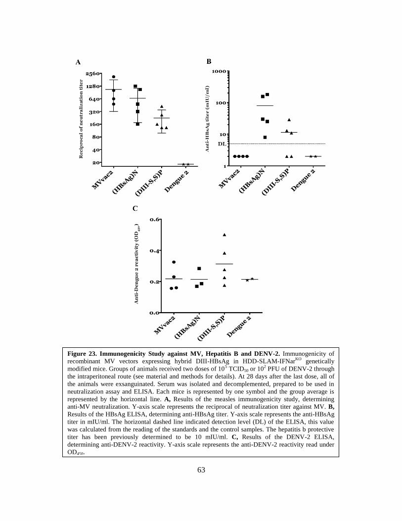
63
Figure 23. Immunogenicity Study against MV, Hepatitis B and DENV-2. Immunogenicity of
recombinant MV vectors expressing hybrid DIII-HBsAg in HDD-SLAM-IFNarKO
genetically
modified mice. Groups of animals received two doses of 105 TCID50 or 10
2 PFU of DENV-2 through
the intraperitoneal route (see material and methods for details). At 28 days after the last dose, all of
the animals were exsanguinated. Serum was isolated and decomplemented, prepared to be used in
neutralization assay and ELISA. Each mice is represented by one symbol and the group average is
represented by the horizontal line. A, Results of the measles immunogenicity study, determining
anti-MV neutralization. Y-axis scale represents the reciprocal of neutralization titer against MV. B,
Results of the HBsAg ELISA, determining anti-HBsAg titer. Y-axis scale represents the anti-HBsAg
titer in mIU/ml. The horizontal dashed line indicated detection level (DL) of the ELISA, this value
was calculated from the reading of the standards and the control samples. The hepatitis b protective
titer has been previously determined to be 10 mIU/ml. C, Results of the DENV-2 ELISA,
determining anti-DENV-2 reactivity. Y-axis scale represents the anti-DENV-2 reactivity read under
OD450.

64
To analyze the anti-HBsAg reactivity of the sera samples, we used a commercial
ELISA kit. We compared the readings from each sera collected from our animals, to a
standard curve constructed with an international reference. The known hepatitis B
protective titer is 10 mIU/ml and the detection level of our ELISA results is shown as DL
(Fig. 23. B). As expected, there was no reactivity detected in sera collected from mice
vaccinated with MVvac2 and DENV-2. We observed that the anti-HBsAg titer for both
MVvac2(HBsAg)N and MVvac2(DIII-S,S)P passed the protective titer of 10 mIU/ml.
However, the sera collected from mice vaccinated with MVvac2(DIII-S,S)P showed an
eight-fold reduction in HBsAg immunogenicity when compared to MVvac2(HBsAg)N.
To analyze the anti-DENV-2 reactivity of our sera samples, we used an ELISA.
Whole dengue 2 viruses was adsorbed to the bottom of the well overnight at 4ºC. After
blocking, 1:50 serum dilutions were added and incubated. We used anti-mouse-HRP to
bind to the left-over antibodies which reacted with DENV-2, and developed the plate
with HRP and 3M TMB-H2SO4. Anti-DENV2 reactivity was only seen in two sera
samples from animals vaccinated with MVvac2(DIII-S,S)P (Fig. 23. C). These two sera
samples were also the samples that showed the highest measles and hepatitis B
immunogenicity from the MVvac2(DIII-S,S)P vaccinated group. As expected, all of the
mice vaccinated with MVvac2 and MVvac2(HBsAg)N did not show any reactivity to
DENV-2. However, we were also not able to observe any reactivity in sera from animal
vaccinated with DENV-2, which is supposed to be the positive control. This might have
been caused by the error found in the doses given during vaccination of these mice,
which happened to be much lower than previously determined. Overall, we saw reactivity
against DENV-2 from sera collected from our vaccinated animals, but an ELISA does not
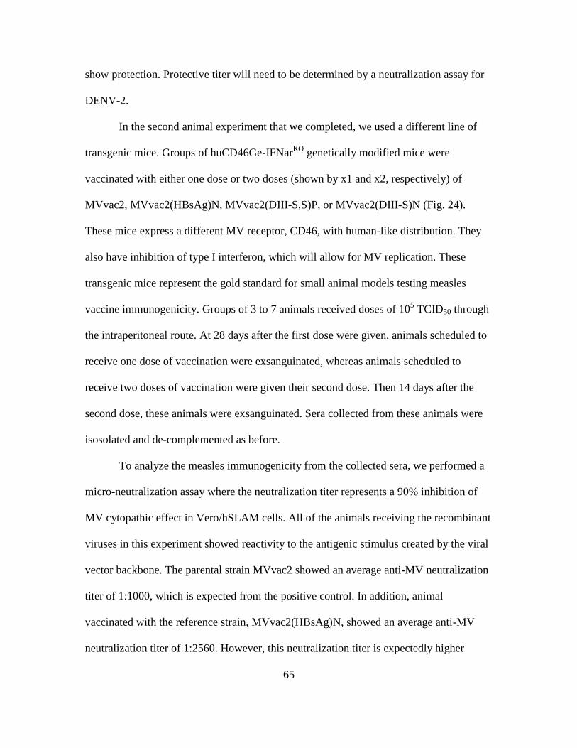
65
show protection. Protective titer will need to be determined by a neutralization assay for
DENV-2.
In the second animal experiment that we completed, we used a different line of
transgenic mice. Groups of huCD46Ge-IFNarKO
genetically modified mice were
vaccinated with either one dose or two doses (shown by x1 and x2, respectively) of
MVvac2, MVvac2(HBsAg)N, MVvac2(DIII-S,S)P, or MVvac2(DIII-S)N (Fig. 24).
These mice express a different MV receptor, CD46, with human-like distribution. They
also have inhibition of type I interferon, which will allow for MV replication. These
transgenic mice represent the gold standard for small animal models testing measles
vaccine immunogenicity. Groups of 3 to 7 animals received doses of 105 TCID50 through
the intraperitoneal route. At 28 days after the first dose were given, animals scheduled to
receive one dose of vaccination were exsanguinated, whereas animals scheduled to
receive two doses of vaccination were given their second dose. Then 14 days after the
second dose, these animals were exsanguinated. Sera collected from these animals were
isosolated and de-complemented as before.
To analyze the measles immunogenicity from the collected sera, we performed a
micro-neutralization assay where the neutralization titer represents a 90% inhibition of
MV cytopathic effect in Vero/hSLAM cells. All of the animals receiving the recombinant
viruses in this experiment showed reactivity to the antigenic stimulus created by the viral
vector backbone. The parental strain MVvac2 showed an average anti-MV neutralization
titer of 1:1000, which is expected from the positive control. In addition, animal
vaccinated with the reference strain, MVvac2(HBsAg)N, showed an average anti-MV
neutralization titer of 1:2560. However, this neutralization titer is expectedly higher
66
because these mice received two doses. The sera sample from mice vaccinated with one
dose of MVvac2(DIII-S,S)P showed an average anti-MV neutralization titer of 1:640,
which is lower than the samples from the mice vaccinated with the parental strain.
However, the sera from the mice that received two doses of MVvac2(DIII-S,S)P showed
an average anti-MV neutralization titer of 1:1280, which reached a titer higher than those
Figure 24. Immunogenicity Study against MV and DENV-2. Immunogenicity of
recombinant MV vectors expressing hybrid DIII-HBsAg in huCD46Ge-IFNarKO
genetically
modified mice. Groups of animals received one (x 1) or two (x 2)doses of 105 TCID50
through the intraperitoneal route (see material and methods for details). At 28 days after the
first dose, mice scheduled to receive only one dose were exsanguinated and mice scheduled
to receive two doses were given their second dose. At 14 days after the second dose, the rest
of the animals were exsanguinated. Serum was isolated and decomplemented, prepared to
be used in neutralization assays and ELISA. Each mice is represented by one symbol and
the group average is represented by the horizontal line. A, Results of the measles
immunogenicity study, determining anti-MV neutralization. Y-axis scale represents the
reciprocal of neutralization titer against MV. B, Results of the DENV-2 immunogenicity
study, determining anti-DENV-2 neutralization. Y-axis scale represents the logarithmic
neutralization index (LNI) against DENV-2. The controls were used to create the baseline
value were sera collected from animal vaccinated with the parental strain, MVvac2, and
reference strain, MVvac2(HBsAg)N. The horizontal dashed line shows the protective level
of 0.7 LNI previously determined for other flaviviruses. Statistically significant differences
are indicated by horizontal line above the experimental groups. Values from PRNT50
neutralization assay is shown at the top of the graph.

67
of the mice vaccinated with MVvac2. In addition, the sera sample from mice vaccinated
with two doses of MVvac2(DIII-S)N showed a higher average anti-MV neutralization
titer of 1:1500. This measles immunogenicity study showed that receiving two doses of
the recombinant viruses can induce similar amount of immune response against measles
as vaccinating with only one dose of MVvac2. Therefore, the problem in reduction of
neutralization titer against MV seen in mice vaccinated with the lower fitness
recombinant virus can possibly be avoided by adding a second dose.
To analyze the reactivity of the collected sera against HBsAg, we used a
commercial ELISA kit, comparing the readings from the sera from our vaccinated mice
to a standard curve constructed with an international reference (data not shown). For this
experiment, the sera of the animals were pooled for each group of mice. Pooled sera from
mice vaccinated with two doses of the reference strain, MVvac2(HBsAg)N, reached a
titer of 648 mIU/ml. Whereas pooled sera from mice vaccinated with two doses of the
recombinant strain, MVvac2(DIII-S,S)P, reached a titer of 787 mIU/ml. In contrast,
pooled sera from animals vaccinated with two doses of MVvac2(DIII-S)N, did not show
significant reactivity. As expected, the pooled sera collected from mice vaccinated with
MVvac2, did not show HBsAg reactivity.
To analyze the reactivity of the collected sera against DENV-2, we performed
neutralization experiments through two different approaches. First, we analyzed the effect
of a 1:5 serum dilution on the infection of BHK-21 cells with a 105 PFU/ml DENV-2
(Fig. 24. B). The reduction in DENV-2 infectivity caused by immune sera reactivity was
recorded as the logarithmic neutralization index (Log10NI). Sera samples collected from
animal vaccinated with the parental strain, MVvac2, and the reference strain,

68
MVvac2(HBsAg)N, were used as the negative controls; their values is used to set the
baseline of the graph (Fig. 24. B). Reactivity against DENV-2 was seen in sera collected
from mice vaccinated with MVvac2(DIII-S,S)P. We observe that those of which received
only one dose had an average of 0.7 LNI, which shows a reduction of 5 times in DENV-2
infection. In addition, those that received two doses of MVvac2(DIII-S,S)P had an
average of 1.3 LNI, which shows a reduction of about 20 times in DENV-2 infection.
Protective titer level against other flaviviruses that has been previously determined is at
0.7 LNI. Therefore, sera collected from the mice vaccinated with two doses of
MVvac2(DIII-S,S)P reached a higher LNI than needed for the protective level. Whereas,
those that only received one dose reach just at the protective level of LNI. However, sera
collected MVvac2(DIII-S)N vaccinated mice did not reach a protective level.
The second neutralization experiment that was performed to analyze the ability of
the mice sera to inhibit DENV-2 infection was the classic 50% plaque reduction
neutralization assay (Fig. 24. B). Two-fold serum dilutions were incubated with 200 PFU
of DENV-2 and the neutralization titer is determined by the dilution at which the sera
have reduced the plaques by half, compared to a control. To determine the PRNT50
through this experiment we used pooled mice sera for each group of mice following their
vaccine schedule. We calculated the PRNT50 from sera pooled from mice that were given
one dose of MVvac2(DIII-S,S)P, two doses of MVvac2(DIII-S,S)P, and two doses of
MVvac2(DIII-S)N to be 1:10, 1:160, and 1:20 respectively. We observed that the
PRNT50 of mice vaccinated with two doses of MVvac2(DIII-S)N is higher than those that
received only one dose of MVvac2(DIII-S,S)P, this is the opposite of the result we
received in the previous experiment. However, this may be due to the small amount of

69
DENV-2 epitopes that were accurately displayed causing borderline immunogenicity
reaction against DENV-2 in this neutralization assay. Overall, the best PRNT50 titer that
we observed through this neutralization assay resulted from the pooled mice sera from
those receiving two doses of MVvac2(DIII-S,S)P.
70
Chapter 4. Discussion
VLP platforms are very attractive vaccine candidates since they offer the
possibility of delivering antigenic material with a structure very similar to a full virion
and are safe because there is no risk of infection. These platforms represent the current
vaccines for hepatitis B virus and human papillomavirus. We decided to use HBsAg as a
platform to display domain III of the envelope protein of DENV-2 because of the
translational strategy used for the expression and assembly of the particulate antigen.
During hepatitis B virus infection, the viral envelope is generated by the expression of
three envelope glycoproteins, the small, medium and large surface antigens. Their coding
sequences have in common a 226 amino acids scaffold towards the carboxyl terminus.
Translation starts from in-frame initiation codons, giving origin to the expression of the
preS1 gene for the large antigen, preS2 gene for the medium antigen, and S gene for the
small antigen. These glycoproteins of 27, 36 and 42 kDa, have the potential to self-
assemble into empty particles upon expression in infected cells with a proportion of
4:1:1. This project explored the possibility of swapping the preS1 and preS2 regions with
the DIII of the DENV-2 envelope glycoprotein and a bridge, creating the hybrid DIII-
HBsAg antigen. Two different recombinant viruses were then engineered using MV as
the viral vector backbone. MV was selected to be used as the viral vector due to its safety
and stability. We used MVvac2, which is the recombinant version of the attenuated
strain, Moraten and Schwartz (genetically identical, despite the fact of being obtained
separately), which is used in the current children’s vaccination schedule. We take
advantage of the MV expression gradient to strategically place our ATUs; where the
amount of transcription is determined by the position of the cistron relative to the 3’ end

71
of the genome. The cistron located towards the 3’ end is transcribed at a higher frequency
due to the graduate attenuation at gene boundaries. This knowledge of the transcription of
MV genome is factored into the engineering of our recombinant viruses. These
recombinant viruses are termed MVvac2(DIII-S,S)P with the insertion of two ATUs and
MVvac2(DIII-S)N with the insertion of only one ATU.
In MVvac2(DIII-S,S)P we co-expressed from a different locus in the MV genome
the HBsAg protein in addition to the hybrid DIII-S, in order to promote the native
assembly of VLPs. The proportion of these two ATUs by MV transcription gradient will
drive a proportion of expression close to 3:1. Therefore, the generation of unmodified
HBsAg, from the cistron located after the N cistron, can be used as a scaffold for the
hybrid DIII-S antigen. The ATU encoding the hybrid DIII-S antigen is inserted
downstream of the P gene. The correct construction of this full-length plasmid,
pB(+)MVvac2(DIII-S,S)P, was confirmed through HindIII digestion and DNA
sequencing. In figure 16, we show the observed bands from samples of the full-length
plasmid digested with HindIII. Compared to the expected band, we conclude that the
correct full-length plasmid was created. In addition, the DNA sequence of the full-length
plasmid was checked for errors, and we conclude that the sequence was accurate.
In MVvac2(DIII-S)N, we only inserted one ATU expressing the hybrid DIII-S to
observe whether VLPs can assemble efficiently without an HBsAg scaffold. The ATU
encoding the hybrid DIII-S antigen is inserted downstream of the N gene and as such we
would have expected a robust expression of the antigen. The correct construction of this
full-length plasmid, pB(+)MVvac2(DIII-S)N, was also confirmed through HindIII
digestion and DNA sequencing. In figure 15, we show the observed bands from one clone

72
of the full-length plasmid. We conclude that the correct full-length plasmid was created
after the result is compared to the expected bands. The DNA sequence also confirmed the
accuracy of the full-length plasmid.
After confirmation of the correct sequences, we move on to rescue the
recombinant viruses through the established reverse genetic techniques for MVs. In the
bottom syncytia image in figure 17, we show the syncytia observed after we successfully
rescued one of our recombinant viruses MVvac2(DIII-S,S)P. Then we continued to
successfully rescue our second recombinant virus MVvac2(DIII-S,S)N (syncytia not
shown). After the recombinant viruses were rescued, the viruses were passaged to create
stock of viral prep. We were able to create a viral prep tittered at 1.8x10⁵ TCID50/ml for
MVvac2(DIII-S,S)P and 5.6x10⁷
TCID50/ml for MVvac2(DIII-S)N. These titers are high
enough for us to move forward with additional experiments, which also include our
animal experiments.
The next step of the experiments was to analyze the replication fitness of the
recombinant MVs compared to the parental strain, MVvac2, and the reference strain,
MVvac2(HBsAg)N. The multiple-step growth kinetics that was performed showed a
decrease in the replication fitness of MVvac2(DIII-S,S). The decrease of viral production
compared to MVvac2 and MVvac2(HBsAg)N was concluded to be result of the two
ATUs inserted into the MV genome. Although the two ATUs were inserted in non-
sequential locus of the genome, the increased size of the genome may also account for the
drop of MV infectivity progression. Furthermore, it makes sense to conclude that more
insertions lead to a dilution of the genes downstream the insertions. Those genes are
important for measles replication (like the polymerase, L gene) In addition, this negative

73
effect on the replication of recombinant MVs have been observed in other research
utilizing MV vectors with multiple insertion of ATUs. On the other hand, this negative
effect of the recombinant virus’ replication fitness was not observed in MVvac2(DIII-
S)N which makes us conclude that the gene DIII-SA is not by itself incompatible with
measles replication.. Our second recombinant virus followed a similar replication fitness
as the parental strain, MVvac2. In addition, it also follows the replication fitness of
MVvac2(HBsAg)N, which also have one ATU encoding HBsAg. This supports the idea
that only one ATU insertion into the MV genome was not able to affect its replication
fitness, whereas the insertion of two ATUs was able to affect it negatively. The results
from the multi-step growth kinetics is statistically valid, since it shows the corroborated
average data from experiments repeated independent of each other.
Next, we performed an ELISA against HBsAg to confirm the production of
HBsAg. This experiment was done as an initial assessment of the possible production of
HBsAg from the recombinant MVs. Hence it was performed only using the supernatants
collected from MVvac2(DIII-S,S)P infected cells, as well as the supernatants from
MVvac2(HBsAg)N infected cells as the control. Samples of the supernatants were
collected at different time-points post-infection. The result from the Anti-HBsAg ELISA
showed the presence of HBsAg in the supernatant from MVvac2(DIII-S,S)P infected
cells. Data above the dashed line (Fig. 19) showed the significant value, which represents
the HBsAg present. However, we were not able to state any quantitative data from this
experiment, because the assay shows only the possibility to discern positive from
negative; due to the sensitivity of this specific ELISA kit. We plan to standardize this
experiment in order to supplement the replication kinetics we collected. Therefore this
74
experiment was a supplement to the replication kinetics we collect. However, we
characterized the production of HBsAg, as well as the hybrid DIII-HBsAg antigen, in a
more precise approach, by protein immunoblots.
The results from the characterization of the expression of hybrid DIII-HBsAg
antigen (Fig. 20) solidified that DIII-S hybrid glycoprotein was indeed expressed in
infected cell extracts. We were able to corroborate that each sample contained the same
amount of protein extract, as shown by the bottom western blot (Fig. 20) that is
developed by the antibody against the N protein of MV. In the experimental sample, there
is a clear upward shift of the double band in the sample collected from the protein
extracts from cells infected with MVvac2(DIII-S,S)P representing the higher size of the
protein with the addition of DIII from DENV-2. This can be easily compared to the
control sample of protein extracts from cells infected with MVvac2(HBsAg)N, which
only show one set of double bands at a lower size. However, the upwardly shifted double
bands corresponding to the glycosylated (37 kDa) and non-glycosylated (34 kDa)
isoforms of the hybrid antigen is shown to be at a lower quantity, by the intensity of the
bands (Fig. 20). We concluded that this was due to the location of the ATU that is
inserted encoding the hybrid DIII-S antigen, which is downstream of the P gene.
Following the MV genome transcription gradient, this means that it was transcribed at a
lower quantity due to its location. In addition, the amount of the native HBsAg
glycosylated (27 kDa) and non-glycosylate (24 kDa) isoforms present in the sample from
the MVvac2(DIII-S,S)P infected cells is similar to the same isoform in the sample
collected from cells infected with the reference strain. Overall, from this initial
characterization experiment, we were able to confirm from our western blots, the
75
successful expression of the hybrid DIII-HBsAg antigen from one of our recombinant
MV, MVvac2(DIII-S,S)P. The next step we took was then to characterize both of our
recombinant MVs for the production of the hybrid antigen.
The characterization for the expression of the hybrid DIII-HBsAg antigen was
repeated with both recombinant MVs. The experiment was performed in the same way as
the previous one. However, the western blots were containing the same samples, were
developed with anti-HBsAg monoclonal antibody (Fig. 21. A) and anti-DIIII DENV-2
monoclonal antibody (Fig. 21. B). As expected, the upward shifted band that was seen in
the sample from cells infected with MVvac2(DIII-S,S)P is present again in this western
blot. We also see again the lower intensity of the bands at 37 kDa and 34 kDa. The bands
corresponding to the native HBsAg can also be seen in the samples form cells infected
with MVvac2(DIII-S,S)P and MVvac2(HBsAg)N. However, bands showing any
reactivity were not seen in the protein extract from cells infected with MVvac2(DIII-S)N,
concluding that the insertion of DIII into HBsAg, in the absence of HBsAg alone is not
compatible with the recognition by our antibody. The reactivity of the anti-HBsAg
antibodies highly depends on the conservation of the semi-native structure of the
protein’s epitopes, which is the reason why this western blot was run under non-reducing
conditions. This monoclonal detects a region on HBsAg that is termed he “a” epitope that
is essential for proper folding of HBsAg. We propose that the sole expression of DIII-S
hybrid antigens leads to an unfolded product that is certainly tagged for degradation.
Therefore, we suggest that the additional native HBsAg is needed to scaffold the DIII-S
antigen, which will result in the correct protein structure that will be recognized by the
antibody. In order to confirm that the expressed hybrid DIII-S antigen also presented the

76
DENV-2 epitopes shown in DIII, a western blot was developed with a monoclonal
antibody specific to DIII of the DENV-2 envelope protein (Fig. 21. B). Here we observed
two bands at 37 kDa and 34 kDa in both samples of protein extracts from cells infected
with MVvac2(DIII-S,S)P and MVvac2(DIII-S)N. These bands correspond to the same
bands in the western blot developed with anti-HBsAg antibodies, which represents the
glycosylated and non-glycosylated isoforms of the hybrid antigen. The intensity of the
bands was observed to be weak in the sample from cells infected with MVvac2(DIII-S)N,
which supports the conclusion that the expressed hybrid antigen degraded and misfolded.
In addition, a western blot containing the same samples was also developed with
antibodies against the N protein of MV to corroborate that the same amount of each
sample were used (data not shown). Therefore, we were able to confirm the expression of
the correct DENV-2 epitope present in DIII from one of our recombinant MV,
MVvac2(DIII-S,S)P. However, we suggest that MVvac2(DIII-S)N may be expressing an
incorrectly folded hybrid DIII-HBsAg.
The next experiment that we performed was done to confirm the successful
construction of hybrid viral-like particles. This was done by using a discontinuous
sucrose gradient to fractionate by density samples collected from supernatants of cells
infected with MVvac2(DIII-S,S)P of MVvac2(HBsAg)N. Due to the inability of the anti-
HBsAg antibody to recognize the hybrid antigen produced by MVvac2(DIII-S)N (Fig.
21. B), this experiment was focused on the first recombinant virus MVvac2(DIII-S,S)P.
MVvac2(HBsAg)N was used as a positive control, and cells were infected with the
reference strain in parallel, because their ability to secrete VLPs following a specific
sucrose density has been previously documented by other researchers. In the western blot
77
of the nine fractions collected from the supernatants of cells infected with MVvac2(DIII-
S,S)P, developed with anti-HBsAg, we observed the upward shifted double bands
corresponding to the hybrid DIII-HBsAg bands in fractions 4 to 5, shown with arrows
(Fig. 22). We also observe the double bands corresponding to the native HBsAg in the
western blots from both the recombinant strain and reference strain fractions; these are
also sequestered in fraction 4 to 5 at a sucrose density of 1.10-1.12 g/ml. Therefore, we
can conclude that the biophysical characteristics of the HBsAg subviral particle were not
changed or affected by the incorporation of DIII, because the hybrid VLPs and the native
HBsAg VLPs were sequestered in the same fractions and at the same density. In addition,
the last western blot that was done (Fig. 22. B) was developed by antibodies against the N
protein of MV, using fractions collected from the supernatants of cells infected with
MVvac2(DIII-S,S)P. This western blot shows that the MV viral particles are sequestered
at more dense fractions, fraction 6 to 9. This supports the notion that HBsAg- and hybrid-
VLPs are secreted independently of the MV viral particles and that the production of the
VLPs did not negatively affect the production of MV viral particles. Overall, we
confirmed the successful incorporation of DIII in the hybrid VLPs, without affecting the
antigenic characteristics of the VLPs.
After we have established that the hybrid antigen DIII-HBsAg can be expressed
by our recombinant MVs and that viable hybrid VLPs can be produced, we moved
forward with the most important experiments in our animal models. Two animal
experiments were performed, using two different types of transgenic mice; sera collected
from these animal experiments were then used for an immunogenicity study. The first
animal experiment was done in HDD-SLAM-IFNarKO
transgenic mice. These mice were

78
vaccinated with the controls: parental strain, MVvac2, reference strain,
MVvac2(HBsAg)N, and the positive control, DENV-2; as well as the experimental
recombinant virus, MVvac2(DIII-S,S)P. In the immunogenicity study against measles,
we observed a four-fold reduction of neutralization in sera from mice vaccinated with
MVvac2(DIII-S,S)P compared to the parental strain, MVvac2 (Fig. 23). However, we
also observed a two-fold reduction of neutralization in sera from mice vaccinated with
MVvac2(HBsAg)N. This data would support the theory of the decrease in viral fitness
based on the amount of ATUs inserted into the MV genome. Insertion of one ATU
resulted in a two-fold reduction in neutralization and the insertion of two ATUs resulted
in a four-fold reduction in neutralization. This will be a valuable knowledge for future
construction of recombinant MVs.
In the immunogenicity study against hepatitis B and DENV-2, we observed
reactivity for both in our experimental sample (Fig. 23). The result of the anti-HBsAg
ELISA showed that the sera from mice vaccinated with MVvac2(DIII-S,S)P were able to
reach the protective titer of 10 mIU/ml. However, the eight-fold reduction in its anti-
HBsAg titer may be caused by the use of HBsAg in creating the hybrid VLP, which may
cause a specific amount of the antigenic epitope of HBsAg to be hidden from the surface
of the VLPs. The result of the homemade ELISA to analyze the reactivity of the sera to
DENV-2, shows two sera samples from mice vaccinated with MVvac2(DIII-S,S)P that
clearly reacted to DENV-2. The sera that came from these two mice were also the ones
that had the highest measles neutralization titer at 1:426 and 1:266, as well as the highest
anti-HBsAg titer at 54 mIU/ml and 13 mIU/ml, from the group of 5 mice vaccinated with
MVvac2(DIII-S,S)P, which supports the notion that a robust anti-HBsAg immunity is

79
accompanied by Dengue 2 recognition. Although the mice vaccinated with DENV-2 was
supposed to be the positive control. We realized at a later time, that these mice were
given a dose of 102 PFU instead of 10
5 PFU, which may have resulted in the lack of
immune response that was induced in these animals. Overall, the ELISA against DENV-2
only shows reactivity of the sera, not quantifying its neutralization capability. Therefore,
a neutralization assay for DENV-2 was performed from the animal sera collected from
the second animal experiment, to present a more accurate data.
In the second animal experiment, huCD46Ge-IFNarKO
transgenic mice were
vaccinated with the controls: the parental strain, MVvac2, and the reference strain,
MVvac2(HBsAg)N; as well as the experimental recombinant MVs: MVvac2(DIII-S,S)P
and MVvac2(DIII-S)N. Different groups of animal received different doses as shown in
figure 24. In the immunogenicity study against measles, we observed a similar reduction
in anti-MV neutralization titer in the sera collected from animals vaccinated with one
dose of MVvac2(DIII-S,S)P, as the previous animal experiment. Again, we believe that
this is caused by the reduced viral replication fitness due to the insertion of two ATUs, as
documented in vitro from our multi-step growth kinetics (Fig. 18). However, we
observed that this negative effect from the weaker viral fitness can be solved by the
administration of a booster dose. The anti-MV neutralization titer of the sera collected
from mice which received two doses of MVvac2(DIII-S,S)P is comparable to that of the
parental strain, MVvac2 (Fig. 23. A). In addition, the other groups of animal which also
received two doses, MVvac2(HBsAg)N and MVvac2(DIII-S)N, also showed an anti-MV
neutralization titer comparable to the animals that was vaccinated with MVvac2.

80
The immunogenicity study against hepatitis B was done through the use of a
commercial ELISA. There were no significant reactivity seen in the pooled sera collected
from animals that received one dose of MVvac2(DIII-S,S)P, two doses of MVvac2(DIII-
S)N, or as expected, one dose of MVvac2 (data not shown). However, we did observed
an anti-HBsAg titer of 787 mIU/ml from mice vaccinated with two doses of
MVvac2(DIII-S,S)P and 648 mIU/ml from mice vaccinated with two doses of
MVvac2(HBsAg)N. As previously witnessed, the administration of two doses of the
recombinant virus highly increases the immune response induced. In the immunogenicity
study against DENV-2, two different types of neutralization assays were performed. The
result of the first approach, recorded in Log10NI, shows a strong neutralization capability
of sera collected from mice vaccinated with two doses of MVvac2(DIII-S,S)P; although
the mice that received only one dose was able to reach the protective titer for reference
flaviviruses (such as YFV and JEV) of 0.7 LNI. This result gave additional support to the
theory that the administration of a booster shot may be needed to induce a completely
protective immune response. On the other hand, the sera from mice vaccinated with two
doses of MVvac2(DIII-S)N did not reach a protective titer. This may be caused by the
improper folding of the hybrid DIII-HBsAg, resulting in the improper construction of the
hybrid VLPs produced by this recombinant virus, as we have previously suggested. The
second neutralization assay done against DENV-2, was the classic PRNT50. Once more
the result of the neutralization capability of the sera collected from the mice that received
two doses of MVvac2(DIII-S,S)P was shown to be the strongest. However, the pooled
sera from mice that received only one dose of MVvac2(DIII-S,S)P had a lower PRNT50
titer than those that received two doses of MVvac2(DIII-S)N. It is possible that this

81
happened due to the presence of a few properly expressed DENV-2 epitopes that was
previously seen in a western blot developed with anti-DIII DENV-2 (Fig. 21. B); this
may have caused a minimal immunogenicity reaction against DENV-2 in this
neutralization assay. Throughout the immunogenicity study, the animals that received
two doses of MVvac2(DIII-S,S)P consistently showed higher immunogenicity against all
three viruses.
The stability of the recombinant viruses could be observed through the presence
of the bands in an immunoblot from samples of different passages. MVvac2(DIII-S,S)P
have been amplified to reach passage 5, and the double bands corresponding to the DIII-S
antigen from these samples can still be observed in an immunoblot. However,
MVvac2(DIII-S)N have only been amplified to passage 3, and this was the passage used
in all of the experiments concerning this recombinant MV. Further experiments are still
needed to fully confirm the stability of the recombinant MVs, which include sequencing
each passage of the virus as well as a serial passaging experiment. The result from these
experiments will then be able to determine whether the viral genome remains unaffected
through multiple passages.
Overall, we were able to confirm the successful construction of the recombinant
MVs and analyzed their capability of eliciting immune response in the immunogenicity
studies. In MVvac2(DIII-S,S)P, we demonstrated the correct assembly of our hybrid
antigen and the release of the hybrid subviral particles in the supernatants of infected
cells. In addition, this recombinant MV was able to induce a protective neutralizing
immune response against all three viruses: MV, HBV, and DENV-2. Although we were
not able to detect the successful secretion of the hybrid antigen from MVvac2(DIII-S)N

82
by anti-HBsAg, we confirmed that its replication fitness mirrored that of the parental
strain, we were able to see it elicit immune response against measles, as well as express
marginally some antigens that are recognized by the anti-DIII DENV-2. The result of our
animal experiments also shows the importance of the vaccination dosage, as the animals
that received two doses of MVvac2(DIII-S,S)P elicited the strongest immune response.
Although we were not able to detect viable hybrid subviral particles secreted from
MVvac2(DIII-S)N, we suggests that this is due to the lack of native HBsAg VLP that was
available to scaffold the hybrid DIII-S antigen. In order to clarify this theory, we will
perform a co-infection experiment using MVvac2(DIII-S)N and MVvac2(HBsAg)N
We decided to use HBsAg to create our hybrid VLP, because HBsAg
independently creates its own VLP and it is also used in an approved human vaccine
against hepatitis B. As shown in our results, the production of additional native HBsAg is
necessary in the accurate assembly of the hybrid VLPs. In addition, our use of MV as the
viral vector serves the purpose of a live adjuvant in the form of a live attenuated virus,
which has been proven to be safe and stable.

83
Chapter 5. Conclusion
This is a proof-of concept for an ideal pediatric vaccine that would protect an
individual against DENV, Hepatitis B virus, and MV. Using MV as a viral vector will
make the production and distribution of this recombinant vaccine to be efficient and cost
effective; mainly due to the system that is already in place for the production and
distribution of MV based vaccines. In addition, this vaccine would be a safe option, since
the Moraten/Schwartz vaccine strain used has been proven to have long-term safety. Our
data confirms immunogenicity in MV susceptible animal models against all three:
DENV, Hepatitis B virus and MV. The heightened immune response seen in animal who
received two doses suggests that to achieve complete protection in humans, booster
vaccination may be necessary.
Chapter 6. Future Perspective
In future experiments, we would like to look for possibilities of increasing the
viral fitness of our recombinant MVs. As well as the possibility of avoiding the decrease
in viral fitness by inserting only one ATU into the MV genome, but inoculating in vivo,
with two recombinant MVs expressing the additional HBsAg needed. To circumvent the
replication problem that having two ATUs originate, we propose to invert the order of
insertion: DIII-S after N and HBsAg after P, which will increase the expression of the
hybrid antigen and conserve a scaffold of HBsAg to construct the particle. Alternatively,
we can infect cells with two separate vectors and collect the supernatant form co-infected
cells. Arguably, if both antigens are expressed in the same syncytia, then hybrid VLPs
will be secreted, it is tempting to think that MV virion with two genome can be
assembled and propose and original solution to multiple insertion.
84
References
1. Rothman AL. 2011. Immunity to dengue virus: a tale of original antigenic sin
and tropical cytokine storms. Nature Reviews Immunology 11:532-543.
2. Acosta EG, Kumar A, Bartenschlager R. 2014. Chapter One - Revisiting
Dengue Virus–Host Cell Interaction: New Insights into Molecular and Cellular
Virology, p. 1-109. In Karl M, Frederick AM (ed.), Advances in Virus Research,
vol. Volume 88. Academic Press.
3. Guzman MG, Harris E. 2015. Dengue. Lancet 385:453-465.
4. Lei H-Y, Yeh T-M, Liu H-S, Lin Y-S, Chen S-H, Liu C-C. 2001.
Immunopathogenesis of dengue virus infection. Journal of Biomedical Science
8:377-388.
5. Kautner I, Robinson MJ, Kuhnle U. 1997. Dengue virus infection:
Epidemiology, pathogenesis, clinical presentation, diagnosis, and prevention. The
Journal of pediatrics 131:516-524.
6. Yauch LE, Shresta S. 2014. Chapter Seven - Dengue Virus Vaccine
Development, p. 315-372. In Karl M, Frederick AM (ed.), Advances in Virus
Research, vol. Volume 88. Academic Press.
7. Wan S-W, Lin C-F, Yeh T-M, Liu C-C, Liu H-S, Wang S, Ling P, Anderson
R, Lei H-Y, Lin Y-S. 2013. Autoimmunity in dengue pathogenesis. Journal of
the Formosan Medical Association 112:3-11.
8. Mackenzie JS, Gubler DJ, Petersen LR. 2004. Emerging flaviviruses: The
spread and resurgence of Japanese encephalitis, West Nile and dengue viruses.
Nature medicine 10:S98-S109.
9. Byron EEM, Koraka P, Albert DMEO. 2009. Dengue Virus Pathogenesis: an
Integrated View. Clinical microbiology reviews 22:564-581.
10. Brandler S, Ruffie C, Najburg V, Frenkiel MP, Bedouelle H, Despres P,
Tangy F. 2010. Pediatric measles vaccine expressing a dengue tetravalent antigen
elicits neutralizing antibodies against all four dengue viruses. Vaccine 28:6730-
6739.
11. Henchal EA, Putnak JR. 1990. The dengue viruses. Clinical microbiology
reviews 3:376-396.
12. Angel RMd, Valle JR-d. 2013. Dengue Vaccines: Strongly Sought but Not a
Reality Just Yet. PLoS Pathogens 9:1-4.

85
13. Whitehorn J, Simmons CP. 2011. The pathogenesis of dengue. Vaccine
29:7221-7228.
14. Murphy BR, Durbin AP, Whitehead SS, Blaney JE. 2007. Prospects for a
dengue virus vaccine. Nature Reviews Microbiology 5:518-528.
15. Edelman R. 2005. Dengue and Dengue Vaccines. The Journal of infectious
diseases 191:650-653.
16. Bentsi-Enchill AD. Long-term safety assessment of live attenuated tetravalent
dengue vaccines: Deliberations from a WHO technical consultation. Vaccine
31:2603-2609.
17. Schmitz J, Roehrig J, Barrett A, Hombach J. 2011. Next generation dengue
vaccines: A review of candidates in preclinical development. Vaccine 29:7276-
7284.
18. Wallace D, Canouet V, Garbes P, Wartel TA. 2013. Challenges in the clinical
development of a dengue vaccine. Current Opinion in Virology 3:352-356.
19. Coller B-AG, Clements DE. 2011. Dengue vaccines: progress and challenges.
Current Opinion in Immunology 23:391-398.
20. Guzman MG, Hermida L, Bernardo L, Ramirez R, Guillen G. 2010. Domain
III of the envelope protein as a dengue vaccine target. EXPERT REVIEW OF
VACCINES 9:137-147.
21. Clements D, Ogata S, Modis Y, Harrison SC. 2004. Structure of the dengue
virus envelope protein after membrane fusion. Nature 427:313-319.
22. Watterson D, Kobe B, Young PR. 2012. Residues in domain III of the dengue
virus envelope glycoprotein involved in cell-surface glycosaminoglycan binding.
Journal of General Virology 93:72-82.
23. Sukupolvi-Petty S, Austin SK, Purtha WE, Oliphant T, Nybakken GE,
Schlesinger JJ, Roehrig JT, Gromowski GD, Barrett AD, Fremont DH,
Diamond MS. 2007. Type- and Subcomplex-Specific Neutralizing Antibodies
against Domain III of Dengue Virus Type 2 Envelope Protein Recognize
Adjacent Epitopes. Journal of Virology 81:12816-12826.
24. Grgacic EVL, Anderson DA. 2006. Virus-like particles: Passport to immune
recognition. Methods 40:60-65.
25. Cobleigh MA, Buonocore L, Uprichard SL, Rose JK, Robek MD. 2010. A
Vesicular Stomatitis Virus-Based Hepatitis B Virus Vaccine Vector Provides
Protection against Challenge in a Single Dose. Journal of Virology 84:7513-7522.

86
26. Thomson S, Haigh O, Gould A, Tindle R. 2008. Genetically modified hepatitis
B surface antigen: A powerful vaccine technology for the delivery of disease-
associated foreign antigens. Current Drug Therapy 3:226-234.
27. Noad R, Roy P. 2003. Virus-like particles as immunogens. Trends in
microbiology 11:438-444.
28. Roldao A, Mellado MCM, Castilho LR, Carrondo MJT, Alves PM. 2010.
Virus-like particles in vaccine development. EXPERT REVIEW OF VACCINES
9:1149-1176.
29. Vreden SG, Verhave JP, Oettinger T, Sauerwein RW, Meuwissen JH. 1991.
Phase I clinical trial of a recombinant malaria vaccine consisting of the
circumsporozoite repeat region of Plasmodium falciparum coupled to hepatitis B
surface antigen. The American journal of tropical medicine and hygiene 45:533-
538.
30. Bisht H, Chugh DA, Raje M, Swaminathan SS, Khanna N. 2002.
Recombinant dengue virus type 2 envelope/hepatitis B surface antigen hybrid
protein expressed in Pichia pastoris can function as a bivalent immunogen.
Journal of biotechnology 99:97-110.
31. Dowling PC, Blumberg BM, Menonna J, Adamus JE, Cook P, Crowley JC,
Kolakofsky D, Cook SD. 1986. Transcriptional Map of the Measles Virus
Genome. Journal of General Virology 67:1987-1992.
32. Brandler S, Tangy F. 2008. Recombinant vector derived from live attenuated
measles virus: Potential for flavivirus vaccines. Comparative Immunology,
Microbiology and Infectious Diseases 31:271-291.
33. Zuniga A, Wang Z, Liniger M, Hangartner L, Caballero M, Pavlovic J, Wild
P, Viret JF, Glueck R, Billeter MA, Naim HY. 2007. Attenuated measles virus
as a vaccine vector. Vaccine 25:2974-2983.
34. del Valle JR, Devaux P, Hodge G, Wegner NJ, McChesney MB, Cattaneo R.
2007. A Vectored Measles Virus Induces Hepatitis B Surface Antigen Antibodies
While Protecting Macaques against Measles Virus Challenge. Journal of Virology
81:10597-10605.
35. Bankamp B, Takeda M, Zhang Y, Xu W, Rota PA. 2011. Genetic
Characterization of Measles Vaccine Strains. Journal of Infectious Diseases
204:S533-S548.
36. Parks CL, Lerch RA, Walpita P, Wang H-P, Sidhu MS, Udem SA. 2001.
Analysis of the Noncoding Regions of Measles Virus Strains in the Edmonston
Vaccine Lineage. Journal of Virology 75:921-933.

87
37. Robert-Guroff M. 2007. Replicating and non-replicating viral vectors for
vaccine development. Current opinion in biotechnology 18:546-556.
38. Rutgers T, Gordon D, Gathoye AM, Hollingdale M, Hockmeyer W,
Rosenberg M, De Wilde M. 1988. Hepatitis B surface antigen as carrier matrix
for the repetitive epitope of the circumsporozoite protein of Plasmodium
falciparum. Bio/Technology 6:1065-1070.
39. Valle JR-d, Hodge G, McChesney MB, Cattaneo R. 2009. Protective Anti-
Hepatitis B Virus Responses in Rhesus Monkeys Primed with a Vectored Measles
Virus and Boosted with a Single Dose of Hepatitis B Surface Antigen. Journal of
virology 83:9013-9017.
40. Radecke F, Spielhofer F, Schneider H, Kaelin K, Huber M, Dotsch C,
Christiansen G, Billeter MA. 1995. Rescue of measles viruses from cloned
DNA. EMBO Journal 14:5773-5784.
41. REED LJ, MUENCH H. 1938. A SIMPLE METHOD OF ESTIMATING
FIFTY PER CENT ENDPOINTS. American Journal of Epidemiology 27:493-
497.
42. Whelan SPJ, Barr JN, Wertz GW. 2004. Transcription and replication of
nonsegmented negative-strand RNA viruses, p. 61-119, vol. 283. SPRINGER-
VERLAG BERLIN, BERLIN.