
Hepatitis B Subviral Envelope Particle Morphogenesis and
10
JOURNAL OF VIROLOGY, Apr. 2007, p. 3842–3851 Vol. 81, No. 8 0022-538X/07/$08.000 doi:10.1128/JVI.02741-06 Copyright © 2007, American Society for Microbiology. All Rights Reserved. Hepatitis B Virus Subviral Envelope Particle Morphogenesis and Intracellular Trafficking Romuald Patient, 1 Christophe Hourioux, 1 Pierre-Yves Sizaret, 1 Sylvie Trassard, 1 Camille Sureau, 2 and Philippe Roingeard 1 * Universite ´ Franc ¸ois Rabelais, INSERM ERI 19, Tours, France, 1 and Laboratoire de Virologie Mole ´culaire, INTS, Paris, France 2 Received 13 December 2006/Accepted 22 January 2007 Hepatitis B virus (HBV) is unusual in that its surface proteins (small [S], medium, and large [L]) are not only incorporated into the virion envelope but they also bud into empty subviral particles in great excess over virions. The morphogenesis of these subviral envelope particles remains unclear, but the S protein is essential and sufficient for budding. We show here that, in contrast to the presumed model, the HBV subviral particle formed by the S protein self-assembles into branched filaments in the lumen of the endoplasmic reticulum (ER). These long filaments are then folded and bridged for packing into crystal-like structures, which are then transported by ER-derived vesicles to the ER-Golgi intermediate compartment (ERGIC). Within the ERGIC, they are unpacked and relaxed, and their size and shape probably limits further progression through the secretory pathway. Such progression requires their conversion into spherical particles, which occurred spon- taneously during the purification of these filaments by affinity chromatography. Small branched filaments are also formed by the L protein in the ER lumen, but these filaments are not packed into transport vesicles. They are transported less efficiently to the ERGIC, potentially accounting for the retention of the L protein within cells. These findings shed light on an important step in the HBV infectious cycle, as the intracellular accumulation of HBV subviral filaments may be directly linked to viral pathogenesis. The human hepatitis B virus (HBV) is the prototype of the mammalian Hepadnaviridae (genus Orthohepadnavirus), a family of small hepatotropic DNA viruses causing acute and chronic liver disease (16). Despite the existence of an effective HBV vaccine, HBV remains a major health problem world- wide, as there is no generally effective treatment for the esti- mated 350 million chronic carriers who have a high risk of liver cirrhosis and hepatocellular carcinoma. A thorough under- standing of HBV morphogenesis and life cycle is thus required for the development of innovative antiviral treatments. The virion (or Dane particle) is a spherical particle, 42 nm in diameter, consisting of an icosahedral nucleocapsid of approx- imately 30 nm in diameter and an envelope composed of three surface proteins and, presumably, lipids of host cell origin. The nucleocapsid and envelope are synthesized and mature sepa- rately in different cellular compartments, subsequently inter- acting to form the virion (5, 26, 34). The three HBV envelope proteins are encoded by a single open reading frame, using three in-frame start codons (21). The large surface protein (L, or p39) is the translation product of the entire open reading frame (389 to 400 amino acid [aa] residues, depending on HBV genotype). The middle surface protein (M, or p30) lacks the N-terminal 119 aa of L (the pre-S1 sequence), and the small surface protein (S, or p24) lacks the N-terminal 55 aa of M (the pre-S2 sequence). These proteins are synthesized at the endo- plasmic reticulum (ER) membrane and have a complex trans- membrane topology (12, 13). They form disulfide-linked dimers with each other, with no detectable partner preference (27, 45). All three proteins have a form glycosylated at residue N146 in the S domain (gp27, gp33, and gp42 for S, M, and L, respectively), but about half the molecules produced remain unglycosylated at this site. The M protein may also be glyco- sylated (ggp36) at N4 in the pre-S2 domain (17). HBV and related viruses are unusual among viruses in that the surface proteins are not only incorporated into virion en- velopes but they also bud to generate empty subviral spherical or filamentous particles without nucleocapsids in an intracel- lular compartment, with these particles being formed in great excess over virions (21). The S protein is the predominant constituent of these subviral particles and is the essential topogenic element for this budding, with no other viral protein required. Indeed, the production of S protein alone in mam- malian cells causes efficient secretion of 20-nm large subviral particles, also called HBsAg particles (11, 29). This material is highly immunogenic and is the basis of most vaccines against hepatitis B (14). The type of particle formed seems to be determined by the ratio of S to L proteins coassembling during morphogenesis. The spherical 20-nm HBsAg particles isolated from the serum of infected patients contain only traces of L protein (21). The coassembly of a higher proportion of L protein with S results in formation of the filamentous form of HBsAg (21). This high L content inhibits particle secretion (32) due to specific retention motifs in this protein (15, 35) and/or the inefficient export of the filamentous particles (39). The L protein plays a key role in virus assembly and infectivity and is therefore present in large proportions in the virion envelope (6, 8, 21). The M protein seems to have no deter- mining influence on particle morphology because this protein is present in all three particles in proportions similar to that for S (21). This protein is also dispensable for virion formation in * Corresponding author. Mailing address: INSERM ERI 19, Labo- ratoire de Biologie Cellulaire, Faculte ´ de Me ´decine de Tours, 10 boulevard Tonnelle ´, F-37032 Tours Cedex, France. Phone: 33 2 47 36 60 71. Fax: 33 2 47 36 60 90. E-mail: [email protected]. Published ahead of print on 31 January 2007. 3842 Downloaded from https://journals.asm.org/journal/jvi on 23 November 2021 by 14.47.75.91.
Transcript of Hepatitis B Subviral Envelope Particle Morphogenesis and
JOURNAL OF VIROLOGY, Apr. 2007, p. 3842–3851 Vol. 81, No. 80022-538X/07/$08.00�0 doi:10.1128/JVI.02741-06Copyright © 2007, American Society for Microbiology. All Rights Reserved.
Hepatitis B Virus Subviral Envelope Particle Morphogenesis andIntracellular Trafficking�
Romuald Patient,1 Christophe Hourioux,1 Pierre-Yves Sizaret,1 Sylvie Trassard,1Camille Sureau,2 and Philippe Roingeard1*
Universite Francois Rabelais, INSERM ERI 19, Tours, France,1 and Laboratoire de Virologie Moleculaire, INTS, Paris, France2
Received 13 December 2006/Accepted 22 January 2007
Hepatitis B virus (HBV) is unusual in that its surface proteins (small [S], medium, and large [L]) are notonly incorporated into the virion envelope but they also bud into empty subviral particles in great excess overvirions. The morphogenesis of these subviral envelope particles remains unclear, but the S protein is essentialand sufficient for budding. We show here that, in contrast to the presumed model, the HBV subviral particleformed by the S protein self-assembles into branched filaments in the lumen of the endoplasmic reticulum(ER). These long filaments are then folded and bridged for packing into crystal-like structures, which are thentransported by ER-derived vesicles to the ER-Golgi intermediate compartment (ERGIC). Within the ERGIC,they are unpacked and relaxed, and their size and shape probably limits further progression through thesecretory pathway. Such progression requires their conversion into spherical particles, which occurred spon-taneously during the purification of these filaments by affinity chromatography. Small branched filaments arealso formed by the L protein in the ER lumen, but these filaments are not packed into transport vesicles. Theyare transported less efficiently to the ERGIC, potentially accounting for the retention of the L protein withincells. These findings shed light on an important step in the HBV infectious cycle, as the intracellularaccumulation of HBV subviral filaments may be directly linked to viral pathogenesis.
The human hepatitis B virus (HBV) is the prototype ofthe mammalian Hepadnaviridae (genus Orthohepadnavirus), afamily of small hepatotropic DNA viruses causing acute andchronic liver disease (16). Despite the existence of an effectiveHBV vaccine, HBV remains a major health problem world-wide, as there is no generally effective treatment for the esti-mated 350 million chronic carriers who have a high risk of livercirrhosis and hepatocellular carcinoma. A thorough under-standing of HBV morphogenesis and life cycle is thus requiredfor the development of innovative antiviral treatments. Thevirion (or Dane particle) is a spherical particle, 42 nm indiameter, consisting of an icosahedral nucleocapsid of approx-imately 30 nm in diameter and an envelope composed of threesurface proteins and, presumably, lipids of host cell origin. Thenucleocapsid and envelope are synthesized and mature sepa-rately in different cellular compartments, subsequently inter-acting to form the virion (5, 26, 34). The three HBV envelopeproteins are encoded by a single open reading frame, usingthree in-frame start codons (21). The large surface protein (L,or p39) is the translation product of the entire open readingframe (389 to 400 amino acid [aa] residues, depending on HBVgenotype). The middle surface protein (M, or p30) lacks theN-terminal 119 aa of L (the pre-S1 sequence), and the smallsurface protein (S, or p24) lacks the N-terminal 55 aa of M (thepre-S2 sequence). These proteins are synthesized at the endo-plasmic reticulum (ER) membrane and have a complex trans-membrane topology (12, 13). They form disulfide-linked
dimers with each other, with no detectable partner preference(27, 45). All three proteins have a form glycosylated at residueN146 in the S domain (gp27, gp33, and gp42 for S, M, and L,respectively), but about half the molecules produced remainunglycosylated at this site. The M protein may also be glyco-sylated (ggp36) at N4 in the pre-S2 domain (17).
HBV and related viruses are unusual among viruses in thatthe surface proteins are not only incorporated into virion en-velopes but they also bud to generate empty subviral sphericalor filamentous particles without nucleocapsids in an intracel-lular compartment, with these particles being formed in greatexcess over virions (21). The S protein is the predominantconstituent of these subviral particles and is the essentialtopogenic element for this budding, with no other viral proteinrequired. Indeed, the production of S protein alone in mam-malian cells causes efficient secretion of 20-nm large subviralparticles, also called HBsAg particles (11, 29). This material ishighly immunogenic and is the basis of most vaccines againsthepatitis B (14). The type of particle formed seems to bedetermined by the ratio of S to L proteins coassembling duringmorphogenesis. The spherical 20-nm HBsAg particles isolatedfrom the serum of infected patients contain only traces of Lprotein (21). The coassembly of a higher proportion of Lprotein with S results in formation of the filamentous form ofHBsAg (21). This high L content inhibits particle secretion(32) due to specific retention motifs in this protein (15, 35)and/or the inefficient export of the filamentous particles (39).The L protein plays a key role in virus assembly and infectivityand is therefore present in large proportions in the virionenvelope (6, 8, 21). The M protein seems to have no deter-mining influence on particle morphology because this proteinis present in all three particles in proportions similar to that forS (21). This protein is also dispensable for virion formation in
* Corresponding author. Mailing address: INSERM ERI 19, Labo-ratoire de Biologie Cellulaire, Faculte de Medecine de Tours, 10boulevard Tonnelle, F-37032 Tours Cedex, France. Phone: 33 2 47 3660 71. Fax: 33 2 47 36 60 90. E-mail: [email protected].
� Published ahead of print on 31 January 2007.
3842
Dow
nloa
ded
from
http
s://j
ourn
als.
asm
.org
/jour
nal/j
vi o
n 23
Nov
embe
r 20
21 b
y 14
.47.
75.9
1.
vitro (6). It is widely accepted that subviral particles self-as-semble at a post-ER/pre-Golgi compartment (ERGIC for ER-Golgi intermediate compartment), together with lipids (24),before their secretion in the constitutive secretory pathway.However, little is known about the molecular mechanisms ofthe transition of the HBsAg from the transmembrane to theparticulate state and the intracellular trafficking of this particle.
We have recently shed new light on hepatitis C virus mor-phogenesis by developing a model based on the production ofhepatitis C virus structural proteins from Semliki Forest virus(SFV)-derived vectors (1, 2). This model made it possible forthe first time to visualize, by electron microscopy (EM), viralassembly and budding at the ER membrane (38). We thereforeadopted a similar approach for studies of HBV subviral enve-lope particle morphogenesis.
MATERIALS AND METHODS
Plasmid construction. The original plasmid pHBV1.5 (generously provided byVolker Bruss), containing an overlength copy of the HBV DNA subtype adw (6),was used to extract the sequences encoding the three HBV envelope proteins.We constructed pSFV1-SHBsadw by amplifying a DNA fragment encoding the Sprotein by PCR from pHBV1.5, using a proofreading Taq DNA polymerase (PfuTurbo DNA polymerase; Stratagene) and primers flanked by BamHI restrictionendonuclease site sequences (underlined): 5�-Sadw, 5�-ATAGGATCCATGGAGAACATCACATCAGGATTCC-3�; 3�-Sadw, 5�-ATAGGATCCTTATTAAATGTATACCCAGAGACAAAAGAAAAT-3�). The start codon and inserted stopcodon are indicated in boldface type. The PCR product was inserted intopGEM-T (pGEM-T Easy Vector System, Promega) and then into the BamHIrestriction site of the pSFV1 vector (Invitrogen). The plasmids pSFV1-MHBsadw
and pSFV1-LHBsadw were obtained in a similar manner, using specific primersflanked by XmaI restriction endonuclease site sequences (underlined): 5�-Madw,5�-ATACCCGGGATGCAGTGGAATTCCA-3�; 5�-Ladw, 5�-ATACCCGGGATGGGAGGTTGGTCAT-3� (for the M and L sequences, respectively); 3�-MLadw,5�-ATACCCGGGTTATTAAATGTATACCCAGAGACAA-3� (for both M and L se-quences). All PCR products (702 bp, 867 bp, and 1,224 bp for S, M, and L, respectively)were verified by DNA sequencing.
Cell culture and RNA transfection. Baby hamster kidney cells (BHK-21) weremaintained in Glasgow minimal essential medium (Invitrogen) supplementedwith 5% fetal bovine serum (ATGC), 8% tryptose phosphate (Sigma), 100 IU/mlpenicillin, and 100 �g/ml streptomycin, in a standard cell incubator, at 37°Cunder an atmosphere containing 5% CO2. For recombinant RNA synthesis,pSFV1 constructs, which contain an SP6 RNA polymerase promoter upstreamfrom the 5� SFV untranslated region, were linearized at the single SpeI sitedownstream from the multiple cloning site. Linear DNA was transcribed in vitro,using SP6 RNA polymerase, according to the standard protocol provided by themanufacturer (Promega). For the negative control, recombinant RNA encoding�-galactosidase (�-Gal) was synthesized from the pSFV3 expression vector (In-vitrogen). BHK-21 cells (106) were trypsinized, washed in phosphate-bufferedsaline (PBS), and mixed with 10 �g of the various recombinant SFV RNAs. Theywere then electroporated, using a single exponential decay pulse at 350 V, 750�F in a Gene Pulser Xcell (Bio-Rad). Immediately after electroporation, cellswere diluted in growth medium, plated in a 75-cm2 culture dish (Falcon), andcultured for 16 h (unless otherwise specified) before analysis. For confocalmicroscopy, electroporated cells were cultured directly on a 12-mm coverslip ina 24-well plate at a density of 105 cells per coverslip.
Western blotting. Sixteen hours after transfection, cells were lysed with 1%NP-40 in Tris-EDTA buffer (1 M Tris, pH 8, 1 mM EDTA) supplemented withprotein inhibitor cocktail (1 mM phenylmethylsulfonyl, 2 �g/ml aprotinin, 2�g/ml leupeptin). The lysates were clarified by centrifugation at 4°C for 20 minat 12,000 � g. An aliquot of the supernatants was processed for the assessmentof protein concentration by the Lowry method. The supernatants were boiled for5 min in disruption buffer (Laemmli buffer containing 5% �-mercaptoethanol).For Western blotting, 10 �g of total proteins was subjected to sodium dodecylsulfate-polyacrylamide gel electrophoresis in a 12% acrylamide gel and trans-ferred to a polyvinylidene difluoride membrane (Hybond P; Amersham). Unsat-urated sites on the polyvinylidene difluoride membranes were blocked with 2%(wt/vol) nonfat milk powder in Tris-buffered saline for 1 h at room temperature.The membranes were then incubated overnight at 4°C with the rabbit polyclonalantibody (PAb) anti-HBsAg (R247) (25), diluted 1:2,000 in blocking buffer. This
antiserum is specific for a linear epitope located between residues 54 and 64 ofthe S domain (D genotype, aym3 subtype). Membranes were washed with 0.3%(vol/vol) Tween in Tris-buffered saline, and incubated for 1 h at room temper-ature with horseradish peroxidase-conjugated goat anti-rabbit antibody (Bio-source), diluted 1:5,000 in blocking buffer. Immunoblots were developed byenhanced chemiluminescence (ECL kit; Amersham) and placed against KodakBiomax Light films for the detection of light emission.
Confocal microscopy. Sixteen hours after transfection, cells grown on cover-slips were washed in PBS and fixed by incubation with 4% paraformaldehyde inPBS for 30 min at room temperature. Free aldehyde groups were blocked byincubation with 100 mM glycine in PBS for 10 min. The cells were then perme-abilized by incubation for 30 min with 0.05% saponin, 0.2% bovine serumalbumin in PBS. They were then incubated for 30 min at room temperature in adark, humid chamber, with mouse monoclonal antibody (MAb) anti-HBsAg(H25B10; ATCC), rabbit PAb against calreticulin (anti-CRT; Stressgen), orrabbit PAb against ERGIC53 (Axxora) diluted 1:100, 1:200, and 1:500, respec-tively, in permeabilization buffer. Cells were washed three times in PBS andincubated with Alexa 488-conjugated goat anti-mouse and Alexa 594-conjugatedgoat anti-rabbit secondary antibodies (Molecular Probes), diluted 1:2,000 and1:5,000, respectively, in permeabilization buffer. Cells were washed three times inPBS and were then mounted in 25 mM Tris, pH 8.8, 5% glycerol, 2.5% 1,4-diazabicyclo[2,2,2]octane, and 10% polyvinylalcohol (molecular weight [MW]range, 31,000 to 50,000; Sigma). The mounted cells were examined with anOlympus Fluoview 500 confocal laser-scanning microscope (Olympus).
Ultrastructural analysis of the transfected cells by EM. Cells were fixeddirectly in the culture dish 16 h after electroporation, by incubation for 48 h in4% paraformaldehyde and 1% glutaraldehyde in 0.1 M phosphate buffer, pH 7.2.In some experiments, cells transfected with the pSFV1-SHBsadw construct weresubjected to kinetic analysis with fixation 4, 8, and 12 h after electroporation.Cells were scraped off with a Cell Scraper (Falcon), washed in PBS, postfixed for1 h with 1% osmium tetroxide, and dehydrated in a graded series of ethanolsolutions. Cell pellets were embedded in Epon resin (Sigma), which was allowedto polymerize for 48 h at 60°C. Ultrathin sections were cut, stained with 5%uranyl acetate and 5% lead citrate, and deposited on EM grids coated withcollodion membrane for examination under a Jeol 1010 transmission electronmicroscope (TEM). For immuno-EM, transfected BHK-21 cells were fixed byincubation in 4% paraformaldehyde in 0.1 M phosphate buffer (pH 7.2) for 3 h.The cell pellet was then dehydrated in a graded series of ethanol solutions at�20°C, using an automatic freezing substitution system (Leica), and embeddedin London Resin white (Electron Microscopy Science). The resin was allowed topolymerize at �25°C, under UV light, for 72 h. Ultrathin sections were blockedby incubation with 1% fraction V bovine serum albumin (Sigma) in PBS andincubated with the rabbit PAb against HBsAg, diluted 1:50 in PBS. Immuno-labeling was detected by incubation with gold-conjugated goat anti-rabbit immu-noglobulin G (IgG) antibodies (British Biocell International) diluted 1:100 inPBS. Ultrathin sections were cut and stained as described above and observedwith a Jeol 1010 TEM.
HBV subviral envelope particle purification and negative staining by EM.Sixteen hours after transfection, cells were collected from 20 75-cm2 culturedishes, treated with trypsin, pooled, and resuspended in Tris-NaCl buffer (10 mMTris, pH 8, 140 mM NaCl) supplemented with protein inhibitor cocktail (1 mMphenylmethylsulfonyl fluoride, 2 �g/ml aprotinin, 2 �g/ml leupeptin). Cells werelysed by heat shock (2 min in liquid nitrogen, followed immediately by 2 min at37°C, repeated) and homogenized for 10 min on ice. The samples were centri-fuged at 4°C, for 15 min, at 15,000 rpm in an SW41 rotor (Beckman), and thesupernatants were concentrated with an Amicon Ultracell-100k device (Milli-pore). They were then layered onto the top of a discontinuous sucrose gradient(25 to 60% in 20 mM Tris, pH 8) and centrifuged at 4°C for 16 h at 28,000 rpmin an SW41 rotor (Beckman). The collected fractions were quantified by immu-nocapture enzyme-linked immunosorbent assay (ELISA), using Maxisorp plates(Nunc) coated with the mouse anti-HBs MAb H25B10 (ATCC) with the biotin-ylated mouse anti-HBs MAb H25B10 as a detection reagent. Positive fractionswere pooled and dialyzed, using a Slide-A-Lyser 10,000-MW-cutoff dialysis cas-sette (Pierce Perbio), against 20 mM Tris, pH 8, at 4°C and then concentratedusing an Amicon Ultracell-100k (Millipore) device. This crude preparation (10�l) was then deposited on EM carbon-coated grids, negatively stained with 1%uranyl acetate, and analyzed under the TEM. For immunogold labeling, 10 �l ofthe preparation was first incubated overnight at 4°C with the rabbit anti-HBsPAb R247, diluted 1:100 in PBS. It was then incubated for 3 h at room temper-ature with gold-conjugated goat anti-rabbit IgG antibodies (British Biocell In-ternational) diluted 1:50 in PBS. Negative staining was then carried out asdescribed above.
HBV subviral envelope particles were further purified from the initial crude
VOL. 81, 2007 HBsAg PARTICLE MORPHOGENESIS AND TRAFFICKING 3843
Dow
nloa
ded
from
http
s://j
ourn
als.
asm
.org
/jour
nal/j
vi o
n 23
Nov
embe
r 20
21 b
y 14
.47.
75.9
1.
preparation by anti-HBs affinity chromatography. Briefly, the anti-HBs MAbH25B10 was coupled through carbohydrates present in its Fc region, to resin, bymild reduction of the carbohydrate moiety with sodium meta-periodate, followedby covalent coupling to hydrazide gel (CarboLink coupling gel; Pierce). Forchromatography, the column was equilibrated with PBS and concentrated sam-ples were loaded onto the column at a flow rate of 1 ml/min, with monitoring ofabsorbance at 280 nm. Once the samples were loaded, the column was washedwith PBS until absorbance reached baseline values. Immunoadsorbed HBsAgparticles were eluted with 0.1 M citrate, pH 2.9, and then collected in 1-mlfractions in tubes containing 0.115 ml of 1 M Tris base for rapid neutralizationof the eluted material. The collected fractions were quantified with the immu-nocapture ELISA described above. Positive fractions were pooled and dialyzed,using a Slide-A-Lyser 10,000-MW-cutoff dialysis cassette (Pierce Perbio), against20 mM Tris, pH 8, at 4°C. They were then concentrated using an AmiconUltracell-100k (Millipore) device. This final preparation was negatively stainedand examined under the TEM as described above.
HBsAg quantification in the cell supernatants. The amount of HBsAg presentin the supernatant of cells transfected with the various constructs was quantifiedwith the ELISA described above, using sequential dilutions of a recombinantHBsAg (HBsAg adw R86872; BioDesign) as the standard.
RESULTS
Ultrastructural changes induced by the production of theHBV S envelope protein. Sixteen hours after transfection withthe SFV recombinant construct expressing the DNA sequenceencoding the HBV S protein, BHK-21 cells were harvested andlysed for Western blot analysis (Fig. 1, lane 2). Consistent withprevious studies, the S protein was detected in similar quanti-ties of the unglycosylated (p24) and glycosylated (gp27) forms.
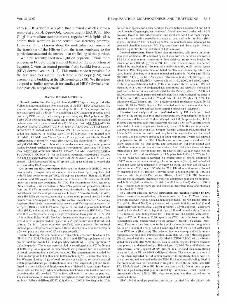
FIG. 1. Analysis of HBV envelope protein production in BHK-21cells transfected with pSFV1-SHBsadw, pSFV1-MHBsadw, or pSFV1-LHBsadw. Western blots showed that the transfection with the pSFV1-SHBsadw construct led to the production of S protein in large amounts,with equal proportions of the unglycosylated (p24) and glycosylated(gp27) forms. Transfection with pSFV1-MHBsadw led to the produc-tion within cells of the two main forms of the glycosylated M protein(ggp36 and gp33) but only trace amounts of the unglycosylated form(p30), with small amounts of the two forms of the HBV S envelopeprotein, p24 and gp27. Transfection with pSFV1-LHBsadw led to theproduction of the L protein in its unglycosylated (p39) and glycosylated(gp42) forms and to the production of smaller amounts of the twoglycosylated forms of M. ELISA showed that HBsAg levels were twiceas high in the supernatants of cells transfected with pSFV1-MHBsadw
than in the supernatants of cells transfected with pSFV1-SHBsadw andthat no HBsAg was present in the supernatants of cells transfectedwith the pSFV1-LHBsadw and �-Gal constructs. Molecular mass mark-ers (M) are indicated on the left of the blot.
FIG. 2. Subcellular localization, by confocal microscopy, of theHBV envelope proteins in BHK-21 cells transfected with pSFV1-SHBsadw, pSFV1-MHBsadw, or pSFV1-LHBsadw. All three proteinscolocalized at least partly with CRT, a specific ER marker, and withERGIC53, a specific ERGIC marker, in the perinuclear area. TheHBV M protein had a more diffuse subcellular distribution than the Sand L proteins. The HBV L protein was found mostly colocalized withCRT and, to a lesser extent, with ERGIC53. Cells transfected with arecombinant SFV RNA encoding the �-Gal were used as a negativecontrol.
3844 PATIENT ET AL. J. VIROL.
Dow
nloa
ded
from
http
s://j
ourn
als.
asm
.org
/jour
nal/j
vi o
n 23
Nov
embe
r 20
21 b
y 14
.47.
75.9
1.
Confocal microscopy on cells grown on glass coverslips showedthat the S protein colocalized with CRT, a specific ER marker,and with the ERGIC marker ERGIC53, mostly in the perinu-clear area (Fig. 2). Cells transfected with �-Gal RNA gave no
signal with anti-HBs antibodies on Western blots or on confo-cal microscopy (Fig. 1, lane 1, and Fig. 2).
Cells producing the S protein displayed significant ultra-structural modifications on EM (Fig. 3 and 4) with respect to
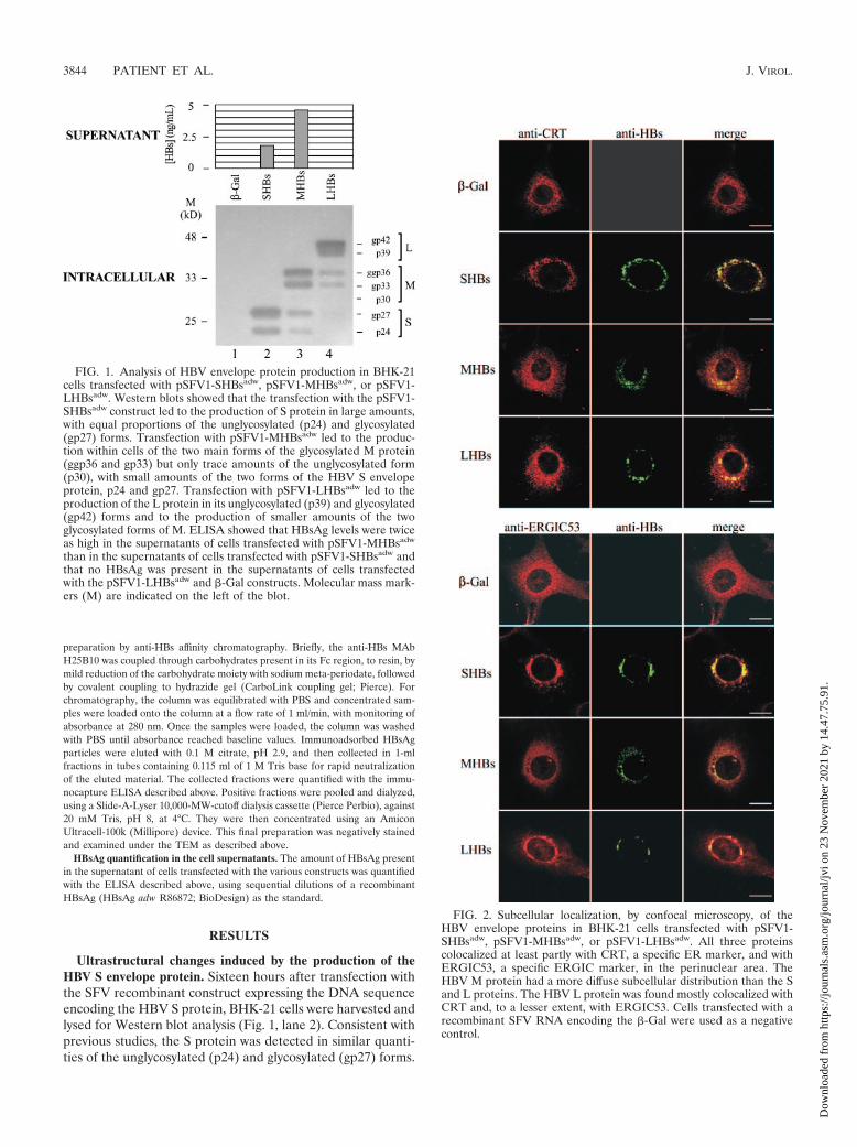
FIG. 3. Early ultrastructural changes in BHK-21 cells producing the HBV S protein. Numerous vesicles 0.2 to 0.3 �m in diameter (shortarrows), packed with 22-nm large filaments, were observed in the perinuclear area. These vesicles originated from the ER, as they were frequentlyobserved budding from the nuclear envelope (panel A and its inset) or from membranes carrying ribosomes (indicated by the long arrows in panelB). The filaments packed in these vesicles appeared in lengthwise (open arrows) or crosswise (closed arrows) sections in crystal-like structures (seea high magnification of these packed filaments in the inset in panel C). The specificity of these filaments was studied by immuno-EM using therabbit anti-HBs PAb. Modifications to the fixation and embedding procedures required for this specific immunostaining method resulted in cellstructures and filaments being less well preserved than in cells embedded in Epon resin and subjected to standard EM procedures, but intense goldlabeling restricted to these vesicles and predominantly found in the perinuclear area was observed (inset in panel D). No specific ultrastructuralchanges were observed in cells producing �-Gal (not shown on figures). Bars, 0.2 �m. N, nucleus; Cy, cytoplasm.
VOL. 81, 2007 HBsAg PARTICLE MORPHOGENESIS AND TRAFFICKING 3845
Dow
nloa
ded
from
http
s://j
ourn
als.
asm
.org
/jour
nal/j
vi o
n 23
Nov
embe
r 20
21 b
y 14
.47.
75.9
1.

the cells producing �-Gal (not shown on figures). Intracellularvesicles, packed with filaments, were abundantly observed inthe perinuclear area of these cells (Fig. 3). These vesiclesclearly originated from the ER, as they were frequently ob-served budding from the nuclear envelope (Fig. 3A) or frommembranes with ribosomes at their surface (Fig. 3B). They
were homogeneous in size (between 0.2 and 0.3 �m in diam-eter) and were filled with packed and presumably bridgedfilaments, 22 nm in diameter, which were observed in length-wise or crosswise sections in crystalline structures (Fig. 3C andD). The specificity of the content of these vesicles was deter-mined by immunogold labeling with the rabbit PAb against
FIG. 4. Late ultrastructural changes in BHK-21 cells producing the HBV S protein. Very large cisternae with smooth membranes, 1 to 2 �min diameter, were found to contain interlaced relaxed 22-nm-diameter filaments (A). The ends of these filaments appeared to be electron dense(see a high magnification of these filaments in the inset in panel A). The fusion of vesicles transporting packed filaments with the membranes ofcisternae was frequently observed (large arrows in panels B, C, and D), suggesting that the lumina of these cisternae contained material providedby the vesicles with the packed filaments. Filaments in the lumen were sometimes relaxed to one end, with the other end remaining associated witha crystal-like structure and other filaments (small arrows in panels A, C, and D). These ultrastructural changes were never observed in cellsproducing �-Gal (not shown on figures). Bars, 0.2 �m. Ci, cisterna; Cy, cytoplasm.
3846 PATIENT ET AL. J. VIROL.
Dow
nloa
ded
from
http
s://j
ourn
als.
asm
.org
/jour
nal/j
vi o
n 23
Nov
embe
r 20
21 b
y 14
.47.
75.9
1.
HBsAg. Modifications to the fixation and embedding proce-dures required for this specific immunostaining method (par-ticularly the absence of glutaraldehyde and osmium tetroxide),resulted in the cell structures and filaments being less wellpreserved than in cells embedded in Epon resin according tostandard EM methods. However, intense gold labeling re-stricted to these vesicles was observed in the perinuclear area(Fig. 3D, inset). We carried out a time course analysis and
found that these vesicles became detectable as early as 12 hafter transfection and that their frequency peaked 16 h aftertransfection.
We also observed very large cisternae (1 to 2 �m in diam-eter) in some cells. These cisternae were delimited by smoothmembranes and contained relaxed long filaments (Fig. 4).Their lumina presumably contained material from the smallervesicles described above. Indeed, fusion with the cisternae of
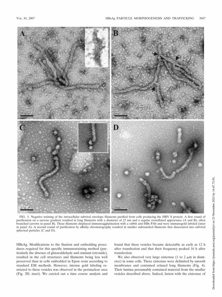
FIG. 5. Negative staining of the intracellular subviral envelope filaments purified from cells producing the HBV S protein. A first round ofpurification on a sucrose gradient resulted in long filaments with a diameter of 22 nm and a regular crenellated appearance (A and B), oftenbranched (arrows in panel B). These filaments displayed immunoagglutination with a rabbit anti-HBs PAb and were immunogold labeled (insetin panel A). A second round of purification by affinity chromatography resulted in smaller unbranched filaments that dissociated into subviralspherical particles (C and D).
VOL. 81, 2007 HBsAg PARTICLE MORPHOGENESIS AND TRAFFICKING 3847
Dow
nloa
ded
from
http
s://j
ourn
als.
asm
.org
/jour
nal/j
vi o
n 23
Nov
embe
r 20
21 b
y 14
.47.
75.9
1.
vesicles containing 22-nm packed filaments was frequently ob-served (Fig. 4B, C, and D). The 22-nm filaments in the luminaof the cisternae were mostly fully relaxed, but in some cases,the filaments were relaxed at one end but remained bridged ina crystalline structure with other filaments at the other (Fig.4A, C, and D). When relaxed, the end of the filament waselectron dense (Fig. 4A, inset). These cisternae were observedin cells fixed 16 h after transfection but not in cells fixed 4, 8,or 12 h after transfection.
Negative staining of intracellular subviral particles formedby the HBV S envelope protein. Initial analysis of the crude celllysate subjected to centrifugation on a sucrose gradient led tothe observation of long filaments 22 nm in diameter (Fig. 5Aand B). These filaments were well organized, with negativestaining revealing a regular crenellated appearance (Fig. 5Aand B). They were of various sizes and were often branched(Fig. 5B). The longest unbranched filament found in this prep-aration was 1.2 �m in length (not shown on the figure). Thefilaments had bulging extremities, probably accounting for theelectron-dense appearance of these extremities in ultrathinsections (see our comment concerning the inset in Fig. 4Aabove). The specificity of these filaments was confirmed byimmunogold labeling. Following prior incubation with the rab-bit anti-HBs PAb, the filaments were immunoagglutinated andlabeled with gold-conjugated goat anti-rabbit IgG antibodies(Fig. 5A, inset). Following purification by affinity chromatog-raphy, these filaments were systematically unbranched andmuch smaller (less than 0.3 �m in length), and their endsshowed a clear tendency to dissociation into subviral sphericalparticles (Fig. 5C and D).
Ultrastructural changes induced by the production of theHBV M or L envelope proteins. Sixteen hours after transfec-tion with the recombinant SFV constructs containing the DNAsequence of the HBV M or L proteins, BHK-21 cells wereharvested and lysed for Western blot analysis (Fig. 1). Expres-sion of the pSFV1-MHBsadw construct gave large amounts ofglycosylated M protein in cells, with equal amounts of the twomain forms (ggp36 and gp33) but only traces of the unglyco-sylated form (p30). This construct also yielded small amountsof the two forms of the HBV S envelope protein, p24 and gp27(Fig. 1, lane 3). Expression of the pSFV1-LHBsadw constructyielded large amounts of L protein, with similar amounts of theunglycosylated (p39) and glycosylated (gp42) forms. In con-trast to what was observed for pSFV1-MHBsadw, the expres-sion of pSFV1-LHBsadw resulted in the production of only tinyamounts of S proteins. However, expression of this constructyielded two glycosylated forms of M (Fig. 1, lane 4). ELISAshowed that HBsAg levels were higher in the supernatant ofcells transfected with the pSFV1-MHBsadw construct than inthat of cells transfected with the pSFV1-SHBsadw construct(4.6 ng/ml and 1.8 ng/ml, respectively), whereas no HBsAg wasdetected in the supernatant of cells transfected with thepSFV1-LHBsadw and �-Gal constructs (Fig. 1). Immunocyto-chemistry and confocal microscopy showed that the HBV Mprotein was partly colocalized with CRT and ERGIC53 (Fig.2) and that the subcellular distribution of this protein was morediffuse than that of the HBV S protein (Fig. 2). The HBV Lprotein colocalized mostly with CRT and, to a lower extent,with ERGIC53, in the perinuclear area of the cells (Fig. 2).
Despite an intensive search and repeated experiments, no
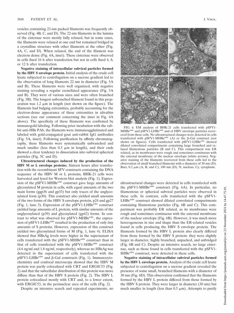
ultrastructural changes were detected in cells transfected withthe pSFV1-MHBsadw construct (Fig. 6A). In particular, nofilamentous or spherical subviral particles were observed inthese cells. In contrast, cells transfected with the pSFV1-LHBsadw construct showed dilated convoluted compartmentscontaining filamentous particles (Fig. 6B and C). This com-partment was probably ER related, as its membranes wererough and sometimes continuous with the external membraneof the nuclear envelope (Fig. 6B). However, it was much moreheterogeneous in size and shape than the ER-related vesiclesfound in cells producing the HBV S envelope protein. Thefilaments formed by the HBV L protein also clearly differedfrom those formed by the HBV S protein: they were slightlylarger in diameter, highly branched, unpacked, and unbridged(Fig. 6B and C). Despite an intensive search, no large cister-nae, such as those found in cells transfected with the pSFV1-SHBsadw construct, were detected in these cells.
Negative staining of intracellular subviral particles formedby the HBV L envelope protein. Analysis of the crude cell lysatesubjected to centrifugation on a sucrose gradient revealed thepresence of some small, branched filaments with a diameter of30 nm (Fig. 6D). This observation confirmed that the filamentsformed by the HBV L protein differed from those formed bythe HBV S protein. They were larger in diameter (30 nm) butmuch smaller in length (less than 0.5 �m). Attempts to purify
FIG. 6. EM analysis of BHK-21 cells transfected with pSFV1-MHBsadw and pSFV1-LHBsadw and of HBV envelope particles recov-ered from these cells. No ultrastructural changes were detected in cellstransfected with pSFV1-MHBsadw (A) or the �-Gal construct (notshown on figures). Cells transfected with pSFV1-LHBsadw showeddilated convoluted compartments containing large branched and re-laxed filamentous particles (B and C). This compartment was ERrelated, as its membranes were rough and sometimes continuous withthe external membrane of the nuclear envelope (white arrows). Neg-ative staining of the filaments recovered from these cells led to theobservation of small branched filaments with a diameter of 30 nm (D).Bars, 0.2 �m (A, B, and C), 100 nm (D). N, nucleus; Cy, cytoplasm.
3848 PATIENT ET AL. J. VIROL.
Dow
nloa
ded
from
http
s://j
ourn
als.
asm
.org
/jour
nal/j
vi o
n 23
Nov
embe
r 20
21 b
y 14
.47.
75.9
1.

these filaments further by affinity chromatography were unsuc-cessful, probably due to their low rate of recovery from the celllysate and/or their weak binding to the anti-HBs antibody withwhich the column was coated. We also analyzed a lysate fromcells producing the M protein and confirmed the absence ofany filamentous or spherical subviral particles in these cells, atleast in sufficient amounts for detection by negative EM stain-ing (data not shown).
DISCUSSION
Despite the existence of hepatoma cell lines that can produceinfectious virus after transfection with the viral genome (40, 43)and the recent establishment of a cell line that can be infected bythe virus in vitro (20), little is known about the morphogenesis ofHBV. This may be due to the low rate of HBV production inthese cells, as it has been estimated that an individual hepatocytereleases only 1 to 10 viruses per day during the productive phaseof infection in vivo (30). HBV subviral envelope particles canreach concentrations 10,000 times higher than that of virions inthe serum of infected patients, but their assembly and intracellu-lar transport also remain poorly understood (4).
As the production of the S envelope protein alone is suffi-cient for the secretion of spherical HBV subviral envelopeparticles, it is widely thought that the filamentous shape ofthese particles results from the coproduction of the S and Lproteins (7, 21). Our EM study showed that the production ofthe HBV S protein alone also led, at least initially, to themorphogenesis of filamentous subviral particles. The existenceof this phenomenon was suggested in early EM studies onChinese hamster ovary (CHO) cells producing the HBV Sprotein (31), but it has not been studied further in the last 20years. The filaments induced by the HBV S protein in ourstudy seemed to be extremely long (up to 1.2 �m) and occa-sionally branched, a feature that was also considered specific tofilaments induced by HBV L protein overproduction in a trans-genic mouse model (9).
Our study shows that the morphogenesis of these filamentstakes place in the ER. The currently accepted model of thesubviral HBV particle formation assumes that this processtakes place at the ERGIC membrane, resulting in budding ofthe particle into the lumen of this compartment (4). Indeed,biochemical and immunocytochemical studies have suggestedthat the S protein initially forms dimers, catalyzed by proteindisulfide isomerase (PDI) in the ER compartment, before itstransport in vesicles, as transmembrane dimers, to the ERGIC(24). The absence of PDI and the presence of different mole-cules in the lumen of the ERGIC would thus lead to reorga-nization of the interdimer disulfide bridges, an essential step inthe assembly of the subviral particles (24). However, the for-mation of interdimer disulfide bridges may not be required forparticle formation because most S proteins in particles freshlysecreted from transfected cells are dimeric and S mutants withmultiple cysteine-to-serine substitutions nonetheless form par-ticles (27, 45). However, direct interactions within S proteinsubunits are probably required for the formation of subviralenvelope particles. Coproduction of the human HBV S proteinwith its counterpart from an animal hepadnavirus showed thatthe human and duck HBV S proteins did not form mixed
subviral particles, despite clear structural and sequence simi-larities, whereas human and woodchuck HBV S, which aremore closely related, coassembled efficiently (18). It shouldalso be pointed out that the current model for subviral HBVparticle formation is based principally on studies including noultrastructural analysis of the transfected cells (24). Our EMobservations clearly show that the HBV subviral particleformed by the S protein assembles in the ER compartment.Indeed, this phenomenon clearly occurred at ER-related mem-branes, as the membranes had ribosomes on their surfaceand/or were continuous with the outer membrane of the nu-clear envelope. The HBV subviral particles with a filamentousshape formed by the S protein were initially packed into ves-icles budding from the ER and were then delivered by thesevesicles to large cisternae, in which they were unpacked andrelaxed. These large cisternae had smooth membranes andprobably corresponded to the ERGIC compartment. Unfortu-nately, it was technically difficult to confirm this hypothesis byimmunogold labeling with an anti-ERGIC antibody. The useof Epon resin and standard EM techniques was required tovisualize the filaments in this compartment, but the resultsobtained with anti-ERGIC antibody on ultrathin sections ofEpon-embedded cells were unsatisfactory. The HBV subviralfilaments formed by the S protein and purified from cell lysatesappeared on negative staining to be much longer (up to 1.2�m) than expected from the size of the intracellular vesicles inwhich they were packed (0.2 to 0.3 �m). In addition, theseparticles were often branched. This indicates that they wereprobably folded in the vesicles during intracellular traffickingbetween the ER and the ERGIC. This folding was properlyorganized, as these packed filaments had a regular, crystal-likestructure and were presumably bridged in the vesicles. It hasbeen suggested that chaperones, such as calnexin (36, 44) orBiP (10), are involved in the assembly and morphogenesis ofthe HBV subviral envelope particles. The ERp57 protein,which is a close homologue of PDI, has also been shown tohave chaperone activities in conjunction with calnexin andCRT, forming transient mixed disulfide bridges with cellularproteins bearing cysteines (22, 23). These ER-resident proteinsmay therefore assist with the folding and packing of the HBVsubviral envelope filaments during their transport in ER-de-rived vesicles. The concentrations of these proteins are lowerin the ERGIC, which might result in the release of the fila-ments from these crystal-like structures, as suggested by ourEM observations showing the relaxation of filaments in thiscompartment.
We were unable to identify the morphological events follow-ing the release of the HBV S subviral filaments in the ERGIC.This is not entirely surprising, as the HBV S protein remains inpre-Golgi compartments for almost all of its long lifetimewithin the cell (31). The high-mannose oligosaccharide chainsadded to the S protein are converted to the complex form justbefore secretion, indicating that this protein is rapidly trans-ported to the cell surface through the Golgi apparatus and thefinal constitutive pathway of vesicular transport (31). It hasbeen suggested that the long half-time of HBV S protein se-cretion may be accounted for by the rate-limiting step of itsassembly into a subviral particle (41). However, we show herethat the assembly of the HBV S protein into filamentous par-ticles is an early event, followed by the transport of these
VOL. 81, 2007 HBsAg PARTICLE MORPHOGENESIS AND TRAFFICKING 3849
Dow
nloa
ded
from
http
s://j
ourn
als.
asm
.org
/jour
nal/j
vi o
n 23
Nov
embe
r 20
21 b
y 14
.47.
75.9
1.
filaments toward a compartment probably corresponding tothe ERGIC. The rate-limiting step in secretion thereforeseems to be the transfer of these particles from the ERGIC tothe Golgi apparatus. The filamentous shape of these particlesmay limit the size of the particles that can be accommodatedwithin transport vesicles. The rate of transport would then bedetermined by the rate of conversion of a filamentous particleto a more spherical particle. Our observations of HBV S fila-ment purification by affinity chromatography and the analysisof these filaments by negative staining provide support for thishypothesis, demonstrating that these filaments can be sponta-neously converted into spherical particles. Subviral sphericalparticles purified from the serum of HBV S-transgenic micewere recently reconstructed by cryo-EM and were found to bevery heterogeneous in size (19). This may be due to theirformation by dissociation from a precursor filamentous parti-cle. However, it should be noted that extremely long filamentscould be transported from the ER to the ERGIC within smalltransport vesicles. This may be due to the presence of specificchaperones in the ER compartment, making it possible to foldand to cross-link these long filaments in transport vesicles. Inour system, only tiny amounts of HBsAg were secreted fromthe transfected cells. This low level of secretion may be ac-counted for by the high toxicity of the SFV expression system,making it impossible to culture cells for longer than 24 h,together with the long half-time of HBsAg secretion. However,HBV subviral particles collected from the supernatant of trans-fected cells were purified by affinity chromatography and stud-ied by negative staining. Few particles were recovered fromthese experiments, but these secreted HBV subviral particlesappeared to be spherical, suggesting that the conversion of thefilaments to spheres is a prerequisite for their secretion (datanot shown). A construct bearing the HBV S gene under thecontrol of the cytomegalovirus promoter was found to give a10-fold-more-efficient HBsAg secretion than the SFV expres-sion vector, but ultrathin sections of the transfected cellsshowed no morphological events typical of subviral envelopeparticle assembly and trafficking. However, the quantificationof intracellular HBsAg by ELISA showed an amount similar tothe two expression systems (data not shown). The visualizationof the subviral envelope particle assembly with the SFV RNAvectors may be due to a more rapid protein synthesis withthis system, leading to a major intracellular accumulation ofHBsAg in the early compartments of the secretory pathway.Also, our efforts to visualize this phenomenon in hepatomacells such as the Huh7 cell line were unsuccessful, but we havepreviously observed that the SFV vectors are less efficient inthese cells (2).
Many 0.2- to 0.3-�m vesicles packed with HBV S filamentswere seen in the perinuclear area of the cells, but we observedno extrusion of HBV S filaments from the membranes of thesevesicles, despite intensive EM analysis of the cells 8, 12, and16 h after transfection. Previous studies have shown that thesubviral particles contain only 25% lipid (in terms of weight)(17), so the lipids are unlikely to be organized as in a conven-tional membrane bilayer. The absence of a lipid bilayer inspherical HBV S particles was recently confirmed by cryo-EManalysis (19). Thus, the process resulting in delivery of theHBV S filament to the ER lumen may differ from conventionalviral budding. Some EM observations such as those presented
in Fig. 3A and B suggest that HBV S filaments are assembledde novo in the ER lumen and that a cluster of packagedfilaments initiates the budding of a forthcoming transport ves-icle. Further investigation is required to confirm or reject thishypothesis.
The production of HBV M and L proteins with an SFVvector had contrasting effects. HBV M protein production didnot lead to formation of intracellular filamentous or sphericalsubviral envelope particles, although the pSFV1-MHBsadw
construct also produced the HBV S protein. This lack of par-ticle formation may be accounted for by the much lower levelof HBV S protein production induced by the pSFV1-MHBsadw
construct than by pSFV1-SHBsadw. However, HBsAg levelswere higher in the supernatants of cells transfected with thepSFV1-MHBsadw construct than in the supernatants of cellstransfected with the pSFV1-SHBsadw construct. Confocal mi-croscopy also showed the subcellular distribution of HBsAg inthe cytoplasma of these cells to be more diffuse. This mayreflect a specific role of the M protein in enhancing HBVsubviral particle trafficking through the secretory pathway. TheM protein is dispensable for virion or subviral envelope parti-cle formation, but an M protein lacking its specific N-linkedcarbohydrates may inhibit virion (3, 28) or subviral envelopeparticle (42, 44) secretion. The coproduction of a fully glyco-sylated M with S may therefore promote trafficking of the HBVsubviral envelope particles toward the cell surface, as previ-ously suggested (44). Alternatively, we cannot exclude that theHBV M protein could have a specific cytotoxic effect leading toHBsAg release in the cell supernatant. This phenomenon, to-gether with lower levels of S production, may account for ourfailure to detect HBV subviral particles in sections of cellstransfected with pSFV1-MHBsadw. In contrast, the HBV Lprotein production led to the morphogenesis of small branchedfilaments that seemed to accumulate in the lumen of a convo-luted, ER-related compartment. Similar ultrastructural obser-vations have been reported with Huh7 hepatoma cells produc-ing HBV L protein alone (46), but our study allowed acomparison of the morphogenesis of HBV L filaments directlywith that of HBV S filaments. Unlike the HBV S filaments, theHBV L filaments were never found packed and bridged insmall transport vesicles and were not targeted to a distal com-partment. However, we cannot exclude the possibility thatHBV L filaments are transported from the ER to the ERGIC;although if such transport does occur, it may be less efficientfor these filaments than for HBV S filaments. This hypothesisis supported by the colocalization of HBV L with ERGIC53 onconfocal microscopy, which was found to be weaker than thatfor the HBV S and M proteins. These observations may reflectdifferent affinities of these proteins for the cellular chaperones,which may subsequently contribute to intracellular retention ofthe HBV L protein.
In conclusion, our study sheds new light on the mechanismsinvolved in HBV subviral envelope particle morphogenesis andintracellular trafficking through the secretory pathway. As theintracellular accumulation of subviral envelope filaments maybe directly linked to the pathogenesis of HBV (9, 33, 37),improvements in our understanding of these mechanismsmight facilitate the design of new antiviral strategies in futurestudies.
3850 PATIENT ET AL. J. VIROL.
Dow
nloa
ded
from
http
s://j
ourn
als.
asm
.org
/jour
nal/j
vi o
n 23
Nov
embe
r 20
21 b
y 14
.47.
75.9
1.
ACKNOWLEDGMENTS
We thank Didier Leturcq, R.W. Johnson Pharmaceutical ResearchInstitute in San Diego, for his invaluable help in setting up the affinitychromatography system. We thank Volker Bruss, University ofGottingen, for the gift of the pHBV1.5 plasmid.
Our research is supported by the Region Centre (Equipe ESPRI)and ANRS. R.P. was supported by a fellowship from the FrenchMinistry of Research.
Our data were generated with the help of the RIO Electron Micros-copy Facility of Francois Rabelais University.
REFERENCES
1. Blanchard, E., D. Brand, S. Trassard, A. Goudeau, and P. Roingeard. 2002.Hepatitis C virus-like particle morphogenesis. J. Virol. 76:4073–4079.
2. Blanchard, E., C. Hourioux, D. Brand, M. Ait-Goughoulte, A. Moreau, S.Trassard, P. Y. Sizaret, F. Dubois, and P. Roingeard. 2003. Hepatitis Cvirus-like particle budding: role of the core protein and importance of itsAsp111. J. Virol. 77:10131–10138.
3. Block, T. M., X. Lu, A. Mehta, J. Park, B. S. Blumberg, and R. Dwek. 1998.Role of glycan processing in hepatitis B virus envelope protein trafficking.Adv. Exp. Med. Biol. 435:207–216.
4. Bruss, V. 2004. Envelopment of the hepatitis B virus nucleocapsid. VirusRes. 106:199–209.
5. Bruss, V. 1997. A short linear sequence in the pre-S domain of the largehepatitis B virus envelope protein required for virion formation. J. Virol.71:9350–9357.
6. Bruss, V., and D. Ganem. 1991. The role of envelope proteins in hepatitis Bvirus assembly. Proc. Natl. Acad. Sci. USA 88:1059–1063.
7. Bruss, V., E. Gerhardt, K. Vieluf, and G. Wunderlich. 1996. Functions of thelarge hepatitis B virus surface protein in viral particle morphogenesis. Inter-virology 39:23–31.
8. Bruss, V., and K. Vieluf. 1995. Functions of the internal pre-S domain of thelarge surface protein in hepatitis B virus particle morphogenesis. J. Virol.69:6652–6657.
9. Chisari, F. V., P. Filippi, J. Buras, A. McLachlan, H. Popper, C. A. Pinkert,R. D. Palmiter, and R. L. Brinster. 1987. Structural and pathological effectsof synthesis of hepatitis B virus large envelope polypeptide in transgenicmice. Proc. Natl. Acad. Sci. USA 84:6909–6913.
10. Cho, D. Y., G. H. Yang, C. J. Ryu, and H. J. Hong. 2003. Molecular chap-erone GRP78/BiP interacts with the large surface protein of hepatitis B virusin vitro and in vivo. J. Virol. 77:2784–2788.
11. Dubois, M. F., C. Pourcel, S. Rousset, C. Chany, and P. Tiollais. 1980.Excretion of hepatitis B surface antigen particles from mouse cells trans-formed with cloned viral DNA. Proc. Natl. Acad. Sci. USA 77:4549–4553.
12. Eble, B. E., V. R. Lingappa, and D. Ganem. 1986. Hepatitis B surfaceantigen: an unusual secreted protein initially synthesized as a transmem-brane polypeptide. Mol. Cell. Biol. 6:1454–1463.
13. Eble, B. E., V. R. Lingappa, and D. Ganem. 1990. The N-terminal (pre-S2)domain of a hepatitis B virus surface glycoprotein is translocated acrossmembranes by downstream signal sequences. J. Virol. 64:1414–1419.
14. Eddleston, A. 1990. Modern vaccines. Hepatitis. Lancet 335:1142–1145.15. Gallina, A., E. Gazina, and G. Milanesi. 1995. A C-terminal PreS1 sequence
is sufficient to retain hepatitis B virus L protein in 293 cells. Virology 213:57–69.
16. Ganem, D., and A. M. Prince. 2004. Hepatitis B virus infection–naturalhistory and clinical consequences. N. Engl. J. Med. 350:1118–1129.
17. Gavilanes, F., J. M. Gonzalez-Ros, and D. L. Peterson. 1982. Structure ofhepatitis B surface antigen. Characterization of the lipid components andtheir association with the viral proteins. J. Biol. Chem. 257:7770–7777.
18. Gerhardt, E., and V. Bruss. 1995. Phenotypic mixing of rodent but not avianhepadnavirus surface proteins into human hepatitis B virus particles. J. Vi-rol. 69:1201–1208.
19. Gilbert, R. J., L. Beales, D. Blond, M. N. Simon, B. Y. Lin, F. V. Chisari, D. I.Stuart, and D. J. Rowlands. 2005. Hepatitis B small surface antigen particlesare octahedral. Proc. Natl. Acad. Sci. USA 102:14783–14788.
20. Gripon, P., S. Rumin, S. Urban, J. Le Seyec, D. Glaise, I. Cannie, C.Guyomard, J. Lucas, C. Trepo, and C. Guguen-Guillouzo. 2002. Infection ofa human hepatoma cell line by hepatitis B virus. Proc. Natl. Acad. Sci. USA99:15655–15660.
21. Heermann, K. H., U. Goldmann, W. Schwartz, T. Seyffarth, H. Baumgarten,
and W. H. Gerlich. 1984. Large surface proteins of hepatitis B virus con-taining the pre-s sequence. J. Virol. 52:396–402.
22. Helenius, A., and M. Aebi. 2001. Intracellular functions of N-linked glycans.Science 291:2364–2369.
23. Helenius, A., and M. Aebi. 2004. Roles of N-linked glycans in the endoplas-mic reticulum. Annu. Rev. Biochem. 73:1019–1049.
24. Huovila, A. P., A. M. Eder, and S. D. Fuller. 1992. Hepatitis B surfaceantigen assembles in a post-ER, pre-Golgi compartment. J. Cell Biol. 118:1305–1320.
25. Jenna, S., and C. Sureau. 1999. Mutations in the carboxyl-terminal domainof the small hepatitis B virus envelope protein impair the assembly ofhepatitis delta virus particles. J. Virol. 73:3351–3358.
26. Le Pogam, S., and C. Shih. 2002. Influence of a putative intermolecularinteraction between core and the pre-S1 domain of the large envelopeprotein on hepatitis B virus secretion. J. Virol. 76:6510–6517.
27. Mangold, C. M., F. Unckell, M. Werr, and R. E. Streeck. 1995. Secretion andantigenicity of hepatitis B virus small envelope proteins lacking cysteines inthe major antigenic region. Virology 211:535–543.
28. Mehta, A., X. Lu, T. M. Block, B. S. Blumberg, and R. A. Dwek. 1997.Hepatitis B virus (HBV) envelope glycoproteins vary drastically in theirsensitivity to glycan processing: evidence that alteration of a single N-linkedglycosylation site can regulate HBV secretion. Proc. Natl. Acad. Sci. USA94:1822–1827.
29. Moriarty, A. M., B. H. Hoyer, J. W. Shih, J. L. Gerin, and D. H. Hamer. 1981.Expression of the hepatitis B virus surface antigen gene in cell culture byusing a simian virus 40 vector. Proc. Natl. Acad. Sci. USA 78:2606–2610.
30. Nowak, M. A., S. Bonhoeffer, A. M. Hill, R. Boehme, H. C. Thomas, and H.McDade. 1996. Viral dynamics in hepatitis B virus infection. Proc. Natl.Acad. Sci. USA 93:4398–4402.
31. Patzer, E. J., G. R. Nakamura, C. C. Simonsen, A. D. Levinson, and R.Brands. 1986. Intracellular assembly and packaging of hepatitis B surfaceantigen particles occur in the endoplasmic reticulum. J. Virol. 58:884–892.
32. Persing, D. H., H. E. Varmus, and D. Ganem. 1986. Inhibition of secretionof hepatitis B surface antigen by a related presurface polypeptide. Science234:1388–1391.
33. Phillips, M. J., R. Cameron, M. A. Flowers, L. M. Blendis, P. D. Greig, I.Wanless, M. Sherman, R. Superina, B. Langer, and G. A. Levy. 1992. Post-transplant recurrent hepatitis B viral liver disease. Viral-burden, steatoviral,and fibroviral hepatitis B. Am. J. Pathol. 140:1295–1308.
34. Poisson, F., A. Severac, C. Hourioux, A. Goudeau, and P. Roingeard. 1997.Both pre-S1 and S domains of hepatitis B virus envelope proteins interactwith the core particle. Virology 228:115–120.
35. Prange, R., A. Clemen, and R. E. Streeck. 1991. Myristylation is involved inintracellular retention of hepatitis B virus envelope proteins. J. Virol. 65:3919–3923.
36. Prange, R., M. Werr, and H. Loffler-Mary. 1999. Chaperones involved inhepatitis B virus morphogenesis. Biol. Chem. 380:305–314.
37. Roingeard, P. 2003. Vacuolization in hepatitis B virus-infected hepatocytes.Hepatology 37:1223–1224.
38. Roingeard, P., C. Hourioux, E. Blanchard, D. Brand, and M. Ait-Goughoulte.2004. Hepatitis C virus ultrastructure and morphogenesis. Biol. Cell 96:103–108.
39. Roingeard, P., and C. Sureau. 1998. Ultrastructural analysis of hepatitis Bvirus in HepG2-transfected cells with special emphasis on subviral filamentmorphogenesis. Hepatology 28:1128–1133.
40. Sells, M. A., M. L. Chen, and G. Acs. 1987. Production of hepatitis B virusparticles in Hep G2 cells transfected with cloned hepatitis B virus DNA.Proc. Natl. Acad. Sci. USA 84:1005–1009.
41. Simon, K., V. R. Lingappa, and D. Ganem. 1988. Secreted hepatitis B surfaceantigen polypeptides are derived from a transmembrane precursor. J. CellBiol. 107:2163–2168.
42. Sureau, C., C. Fournier-Wirth, and P. Maurel. 2003. Role of N glycosylationof hepatitis B virus envelope proteins in morphogenesis and infectivity ofhepatitis delta virus. J. Virol. 77:5519–5523.
43. Sureau, C., J. L. Romet-Lemonne, J. I. Mullins, and M. Essex. 1986. Pro-duction of hepatitis B virus by a differentiated human hepatoma cell lineafter transfection with cloned circular HBV DNA. Cell 47:37–47.
44. Werr, M., and R. Prange. 1998. Role for calnexin and N-linked glycosylationin the assembly and secretion of hepatitis B virus middle envelope proteinparticles. J. Virol. 72:778–782.
45. Wounderlich, G., and V. Bruss. 1996. Characterization of early hepatitis Bvirus surface protein oligomers. Arch. Virol. 141:1191–1205.
46. Xu, Z., V. Bruss, and T. S. Yen. 1997. Formation of intracellular particles byhepatitis B virus large surface protein. J. Virol. 71:5487–5494.
VOL. 81, 2007 HBsAg PARTICLE MORPHOGENESIS AND TRAFFICKING 3851
Dow
nloa
ded
from
http
s://j
ourn
als.
asm
.org
/jour
nal/j
vi o
n 23
Nov
embe
r 20
21 b
y 14
.47.
75.9
1.


