Imaging and Doppler of Portal Hypertension · Imaging and Doppler of Portal Hypertension Myron A....
18
11/14/2016 1 Imaging and Doppler of Portal Hypertension Myron A. Pozniak, MD Professor of Radiology University of Wisconsin Madison, Wisconsin Nothing to disclose relevant to this presentation. Imaging and Doppler of Portal Hypertension Review the normal hepatic Doppler flow profiles Recognize the hemodynamic changes of portal hypertension Recognize the common and unusual pathways of porto-systemic shunting Objectives: Vascular Flow Profiles • Inflow • Hepatic artery • Portal vein • Outflow • Hepatic veins Hepatic artery • Normal waveform • Brisk upstroke in systole • RI 60-70% • Diastolic velocity <20 cm/sec Portal vein • Relatively uniform velocity. • Some periodicity OK, but not too much. • Velocity just under 20 cm/sec in a fasting patient
Transcript of Imaging and Doppler of Portal Hypertension · Imaging and Doppler of Portal Hypertension Myron A....

11/14/2016
1
Imaging and Doppler of Portal Hypertension
Myron A. Pozniak, MD
Professor of Radiology
University of Wisconsin
Madison, Wisconsin
Nothing to disclose relevant to this presentation.
Imaging and Doppler of Portal Hypertension
Review the normal hepatic Doppler flow profiles
Recognize the hemodynamic changes of portal hypertension
Recognize the common and unusual pathways of porto-systemic shunting
Objectives:Vascular Flow Profiles
• Inflow
• Hepatic artery
• Portal vein
• Outflow
• Hepatic veins
Hepatic artery
• Normal waveform
• Brisk upstroke in systole
• RI 60-70%
• Diastolic velocity <20 cm/sec
Portal vein
• Relatively uniform velocity.
• Some periodicity OK, but not too much.
• Velocity just under
20 cm/sec in a fasting patient

11/14/2016
2
Portal flow basically percolates through the liver
Very small pressure gradient
The liver vascular index
• Relates portal vein velocity to hepatic artery velocity
• PV velocity near arterial end diastolic velocity
The liver vascular index
• Relates portal vein velocity to hepatic artery velocity OutflowThe Hepatic Veins
Caudate Veins Hepatic vein laminar flow dynamics

11/14/2016
3
Hep Vein
Velocity
Tracing
ECG
TricuspidM-Mode
AC
SV D
Hepatic Vein Flow Dynamics Venous waveform terminology
• Pulsatility
• Periodicity
• Phasicity
Periodicity
Normal hepatic blood flow
• 25% of cardiac output
• 1.5 Liters per minute
• Portal inflow 2/3; arterial inflow 1/3
• 90% of Oxygen via Hepatic Artery
• The Artery supplies the disease process.
The Diseased Liver
An earlier indicator: The altered liver vascular index
}
Increased hepatic arterial flow / Decreased portal vein flow
The altered liver vascular index• Initially reported to be highly sensitive and specific for
diagnosis of Hepatocellular Carcinoma (HCC)
• Many other causes
Iwao T, et al. Value of Doppler ultrasound parameters of portal vein and hepatic artery in
the diagnosis of cirrhosis and portal hypertension. Am J Gastroenterol 1997;92:1012-1017.

11/14/2016
4
Altered porta-hepatis hemodynamics
• Diffuse hepatocellular disorder • Hepatitis – viral, chemical, alcoholic
• Focal lesions• Lymphoma• Metastatic disease• Hepatitis• Etc.
• Non-specific• It’s not really compensatory
Altered porta-hepatis hemodynamics
• How do you report this liver?
• Verifies the “starry sky” liver as abnormal
With just the right degree of liver disease main PV flow may be relatively stagnant.
Don’t call it thrombosed.
Valsalva maneuver then release can flip PV flow from fugal to petal.
Good trick to avoid thrombosis overcall.
Valsalva Release
Nomenclature
• Reversed portal flow is hepatofugal
• Normal portal flow is hepatopetal
• Not – hepatopedal
• As in: centrifugal force / centripetal force
The degree of main PV flow reversal correlates with the severity of the liver disease.
Except in the presence of a paraumbilical vein
11/14/2016
5
Increased Periodicity of Portal vein flow
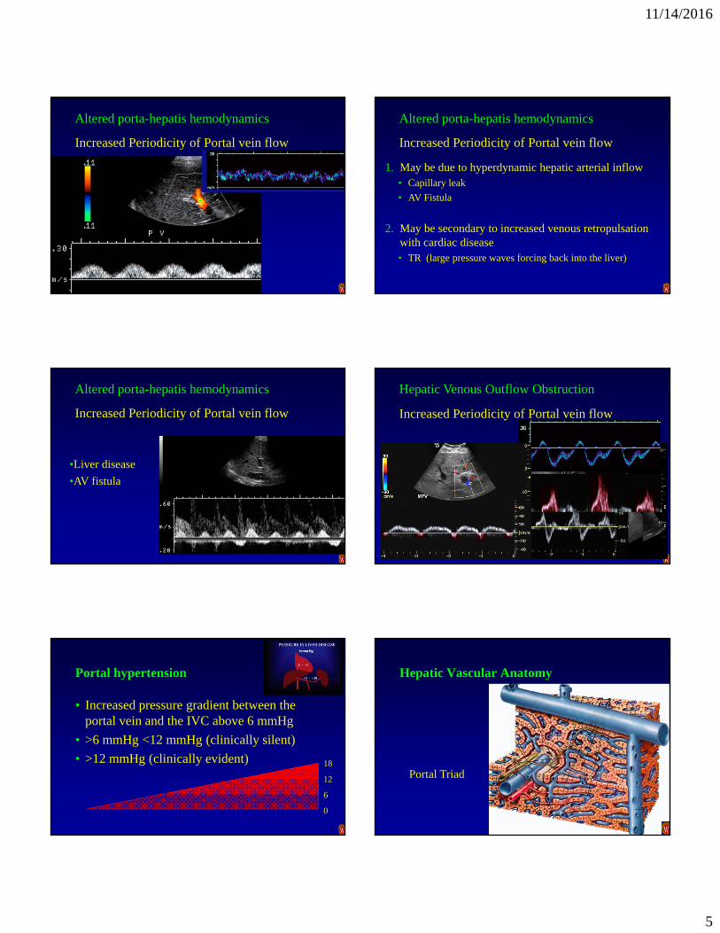
Altered porta-hepatis hemodynamics
Increased Periodicity of Portal vein flow
1. May be due to hyperdynamic hepatic arterial inflow• Capillary leak
• AV Fistula
2. May be secondary to increased venous retropulsationwith cardiac disease• TR (large pressure waves forcing back into the liver)
Altered porta-hepatis hemodynamics
•Liver disease
•AV fistula
Increased Periodicity of Portal vein flow
Altered porta-hepatis hemodynamics
•Right cardiac issues
•Tricuspid regurgitation
•Right ventricular dysfunction
Increased Periodicity of Portal vein flow
Hepatic Venous Outflow Obstruction
Portal hypertension
• Increased pressure gradient between the portal vein and the IVC above 6 mmHg
• >6 mmHg <12 mmHg (clinically silent)
• >12 mmHg (clinically evident) 18
12
6
0
Hepatic Vascular Anatomy
Portal Triad
11/14/2016
6
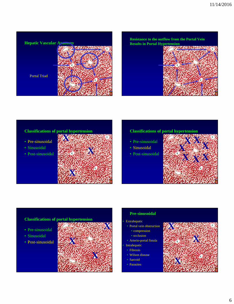
Hepatic Vascular Anatomy
Portal Triad
Resistance to the outflow from the Portal VeinResults in Portal Hypertension
Classifications of portal hypertension
• Pre-sinusoidal
• Sinusoidal
• Post-sinusoidal XX
X
Classifications of portal hypertension
• Pre-sinusoidal
• Sinusoidal
• Post-sinusoidal
X XXXXX
X
Classifications of portal hypertension
• Pre-sinusoidal
• Sinusoidal
• Post-sinusoidal X
X
X
XX
X
Pre-sinusoidal
• Extrahepatic
• Portal vein obstruction
• compression
• occlusion
• Arterio-portal fistula
• Intrahepatic
• Fibrosis
• Wilson disease
• Sarcoid
• Parasites
11/14/2016
7
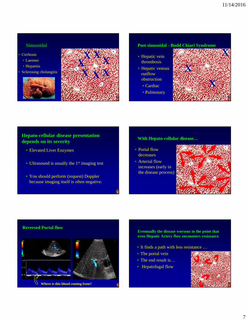
Sinusoidal
• Cirrhosis
• Laennec
• Hepatitis
• Sclerosing cholangitis
X XXXXX
X
Post-sinusoidal - Budd Chiari Syndrome
• Hepatic vein thrombosis
• Hepatic venous outflow obstruction
• Cardiac
• Pulmonary
X
X
X
Hepato-cellular disease presentation depends on its severity
• Elevated Liver Enzymes
• Ultrasound is usually the 1st imaging test
• You should perform (request) Doppler because imaging itself is often negative.
With Hepato-cellular disease…
• Portal flow decreases
• Arterial flow increases (early in the disease process)
Reversed Portal flow
Where is this blood coming from?
Eventually the disease worsens to the point that even Hepatic Artery flow encounters resistance.
• It finds a path with less resistance …
• The portal vein
• The end result is…
• Hepatofugal flow
11/14/2016
8
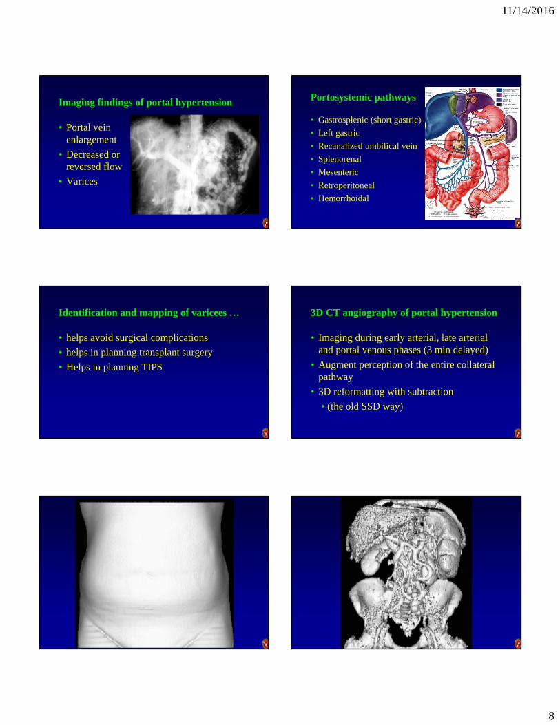
Imaging findings of portal hypertension
• Portal vein enlargement
• Decreased or reversed flow
• Varices
Portosystemic pathways
• Gastrosplenic (short gastric)
• Left gastric
• Recanalized umbilical vein
• Splenorenal
• Mesenteric
• Retroperitoneal
• Hemorrhoidal
Identification and mapping of varicees …
• helps avoid surgical complications
• helps in planning transplant surgery
• Helps in planning TIPS
3D CT angiography of portal hypertension
• Imaging during early arterial, late arterial and portal venous phases (3 min delayed)
• Augment perception of the entire collateral pathway
• 3D reformatting with subtraction
• (the old SSD way)

11/14/2016
9

11/14/2016
10
42 y/o liver transplant candidate
Short Gastric Varix Both Short and Left Gastric Varicees
55 y/o liver transplant candidate
Both Short and Left Gastric Varicees Short Gastric Varix
Left Gastric Varix
Liver transplant recipient
Collateral steal syndrome

11/14/2016
11
Esophageal varix
Left Gastric VarixThe Ultrasound window to the….
Left gastric varix Short gastric varix
Midline Longitudinal
Recanalized paraumbilical vein
When flow arrives at the umbilicus, it is still not back to the systemic circulation

11/14/2016
12
Recanalized umbilical vein Drainage pathways
• Caput Medusa
• Inferior epigastric to external iliac
• Superficial circumflex iliac vein
• Substernal veins
• Anywhere it can
Caput Medusa
Caput Medusa But a Caput Medusa is rarely seen
Paraumbilical
Inferior epigastric
External iliac
IVC
Rec. Umbilical to Right Inferior Epigastric Varix
11/14/2016
13
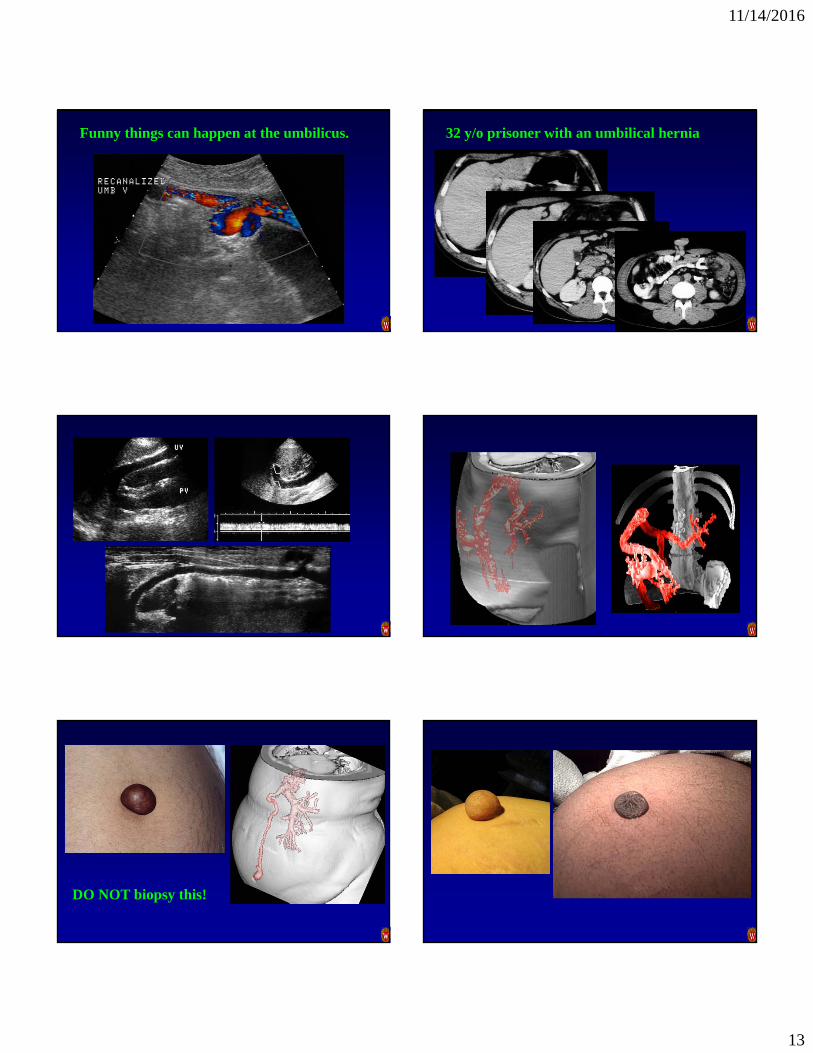
Funny things can happen at the umbilicus. 32 y/o prisoner with an umbilical hernia
DO NOT biopsy this!

11/14/2016
14
Varices may complicate the surgical approach to underlying pathology
52 y/o female with LLQ pain, fever, elevated white count
• Clinical diagnosis - diverticulitis
• Past medical history
• Liver disease
• Portal hypertension - Apparently resolved
Pericholecystic varices
• Rare
• Commonly associated with portal vein thrombosis
What do you think of the Portal Vein?
Pericholecystic varices

11/14/2016
15
S K
Spleno-renal Collateral Spleno-renal Collateral
Spleno-renal varices are rarely direct
They often involve the gonadal vein.
… or a mesenteric veinIMV SMV
… or an adrenal vein
Spleno-external-iliac collateral (via the panus)
Spleno-renal-mesenteric collateral pathways can be very convoluted

11/14/2016
16
IMV to IVC via a Lumbar Vein
… or the collateral pathway can be very direct
39 y/o female with pelvic mass on physical exam
IMV to Left Gonadal Vein
IMV to hemorrhoidal varix PC VIPRPhase Contrast Vastly Under-sampled
Isotropic Projection Imaging
• Novel '4D MR Flow' technique developed at UW Madison
•Currently very computer intensive, but improving
MR PC VIPR
• Helical flow in the Portal Vein
Normal Variant
MR PC VIPR
• Portal Vein Thrombosis
• Reversed Lt PV
• Reversed SMV

11/14/2016
17
… two more details… Cavernous Transformation of the Portal vein
Cavernous Transformation of the Portal vein What do you think of this Portal Vein?
Tumor Thrombus
ConclusionsPortal Hypertension
• Variceal pathways can be just about anywhere
• Pre-transplant shunt identification is critical to transplant survival
• An unsuspected varix can ruin a good surgeon’s day
ConclusionsPortal hypertension (cont.)
• The Caput Medusae is only present in a small percentage of recanalized paraumbilical varices
• When you think you have a cystic mass -don’t forget to turn on the Doppler

11/14/2016
18
Thank you
![BMC Medical Imaging - MedPage Today · 2009. 7. 30. · Transcranial Doppler ultrasonography predicts ... hypertension [9, 10], weakness [2, 9, 10], speech ... TCD diagnosis of intracranial](https://static.fdocuments.net/doc/165x107/6102368f5c8aa16a7f22c4af/bmc-medical-imaging-medpage-today-2009-7-30-transcranial-doppler-ultrasonography.jpg)


![April 25th 2007 Intraoperative Doppler probe from …€¦ · Web view[1] British Hypertension Society Protocol: O’Brien E, Petrie J, Littler W et al (1993) The British Hypertension](https://static.fdocuments.net/doc/165x107/5f0243397e708231d4036391/april-25th-2007-intraoperative-doppler-probe-from-web-view-1-british-hypertension.jpg)