ID-FISH PLASMODIUM GENUS TEST KIT ID-FISH...
6
ID-FISH PLASMODIUM GENUS TEST KIT ID-FISH PLASMODIUM FALCIPARUM AND P. VIVAX COMBO TEST KIT INDICATIONS FOR USE ID-FISH Plasmodium Genus Test Kit (PlasG) and ID-FISH Plasmodium falciparum and P. vivax Combo Test Kit (PlasFV) are intended for in vitro diagnostic use in the clinical laboratory for detection of Plasmodium species in human venous whole blood (EDTA) samples from patients suspected of Plasmodium infection. The test kits are intended to aid in the diagnosis of malaria and to aid in the differential diagnosis of P. falciparum and P. vivax infection. The test kits should be used only on samples from patients with a clinical history, signs and symptoms consistent with malaria, and are not intended as a screen for asymptomatic patients. The ID-FISH Plasmodium Genus Test Kit is a qualitative test for detection of malaria parasites in blood smears. Positive results should be supplemented with the Plasmodium species specific test kit, ID-FISH Plasmodium falciparum and P. vivax Combo Test Kit for identification and differentiation of Plasmodium falciparum and Plasmodium vivax. The results of these test kits should be used in conjunction with other diagnostic test results. Clinical performance has not been established for P. ovale, P. malariae, or P. knowlesi. For in vitro diagnostic use only For professional use only For prescription use only SUMMARY AND EXPLANATION Human malaria a serious, often fatal, parasitic disease is caused by four Plasmodium species, P. falciparum, P. vivax, P. malariae and P. ovale that are transmitted from human to human by mosquito vectors of the genus Anopheles. The non-human primate malaria parasites P. knowlesi, P. cynomolgi, P. brasilianum and P. simium can also naturally infect humans to cause serious disease, and their accurate identification at the species level commonly requires PCR-based methods [1]. According to the WHO, there were approximately 198 million cases of malaria and an estimated 584,000 deaths in the world in 2013 [2]. About 3.3 billion people are at risk of malaria transmission. Africa is the most affected continent with more than 90% of all malaria deaths. South East Asia and the Eastern Mediterranean Region represent 6% and 3% of all the malaria death cases, respectively [3]. WHO recommends that anti-malarial treatment should only be limited to confirmed positive cases. However, treatment based on clinical suspicion should be considered when a parasitological diagnosis is not accessible within two hours of sample collection [3]. Microscopic examination of Giemsa stained thick and thin blood smears is the most wide-spread technique used for malaria diagnosis [4]. An experienced microscopist can detect densities as low as 5–10 parasites per μl of blood by this method, but the detection capabilities of a typical microscopist might be more realistically 100 or more parasites per μl of blood [5, 6]. Although Giemsa stained smears can be used to assess the parasite load as well as to monitor antimalarial treatment, it has limited sensitivity and specificity and requires experienced pathologist for accurate readings. Malaria antigen-based rapid diagnostic tests (RDTs) detect parasite specific antigens or enzymes by immunochromatography These tests although rapid, are not as specific as Giemsa and do not quantify malaria parasites (5,7-11). PCR is a very sensitive diagnostic method for detection and species identification of malaria parasites. Currently there are no FDA approved PCR tests and therefore in the US PCR is usually used for cases where blood film diagnosis is inconclusive or for identification of Plasmodium species in reference laboratories and health departments [12]. Giemsa stained smear microscopy when compared with PCR has sensitivity between 50% [13] and 93% [14]. Despite being more accurate, PCR is time-consuming and expensive. Therefore it is generally not used in the initial diagnosis and treatment of patients with malaria [4]. Fluorescence in situ hybridization (FISH) is a cytogenetic technique used to detect and localize the specific nucleic acid (DNA or RNA) sequences by hybridizing with complementary sequences labeled with fluorescent probes. In 1989, Delong et al. demonstrated that FISH can be used to detect a single microbial cell using fluorescent labeled oligodeoxynucleotides complementary to 16S ribosomal RNA (rRNA), and that with appropriate probes labeled with different fluorescent dyes, FISH can distinguish closely related organisms [15]. In the last 25 years there have been several publications describing FISH technology for the detection and differentiation of infectious agents in cultures and clinical samples [15- 27] Ribosomal RNA (rRNA) was used as a target for developing FISH assays for diagnosing malaria for the following reasons: (1) rRNA is highly abundant in the cell cytoplasm and therefore can be visualized using sequence-specific fluorescence probes under a microscope, without target sequence amplification, and (2) as a consequence of variable nucleotide sequence conservation, it is possible to find short nucleotide stretches that are unique to the genus, species, sub-species or strain [28]. PRINCIPLES OF THE PROCEDURES Fluorescent in situ hybridization is a microscopic technique in which a DNA probe is labeled with a fluorescent dye and then hybridized on a slide with a target ribosomal RNA of the pathogen in a clinical sample. Venous whole blood from a patient (EDTA preserved) suspected of malaria is smeared onto a glass slide with fixative, hybridized with a fluorescently-labeled DNA probe in the PlasG complimentary to the rRNA of Plasmodium genus, and probes in the PlasFV complimentary to P. falciparum and P. vivax rRNA. Plasmodium species, if present, will appear green with the Plasmodium genus probe; P. falciparum will appear green with the P. falciparum probe, and P. vivax will appear orange with the P. vivax probe. Both asexual and sexual forms of Plasmodium, P. falciparum and P. vivax are detected by the PlasG, P. falciparum and P. vivax probes respectively. Non-Plasmodium organisms will not give any signal when viewed under the fluorescent filter. Since ribosomal RNA is present in the cytoplasm of the parasite and is visible within red cell halos, the entire parasite will give a fluorescent signal. Unlike immunoassays and PCR assays, the assay detects ribosomal RNA within the cell; the parasite must be actively infecting the patient’s red blood cells to produce a positive result. REAGENTS AND MATERIALS Equipment, reagents and consumables required and can be purchased from ID-FISH Technology Inc.: 1. LED Fluorescent Unit (Part. No. IDF001) 2. 100x Oil Objective (Part No. IDF002) 3. SPR – Smear Preparation Reagent (Part. No. SPR03) 4. Dual Band Filter Set specific to ID-FISH hybridization probes (Part. No. IDF004). 5. Staining Dish Set – Stainless-steel staining dish and slide holder (Part. No. IDF005) 6. Stainless steel Drying Rack with cover (Part. No. IDF006) 7. Glass cover slips – 100 ea. 22x22 mm, thickness 0.15mm (Part No. IDF008) 8. Plastic Cover slips – 100 ea. (Part. No. IDF009) Equipment, reagents and consumables required but not provided: 1. 37±1º C Incubator 2. Hybridization box (slide box) 3. 45-50ºC Slide warmer (optional) 4. Pipettes 1-20 μl, 20 to 200 μl and 200-1000 μl 5. Clean glass microscope slides 6. 1.5 ml micro centrifuge tubes 7. Negative Control Smears (made with non-malarial EDTA blood less than 7 days old and SPR) 8. Positive Control Smears (made with EDTA blood infected with Pf or Pv parasites less than 7 days old with SPR) 9. ≥99.8% Methanol 10. Water, deionized or distilled 11. Immersion oil - Must comply with the microscope objective and be low-fluorescing oil. PRECAUTIONS For in vitro diagnostic and professional use only. Rx only. Safety precautions 1. The Hyb B contains guanidine thiocyanate and Igepal. May cause eye, skin and respiratory irritation. Harmful if swallowed. In case of eye contact, rinse eyes immediately with water. Seek medical advice. 2. SPR03 contains <5% formaldehyde. May cause harm to the unborn child. Keep out of reach of children. Formaldehyde is a carcinogen. May cause eye, skin and respiratory irritation. May be fatal if inhaled, swallowed or absorbed through skin. Wear gloves and eye protection. Unused reagent should be discarded in hazardous waste. 3. Material Safety Data Sheets are available upon request. 4. Establish biosafety precautions in handling human blood specimens and microbiological hazards. Only qualified personnel should perform venipuncture procedures. 5. Follow standard biological safety precautions. Do not eat, drink, smoke, apply cosmetics, insert contact lenses, store or prepare food within the designated work area. 6. Dispose of reagents in accordance with federal, state and local regulations. Technical Precautions 1. Do not use reagents beyond the expiration dates printed on labels. 2. Reagents are provided at fixed concentrations. Assay performance may be affected if the reagents are modified in any way or are not stored under the recommended conditions as detailed in "Storage and Preparation of Kit Components". Do not mix reagents between different kits. 3. Avoid microbial and parasite contamination of reagents. 4. Avoid any cross-contamination of samples and reagents, as this may give erroneous results. 5. Do not allow pipet tips to touch the smear, or allow tips to contaminate the reagents. 6. Do not allow dropper bottle tip to touch the smear as this may cause cross contamination of material between slides 7. It is important that the microscope is functioning properly. Make sure that the microscope bulb is correctly adjusted and has not aged beyond its specified lifetime. 8. Do not use filters other than the ID-FISH Dual Band Filter set (Part. No. IDF004) STORAGE AND STABILITY ID-FISH Test Kits and reagents are stable until the expiration dates marked on their packaging and containers when stored as specified. Plasmodium Hyb A Shelf life of 24 months from the date of manufacturing when store at 15-30 o C in the dark. Plasmodium Hyb B Shelf life of 24 months from the date of manufacturing when store at 15-30 o C in the dark. Plasmodium Hyb A + Hyb B (reconstituted) In-use life of 6 months from the date of kit reconstitution when store 15-30 o C in the dark. Plasmodium Wash Buffer Shelf life of 24 months from the date of manufacturing when store at 15-30 o C in the dark. In-use life of 6 months from the date of kit reconstitution when store at 15-30 o C in the dark. Plasmodium Rinse Buffer Shelf life of 24 months from the date of manufacturing when store at 15-30 o C in the dark. In-use life of 6 months from the date of kit reconstitution when store at 15-30 o C in the dark. Plasmodium Counterstain Shelf life of 24 months from the date of manufacturing when store at 15-30 o C in the dark. In-use life of 6 months from the date of kit reconstitution when store at 15-30 o C in the dark. ID-FISH Plasmodium Genus Test Kit (50 assays per kit) PlasGK04 (Part. No.) Plasmodium Genus Hyb A PlasGHA03 Plasmodium Hyb B PlasHB03 5x Plasmodium Wash Buffer PlasWB03 10x Plasmodium Rinse Buffer PlasRB03 Plasmodium Counterstain PlasC03 ID-FISH Plasmodium falciparum and P. vivax Combo Test Kit (50 assays per kit) PlasFVK04 (Part. No.) Plasmodium falciparum / P. vivax Hyb A PlasFVHA03 Plasmodium Hyb B PlasHB03 5x Plasmodium Wash Buffer PlasWB03 10x Plasmodium Rinse Buffer PlasRB03 Plasmodium Counterstain PlasC03
Transcript of ID-FISH PLASMODIUM GENUS TEST KIT ID-FISH...
ID-FISH PLASMODIUM GENUS TEST KIT
ID-FISH PLASMODIUM FALCIPARUM AND P. VIVAX COMBO TEST KIT
INDICATIONS FOR USE ID-FISH Plasmodium Genus Test Kit (PlasG) and ID-FISH Plasmodium falciparum and P. vivax Combo Test Kit (PlasFV) are intended for in vitro diagnostic use in the clinical laboratory for detection of Plasmodium species in human venous whole blood (EDTA) samples from patients suspected of Plasmodium infection. The test kits are intended to aid in the diagnosis of malaria and to aid in the differential diagnosis of P. falciparum and P. vivax infection. The test kits should be used only on samples from patients with a clinical history, signs and symptoms consistent with malaria, and are not intended as a screen for asymptomatic patients. The ID-FISH Plasmodium Genus Test Kit is a qualitative test for detection of malaria parasites in
blood smears. Positive results should be supplemented with the Plasmodium species specific test kit, ID-FISH Plasmodium falciparum and P. vivax Combo Test Kit for identification and differentiation of
Plasmodium falciparum and Plasmodium vivax. The results of these test kits should be used in conjunction with other diagnostic test results. Clinical performance has not been established for P. ovale, P. malariae, or P. knowlesi. For in vitro diagnostic use only For professional use only For prescription use only
SUMMARY AND EXPLANATION Human malaria a serious, often fatal, parasitic disease is caused by four Plasmodium species, P. falciparum, P. vivax, P. malariae and P. ovale that are transmitted from human to human by mosquito vectors of the genus Anopheles. The non-human primate malaria parasites P. knowlesi, P. cynomolgi, P. brasilianum and P. simium can also naturally infect humans to cause serious disease, and their accurate identification at the species level commonly requires PCR-based methods [1]. According to the WHO, there were approximately 198 million cases of malaria and an estimated 584,000 deaths in the world in 2013 [2]. About 3.3 billion people are at risk of malaria transmission. Africa is the most affected continent with more than 90% of all malaria deaths. South East Asia and the Eastern Mediterranean Region represent 6% and 3% of all the malaria death cases, respectively [3]. WHO recommends that anti-malarial treatment should only be limited to confirmed positive cases. However, treatment based on clinical suspicion should be considered when a parasitological diagnosis is not accessible within two hours of sample collection [3].
Microscopic examination of Giemsa stained thick and thin blood smears is the most wide-spread technique used for malaria diagnosis [4]. An experienced microscopist can detect densities as low as 5–10 parasites per μl of blood by this method, but the detection capabilities of a typical microscopist might be more realistically 100 or more parasites per μl of blood [5, 6]. Although Giemsa stained smears can be used to assess the parasite load as well as to monitor antimalarial treatment, it has limited sensitivity and specificity and requires experienced pathologist for accurate readings. Malaria antigen-based rapid diagnostic tests (RDTs) detect parasite specific antigens or enzymes by immunochromatography These tests although rapid, are not as specific as Giemsa and do not quantify malaria parasites (5,7-11). PCR is a very sensitive diagnostic method for detection and species identification of malaria parasites. Currently there are no FDA approved PCR tests and therefore in the US PCR is usually used for cases where blood film diagnosis is inconclusive or for identification of Plasmodium species in reference laboratories and health departments [12]. Giemsa stained smear microscopy when compared with PCR has sensitivity between 50% [13] and 93% [14]. Despite being more accurate, PCR is time-consuming and expensive. Therefore it is generally not used in the initial diagnosis and treatment of patients with malaria [4]. Fluorescence in situ hybridization (FISH) is a cytogenetic technique used to detect and localize the specific nucleic acid (DNA or RNA) sequences by hybridizing with complementary sequences labeled with fluorescent probes. In 1989, Delong et al. demonstrated that FISH can be used to detect a single microbial cell using fluorescent labeled oligodeoxynucleotides complementary to 16S ribosomal RNA (rRNA), and that with appropriate probes labeled with different fluorescent dyes, FISH can distinguish closely related organisms [15]. In the last 25 years there have been several publications describing FISH technology for the detection and differentiation of infectious agents in cultures and clinical samples [15- 27] Ribosomal RNA (rRNA) was used as a target for developing FISH assays for diagnosing malaria for the following reasons: (1) rRNA is highly abundant in the cell cytoplasm and therefore can be visualized using sequence-specific fluorescence probes under a microscope, without target sequence amplification, and (2) as a consequence of variable nucleotide sequence conservation, it is possible to find short nucleotide stretches that are unique to the genus, species, sub-species or strain [28].
PRINCIPLES OF THE PROCEDURES Fluorescent in situ hybridization is a microscopic technique in which a DNA probe is labeled with a fluorescent dye and then hybridized on a slide with a target ribosomal RNA of the pathogen in a clinical sample. Venous whole blood from a patient (EDTA preserved) suspected of malaria is smeared onto a glass slide with fixative, hybridized with a fluorescently-labeled DNA probe in the PlasG complimentary to the rRNA of Plasmodium genus, and probes in the PlasFV complimentary to P. falciparum and P. vivax rRNA. Plasmodium species, if present, will appear green with the Plasmodium genus probe; P. falciparum will appear green with the P. falciparum probe, and P. vivax will appear orange with the P. vivax probe. Both asexual and sexual forms of Plasmodium, P. falciparum and P. vivax are detected by the PlasG, P. falciparum and P. vivax probes respectively. Non-Plasmodium organisms will not give any signal when viewed under the fluorescent filter. Since ribosomal RNA is present in the cytoplasm of the parasite and is visible within red cell halos, the entire parasite will give a fluorescent signal. Unlike immunoassays and PCR assays, the assay detects ribosomal RNA within the cell; the parasite must be actively infecting the patient’s red blood cells to produce a positive result.
REAGENTS AND MATERIALS
Equipment, reagents and consumables required and can be purchased from ID-FISH Technology Inc.:
1. LED Fluorescent Unit (Part. No. IDF001) 2. 100x Oil Objective (Part No. IDF002) 3. SPR – Smear Preparation Reagent (Part. No. SPR03) 4. Dual Band Filter Set specific to ID-FISH hybridization probes (Part. No. IDF004). 5. Staining Dish Set – Stainless-steel staining dish and slide holder (Part. No. IDF005) 6. Stainless steel Drying Rack with cover (Part. No. IDF006) 7. Glass cover slips – 100 ea. 22x22 mm, thickness 0.15mm (Part No. IDF008) 8. Plastic Cover slips – 100 ea. (Part. No. IDF009)
Equipment, reagents and consumables required but not provided: 1. 37±1º C Incubator 2. Hybridization box (slide box) 3. 45-50ºC Slide warmer (optional) 4. Pipettes 1-20 µl, 20 to 200 µl and 200-1000 µl 5. Clean glass microscope slides 6. 1.5 ml micro centrifuge tubes 7. Negative Control Smears (made with non-malarial EDTA blood less than 7 days old and SPR) 8. Positive Control Smears (made with EDTA blood infected with Pf or Pv parasites less than 7
days old with SPR) 9. ≥99.8% Methanol 10. Water, deionized or distilled 11. Immersion oil - Must comply with the microscope objective and be low-fluorescing oil.
PRECAUTIONS For in vitro diagnostic and professional use only. Rx only. Safety precautions
1. The Hyb B contains guanidine thiocyanate and Igepal. May cause eye, skin and respiratory irritation. Harmful if swallowed. In case of eye contact, rinse eyes immediately with water. Seek medical advice.
2. SPR03 contains <5% formaldehyde. May cause harm to the unborn child. Keep out of reach of children. Formaldehyde is a carcinogen. May cause eye, skin and respiratory irritation. May be fatal if inhaled, swallowed or absorbed through skin. Wear gloves and eye protection. Unused reagent should be discarded in hazardous waste.
3. Material Safety Data Sheets are available upon request. 4. Establish biosafety precautions in handling human blood specimens and microbiological
hazards. Only qualified personnel should perform venipuncture procedures. 5. Follow standard biological safety precautions. Do not eat, drink, smoke, apply cosmetics, insert
contact lenses, store or prepare food within the designated work area. 6. Dispose of reagents in accordance with federal, state and local
regulations. Technical Precautions
1. Do not use reagents beyond the expiration dates printed on labels. 2. Reagents are provided at fixed concentrations. Assay performance may be affected if the
reagents are modified in any way or are not stored under the recommended conditions as detailed in "Storage and Preparation of Kit Components". Do not mix reagents between different kits.
3. Avoid microbial and parasite contamination of reagents. 4. Avoid any cross-contamination of samples and reagents, as this may give erroneous results. 5. Do not allow pipet tips to touch the smear, or allow tips to contaminate the reagents. 6. Do not allow dropper bottle tip to touch the smear as this may cause cross contamination of
material between slides 7. It is important that the microscope is functioning properly. Make sure that the microscope bulb is
correctly adjusted and has not aged beyond its specified lifetime. 8. Do not use filters other than the ID-FISH Dual Band Filter set (Part. No. IDF004)
STORAGE AND STABILITY ID-FISH Test Kits and reagents are stable until the expiration dates marked on their packaging and
containers when stored as specified. Plasmodium Hyb A Shelf life of 24 months from the date of manufacturing when store at 15-30
oC in the dark.
Plasmodium Hyb B Shelf life of 24 months from the date of manufacturing when store at 15-30
oC in the dark.
Plasmodium Hyb A + Hyb B (reconstituted) In-use life of 6 months from the date of kit reconstitution when store 15-30
oC in the dark.
Plasmodium Wash Buffer Shelf life of 24 months from the date of manufacturing when store at 15-30
oC in the dark.
In-use life of 6 months from the date of kit reconstitution when store at 15-30oC in the dark.
Plasmodium Rinse Buffer Shelf life of 24 months from the date of manufacturing when store at 15-30
oC in the dark.
In-use life of 6 months from the date of kit reconstitution when store at 15-30oC in the dark.
Plasmodium Counterstain Shelf life of 24 months from the date of manufacturing when store at 15-30
oC in the dark.
In-use life of 6 months from the date of kit reconstitution when store at 15-30oC in the dark.
ID-FISH Plasmodium Genus Test Kit (50 assays per kit)
PlasGK04 (Part. No.)
Plasmodium Genus Hyb A PlasGHA03
Plasmodium Hyb B PlasHB03
5x Plasmodium Wash Buffer PlasWB03
10x Plasmodium Rinse Buffer PlasRB03
Plasmodium Counterstain PlasC03
ID-FISH Plasmodium falciparum and P. vivax Combo Test Kit (50 assays per kit)
PlasFVK04 (Part. No.)
Plasmodium falciparum / P. vivax Hyb A PlasFVHA03
Plasmodium Hyb B PlasHB03
5x Plasmodium Wash Buffer PlasWB03
10x Plasmodium Rinse Buffer PlasRB03
Plasmodium Counterstain PlasC03

QUALITY CONTROL Control material should be tested in accordance with the guidelines or requirements of local, state, and/or federal regulations or accrediting organizations. To monitor the assay, reagent performance and day-to-day variation, positive controls for P. falciparum (PF) or P. vivax (PV) along with a negative control (non-malarial blood) must be tested with each run. Negative control slides can be prepared from EDTA whole blood collected from normal subjects. Positive control slides can be prepared from previously positive patient blood samples. Control slides should be prepared in the same manner as patient samples and they are stable for five year if stored in a dry place at room temperature. The positive control must show fluorescing parasites (green or orange depending on the kit used) within red blood cell halos and the negative control must show no parasites within red cells for the test to be valid. Record control reactions and document control failures. Do not use kit if controls do not perform accurately.
SPECIMEN COLLECTION AND HANDLING Specimen Type
Whole blood collection should be performed immediately upon first suspicion of malaria and, preferably, prior to antimalarial drug treatment. Specimen collection
Collect venous blood aseptically by standard venipuncture procedure, into EDTA vacutainer tube. For vacutainer tubes, assure that sufficient blood is collected to fill the tube adequately for correct ratio of blood to EDTA. To prevent clotting, mix blood thoroughly with the anticoagulant by inverting the tube several times. If slides cannot be prepared immediately, store the tubes at 4°C for no longer than 4 days from the date of collection, prior to testing. Note: If regulatory agencies require submission of smears from positive patients, or if microscopy by Giemsa/ Wright staining is desired, smears for stains should be prepared within one hour of collection without addition of SPR. Criteria for Unacceptable Blood Specimens
Clotted blood.
Grossly hemolysed whole blood.
Inadequately filled tube.
Care should be used to avoid contamination of one sample with another sample.
Preparation of Smears
See page 5 schematic
Criteria for Unacceptable Smears
Smears that are poorly made.
Smears made from frozen blood.
Smears that are stored frozen.
Smears made from grossly hemolysed blood.
Thick smears are not appropriate for the assay
Storage of Smears
Methanol fixed, air-dried smears are stored at room temperature with desiccant. They are stable for 5 years if stored in a cool dark place.
TEST PROCEDURE
(See page 5 schematic) FISH assay should be performed in a humid environment to prevent slides from drying out. One can hybridize slides in a humid hybridization box placed in an incubator. This can be done by placing wet paper towels in a slide box to create a humid Hybridization box and place box in an incubator set at 37±1
o C.
Assure that all reagents are prepared and warmed to room temperature. Label slides appropriately for the specific hybridization reagent being tested. Hybridization reaction
Add 12 µl hybridization mixes (in vial labeled Hyb A) onto each sample smear.
Cover smear with plastic cover slip. Avoid creating bubbles and disturbing smear.
Place slides in the hybridization box in 37±1oC incubator, to shield slides from light.
Incubate slides in the dark for 15 minutes. Wash step:
One at a time, process each slide as follows:
Remove hybridization box from the incubator.
Remove slide from the hybridization box. Gently remove cover slip from slide.
Place slide on rack of staining dish.
IMMEDIATELY add 1ml of Wash Buffer to top of smear. DO NOT LET SMEAR DRY.
Repeat procedure for all slides. Once all slides have been removed from hybridization box and the wash buffer added, allow slides to sit for 2-5 minutes. DO NOT LET SMEARS DRY.
Tilt slides to remove buffer.
Add 1ml Wash Buffer to all slides.
Allow buffer to sit on slides for 2-5 minutes.
Tilt slides to remove buffer. DO NOT LET SMEARS DRY.
Rinse step:
Add 1 ml of Rinse buffer to each slide.
Allow buffer to sit on slides for 2-5 min.
Tilt slides to remove buffer.
Air dry slides in the dark for 15-30 min, or until dried.
Mounting:
Once dried, add 30 µl of Counterstain to each smear and cover with glass cover slips. Avoid creating bubbles.
Store slides in dark.
Slides are ready for microscopic viewing after 15 min but should be viewed within 4 hours.
Do not expose the slides to direct sun light or other strong light sources as this may lead to fluorescence quenching.
INTERPRETATION AND REPORTING OF RESULTS
ID-FISH Plasmodium Tests are qualitative tests. Examine each slide using oil immersion (100x) objective and using the dual filter and a fluorescence microscope. Tests are only valid when positive and negative control slides are included in each run and the control results as described below. Positive Result: PlasG - Shows green fluorescence for P. falciparum, P. vivax, P. malariae, P. ovale and P. knowlesi
parasites (asexual and sexual forms of the parasite). If the smear is positive by the PlasG, further testing can be performed to determine whether the smear is positive for either P. falciparum or P. vivax using the PlasFV.
PlasFV - P. falciparum will fluoresce green and P. vivax will fluoresce orange. Negative Result: PlasG - No visible parasite-like green fluorescence for 300 fields. PlasFV - No visible parasite-like green or orange fluorescence for 300 fields.
LIMITATIONS AND INTERFERENCES
1. A positive test indicates the presence of malaria parasites in the blood specimens. Results should be used in conjunction with other clinical findings to establish a diagnosis.
2. Successive films every 8 hours for up to three days are sometimes needed for diagnosis.
3. A negative test result does not exclude the possibility of the presence of malaria parasite. This may occur when the parasite level in the sample is below the limit of detection level of the test. A physician, in light of other laboratory results and clinical findings, should interpret test results.
4. Proper specimen collection and processing are essential to achieve optimal performance of the test. See Specimen Collection and Handling section.
5. The type and condition of the instrumentation used will influence the visual appearance of the image obtained. The fluorescence may vary due to the type of microscope employed, the light source and the level of rRNA in the cells. Each laboratory should establish its own criteria for reading the results using appropriate controls.
6. Reading smears with a fluorescence filter other than supplied by ID-FISH can result in erroneous results.
7. The product has not been validated with specimens other than venous whole blood (EDTA). 8. Patients being treated with anti-malaria agents may have a negative FISH result, while Giemsa
and PCR result can remain positive for a longer period of time after treatment (based on in-
house data from sequential samples from untreated and treated patients).
LABELS
Store Product Away from direct sunlight
Store product between 15oC to 30
oC
Keep dry
This product is non-sterile
This product is sufficient for 50 assays
Instruction for product use
Use By
Manufacturing part number
Lot number
In Vitro Diagnostics Use Only
Authorized representative in the European Community

PERFORMANCE CHARACTERISTICS Samples from Kenya, India and Peru with high prevalence rates for malaria were used for sensitivity studies. In Kenya, the most common species is P. falciparum. In India and Peru, both P. vivax and P. falciparum are common. P. malariae and P. ovale are infrequently found in Peru and Kenya. Samples were tested at the collection sites by Giemsa. For the specificity study, 150 samples from patients from US with malaria–like symptoms or confirmed parasite and other blood diseases were tested. CLINICAL SENSITIVITY AND SPECIFICITY The clinical performance of the ID-FISH Plasmodium Genus (PlasG) and the ID-FISH Plasmodium falciparum and P. vivax Combo (PlasFV) Test Kits were evaluated in a multi-center study conducted in malaria endemic regions and United States. The FISH results were compared to Giemsa malaria microscopy results. PCR with bi-directional sequencing with a minimum of 200 contiguous bases with a PHRED Quality value of 20 or higher was used for discrepant analysis. A total of 1067 patient samples were tested; 917 patient samples from regions endemic for malaria (India, Kenya and Peru) and 150 patient samples from the United States (USA), a country not endemic for malaria. An additional 21 samples from Kenya were excluded from analysis as per the study protocol because the patients had received malaria treatment within the six weeks prior to sample collection. EDTA venous whole blood sample smears prepared as per ID-FISH protocol were evaluated by the ID-FISH Plasmodium Genus Tests Kit and the ID-FISH Plasmodium falciparum and P. vivax Combo Test Kit. The results of PlasG for detection of Plasmodium species and of PlasFV for detection and differentiation of P. falciparum or P. vivax infections is summarized below. All 150 patient samples from the USA tested blindly at ID-FISH Technology Inc. were negative by Giemsa microscopy and negative by PlasG and PlasFV FISH. Therefore only data for the 917 samples from malaria endemic regions is shown in tables below. Performance of the kits on Kenya and India samples were evaluated together, since samples from both these sites included all samples submitted for diagnosis of malaria at the collection sites (Table 1a,1b and 2c). No P. vivax samples were observed among the Kenya samples. Peru Samples (n=212) were evaluated separately because of the 122 positives, for 100 new comers that were Giemsa positive samples, the same number of Giemsa negatives were selectively enrolled for each day (Tables 2a, 2b and 2c). Data on overall sensitivity of FISH assays as compared to Giemsa is shown in Table (3a, 3b and 3c) Table 1a: PlasG FISH Assay Performance on combined Kenya and India samples as compared to Giemsa
P-Genus - Overall
Reference Method Giemsa
95% Confidence Interval
Lower limit
Upper Limit
+ - Sensitivity 95.7% 92.8% 97.6%
PlasG + 314
b 27
a Specificity 92.6% 89.4% 95.0%
- 14 340 PPV 92.1% 88.6% 94.6%
695 328 367 NPV 96.0% 93.3% 97.7%
a Of the 27 samples with Giemsa negative and FISH positive results, 20 samples were positive for
Plasmodium species DNA by PCR/sequencing: 13 P. falciparum DNA positive, 6 P. vivax DNA positive, and 1 P. malariae DNA positive. b Of the 314 concordant Giemsa positive and FISH PlasG positive results, 2 samples were
positive for P. ovale DNA and 1 sample was positive for P. malariae DNA by PCR/sequencing.
Table 1b: PlasFV Combo FISH Assay Performance for P. falciparum Compared to Giemsa
P. falciparum - Overall
Reference Method Giemsa
95% Confidence Interval
Lower Limit
Upper Limit
+ - Sensitivity 97.4% 93.2% 99.2%
PF-PV
FISH
+ 152 21a Specificity 96.1% 94.0% 97.5%
- 4 518 PPV 87.9% 81.8% 92.2%
695 156 539 NPV 99.2% 97.9% 99.8%
a Of the 21 samples with Giemsa negative and FISH positive results, 15 samples were positive for
P. falciparum DNA by PCR/sequencing. Table 1c: PlasFV Combo FISH Assay Performance for P. vivax on combined Kenya and India samples as Compared to Giemsa
P. vivax - Overall
Reference Method Giemsa
95% Confidence Interval
Lower Limit
Upper Limit
+ - Sensitivity 91.2% 85.7% 94.8%
PF-PV
FISH
+ 156 9a Specificity 98.3% 96.6% 99.2%
- 15b 515 PPV 94.5% 89.6% 97.3%
695 171 524 NPV 97.2% 95.3% 98.3%
a Of the 9 samples with Giemsa negative and FISH positive results, 6 samples were positive for
P. vivax by PCR/sequencing. b Of the 15 samples that were positive for P. vivax by Giemsa and negative for P. vivax by the FISH
PlasFV kit, 4 samples were positive for Plasmodium sp. by the FISH PlasG kit. All 4 samples were positive for P. falciparum DNA by PCR/sequencing; however, dual infection with P. vivax could not be ruled out by this method. 3 of the 4 sample were positive for P. falciparum by the FISH PlasFV kit. Overall Result Summary and Conclusion PlasG FISH kit
The sensitivity and specificity of PlasG FISH 95.7% and 92.6% respectively as compared to Giemsa. The Positive Predictive Value and Negative Predictive Value is 92.1% and 96% respectively as compared to Giemsa. PlasFV FISH – P. falciparum
The sensitivity and specificity of PlasFV FISH for P. falciparum is 97.4% and 96.1% as compared to Giemsa. The Positive Predictive Value and Negative Predictive Value is 87.9% and 99.2% respectively as compared to Giemsa. PlasFV FISH – P. vivax The sensitivity and specificity of PlasFV FISH for P. vivax is 91.2% and 98.3% respectively as compared to Giemsa. The Positive Predictive Value and Negative Predictive Value is 94.5% and 97.2% respectively as compared to Giemsa.
Table 2a: Peru - PlasG FISH Assay Performance Compared to Giemsa
Peru P-Genus Giemsa
95% Confidence Interval
Lower Limit
Upper Limit
+ - PPA 95.9% 90.2% 98.5%
PlasG
+ 117 0 NPA 100.0% 95.4% 100.0%
- 5 100
222 122 100
Table 2b: Peru - PlasFV Combo FISH Assay Performance for P. falciparum Compared to Giemsa
Peru P. falciparum
Reference Method Giemsa
95% Confidence Interval
Lower Limit
Upper Limit
+ - PPA 100.0% 46.3% 100.0%
PlasFV + 5 0 NPA 100.0% 97.8% 100.0%
- 0 217
222 5 217
Table 2c: Peru - PlasFV Combo FISH Assay Performance for P. vivax Compared to Giemsa
Peru P. vivax
Reference Method Giemsa
95% Confidence Interval
Lower Limit
Upper Limit
+ - PPA 90.6% 83.4% 95.0%
PlasFV + 106 0 NPA 100.0% 95.6% 100.0%
- 11a 105
222 117 105 a Of the 11 samples that were Giemsa positive and FISH PlasFV negative for P. vivax, 7 samples were
positive for Plasmodium sp. by the FISH PlasG kit. Peru - Result Summary and Conclusion PlasG FISH kit: The positive percent agreement and negative percent agreement of PlasG FISH is 95.9% and 100% respectively as compared to Giemsa. PlasFV FISH – P. falciparum
The positive percent agreement and negative percent agreement of PlasFV FISH for P. falciparum is 100% and 100% respectively as compared to Giemsa. PlasFV FISH – P. vivax The positive percent agreement and negative percent agreement of PlasFV FISH for P. vivax is 90.6% and 100% respectively as compared to Giemsa. Sensitivity of FISH Assays as compared to Giemsa Table 3a: Performance of PlasG FISH Assay at different levels of parasites
Parasites/µl Giemsa FISH G+F Sensitivity
95% Confidence Interval
Lower Limit Upper Limit
>10000 63 63 63 100.0% 92.8% 100.0%
5001 - 1000 99 99 99 100.0% 95.3% 100.0%
1001 - 5000 118 113 113 95.8% 89.9% 98.4%
501 - 1000 63 62 62 98.4% 90.3% 99.9%
101 - 500 75 71 71 94.7% 86.2% 98.3%
<100 32 23 23 71.9% 53.0% 85.6%
Total 450 431 431 95.8% 93.4% 97.4%
33 Giemsa negative samples were positive for Plasmodium DNA by PCR. Of these 20 were positive by FISH
As shown in Table 3a, overall sensitivity of FISH compared to Giemsa was 100% for Plasmodium genus at >5000 parasites per µl blood. Between 101 and 5000 parasites per µl blood, the sensitivity is 94.7% – 98.4%. Below 100 parasites per µl blood the sensitivity is 72 % Table.3b: Performance of PlasFV FISH Assay for P. falciparum at different levels of parasites
Parasites/µl Giemsa FISH G+F Sensitivity
95% Confidence Interval
Lower Limit Upper Limit
>10000 38 38 38 100.0% 88.6% 100.0%
5001 - 1000 17 17 17 100.0% 77.1% 100.0%
1001 - 5000 37 37 37 100.0% 88.3% 100.0%
501 - 1000 26 26 26 100.0% 84.0% 100.0%
101 - 500 30 28 28 93.3% 76.5% 98.8%
<100 13 11 11 84.6% 53.7% 97.3%
Total 161 157 157 97.5% 93.4% 99.2%
24 Giemsa negative samples were positive for P. falciparum DNA by PCR. Of these 13 were positive by FISH

As shown in table 3b, overall sensitivity of PF-PV FISH for P. falciparum as compared to Giemsa was 100% at >500 parasites per µl blood. Between 101 and 500 parasites per µl blood, the sensitivity is 93.3%. Below 100 parasites per µl blood the sensitivity is 84.6 %. Table.3c: Performance of PlasFV FISH Assay for P. vivax at different levels of parasites
Parasites/µl Giemsa FISH G+F Sensitivity
95% Confidence Interval
Lower Limit Upper Limit
>10000 25 25 25 100.0% 83.4% 100.0%
>5000 81 79 79 97.5% 90.5% 99.6%
>1000 81 75 75 92.6% 84.0% 97.0%
>500 37 31 31 83.8% 67.3% 93.2%
>100 45 40 40 88.9% 75.2% 95.8%
<100 19 12 12 63.2% 38.6% 82.8%
Total 288 262 262 91.0% 86.9% 93.9%
8 Giemsa negative samples were positive for P. vivax DNA by PCR. Of these 6 were positive by FISH
As shown in table 3c, overall sensitivity of PF-PV FISH for P. vivax as compared to Giemsa was 100% at >10000 parasites per µl blood. Between 1001 and 10000 parasites per µl blood, the sensitivity is 92.6% – 97.5%. Between 101 and 1000 parasites per µl blood, the sensitivity is between 83.8 - 88.9%. Below 100 parasites per µl blood the sensitivity was 63.2%. ANALYTICAL SENSITIVITY Limit of detection is defined as the number of parasites at which 95% or more samples give a positive result when at least 20 samples are tested. P. falciparum infected blood was a left over coded EDTA venous blood sample from a 5 year old female patient living in a village on the shore of Lake Victoria, Kenya. The estimated parasitemia, by Giemsa was 18,375 parasites per µl/blood. The sample was diluted in control blood as follows: 1:16, 1:32, 1:64, 1:128 and 1:256. 60 smears were prepared from neat patient blood and each of the diluted blood samples. P. vivax infected blood was from a 35 year old male patient living in Mangalore, India. The estimated parasitemia, by Giemsa was 5040 parasites per µl/blood. The sample was diluted in EDTA venous control blood as follows: 1:5, 1:10, 1:20, 1:40, 1:80, 1:160 and 1:320. 60 smears were prepared from neat patient blood and each of the diluted blood samples. 3 smears each of P. falciparum positive blood and P. vivax positive blood were tested from the neat sample and at each dilution over 3 days. The highest dilution at which all 3 samples gave a positive result was considered preliminary Limit of Detection. Additional 20 smears were tested at this dilution to confirm a detection rate of greater than or equal to 95%. Based on the data, the limit of detection for the ID-FISH Plasmodium Genus Test Kit is: P. falciparum 143 parasites/µl blood and for P. vivax 126 parasites/µl blood; and for the ID-FISH Plasmodium falciparum and P. vivax Combo Test Kit: P. falciparum 143 parasites/µl blood and P. vivax 126 parasites/µl blood. SPECIES INCLUSION FOR PlasG
A total of 12 samples, 10 clinical samples positive for Plasmodium species, one monkey blood positive for P. knowlesi and one mouse blood positive for P. berghei were tested with ID FISH Plasmodium Genus Test Kit and P. falciparum and P. vivax combo Test Kits. All Plasmodium species - P. falciparum, P. vivax, P. malariae, P. ovale, P. knowlesi and P. berghei were detected by PlasG. P. falciparum and P. vivax were correctly detected as P.falciparum and P. vivax by the PlasFV. INTERFERENCE STUDY Interference from substances introduced into the blood:
The following exogenous agents are often present in whole blood of patients infected with malarial parasites. Using concentrations that are considered high therapeutic levels, these agents were spiked into Plasmodium positive human blood samples and control blood samples. The effects of these agents on PlasG and PlasFV were studied after the agent was added; the effect of subsequent clinical metabolites was not studied. All of the Plasmodium species were detected with the expected PlasG and PlasFV kits probes at the concentrations tested. No false positive results were observed (Table.4).
Table.4 Exogenous analytes interference results
Agent Concentration
Anti-inflammatory
Acetaminophen 1 mg/ml
Aspirin 1 mg/ml
Ibuprofen 1 mg/ml
Anti-malarial agents
Artesunate 1 mg/ml
Chloroquine 1 mg/ml
Doxycycline 1 mg/ml
Primaquine 1 mg/ml
Quinine 1 mg/ml
Sulfadoxine 1 mg/ml
Antimicrobial agents
Amoxicillin 100 µg/ml
Cephalexin 100 µg/ml
Ciprofloxacin 100 µg/ml
Erythromycin 100 µg/ml
Interference from Endogenous Blood components introduced into the blood: To assess the effect of endogenous analytes in blood, Plasmodium positive and control human blood samples were spiked with the concentrations listed inTable.5 and then tested by PlasG and PlasFV. Each Plasmodium positive sample was detected by the expected PlasG and PlasFV kits probes in the presence of the potential interferents. No false positive results were observed in the uninfected control blood samples.
Table.5 Endogenous analytes interference results
Blood component Concentration
Triglycerides 37 mmol/L
Hemoglobin 2 g/L
Bilirubin 342 µmol/L
Serum albumin 60 g/L
Interference from Unrelated Medical Conditions:
To assess the effect of unrelated medical conditions on the specificity of the assay, 76 blood specimens from patients with known medical conditions were tested with PlasG and PlasFV. The medical conditions include HIV positive patient samples and samples from patient with positive antibody test results for:
- Tick borne diseases (B.burgdorferi, B. hensalae, Rickettsia spp. and Ehrlichia spp.)
- Autoimmune diseases (Antiphospholipid Antibody Syndrome, Lupus and Rheumatoid
Arthritis)
None of the samples tested positive with PlasG and PlasFV, demonstrating that the performance of PlasG and PlasFV was not affected by unrelated medical condition. ANALYTICAL SPECIFICITY (CROSS –REACTIVITY) Interference from pathogens
29 pathogens, 7 bacterial cultures, 11 parasites and 11 viruses found in human blood were collected from animal or human hosts or in vitro cultures, as listed in the table below. No off-target organism produced a positive result with PlasG or PlasFV kits at the tested concentrations. All on-target Plasmodium species were detected with the expected PlasG and PlasFV kits probes.
PATHOGENIC MICROORGANISMS SOURCE
Bacteria
Anaplasma phagocytophilum HOPKINS
Bartonella henselae IGeneX
Borrelia burgdorferi IGeneX
Ehrlichia chaffeensis HOPKINS
Leptospira interrogans ATCC
Rickettsia rickettsii FULLER
Rickettsia typhi FULLER
Viruses
Bwamba Fever Virus KEMRI-KIS
Chikungunya virus KEMRI-KIS
Cytomegalovirus (CMV) UCSF
Dengue virus KEMRI-KIS
Epstein-Barr virus (EBV) UCSF
Hepatitis B IGeneX
HIV-1 UCSF
Influenza A UCSF
Influenza B UCSF
West Nile virus KEMRI-KIS
Yellow Fever virus KEMRI-KIS
Parasites
Leishmania major(amastigotes) KEMRI-KIS
Leishmania major(promastigotes)
KEMRI-KIS
Babesia microti AS Inc
Babesia duncani AS Inc
Trypanosoma cruzi IGeneX
Plasmodium berghei KEMRI-NBO
Plasmodium falciparum KEMRI-KIS
Plasmodium malariae KEMRI-KIS
Plasmodium knowlesi ATCC
Plasmodium ovale KEMRI-KIS
Plasmodium vivax KMC
HIV-1 and Plasmodium species were tested at > 10
4 organisms per milliliter. All other parasites and
bacteria were tested at concentrations greater than 106 organisms per milliliter and viruses were tested
at greater than 105 PFU or copies per milliliter.
Samples were received from the following sites:
1. ATCC: American Tissue Type Culture, Manassas, VA 20110, USA 2. AS Inc.: Antibody Systems Inc. Hurst, TX 76054, USA 3. FULLER: Focus Diagnostics, Cypress, CA 90630, USA 4. HOPKINS: John Hopkins university, Baltimore, MD, USA 5. KEMRI-KIS: Kenya Medical Research Institute, Kisumu, Kenya 6. KEMRI-NBO: Kenya Medical Research Institute, Nairobi, Kenya 7. KMC: Kasturba Medical College, Mangalore, India 8. UCSF: University of California, San Francisco, CA 94143, USA 9. USF: University of South Florida, Tampa, FL 33620, USA 10. IGeneX: IGeneX Inc., Palo Alto, CA 94303, USA.
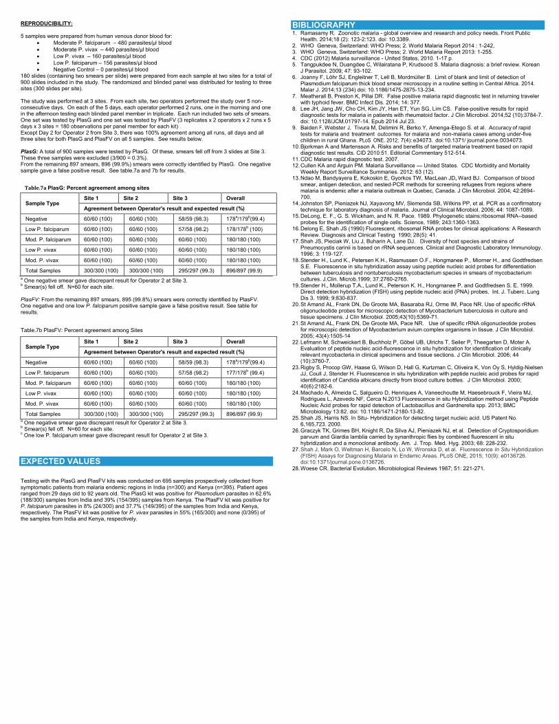
REPRODUCIBILITY: 5 samples were prepared from human venous donor blood for:
Moderate P. falciparum – 480 parasites/µl blood
Moderate P. vivax – 440 parasites/µl blood
Low P. vivax – 160 parasites/µl blood
Low P. falciparum – 156 parasites/µl blood
Negative Control – 0 parasites/µl blood 180 slides (containing two smears per slide) were prepared from each sample at two sites for a total of 900 slides included in the study. The randomized and blinded panel was distributed for testing to three sites (300 slides per site). The study was performed at 3 sites. From each site, two operators performed the study over 5 non-consecutive days. On each of the 5 days, each operator performed 2 runs, one in the morning and one in the afternoon testing each blinded panel member in triplicate. Each run included two sets of smears. One set was tested by PlasG and one set was tested by PlasFV (3 replicates x 2 operators x 2 runs x 5 days x 3 sites = 180 observations per panel member for each kit) . Except Day 2 for Operator 2 from Site 3, there was 100% agreement among all runs, all days and all three sites for both PlasG and PlasFV on all 5 samples. See results below. PlasG: A total of 900 samples were tested by PlasG. Of these, smears fell off from 3 slides at Site 3. These three samples were excluded (3/900 = 0.3%). From the remaining 897 smears, 896 (99.9%) smears were correctly identified by PlasG. One negative sample gave a false positive result. See table.7a and 7b for results.
a One negative smear gave discrepant result for Operator 2 at Site 3.
b Smear(s) fell off. N=60 for each site.
PlasFV: From the remaining 897 smears, 895 (99.8%) smears were correctly identified by PlasFV. One negative and one low P. falciparum positive sample gave a false positive result. See table for results.
Table.7b PlasFV: Percent agreement among Sites
a One negative smear gave discrepant result for Operator 2 at Site 3.
b Smear(s) fell off. N=60 for each site.
c One low P. falciparum smear gave discrepant result for Operator 2 at Site 3.
EXPECTED VALUES
Testing with the PlasG and PlasFV kits was conducted on 695 samples prospectively collected from symptomatic patients from malaria endemic regions in India (n=300) and Kenya (n=395). Patient ages ranged from 29 days old to 92 years old. The PlasG kit was positive for Plasmodium parasites in 62.6% (188/300) samples from India and 39% (154/395) samples from Kenya. The PlasFV kit was positive for P. falciparum parasites in 8% (24/300) and 37.7% (149/395) of the samples from India and Kenya, respectively. The PlasFV kit was positive for P. vivax parasites in 55% (165/300) and none (0/395) of the samples from India and Kenya, respectively.
BIBLIOGRAPHY 1. Ramasamy R. Zoonotic malaria - global overview and research and policy needs. Front Public
Health. 2014;18 (2): 123-2:123. doi: 10.3389. 2. WHO Geneva, Switzerland: WHO Press; 2. World Malaria Report 2014 : 1-242, 3. WHO Geneva, Switzerland: WHO Press; 2. World Malaria Report 2013: 1-255. 4. CDC (2012) Malaria surveillance - United States, 2010. 1-17 p. 5. Tangpukdee N, Duangdee C, Wilairatana P, Krudsood S. Malaria diagnosis: a brief review. Korean
J Parasitol. 2009; 47: 93-102. 6. Joanny F, Löhr SJ, Engleitner T, Lell B, Mordmüller B. Limit of blank and limit of detection of
Plasmodium falciparum thick blood smear microscopy in a routine setting in Central Africa. 2014. Malar J. 2014;13 (234) doi: 10.1186/1475-2875-13-234.
7. Meatherall B, Preston K, Pillai DR. False positive malaria rapid diagnostic test in returning traveler with typhoid fever. BMC Infect Dis. 2014; 14: 377.
8. Lee JH, Jang JW, Cho CH, Kim JY, Han ET, Yun SG, Lim CS. False-positive results for rapid diagnostic tests for malaria in patients with rheumatoid factor. J Clin Microbiol. 2014;52 (10):3784-7. doi: 10.1128/JCM.01797-14. Epub 2014 Jul 23.
9. Baiden F, Webster J, Tivura M, Delimini R, Berko Y, Amenga-Etego S. et al. Accuracy of rapid tests for malaria and treatment outcomes for malaria and non-malaria cases among under-five children in rural Ghana. PLoS ONE. 2012; 7(4): e34073. doi:10.1371/ journal.pone.0034073.
10. Bjorkman A and Martensson A. Risks and benefits of targeted malaria treatment based on rapid diagnostic test results. CID 2010:51. Editorial Commentary 512-514.
11. CDC Malaria rapid diagnostic test. 2007. 12. Cullen KA and Arguin PM. Malaria Surveillance — United States. CDC Morbidity and Mortality
Weekly Report Surveillance Summaries. 2012: 63 (12). 13. Ndao M, Bandyayera E, Kokoskin E, Gyorkos TW, MacLean JD, Ward BJ. Comparison of blood
smear, antigen detection, and nested-PCR methods for screening refugees from regions where malaria is endemic after a malaria outbreak in Quebec, Canada. J Clin Microbiol. 2004; 42:2694-700.
14. Johnston SP, Pieniazek NJ, Xayavong MV, Slemenda SB, Wilkins PP, et al. PCR as a confirmatory technique for laboratory diagnosis of malaria. Journal of Clinical Microbiol. 2006; 44: 1087-1089.
15. DeLong, E. F., G. S. Wickham, and N. R. Pace. 1989. Phylogenetic stains:ribosomal RNA--based probes for the identification of single cells. Science, 1989; 243:1360-1363.
16. Delong E, Shah JS (1990) Fluorescent, ribosomal RNA probes for clinical applications: A Research Review. Diagnosis and Clinical Testing 1990; 28(5): 41.
17. Shah JS, Pieciak W, Liu J, Buharin A, Lane DJ. Diversity of host species and strains of Pneumocystis carinii is based on rRNA sequences. Clinical and Diagnostic Laboratory Immunology. 1996; 3: 119-127.
18. Stender H., Lund K., Petersen K.H., Rasmussen O.F., Hongmanee P., Miorner H., and Godtfredsen S.E. Fluorescence in situ hybridization assay using peptide nucleic acid probes for differentiation between tuberculosis and nontuberculosis mycobacterium species in smears of mycobacterium cultures. J.Clin. Microb.1999; 37:2760-2765.
19. Stender H., Mollerup T.A., Lund K., Peterson K. H., Hongmanee P. and Godtfredsen S. E. 1999. Direct detection hybridization (FISH) using peptide nucleic acid (PNA) probes. Int. J. Tuberc. Lung Dis 3. 1999; 9:830-837.
20. St Amand AL, Frank DN, De Groote MA, Basaraba RJ, Orme IM, Pace NR. Use of specific rRNA oligonucleotide probes for microscopic detection of Mycobacterium tuberculosis in culture and tissue specimens. J Clin Microbiol. 2005;43(10):5369-71.
21. St Amand AL, Frank DN, De Groote MA, Pace NR. Use of specific rRNA oligonucleotide probes for microscopic detection of Mycobacterium avium complex organisms in tissue. J Clin Microbiol. 2005; 43(4):1505-14
22. Lefmann M, Schweickert B, Buchholz P, Göbel UB, Ulrichs T, Seiler P, Theegarten D, Moter A. Evaluation of peptide nucleic acid-fluorescence in situ hybridization for identification of clinically relevant mycobacteria in clinical specimens and tissue sections. J Clin Microbiol. 2006; 44 (10):3760-7.
23. Rigby S, Procop GW, Haase G, Wilson D, Hall G, Kurtzman C, Oliveira K, Von Oy S, Hyldig-Nielsen JJ, Coull J, Stender H. Fluorescence in situ hybridization with peptide nucleic acid probes for rapid identification of Candida albicans directly from blood culture bottles. J Clin Microbiol. 2000; 40(6):2182-6.
24. Machado A, Almeida C, Salgueiro D, Henriques A, Vaneechoutte M, Haesebrouck F, Vieira MJ, Rodrigues L, Azevedo NF, Cerca N.2013 Fluorescence in situ Hybridization method using Peptide Nucleic Acid probes for rapid detection of Lactobacillus and Gardnerella spp. 2013; BMC Microbiology 13:82. doi: 10.1186/1471-2180-13-82.
25. Shah JS, Harris NS. In Situ- Hybridization for detecting target nucleic acid. US Patent No. 6,165,723. 2000.
26. Graczyk TK, Grimes BH, Knight R, Da Silva AJ, Pieniazek NJ, et al. Detection of Cryptosporidium parvum and Giardia lamblia carried by synanthropic flies by combined fluorescent in situ hybridization and a monoclonal antibody. Am. J. Trop. Med. Hyg. 2003; 68: 228-232.
27. Shah J, Mark O, Weltman H, Barcelo N, Lo W, Wronska D, et al. Fluorescence In Situ Hybridization (FISH) Assays for Diagnosing Malaria in Endemic Areas. PLoS ONE, 2015; 10(9): e0136726. doi:10.1371/journal.pone.0136726.
28. Woese CR. Bacterial Evolution. Microbiological Reviews 1987; 51: 221-271.
Table.7a PlasG: Percent agreement among sites
Sample Type Site 1 Site 2 Site 3 Overall
Agreement between Operator's result and expected result (%)
Negative 60/60 (100) 60/60 (100) 58/59 (98.3) 178a/179
b(99.4)
Low P. falciparum 60/60 (100) 60/60 (100) 57/58 (98.2) 178/178b (100)
Mod. P. falciparum 60/60 (100) 60/60 (100) 60/60 (100) 180/180 (100)
Low P. vivax 60/60 (100) 60/60 (100) 60/60 (100) 180/180 (100)
Mod. P. vivax 60/60 (100) 60/60 (100) 60/60 (100) 180/180 (100)
Total Samples 300/300 (100) 300/300 (100) 295/297 (99.3) 896/897 (99.9)
Sample Type Site 1 Site 2 Site 3 Overall
Agreement between Operator's result and expected result (%)
Negative 60/60 (100) 60/60 (100) 58/59 (98.3) 178a/179
b(99.4)
Low P. falciparum 60/60 (100) 60/60 (100) 57/58 (98.2) 177/178b (99.4)
Mod. P. falciparum 60/60 (100) 60/60 (100) 60/60 (100) 180/180 (100)
Low P. vivax 60/60 (100) 60/60 (100) 60/60 (100) 180/180 (100)
Mod. P. vivax 60/60 (100) 60/60 (100) 60/60 (100) 180/180 (100)
Total Samples 300/300 (100) 300/300 (100) 295/297 (99.3) 896/897 (99.9)
http://www.ncbi.nlm.nih.gov/pubmed/?term=Salgueiro%20D%5BAuthor%5D&cauthor=true&cauthor_uid=23586331
http://www.ncbi.nlm.nih.gov/pubmed/?term=Henriques%20A%5BAuthor%5D&cauthor=true&cauthor_uid=23586331
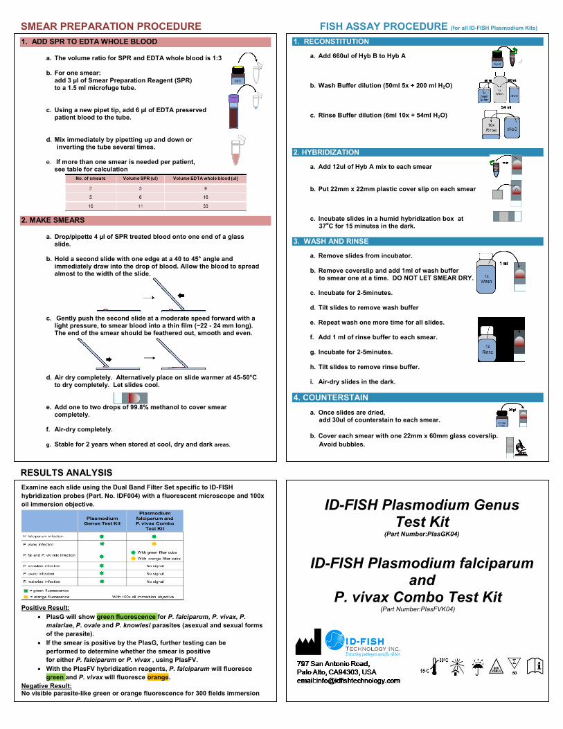
SMEAR PREPARATION PROCEDURE FISH ASSAY PROCEDURE (for all ID-FISH Plasmodium Kits)
RESULTS ANALYSIS
1. RECONSTITUTION
a. Add 660ul of Hyb B to Hyb A
b. Wash Buffer dilution (50ml 5x + 200 ml H2O) c. Rinse Buffer dilution (6ml 10x + 54ml H2O)
2. HYBRIDIZATION
a. Add 12ul of Hyb A mix to each smear b. Put 22mm x 22mm plastic cover slip on each smear c. Incubate slides in a humid hybridization box at 37
oC for 15 minutes in the dark.
3. WASH AND RINSE
a. Remove slides from incubator.
b. Remove coverslip and add 1ml of wash buffer to smear one at a time. DO NOT LET SMEAR DRY. c. Incubate for 2-5minutes. d. Tilt slides to remove wash buffer e. Repeat wash one more time for all slides. f. Add 1 ml of rinse buffer to each smear. g. Incubate for 2-5minutes. h. Tilt slides to remove rinse buffer. i. Air-dry slides in the dark.
4. COUNTERSTAIN
a. Once slides are dried, add 30ul of counterstain to each smear.
b. Cover each smear with one 22mm x 60mm glass coverslip.
Avoid bubbles.
c. Let slides sit for 15 mins before viewing under microscope.
1. ADD SPR TO EDTA WHOLE BLOOD
a. The volume ratio for SPR and EDTA whole blood is 1:3
b. For one smear:
add 3 µl of Smear Preparation Reagent (SPR) to a 1.5 ml microfuge tube.
c. Using a new pipet tip, add 6 µl of EDTA preserved
patient blood to the tube.
d. Mix immediately by pipetting up and down or inverting the tube several times.
e. If more than one smear is needed per patient,
see table for calculation
2. MAKE SMEARS
a. Drop/pipette 4 µl of SPR treated blood onto one end of a glass slide.
b. Hold a second slide with one edge at a 40 to 45° angle and immediately draw into the drop of blood. Allow the blood to spread almost to the width of the slide.
c. Gently push the second slide at a moderate speed forward with a light pressure, to smear blood into a thin film (~22 - 24 mm long). The end of the smear should be feathered out, smooth and even.
d. Air dry completely. Alternatively place on slide warmer at 45-50°C to dry completely. Let slides cool.
e. Add one to two drops of 99.8% methanol to cover smear completely.
f. Air-dry completely.
g. Stable for 2 years when stored at cool, dry and dark areas.
Examine each slide using the Dual Band Filter Set specific to ID-FISH
hybridization probes (Part. No. IDF004) with a fluorescent microscope and 100x
oil immersion objective.
Positive Result:
PlasG will show green fluorescence for P. falciparum, P. vivax, P.
malariae, P. ovale and P. knowlesi parasites (asexual and sexual forms
of the parasite).
If the smear is positive by the PlasG, further testing can be
performed to determine whether the smear is positive
for either P. falciparum or P. vivax , using PlasFV.
With the PlasFV hybridization reagents, P. falciparum will fluoresce
green and P. vivax will fluoresce orange.
Negative Result: No visible parasite-like green or orange fluorescence for 300 fields immersion
(100x).
ID-FISH Plasmodium Genus Test Kit
(Part Number:PlasGK04)
ID-FISH Plasmodium falciparum
and P. vivax Combo Test Kit
(Part Number:PlasFVK04)