HUMAN PARVOVIRUS INFECTIONS Welcome to Jianming's Homepage
26
Chapter 27 HUMAN PARVOVIRUS INFECTIONS Stuart P. Adler William C. Koch Microbiology 868 Pathogenesis: General Aspects 869 Epidemiology and Transmission 870 Global Distribution Seasonality and Periodicity Scroprevalence by Age Scroprevalence by Gender Seroprcvalence by Race Incidence Ki\k F,rctorb for Acquisition I-lospital Transmission Route5 of Viral S p r e d Risk ot BI9 Acquisition for Women of Childbearing Age Clinical Manifestations Other than Intrauterine Infection 874 Erythema Infectiosum ‘Iransient Aplastic Crisis Arthropathy Infection in the Imniunocompromisrd Host Other Ilermatologic Syndromes (:cntral Nervous System Infection and Neurologic Ilisorders ILnal I )isease Diagnosis: General Approach and Laboratory Methods 876 Epidemiology of 619 Infections and Risk of Acquisition in the Pregnant Woman 877 l’revalence and Incidence in the United States Prevalence and Incidence in Other Countries Clinical Manifestations of 619 Infections in the Pregnant Woman 878 Intrauterine Transmission Rates, Clinical Manifestations, and Fetal Outcomes 879 Fetd Ikath Asymptomatic Fetd Infection Birth Defects hleconium Ileus and Peritonitis Fetal IIydrops Fetd Outcome in Relation to Maternal hlnnifestations Pathogenesis of Infection in the Fetus Fetal Immune Responses to B19 Pdhogtwsis of BI 9 Hydrops Pathology in the Fetus 882 Anatomic and Histologic Features Placentd Heart 0 the r Orga 11 s Diagnostic Evaluation and Management of the Woman and Fetus Exposed to or Infected by 619 during Pregnancy 883 Prevalence of Erythema Infectiosum History of Exposure Clinical Features Suggesting B19 Infection in the Pregnant Woman I.aboratory Diagnosis in the Pregnant Woman Fetal Monitoring Fctd Therapy 881 Differential Diagnosis 885 Prognosis 885 Prevention 886 General Measures Vaccine Development The parvoviruses are a family of small, single-stranded DNA viruses that have a wide cellular tropism and broad host range, causing infection in invertebrate and vertebrate species from insects to mammals. Although many are important veterinary pathogens, there is only one proven human pathogen in the family, the human parvovirus B19. The virus is most commonly referred to as parvovirus B19, or simply B19. A new genus and name have been proposed for this virus: erythrovirus B19,’ based on its cellular tropism for erythroid lineage cells and to distinguish it from the other mammalian parvoviruses. Compared with most other common human viruses, B19 is a relatively new pathogen, but since its initial description, B19 has come to be associated with a variety of seemingly diverse clinical syndromes in a number of different patient populations (Table 27-1). Although the list of clinical mani- festations caused by B19 infection is probably not yet complete, some proposed relationships such as to rheumatologic disease and neurologic disorders remain contr~versial.~’~ Parvovirus B19 was accidentally discovered by Cossart and associates4 in 1975 as an anomalous band of precipitation while screening blood donor serum for hepatitis B antigen by counterimmunoelectrophoresis. The name B19 refers to the donor unit from which it was originally isolated. Initial analysis of the new virus revealed it had physical features characteristic of the known parvovir~ses,~ allowing classifi- cation in this family. Because the donors from which it was originally isolated were asymptomatic, B19 infection was not initially associated with any illness, and for the next several years after its description, it was a virus in search of a disease. In 1981, Pattisod found a high prevalence of antibodies to this virus in the serum of children hospitalized with the transient aplastic crisis of sickle cell disease and proposed B19 as the viral cause of this clinically well-described event. Sergeant and colleagues7later confirmed this association in population studies of sickle cell patients in Jamaica. It was not until 1983, 8 years after its initial description, that Anderson proposed B19 as the cause of the common child- hood exanthem known as erythema infectiosum (EI), or fifth disease, that the virus was linked to its most common clinical manifestation: This benign rash illness of children had been clinically recognized and well described for many decades, but the cause was unknown. The namefifth disease
Transcript of HUMAN PARVOVIRUS INFECTIONS Welcome to Jianming's Homepage
Chapter 27 HUMAN PARVOVIRUS INFECTIONS Stuart P. Adler William C. Koch
Microbiology 868
Pathogenesis: General Aspects 869
Epidemiology and Transmission 870
Global Distribution Seasonality and Periodicity Scroprevalence by Age Scroprevalence by Gender Seroprcvalence by Race Incidence Ki\k F,rctorb for Acquisition I-lospital Transmission Route5 of Viral S p r e d Risk o t B I 9 Acquisition for Women of Childbearing Age
Clinical Manifestations Other than Intrauterine Infection 874
Erythema Infectiosum ‘Iransient Aplastic Crisis Arthropathy Infection in the Imniunocompromisrd Host Other Ilermatologic Syndromes (:cntral Nervous System Infection and Neurologic Ilisorders ILnal I )isease
Diagnosis: General Approach and Laboratory Methods 876
Epidemiology of 619 Infections and Risk of Acquisition in the Pregnant Woman 877
l’revalence and Incidence in the United States Prevalence and Incidence in Other Countries
Clinical Manifestations of 619 Infections in the Pregnant Woman 878
Intrauterine Transmission Rates, Clinical Manifestations, and Fetal Outcomes 879
Fetd Ikath Asymptomatic Fetd Infection Birth Defects hleconium Ileus and Peritonitis Fetal IIydrops Fetd Outcome in Relation to Maternal hlnnifestations
Pathogenesis of Infection in the Fetus
Fetal Immune Responses to B19 Pdhogtwsis of B I 9 Hydrops
Pathology in the Fetus 882
Anatomic and Histologic Features Placentd Heart 0 the r Orga 11 s
Diagnostic Evaluation and Management of the Woman and Fetus Exposed to or Infected by 619 during Pregnancy 883
Prevalence of Erythema Infectiosum History of Exposure Clinical Features Suggesting B19 Infection in the Pregnant Woman I.aboratory Diagnosis in the Pregnant Woman Fetal Monitoring Fctd Therapy
881
Differential Diagnosis 885
Prognosis 885
Prevention 886
General Measures Vaccine Development
The parvoviruses are a family of small, single-stranded DNA viruses that have a wide cellular tropism and broad host range, causing infection in invertebrate and vertebrate species from insects to mammals. Although many are important veterinary pathogens, there is only one proven human pathogen in the family, the human parvovirus B19. The virus is most commonly referred to as parvovirus B19, or simply B19. A new genus and name have been proposed for this virus: erythrovirus B19,’ based on its cellular tropism for erythroid lineage cells and to distinguish it from the other mammalian parvoviruses.
Compared with most other common human viruses, B19 is a relatively new pathogen, but since its initial description, B19 has come to be associated with a variety of seemingly diverse clinical syndromes in a number of different patient populations (Table 27-1). Although the list of clinical mani- festations caused by B19 infection is probably not yet complete, some proposed relationships such as to rheumatologic disease and neurologic disorders remain contr~versial.~’~
Parvovirus B19 was accidentally discovered by Cossart and associates4 in 1975 as an anomalous band of precipitation while screening blood donor serum for hepatitis B antigen by counterimmunoelectrophoresis. The name B19 refers to the donor unit from which it was originally isolated. Initial analysis of the new virus revealed it had physical features characteristic of the known parvovir~ses,~ allowing classifi- cation in this family. Because the donors from which it was originally isolated were asymptomatic, B19 infection was not initially associated with any illness, and for the next several years after its description, it was a virus in search of a disease. In 1981, Pattisod found a high prevalence of antibodies to this virus in the serum of children hospitalized with the transient aplastic crisis of sickle cell disease and proposed B19 as the viral cause of this clinically well-described event. Sergeant and colleagues7 later confirmed this association in population studies of sickle cell patients in Jamaica. It was not until 1983, 8 years after its initial description, that Anderson proposed B19 as the cause of the common child- hood exanthem known as erythema infectiosum (EI), or fifth disease, that the virus was linked to its most common clinical manifestation: This benign rash illness of children had been clinically recognized and well described for many decades, but the cause was unknown. The namefifth disease

868 Section I11 Viral Infections
Table 27-1 Clinical Manifestations of Parvovirus B19 Infection
Diseases Primary Patient Groups ~~~~~~~ ~~~
Diseases Associated with Acute Infection Erythema infectiosum (fifth disease) Normal children Polyarthropathy Normal adolescents and adults Transient aplastic crisis Patients with hemolytic anemia or accelerated erythropoiesis, or both Papular-purpuric "gloves and socks" syndrome Normal adolescents and adults Diseases Associated with Chronic Infection Persistent anemia (red cell aplasia) Nonimmune fetal hydrops Congenital anemia Chronic arthropathy Infection-associated hemophagocy-tosis Vasculitis or purpura Myoca rd it is
Immunodeficient or immunocompromised children and adults Intrauterine infection Intrauterine infection Rare patients with B19-induced joint disease Normal or immunocompromised patients Normal adults and children Intrauterine infection, normal infants and children, immunocompromised
patients
Data from Brown KE, Young NS. Parvovirus B19 infection and hematopoiesis. Blood Rev 9:176, 1995.
was derived from the 19th century practice of numbering the common exanthems of childhood, and EI was the fifth rash designated in this scheme and the only one for which this numeric designation has persisted in clinical practice.' The others in the series included measles, scarlet fever, rubella, and Filatov-Dukes disease (a mild variant of scarlet fever that is no longer recognized).
The possibility of fetal disease associated with EI was considered long before the viral origin was known, primarily because of comparison with rubella and the incidence of congenital rubella syndrome after community epidemics.'0-12 Advances in knowledge of the virology of other animal parvoviruses and their known propensity to cause disease in fetuses and newborn animals further fueled this concern.13 This suspicion was confirmed in 1984, when two of B19 infection in pregnant women associated with adverse fetal outcomes appeared and were later followed by a larger report of a series of cases of nonimmune hydrops fetalis caused by intrauterine infections with B19.I6 Over the ensuing decade, a variety of clinical manifestations associated with acute and chronic infections have been attributed to this virus in different patient groups (see Table 27-1).
Since the initial reports of fetal infection, knowledge of the epidemiology, pathophysiology, and short-term outcome of fetal and neonatal infection with B19 has increased immensely because of several large, population-based ~tudies.'~.'~ B19 infection during pregnancy has probably been the subject of more such studies than any of the other manifestations, with the possible exception of the transient aplastic crisis of sickle cell disease. However, there is still much to be learned regarding the long-term outcome of fetal infection, the unusual clinical manifestations of infection in neonates, and the immunologic response to infection. The potential for prevention through vaccine development is a topic of current interest and ongoing research.
MICROBIOLOGY
Like other members of the family Parvoviridae, B19 is a small, nonenveloped, single-stranded DNA virus. The taxon-
omy for this family has been revised to include two sub- families22323: the Densovirinae, which are insect viruses; and the Parvovirinae, which infect vertebrates. The Parvovirinae subfamily is composed of three genera: Dependovirus, Parvovirus, and Erythrovirus. The dependoviruses require co-infection with another, unrelated helper virus (i.e., adeno- virus or herpesvirus) to complete their life cycle. There are some dependoviruses that infect humans (e.g., adeno- associated viruses), but the infection is asymptomatic and without consequence. In contrast to the dependoviruses, members of the genera Parvovirus and Erythrovirus are able to replicate autonomously. Previously included in the genus Parvovirus, B19 is now classified as an Erythrovirus (i.e., Erythrovirus B19). The genus Erythrovirus consists of only two members, B19 and a simian parvovirus (SPV) that has a similar genomic organization as B19 and has a simiiar tropism for erythroid cells.24 Although a number of parvoviruses are pathogenic to other mammals (e.g., canine parvovirus, feline panleukopenia virus), B19 is the only parvovirus that causes disease in humans.
There is only one recognized serotype of B19. Minor variations in the nucleotide sequence occur among different B19 viral isolates from different geographic areas, but they have not been definitely shown to affect clinical patterns of infection or path~genicity.'~-~~ Two isolates of human parvo- virus, V9 and V6, whose nucleotide sequence differs signifi- cantly (>lo%) from B19 have been Both were isolated from patients with a transient red cell aplasia indis- tinguishable clinically from the typical B19-induced aplastic crisis. The clinical significance of these variants and whether they represent different genotypes or merely geographic variants of B19 remains a topic of debate.27330
The B19 genome is very small ( ~ 5 . 6 kb) and contained within an icosahedral protein capsid. The capsid structure and lack of an envelope make the virus very resistant to heat and detergent inactivation, features that appear to be important in transmission. The genome appears to encode only three proteins. Two are capsid proteins, designated VP1 and VP2. VP2 is smaller but more abundant and makes up approximately 96% of the capsid protein. VP1 is larger and constitutes about 4% of the capsid but contains a unique

Chapter 27 Human Parvovirus Infections 869
region that extends out from the capsid surface and serves as the attachment site for the cellular receptor?l VP2 has the unique ability to self-assemble into capsids that are morpho- logically and antigenically similar to B19 viruses when expressed in cell culture systems in v i t r ~ . ~ ’ , ~ ~ When present with VP1, the capsids incorporate both proteins, but VP1 alone does not self-as~emble.~~
The third gene product is a nonstructural protein designated NS1. The function of this protein is not entirely clear, but it is involved in regulation of the viral promoter and appears to have a role in DNA repli~ation.’~ Studies of NS1 have been hampered by the observation that it appears to be toxic to cells by an unknown mechanism.34 Studies have further suggested that production of NS1 can lead to programmed cell death (i.e., apoptosis) mediated by stimulation of cytokine p r o d ~ c t i o n . ~ ~ . ~ ~
Because of its limited genomic complement, B19 requires a mitotically active host cell for replication. It can replicate only in certain erythroid lineage cells stimulated by erythro- poietin, such as erythroid precursors found in bone marrow, fetal liver, umbilical cord blood, and a few erythroleukemic cell line^.'^,^'-^^ B19 cannot be propagated in standard cell cultures,4’ a fact that previously limited the availability of viral products for development of diagnostic assays. Much of this limitation has been overcome by the development of molecular methods for the detection of viral nucleic acid, but reliable commercial serologic assays are still somewhat limited.
The cellular receptor for the virus has been identified as globoside, a neutral glycosphingolipid found on erythrocytes, where it represents the P blood group antigen.43 This receptor is necessary for viral infection to occur, and individuals who lack this antigen (p phenotype) are naturally immune to B19 infection.44 The P antigen is present on other cells such as endothelial cells, fetal myocardial cells, placenta, and mega- karyocyte~.~~ The tissue distribution of this receptor may explain some of the clinical manifestations of infection with this virus (discussed later).
Although the P antigen is necessary for B19 viral infection, it is not sufficient, because some cells, particularly non- erythroid tissues, that express the receptor are not capable of viral infection!’ A co-receptor has been described on cells that are permissive for B19 infection!6 The hypothesis is that the globoside receptor is necessary for viral attachment but that the co-receptor somehow allows for viral entry into the cell, where viral replication can occur. If confirmed, this may provide an alternative explanation of the pathogenesis of infection in nonerythroid tissues that express globoside without the co-receptor.
PATHOGENESIS: GENERAL ASPECTS
Parvovirus B19 requires a mitotically active host cell to complete its full replicative life cycle. The primary target for B19 infection appears to be erythroid progenitor cells that are near the pronormoblast stage of development. The virus can be propagated only in human erythroid progenitor cells from bone marrow, umbilical cord blood, fetal liver, peripheral blood, and a few erythroid leukemic cell lines.47 B19 lytically infects these cells, with progressive loss of targeted cells as infection proceeds. In vitro hematopoietic
assays demonstrate that B19 suppresses formation of erythroid colony-forming units and this effect can be reversed by addition of serum containing anti-B19 immunoglobulin G (IgG) antibodies!8 The virus has little to no effect on the myeloid cell line in vitro, but it inhibits megakaryocyto- poiesis in vitro without viral replication or cell l~s i s .*~
Clinically, this situation is best illustrated in the transient aplastic crisis of sickle cell disease. Patients have fever, weak- ness, and pallor on presentation, with a sudden and severe drop in their reticulocyte counts. This cessation of red blood cell production, coupled with shortened red blood cell survival because of hemolysis, produces a profound anemia. Examination of the bone marrow typically reveals hypo- plasia of the erythroid cell line and a maturational arrest; giant pronormoblasts are often seen with intranuclear viral inclusion^.^^ With development of specific antibodies, viral infection is controlled, and reticulocyte counts begin to rise.
Evaluation of infection in normal volunteers has shown similar hematologic changes, but because of the longer life of red blood cells, these changes are clinically insignificant.” Adult volunteers inoculated intranasally with B 19 developed viremia after 5 to 6 days with a mild illness. Their reticulo- cyte counts fell to undetectable levels, and this was accom- panied by a modest fall in hemoglobin and hematocrit levels. Platelets and granulocyte counts also declined. Specific anti- body production with immunoglobulin M (IN) was followed by IgG, and viremia was cleared rapidly. A second-phase illness developed at 17 to 18 days with rash and arthralgias but without fever, and hematologic indices had returned to normal.
The tissue distribution of the cellular receptor for the virus (P antigen) may explain the predominance of hemato- logic findings associated with B19 infection. Its presence on other tissues may help to explain other clinical manifestations, such as myocardial disease, congenital infection, and vasculitis syndromes. Although the cellular receptor is present and the virus can attach, unlike erythroid cells, these cells are non- permissive for viral replication; the virus is unable to under- go a complete life cycle with the resultant lysis of the host cells. Instead, interaction in these tissues leads to accumu- lation of the nonstructural protein NS1. This protein is essential for viral replication and has a variety of proposed functionsz3 but appears to be toxic to most mammalian cell lines when present in excess.34 NS1 has been associated with a p o p t ~ s i s . ~ ~ , ~ ~ NS1 also has been linked to production of tumor necrosis factor-a and interleukin-6, a potent pro- inflammatory c y t ~ k i n e . ~ ” ~ ~ ” ~ This may lead to cellular injury through cytokine pathways and provide another mechanism aside from lytic infection for some of the clinical manifestations.
Chronic infections in immunocompromised patients develop when patients are unable to mount an adequate neutralizing antibody response. These infections are charac- terized by viral persistence in serum or bone marrow and lack of detectable circulating antibody. Clinical mani- festations include chronic anemia or red cell aplasia and may include granulocytopenia and thrombocytopenia. The mechanism for the leukopenia and thrombocytopenia is not known, although it has been shown49 that B19 causes disturbances in megakaryocytic replication when infected in vitro.

870 Section 111 Viral Infections
EPIDEMIOLOGY AND TRANSMISSION
B19 is a highly contagious and common infection world- wide. In the United States, 60% or more of white adults are seropositive (i.e., have IgG antibodies to B19 in their sera). This indicates a previous infection, usually one acquired in childhood. Among African Americans, the rate of sero- positivity is lower, about 30%.19 Transmission of B19 from person to person probably occurs by droplets from oral or nasal secretions. This is suggested by the rapid transmission among those in close physical contact, such as schoolmates or family members, and from a study of healthy volunteers experimentally infected with B19, in whom virus was found in blood and nasopharyngeal secretions for several days beginning 1 or 2 days before symptoms a~peared.~’ In the volunteer study, no virus was detected in urine or stool.
Given the highly contagious nature of B19 infections, it is not surprising that most outbreaks occur in elementary schools and occasionally child-care centers. Susceptible (seronegative) adult school personnel are at high risk for acquiring the infection from students.” Some outbreaks in schools may be seasonal (often late winter and spring) and epidemic, with many children and staff acquiring the infection and developing symptoms of EI. At other times, the infection is often endemic, with transmission occurring slowly and with only a few persons manifesting symptoms.
Global Distribution B19 infections occur worldwide. Serologic evidence of B19 infection has been found everywhere studied, including developed countries, undeveloped countries, urban and rural areas, and isolated island population^.^^-^^ The diseases and associated signs and symptoms are the same worldwide. No important strain or antigenic differences have been detected, and serologic assays are independent of the source or location of patient serum. Disease caused by B 19 appears to be unrelated to specific viral genotypes, although analysis of the antigenic variation or nucleotide sequences of widely dispersed B19 isolates shows some heterogeneity of unknown ~ignificance.~~*~~*~~-~’
Seasonality and Periodicity Transmission of B 19 continues throughout the year, but there are seasonal variations in transmission rates. Outbreaks of EI most often occur in winter and spring in temperate climates and less frequently in fall and summer.62-64 In schools or daycare centers, outbreaks of EI may persist for months, usually starting in late winter or early spring and ending with summer vacation. Figure 27- 1 highlights multiyear out- breaks of B19 exposure among pregnant women and the associated seasonal variation in Pittsburgh, Pennsylvania. Most cases occurred in late spring and summer of each year.
In Jamaica, an island nation, careful studies of those with sickle cell disease show that epidemics of transient aplastic crises occurred about every 5 years, with little disease occurring inside this interval.65 Epidemics of B 19 infections at 5-year intervals were also observed in Rio de Janeiro, Brazil.” In Japan, age-related serologic evaluation of stored serum samples showed no evidence for B19 epidemics over a 10-year peri0d.6~ The prevalence of IgG antibodies to B19 among three tribes of South American Indians living in remote regions of Brazil was very low (< 1 l%), and was zero for those younger than 30 years in one tribe?5 However, school nursing records in Iowa over 14 years identified cases of EI every year but
Seroprevalence by Age In numerous studies of B19 infection based on serologic testin the seroprevalence of B 19 infection increases with
seroprevalence in Richmond, Virginia.75 Transplacentally acquired maternal antibodies are undetectable by 1 year of age. In children younger than 5 years, the prevalence of IgG antibodies to B19 is usually less than 5%. The greatest increase in seroprevalence and B19 infection occurs between 5 and 20 years of age. By age 20 years, the seroprevalence of B19 infection rises from about 5% to almost 40%. Afterward, without regard to risk factors, B19 seroprevalence increases slowly. In adult blood donors, the seroprevalence of IgG antibodies to B19 ranges from 29% to 79% with a median of 45%.76-82 By age 50, the seroprevalence may be greater than
age.436 8 , . v69 74 Figure 27-2 shows the age-dependent increase in
Figure 27-1 from Harger J, Koch W, Harger GF. Prospective evaluation of 618 pregnant women exposed to parvovirus 619: risks and symptoms. Obstet Gynecol 91:413, 1998.)
Seasonal variation in reported parvovirus 619 exposures in pregnant women. Each month is indicated by its first letter. (Data

Chapter 27 Human Parvovirus Infections 871
Incidence In tests of serum from random blood donors for evidence of recent B19 infection using detection of viral antigens or DNA, the rate using antigen detection of infection is 0 to 2.6 cases per 10,000 individuals tested, with a median of 1 per 10,000; using DNA detection, the rate is 0 to 14.5 per 10,000, with a median of 2 per 10,000.86-91 When IgM antibodies to B19 are used to detect recent infection, the rate has been zero, but all studies included fewer than 1000 As for seroprevalence, women may have a greater risk for infection during outbreaks of EI. During an epidemic of EI in Port Angeles, Washington, the attack rate for women was 15.6%, more than twice the rate of 7.4% for men.”
In Spain and Chile, children have the highest rates of B19 infection, which is true for groups of children 0 to 4 years old and for those 5 to 9 years A study of 633 children with sickle cell disease followed at the Children’s Hospital in Philadelphia between 1996 and 2001 found that 70% were seronegative (i.e., susceptible), and during this period, 110 patients developed B19 infections, for an incidence of 11.3 per 100 patients per year.96 Among the 110 patients infected, there were 68 episodes of transient aplastic crisis, characterized by an acute exacerbation of anemia, acute chest syndrome, pain, and fever. The high incidence of disease among these patients emphasizes the need for a vaccine against a parvo- virus B19.
Age (Y)
Figure 27-2 to B19 by age. The sample includes 283 subjects from 11 1 families. Subjects were one twin of each twin pair, nontwin parents, and the oldest child of each family. (Data from Adler SP, Koch W. Human parvovirus B19 infections in women of childbearing age and within families. J Pediatr Infect Dis 8233, 1989.)
Percentage of family subjects positive for IgG antibody
75%. Similar results on the age-related seroprevalence of B19 infections were observed in India.83
Seroprevalence by Gender In most studies, the prevalence of antibodies to B19 in sera obtained from men and women is similar.” At least four studies, however, have reported that women have a higher rate of 1319 infection than men.19,75,77,84 In one study of adult blood donors, the proportion of women who were sero- positive, 47.5%, was 1.5 times higher than for men. The prevalence of IgG antibodies averaged 51% for women of all ages, compared with 38% for males in one of two family studies in Richmond, Virginia, and 64% for women and 50% for men in the ~ t h e r . ’ ~ . ~ ~ In Taiwan, the prevalence of IgG antibodies to B19 among females was significantly higher than among males (36.4% versus 29.4%, P < .001).85 The most likely explanation for the higher rates of B19 infection among women compared with men is that women are likely to have more frequent contact with children, especially school- aged children, who are the major sources of B19 transmission because of school attendance. For adults, contact with school- aged children is the major risk factor for B19 infection.”
Seroprevalence by Race In the United States, there are significant differences in the seroprevalence to B19 between blacks and whites. For example in Richmond, Virginia, approximately 60% of whites are seropositive, compared with 45% of bla~ks.’~ The reasons for the lower rate of infection among blacks are unknown, but may reflect the fact that students in Richmond schools are predominantly African American.
Risk Factors for Acquisition B19 is efficiently transmitted among those residing in the same home, with attack rates based on the development of signs and symptoms of EI ranging from 17% to 30%.’0,97 Using serologic testing to identify asymptomatic infection and to exclude immune individuals, the secondary attack rate for susceptible household contacts is 50%. Most secondary cases of EI or aplastic crisis in the home occur 6 to 12 days after the index case.’O’ 97-100 A serologic study of pregnant Danish women indicated that seropositivity was significantly correlated with increasing number of siblings, having a sibling of the same age, number of own children, and occupational exposure of children.’”
During epidemics, B19 transmission is widespread among school-aged children. Studies of school or classroom out- breaks of EI with at least one serologically confirmed case of acute B19 infection revealed student infection rates ranging from 1% to 62% based on the occurrence of a rash illness. The median rate for all studies was 23%.’02-’09 Because asymptomatic infections are common and other signs and symptoms of EI may be mild and overlooked, these studies undoubtedly underestimate the true incidence of infection. Studies of students using serologic assays to identify B19 infection during outbreaks report infection rates of between 34% and 72%, with most not associated with a rash illness.103,108J09 The higher rates of infection occur in elementary schools and daycare centers compared with secondary schools and in boarded school students compared with nonboarded student^.'^^-'^^
During school epidemics, employees in contact with children have the highest rates of infection compared with community controls. The attack rate based on detection of rash illness or arthropathy may be relatively low (12% to
872 Section 111 Viral Infections
25%).lo3,Io7 However, the seroprevalence of B19 IgG anti- bodies to B19 in school employees is greater than adult community controls and ranges between 50% and 75%.'9,'09,'10 When serologic testing is used to identify employees with asymptomatic infection and to exclude immune employees, the attack rate among the susceptibles is usually very high. In four school outbreaks where serologic testing was used, the attack rate varied from 19% to 84%, and the frequency of asymptomatic infection was greater than 50% in all but one o u ~ ~ r e ~ ~ 1 0 3 ~ 1 0 6 ~ 1 0 9 . " 0 The highest infection rates occurred among susceptible elementary school teachers compared with middle and high school teachers, and this may reflect exposure to more infected children or a greater likelihood of contact with respiratory secretions in younger ~hi"ren.'~'*''~ During a community-wide outbreak of EI in Connecticut in 1988, the infection rate among susceptible women was 16% for school teachers, 9% for daycare workers and homemakers, but only 4% for other women working outside the home."'
The risk of infection may be increased for school employees even in the absence of recognized epidemics of EI. In a study of 927 susceptible school employees conducted during a 3.5-year period when no community outbreaks were detected, the annual incidence of specific IgG seroconversion was 2.9%, compared with 0.4% for a control population of 198 hospital employees.'' The rate of 3.4% was higher for school employees with jobs involving direct contact with children compared with only 0.6% observed for persons with other job classifications. Most of the individuals who seroconverted did not recall an illness characterized by rash or arthropathy.
Salivary antibodies can be used to detect IgG and IgM antibodies to B19 because serum antibodies passively diffuse into saliva. Testing saliva for antibodies to B19 was useful in documenting outbreaks in schools and households. In an outbreak in England, school attack rates varied from 8% to 50%, including an attack rate of 45% for the teaching staff.'" The household transmission attack rate was 45% for 11 susceptible individuals. These rates are similar to what has been previously observed."'
Crowding and low socioeconomic status are not proven risk factors for B19 infection. However, these factors are suggested by the observation that in Rio de Janeiro, the seroprevalence of IgG antibodies to B19 is 35% in children age 5 years or younger, but in Niger, it was 90% by 2 years of age.54,70
Hospital Transmission B19 can be transmitted from infected patients to hospital workers."' Most investigations reveal that hospital trans- mission of B19 is common and includes direct patient-to- patient transmission and indirect transmission from materials or specimens known to contain B19 to laboratory pers~nnel . "~~"~ One patient with sickle cell anemia became ill with aplastic crisis 9 to 11 days after contact in the hospital with a patient with hereditary spherocytosis hospitalized for aplastic crisis; B19 infection was confirmed in both.L16 An outbreak of EI occurred on a pediatric ward where 13 (26%) of 50 children developed a rash illness.'I7 B19 seroconversion occurred in 5 (71%) of 7 children with rash illness and in 9 (35%) of 26 children who were asymptomatic. Transmission from patient to health care worker occurred twice in one hospital after admission of patients with aplastic crisis."' In
the first case, 4 (36%) of 1 1 susceptible employees with close contact had IgM antibodies to B19, indicating recent infection; in the second case, 10 (48%) of 21 employees had specific IgM antibodies to B19 or seroconverted from IgG negative to positive. Eleven (79%) of 14 were symptomatic with rash or arthropathy. Another study of an outbreak of EI among health care workers on a pediatric ward found that 10 (33%) of 30 susceptible health care workers had serologic evidence of acute B19 infection, along with 2 (17%) of 12 immunocompromised patients being cared for on the ~ a r d . " ~ * " ~ The two infected patients were not symptomatic, but analysis of preexisting sera showed they acquired B19 while hospitalized. Onset of symptoms among the employees was temporally clustered, indicating a chronic source such as an immunocompromised patient or person-to-person trans- mission.
Studies in Hong Kong identified three immunocom- promised patients who appeared to transmit genetically identical strains of B19 from patient to patient."' At least one of these three patients appeared to be able to transmit the virus over many months. Immunocompromised patients often have chronic infections and therefore may be infectious for long periods. DNA sequence analysis was also used in Japan to document B19 transmission between hospital staff members, including nursing staff, office workers, and a physiotherapist.12'
Other investigations have observed little or no risk for hospital transmission. No evidence of patient-to-employee transmission was found among 10 susceptible health care workers with frequent contact with a chronically infected patient hospitalized for 24 days before institution of isolation precautions.12' Transmission to hospital employees did not occur after exposure to a parvovirus B19-infected mother, her infected stillborn fetus, and contaminated objects in the hospital room.'22 During a community outbreak of B19, none of 17 susceptible pregnant health care workers with possible exposure had serologic evidence (IgM antibodies to B19) of a recent infecti~n."~In a case-control study of hospital transmission, serologic testing was used to determine the infection rates among personnel exposed to patients with sickle cell disease and transient aplastic crisis before the subjects being placed in is01ation.l~~ Only 1 of 32 susceptible exposed hospital workers acquired a B 19 infection, compared with 3 of 37 susceptible workers not exposed. Results of this study suggested that hospital workers who cared for patients with aplastic crisis were not at an increased risk for B19 acquisition.
Two prospective studies from one institution determined the incidence of infection in health care workers during endemic (nonepidemic) periods. The first study found the annual seroconversion rate to be 1.4% for 124 susceptible female health care workers followed for an average of 1.7 years. In a subsequent study of 198 susceptible hospital employees, the annual rate was 0.4%, compared with 2.9% for school employees."
Taken as a whole, the evidence indicates that B19 may be highly contagious in the hospital, although perhaps not in every circumstance. Many potential variables may affect rates of transmission from patients to staff, including the type of patient (immunocompromised or not), the duration of B19 infection at the time of hospitalization, and potentially, the viral load of the infected patient. Patients with erythrocyte

Chapter 27 Human Parvovirus Infections 873
aplasia or others with suspected EI or B19 infection should be presumed to have a B19 infection until proved otherwise. These patients should receive respiratory and contact isolation while hospitalized.
Routes of Viral Spread Person-to-person spread of B19 probably occurs through contact with respiratory secretions. Viral DNA is present
at levels similar to those in blood, and in a volunteer study, infection was initiated by intranasal inoculation of B19.50,’26 B19 cannot be detected in columnar epithelial cells of the large airways.’27 Indirect evidence suggests B19 is not transmitted by aerosols. Viruses such as measles and influenza that are transmitted by aerosols are rapidly spread during outbreaks, but new cases of EI are spread out over many months during school outbreaks, suggesting that B19 transmission is inefficient. B19 DNA may be found in the urine, but it is unlikely that this is associated with infectious virus.
The only well-documented routes of spread for B19 are vertically from mother to fetus and from parented trans- fusion with contaminated blood products or needles. Vertical transmission is discussed later. Transmission of B19 by trans- fusion occurs but is rare because of the low prevalence of B19 viremia among donors of blood and blood products; however, the risk increases for pooled blood products. 1’8-131
For example, B19 DNA is frequently found in clotting factor concentrates, including products treated with solvents and detergents, steam, or monoclonal antibodies, and even treated products may be i n f e c t i ~ u s . ~ ~ ” ’ ~ ” ~ ~ ~ ~ ~ ~ S eroprevalence of IgG antibodies to B19 is high among hemophiliacs compared with age-matched controls and is higher for those who received frequent infusions of clotting factors prepared from large donor pools compared with those prepared from small donor P O O ~ S . ~ ~ ~
Parvoviruses are resistant to chemical inactivation. In one hospital, B19 transmission occurred without recognized direct patient contact, suggesting possible transmission by fomites or environmental contamination.”’ That B19 is transmitted by fomites has not been directly established, but considering the stability of related animal parvoviruses, this possibility exists. B 19 DNA, not infectious virus, was found in a study of a suspected nosocomial outbreak in a maternity ~ a r d . ’ ’ ~ B19 DNA was detected by polymerase chain reaction (PCR) on the hands of the mother of a stillborn fetus infected with B19 and on the sink handles in her hospital room. Samples from countertops, an intravenous pump, and telephone were also positive by a sensitive nested-PCR DNA technique. PCR is so sensitive that minute quantities of DNA can be detected by this technique, and the presence of B19 DNA on surfaces does not imply that these surfaces are sources of infection. Infected fetal tissues and placental or amniotic fluids are more likely sources of infection for health care workers than fomites.
in saliva50.108,L?4,125
Risk of B19 Acquisition for Women of Childbearing Age We completed a large epidemiologic study” to determine the relative risk of B19 acquisition for women of childbearing age in daily contact with children, including nurses, daycare
employees, and teachers at all levels. We identified risk factors for B19 infections for hospital and school employees during an endemic period. We monitored by serologic testing 2730 employees of 135 schools in three school systems and 75 1 employees of a hospital, all in Richmond, Virginia. Six ty percent were initially seropositive. After adjusting for age, race, and gender, risk factors for seropositivity were contact with children 5 to 18 years old at home or at work and employment in elementary schools. Over 42 months, only 1 of 198 susceptible hospital employees seroconverted (0.42% annual rate), compared with 62 of 927 (2.93% annual rate) school employees (relative risk = 6.9). Four factors associated with seroconversion were employment at elementary schools, contact with children 5 to 11 years of age at home, contact at work with children 5 to 18 years old; and age younger than 30 years. Those in daily contact with school-aged children had a fivefold increased annual occupational risk for B19 infection. l9
Several observations indicate that B 19 infections were endemic but not epidemic or pandemic in the Richmond area during the 42-month prospective evaluation.” First, few cases of B19 infection were reported by the school nurses, and no cluster of cases was observed at any single school or group of schools. Second, the seroconversion rates during each of three consecutive study periods were the same for all groups or subgroups. Third, for employees, B19 infections were not clustered at individual schools or groups of schools. Fourth, the infection rates we observed among employees, even for those teaching elementary school, were less than those observed for the 1988 Connecticut epidemic, in which 46 infections occurred among 236 susceptible individuals exposed in the schools, for a minimum annual infection rate of 19%.‘09 In a study of secondary B19 infections among exposed household members, rates ranged from 30% to
Persons with B19 infections are often asymptomatic or have no rash, and low-level endemics can go unnoticed. We observed that 28 of 60 infected employees were asymptomatic and that only 20 knew of a specific exposure. In a study of 52 household contacts of patients with B19 infections during an Ohio epidemic, infections without a rash occurred in 15 (94%) of 16 blacks and 17 of 35 (47%) whites, and completely asymptomatic infections occurred in 11 (69%) of 16 blacks and 6 (20%) of 30 whites.98 During the Connecticut out- break, 5 (8%) of 65 teachers who were never exposed to a child with a rash became infected.’09 Observations of high secondary attack rates during epidemics and the high rates of rashless or asymptomatic infections provide strong evidence that even during periods when EI is inapparent in the community, school or hospital personnel in contact with children have a significant occupational risk for B19 infections.
Contact with elementary school-aged children, whether at home or at work, may be the most important risk factor for B19 acquisition. When seropositivity for those with children at home was stratified by the child’s age, the associ- ation between seropositivity and children at home was significant (P < .05) when all children between 5 and 18 years old were included, and for seroconversion, the significant association was with elementary school-aged children at home.” The low seroprevalence and seroconversion rate among hospital employees without known contact with

874 Section I11 Viral Infections
children indicates that this group has a low occupational risk for acquiring B19 infections.
The major conclusions from these studies were that when EI is inapparent in the community, school or hospital personnel in contact with children still have a significant occupational risk for B19 infections and that school employees have an approximately twofold greater risk of acquiring B19 from children at work than from elementary school-aged children at home. We also found that hospital employees without contact with children have a low risk for acquiring B19.
Using the Richmond data and assuming that on average 50% of pregnant women are immune, we estimate that 1% to 4% of susceptible women will become infected during pregnancy during endemic periods. If the rate of fetal death after maternal infection is as high as 5% to 10% (discussed later), the occupational risk of fetal death for a pregnant woman with unknown serologic status wdl be between 1 in 500 and 1 in 4000. These rates are so low that during endemic periods, they do not justrfy intervention such as serologic testing for pregnant women, furloughing workers, or temporary transfer of pregnant seronegative employees to administrative or other positions without child contact.
Knowing B19 infection rates during endemic periods may be more important than knowing rates during epidemic periods. In the United States, B19 infections are endemic most of the time. Because more than 75% of B19 infections are inapparent, most women who acquire B19 infection during pregnancy do so during endemic periods, not during epidemics. For establishing public health policy and assessing the potential importance of immunizing against B19, knowing that for seronegative women the endemic rate is between 1% and 4% is more important than knowing epidemic rates.
CLINICAL MANIFESTATIONS OTHER THAN INTRAUTERINE INFECTION
Erythema lnfectiosum The most common clinical manifestation of infection with parvovirus B19 is EI, or fifth disease, a well-known rash illness of children. EI begins with a mild prodromal illness consisting of low-grade fever, headache, malaise, and upper respiratory tract symptoms. This prodrome may be so mild as to go unnoticed. The hallmark of the illness is the charac- teristic exanthem. The rash usually occurs in three phases, but these are not always disting~ishable.'~~'"~~~~~ The initial stage consists of an erythematous facial flushing described as a slapped-cheek appearance. In the second stage, the rash spreads quickly to the trunk and proximal extremities as a diffuse macular erythema. Central clearing of macular lesions occurs promptly, giving the rash a lacy, reticulated appearance. Palms and soles are usually spared, and the rash tends to be more prominent on the extensor surfaces. Affected children at this point are afebrile and feel well. Adolescents and adult patients often complain of pruritus or arthralgias concurrent with the rash. The rash resolves spontaneously, but it typically may recur over the course of 1 to 3 weeks in response to a variety of environmental stimuli such as sunlight, heat, exercise, and stress.136
Lymphadenopathy is not a consistent feature but has been reported in association with E197 and as sole manifestations of i n f e ~ t i o n . ' ~ ~ . ' ~ ~ A mononucleosis-like illness associated with confirmed B19 infections has occasionally been reported, but B19 does not typically cause a mono-like illness. Atypical rashes not recognizable as classic EI have also been associated with acute B19 infections; these include morbilliform, vesi- culopustular, desquamative, petechial, and purpuric rashes.'
Asymptomatic infection with B19 also occurs commonly in children and adults. In studies of large outbreaks, asymp- tomatic infection is reported in approximately 20% to 30% of serologically proven case^.^^'^^
Transient Aplastic Crisis Transient aplastic crisis was the first clinical illness to be definitively linked to infection with B19. An infectious origin had been suspected for this condition because it usually occurred only once in a given patient, had a well-defined course and duration of illness, and tended to occur in clusters within families and comrnunitie~. '~~ Attempts to link it to infection with any particular agent had repeatedly failed until 1981, when Pattison and colleagues6 reported six positive tests for B19 (seroconversion or antigenemia) among 600 admissions to a London hospital. All six were children with sickle cell anemia admitted with aplastic crisis. This associ- ation was confirmed by studies of an outbreak of aplastic crisis in the population with sickle cell disease in Jama i~a .~
In contrast to EI, patients with a transient aplastic crisis are ill at presentation with fever, malaise, and signs and symptoms of profound anemia (e.g., pallor, tachypnea, tachy- cardia). These patients rarely have a rash.1007140 The acute infection causes a transient arrest of erythropoiesis with a profound reticulocytopenia. Given the short half-life of these patients' red cells and their dependence on active erythropoiesis to counterbalance their increased red cell turnover, this leads to a sudden and potentially life-threatening decline in serum hemoglobin. Children with sickle hemoglobinopathies may also develop a concurrent vaso-occlusive pain crisis, which may further complicate the clinical picture.
Although such transient aplastic crises are most commonly associated with sickle cell anemia, any patient with a con- dition of increased red cell turnover and accelerated erythro- poiesis can experience a similar transient red cell aplasia with B19 infection. B19-induced aplastic crises have been described in many hematologic disorders, including other hemoglobinopathies (e.g., thalassemia, sickle-C hemoglobin); red cell membrane defects (e.g., hereditary spherocytosis, stomatocytosis); enzyme deficiencies (e.g., pyruvate kinase deficiency, glucose-6-phosphate dehydrogenase deficiency); antibody-mediated red cell destruction (e.g., autoimmune hemolytic anemia); and decreased red cell production (e.g., iron deficiency, blood ~ O S S ) . ~ ~ , ' ~ ~ B19 is not a significant cause of transient erythroblastopenia of childhood, another con- dition of transient red cell hypoplasia that usually occurs in younger, hematologically normal children and follows a more indolent course.47
Leukopenia and thrombocytopenia may occur during a transient aplastic crisis, but the incidence varies with the underlying condition. In a French study of 24 episodes of aplastic crisis (mostly in individuals with hereditary sphero- cytosis), 35% to 40% of patients were leukopenic or thrombo-

Chapter 27 Human Parvovirus Infections 875
cytopenic, compared with 10% to 15% reported in a large U.S. study of mostly sickle cell These transient declines in leukocyte count or platelets follow a time course similar to that for reticulocytopenia, although they are not as severe and recovery occurs without clinical sequelae. The relative preservation of leukocyte and platelet counts in sickle cell anemia compared with other hereditary hemolytic anemias presumably is caused by the functional asplenia associated with sickle cell disease:'
As observed in experimental infection in human volunteers, B19 infection in normal subjects does result in a fall in the reticulocyte count, but because of the normal red cell half-life, this is not clinically significant or noticeable. Various degrees of leukopenia and thrombocytopenia also occur after natural B 19 infection in hematologically normal patients." Some cases of idiopathic thrombocytopenic purpura (ITP) and cases of childhood neutropenia have been reported in association with acute B19 Aside from these few anecdotal reports, larger studies have not confirmed B19 as a common cause of ITP or chronic neutropenia in ~hildren.4~
Arthropathy Joint symptoms are reported by up to 80% of adolescents and adults with B19 infection, whereas joint symptoms are uncommon in children. "J'' Arthritis or arthralgia may occur in association with the symptoms of typical EI or be the only manifestation of infection. Females are more frequently affected with joint symptoms than males.'0"02
The joint symptoms of B19 infection usually manifest as the sudden onset of a symmetric peripheral polyarthropathy.'44 The joints most often affected are the hands, wrists, knees, and ankles, but the larger joints can also be i n v ~ l v e d . ' ~ ~ ~ ' ~ ~ The joint symptoms have a wide range of severity, from mild morning stiffness to frank arthritis with the classic combi- nation of erythema, warmth, tenderness, and swelling. Like the rash of EI, the arthropathy has been presumed to be immunologically mediated because the onset of joint symp- toms occurs after the peak of viremia and coincides with the development of specific IgM and IgG antibodies?' Rheumatoid factor may also be transiently positive, leading to some diag- nostic confusion with rheumatoid arthritis (RA) in adult patients.'46 Fortunately, there is no joint destruction, and in most patients, joint symptoms resolve within 2 to 4 weeks. For some patients, joint discomfort may last for months or, in rare individuals, for years. The role of B19 in these more chronic arthropathies is not clear.
The arthritis associated with B19 infection may persist long enough to satisfy clinical diagnostic criteria for RA or juvenile rheumatoid arthritis ( JRA).85'93,'45n 147~148 This has led some to suggest that B19 might be the etiologic agent of these conditiom2 This speculation has been supported by the detection of B19 DNA in synovial tissue from patients with RA and reports of increased seropositivity among patients with these c ~ n d i t i o n s . ~ ~ ~ ~ ~ ~ ~ ' ~ ' Lat er findings of DNA from other viruses in addition to B19 in synovial tissue from patients with arthritis and the finding of B19 DNA in syno- vium from persons without arthritis suggest that this may be a nonspecific effect of inflarnmati~n.'~'"~~ A review of the accumulated evidence on this topic has concluded that B19 is unlikely to be a primary cause in these rheumatic diseases
but may be one of several viral triggers capable of initiating joint disease in genetically predisposed indi~idiua1s.l~~
Infection in the lmmunocompromised Host Patients with impaired humoral immunity are at risk for developing chronic and recurrent infections with B 19. Persistent anemia, sometimes profound, with reticulocyto- penia is the most common manifestation of such infections, which may also be accompanied by neutropenia, thrombo- cytopenia, or compete marrow suppression. Chronic infections with B19 occur in children with cancer who receive cytotoxic ~hemotherapy, '~~, '~~ children with congenital immunodefi- ciency states,'57 children and adults with acquired immuno- deficiency syndrome (AIDS),'58 and transplant recipient^,'^^ and they may even occur in patients with more subtle defects in immunoglobulin production who are able to produce measurable antibodies to B19 but are unable to generate adequate neutralizing antibodies. 16'
B 19 has also been linked to viral-associated hemophago- cytic syndrome (VAHS),'55*'61 more generally referred to as infection-associated hemophagocytic syndrome (IAHS). This condition of histiocytic infiltration of bone marrow and associated cytopenias usually occurs in immunocompromised patients. B19 is only one of several viruses that have been implicated as causing VAHS. IAHS is considered a nonspecific response to a variety of viral and bacterial insults rather than a specific manifestation of a single pathogen.
Infections in the immunocompromised host can lead to chronic infection. This is most often manifested as chronic anemia (i.e., red cell aplasia), but various degrees of cytopenia have been described, ranging from thrombocytopenia or neutropenia to complete bone marrow failure. 14' Patients with an inability to produce neutralizing antibodies are at greatest risk, and this complication of B19 infection has been described in children with congenital immunodeficiency syn- dromes, patients on cytoreductive chemotherapy, transplant recipients on immunosuppressive therapy, and adults and children with AIDS.'40
Increased recognition of B19 infection in solid-organ transplant recipients led to several reports.'62-164 Although most such infections are manifested as the typical persistent anemia, an association of B19 viremia with acute graft rejection has been de~cribed. '~~
Other Dermatologic Syndromes Vasculitis and Purpura
A variety of atypical skin eruptions has been associated with B19 infections. Most of these are petechial or purpuric in nature, often with evidence of vasculitis in those that report skin biopsy results, and the eruptions may resemble the rash of other connective tissue diseases.','" There are reports of confirmed acute B19 infections associated with non- thrombocytopenic purpura and vasculitis, including several cases clinically diagnosed as Henoch-Schonlein p~rpura!~~'~' an acute leukocytoclastic vasculitis of unknown origin in children. Chronic B19 infection has also been associated with necrotizing vasculitis, including cases of polyarteritis nodosa and Wegener's granulomatosis.'68 These patients had no underlying hematologic disorder and were generally not
876 Section I11 Viral Infections
anemic at diagnosis. The pathogenesis is unknown, but these details may suggest an endothelial cell infection, as occurs with some other viruses such as rubella.
Information from biopsy of rashes temporally associated with B19 infection is limited, although several reports have appeared. B19 capsid antigens and DNA were found in a skin biopsy from a patient with EI, and this observation lends support to a role for B19 in these vascular d i~orders . '~~ Rashes resembling those of systemic lupus erythematosus, Henoch-Schonlein purpura, and other connective tissue disorders have been de~c r ibed . '~ . ' ~~ In a controlled study of 27 children with Henoch-Schonlein purpura, B19 was not a common cause.'" Only 3 of 27 children had detectable B19 IgM antibodies indicating a recent infection. The role of B19 in these conditions remains speculative.
Papular-Purpuric "Gloves and Socks It Syndrome Papular-purpuric "gloves and socks" syndrome (PPGSS) is a distinctive, self-limited dermatosis first described in the dermatologic literature in 1990.'72 The syndrome is charac- terized by fever, pruritus, and painful edema and erythema localized to the distal extremities in a distinct glove and sock distribution. The distal erythema is usually followed by petechiae, and oral lesions often develop. Resolution of all symptoms usually occurs in 1 to 2 weeks. A search for sero- logic evidence of viral infection led to the discovery of an association with acute B19 infection in many of these patients, based on demonstration of specific IgM or seroconversion. This association has been further confirmed with subsequent reports and demonstration of B19 DNA in skin biopsy samples and sera from these patient^.""'^^ Initially described in adults, a number of children with this condition were subsequently de~cribed. '~~ There appears to be sufficient evidence to suggest that PPGSS is a rare but distinctive mani- festation of primary, acute infection with parvovirus B19, occurring mainly in young adults but also affecting children.
Central Nervous System Infection and Neurologic Disorders Although a variety of neurologic symptoms and disorders have been described in patients clinically diagnosed as having EI or laboratory-confirmed B19 infection: the issue of whether B19 causes central nervous system (CNS) infection or is etiologic for other neurologic conditions remains un- resolved. Cases of meningi t i~, '~ '* '~~ en~ephalitis,'~' and en~ephalopathy'~' caused by B19 infection have been reported. Many of these cases were determined during out- breaks of EI from older reports based on clinical diagnosis only, before reliable laboratory tests for B19 were available. In one study, headache was reported in as many as 32% of children with rash illne~s.'~' However, there are no controlled comparative studies to evaluate the frequency of signs or symptoms suggestive of meningeal inflammation or CNS infection in B19 infection. Cerebrospinal fluid (CSF) abnor- malities such as pleocytosis and increased levels of CSF protein have been reported in some patients with meningismus or altered level of consciousness associated with EL2 B19 DNA has been detected in CSF using PCR in several cases of serologically confirmed acute B19 infection with meningoencephalitis or en~ephalopathy.'~~~''' However, most of these reported patients were also viremic at the time,
and the possibility that the CSF PCR was positive because of contamination from blood could not be completely excluded.
Disorders of the peripheral nervous system have included brachial plexus neuropathy,18' extremity paresthesias and dysesthesia~, '~~ myasthenia-like weakness,la4 and carpal tunnel syndrome."' The onset of most of these peripheral nerve symptoms has been coincident with the onset of rash and or joint pain at a time when the patient should have a brisk immune response, suggesting that the neurologic abnor- malities could be immunologically mediated? In the course of one well-described outbreak of EI among intensive care nurses, numbness and tingling of the fingers were reported by 54% of the 13 B19-infected nurses.'83 The neurologic symptoms persisted for more than 1 year in three of the nurses, and one had low levels of B19 DNA in serum for more than 3 years in association with recurrent episodes of paresthesias. She was never anemic and had no demonstrable immunodeficiency.Ia6 Although these cases are suggestive, the role of B19 in neurologic disease and CNS infection will remain unresolved until the pathogenesis of the viral infection in these conditions can be
Renal Disease Reports of renal disease after B19 infection, previously rare, have increased within the past few year^.'^^‘'^^ Most have been case reports of glomerulonephritis or focal glomerulo- sclerosis temporally related to an acute B19 infection. Immune complex deposition has been demonstrated in renal tissue, and B19 DNA occasionally can be found in renal tissue by PCR.'" Renal failure is rarely reported. The virus is not known to infect kidney cells in vitro, and its presence in renal tissue may reflect filtration of the viremia of acute infection. B19 DNA has been detected in urine in studies of infants with evidence of intrauterine infections. B19 antigens may trigger an immune complex-mediated nephritis, but this may be a nonspecific effect, and further study is necessary to define the relationship between B19 infection and the potential for renal disease.
DIAGNOSIS: GENERAL APPROACH AND LABORATORY METHODS
The diagnosis of EI is usually based on the clinical recog- nition of the typical exanthem, a benign course, and exclusion of similar conditions. Rarely is laboratory confirmation necessary. A presumptive diagnosis of a B19-induced transient aplastic crisis in a patient with known sickle cell disease (or other condition associated with chronic hemolysis) is based on an acute febrile illness, a sudden and severe decline in the serum hemoglobin level, and an absolute reticulocytopenia. Likewise, a clinical diagnosis of PPGSS can be based on the characteristic skin eruption in the distinct acral distribution.
Specific laboratory diagnosis depends on identification of B19 antibodies, viral antigens, or viral DNA. In the immuno- logically normal patient, determination of anti-B19 IgM is the best marker of recent or acute infection on a single serum sample. IgM antibodies develop rapidly after infection and are detectable for as long as 6 to 8 weeks.'92 Specific IgG antibodies become detectable a few days after I@¶ and
Chapter 27 Human Parvovirus Infections 877
persist for years and probably for life. Seroconversion from an IgG-negative to IgG-positive status on paired sera confirms a recent infection. Anti-B19 IgG, however, primarily serves as a marker of past infection or immunity. Patients with EI or acute B 19 arthropathy are usually IgM positive, and a diagnosis usually can be made from a single serum sample. Patients with B19-induced aplastic crisis may present before anti- bodies are detectable; however, IgM will be detectable within 1 to 2 days of presentation, and IgG will follow within days.’O0
The availability of serologic assays for B19 had previously been limited by the lack of a reliable and renewable source of antigen for diagnostic studies. The development of recombinant cell lines that express B19 capsid proteins have provided more reliable sources of antigen suitable for use in commercial test kit^.'^^,'^^ Several commercial kits are available for detection of B19 antibodies, but they employ a variety of different antigens (e.g., recombinant capsid proteins, fusion proteins, synthetic peptides), and their performance in large studies has varied.’93 Based on studies of the humoral immune response to the various B19 viral antigens, it appears to be important to have serologic assays based on intact capsids that provide conformational epitopes. Antibody responses to these antigens are more reliable and longer lasting than are responses to the linear epitopes used in some assays.’95 Only one commercial assay based on such capsids has received Food and Drug Administration approval in the United States’96; other commercial assays for this purpose are considered research tests. Until serologic tests are more standardized and results more consistent, some knowledge of the assay and antigens used will be necessary for proper interpretation of B19 antibody test results.
In immunocompromised or immunodeficient patients, serologic diagnosis is unreliable because humoral responses are impaired, and methods to detect viral particles or viral DNA are necessary to make the diagnosis of a B19 infection. Because the virus cannot be isolated on routine cell cultures, viral culture is not useful. Detection of viral DNA by DNA hybridization technique^'^^ or by PCR’983’99 is useful in these patients. Both techniques can be applied to a variety of clinical specimens, including serum, amniotic fluid, fresh tissues, bone marrow, and paraffin-embedded tissues.14
Histologic examination is also helpful in diagnosing B19 infection in certain situations. Examination of bone marrow aspirates in anemic patients typically reveals giant pronormo- blasts or “lantern cells” against a background of general erythroid hypoplasia. However, the absence of such cells does not exclude B 19 infection.2003201 Electron microscopy has proved useful and may reveal viral particles in serum of some infected patients and cord blood or tissues of hydropic infants (discussed later).
EPIDEMIOLOGY OF B19 INFECTIONS AND RISK OF ACQUISITION IN THE PREGNANT WOMAN
Prevalence and Incidence in the United States We have completed three studies” using complementary strategies to determine the incidence of human parvovirus B19 infection during pregnancy. First, using the data from a study of school personnel, we estimated the average B19
infection rate among pregnant school personnel. Of the 60 individuals who seroconverted during that study, 8 (13%) were pregnant. However, not all pregnant women in the school system participated in the study. Although we had data on the pregnancy rates for the female school personnel who participated, these volunteers may have been biased toward younger females, raising the possibility that their preg- nancy rates may not have been representative of all school employees. Of approximately 11,637 total school employees in Richmond,Virginia, we enrolled 2730 (24%) in our study. To determine whether the sample enrolled was representative, we performed a random survey of 733 school employees at the schools studied. The results provided strong evidence that the seroprevalence and annual infection rates observed among study subjects were representative and applicable to the entire school employee population.” Assuming no seasonality to B19 infections (none was observed) and that pregnancy does not affect susceptibility, we predicted that without regard to risk factors, seronegative pregnant personnel have an average annual infection rate of 3%, for a rate of 2.25% per pregnancy.”
Second, in Richmond from 1989 to 1991, we collected sera from 1650 pregnant women from a lower socioeconomic group who attended a high-risk pregnancy clinic for patients without medical insurance. This group was 80% African American, with an average maternal age of 24 years. We randomly selected a subset of 395 women for serotesting and monitoring, 35% of whom were seropositive. Of the 256 seronegative women, 2 (0.8%) seroconverted, for an annual rate of 1.7%. This rate was similar to the rate observed among low-risk and African American school personnel in Richmond.”
We also obtained serial sera from a large number of private practice obstetric patients from Birmingham, Alabama.202 From this serum bank, we randomly selected 200 patients per year over 4 years (1987 to 1990). No significant differences were observed by year among the 800 patients (average age was 27 years and 88% were white), and 46% were seropositive overall. Of 413 seronegative women serially tested over the 4 years, 5 seroconverted. Overall, the annual seroconversion rate was 2%. Combining data from the studies of pregnant women done in Richmond and Birmingham, we observed that 7 of 669 seronegative women seroconverted during preg- nancy, for a rate of 1% per pregnancy (95% CI, 0.3% to 21%).
Prevalence and Incidence in Other Countries In numerous studies conducted worldwide, for pregnant women and women of reproductive age, the seroprevalence of IgG antibodies to B19 has varied from 16% to 72%, with most estimates falling between 35% and 55%.69v74,753124 In Denmark, a serologic survey of 31,000 pregnant Danish women found 65% had evidence of past infection’”; the seroprevalence of IgG antibody among 1610 pregnant women in Barcelona was 35.03%*03; 81% of pregnant Swedish women had parvovirus a n t i b o d i e ~ ; ~ ~ ~ ~ ~ ~ and in Japan, the seropre- valence of IgG antibodies to B19 was 26% for women between the ages of 21 and 30, and 44% for women between the ages of 31 and 40?4 The prevalence of IgG antibodies to B19 in cord blood from normal newborns provides estimates of maternal immunity ranging from 50% to 75%.17%205p206
878 Section I11 Viral Infections
Without regard to maternal age or other potential risk factors, a South African study found that 64 (3.3%) of 1967 pregnant women acquired B19 infection during pregnancy, and another in Barcelona found that 60 (3.7%) of 1610 preg- nant women became infected with B19 during pregnancy?0J03 Seroconversion rates among susceptible pregnant Danish women during endemic and epidemic periods were 1.5% and 13%, respectively. In Denmark, risk of infection increased with the number of children in the household and having children 6 to 7 years old resulted in the highest rate of sero- conversion, and nursery school teachers had a threefold increased risk of acute infection.lOl Extrapolating to a 40-week period places the infection rate during pregnancy among susceptible women at approximately 1.1%, with a range of 1% to 4%, depending on risk factors. The Danish and Barcelona data are similar to those obtained in Richmond, Virginia.”
A few studies have tried to estimate the infection rate based on the prevalence of IgM antibodies to B19 in preg- nancy or in women of reproductive age. Although B19-specific IgM is an accurate diagnostic test for recent infection, it is a poor test for epidemiologic studies. B 19-specific IgM persists for only a few months and therefore underestimates the maternal infection rate because women who have had a B19 infection 6 to 9 months before testing are not detected. Another problem with IgM surveys is that most studies have surveyed high-risk populations such as women with rash illness, possible exposure to cases of EI, or recent diagnosis of adverse reproductive outcomes. Sampling high-risk popu- lations biases the results toward rates higher than would be observed in population-based studies. A few studies used B19-specific IgM to test pregnant women or women of repro- ductive age who did not have risk factors. The observed range in these studies was 0% to 2.6%.’7*205,207 For susceptible women with B19-specific IgM in populations known to be at increased risk, the prevalence of IgM has ranged from 0% to
In countries other than the United States, the prevalence of IgG antibodies to B19 among pregnant women and women of reproductive age varies widely and probably reflects exposure during prior epidemics. Studies of infections during pregnancy are fraught with potentially confounding variables such as IgM testing, which lacks sensitivity, and biases introduced by selection criteria for the population studied. Despite these problems, it is likely that the risk for B19 infection during pregnancy in other countries is similar to the risk observed in the United States.
1 2 . 5 ~ ~ . 17,74,124,208,209
CLINICAL MANIFESTATIONS OF B19 INFECTIONS IN THE PREGNANT WOMAN
The symptoms reported by pregnant women with a proven recent B19 infection are usually vague and nonspecific, and serologic confirmation is essential to establish the diagnosis. The signs and symptoms of classic EI in children are signifi- cantly different in adults; the sunburned or slapped-cheek facial rash common in children rarely occurs in adults. Malaise is a common feature of B19 infection in children and in adults, but it is nonspecific. In pregnant women and adolescents, the most characteristic symptom is symmetrical arthralgias, occasionally with signs of arthritis and usually involving the small (distal) joints of hands, wrists, and feet.
The proportion of pregnant women with serologically proven B19 infection who are asymptomatic varies with the inclusion criteria in the few studies that address symptoms. In a cohort of 1610 pregnant women studied in Barcelona, the sera of 30 women had IgM antibodies to B19 at the first prenatal visit, and another 30 seroconverted during preg- nancy.” Of these 60 women, only 18 (30%) reported any combination of fever, rash, and arthralgias, and 70% were asymptomatic. The investigators did not report when questions about symptoms were asked in relation to the serologic results, and no comment was made about the distri- bution of symptoms nor about which joints were affected by the arthralgias.20 Similarly, during an epidemic of EI in Connecticut, fully 69% of nonpregnant adults with sero- logically proven B19 infection were asymptomatic. In this study, symptoms were assessed by mailed questionnaires after the women were provided their serologic result^.'^^"^^ In a British multicenter study, only 6 (3%) of 184 patients were asymptomatic, but the population was ascertained largely by recruiting women with typical symptoms, and this study therefore is not comparable to the others.”
We studied 618 pregnant women in Pittsburgh with known exposure to someone with a rash illness highly suggestive of EI.’” Each exposed patient was questioned about symptoms before serologic testing. Only 33% of the 52 women with serologically proven B 19 infection reported no symptoms, and the remaining 67% reported rash, fever, arthralgias, coryza, or malaise, or some combination of these symptoms.*” Malaise, although a very vague and nonspecific findin was reported by 27 (52%) of the 52 infected women!” In contrast, only 5.5% of 307 exposed but not susceptible (IgG- seropositive and IgM-seronegative) women reported this symptom. After malaise, symmetrical arthralgias were the second most common symptom reported. Of the 618 known exposures in pregnant women, 24 (46%) of the 52 infected pregnant women reported arthralgias, compared with 11 (3.6%) of 307 immune women and 12 (4.6%) of 259 susceptible but uninfected women (P < .0001).210 Of the 24 women with arthralgias in this study, 23 also reported malaise, 16 had rash, 7 had coryza, and 7 had fever. Among the 24 IgM-positive women with arthralgias, the symmetrical joints most commonly affected by pain, swelling, and erythema were the knees (75%), followed by wrists (71%), fingers (63%), ankles (42%), feet (29%), elbows (29%), shoulders (17%), hips (13%), and back and neck (8%). Only 2 of the 24 had only one set of joints involved, and very few other women reported monarticular pain or swelling. In most women, the arthralgias were easily controlled by anti- inflammatory drugs and lasted only 1 to 5 days. However, arthralgias occasionally lasted 10 to 14 days and, in some women, were so painful that they were incapacitated for 2 to 3 days.
The high frequency of arthralgia in pregnant women with B 19 infection is consistent with reports that distal arthralgias and arthritis are the most frequent finding in adults with EI. The frequency of arthralgias among nonpregnant adults with proven B19 infection in the Torrington, Connecticut, epidemic was 24% (1 1 of 46 adults), compared with 12% (61 of 512 adults) in adults without B19 infection ( P < .05).’09 In another Connecticut study, arthralgias occurred significantly more often (26%) in 19 adults with IgM antibodies to B19 than in 460 adults (7%) who lacked IgM antibodies to B19

Chapter 27 Human Parvovirus Infections 879
( P < .01).123 Arthralgias were even more common during outbreaks in Ireland; they occurred in 79% of 47 recently infected women and men. Ninety-three percent of those with arthralgias reported that their knees were
Rash is less frequent in pregnant women than in children with EI, and the rash in pregnant women is not charac- teristic. In one report of the Connecticut epidemic, rashes occurred in 6 (13%) of 46 infected adults, compared with 49 (10%) of 512 individuals who were uninfected. In another report, rashes occurred in 3 (16%) of 19 infected adults, compared with 33 (7%) of 460 uninfected individuals. This difference is not significant ( P = .16) and may represent random variation."' In contrast to the classic curtain lace rash in children, pregnant women (80%) often have a maculo- papular rash that rarely involves the face and may even be urticaria1 or morbilliform. In adults, these rashes are rarely pruritic and usually resolve within 1 to 5 days.
In the Pittsburgh series, coryza was reported by 23% of the 52 B19-infected pregnant women but was reported in only 6.8% of the 307 previously infected women and 5.8% of the 259 seronegative women?" This difference was signifi- cant (P < .0001), but the nonspecific nature of coryza in pregnant women means this symptom alone is not diag- nostically helpful.
In the Pittsburgh series, a temperature of 38.0"C or higher occurred in 19% of the 52 IgM B19-infected women, compared ( P < .0001) with 2.6% of 307 previously infected patients and 3.1% of 259 susceptible, noninfected patients.'" In 9 of 10 women with fever, at least one other symptom was present. No woman's temperature exceeded 38.9"C. In 16 uninfected women with fever, all had at least one other symptom, and they had temperatures up to 4O.O0C, suggesting that a temperature of more than 39.0" C in a pregnant woman indicates infections other than B19. In a London outbreak of B19 infection, 7 of 10 infected adults had a an elevated temperat~re."~ In the Connecticut epidemic, fever was reported in 15% of the 46 infected individuals and in 16% of the 512 uninfected individual^.'^' Pregnant women with fever are likely to seek medical attention.
Occasionally, pregnant women infected with B 19 develop rapidly increasing fundal height, preterm labor, or even preeclampsia. Such symptoms are nonspecific and rarely indicate B19 infection.
INTRAUTERINE TRANSMISSION RATES, CLINICAL MANIFESTATIONS, AND FETAL OUTCOMES
Primary maternal infection with B19 during gestation has been associated with adverse outcomes such as nonimmune hydrops fetalis, intrauterine fetal death, asymptomatic neonatal infection, and normal delivery at term.14*15 Initial reports of fetal hydrops related to maternal B19 infection were anecdotal and retrospective, suggesting rates of adverse outcomes as high as 26% and generating concern that B19 may be more fetotropic than rubella or cytornegalo~irus.~'~~''~ Subsequent reports of normal births after documented maternal B19 infection made clear the need for better estimates of the rate of intrauterine transmission and the risk of adverse outcome^?'^^'^
Fetal Death
B19 was first linked to fetal death in 1984.15 As anticipated based on the epidemiology of B19 transmission, the per- centage of fetal deaths attributable to B19 varies, probably depending on the frequency of B19 infections in the popu- lation being studied.
Prospective studies report rates of intrauterine viral trans- mission ranging from 25% to 50%.'8,216,217 Initial studies indicated that the risk of an adverse fetal outcome after a recent maternal infection was less than 10% (probably much less) and greatest in the first 20 weeks of pregnancy.'44 A large, prospective study in the United Kingdom identified 186 pregnant women with confirmed B19 infections during an epidemic and followed them to term." There were 30 (16%) fetal deaths, with as many as 17 (9%) estimated to be caused by B19 on the basis of DNA studies of a sample of the abortuses. Most of the fetal deaths occurred in the first 20 weeks, with an excess fetal loss occurring in the second trimester.'* The intrauterine transmission rate was estimated at 33% based on analysis of the abortuses, fetal IgM in cord blood, and persistence of B19 IgG at a 1-year follow-up assessment of the infants. A smaller study of 39 pregnancies complicated by maternal B19 infection and followed to term found two fetal deaths (fetal loss rate of 5%), one (3%) of which was attributable to B19 and occurred at 10 weeks' gestation.'I6 A prospective study conducted by the Centers for Disease Control and Prevention identified 187 pregnant women with B19 infection and compared their outcomes to 753 matched The overall fetal loss rate in the infected group was 5.9%, with 10 of 11 occurring before the 18th week of gestation, compared with a 3.5% fetal loss rate in the control group, suggesting a fetal loss rate of 2.5% attributable to B19 infection. In a prospective Spanish study during an endemic period, 1610 pregnant women were screened for B19 infection, and 60 (3.7%) were identified.20 There were five abortions among this group, but only one ( 1.7%) was caused by B 19 based on histologic and virologic analysis of fetal samples. The incidence of vertical trans- mission was estimated at 25% based on serologic evaluation of the infants at delivery and at 1 year of age. In a similar prospective study of an obstetric population, 1967 pregnant women were screened, and 64 (3.3%) identified as recently infected.203 Among this group, no adverse effects were seen by serial ultrasound examinations, and no case of fetal hydrops was identified; one abortion occurred, but the fetus was not examined for evidence of B19 infection (maximal fetal loss attributable = 1.6%).
In a case-control study of 192 women with fetal deaths, with one half occurring before 20 weeks' gestation and one half after, there was serologic evidence of acute B19 infection in 1% of both case and control groups." The prevalence of IgG antibodies was similar. In this study, the percentage of fetal deaths attributed to B19 infection was unlikely to exceed 3% in cases not selected for parvovirus exposure.
In another study, 5 (6.3%) of 80 women with spontaneous abortions between 4 and 17 weeks' gestation had IgM anti- bodies to B19 compared with 2 (2%) of 100 controls, but this difference was not statistically significant.20' These investigators studied the aborted fetuses of the five sero- positive cases and found B19 DNA in only two.

880 Section I11 Viral Infections
In a prospective study of 39 pregnant women infected with B19 during a community-wide outbreak in Connecticut, there were two fetal deaths, and only one (3%) was attri- butable to B19 Among women followed prospectively and who acquired B19 infection during preg- nancy, there was no evidence of fetal damage in 43 in Virginia and 52 in Pittsburgh, and one fetal loss among 56 pregnancies in women from Barcelona.20v2’.210
Two Chinese studies found fetal B19 infection frequently associated with fetal The first study in China found that of 116 spontaneously aborted fetuses tested for B19 DNA, 27.3% were positive for parvovirus B19, but only 4% ( 1 of 25) of nonaborted fetal tissues in the control group tested positive.’” This difference was significant. It was unknown when these samples were collected whether B19 was endemic or epidemic in the community.
Similarly a second Chinese study examined 175 biopsy tissues from spontaneous abortions from 1994 to 1995 and found that 25% were positive for B19 DNA in the fetal ti~sues.2’~ A control group of 40 fetal tissues came from induced abortions, and only 2 (5%) were positive. This dif- ference was not statistically significant but did support the observation that in China, B19 may be an important cause of fetal death, especially if B19 is epidemic in the community.
In contrast to the Chinese studies, a study from the Netherlands of fetal and placenta tissue from 273 cases of first and second trimester fetal loss were tested for serologic or virologic evidence of B19 infection.220 Of the 273 cases, 149 were from seronegative women, and the fetal deaths for these women were considered unrelated to B19 infection. In only two of the remaining 124 cases (0.7% of all 273 cases) did the mothers have IgM antibodies to B19 at the time of abortion. This study indicates that B19 infection was a rare cause of fetal loss during the first and second trimesters. No congenital anomalies were observed among the fetal tissues examined.
A study of 1047 pregnant women in Kuwait obtained maternal blood samples in the first, second, and third tri- mesters and tested them for serologic evidence of the recent B19 Forty-seven percent of the mothers were seronegative, and among these, the incidence of serocon- version was 16.5%. Among the women who seroconverted to a B19-positive status, the rate of fetal loss was 5.4%. All the fetal deaths occurred in the first two trimesters, suggesting that fetal death after maternal B19 infection is common, particularly during the first and second trimesters.
A report from Toledo, Ohio, describes five unexpected fetal deaths that occurred in the second trimester.”’ Only one of the fetuses was hydropic, but all five had viral inclusions in the liver, and all five women were seropositive for B 19.
Third trimester fetal deaths have also been reported. A Swedish study of fetal deaths among 33,759 pregnancies found 93 cases of third-trimester fetal deaths, and of these, 7 (7.5%) had detectable B19 DNA in frozen placental tis~ue.’’~ None of the seven fetuses was hydropic. The investigators suggested B19 occasionally caused third-, second-, and first- trimester fetal death.
A study of 13 pregnant women who acquired B19 infection during pregnancy and in whom the time of acquisition was known was completed in Japan.224 Nonimmune hydrops occurred in three fetuses whose mothers acquired B19 infection in the first half of pregnancy. Spontaneous abortion without hydrops and intrauterine growth retardation occurred in
two fetuses whose mothers also developed B19 infection during the first half of pregnancy. The remaining eight fetuses, whose mothers acquired infection in the first or second half of pregnancy, were asymptomatic, although human parvovirus B19 DNA was detected in the immune serum of all of the infants. These results suggest that B19 transmission to the fetus is common and that death may occur in almost one half of the fetuses of infected mothers.
A Swedish study of 92 pregnancies for which there was an unexpected fetal death occurring after 22 weeks’ gestation found B19 DNA in 13 (14%) of the 92 fetuses.225 Only 2 of the 13 were hydropic. The Swedish study suggests that B19 can infect the fetus in the third trimester and result in fetal death or hydrops, or both. This observation was confirmed in a larger study from Sweden, in which 47 cases of fetal deaths occurring after 22 weeks’ gestation were identified and compared with 53 normal Seven of the 43 intrauterine fetal deaths were positive for parvovirus B19 DNA, whereas B19 DNA was not detected in any of the normal pregnancies.
In summary, B19 is a likely cause of first-, second-, and third-trimester fetal death, and most infected infants are not hydropic. The estimates of fetal deaths attributable to B19 range from 0% to 27%, making it difficult to assess the precise increase in fetal mortality attributable to B19.
Asymptomatic Fetal Infection Although the published prospective studies of B19 infection in pregnancy have varied in their estimates of adverse fetal outcome and rates of vertical transmission, it is clear that most women infected during pregnancy deliver normal- appearing infants at term. Some of these infants have asymptomatic infection^.^^' Results of a prospective study that combined serologic with virologic markers of infection suggest that the rate of intrauterine transmission is very high.” In this study, 43 pregnant women with a confirmed B19 infection were followed to delivery. The infants were tested at birth and at intervals throughout the first year of life for IgM and IgG to B19 and by PCR for viral DNA in serum, urine, or saliva. No fetal losses or cases of fetal hydrops were observed in this study, although the rate of intrauterine viral transmission was 51%.’l
Birth Defects There is circumstantial evidence that intrauterine B19 infection may occasionally cause birth defects. The first case was reported in 1987.’” A fetus aborted at 11 weeks’ gestation was described with striking ocular abnormalities, including microphthalmia, aphakia, and dysplastic changes of the cornea, sclera, and choroid of one eye and retinal folds and degeneration of the lens in the other eye.229,230 The mother had a history of a rash illness with arthropathy at 6 weeks that was serologically confirmed.
There have been few additional reports of malformations or developmental abnormalities in aborted fetuses or live- born infants after intrauterine infection, and most of these cases could not be unequivocally attributed to infection with
However, three live-born infants had severe CNS abnormalities after serologically confirmed maternal B19 infe~t ion.’~~’’~~ Subsequent case reports have also identified
~19.231-238
Chapter 27 Human Parvovirus Infections 881
CNS manifestations, including mild to moderate hydro- cephalus with CNS scarring associated with fetal B19 infe~tion.’~’ These reports suggest possible long-term neuro- logic sequelae in surviving infants that may not be apparent at birth.
There are no other data suggesting that B19 is an important cause of birth defects in live-born infants. In an uncontrolled study of 243 infants younger than 4 months with birth defects, none had IgM antibodies to B19 detected.’05 In a controlled study of 57 infants with structural abnor- malities or stigmata of congenital infection, specific IgM was not detected in cord blood of any of the affected infants or of the matched normal newborn control^.'^ There are no data suggesting that structural defects are common in new- borns after maternal B19 infection. During a large community- wide outbreak of EI, there was no increase in congenital malformations compared with the periods before and after the epidemi~.’~’ In the British study of maternal infections during pregnancy, outcomes were available for 186 patients; anencephaly was reported in 1 of the 30 fatal cases but not attributed to B19 infection, and hypospadias was present in 2 of the 156 live-born infants.” No new anomalies or serious neurodevelopmental problems were detected in the 114 infants followed clinically for at least 1 year?” In another prospective, but uncontrolled study of 39 pregnancies with maternal B19 infection, hypospadias was reported in 1 of the 37 live-born infants, and no abnormalities were reported in the one fatal case for which tissues were available?16
Meconium lleus and Peritonitis Meconium ileus and peritonitis have been associated with maternal B19 infection in a few report~.’~’’’~~ Three infants with congenital anemia after maternal infection and intra- uterine hydrops have been rep~rted.’~ All three had abnor- malities identified on bone marrow examination and B19 DNA detected in bone marrow by PCR.
Fetal Hydrops Although parvovirus B19 infection in utero may cause nonimmune hydrops fetalis, it is one of many causes of this syndrome and probably accounts for only 10% to 15% of fetal hydrops cases.14o Hydrops fetalis is rare, occurring in only 1 of 3000 births; and in 50% of cases, the cause is unknown. In a study of 50 fetuses, B19 DNA was detected by in situ hybridization in the tissues of 4 fetuses, but most cases were caused by chromosomal or cardiovascular abnor- malitie~.’~~ In another study, B19 DNA was demonstrated in 4 of 42 cases of nonimmune hydrops fetalis.244
However, B 19 infection is frequently associated with nonimmune fetal hydrops during local epidemics of EI. Ten cases of B19-associated hydrops, representing 8% of all cases of nonimmune hydrops and 27% of anatomically normal cases of nonimmune hydrops, occurred over 17 years in a hospital series from England?32 In a consecutive series of 72 patients with nonimmune hydrops from Germany, 3 (4.2%) had B19 infe~tion.2~’ In a series of 673 fetal and neonatal autopsies conducted over 6 years in Rhode Island, 32 (0.7%) cases of hydrops were identified, and 5 (16%) of these had histologic and laboratory evidence of B 19 infection.2463247 In the British study, 1 of the 156 live-born infants had been
diagnosed with intrauterine hydrops and recovered after intrauterine transfusion; and of the six fatal cases that were positive for B19 DNA, hydrops was identified in one of three fatal cases with laboratory confirmed intrauterine in- f ec t i~n . ’* ’~~~ Postmortem examination may not be able to identify hydrops in fetal death occurring in early pregnancy. In summary, published reports suggest that nonimmune hydrops is not a common manifestation of fetal infection with B19.
Fetal Outcome in Relation to Maternal Manifestations There are no data suggesting that the clinical manifestations of B19 infection in the mother influences the pregnancy outcome. There is evidence for an association between the B19-affected fetus and maternal hypertension. Pregnancy- induced hypertension, preeclampsia, and eclampsia have been reported for some women with B19-associated fetal hydrops, and there is a record of improvement with spon- taneous resolution of hydrops in one case.16~232,245~260-262 Hypertension of pregnancy may be caused by poor feto- placental perfusion, and there is an increased risk in pregnancies complicated by hydrops. It is unknown whether there is an increased frequency of hypertensive disorders among B 19-infected women compared with uninfected women or whether more careful monitoring of B19-infected women to detect findings of preeclampsia would be useful in identifying women at increased risk of B19-associated fetal hydrops. Long-term outcomes of live-born infants infected in utero with B19 are discussed in the “Prognosis” section of this chapter.
PATHOGENESIS OF INFECTION IN THE FETUS
Fetal Immune Responses to B19 When serologic and virologic markers of infection have been examined, fetal immune responses to B19 are ~ a r i a b l e ? ’ ~ ’ ~ ~ ~ ~ ~ B19-specific IgM in cord blood is a recognized marker of fetal infection, but sensitivity can be increased by adding other markers such as IgA, PCR positivity, and persistence of B19 IgG at 1 year of age.21*’44~252 Infants exposed to B19 earlier in gestation may be less likely to demonstrate a positive IgM response because of immaturity of the fetal immune system, whereas the IgM response of infants exposed late in gestation may be delayed because of interference by passively acquired maternal antibodies. In one study, only two of nine infected infants whose exposure occurred in the first 14 weeks of pregnancy were B19 IgM-positive at delivery, whereas all four infected infants exposed in the third trimester had B19-specific IgM in cord blood.*’ Serum IgA, like IgM, does not cross the placenta, and for some other congenital viral infections, such as rubella and human immunodeficiency virus (HIV), virus-specific IgA responses in cord blood has been used to provide evidence of intrauterine infection.253 In the only study of B19 that examined this marker, B19 IgA in cord blood was associated with maternal infection with B19, and for a few infants, this was the only marker of intra- uterine infection?’
The fetal immune response to B19 may be important for preventing B19-induced red cell aplasia in the fetus. This
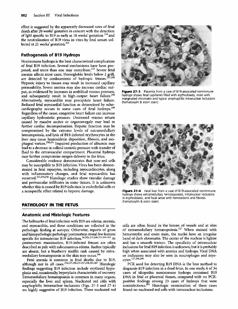
882 Section I11 Viral Infections
effect is suggested by the apparently decreased rates of fetal death after 20 weeks' gestation in concert with the detection of IgM specific to B19 as early as 18 weeks' gestation 254 and the neutralization of B19 virus in vitro by fetal serum col- lected at 21 weeks' ge~tation.2~~
Pathogenesis of B19 Hydrops Nonimmune hydrops is the best-characterized complication of fetal B19 infection. Several mechanisms have been pro- posed, and more than one may contribute.232 Severe fetal anemia affects most cases. Hemoglobin levels below 2 g/dL are detected by cordocentesis of hydropic Hypoxic injury to tissues may result in increased capillary permeability. Severe anemia may also increase cardiac out- put, as evidenced by increases in umbilical venous pressure, and subsequently result in high-output heart failure.259 Alternatively, myocarditis may precipitate heart failure. Reduced fetal myocardial function as determined by echo- cardiography occurs in some cases of fetal h y d r o p ~ . ~ ~ ~ Regardless of the cause, congestive heart failure can increase capillary hydrostatic pressure. Decreased venous return caused by massive ascites or organomegaly may lead to further cardiac decompensation. Hepatic function may be compromised by the extreme levels of extramedullary hematopoiesis, and lysis of B19-infected erythrocytes in the liver may cause hemosiderin deposition, fibrosis, and eso- phageal v a r i ~ e s . ~ ~ ~ ~ ~ ~ ' Impaired production of albumin may lead to a decrease in colloid osmotic pressure with transfer of fluid to the extravascular compartment. Placental hydrops may further compromise oxygen delivery to the fetus.
Considerable evidence demonstrates that non-red cells may be susceptible to B19 infection. Virus has been demon- strated in fetal myocytes, including myocardiocytes, along with inflammatory changes, and fetal myocarditis has occurred.'27*234,256 Histologic studies show vascular damage and perivascular infiltrates in some tissues. It is unknown whether this is caused by B19 infection in endothelial cells or a nonspecific effect related to hypoxic damage.
PATHOLOGY IN THE FETUS
Anatomic and Histologic Features The hallmarks of fetal infection with B19 are edema, anemia, and myocarditis, and these conditions are reflected in the pathologic finding at autopsy. Otherwise, reports of gross and histopathologic pathology postmortem reveal few features specific for intrauterine B19 infection.163232* 235*246*251,260-265 At postmortem examination, B 1 !+infected fetuses are often described as pale with subcutaneous edema. Rashes typically are absent, but a blueberry muffin rash caused by extra- medullary hematopoiesis in the skin may occur.266
Fetal anemia is common in fetal deaths due to B19, although not in all cases.234,237,250.251.257,258,263,267 Histologic findings suggesting B19 infection include erythroid hypo- plasia and, occasionally, hyperplasia characteristic of recovery. Extramedullary hematopoiesis is common in many organs, especially the liver and spleen. Nucleated red cells with amphophilic intranuclear inclusions (Figs. 27-3 and 27-4) are highly suggestive of B19 infection. These nucleated red
Figure 27-3 Placenta from a case of B19-associated nonimmune hydrops shows fetal capillaries filled with erythroblasts, most with marginated chromatin and typical amphophilic intranuclear inclusions (hematoxylin & eosin stain).
Figure 2 7 4 Fetal liver from a case of B19-associated nonimmune hydrops shows extramedullary hematopoiesis, intranuclear inclusions in erythroblasts, and focal areas with hemosiderin and fibrosis (hematoxylin & eosin stain).
cells are often found in the lumen of vessels and at sites of extramedullary hematopoie~is.'~~ When stained with hematoxylin and eosin stain, the nuclei have an irregular band of dark chromatin. The center of the nucleus is lighter and has a smooth texture. The specificity of intranuclear inclusions for fetal B19 infection is unknown, but it is probably high when associated with anemia and hydrops. Viral DNA or inclusions may also be seen in macrophages and myo-
PCR used for detecting B19 DNA is the best method to diagnosis B19 infection in a dead fetus. In one study, 6 of 34 cases of idiopathic nonimmune hydrops contained B 19 DNA in fetal or placental tissues, compared with no PCR- positive findings among 23 cases of hydrops that were n o n i n f e c t i o ~ s . ~ ~ ~ Histologic examination of these cases found no nucleated red cells with intranuclear inclusions.
cytes. 127,256,268

Chapter 27 Human Parvovirus Infections 883
Placenta B19 infection of the placenta probably precedes fetal infection. The placenta is usually abnormal when associated with fetal death due to B19. Grossly the placenta is often enlarged and edematous. Histologically, the placenta also contains nucleated red blood cells with typical intranuclear inclusions (see Fig. 27-4). Foci of red cell production also occur in the placenta, as does vascular i n f l a m m a t i ~ n ? ~ ~ ' ~ ~ ~ ~ ~ ~ In one vasculitis of villous capillaries or stem arteries occurred in 9 of 10 placentas. The tissues demonstrated swelling of endothelial cells, fragmentation of endothelial cell nuclei, and fibrin thrombi. B19 DNA occurs in endo- thelial cells of patients with myocarditis and in patients with cutaneous lesions but has not been sought in placental endo- thelial cells. The human placenta contains a B19 receptor, the neutral glycosphingolipid (globoside), on the villous trophoblast layer of the placenta, and the concentration of the globoside decreases with advancing pregnancy.270 The highest concentration occurs in the first trimester, with diminished reactivity occurring in the second trimester. The presence of this globoside in the placenta provides a mechanism by which the virus infects the placenta and fetus. It also may explain why there is a difference in fetal outcome associated with gestational age. Maternal infections in late pregnancy have a better prognosis than those occurring early in pregnancy. In addition to B19 receptors, there is a B19- induced inflammatory response in the placenta, characterized by a significant number of CD3' T cells and the inflammatory cytokine in te r le~kin-2 .~~~
Heart The anemia associated with B19 infection is caused by a specific viral tropism for progenitor erythroid cells, specifically P antigen, which is found on these However, clinical and laboratory evidence suggests that B19 has a wider tropism than for e ry thr~blas t s .~~~ Fetal myocardial cells contain P antigen.273 Direct infection of myocardial cells after fetal B19 infection of extramedullary erythroid progenitor cells has been demonstrated by in situ DNA hybridization or electron m i ~ r o s ~ o p y . ~ ~ ~ ~ ~ ~ ' ~ ~ ~ ~ ~ ~ , ~ ~ ~ B 19 myocarditis is also associated with acute lymphocytic infiltration. Case reports have described at least eight fetuses, five children, and four adults with myocarditis associated with a concurrent B 19
B19 causes acute and chronic myocarditis in infants. Myocarditis and the cardiac enlargement found in some B19-infected fetuses with hydrops suggest that B19 is pathogenic for the myocar~~um~127~232-235~246~254~256~276~279 In infected fetuses, the heart may be normal or symmetrically enlarged, suggesting congestive heart failure. Pericardial effusions are common. Myocytes with intranuclear inclusions occur infrequently. Mononuclear cell infiltrates occur occasionally, and B19 DNA, not associated with cells, can be found in the lumen of large vessels. Focal areas with dystrophic calcification or fibroelastosis have occurred as a response to injury.
One case-control study8' examined the relationship between congenital heart disease and B19 infection. Five of 29 cases of congenital heart disease had parvovirus B19 DNA detected in cardiac tissue using PCR, compared with none
infection. 274,276-278
of 30 matched controls. This difference was significant (P < .02). Other infections, including herpes simplex virus, cytomegalovirus, rubella, and toxoplasmosis, were excluded. Additional studies testing for B19 infection of congenital heart disease are appropriate.
Other Organs Numerous other anatomic abnormalities have been associated with B19 infection of the fetus. Their occurrences, however, are so infrequent that it is unlikely that they are related to B19 infection. These associated abnormalities include dystrophic calcification of the brain and adrenal glands; anencephaly and ventriculomegaly; pulmonary hypoplasia; hypospadias; cleft lip; meconium peritonitis; corneal opacification and an ioedema; and thymic abnor- malities.16,18,127,23~-233, 235-239,264$81-283
DIAGNOSTIC EVALUATION AND MANAGEMENT OF THE WOMAN AND FETUS EXPOSED TO OR INFECTED BY B19 DURING PREGNANCY
Management of a pregnant woman exposed to B 19 requires knowledge of the prevailing status of EI in the community, a detailed history of the exposure, knowledge of characteristic symptoms and signs of maternal EI and B19 infection in the fetus, appropriate laboratory tests needed to confirm maternal and fetal infection, knowledge of the methods for monitoring the fetus at risk for nonimmune hydrops, knowledge of therapeutic approaches for treating the hydropic fetus, and information about the prognosis of maternal and fetal infection and the expected outcomes for the therapeutic intervention.
Prevalence of Erythema lnfectiosum The community health or school health departments may know whether EI is epidemic in the community, increasing the probability of primary infection in susceptible pregnant women.
History of Exposure Pregnant women who are potentially exposed to someone with EI should be asked about the type of exposure, including duration (brief or prolonged) and location (household or workplace, indoor or outdoor), and contact with respiratory secretions. Exposure to a child within the household constitutes the highest risk.
Did the contact have symptoms typical of EI, including a low-grade fever and a slapped-cheek rash that soon spread to the trunk or limbs in a lacy pattern? Did the rash disappear and then reappear when the child was warm from exercise or bathing? Had the child been exposed to any known source of EI, such as an outbreak in school, pre- school, a daycare center, a family gathering, a play group, or church nursery? Was the child evaluated by a physician familiar with viral exanthems?

884 Section I11 Viral Infections
Clinical Features Suggesting B19 Infection in the Pregnant Woman
The examiner should consider whether the mother's signs and symptoms are compatible with B19 infection in adults, including at least one or more of the following: malaise, arthralgia, rash, coryza, or fever higher than 38" C. Pregnant women with such symptoms, especially malaise with symmetrical arthralgias in the hands, wrists, knees, or feet, should be considered at high risk and tested for recent B19 infection. In Barcelona, however, Gratecos and colleagues" found that only 30% of 60 IgM-positive women recalled any such symptoms.
Pregnant women without such systemic symptoms but with a rapidly enlarging uterus (i.e., fundal height exceeding dates by more than 3 cm), an elevated serum a-fetoprotein level, preterm labor, or decreased fetal movement should be asked about B 19 exposure. If ultrasonography reveals evidence of hydrops fetalis or the fetus has ascites, pleural or pericardial effusion, skin thickening, polyhydramnios, or placentomegaly, maternal B19 testing is appropriate.
Laboratory Diagnosis in the Pregnant Woman With evidence of maternal B19 exposure or maternal disease, maternal serum should be tested for IgG and IgM antibodies to B19. If there is probable or possible exposure, the first serum sample should be drawn at least 10 days after the exposure. Because fetal morbidity is unlikely to occur within 2 weeks of exposure, immediate serologic testing is appro- priate for a woman or fetus with symptoms or signs of B19 infection.
An initial serum sample that is IgG positive but IgM negative indicates a previous maternal infection, and additional testing is unnecessary. The IgM assay is sensitive, with few false-negative reactions. An initial serum sample that is negative for IgM and IgG indicates no previous maternal infection, and B19 infection is not responsible for maternal symptoms and signs or for hydrops fetalis.
If the IgM result is positive, a recent B19 infection is established regardless of the IgG titer. A concomitant negative IgG titer means an early B19 infection without time for IgG to be detectable. Detection of maternal viremia by PCR for B19 DNA is also diagnostic of B19 infection. Viremia may precede the development of IgM antibodies by 7 to 14 days and may persist for several months after a primary infection.
With a positive maternal IgM result, the fetus must be examined for signs of hydrops fetalis by ultrasonography within 24 to 48 hours. If the gestational age is less than 18 weeks, the absence of hydrops may not be reassuring, because hydrops can appear later. Because several cases of severe hydrops fetalis spontaneously reverting to normal over 3 to 6 weeks have been reported, advice about pregnancy termination is d i f f i c ~ l t . ~ ~ ~ , ~ ~ ' , ~ ' ~
Fetal Monitoring For a fetal gestational age of more than 20 weeks, initial negative ultrasound results demand sonograms to be repeated weekly to detect hydrops. The number of weekly sonograms that should be performed is controversial. Rodis and associates233 originally suggested continuing weekly scans for
Table 27-2 Fetal Deaths from B19 Infection
Infection to Death Interval (weeks)
Gestational Age at Death (weeks)
1 10 13 4 4 4 9 7 8 1 3 6
10-19 5
(1 0)" (4) (1 1) (7) (4) (3) (8)
Fetal Weight at Death (grams) Reference
3810 15 NR 298
409 16 161 16 420 299 695 300 580 300 300 30 1 236 286
NR 302 NR 302 NR 302 NR 303 NR 303
289 Hydrops, 3320 290 Hydrops, 31 11 291 Hydrops fetalis 158 Hydrops, 1495 158 Hydrops, 3550 303 Hydrops fetalis 140
aNumbers in parentheses refer to intervals between exposure or onset of symptoms and the diagnosis of hydrops fetalis. bNumbers in parentheses refer to gestational age at the time of diagnosis of hydrops fetalis.
NR, not reported.
6 to 8 weeks after exposure, and they reported a fetal death as late as 23 weeks' gestation after maternal fever and arthralgias in the first trimester.12' The interval between maternal B19 infection and fetal morbidity is uncertain. Based on this report, others recommended weekly sonograms for 14 weeks after maternal B19 infection.284 This approach often appeals to pregnant women fearful about fetal death, but it is time consuming and expensive.
The duration of monitoring for hydrops fetalis may be best determined by examination of the interval between maternal exposure or symptoms of B19 infection and the appearance of hydrops fetalis or fetal death. Table 27-2 summarizes reports with adequate information to evaluate the interval, which include 14 intervals between maternal B19 exposure or infection and fetal death and 7 intervals between maternal exposure or infection and the first diagnosis of hydrops fetalis. The intervals range from 1 to 19 weeks, with a median of 6 weeks. Seventeen (81%) of 21 cases developed between 3 and 11 weeks. Because 11 of the 21 cases developed between 4 and 8 weeks after maternal exposure or infection, this is the most common interval between infection and the detection of fetal hydrops. Based on these observations, weekly ultrasound monitoring of the fetus for 12 weeks after maternal exposure is optimal but cannot detect all delayed cases and may be expensive. Such frequent scanning may not be considered cost-effective because the incidence of hydrops after maternal B19 infection is low in many studies. In our study, none of the 52 fetuses born to B19-IgM-positive pregnant women developed hydrops fetalis; however, the 95% confidence interval based on our sample size ranged from 0% to 8.6% for the risk of hydrops fetalis.2'0 Other studies using maternal symptoms as

Chapter 27 Human Parvovirus Infections 885
criteria for maternal B19 infection have suggested a 9% incidence of fetal death due to B19 in B19-IgM-positive women.I8
Serial maternal serum a-fetoprotein (MSAFP) measure- ments may monitor the fetus in B19-infected women.285 One report found elevated MSAFP levels in five B19-IgM-positive pregnancies associated with fetal death, but no fetal deaths in 11 IgM-positive women with B19 infection but normal MSAFP values.286 A fatal case of B19-associated fetal death, discovered because of an elevated MSAFP level at 16 weeks in a routine test in an asymptomatic woman, has been described.16 In adding a seventh case of fetal death associated with elevated MSAFP levels in B19-IgM-positive women, Bernstein and C a p e l e ~ s ~ ~ ~ suggested using the MSAFP values to indicate a good fetal prognosis.
A German study8’ found that neither MSAFP nor human gonadotropin levels were markers of B19-infected pregnancies, although both were frequently elevated when complications occurred. The study included 35 pregnant women with fetal complications associated with B19; significant elevations of MSAFP levels occurred in 13 of 35 women, and elevations of human gonadotropin concentrations occurred in 25 of 35. The investigators tested 137 sera from 65 pregnant women without acute parvovirus infection and no fetal complications. Of the 30 women without fetal complications, there were significant elevations of MSAFP levels in only 2 women, and elevations of human gonadotropin levels occurred in only 5 women. Neither protein was a marker for a poor pregnancy outcome early on, but levels were frequently elevated when complications developed. Despite these results, there is insufficient experience using MSAFP concentrations, and MSAFP measurements at any gestational age are relatively nonspecific indicators of fetal well-being.
Electronic fetal monitoring is ineffective in detecting hydrops fetalis and predicting the outcome of pregnancy in B19-IgM-positive women. Contraction stress tests and “non- stress” tests are not accurate predictors of fetal well-being in cases of fetal anemia or hydrops fetalis. Similarly, fetal assess- ment with estriol measurements or other biochemical markers have no documented role in cases of hydrops fetalis. Because fetal sonograms are as readily available and provide rapid specific information about hydrops fetalis, ultrasound is the best method to monitor the fetus after maternal B19 infection.
Fetal Therapy If hydrops fetalis is detected before 18 weeks, there is no effective intervention. Other causes of hydrops, such as chromosomal disorders or anatomic abnormalities, should be assessed. If at 18 weeks‘ gestation the fetus is still viable as determined by ultrasound examination, consideration can be given to percutaneous umbilical blood sampling (PUBS), also called cordocentesis. At 18 weeks‘ gestation, the umbilical vein diameter is about 4 mm, which is the minimum size required for successful PUBS. Fetal blood should be obtained for the hematocrit, reticulocyte count, platelet count, leukocyte count, antiparvovirus B19 IgM, karyotype, and tests for B19 DNA by PCR. The hematocrit must be determined immediately, and if fetal anemia exists, intrauterine intra- vascular fetal transfusion is performed with the same needle puncture.
If the fetus is between 18 and 32 weeks’ gestation when hydrops fetalis is detected, fetal transfusion should be considered. Many successful cases of fetal transfusion for B19-induced hydrops fetalis have been reported, and some have long-term follow-up, but the success rate of the procedure remains unknown.288-294
Two or three separate transfusions are usually required before resolution of the fetal anemia and hydrops fetalis, increasing the 1% to 2% risk of each single PUBS procedure. Resolution of the hydrops usually occurs 3 to 6 weeks after the first transfusion. Although spontaneous resolution has been reported, it seems appropriate not to risk an un- certain outcome, because the longer the fetal transfusion is delayed, the less likely it is to be successful and the worse the potential harm to the fetus caused by continued fetal
For fetuses of 32 weeks’ gestation or older when hydrops is discovered, immediate delivery with neonatal exchange transfusion, thoracentesis, and paracentesis as indicated usually is the safest management.
hypoxia.231, 249,284,289-291
DIFFERENTIAL DIAGNOSIS
Recalling that the hallmarks of fetal infection with B19 are anemia, hydrops, and myocarditis helps in compiling a dif- ferential diagnosis. For infants with anemia, the differential diagnosis includes all the known causes, including fetal- maternal transfusion, intracranial bleeding, blood group incompatibilities, congenital anemias such as Diamond- Blackfan syndrome, nutritional deficiencies, and inborn metabolic errors. Fetal hydrops and fetal and placental edema may be associated with other congenital infections, particularly congenital syphilis, chromosomal abnormalities, immune hydrops associated with blood group incompatibilities, hypothyroidism, and heart or renal failure, or both.
PROGNOSIS
Pregnant women can be reassured about the relatively low risk of fetal morbidity resulting from exposure to B19. About one half of women already are seropositive. The seronegative maternal B19 infection rate ranges from about 29% for exposures by the woman’s own children to about 10% to 18% for other exposures. The expected fetal morbidity and mortality risk is about 2% (1 of 50). The overall risk of fetal death varies from 0.3% ( Y2 x Yl0 x 1/50 = 3/Iooo) to a mere 0.1 %
Live-born infants infected in utero may die shortly after birth. Two infants born prematurely at 24 and 35 weeks’ gestation developed an illness characteristic of congenital viral infection, including placentomegaly, petechial rash, edema, hepatomegaly, anemia, thrombocytopenia, and respiratory insufficiency, and both died p~stnatally.’~~ Both infants had nuclear inclusions in erythroid precursor cells, and PCR confirmed the presence of parvoviral DNA in one of the infants.
Data regarding the long-term outcomes of live-born children infected in utero or born of mothers infected during pregnancy are very limited. In one study, 113 pregnant women with B19 infection during pregnancy and a control
(1/2 x 1/10 x 1/50 = 1 / I ~ ~ ) . 2 1 0

886 Section 111 Viral Infections
group of immune women were questioned about the health and development of their children when the median age of the children was 4 years for both The incidence of developmental delays in speech, language, information pro- cessing, and attention was similar between the study group and the controls (7.3% versus 7.5%). Two cases of cerebral palsy were found in the study group, compared with none in the controls. Although not statistically significant, this 2% incidence of cerebral palsy in the infected group is 10-fold higher than the reported national incidence.242
In a British study of 427 pregnant women with B19 infection and 367 of their surviving infants, 129 surviving infants were reassessed when 7 to 10 years old.296 The follow- up included questionnaires to obstetricians and general practitioners about the outcome of pregnancy and health of surviving infants. Maternal infection was confirmed by B19-specific IgM assay or IgG seroconversion. An excess rate of fetal loss was confined to the first 20 weeks’ gestation and averaged 9%. There were seven cases of fetal hydrops with maternal infections between 9 and 20 weeks’ gestation. There were no abnormalities attributable to B19 infection found at birth in surviving infants. No late effects were observed when the children were 7 to 10 years old. This study concluded that approximately 1 in 10 women infected before 20 weeks‘ gestation would have a fetal loss due to B19, that the risk of an adverse outcome of pregnancy beyond this stage was un- likely, and that infected women could be reassured that the risk of congenital abnormality due to B19 is less than 1% and that long-term development would be normal.
One study used IQ testing and standard neurodevelop- mental tests to assess 20 children who had parvovirus- induced fetal hydrops and intrauterine transfusion of packed red cells?” Testing of the 20 children when they were between 13 months and 9 years old revealed that all of their results ranged within two standard deviations of a population norm. There was no significant developmental delay. This study concluded that children who survived successful intrauterine transfusion from B19 anemia and hydrops had a good neuro- developmental prognosis.
PREVENTION
General Measures Because B19 is usually endemic in most communities, what is appropriate management for pregnant women with daily contact with children? The prevalence of seropositivity (immunity) to B 19 among pregnant women varies according to geographic location, sex, age, and race. Assuming that on average 50% of pregnant women are immune; that during endemic periods, between 1% and 4% of susceptible women become infected during pregnancy; and that the rate of fetal death after maternal infection is 2%, the occupational risk of fetal death for a pregnant woman with unknown serologic status is between 1 in 1000 and 1 in 2500. These low rates do not justify intervention such as serologic testing for pregnant women, furloughing pregnant workers, or temporarily trans- ferring pregnant seronegative employees to administrative or other positions without child contact. During epidemic periods in specific schools, when the infection rates may be 5- to 20-fold higher, serologic testing or temporary transfer
of pregnant employees may occasionally be appropriate, and some very anxious women may choose to leave the workplace.
Given the low risk for individual pregnant women, sero- negative women should not send their own children away, and schools and daycare centers cannot stop B19 outbreaks by excluding children with rash illnesses because B19 is transmissible before the rash appears. Whether B19 can be transmitted by breast-feeding is unknown.
Vaccine Development For most women, fetal B19 infections during pregnancy occur from exposure to school-aged children at home rather than from occupational exposure. Given the highly com- municable and endemic nature of the infection, the broad spectrum of illness that B19 causes, and the large portion of population (30% to 50%) who are susceptible, an effective B19 vaccine, preferably administered in infancy, is appro- priate, and at least one vaccine is being de~eloped.’~’ This vaccine is composed of the major B19 capsid proteins VP1 and VP2 and administered with a squalene adjuvant, MF59. After testing in a limited number of subjects, the vaccine appears to be safe and induces neutralizing antibodies. Studies using volunteers challenged with wild-type B19 should be able to assess efficacy. A vaccine that induces sustained neutralizing antibody IgG levels to B19 should be effective given that prior immunity to B19 protects against reinfection.
Acknowledgments
We are grateful to Dr. James H. Harger for his years of collaboration and helpful assistance.
REFERENCES 1.
2.
3.
4.
5.
6.
7.
8.
9.
10.
11.
12.
Murphy FA, Fauquet CM, Bishop DHL, et al (eds). Virus Taxonomy. Sixth Report of the International Committee on Taxonomy ofviruses. New York, Springer-Verlag, 1995. Torok TJ. Unusual clinical manifestations reported in patients with parvovirus B19 infection. In Anderson LJ, Young NS (eds). Human Parvovirus B19. Monographs in Virology, vol. 20. Basel, Karger, 1997,
Koch WC. Fifth (human parvovirus B19) and sixth (herpesvirus 6) diseases. Curr Opin Infect Dis 14343,2001. Cossart YE, Field AM, Cant B, Widdows D. Parvovirus-like particles in human sera. Lancet 1:72, 1975. Summers J, Jones SE, Anderson MJ. Characterization of the genome of the agent of erythrocyte aplasia permits its classification as a human parvovirus. J Gen Virol64:2527, 1983. Pattison JR, Jones SE, Hodgson J. Parvovirus infections and hypo- plastic crisis in sickle-ceu anaemia. Letter. Lancet 1:664, 1981. Sejeant GR, Topley JM, Mason K, et al. Outbreak of aplastic crises in sickle cell anaemia associated with parvovirus-lke agent. Lancet 2595, 1981. Anderson MI, Jones SE, Fisher-Hoch SP, et al. Human parvovirus, the cause of erythema infectiosum (fifth disease)? Letter. Lancet 1:1378, 1983. Thurn J. Human parvovirus B19: historical and clinical review. Rev Infect Dis 10:1005, 1988. Ager EA, Chin TDY, Poland JD. Epidemic erythema infectiosum. N Engl J Med 2751326,1966. Cramp JE, Armstrong BDJ. Erythema infectiosum: no evidence of teratogenicity. Br Med J 2:1031, 1977. Pattison IR. B19 Virus infections in pregnancy. In Pattison JR (ed). Parvoviruses and Human Disease. Boca Raton, Fla, CRC Press, 1988, pp 133-138.
pp 61-92.
Chapter 27 Human Parvovirus Infections 887
13. Siege1 G. Patterns of parvovirus disease in animals. In Pattison JR (ed). Parvoviruses and Human Disease. Boca Raton, Fla, CRC Press, 1988, pp 43-68.
14. Brown T, h a n d A, Ritchie LD, et al. Intrauterine parvovirus infection associated with hydrops fetalis. Lancet 2:1033, 1984.
15. Knott PD, Welply GAC, Anderson MJ. Serologically proved intra- uterine infection with parvovirus. Letter. BMJ 289:1960, 1984.
16. Anand A, Gray ES, Brown T, et al. Human parvovirus infection in pregnancy and hydrops fetalis. N Engl J Med 316183,1987.
17. Kinney JS, Anderson LJ, Farrar J, et al. Risk of adverse outcomes of pregnancy after human parvovirus B19 infection. J Infect Dis 157:663, 1988.
18. Hall SM, Public Health Laboratory Service Working Party on Fifth Disease. Prospective study of human parvovirus (B19) infection in pregnancy. Br Med J 3001166,1990.
19. Adler SP, Manganello AM, Koch WC, et al. Risk of human parvovirus B19 infections among school and hospital employees during endemic periods. J Infect Dis 168361,1993.
20. Gratacos E, Torres P-J, Vidal J, et al. The incidence of human parvovirus B19 infection during pregnancy and its impact on perinatal outcome. J Infect Dis 171:1360, 1995.
21. Koch WC, Harger JH, Barnstein B, Adler SP. Serologic and virologic evidence for frequent intrauterine transmission of human parvovirus B10 with a primary maternal infection during pregnancy. Pediatr Infect Dis 17:489, 1998.
22. Pringle CR.Virus taxonomy update. Arch Virol 133:491, 1993. 23. Astell CR, Weixing L, Brunstein J, St Arnand J. B19 parvovirus: bio-
chemical and molecular features. In Anderson LJ, Young NS (eds). Monographs in Virology, vol. 20. Human Parvovirus B19. Basel, Karger,
24. O'Sullivan MG, Anderson DC, Fikes JD, et al. Identification of a novel simian parvovirus from cynomolgus monkey with severe anemia: a paradigm for human B19 parvovirus infection. J Clin Invest 93:1571, 1994.
25. Mori J, Beattie P, Melton DW, et al. Structure and mapping of the DNA of human parvovirus B19. J Gen Virol68:2797,1987.
26. Umene K, Nunoue T. Genetic diversity of human parvovirus B19 determined using a set of restriction endonucleases recognizing four or five base pairs and partial nudeotide sequencing: use of sequence variability in virus classification. J Gen Virol72:1997,1991.
27. Gallinella G,Venturoli S, Manaresi E, et al. B19 virus genome diversity: epidemiological and clinical correlations. J Clin Virol769 124,2003.
28. Nguyen QT, Sifer C, Schneider V, et al. Novel human erythrovirus associated with transient aplastic crisis. J Clin Microbiol372483, 1999.
29. Nguyen QT, Wong S, Heegaard ED, Brown KE. Identification and characterization of a second human erythrovirus variant, A6. Virology 30:374,2002.
30. Servant A, Laperche S, Lallemand F, et al. Genetic diversity within human erythroviruses: identification of three genotypes. J Virol 7 6 9124,2002.
34. Ozawa K, Ayub J, Kajigaya S, et al. The gene encoding the nonstructural protein of B19 (human) parvovirus may be lethal in transfected cells. J Virol62:2884, 1988.
35. Moffat S, Yaegashi N, Tada K, et al. Human parvovirus B19 nonstructural protein NSI induces apoptosis in erythroid lineage cells. JVirol72:3018,1998.
36. Sol N, Le Junter J, Vassias I, et al. Possible interactions between the NS- 1 protein and tumor necrosis factor alpha pathways in erythroid cell apoptosis induced by parvovirus B19. J Virol73:8762,1999.
37. Ozawa K, Kurtzman G,Young N. Replication of the B19 parvovirus in human bone marrow cell cultures. Science 233:883,1986.
38. Srivastava A, Lu L. Replication of B19 parvovirus in highly enriched hematopoietic progenitor cells form normal human bone marrow. J Virol62:3059, 1988.
39. Yaegashi N, Shiraishi H, Takeshita T, et al. Propagation of human parvovirus B19 in primary culture of erythroid lineage cells derived from fetal liver. J Virol63:2422, 1989.
40. Takahahsi T, Ozawa K, Mitani K, et al. B19 parvovirus replicates in erythroid leukemic cells in vitro. J Infect Dis 160548,1989.
41. Miyagawa E, Yoshida T, Yamaguchi K, et al. Infection of the erythroid cell line KU812Ep6 with human parvovirus B19 and its application to titration of B19 infectivity. J Virol Methods 83:45,1999.
42. Brown KE, Young NS, Liu JM. Molecular, cellular and clinical aspects of parvovirus B19 infection. Crit Rev Oncol Hematol 161,1994.
43. Brown KE, Anderson SM, Young NS. Erythrocyte P antigen: cellular receptor for B19 parvovirus. Science 262:114, 1993.
1997, pp 16-41.
44. Brown KE, Hibbs JR, Gallinella G, et al. Resistance to parvovirus B19 infection due to a lack of virus receptor (erythrocyte P antigen). N Engl J Med 3301192,1994.
45. Weigel-Kelley KA, Yoder MC, Srivastava A. Recombinant human parvovirus B19 vectors: erythrocyte P antigen is necessary but not sufficient for successful transduction of human hematopoietic cells. J Virol 75:4110,2001.
46. Weigel-Kelley KA, Yoder MC, Srivastava A. Alpha5betal integrin as a cellular coreceptor for human parvovirus B19: requirement of functional activation of beta 1 integrin for viral entry. Blood 1023927, 2003.
47. Brown KE, Young NS. Parvovirus B19 infection and hematopoiesis. Blood Rev 9176, 1995.
49. Srivastava A. Bruno E, Briddell R, et al. Parvovirus B19-induced perturbation of human megakaryocytopoiesis in vitro. Blood 761997, 1990.
48. Young N. Hematologic and hematopoietic consequences of 819 infection. Semin Hematol25:159, 1988.
50. Anderson MJ, Higgins PG, Davis LR, et al. Experimental parvovirus infection in humans. J Infect Dis 152:257, 1985.
51. Moffat S, Tanaka N, Tada K, et al. A cytotoxic nonstructural protein, NSI, of human parvovirus B19 induces activation of interleukin-6 gene expression. J Virol708485, 1996.
52. Teuscher T, Baillod B, Holzer BR. Prevalence of human parvovirus B19 in sickle cell disease and healthy controls. Trop Geogr Med 43:108, 1991.
53. Schwarz TF, Giirtler LG, Zoulek G, et al. Seroprevalence of human parvovirus B19 infection in SSo Tom6 and Principe, Malawi and Mascarene Islands. Zentralbl Bakteriol271:231, 1989.
54. Jones PH, Pickett LC, Anderson MJ, Pasvol G. Human parvovirus infection in children and severe anaemia seen in an area endemic for malaria. J Trop Med Hyg 93:67, 1990.
55. de Freitas RB, Wong D, Boswell F, et al. Prevalence of human parvo- virus (B19) and rubella virus infections in urban and remote rural areas in northern Brazil. J Med Virol32:203, 1990.
56. Brown CS, Jensen T, Meloen RH, et al. Localization of an immuno- dominant domain on baculovirus-produced parvovirus B19 capsids: correlation to a major surface region on the native virus particle. J Virol66:69, 1992.
57. Morey AL, ONeill HJ, Coyle PV, Fleming KA. Immunohistologica\ detection of human parvovirus B19 in formalin-fixed, paraffin- embedded tissues. J Pathol 166105,1992.
58. Loughrey AC, O'Neill HJ, Coyle PV, DeLeys R. Identification and use of a neutralizing epitope of parvovirus B19 for the rapid detection of virus infection. J Med Virol39:97, 1993.
59. Morinet F, Tratschin JD, Perol Y, Siegl G. Comparison of 17 isolates of the human parvovirus B19 by restriction enzyme analysis. Arch Virol 90165,1986.
60. Umene K, Nunoue T. The genome type of human parvovirus B19 strains isolated in Japan during 1981 differs from types detected in 1986 to 1987: a correlation between genome type and prevalence. J Gen Viiol71:983, 1990.
61. Umene K, Nunoue T. Partial nucleotide sequencing and characterization of human parvovirus B19 genome D N A from damaged human fetuses and from patients with leukemia. J Med Virol39333,1993.
62. Lawton AL, Smith RE. Erythema infectiosum: a clinical study of an epidemic in Branford, Connecticut. Arch Intern Med 4228, 193 1.
63. Chargin L, Sobel N, Goldstein H. Erythema infectiosum: report of an extensive epidemic. Arch Dermatol Syphilol47:467, 1943.
64. Galvon FAC. An outbreak of erythema infectiosum-Nova Scotia. Can Dis Wkly Rep 969,1983.
65. Serjeant GR, Sejeant BE, Thomas PW, et al. Human parvovirus infection in homozygous sickle cell disease. Lancet 341:1237,1993.
66. Oliveira SA, Carnacho LA, Pereira AC, et al. Clinical and epidemio- logical aspects of human parvovirus B19 infection in an urban area in Brazil (Niteroi city area, State of Rio de Janeiro, Brazil). Mem Inst Oswaldo Cruz 92965,2002.
67. Ymashita K, MatsunagaY, Taylor-Wiedeman J, Yamazaki S. A significant age shift of the human parvovirus B19 antibody prevalence among young adults in Japan observed in a decade. Jpn J Med Sci Biol45:49, 1992.
68. Naides SJ. Erythema infectiosum (fifth disease) occurrence in Iowa. Am J Public Health 78:1230, 1988.
69. Cohen BJ, Buckley MM. The prevalence of antibody to human parvovirus B19 in England and Wales. J Med Microbiol 25:151, 1988.
888 Section I11 Viral Infections
70. Nascimento JP, Buckley MM, Brown KE, Cohen BJ. The prevalence of antibody to human parvovirus B19 in Rio de Janeiro, Brazil. Rev Inst Med Trop Sao Paul0 3241,1990.
71. Edelson RN, Altman RA. Erythema infectiosum: a statewide outbreak. J Med Soc N J 67S05.1970.
72. Werner GH, Brachman PS, Ketler A, et al. A new viral agent associated with erythema infectiosum. Ann N Y Acad Sci 67:338,1957.
73. Greenwald P, Bashe WJ Jr. An epidemic of erythema infectiosum. Am J Dis Child 107:30,1964.
74. Yaegashi N, Okamura K, Hamazaki Y, et al. Prevalence of anti-human parvovirus antibody in pregnant women. Nippon Sanka Fujinka Gakkai Zasshi 42:162, 1990.
75. Koch WC, Adler SP. Human parvovirus B19 infections in women of childbearing age and within families. Pediatr Infect Dis J 8:83, 1989.
76. Cohen BJ, Mortimer PP, Pereira MS. Diagnostic assays with mono- clonal antibodies for the human serum parvovirus-like virus (SPLV). J Hyg (Lond) 91:113,1983.
77. Schwarz TF, Roggendorf M, Deinhardt F. Haufigkeit der parovirus- B19-infektionen. Seroepidemiologishe untersuchungen. Dtsch Med Wochenschr 112:1526,1987.
78. Bartolomei Corsi 0, Assi A, Morfini M, et al. Human parvovirus infection in haemophiliacs first infused with treated clotting factor concentrates. J Med Virol 25165, 1988.
79. Eiffert H, Kochel HG, Heuer M, et al. Expression of an antigenic polypeptide of the human parvovirus B19. Med Microbiol Immunol 179169, 1990.
80. Brown CS, van Bussel MJA, Wassenaar ALM, et al. An immuno- fluorescence assay for the detection of parvovirus B19 IgG and IgM antibodies based on recombinant viral antigen. J Virol Methods 29:53, 1990.
81. Rollag H, Patou G, Pattison JR, et al. Prevalence of antibodies against parvovirus B19 in Norwegians with congenital coagulation factor defects treated with plasma products from small donor pools. Scand J Infect Dis 23:675, 1991.
82. Salimans MMM, van Bussel MIA, Brown CS, Spaan WJM. Recombinant parvovirus B19 capsids as a new substrate for detection of B19-specific IgG and IgM antibodies by an enzyme-linked immunosorbent assay. J Virol Methods 39247,1992.
83. Abraham M, Rudraraju R, Kannangai R, et al. A pilot study on the seroprevalence of parvovirus B19 infection. Indian J Med Res 115:139, 2002.
84. Schwarz TF, Hottentrager B, Roggendorf M. Prevalence of antibodies to parvovirus B19 in selected groups of patients and healthy individuals. Int J Med Microbiol Virol Parasitol Infect Dis 276:437,1992.
85. Lin KH, You SL, Chen CJ, et al. Seroepidemiology of human parvo- virus B19 in Taiwan. J Med Virol57:169,1999.
86. CouroucC AM, Ferchal F, Morinet F, et al. Parvovirus (SPLV) et antigene Aurillac. Rev Fr Transfus Immunohematol27:5, 1984.
87. Cossart Y. Parvovirus B19 finds a disease. Letter. Lancet 2:988, 1981. 88. ONeill HJ, Coyle PV. Two anti-parvovirus B19 IgM capture assays
incorporating a mouse monoclonal antibody specific for B19 viral capsid proteins VPl and VP2. Arch Virol 123:125, 1992.
89. Cohen BJ, Field AM, Gudnadottir S, et al. Blood donor screening for parvovirus B19. J Viol Methods 30233,1990.
90. da Silva Cruz A, Serpa MJA, Barth OM, Nascimento JP. Detection of the human parvovirus B19 in a blood donor plasma in Rio de Janeiro. Mem Inst Oswaldo Cruz 84279,1989.
91. McOmish F, Yap PL, Jordan A, et al. Detection of parvovirus B19 in donated blood a model system for screening by polymerase chain reaction. J Clin Microbiol31:323, 1993.
92. Yaegashi N, Shiraishi H,Tada K, et al. Enzyme-linked immunosorbent assay for IgG and IgM antibodies against human parvovirus B19 use of monoclonal antibodies and viral antigen propagated in vitro. J Viol Methods 26171,1989.
93. Naides SJ, Scharosch LL, Foto F, Howard EJ. Rheumatologic mani- festations of human parvovirus B19 infection in adults. Arthritis Rheum 33:1297,1990.
94. Martinez-Campillo F, Lopez 1; Verdu M, et al. Parvovirus B19 outbreak in a rural community in Alicante. Enferm Infect Microbiol Clin 20376,2002.
95. Abarca K. Cohen BJ, Vial PA. Seroprevalence of parvovirus 819 in urban Chilean children and young adults, 1990 and 1996. Epidemiol Infect 128:59,2002.
96. Smith-Whitley K, Zhao H, Hodinka RL, et al. Epidemiology of human parvovirus B19 in children with sickle cell disease. Blood 103:422, 2004.
97. Plummer FA, Hammond GW, Forward K, et al. An erythema infectiosum-like illness caused by human parvovirus infection. N Engl J Med 313:74, 1985.
98. Chorba T, Coccia P, Holman RC, et al. The role of parvovirus B19 in aplastic crisis and erythema infectiosum (fifth disease). J Infect Dis 154383,1986.
99. Mortimer PP. Hypothesis: the aplastic crisis of hereditary spherocytosis is due to a single transmissible agent. J Clin Pathol36:445, 1983.
100. Saarinen UA, Chorba TL, Tattersall P, et al. Human parvovirus B19 induced epidemic red-cell aplasia in patients with hereditary hemolytic anemia. Blood 67:1411, 1986.
101. Valeur-Jensen A, Pedersen CB, Westergaard T, et al. Risk factors for parvovirus B19 infection in pregnancy. JAMA 281:1099, 1999.
102. Anderson MJ, Lewis E, Kidd IM, et al. An outbreak of erythema infectiosum associated with human parvovirus infection. J Hyg (Lond) 93535, 1984.
103. Tuckerman JG, Brown T, Cohen BJ. Erythema infectiosum in a village primary school: clinical and virological studies. J R Coll Gen Pract 36267,1986.
104. Morgan-Capner P, Wright J, Longley JP, Anderson MJ, Sex ratio in outbreaks of parvovirus B19 infection. Letter. Lancet 298, 1987.
105. Mansfield F. Erythema infectiosum: slapped face disease. Aust Fam Physician 17:737, 1988.
106. Woolf AD, Campion GV, Chishick A, et al. Clinical manifestations of human parvovirus B19 in adults. Arch Intern Med 1491153,1989.
107. Turner A, Olojugba 0. Erythema infectiosum in a primary school: investigation of an outbreak in Bury. Public Health 103:391,1989.
108. Grilli EA, Anderson MJ, Hoskins TW. Concurrent outbreaks of influenza and parvovirus B19 in a boys' boarding school. Epidemiol Infect 103:359, 1989.
109. Gillespie SM, Cartter ML, Asch S, et al. Occupational risk of human parvovirus B19 infection for school and day-care personnel during an outbreak of erythema infectiosum. JAMA 263:2061,1990.
1 10. Anderson LJ, Gillespie SM, Torok TJ, et al. Risk of infection following exposures to human parvovirus B19. Behring Inst Mitt 85:60,1990.
111. Rice PS, Cohen BJ. A school outbreak of parvovirus B19 infection investigated using salivary antibody assays. Epidemiol Infect 6:33 1, 1996.
112. Bell LM, Naides SJ, Stoffman P, et al. Human parvovirus B19 infection among hospital staff members after contact with infected patients. N Engl J Med 321:485, 1989.
113. Pillay D, Patou G, Hurt S, et al. Parvovirus B19 outbreak in a children's ward. Lancet 339:107, 1992.
114. Cohen BJ, Courouce AM, Schwarz TF, et al. Laboratory infection with parvovirus B19. Letter. J Clin Pathol41:1027,1988.
115. Shiraishi H, Sasaki T, Nakamura M, et al. Laboratory infection with human parvovirus B19. Letter. J Infect 22:308, 1991.
116. Evans JPM, Rossiter MA, Kumaran TO, et al. Human parvovirus aplasia: case due to cross infection in a ward. BMJ 288:681, 1984.
117. Ueda K, Akeda H, Tokugawa K, Nishima S. Human parvovirus infection. Letter. N Engl J Med 314645, 1986.
118. Pillay D, Patou G, Rees L, Griffiths PD. Secondary parvovirus B19 infection in an immunocompromised child. Pediatr Infect Dis J 10623,1991.
119. Lui SL, Luk WK, Cheung CY, et al. Nosocomial outbreak of parvovirus B19 infection in a rend transplant unit. Transplantation 71:59,2001.
120. Miyamoto K, Ogami M, Takahashi Y, et al. Outbreak of human parvovirus B19 in hospital workers. J Hosp Infect 45:238,2000.
121. Koziol DE, Kurtzman G, Ayub JA, et al. Nosocomial human parvo- virus B19 infection: lack of transmission from a chronically infected patient to hospital staff. Infect Control Hosp Epidemiol 13:343,1992.
122. Dowell SF, Torok TJ, Thorp JA, et al. Parvovirus B19 infection in hospital workers: community or hospital acquisition? J Infect Dis 172:1076, 1995.
123. Carter ML, Farley TA, Rosengren S, et al. Occupational risk factors for infection with parvovirus B19 among pregnant women. J Infect Dis 163:282, 1991.
124. Ray SM, Erdman DD, Berschling JD, et al. Nosocomial exposure to parvovirus B19: low risk of transmission to healthcare workers. Infect Control Hosp Epidemiol 18:109, 1997.
125. Patou G, Pillay D, Myint S, Pattison J. Characterization of a nested polymerase chain reaction assay for detection of parvovirus B19. J Clin Microbiol31:540, 1993.
126. Potter CG, Potter AC, Hatton CSR, et al. Variation of erythroid and myeloid precursors in the marrow of volunteer subjects infected with human parvovirus (B19). J Clin Invest 79:1486,1987.
Chapter 27 Human Parvovirus Infections 889
127. Morey AL, Porter HJ, Keeling JW, Fleming KA. Non-isotopic in situ hybridisation and immunophenotyping of infected cells in investigation of human fetal parvovirus infection. J Clin Pathol45:673,1992.
128. Mortimer PP, Luban NLC, Kelleher JF, Cohen BJ. Transmission of serum parvovirus-like virus by clotting-factor concentrates. Lancet 2:482, 1983.
129. Lyon DJ, Chapman CS, Martin C, et al. Symptomatic parvovirus B19 infection and heat-treated factor IX concentrate. Letter. Lancet 1: 1085, 1989.
130. Williams MD, Cohen BJ, Beddall AC, et al. Transmission of human parvovirus B19 by coagulation factor concentrates. Vox Sang 58:177, 1990.
131. Morfini M, Longo G, Rossi Ferrini P, et al. Hypoplastic anemia in a hemophiliac first infused with a solventldetergent treated factor VIII concentrate: the role of human B19 parvovirus. Letter. Am J Hematol 39149, 1992.
132. Zakrzewska K, Azzi A, Patou G, et al. Human parvovirus B19 in clotting factor concentrates: B19 DNA detection by the nested poly- merase chain reaction. Br J Haematol81:407,1992.
133. Schwarz TF, Roggendorf M, Hottentrager B, et al. Removal of parvo- virus B19 from contaminated factor VIII during fractionation. J Med Virol 35:28, 1991.
134. Azzi A, Ciappi S, Zakrzewska K, et al. Human parvovirus B19 infection in hemophiliacs infused with two high purity, virally attenuated factor VIII concentrates. Am J Hematol39228,1992.
135. Anderson MJ. Rash illness due to B19 virus. In Pattison JR (ed). Parvoviruses and Human Disease, B o a Raton, Fla, CRC Press, 1988,
136. Anderson LJ. Role of parvovirus B19 in human disease. Pediatr Infect Dis 6:711, 1987.
137. Garcia-Tapia AM, Fernandez-Guitierrez del Alamo C, Giron JA, et al. Spectrum of parvovirus B19 infection: analysis of an outbreak of 43 cases in Cadiz, Spain. Clin Infect Dis 21:424, 1995.
138. Zerbini M, Musiani M, Venturoli S, et al. Different syndromes associated with B19 parvovirus viraemia in paediatric patients: report of four cases. Eur J Pediatr 151:815, 1992.
139. Tsuda H, MaedaY, Nakagawa K. Parvovirus B19-related lymphadeno- pathy. Br J Haematol85631,1993.
140. Brown KE. Human parvovirus B19 epidemiology and clinical mani- festations. In Anderson LJ, Young NS (eds). Monographs in Virology, vol. 20. Basel, Karger, 1997, pp 42-60.
141. Lefrere J-J, Courouce A-M, Soulier JP, et al. Henoch-Schonlein purpura and human parvovirus infection. Pediatrics 78183,1986.
142. Saunders PWG, Reid MM, Cohen BJ. Human parvovirus induced cytopenias: a report of five cases. Br J Haematol63:407, 1986.
143. Lefrere JJ, Courouce AM, Kaplan C. Parvovirus and idiopathic thrombocytopenic purpura. Letter. Lancet 1:279,1989.
144. Torok TJ. Parvovirus B19 and human disease. Adv Int Med 32431, 1992.
145. White DG, Woolf AD, Mortimer PP, et al. Human parvovirus arthropathy. Lancet 1:419, 1985.
146. Naides SJ, Field EH. Transient rheumatoid factor positivity in acute human parvovirus B19 infection. Arch Intern Med 148:2587,1988.
147. Reid DM, Reid TMS, Brown T, et al. Human parvovirus-associated with arthritis: a clinical and laboratory description. Lancet 1:422, 1985.
148. Nocton JJ, Miller LC, Tucker LB, Schaller JG. Human parvovirus B19- associated arthritis in children. J Pediatr 122:186, 1993.
149. Dijkmans BA, van Elsacker-Niele Am, Salimans MMM, et al. Human parvovirus B19 DNA in synovial fluid. Arthritis Rheum 31:279, 1988.
150. Sad JG, Stendle M, Emsele H, et al. Persistence of B19 parvovirus in synovial membranes of patients with rheumatoid arthritis. Rheuma- tology 12147,1992.
151. Mimori A, Misaki Y, Hachiya T, et al. Prevalence of antihuman parvovirus B19 IgG antibodies in patients with refractory rheumatoid arthritis and polyarticular juvenile rheumatoid arthritis. Rheumatol Int 1487,1994.
152. Soderlund M, von Essen R, Haapasaari J, et al. Persistence of parvo- virus B19 DNA in synovial membranes of young patients with and without chronic arthropathy. Lancet 349:1063,1997.
153. Stahl HD, Hubner B, Seidl B, et al. Detection of multiple viral DNA species in synovial tissue and fluid of patients with early arthritis. Ann Rheum Dis 59342,2000.
154. Kerr JR. Pathogenesis of human parvovirus B19 in rheumatic disease. Ann Rheum Dis 59672,2000.
pp 93-104.
155. Koch WC, Massey G, Russell EC, et al. Manifestations and treatment of human parvovirus B19 infection in immunocompromised patients. J Pediatr 116355, 1990.
156. Van Horn DK, Mortimer PP, Young N, et al. Human parvovirus- associated red cell aplasia in the absence of hemolytic anemia. Am J Pediatr Hematol Oncol8:235, 1986.
157. Kurtzman GJ, Ozawa K, Cohen B, et al. Chronic bone marrow failure due to persistent B19 parvovirus infection. N Engl J Med 317:287,1987.
158. Frickhofen N, Abkowitz JL, Safford M, et al. Persistent B19 parvovirus infection in patients infected with human immunodeficiency virus type 1 (HIV-1): a treatable cause of anemia in AIDS. Ann Intern Med 113:926,1990.
159. Weiland HT, Salimans MMM, Fibbe WE, et al. Prolonged parvovirus B19 infection with severe anaemia in a bone marrow transplant recipient. Letter. Br J Haematol 71:300, 1989.
160. Kurtzman G, Frickhofen N, Kimball J, et al. Pure red-cell aplasia of ten years’ duration due to persistent parvovirus B19 infection and its cure with immunoglobulin therapy. N Engl J Med 321:519,1989.
161. Muir K, Todd WTA, Watson WH, et al. Viral-associated haemophago- cytosis with parvovirus B19-related pancytopenia. Lancet 3391 139, 1992.
162. Wong TY, Chan PK, Leung CB, et al. Parvovirus B19 infection causing red cell aplasia in renal transplantation on tacrolimus. Am J Kidney Dis 341 119,1999.
163. Geetha D, Zachary JB, Baldado HM, et al. Pure red cell aplasia caused by parvovirus B19 infection in solid organ transplant recipients: a case report and review of the literature. Clin Transplant 14586,2000.
164. Pamidi S, Friedman K, Kampalath B, et al. Human parvovirus infection presenting as persistent anemia in renal transplant recipients. Trans- plantation 69:2666,2000.
165. Zolnourian ZR, Curran MD, Rima BK, et al. Parvovirus B19 in kidney transplant patients. Transplantation 692198,2000.
166. Seishima M, Kanoh H, Izumi T. The spectrum of cutaneous eruptions in 22 patients with isolated serological evidence of infection by parvo- virus B19. Arch Dermatol 135:1556, 1999.
167. Lefrere JJ, Courouce AM, Bertrand Y, et al. Human parvovirus and aplastic crisis in chronic hemolytic anemias: a study of 24 observations. Am J Hematol23:271, 1986.
168. Finkel TH, Torok TJ, Ferguson PJ, et al. Chronic parvovirus B19 infection and systemic necrotising vasculitis: opportunistic infection or aetiological agent? Lancet 343:1255, 1994.
169. Schwarz TF, Wiersbitzky S, Pambor M. Case report: detection of parvovirus B19 in skin biopsy of a patient with erythema infectiosum. J MedVirol43:171,1994.
170. Magro CM, Dawood MR, Crowson AN. The cutaneous manifestations of human parvovirus B19 infection. Hum Path01 31:488,2000.
171. Ferguson PJ, Saulsbury FT, Dowell SF, et al. Prevalence of human parvovirus B19 infection in children with Henoch-Schonlein purpura. Arthritis Rheum 39880,1996.
172. Smith PT, Landry ML, Carey H, et al. Papular-purpuric gloves and socks syndrome associated with acute parvovirus B19 infection: case report and review. Clin Infect Dis 22164, 1997.
173. Grilli R, Izquierdo MJ, Farina MC, et al. Papular-purpuric “gloves and socks” syndrome: polymerase chain reaction demonstration of parvo- virus B19 DNA in cutaneous lesions and sera. J Am Acad Dermatol 41:793, 1999.
174. Saulsbury FT. Petechial gloves and socks syndrome caused by parvovirus B19. Pediatr Dermatol 15:35, 1998.
175. Brass C, Elliott LM, Stevens DA. Academy rash. A probable epidemic of erythema infectiosum (“fifth disease”). JAMA 248:568, 1982.
176. Tsuji A, Uchida N, Asamura S, et al. Aseptic meningitis with erythema infectiosum. Eur J Pediatr 149:449, 1990.
177. Balfour HH Jr, Schiff GM, Bloom JE. Encephalitis associated with erythema infectiosum. JAMA 77133,1970.
178. Hall CB, Horner FA. Encephalopathy with erythema infectiosum. Am J Dis Child 131:65, 1977.
179. Okumura A, Ichikawa T. Aseptic meningitis caused by human parvovirus B19. Arch Dis Child 68784,1993.
180. Cassinotti P, Schultze D, Schlageter P, et al. Persistent human parvovirus B19 infection following an acute infection with meningitis in an immunocompetent patient. Eur J Clin Microbiol Infect Dis 12:701,1993.
181. Watanabe T, Satoh M, OdaY. Human parvovirus B19 encephalopathy. Arch Dis Child 7071,1994.
182. Walsh KJ, Armstrong RD, Turner AM. Brachial plexus neuropathy associated with human parvovirus infection. Br Med J 296896,1988.

890 Section 111 Vird Infections
183. Faden H, Gary GW Jr, Korman M. Numbness and tingling of fingers associated with parvovirus B19 infection. J Infect Dis 161:354, 1990.
184. Dereure 0, Montes B, Guilhou JJ. Acute generalized livedo reticularis with myasthenia-like syndrome revealing parvovirus B19 primary infection. Arch Dermatol 131:744,1995.
185. Samii K, Cassinotti P, de Freudenreich J, et al. Acute bilateral carpal tunnel syndrome associated with human parvovirus B19 infection. Clin Infect Dis 22:162, 1996.
186. Faden H, Gary GW Jr, Anderson LJ. Chronic parvovirus infection in a presumably immunologically healthy woman. CLin Infect Dis 15:595, 1992.
187. Barah F,Vallely PJ, Cleator GM, Kerr JR. Neurological manifestations of human parvovirus B19 infection. Rev MedVirol 13:185,2003.
188. Nakazawa T, Tomosugi N, Sakamoto K, et al. Acute glomerulonephritis after human parvovirus B19 infection. Am J Kidney Dis 35:E31,2000.
189. Komatsuda A, Ohtani H, Nimura T, et al. Endocapillary proliferative glomerulonephritis in a patient with parvovirus B19 infection. Am J Kidney Dis 36851,2000.
190. Diaz F, Collazos J. Glomerulonephritis and Henoch-Schonlein purpura associated with acute parvovirus B19 infection. Clin Nephrol53:237, 2000.
191. Tanawattanacharoen S, Falk RJ, Jennette JC, Kopp JB. Parvovirus B19 DNA in kidney tissue of patients with focal segmental glomerulo- sclerosis. Am J Kidney Dis 35:1166,2000.
192. Anderson LJ, Tsou C, Parker RA, et al. Detection of antibodies and antigens of human parvovirus B19 by enzyme-linked immunosorbent assay. J Clin Microbiol24522, 1986.
193. &hen BJ, Bates CM. Evaluation of 4 commercial test kits for parvo- virus B19-specific IgM. J Virol Methods 55:11, 1995.
194. Koch WC. A synthetic parvovirus B19 capsid protein can replace viral antigen in antibody-capture enzyme immunoassays. J Virol Methods 55:67, 1995.
195. Jordan ]A. Comparison of a baculovirus-based VP2 enzyme immuno- assay (EM) to an Escherichia coli-based VP1 EL4 for detection of human parvovirus B19 immunoglobulin M and immunoglobulin G in sera of pregnant women. J C h Microbiol38:1472,2000.
196. Doyle S, Kerr S, O’Keeffe G, et al. Detection of parvovirus B19 IgM by antibody capture enzyme immunoassay: receiver operating charac- teristics analysis. J Viol Methods 90143,2000.
197. Clewly JP. Detection of human parvovirus using a molecularly cloned probe. J MedVirol 15:383, 1985.
198. Clewly JP. Polymerase chain reaction assay of parvovirus B19 DNA in clinical specimens. J C h Microbiol27:2647, 1989.
199. Koch WC, Adler SP. Detection of human parvovirus B19 DNA by using the polymerase chain reaction. J Clin Microbiol2865, 1990.
200. Heegard ED, Hasle H, Clausen N, et al. Parvovirus B19 infection and Diamond-Blackfan anemia. Acta Pediatr 85:299, 1996.
201. Crook TW, Rogers BB, McFarland RD, et al. Unusual bone marrow manifestations of parvovirus B19 infection in immunocompromised patients. Hum Pathol31:161,2000.
202. Adler SP, Harger JH, Koch WC. Infections due to human parvovirus B19 during pregnancy. M Martens, S Faro, D Soper (eds). Infectious Diseases in Women. Philadelphia, WB Saunders, 2001, pp 100-1 15.
203. Schoub BD, Blackburn NK, Johnson S, et al. Primary and secondary infection with human parvovirus B19 in pregnant women in South Africa. South Afr Med J 83:505,1993.
204. Skjoldebrand-Sparre L, Fridell E, Nyman M, Wahren B. A prospective study of antibodies against parvovirus B19 in pregnancy. Acta Obstet Gynecol S a n d 75:336,1996.
205. Mortimer PP, Cohen BJ, Buckley MM, et al. Human parvovirus and the fetus. Letter. Lancet 2:1012, 1985.
206. Wiersbitzky S, Schwarz TF, Bruns R, et al. Seropravalenz von Antikarpern gegen das humane parvovirus B19 (Ringelrotelnlerythema infectiosum) in der DDR-Bevolkerung. Kinderarztl Prax 58: 185,1990.
207. Barros de Freitas R, Buarque de Gusmao SR, Durigon EL, Linhares AC. Survey of parvovirus B19 infection in a cohort of pregnant women in Belem, Brazil. Braz J Infect Dis 3:6, 1999.
208. Enders G, Biber M. Parvovirus B19 infections in pregnancy. Behring Inst Mitt 85:74, 1990.
209. Rogers BB, Singer DB, Mak SK, et al. Detection of human parvovirus B19 in early spontaneous abortuses using serology, histology, electron microscopy, in situ hybridization, and the polymerase chain reaction. Obstet Gynecol 81:402, 1993.
210. Harger JH, Adler SP, Koch WC, et aI. Prospective evaluation of 618 pregnant women exposed to parvovirus B19 risks and symptoms. Obstet Gynecol 91:413, 1998.
21 1. Kerr JR, Curran MD, Moore JE. Parvovirus B19 infection-persistence and genetic variation. Scand J Infect Dis 27551, 1995.
212. Schwarz TF, Roggendorf M, Hottentrager B, et al. Human parvovirus B19 infection in pregnancy. Letter. Lancet 2566,1988.
213. Gray ES, Anand A, Brown T. Parvovirus infections in pregnancy. Letter. Lancet 1:208, 1986.
214. Brown T, Ritchie LD. Infection with parvovirus during pregnancy. Br Med J 290:559, 1985.
215. Kinney JS, Anderson LJ, Farrar J, et al. Risk of adverse outcomes of pregnancy after human parvovirus B19 infection. J Infect Dis 157:663, 1988.
216. Rodis JF, Quinn DL, Gary GW Jr, et al. Management and outcomes of pregnancies complicated by human B19 parvovirus infection: a prospective study. Am J Obstet Gynecol 163:1168, 1990.
217. Torok TJ, Anderson LJ, Gary GW, et al. Reproductive outcomes following human parvovirus B19 infection in pregnancy. Program and Abstracts of 31st Interscience Conference on Antimicrobial Agents and Chemotherapy (ICAAC), Chicago. Washington, DC, American Society for Microbiology, 1991, p 328 (abstract 1374).
218. Xu D, Zhang G, Wang R. The study on detection of human parvovirus B19 DNA in spontaneous abortion tissues. Zhonghua Shi Yan He Lin Chuang Bing Du Xue Za Zhi 12:158,1998.
219. Wang R, Chen X, Han M. Relationship between human parvovirus B19 infection and spontaneous abortion. Zhonghua Fu Chan Ke Za Zhi 32541, 1997.
220. De Krijger RR, van Elsacker-Niele AM, Mulder-Staple A, et al. Detection of parvovirus B19 infection in first and second trimester fetal loss. Pediatr Pathol Lab Med 18:23, 1998.
22 1. Makhseed M, Pacsa A, Ahmed MA, Essa SS. Pattern of parvovirus B19 infection during different trimesters of pregnancy in Kuwait. Infect Dis Obstet Gynecol 7:287, 1997.
222. Lowden E, Weinstein L. Unexpected second trimester pregnancy loss due to maternal parvovirus B19 infection. South Med J 90:702, 1997.
223. Skjoldebrand-Sparre L, Tolfvenstam T, Papadogiannakis N, et al. Parvovirus B19 infection: association with third-trimester intrauterine fetal death. BJOG 107:476,2000.
224. Nunoue T, Kusuhara K, Hara T. Human fetal infection with parvo- virus B19: maternal infection time in gestation, viral persistence and fetal prognosis. Pediatr Infect Dis J 21:1133,2002.
225. Norbeck 0, Papadogiannakis N, Peterson K, et al. Revised clinical presentation of parvovirus B19-associated intrauterine fetal death. Clin Infect Dis 35:1032,2002.
226. Tolfvenstam T, Papadogiannakis N, Norbeck 0, et al. Frequency of human parvovirus B19 infection in intrauterine fetal death. Lancet 357:1494,2001.
227. Koch WC, Adler SP, Harger 1. Intrauterine parvovirus B19 infection may cause an asymptomatic or recurrent postnatal infection. Pediatr Infect Dis J 12:747, 1993.
228. Weiland HT, Vermey-Keers C, Salimans MM, et al. Parvovirus B19 associated with fetal abnormality. Letter. Lancet 1:682, 1987.
229. Hartwig NG, Vermeij-Keers C, Van Elsacker-Niele AMW, Gleuren GJ. Embryonic malformations in a case of intrauterine parvovirus B19 infection. Teratology 39295, 1989.
230. Hartwig NG, Vermeij-Keers C, Versteeg J. The anterior eye segment in virus induced primary congenital aphakia. Acta Morphol Need Scand
231. Zerbini M, Musiani M, Gentilomi G, et al. Symptomatic parvovirus B19 infection of one fetus in a twin pregnancy. Clin Infect Dis 17:262, 1993.
232. Morey AL, Keeling JW, Porter HJ, Fleming KA. Clinical and histo- pathological features of parvovirus B19 infection in the human fetus. Br J Obstet Gynaecol 99:566, 1992.
233. Rodis IF, Hovick TI Jr, Quinn DL, et al. Human parvovirus infection in pregnancy. Obstet Gynecol72:733, 1988.
234. Naides SJ, Weiner CP. Antenatal diagnosis and palliative treatment of non-immune hydrops fetalis secondary to fetal parvovirus B19 infection. Prenat Diagn 9105, 1989.
235. Katz VL, Chescheir NC, Bethea M. Hydrops fetalis from B19 parvo- virus infection. J Perinatol 10366, 1990.
236. Bloom MC, Rolland M, Bernard JD, et al. Infection materno-foetale a parvovirus associee a une peritonite meconiale anetnatale. Arch Fr Pediatr 47:437, 1990.
237. Bernard ID, Berrebi A, Sarramon MF, et al. Infection materno-foetale a parvovirus humain B19: a propos de deux observations. 1 Gynecol Obstet Biol Reprod 20855, 1991.
26:283,1988-1989.

Chapter 27 Human Parvovirus Infections 891
238. Schwarz TF, Nerlich A, Hottentrager B, et al. Parvovirus B19 infection of the fetus: histology and in situ hybridization. Am J Clin Pathol 96121,1991.
239. Conry JA, Torok T, Andrews PI. Perinatal encephalopathy secondary to in utero human parvovirus B-19 (HPV) infection. Neurology 43(Suppl):A346, 1993 (abstract 736s).
240. TGrok TT. Human parvovirus B19. In Remington J, Klein J (eds). Infectious Diseases of the Fetus and Newborn Infant, 5th ed. Philadelphia, WB Saunders, 2001, pp 779-811.
241. Katz VL, McCoy MC, Kuller JA, Hansen WF. An association between fetal parvovirus B19 infection and fetal anomalies: a report of two cases. Am J Perinatol 13:43, 1996.
242. Rodis JF, Rodner C, Hansen AA, et al. Long-term outcome of children following maternal human parvovirus B19 infection. Obstet Gynecol 91:125, 1998.
243. Porter HJ, Khong TY, Evans MF, et al. Parvovirus as a cause of hydrops fetalis: detection by in situ DNA hybridisation. J Clin Pathol 41:381,1988.
244. Yaegashi N, Okamura K, Yajima A, et al. The frequency of human parvovirus B19 infection in nonimmune hydrops fetalis. J Perinat Med 22:159, 1994.
245. Gloning KP, Schramm T, Brusis E, et al. Successful intrauterine treat- ment of fetal hydrops caused by parvovirus B19 infection. Behring Inst Mitt 85:79, 1990.
246. Rogers BB, Mark Y, Oyer CE. Diagnosis and incidence of fetal parvo- virus infection in an autopsy series. I. Histology. Pediatr Pathol13:371, 1993.
247. MarkY, Rogers BB, Oyer CE. Diagnosis and incidence of fetal pawovirus infection in an autopsy series. 11. DNA amplification. Pediatr Pathol 13:381, 1993.
248. Peters MT, Nicolaides KH. Cordocentesis for the diagnosis and treat- ment of human fetal parvovirus infection. Obstet Gynecol75501,1990.
249. Pryde PG, Nugent CE, Pridjian G, et al. Spontaneous resolution of nonimmune hydrops fetalis secondary to human parvovirus B19 infection. Obstet Gynecol 79859, 1992.
250. Metzman R, Anand A, DeGiulio PA, Knisely AS. Hepatic disease associated with intrauterine parvovirus B19 infection in a newborn premature infant. J Pediatr Gastroenterol Nutr 9: 112,1989.
251. Franciosi RA, Tattersall P. Fetal infection with human parvovirus B19. Hum Pathol 19:489,1988.
252. Zerbini M, Musiani M, Gentilomi G, et al. Comparative evaluation of virological and serological methods in prenatal diagnosis of parvovirus B19 fetal hydrops. J Clin Microbiol34603,1996.
253. Lewis DB, Wilson CB. Developmental immunology and role of host defenses in neonatal susceptibility to infection. In Remington JS, Klein JO (eds). Infectious Diseases of the Fetus and Newborn Infant, 4th ed. Philadelphia, WB Saunders, 1995, pp 20-98.
254. Torok TJ, Wang Q-Y, Gary GW Jr, et al. Prenatal diagnosis of intra- uterine infection with parvovirus B19 by the polymerase chain reaction technique. Clin Infect Dis 14149, 1992.
255. Morey AL, Patou G, Myint S, Fleming KA. In vitro culture for the detection of infectious human parvovirus B19 and B19-specific antibodies using foetal haematopoietic precursor cells. J Gen Virol 73:3313, 1992.
256. Porter HJ, Quantrill AM, Fleming KA. B19 parvovirus infection of myocardial cells Letter. Lancet 1:535, 1988.
257. Carrington D, Gilmore DH, Whittle MJ, et al. Maternal serum alpha- fetoprotein-a marker of fetal aplastic crisis during intrauterine human parvovirus infection. Lancet 1:433,1987.
258. Anderson MJ, Khousam MN, Maxwell DJ, et d. Human parvovirus B19 and hydrops fetalis. Letter. Lancet 1:535, 1988.
259. Sahakian V, Weiner CP, Naides SJ, et al. Intrauterine transfusion treat- ment of nonimmune hydrops fetalis secondary to human parvovirus B19 infection. Am J Obstet Gynecol 1641090, 1991.
260. Nerlich AG, Schwarz TF, Hillemanns P, et al. Pathomorphologie der fetalen parvovirus-B19-infektion. Pathologe 12204, 1991.
261. Berry PJ, Gray ES, Porter HJ, Burton BA. Parvovirus infection of the human fetus and newborn. Semin Diagn Path01 94,1992.
262. Caul EO, Usher MJ, Burton PA. Intrauterine infection with human parvovirus B19 a light and electron microscopy study. J Med Virol 2455,1988.
263. Maeda H, Shimokawa H. Satoh S, et al. Nonirnmunologic hydrops fetalis resulting fiom intrauterine human parvovirus B-I9 infection: report of two cases. Obstet Gynecol 72:482, 1988.
264. van Elsacker-Niele AMW, Salimans MMM, Weiland HT, et al. Fetal pathology in human parvovirus B19 infection. Br J Obstet Gynaecol 96768,1989.
265. Bonneau D, Berthier M, Markhaud M, et al. L’infection a parvovirus B19 au cows de la grossesse. J Gynecol Obstet Biol Reprod 20:1109, 1991.
266. Glaser C, Tannenbaum 7. Newborn with hydrops and a rash. Pediatr Infect Dis J 11:980,984, 1992.
267. Nerlich A, Schwarz TF, Roggendorf M, et al. Parvovirus B19-infected erythroblasts in fetal cord blood. Letter. Lancet 337:310, 1991.
268. Morey AL, Fleming KA. Immunophenotyping of fetal hernatopoietic cells permissive for human parvovirus B19 replication in vitro. Br J Haematol82:302, 1992.
269. Jordan JA. Identification of human parvovirus B19 infection in idio- pathic nonimmune hydrops fetalis. Am J Obstet Gynecol174:37,1996.
270. Jordan JA, DeLoia JA. Globoside expression within the human placenta. Placenta 20103, 1999.
271. Jordan JA, Huff D, DeLoia JA. Placental cellular immune response in women infected with human parvovirus B19 during pregnancy. Clin Diagn Lab Immunol 8:288,2001.
272. Brown KE. Human parvovirus B19 infections in infants and children. Adv Pediatr Infect Dis 13:101, 1998.
273. Heegaard ED, Hornsleth A. Parvovirus: the expanding spectrum of disease. Acta Paediatr 84109, 1995.
274. Respondek M, Bratosiewicz J, Pertynski T, Liberski PP. Parvovirus particles in a fetal heart with myocarditis: ultrastructural and immuno- histochemical study. Arch Immunol Ther Exp (Warsz) 45:465, 1997.
275. Porter HJ, Quantrill AM, Fleming KA. B19 Parvovirus infection of myocardial cells. Lancet 535, 1988.
276. Nigro G, Bastianon V, Colloridil V, et al. Acute and chronic lympho- cytic myocarditis in infancy is associated with parvovirus 819 infection and high cytokine levels. Clin Infect Dis 31:65,2000.
277. Heegaard ED, Eiskjaer H, Baandrup U, Hornsleth A. Parvovirus B19 infection associated with myocarditis following adult cardiac trans- plantation. Scand J Infect Dis 30607, 1998.
278. Papadogiannakis N, Tolfvenstam T, Fischler B, et al. Active, fulminant, lethal myocarditis associated with parvovirus B19 infection in an infant. Clin Infect Dis 35:1027,2002.
279. Kovacs BW, Carlson DE, Shahbahrami B, Platt LD. Prenatal diagnosis of human parvovirus B19 in nonimmune hydrops fetalis by poly- merase chain reaction. Am J Obstet Gynecol 167461, 1992.
280. Wang X, Zhang G, Han M, et al. Investigation of parvovirus B19 in cardiac tissue from patients with congenital heart disease. Chin Med J (Engl) 112:995, 1999.
281. Humphrey W, Magoon M, O’Shaughnessy R. Severe nonimmune hydrops secondary to parvovirus B-19 infection: spontaneous reversal in utero and survival of a term infant. Obstet Gynecol78:900, 1991.
282. Plachouras N, Stefanidis K, Andronikou S, Lolis D. Severe nonimrnune hydrops fetalis and congenital corneal opacification secondary to human parvovirus B19 infection. A case report. J Reprod Med 44:377, 1999.
283. Miyagawa S, Takahashi Y, Nagai A, et al. Angio-oedema in a neonate with IgG antibodies to parvovirus B19 following intrauterine parvo- virus B19 infection. Br J Derrnatol 143:428,2000.
284. Sheikh AU, Ernest JM, O’Shea M. Long-term outcome in fetal hydrops from parvovirus B19 infection. Am J Obstet Gynecol 167:337,1992.
285. Bernstein IA, Capeless EL. Elevated maternal serum alpha-fetoprotein and hydrops fetalis in association with fetal parvovirus B19 infection. Obstet Gynecol774456,1989.
286. Carrington D, Whittle MJ, Gibson AAM, et al. Maternal serum alpha feto-protein: a marker of fetal aplastic crisis during uterine human parvovirus infection. Lancet 1:433, 1987.
287. Komischke K, Searle K, Enders G. Maternal serum alpha-fetoprotein and human chorionic gonadotropin in pregnant women with acute parvovirus B19 infection with and without fetal complications. Prenat Diagn 121039, 1997.
288. Dembinski J, Haverkamp F, Maara H, et al. Neurodevelopmental outcome after intrauterine red cell transfusion for parvovirus B19- induced fetal hydrops. Br J Obstet Gynaecol 109:1232,2002.
289. Kovacs BW, Carlson DE, Shahbahrami B, et al. Prenatal diagnosis of human parvovirus B19 in nonimmune hydrops fetalis by polymerase chain reaction. Am J Obstet Gynecol 167:461, 1992.
290. Humphrey W, Magoon M, OShaughnessy R. Severe nonimmune hydrops secondary to parvovirus B- 19 infection: spontaneous reversal in utero and survival of a term infant. Obstet Gynecol 78:900, 1991.
291. Morey AL, Nicolini U, Welch CR, et al. Parvovirus B19 infection and transient fetal ascites. Lancet 332496, 1991.
292. Schwartz TF, Roggendorf M, Hottentrager B, et al. Human parvovirus B19 infection in pregnancy. Lancet 2566, 1988.

892 Section I11 Viral Infections
293. Soothill l? Intrauterine blood transfusion for non-immune hydrops fetalis due to parvovirus B19 infection. Lancet 356121,1990.
294. Sahakian V, Weiner CP, Naides SJ, et al. Intrauterine transfusion treatment of nonimmune hydrops fetalis secondary to human parvo- virus B19 infection.Amer J Obstet Gynecol 164:1090, 1991.
295. Vogel H, Kornman M, Ledet SC, et al. Congenital parvovirus infection. Pediatr Pathol Lab Med 17903,1997.
296. Miller E, Fairley CK, &hen BJ, Seng C. Immediate and long term outcome of human parvovirus B19 infection in pregnancy. Br J Obstet Gynaecol 105:14, 1998.
297. Ballou WR, Reed JL, Noble W, et al. Safety and immunogenicity of a recombinant parvovirus B19 vaccine formulated with MF59C.1. J Infect Dis 187675,2003.
298. Bond PR, Caul EO, Usher I, et al. Intrauterine infection with human parvovirus. Letter. Lancet 1:448, 1986.
299. Woernle CH, Anderson LJ, Tattersall P, et al. Human parvovirus B19 infection during pregnancy. J Infect Dis 15617, 1987.
300. Maeda H, Shimokawa H, Satoh S, et al. Nonimmunologic hydrops fetalis resulting from intrauterine human parvovirus B19 infection: report of 2 cases. Obstet Gynecol71:482, 1988.
301. Samra JS, Obhrai MS, Constantine G. Parvovirus infection in preg- nancy. Obstet Gynecol73:832,1989.
302. Mortimer PP, Cohen BJ, Buckley MM, et al. Human parvovirus and the fetus. Lancet 2:1012, 1985.
303. Weiner CP, Naides SJ. Fetal survival after human parvovirus B19 infection: spectrum of intrauterine response in a twin gestation. Am J Perinatol966, 1992.