How I treat disseminated intravascular...
23
1 HOW I TREAT DISSEMINATED INTRAVASCULAR COAGULATION Marcel Levi 1,2 , MD and Marie Scully 2,3 , MD University College London Hospitals NHS Trust, Department of Medicine (1), Cardiometabolic Programme-NIHR UCLH/UC BRC (2), and Department of Haematology (3), London, United Kingdom, correspondence to: Marcel Levi, MD University College London Hospitals 250 Euston Road London NW1 2PG United Kingdom tel. (44) 20 34479890 e-mail: [email protected] word count (text): 4721 Blood First Edition Paper, prepublished online December 18, 2017; DOI 10.1182/blood-2017-10-804096 Copyright © 2017 American Society of Hematology For personal use only. on June 5, 2019. by guest www.bloodjournal.org From
Transcript of How I treat disseminated intravascular...

1
HOW I TREAT
DISSEMINATED INTRAVASCULAR COAGULATION
Marcel Levi1,2, MD and Marie Scully2,3, MD
University College London Hospitals NHS Trust, Department of Medicine (1),
Cardiometabolic Programme-NIHR UCLH/UC BRC (2), and Department of
Haematology (3), London, United Kingdom,
correspondence to:
Marcel Levi, MD
University College London Hospitals
250 Euston Road
London NW1 2PG
United Kingdom
tel. (44) 20 34479890
e-mail: [email protected]
word count (text): 4721
Blood First Edition Paper, prepublished online December 18, 2017; DOI 10.1182/blood-2017-10-804096
Copyright © 2017 American Society of Hematology
For personal use only.on June 5, 2019. by guest www.bloodjournal.orgFrom

2
Abstract
Disseminated intravascular coagulation (DIC) is a condition characterised by systemic
activation of coagulation, potentially leading to thrombotic obstruction of small and
midsize vessels, thereby contributing to organ dysfunction. At the same time, ongoing
consumption of platelets and coagulation proteins results in thrombocytopenia
and low concentrations of clotting factors, which may cause profuse hemorrhagic
complications. DIC is always secondary to an underlying condition, such as severe
infections, solid or hematologic malignancies, trauma, or obstetric calamities. A
reliable diagnosis of DIC can be made through simple scoring algorithms based on
readily available routine hemostatic parameters. The cornerstone of supportive
treatment of this coagulopathy is management of the underlying condition.
Additionally, administration of heparin may be useful and restoration of physiological
anticoagulants has been suggested but has not been proven successful in improving
clinically relevant outcomes so far. In patients with major bleeding or at risk for
hemorrhagic complications administration of platelet concentrate, plasma, or
coagulation factor concentrates should be considered.
For personal use only.on June 5, 2019. by guest www.bloodjournal.orgFrom

3
Introduction A variety of disorders, including severe sepsis, systemic inflammatory conditions,
trauma, and malignant disease, will lead to activation of the coagulation system. In
many cases, this coagulation will not result in clinical complications and will not even
be identified by routine laboratory tests, but can only be detected when sensitive
molecular markers for activation of coagulation pathways are used.1 However, if the
activation of coagulation is sufficiently strong, consumption of platelets and
coagulation proteins may become visible through prolongation of routine clotting tests
and increasing thrombocytopenia. Systemic activation of coagulation in its most
extreme form is known as disseminated intravascular coagulation (DIC). DIC is
classically characterized by the simultaneous occurrence of widespread vascular clot
deposition, compromising an adequate blood supply to various organs, and thereby
contributing to organ failure.2-5 Due to ongoing activation of the coagulation system
and other factors, such as impaired synthesis and increased degradation of coagulation
proteins and protease inhibitors, exhaustion of factors and platelets may occur,
potentially resulting in profuse bleeding from various sites. In addition, high levels of
fibrin degradation products may affect platelet function and fibrin cross-linking and
thereby further contribute to the bleeding tendency.6,7
Extreme bleeding may dominate the clinical picture in patients with DIC; however,
this occurs in only a minority of patients.3 The incidence of major bleeding (i.e.
intracranial, intrathoracic, or intra-abdominal bleeding, or bleeding requiring
transfusion) in patients with DIC was 5-12% in previous studies.8,9 Patients with DIC
and a platelet count of <50x109/l have a 4 to 5-fold higher risk for bleeding as
compared to patients with a higher platelet count.10-12
More common is the occurrence of thrombosis in small and midsize vessels
contributing to organ failure in patients with DIC, with reported ranges from 10-15%
in patients with cancer or trauma up to 40% in patients with sepsis, although these
estimates may not be very precise.13,14
A variety of organs in patients with DIC show intravascular fibrin deposition at
pathological examination related to the clinical dysfunction of the organs.15
Experimental DIC in animals causes intra- and extravascular fibrin deposition in
kidneys, lungs, liver and brain and amelioration of the hemostatic defect improves
organ failure and, in some cases, mortality.15,16 In addition, DIC has been shown to be
an independent and relatively strong predictor of organ dysfunction and mortality in
critically ill patients.9,17 In patients with sepsis and DIC, mortality is almost two times
higher as compared with patients who do not have DIC.
After a general introduction on the settings in which DIC may occur and a brief
overview on current insights in the pathogenesis of DIC, we will use four clinical
cases to highlight the main clinical dilemmas encountered when managing DIC in
clinical practice.
For personal use only.on June 5, 2019. by guest www.bloodjournal.orgFrom

4
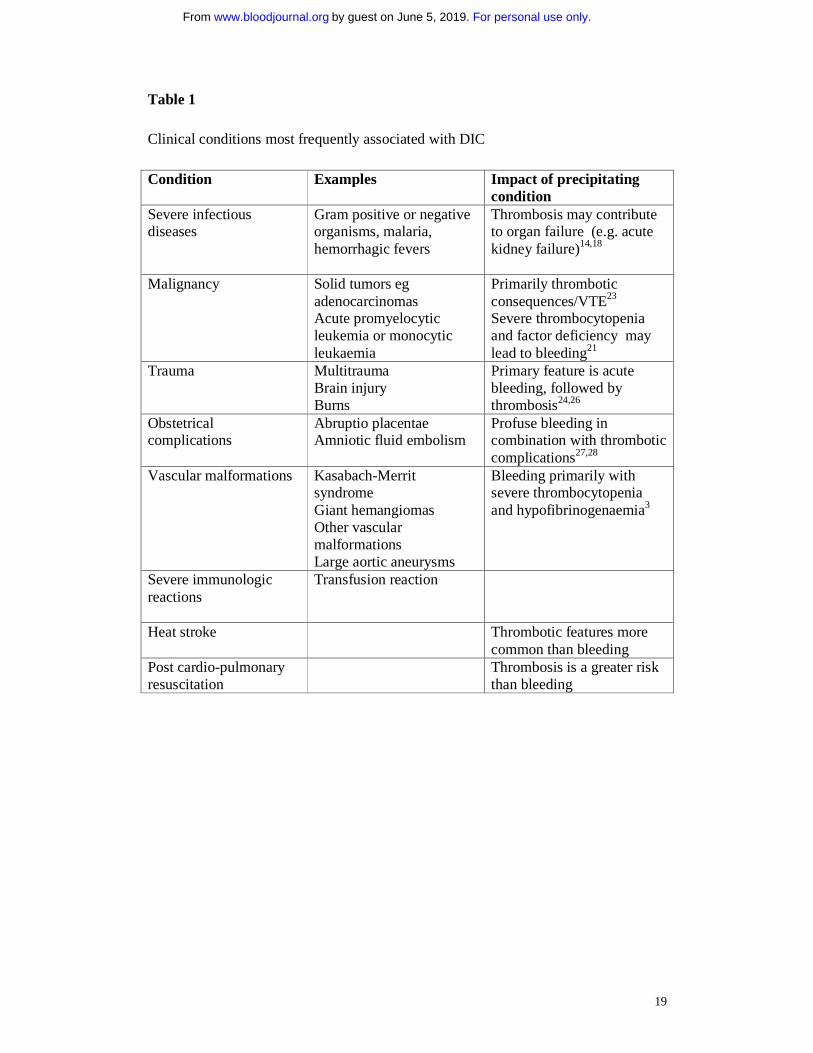
Clinical settings It should be emphasized that DIC is not a disease in itself but is always secondary to
an underlying condition that causes the activation of coagulation. The disorders most
frequently associated with DIC are listed in Table 1.
About 35% of cases of severe sepsis may be complicated by DIC.15,18 Classically,
infection with Gram negative microorganisms has been associated with DIC;
however, the incidence of DIC in patients with Gram positive infections is similar.19
Systemic infections with other microorganisms, including fungi or parasites may lead
to DIC as well.19 For example, high parasitemia primarily of Falciparum malaria,
may be associated with DIC, and high mortality.20 Factors involved in the
development of DIC complicating infections are microbial membrane constituents,
such as lipopolysaccharide or lipoteichoic acid, or exotoxins (e.g. Staphylococcal �-
toxin), evoking a strong immune response and release of cytokines.
Both hematological malignancies and solid tumors may be complicated by DIC due to
expression of procoagulant factors by tumor cells.21 The incidence of DIC in cancer is
not precisely known and may depend on diagnostic criteria used; however, some series,
in particular in patients with metastasized adenocarcinoma or lymphoproliferative
disease, report an incidence of up to 20% in consecutive cases.22 The risk of thrombosis
is greater than bleeding and in severe cases, thromboembolism in conjunction with
bleeding can be seen.23
Severe trauma is another clinical condition commonly associated with DIC.2,24
Systemic cytokine patterns in patients with severe trauma have been shown to be
virtually identical to those of septic patients.25 In addition, release of tissue material
(such as tissue thromboplastin, in particular in patients with head trauma) into the
circulation and endothelial disruption may contribute to the systemic activation of
coagulation. It may be difficult to differentiate DIC from the coagulopathy due to
massive blood loss and the dilutional coagulopathy due to massive transfusion or
infusion of large volumes of crystalloids that may occur in the first hours after major
trauma.26
In obstetric calamities, such as placental abruption and amniotic fluid emboli, acute
and fulminant DIC may occur.27,28 The degree of placental separation in patients with
abruptio placentae correlates with the extent of DIC, suggesting that leakage of tissue
factor from the placental system into the maternal circulation is responsible for the
occurrence of DIC.
For other underlying conditions (Table 1), DIC is a relatively infrequent complication.
In most situations, the severity of the associated systemic inflammatory response in
combination with specific circumstances, such as concomitant infections, will
determine whether severe systemic coagulation activation will occur.
For personal use only.on June 5, 2019. by guest www.bloodjournal.orgFrom

5
Pathogenetic pathways The most important mechanisms leading to the pathological derangement of coagulation
in DIC have been clarified. Initiation and propagation of procoagulant pathways with
simultaneous impairment of natural anticoagulant systems and suppression of
endogenous fibrinolysis as a result of systemic inflammatory activation are leading to
platelet activation and fibrin deposition.15,29 Important mediators that regulate these
processes are cytokines, such as interleukin (IL)-1 and 6 and tumor necrosis factor
(TNF)-�. In addition, recent studies point to a prominent role of intravascular webs
(‘neutrophil-extracellular traps’) consisting of denatured DNA from damaged cells and
entangling neutrophils, plateles, fibrin and kationic proteins, such as histones, in the
development of thrombus deposition.30
Thrombin generation in DIC is initiated through the tissue factor/factor VII(a) pathway
that will activate downstream coagulation factors.31 Tissue factor may be expressed by
activated monocytes but also by vascular endothelial cells or cancer cells. Besides
inflammation causing pro-coagulant effects, the activation of coagulation also
modulates inflammation (Figure 1).
In DIC all physiological anticoagulant pathways are functionally impaired.15 Firstly, a
marked imbalance of TFPI function in relation to the increased tissue factor-dependent
activation of coagulation has been described.32 In addition a significant impairment of
the protein C system may further compromise adequate regulation of thrombin
generation. This is caused by a cytokine-mediated downregulation of thrombomodulin
expression on endothelial cells in combination with decreased synthesis of protein C
and a fall in the concentration of the free fraction of protein S (the essential co-factor of
protein C), together resulting in reduced activation of protein C.33 Lastly, plasma levels
of antithrombin, the most prominent inhibitor of thrombin and factor Xa, are
significantly reduced in DIC, due to a combination of consumption, degradation by
elastase from activated neutrophils, and impaired synthesis.34 In addition, the
endogenous fibrinolytic system is largely shut-off as a result of a sustained rise in the
plasma level of plasminogen activator inhibitor-1 (PAI-1), the principal inhibitor of
plasminogen activation and plasmin generation.2
Patient 1: Diagnosis of DIC in a 68-year-old critically ill man A 68-year-old man is admitted because of respiratory failure to the intensive care unit
three days after a hemicolectomy. His blood pressure is 100/60 mmHg, heart rate
120/min (regular), respiratory rate 28/min and temperature 38.1oC. Arterial blood gas
analysis shows a pO2 of 8.4 kPa (63 mmHg) and oxygen saturation of 84% while on 5
liter supplemental oxygen. Laboratory analysis shows hemoglobin 6.8 mmol/L (11.0
g/dL), leucocytes 9.2x10⁹/L, with 84% neutrophils, a remarkable left shift and some
schistocytes in the blood smear, creatinine 212 umol/L (2.4 mg/dL), bilirubin 18 umol/L
(1.2 mg/dL), lactic acid of 3.3 mmol/L (30 mg/dL), and bicarbonate 18 mmol/L. The
For personal use only.on June 5, 2019. by guest www.bloodjournal.orgFrom

6
patient is intubated and ventilated achieving an oxygen saturation of 98% and
hemodynamically stabilized with crystalloids and intravenous administration of
dopamine. He is further treated with broad spectrum antibiotics and continues
subcutaneous heparin prophylaxis.
Routine coagulation tests show a platelet count of 98x109/L (152x109/l the day before),
prothrombin time (PT) of 17 sec (normal <12 sec), which equals an INR of 1.4,
activated partial thromboplastin time (aPTT) of 43 sec (normal <28 sec), a D-dimer of
7.5 μg/mL (normal: <0.5 μg/mL), and a fibrinogen of 3.5 g/L (normal 1-3 g/L).
Comments about patient 1 This critically ill patient with multiple organ failure and a clinical suspicion of sepsis
shows clear signs of a coagulopathy characterized by a low platelet count, prolonged
global coagulation tests and increased D-dimer. These laboratory abnormalities may be
compatible with DIC, however, some differential diagnostic considerations needs to be
taken into account.
Sepsis per se is clearly associated with thrombocytopenia and the severity of sepsis
correlates with the reduction in platelet count.35 The principal factors that contribute to
thrombocytopenia in patients with sepsis are impaired platelet production, increased
consumption or destruction, or sequestration of platelets in the spleen or along the
endothelial surface. In addition, in a considerable number of patients with sepsis marked
hemophagocytosis may occur. This pathologic process consists of active phagocytosis
of megakaryocytes and other hematopoietic cells by monocytes and macrophages,
hypothetically due to stimulation with high levels of macrophage colony stimulating
factor (M-CSF).36 Platelet activation, consumption, and destruction may also occur at
the vascular surface as a result of extensive endothelial cell-platelet interaction in sepsis,
which may differentially occur in vascular beds of various organs.37 These mechanisms
alone, however, do not explain a prolongation of coagulation times.
The presence of thrombocytopenia and schistocytes in the blood film may point in the
direction of a thrombotic microangiopathy, such as thrombotic thrombocytopenic
purpura or hemolytic-uremic syndrome. However, these syndromes are typically
accompanied by normal clotting times and normal or only slightly elevated D-dimer.38
Schistocytes may also be seen in patients with DIC as a result of enhanced platelet-
vessel wall interaction and formation of microvascular thrombotic obstruction, causing
mechanical damage to erythrocytes. Interestingly, patients with severe sepsis and DIC
may have reduced ADAMTS-13 levels (presumably due to consumption as a result of
the large amount of von Willebrand factor multimers released from perturbed
endiothelial cells) causing some overlap between DIC and thrombotic
microangiopathy.39 Raised VWF levels and associated reduced ADAMTS 13 levels, are
associated with worse outcomes in severe DIC.40
For personal use only.on June 5, 2019. by guest www.bloodjournal.orgFrom

7
Another differential diagnostic consideration for the thrombocytopenia in this case may
be heparin-induced thrombocytopenia (HIT).41 It is likely that our patient was treated
with subcutaneous heparin for some days as perioperative thrombosis prophylaxis.
Patients with HIT may present with arterial and venous thrombosis which may explain a
high D-dimer result. However, HIT is not associated with abnormal global coagulation
times.
Taken together, DIC is the most probable explanation for the coagulopathy in this patient.
Patients with DIC have a low or rapidly decreasing platelet count, prolonged global
coagulation tests, low plasma levels of coagulation factors and inhibitors, and increased
markers of fibrin formation and/or degradation, such as D-dimer or fibrin degradation
products (FDP’s).42 Coagulation proteins with a marked acute phase behavior, such as
factor VIII or fibrinogen, are usually not decreased or may even increase. One of the often
advocated laboratory tests for the diagnosis of DIC, fibrinogen, is therefore not a very
good marker for DIC, except in very severe cases, though sequential measurements can
give some insight. Dynamic changes in coagulation factors and platelets may add
important information. A significant drop in platelet count (as illustrated in the case), a
lengthening duration of clotting assays, or increase in fibrin split products, even still
within the normal range, can indicate an early stage of developing DIC.9
There is no single laboratory test with sufficient accuracy for the diagnosis of DIC. For
the diagnosis of DIC a simple scoring system (Table 2, Figure 2) has been developed by
the International Society on Thrombosis and Hemostasis (ISTH).43,44 The score can be
calculated based on routinely available laboratory tests, i.e. platelet count, prothrombin
time, a fibrin-related marker (usually D-dimer), and fibrinogen. Prospective studies
have shown that the sensitivity of the DIC score is 93 percent, and the specificity is 98
percent.17,45 The severity of DIC according to this scoring system is a strong predictor
for mortality in sepsis.46 Similar scoring systems and diagnostic guidance have been
developed and extensively evaluated in Japan, Italy and the United Kingdom.47-49 The
major difference between the international and Japanese scoring systems seems a
slightly higher sensitivity of the Japanese algorithm, although this may be due to
different patient populations as Japanese series typically include relatively large
numbers of patients with haematological malignancies.
In our patient the platelet count (1 point), prolongation of the PT (1 point) and strongly
increased D-dimer (3 points) leads to a score of 5 points, compatible with a diagnosis of
DIC.
Patient 2: A 63-year old woman with DIC or coagulopathy related to liver disease? A 63-year-old woman, known with longstanding alcohol abuse, presents with
decompensated liver cirrhosis. At physical examination most prominent signs are
hepatic encephalopathy, jaundice, splenomegaly, and ascites. Laboratory test results
show a hemoglobin of 7.0 mmol/L (11.3 g/dL), leucocytes 7.9.2x109/L, platelet count
For personal use only.on June 5, 2019. by guest www.bloodjournal.orgFrom

8
88x109/L (one week before admission 84x109/L), bilirubin 84 umol/L (1.2 mg/dL), and
albumin 28 g/L. Coagulation tests reveal a prothrombin time (PT) of 17 sec (normal <12
sec), activated partial thromboplastin time (aPTT) of 52 sec (normal <28 sec), a D-
dimer of 1.0 μg/mL (normal: <0.5 μg/mL), and a fibrinogen of 2.1 g/L (normal 1-3 g/L).
The question is whether these coagulation abnormalities are due to liver failure-related
coagulopathy or DIC, secondary to an infection.
Comments on patient 2 In patients with severe hepatic failure several changes in coagulation may occur.
More than 75% of patients with cirrhosis present with a platelet count of <150x109/L
and in more than 10% of patients this is <75x109/L, most importantly caused by
sequestration of platelets in the enlarged spleen, reduced levels of thrombopoietin,
and consumption.50,51, In addition, plasma levels of almost all coagulation factors
(except factor VIII and von Willebrand factor) are low, as the liver is the most
important site of coagulation protein synthesis. The combination of thrombocytopenia
and low levels of coagulation factors was traditionally interpreted as a
hypocoagulable state and associated with a high risk of bleeding. However, recent
insights point to a rebalanced hemostatic system in patients with chronic liver failure
as low levels of natural coagulation inhibitors may balance low levels of coagulation
factors and the reduced platelet count may be offset by huigh levels of von Willebrand
factor.52,53
The differential diagnosis between the coagulopathy of liver disease and DIC is
challenging as many laboratory abnormalities point in the same direction. Even more
complex situations may occur when the coagulopathy of liver disease is complicated
by DIC, as patients with severe liver disease may present with infectious
complications (such as bacterial peritonitis) or leakage of endotoxin from the
intestinal compartment that may elicit DIC. However, in most cases the coagulopathy
of liver disease can eventually be distinguished from the presence of DIC.54 Helpful
clues may be that in contrast to patients with DIC in severe liver disease the (low)
platelet count is usually stable and fibrin degradation products (such as D-dimer) are
only mildly elevated, due to the simultaneous presence of fibrinolytic activation and
impairment in this condition.55-57 In addition, clinical signs, such as the presence of
splenomegaly and ascites, may indicate that liver disease rather than DIC is the cause
of the coagulopathy. In the case presented the DIC score was 4 (suggestive of no DIC)
and in combination with the clinical signs and symptoms a diagnosis of coagulopathy
due to severe liver failure was made.
For personal use only.on June 5, 2019. by guest www.bloodjournal.orgFrom

9
Patient 3: Supportive treatment in a 63-year-old man with sepsis and DIC A 63-year-old man presents with severe cholangio-sepsis due to an obstructive stone in
the common bile duct. He is in shock, respiratory insufficient and develops acute renal
failure. Blood cultures are positive for Klebsiella pneumoniae.
Coagulation analysis shows a platelet count of 48x109/L, prothrombin time (PT) of 19
sec (normal <12 sec), which equals an INR of 1.6, activated partial thromboplastin time
(aPTT) of 39 sec (normal <28 sec), a D-dimer of 5.5 μg/mL (normal: <0.5 μg/mL), and
a fibrinogen of 2.8 g/L (normal 1-3 g/L). Based on these findings the DIC score is 6 and
a diagnosis of DIC is established.
Awaiting endoscopic retrograde cholangiopancreaticography (ERCP) and restoration of
bile duct patency, the patient is treated with vasopressors, intubation and mechanical
ventilation and antibiotic treatment is started. The question is what would be the most
appropriate (supportive) treatment for the coagulopathy.
Comments about patient 3 The keystone in the management of DIC is adequate treatment of the underlying
disorder. If the condition causing the DIC is properly dealt with (in the example of the
case with bile duct drainage and antibiotics), the coagulopathy will spontaneously
resolve. However, in some situations adjunctive supportive treatment aimed at the
coagulation system will be required as the coagulopathy may proceed for a while even
after adequate treatment of the underlying condition has been initiated (Figure 2).42,58,59
Low levels of platelets and coagulation factors may increase the risk of bleeding, in
particular in postoperative patients or those planned to undergo an invasive
intervention. However, plasma or platelet substitution therapy should not be instituted
on the basis of laboratory results alone; it is indicated only in patients with active
hemorrhage and in those requiring an invasive procedure or otherwise at risk for
bleeding complications.58,60 The presumed efficacy of treatment with plasma,
fibrinogen, cryoprecipitate, or platelets is not underpinned by randomized controlled
trials but appears to be rational therapy in bleeding patients or in patients at risk for
hemorrhage with a significant deficiency of these hemostatic factors. It may be
required to use large volumes of plasma to restore normal levels of coagulation
factors. Coagulation factor concentrates, such as prothrombin complex concentrate,
may overcome this impediment, but these agents may lack important factors (e.g.
factor V). Previously the use of prothrombin complex concentrates was thought to
aggravate the coagulopathy in DIC due to small traces of activated factors in the
concentrate. It is, however, not very likely that this is still the case for the currently
available concentrates. Specific deficiencies in coagulation factors, such as
fibrinogen, may be corrected by administration of purified coagulation factor
concentrates. Vitamin K must be remembered as a useful non blood product
For personal use only.on June 5, 2019. by guest www.bloodjournal.orgFrom

10
alternative to correcting vitamin K dependent factors and will work within 4-6 hours
following a dose.
Experimental studies have shown that heparin can at least partly inhibit the activation
of coagulation in DIC.61 However, a clinically relevant effect of heparin in patients
with DIC has never been unequivocally demonstrated in controlled clinical trials,
although indirect evidence is accumulating that heparin might be of benefit.62,63 In
addition, there are several studies showing that all critically ill patients need adequate
prophylaxis for venous thromboembolism, usually with (low molecular weight)
heparin.64 Therapeutic doses of heparin are indicated in patients with clinically overt
thromboembolism and may be considered in case of extensive thrombotic
manifestations such as in purpura fulminans or acral ischemia.
Restoration of the levels of physiological anticoagulants in DIC may be a rational
approach and has been extensively evaluated.34 Based on successful preclinical
studies, the use of antithrombin concentrates has been evaluated in randomized
controlled trials in patients with severe sepsis. All trials have shown some beneficial
effect in terms of improvement of laboratory parameters or even improvement in
organ function. An international multicenter, randomized controlled trial with
antithrombin concentrate, however, did not demonstrate a significant reduction in
mortality of septic patients.65 Interestingly, post-hoc subgroup analyses of this study
indicated some benefit in patients who did not receive concomitant heparin and in
those with the most severe coagulopathy.66 Recent propensity-adjusted retrospective
data from Japan demonstrated a significant advantage of antithrombin-treated patients
with severe infection and DIC 67,68. However these observations require prospective
validation.
Adjunctive therapy with activated protein C (APC) has also been widely studied. A
phase III trial of APC concentrate in patients with sepsis was prematurely stopped
because of efficacy in reducing mortality in these patients.69 Notably, patients with
overt DIC benefited most from this treatment.46 However, after a series of negative
trials in specific populations of patients with severe sepsis meta-analyses of published
studies concluded that the basis for treatment with APC was lacking.70 Besides, there
was ambiguity regarding the hemorrhagic risk of APC in patients with severe sepsis.
The last large placebo-controlled trial in patients with severe sepsis and septic shock
was prematurely stopped due to the absence of any significant advantage of APC.71
Subsequently, the manufacturer of APC has withdrawn the product from the market.
A promising intervention that is currently being evaluated is recombinant soluble
thrombomodulin. Preclinical experimental sepsis studies have demonstrated that
soluble thrombomodulin is capable of ameliorating the derangement of coagulation
and may improve organ dysfunction.72,73 Recombinant soluble thrombomodulin was
evaluated in a phase II/III clinical study in 750 patients with sepsis and DIC.74
Twenty-eight-day mortality was 17.8% in the thrombomodulin group and 21.6% in
For personal use only.on June 5, 2019. by guest www.bloodjournal.orgFrom

11
the placebo group. The encouraging results with soluble thrombomodulin are
confirmed by retrospective data in large series of Japanese patients and are
prospectively investigated in a currenty ongoing multicentre phase III trial 75.
Patient 4: A 48-year-old woman with acute promyelocytic leukemia and severe
DIC A 48-year-old female presents with severe persisting epistaxis. She also reports to have
gingival bleeding and widespread ecchymoses for 1 week and unusually severe
menorrhagia. Laboratory analysis shows hemoglobin 5.5 mmol/L (8.9 g/dL), leucocytes
21.7x10⁹/L, with 90% promyelocytes, 4% myelocytes, 3% metamyelocytes and 2%
neutrophils, and a platelet count of 56x109/L. The bone marrow contains few blast-like
cells with scant granules and 72% promyelocytes with Auer rods. Immunophenotyping
show expression of CD9, CD13 and CD33 and is negative for CD34. Fluorescence in
situ hybridization (FISH) of the bone marrow aspirate revealed a t(15;17) translocation
and the PML-RARα fusion gene was confirmed by reverse transcriptase PCR (RT-
PCR), all compatible with a diagnosis of acute promyelocytic leukemia (AML-M3
according to the FAB classification).
Coagulation analysis reveals a prothrombin time (PT) of 20 sec (normal <12 sec), which
equals an INR of 1.7, activated partial thromboplastin time (aPTT) of 59 sec (normal
<28 sec), a D-dimer of 10 μg/mL (normal: <0.5 μg/mL), and a fibrinogen of 0.8 g/L
(normal 1-3 g/L). The plasma levels of plasminogen is 68% (normal 80-120%) and of
�2-antiplasmin 43% (normal 80-100%). Taken together, these results indicate the
presence of DIC in combination with marked hyperfibrinolysis.
Comments about patient 4 There is ample evidence for a pro-coagulant state in virtually all patients with
advanced malignant disease. This may eventually lead to venous thromboembolism or
evolve into DIC.76 DIC can be diagnosed in up to 15% of consecutive patients
presenting with acute leukemia, in particular acute lymphoblastic leukemia and some
reports indicate that the incidence of DIC in acute leukemia patients might further
increase during remission induction with chemotherapy.77
A special manifestation of DIC may occur in some patients with advanced
adenocarcinoma but more frequently in patients with acute promyelocytic leukemia
(AML-M3) and also monocytic leukemias (eg AML-M5) at the time of diagnosis.78
These patients may present with a severe and sometimes life-threatening bleeding
tendency. The hemorrhagic tendency is a consequence of DIC with consumption of
coagulation factors and platelets in combination with striking hyperfibrinolysis
Hyperfibrinolysis can be diagnosed by low levels of plasminogen and α2-antiplasmin,
low levels of fibrinogen (due to plasmin-mediated fibrinogenolysis), and excessively
high levels of fibrin degradation products (e.g. D-dimer).79 Further analysis may
For personal use only.on June 5, 2019. by guest www.bloodjournal.orgFrom
12
reveal remarkably low levels of factor VIII and factor V (which are both degraded by
plasmin), which is in contrast to more conventional types of DIC where plasma levels
of factor VIII are usually relatively preserved.
The coagulopathy associated with acute promyelocytic leukemia is caused by the
expression of tissue factor and cancer procoagulant on leukemic cells in combination
with increased expression of annexin II, causing excessive plasminogen activation
and fibrinolysis.80,81 Release of nonspecific proteases from granules in the leukemia
cells may further contribute to degradation of both fibrin and fibrinogen.
Management of patients who present with acute promyelocytic leukemia and DIC
consists of supportive treatment with platelet transfusion (aiming at a platelet count of
above 30-50x109/L), fresh frozen plasma, and fibrinogen concentrate (guided by the
fibrinogen concentration in the patient’s plasma) and should be maintained throughout
remission induction and disappearance of the coagulopathy.82 In addition, invasive
procedures, such as biopsies or intravenous line placement, should be avoided as
much as possible. In view of the high risk of bleeding and the lack of evidence from
clinical studies the use of heparin is not advocated. Before the introduction of all-trans
retinoic acid and arsenic trioxide in the therapy of acute promyelocytic leukemia,
adjunctive treatment with fibrinolysis inhibitors (such as tranexamic acid) was shown
to be beneficial in small clinical trials.83 However, with these modern treatment
modalities the coagulopathy quickly subsides and inhibition of fibrinolysis is usually
not necessary anymore and may be even harmful in view of the prothrombotic
features of retinoic acid.84
Final considerations Although the understanding of DIC and its underlying pathogenetic pathways has
increased in recent decades, some important questions regarding the proper
management of this coagulopathy remain. Current therapeutic interventions are
mostly supportive and only partly effective, at best resulting in an amelioration of the
derangement of coagulation or more rapid resolution of DIC; however, they have not
been proven to result in an improvement of clinically relevant outcomes such as organ
failure or mortality. As there is increasing circumstantial evidence that heparin may
be beneficial in patients with DIC, a randomized controlled trial of heparin in this
situation is urgently wanted.
Better supportive treatment of DIC may also benefit from advances in early patient
identification and risk stratification. Hypothetically, assays specifically aimed at the
assessment of endothelial cell perturbation in combination with early stage systemic
coagulopathy would be helpful to recognize high-risk patients and would facilitate
early (and thereby potentially more effiacious) intervention. In addition, genetic
variation may play a role in the individual propensity to develop DIC and the severity
of the coagulopathy.85 Gene mutations and polymorphisms have been demonstrated to
For personal use only.on June 5, 2019. by guest www.bloodjournal.orgFrom

13
affect hemostatic changes in DIC. Septic mice with heterozygous protein C deficiency
due to a one allele targeted disruption of the protein C gene, presented with a more
severe DIC and associated inflammatory response.86 The presence of factor V Leiden
heterozygosity has been associated with the incidence and outcome of DIC in patients
with sepsis.87 In addition, the functional 4G/5G polymorphism in the PAI-1 gene
affected plasma levels of PAI-1 and was related to clinical outcome of meningococcal
sepsis and DIC.88 A better understanding of the influence of genomic variation in the
host response leading to DIC could be helpful in identifying patients that are more
susceptible for severe coagulopathy and eventually tailor treatment to the most
vulnerable patients.
Author contribution Both authors together designed the outline of the paper, reviewed the literature, and
wrote the paper
Conflict of interest: None of the authors have a conflict of interest related to this manuscript
For personal use only.on June 5, 2019. by guest www.bloodjournal.orgFrom

14
References 1. Hunt BJ. Bleeding and coagulopathies in critical care. N Engl J Med
2014;370:847-59.
2. Gando S, Levi M, Toh CH. Disseminated intravascular coagulation. Nat Rev Dis Primers 2016;2:16037.
3. Levi M, ten Cate H. Disseminated intravascular coagulation. N Engl J Med 1999;341:586-592.
4. Boral BM, Williams DJ, Boral LI. Disseminated Intravascular Coagulation. Am J Clin Pathol 2016;146:670-680.
5. Levi M. Disseminated intravascular coagulation. Crit Care Med 2007;35:2191-2195.
6. Culasso DE, Donati MB, de Gaetano G, et al. Inhibition of human platelet aggregation by plasmin digests of human and bovine fibrinogen preparations: role of contaminating factor VIII-related material. Blood 1974;44:169-75.
7. Weitz JI, Leslie B, Ginsberg J. Soluble fibrin degradation products potentiate tissue plasminogen activator-induced fibrinogen proteolysis. J Clin Invest 1991;87:1082-90.
8. Bernard GR, Margolis BD, Shanies HM, et al. Extended evaluation of recombinant human activated protein C United States Trial (ENHANCE US): a single-arm, phase 3B, multicenter study of drotrecogin alfa (activated) in severe sepsis. Chest 2004;125:2206-2216.
9. Dhainaut JF, Shorr AF, Macias WL, et al. Dynamic evolution of coagulopathy in the first day of severe sepsis: Relationship with mortality and organ failure. Crit Care Med 2005;33:341-348.
10. Vanderschueren S, De Weerdt A, Malbrain M, et al. Thrombocytopenia and prognosis in intensive care. Crit Care Med 2000;28:1871-1876.
11. Strauss R, Wehler M, Mehler K, et al. Thrombocytopenia in patients in the medical intensive care unit: bleeding prevalence, transfusion requirements, and outcome. Crit Care Med 2002;30:1765-1771.
12. Oppenheim-Eden A, Glantz L, Eidelman LA, et al. Spontaneous intracerebral hemorrhage in critically ill patients: incidence over six years and associated factors. Intensive Care Med 1999;25:63-67.
13. Kushimoto S, Gando S, Saitoh D, et al. Clinical course and outcome of disseminated intravascular coagulation diagnosed by Japanese Association for Acute Medicine criteria. Comparison between sepsis and trauma. Thromb Haemost 2008;100:1099-105.
14. Singh B, Hanson AC, Alhurani R, et al. Trends in the incidence and outcomes of disseminated intravascular coagulation in critically ill patients (2004-2010): a population-based study. Chest 2013;143:1235-1242.
15. Levi M, van der Poll T. Coagulation and sepsis. Thromb Res 2017;149:38-44. 16. van der Poll T, van de Veerdonk FL, Scicluna BP, et al. The
immunopathology of sepsis and potential therapeutic targets. Nat Rev Immunol 2017;17:407-420.
17. Bakhtiari K, Meijers JC, de Jonge E, et al. Prospective validation of the international society of thrombosis and maemostasis scoring system for disseminated intravascular coagulation. Crit Care Med 2004;32:2416-2421.
18. Angus DC, van der Poll T. Severe sepsis and septic shock. N Engl J Med 2013;369:840-851.
For personal use only.on June 5, 2019. by guest www.bloodjournal.orgFrom
15
19. Kinasewitz GT, Yan SB, Basson B, et al. Universal changes in biomarkers of coagulation and inflammation occur in patients with severe sepsis, regardless of causative micro-organism. Crit Care 2004;8:R82-90.
20. O'Sullivan JM, Preston RJ, O'Regan N, et al. Emerging roles for hemostatic dysfunction in malaria pathogenesis. Blood 2016;127:2281-8.
21. Falanga A, Marchetti M, Vignoli A. Coagulation and cancer: biological and clinical aspects. J Thromb Haemost 2013;11:223-233.
22. Colman RW, Rubin RN. Disseminated intravascular coagulation due to malignancy. Semin Oncology 1990;17:172-186.
23. Feinstein DI. Disseminated intravascular coagulation in patients with solid tumors. Oncology (Williston Park) 2015;29:96-102.
24. Gando S. Disseminated intravascular coagulation in trauma patients. Semin Thromb Hemost 2001;27:585-592.
25. Gando S, Nakanishi Y, Tedo I. Cytokines and plasminogen activator inhibitor-1 in posttrauma disseminated intravascular coagulation: relationship to multiple organ dysfunction syndrome. Crit Care Med 1995;23:1835-1842.
26. Cap A, Hunt BJ. The pathogenesis of traumatic coagulopathy. Anaesthesia 2015;70 Suppl 1:96-4.
27. Thachil J, Toh CH. Disseminated intravascular coagulation in obstetric disorders and its acute haematological management. Blood Rev 2009;23:167-176.
28. Erez O. Disseminated intravascular coagulation in pregnancy - Clinical phenotypes and diagnostic scores. Thromb Res 2017;151 Suppl 1:S56-s60.
29. Simmons J, Pittet JF. The coagulopathy of acute sepsis. Curr Opin Anaesthesiol 2015;28:227-36.
30. McDonald B, Davis RP, Kim SJ, et al. Platelets and neutrophil extracellular traps collaborate to promote intravascular coagulation during sepsis in mice. Blood 2017;129:1357-1367.
31. Versteeg HH, Heemskerk JW, Levi M, et al. New fundamentals in hemostasis. Physiol Rev 2013;93:327-358.
32. Osterud B, Bjorklid E. The tissue factor pathway in disseminated intravascular coagulation. Semin Thromb Hemost 2001;27:605-617.
33. Esmon CT. Role of coagulation inhibitors in inflammation. Thromb Haemost 2001;86:51-56.
34. Levy JH, Sniecinski RM, Welsby IJ, et al. Antithrombin: anti-inflammatory properties and clinical applications. Thromb Haemost 2016;115:712-28.
35. Mavrommatis AC, Theodoridis T, Orfanidou A, et al. Coagulation system and platelets are fully activated in uncomplicated sepsis. Crit Care Med 2000;28:451-457.
36. Francois B, Trimoreau F, Vignon P, et al. Thrombocytopenia in the sepsis syndrome: role of hemophagocytosis and macrophage colony-stimulating factor. Am J Med 1997;103:114-120.
37. Levi M, Lowenberg EC. Thrombocytopenia in critically ill patients. Semin Thromb Hemost 2008;34:417-424.
38. Scully M, Goodship T. How I treat thrombotic thrombocytopenic purpura and atypical haemolytic uraemic syndrome. Br J Haematol 2014;164:759-66.
39. Azfar MF, Khan MF, Habib SS, et al. Prognostic value of ADAMTS13 in patients with severe sepsis and septic shock. Clin Invest Med 2017;40:E49-e58.
For personal use only.on June 5, 2019. by guest www.bloodjournal.orgFrom
16
40. Habe K, Wada H, Ito-Habe N, et al. Plasma ADAMTS13, von Willebrand factor (VWF) and VWF propeptide profiles in patients with DIC and related diseases. Thromb Res 2012;129:598-602.
41. Warkentin TE. Heparin-induced thrombocytopenia in critically ill patients. Semin Thromb Hemost 2015;41:49-60.
42. Levi M, Toh CH, Thachil J, et al. Guidelines for the diagnosis and management of disseminated intravascular coagulation. Br J Haematol 2009;145:24-33.
43. Taylor FB, Jr, Toh CH, Hoots WK, et al. Towards definition, clinical and laboratory criteria, and a scoring system for disseminated intravascular coagulation. Thromb Haemost 2001;86:1327-1330.
44. Toh CH, Hoots WK. The scoring system of the Scientific and Standardisation Committee on Disseminated Intravascular Coagulation of the International Society on Thrombosis and Haemostasis: a five year overview. J Thromb Haemost 2007;5:604-606.
45. Gando S, Iba T, Eguchi Y, et al. A multicenter, prospective validation of disseminated intravascular coagulation diagnostic criteria for critically ill patients: comparing current criteria. Crit Care Med 2006;34:625-631.
46. Dhainaut JF, Yan SB, Joyce DE, et al. Treatment effects of drotrecogin alfa (activated) in patients with severe sepsis with or without overt disseminated intravascular coagulation. J Thromb Haemost 2004;2:1924-1933.
47. Gando S, Saitoh D, Ogura H, et al. A multicenter, prospective validation study of the Japanese Association for Acute Medicine disseminated intravascular coagulation scoring system in patients with severe sepsis. Crit Care 2013;17:R111.
48. Wada H, Thachil J, Di Nisio M, et al. Guidance for diagnosis and treatment of DIC from harmonization of the recommendations from three guidelines. J Thromb Haemost 2013;11:761-7.
49. Di Nisio M, Baudo F, Cosmi B, et al. Diagnosis and treatment of disseminated intravascular coagulation: guidelines of the Italian Society for Haemostasis and Thrombosis (SISET). Thromb Res 2012;129:e177-84.
50. Lisman T, Leebeek FW. Hemostatic alterations in liver disease: a review on pathophysiology, clinical consequences, and treatment. Dig Surg 2007;24:250-8.
51. Afdhal N, McHutchison J, Brown R, et al. Thrombocytopenia associated with chronic liver disease. J Hepatol 2008;48:1000-7.
52. Lisman T, Porte RJ. Rebalanced hemostasis in patients with liver disease: evidence and clinical consequences. Blood 2010;116:878-85.
53. Tripodi A, Mannucci PM. The coagulopathy of chronic liver disease. N Engl J Med 2011;365:147-56.
54. Ben-Ari Z, Osman E, Hutton RA, et al. Disseminated intravascular coagulation in liver cirrhosis: fact or fiction? Am J Gastroenterol 1999;94:2977-82.
55. Kim SY, Kim JE, Kim HK, et al. Higher prognostic value of soluble fibrin complexes than D-dimer and fibrin degradation product for disseminated intravascular coagulation in patients with liver cirrhosis. Blood Coagul Fibrinolysis 2013;24:150-6.
56. Roberts LN, Patel RK, Arya R. Haemostasis and thrombosis in liver disease. Br J Haematol 2010;148:507-21.
For personal use only.on June 5, 2019. by guest www.bloodjournal.orgFrom

17
57. Drolz A, Horvatits T, Roedl K, et al. Coagulation parameters and major bleeding in critically ill patients with cirrhosis. Hepatology 2016;64:556-68.
58. Squizzato A, Hunt BJ, Kinasewitz GT, et al. Supportive management strategies for disseminated intravascular coagulation. An international consensus. Thromb Haemost 2016;115:896-904.
59. Wada H, Matsumoto T, Yamashita Y. Diagnosis and treatment of disseminated intravascular coagulation (DIC) according to four DIC guidelines. J Intensive Care 2014;2:15.
60. Wada H, Thachil J, Di NM, et al. Guidance for diagnosis and treatment of DIC from harmonization of the recommendations from three guidelines. J Thromb Haemost 2013:10.
61. Ding R, Zhao D, Guo R, et al. Treatment with unfractionated heparin attenuates coagulation and inflammation in endotoxemic mice. Thromb Res 2011;128:e160-5.
62. Zarychanski R, Abou-Setta AM, Kanji S, et al. The efficacy and safety of heparin in patients with sepsis: a systematic review and metaanalysis. Crit Care Med 2015;43:511-8.
63. Li X, Ma X. The role of heparin in sepsis: much more than just an anticoagulant. Br J Haematol 2017.
64. Boonyawat K, Crowther MA. Venous thromboembolism prophylaxis in critically ill patients. Semin Thromb Hemost 2015;41:68-74.
65. Warren BL, Eid A, Singer P, et al. Caring for the critically ill patient. High-dose antithrombin III in severe sepsis: a randomized controlled trial. JAMA 2001;286:1869-1878.
66. Kienast J, Juers M, Wiedermann CJ, et al. Treatment effects of high-dose antithrombin without concomitant heparin in patients with severe sepsis with or without disseminated intravascular coagulation. J Thromb Haemost 2006;4:90-97.
67. Tagami T, Matsui H, Horiguchi H, et al. Antithrombin and mortality in severe pneumonia patients with sepsis-associated disseminated intravascular coagulation: an observational nationwide study. J Thromb Haemost 2014;12:1470-1479.
68. Hayakawa M, Kudo D, Saito S, et al. Antithrombin Supplementation and Mortality in Sepsis-Induced Disseminated Intravascular Coagulation: A Multicenter Retrospective Observational Study. Shock 2016;46:623-631.
69. Bernard GR, Vincent JL, Laterre PF, et al. Efficacy and safety of recombinant human activated protein C for severe sepsis. N Engl J Med 2001;344:699-709.
70. Marti-Carvajal A, Salanti G, Cardona AF. Human recombinant activated protein C for severe sepsis. Cochrane Database Syst Rev 2007:CD004388.
71. Ranieri VM, Thompson BT, Barie PS, et al. Drotrecogin Alfa (Activated) in Adults with Septic Shock. N Engl J Med 2012;366:2055-2064.
72. Uchiba M, Okajima K, Murakami K, et al. Recombinant thrombomodulin prevents endotoxin-induced lung injury in rats by inhibiting leukocyte activation. Am J Physiol 1996;271:L470-L475.
73. Okamoto T, Tanigami H, Suzuki K, et al. Thrombomodulin: a bifunctional modulator of inflammation and coagulation in sepsis. Crit Care Res Pract 2012;61:45.
74. Vincent JL, Ramesh MK, Ernest D, et al. A randomized, double-blind, placebo-controlled, Phase 2b study to evaluate the safety and efficacy of recombinant human soluble thrombomodulin, ART-123, in patients with
For personal use only.on June 5, 2019. by guest www.bloodjournal.orgFrom

18
sepsis and suspected disseminated intravascular coagulation. Crit Care Med 2013;41:2069-2079.
75. Yamakawa K, Aihara M, Ogura H, et al. Recombinant human soluble thrombomodulin in severe sepsis: a systematic review and meta-analysis. J Thromb Haemost 2015;13:508-519.
76. Levi M. Management of cancer-associated disseminated intravascular coagulation. Thromb Res 2016;140 Suppl 1:S66-70.
77. Barbui T, Falanga A. Disseminated intravascular coagulation in acute leukemia. Semin Thromb Hemost 2001;27:593-604.
78. Arbuthnot C, Wilde JT. Haemostatic problems in acute promyelocytic leukaemia. Blood Rev 2006;20:289-97.
79. Avvisati G, ten Cate JW, Sturk A, et al. Acquired alpha-2-antiplasmin deficiency in acute promyelocytic leukaemia. Br J Haematol 1988;70:43-48.
80. Falanga A, Schieppati F, Russo D. Cancer Tissue Procoagulant Mechanisms and the Hypercoagulable State of Patients with Cancer. Semin Thromb Hemost 2015;41:756-64.
81. Stein E, McMahon B, Kwaan H, et al. The coagulopathy of acute promyelocytic leukaemia revisited. Best Pract Res Clin Haematol 2009;22:153-163.
82. Dohner H, Estey EH, Amadori S, et al. Diagnosis and management of acute myeloid leukemia in adults: recommendations from an international expert panel, on behalf of the European LeukemiaNet. Blood 2010;115:453-74.
83. Avvisati G, ten Cate JW, Buller HR, et al. Tranexamic acid for control of haemorrhage in acute promyelocytic leukaemia. Lancet 1989;2:122-124.
84. Falanga A, Rickles FR. Pathogenesis and management of the bleeding diathesis in acute promyelocytic leukaemia. Best Pract Res Clin Haematol 2003;16:463-82.
85. Texereau J, Pene F, Chiche JD, et al. Importance of hemostatic gene polymorphisms for susceptibility to and outcome of severe sepsis. Crit Care Med 2004;32:S313-S319.
86. Levi M, Dorffler-Melly J, Reitsma PH, et al. Aggravation of endotoxin-induced disseminated intravascular coagulation and cytokine activation in heterozygous protein C deficient mice. Blood 2003;101:4823-4827.
87. Schouten M, van 't Veer C, van der Poll T, et al. Effect of the factor V Leiden mutation on the incidence and outcome of severe infection and sepsis. Neth J Med 2012;70:306-310.
88. Hermans PW, Hibberd ML, Booy R, et al. 4G/5G promoter polymorphism in the plasminogen-activator-inhibitor-1 gene and outcome of meningococcal disease. Meningococcal Research Group. Lancet 1999;354:556-560.
For personal use only.on June 5, 2019. by guest www.bloodjournal.orgFrom
19
Table 1 Clinical conditions most frequently associated with DIC Condition Examples Impact of precipitating
condition Severe infectious diseases
Gram positive or negative organisms, malaria, hemorrhagic fevers
Thrombosis may contribute to organ failure (e.g. acute kidney failure)14,18
Malignancy Solid tumors eg adenocarcinomas Acute promyelocytic leukemia or monocytic leukaemia
Primarily thrombotic consequences/VTE23 Severe thrombocytopenia and factor deficiency may lead to bleeding21
Trauma
Multitrauma Brain injury Burns
Primary feature is acute bleeding, followed by thrombosis24,26
Obstetrical complications
Abruptio placentae Amniotic fluid embolism
Profuse bleeding in combination with thrombotic complications27,28
Vascular malformations Kasabach-Merrit syndrome Giant hemangiomas Other vascular malformations Large aortic aneurysms
Bleeding primarily with severe thrombocytopenia and hypofibrinogenaemia3
Severe immunologic reactions
Transfusion reaction
Heat stroke
Thrombotic features more common than bleeding
Post cardio-pulmonary resuscitation
Thrombosis is a greater risk than bleeding
For personal use only.on June 5, 2019. by guest www.bloodjournal.orgFrom

20
Table 2
Scoring algorithm for the diagnosis of DIC
1. platelet count
• >100 = 0
• <100 = 1
• <50 = 2
2. level of fibrin markers (e.g. D-dimer, fibrin degradation products)
� no increase= 0
� increased but < 5x upper limit of normal= 2
� strong increase (> 5x upper limit of normal)= 3
3. prolonged prothrombin time *
� < 3 sec.= 0
� > 3 sec. but < 6 sec.= 1
� > 6 sec. = 2
4. fibrinogen level
� > 1.0 g/L = 0
� < 1.0 g/L =1
This scoring system is only appropriate in patients with an underlying disorder that can be associated with DIC. A score of 5 points or more is compatible with DIC. Note that if the score is less than 5, consider repeating after 1-2 days.43 *): If prothrombin time values are only available as International Normalized Ratio (INR), an INR value of >1.3 or >1.5 will generate 1 or 2 points, respectively (assuming the ISI value of the thromboplastin reagens used is close to 1)
For personal use only.on June 5, 2019. by guest www.bloodjournal.orgFrom

21
Figure 1. Mononuclear cells express tisue factor resulting in thrombin generation and
subsequent fibrinogen to fibrin conversion, which in combination with increased
platelet vessel wall interaction and activation of platelets contribute to microvascular
clot formation. P-selectin released from activated platelets further upregulates tissue
factor expression.
Binding of fibrin to Toll-like receptor 4 and activated coagulation proteases to
specific protease activated receptors (PAR’s) on inflammatory cells (dotted lines)
modulates inflammation through the further release of pro-inflammatory cytokines.
Cytokines play also a key role in the suppression of endogenous fibrinolysis and
impairment of physiological anticoagulant pathways, such as the activated protein C
pathway.
For personal use only.on June 5, 2019. by guest www.bloodjournal.orgFrom

22
Figure 2. Flow chart for the diagnostic and therapeutic management of disseminated
intravascular coagulation (DIC). PT: prothrombin time; aPTT: activated partial
thromboplastin time; LMW: low molecular weight.
For personal use only.on June 5, 2019. by guest www.bloodjournal.orgFrom

doi:10.1182/blood-2017-10-804096Prepublished online December 18, 2017;
Marcel Levi and Marie Scully How I treat disseminated intravascular coagulation
http://www.bloodjournal.org/site/misc/rights.xhtml#repub_requestsInformation about reproducing this article in parts or in its entirety may be found online at:
http://www.bloodjournal.org/site/misc/rights.xhtml#reprintsInformation about ordering reprints may be found online at:
http://www.bloodjournal.org/site/subscriptions/index.xhtmlInformation about subscriptions and ASH membership may be found online at:
digital object identifier (DOIs) and date of initial publication. indexed by PubMed from initial publication. Citations to Advance online articles must include final publication). Advance online articles are citable and establish publication priority; they areappeared in the paper journal (edited, typeset versions may be posted when available prior to Advance online articles have been peer reviewed and accepted for publication but have not yet
Copyright 2011 by The American Society of Hematology; all rights reserved.Hematology, 2021 L St, NW, Suite 900, Washington DC 20036.Blood (print ISSN 0006-4971, online ISSN 1528-0020), is published weekly by the American Society of
For personal use only.on June 5, 2019. by guest www.bloodjournal.orgFrom


![Disseminated Intravascular Dic Auto Saved]](https://static.fdocuments.net/doc/165x107/577d229f1a28ab4e1e97d81f/disseminated-intravascular-dic-auto-saved.jpg)