
Sepsis and disseminated intravascular coagulation · Sepsis and disseminated intravascular...
8
REVIEW Open Access Sepsis and disseminated intravascular coagulation Kohji Okamoto 1,2* , Toshihisa Tamura 2 and Yusuke Sawatsubashi 2 Abstract Sepsis is frequently complicated by coagulopathy and, in about 35 % of severe cases, by disseminated intravascular coagulation (DIC). In Japan, aggressive treatment of septic DIC is encouraged using antithrombin and recombinant thrombomodulin. The macrophages, monocytes, and neutrophils are a source of TF and participate in the direct activation of the coagulation cascade in the early phases of sepsis. And activated factor X (FXa), which is involved in hemostasis, thrombogenesis, inflammation, and cellular immune responses, induces TF expression in human peripheral monocytes and, conversely, that inhibition of FXa activity reduces TF expression. Both inflammation and coagulation play an important role in DIC due to sepsis. In addition to inflammatory cytokines (TNF-α, IL-1 and so on), HMGB1 has recently been shown to mediate the lethal late phase of sepsis and caused coagulopathy. TM not only binds HMGB1 but also aids the proteolytic cleavage of HMGB1 by thrombin. There have been many reports of the efficacy of recombinant TM and antithrombin for treatment of septic DIC from Japan. Further investigation of the efficacy of recombinant TM and AT in countries other than Japan, as well as the monitoring of medical costs incurred during hospitalization, will help validate the use of TM and AT for treatment of septic DIC. Keywords: Sepsis, Disseminated intravascular coagulation (DIC), HMGB1, Antithrombin, Thrombomodulin Introduction Sepsis is a clinical syndrome defined as a systemic re- sponse to infection. It is frequently complicated by coag- ulopathy [1] and, in about 35 % of severe cases, by disseminated intravascular coagulation (DIC) [2–4]. In the European Union and the USA, the 2012 guidelines of the Surviving Sepsis Campaign do not recommend treatment for septic DIC [5, 6]. In contrast, in Japan, ag- gressive treatment of septic DIC is encouraged [7–9]. It is not an exaggeration to state that Japan is one of the countries that most effectively treats patients with septic DIC. In this article, we review the mechanisms that underlie the interaction between sepsis and DIC and, by highlighting our findings, the effects of sepsis on the co- agulation system. Review Sepsis-induced DIC During sepsis, inflammation diffusely activates the co- agulation system, consuming multiple clotting factors and resulting in DIC [10, 11]. In systemic inflammatory response syndromes caused by infection, both perturbed endothelial cells and activated mononuclear cells pro- duce proinflammatory cytokines that promote coagula- tion [12, 13]. Proteins expressed on these cells initiate coagulation. Thrombin elicits the production of mono- cyte chemoattractant protein 1 and interleukin (IL)-6 in monocytes, fibroblasts, and mesothelial cells, and the production of IL-6 and IL-8 in vascular endothelial cells by interacting with protease-activated receptors (PARs) 1, 3, and 4. Via PAR 2, factor Xa, and the tissue factor-VIIa complex also upregulate IL-6 and IL-8 in vascular endothelial cells [14–16]. In addition, the inhib- ition of physiologic anticoagulant mechanisms and fibrinolysis by endothelial cells causes intravascular fi- brin deposition. Initiation of the extrinsic coagulation protease cascade requires tissue factor (TF), a 47-KDa transmembrane glycoprotein [17]. We reported that macrophages, * Correspondence: [email protected] 1 Department of Surgery, Center for Gastroenterology and Liver Disease, Kitakyushu City Yahata Hospital, 4-18-1 Nishihon-machi, Yahatahigashi-ku, Kitakyushu 805-8534, Japan 2 Department of Surgery 1, School of Medicine, University of Occupational & Environmental Health, 1-1 Iseiogaka, Yahatanishi-ku, Kitakyushu 807-8555, Japan © 2016 Okamoto et al. Open Access This article is distributed under the terms of the Creative Commons Attribution 4.0 International License (http://creativecommons.org/licenses/by/4.0/), which permits unrestricted use, distribution, and reproduction in any medium, provided you give appropriate credit to the original author(s) and the source, provide a link to the Creative Commons license, and indicate if changes were made. The Creative Commons Public Domain Dedication waiver (http://creativecommons.org/publicdomain/zero/1.0/) applies to the data made available in this article, unless otherwise stated. Okamoto et al. Journal of Intensive Care (2016) 4:23 DOI 10.1186/s40560-016-0149-0
Transcript of Sepsis and disseminated intravascular coagulation · Sepsis and disseminated intravascular...

REVIEW Open Access
Sepsis and disseminated intravascularcoagulationKohji Okamoto1,2*, Toshihisa Tamura2 and Yusuke Sawatsubashi2
Abstract
Sepsis is frequently complicated by coagulopathy and, in about 35 % of severe cases, by disseminated intravascularcoagulation (DIC). In Japan, aggressive treatment of septic DIC is encouraged using antithrombin and recombinantthrombomodulin. The macrophages, monocytes, and neutrophils are a source of TF and participate in the directactivation of the coagulation cascade in the early phases of sepsis. And activated factor X (FXa), which is involved inhemostasis, thrombogenesis, inflammation, and cellular immune responses, induces TF expression in human peripheralmonocytes and, conversely, that inhibition of FXa activity reduces TF expression. Both inflammation and coagulationplay an important role in DIC due to sepsis. In addition to inflammatory cytokines (TNF-α, IL-1 and so on), HMGB1 hasrecently been shown to mediate the lethal late phase of sepsis and caused coagulopathy. TM not only binds HMGB1but also aids the proteolytic cleavage of HMGB1 by thrombin. There have been many reports of the efficacy ofrecombinant TM and antithrombin for treatment of septic DIC from Japan. Further investigation of the efficacyof recombinant TM and AT in countries other than Japan, as well as the monitoring of medical costs incurred duringhospitalization, will help validate the use of TM and AT for treatment of septic DIC.
Keywords: Sepsis, Disseminated intravascular coagulation (DIC), HMGB1, Antithrombin, Thrombomodulin
IntroductionSepsis is a clinical syndrome defined as a systemic re-sponse to infection. It is frequently complicated by coag-ulopathy [1] and, in about 35 % of severe cases, bydisseminated intravascular coagulation (DIC) [2–4]. Inthe European Union and the USA, the 2012 guidelinesof the Surviving Sepsis Campaign do not recommendtreatment for septic DIC [5, 6]. In contrast, in Japan, ag-gressive treatment of septic DIC is encouraged [7–9]. Itis not an exaggeration to state that Japan is one of thecountries that most effectively treats patients with septicDIC. In this article, we review the mechanisms thatunderlie the interaction between sepsis and DIC and, byhighlighting our findings, the effects of sepsis on the co-agulation system.
ReviewSepsis-induced DICDuring sepsis, inflammation diffusely activates the co-agulation system, consuming multiple clotting factorsand resulting in DIC [10, 11]. In systemic inflammatoryresponse syndromes caused by infection, both perturbedendothelial cells and activated mononuclear cells pro-duce proinflammatory cytokines that promote coagula-tion [12, 13]. Proteins expressed on these cells initiatecoagulation. Thrombin elicits the production of mono-cyte chemoattractant protein 1 and interleukin (IL)-6in monocytes, fibroblasts, and mesothelial cells, andthe production of IL-6 and IL-8 in vascular endothelialcells by interacting with protease-activated receptors(PARs) 1, 3, and 4. Via PAR 2, factor Xa, and the tissuefactor-VIIa complex also upregulate IL-6 and IL-8 invascular endothelial cells [14–16]. In addition, the inhib-ition of physiologic anticoagulant mechanisms andfibrinolysis by endothelial cells causes intravascular fi-brin deposition.Initiation of the extrinsic coagulation protease cascade
requires tissue factor (TF), a 47-KDa transmembraneglycoprotein [17]. We reported that macrophages,
* Correspondence: [email protected] of Surgery, Center for Gastroenterology and Liver Disease,Kitakyushu City Yahata Hospital, 4-18-1 Nishihon-machi, Yahatahigashi-ku,Kitakyushu 805-8534, Japan2Department of Surgery 1, School of Medicine, University of Occupational &Environmental Health, 1-1 Iseiogaka, Yahatanishi-ku, Kitakyushu 807-8555,Japan
© 2016 Okamoto et al. Open Access This article is distributed under the terms of the Creative Commons Attribution 4.0International License (http://creativecommons.org/licenses/by/4.0/), which permits unrestricted use, distribution, andreproduction in any medium, provided you give appropriate credit to the original author(s) and the source, provide a link tothe Creative Commons license, and indicate if changes were made. The Creative Commons Public Domain Dedication waiver(http://creativecommons.org/publicdomain/zero/1.0/) applies to the data made available in this article, unless otherwise stated.
Okamoto et al. Journal of Intensive Care (2016) 4:23 DOI 10.1186/s40560-016-0149-0
monocytes, and neutrophils are a source of TF in sepsisanimal models and participate in the direct activation ofthe coagulation cascade in the early phases of sepsis[18–20]. We also showed that activated factor X (FXa),which is involved in hemostasis, thrombogenesis, inflam-mation, and cellular immune responses, induces TFexpression in human peripheral monocytes and, con-versely, that inhibition of FXa activity reduces TF ex-pression in an experimental model of rat endotoxemia[21]. Our results indicate that FXa directly modulates TFexpression and that both inflammation and coagulationplay an important role in DIC due to sepsis. Develop-ment of a procoagulant state in sepsis, due to aberrantexpression of tissue factor (TF) and sharp decrease of itsmajor inhibitor tissue factor pathway inhibitor (TFPI),could lead to microthrombotic organ failure [22]. TFPIis a major inhibitor of the TF-FVIIa-initiated coagulationin vivo. Tang et al. [22] and Gando S et al. [23] suggestedthat during early sepsis, the available TFPI might not ad-equately balance the increased TF-dependent coagula-tion activation. Moreover Tang et al. suggested thatplasmin might be partly responsible for proteolytic deg-radation of TFPI in the early stages of sepsis.In addition to inflammatory cytokines, other factors
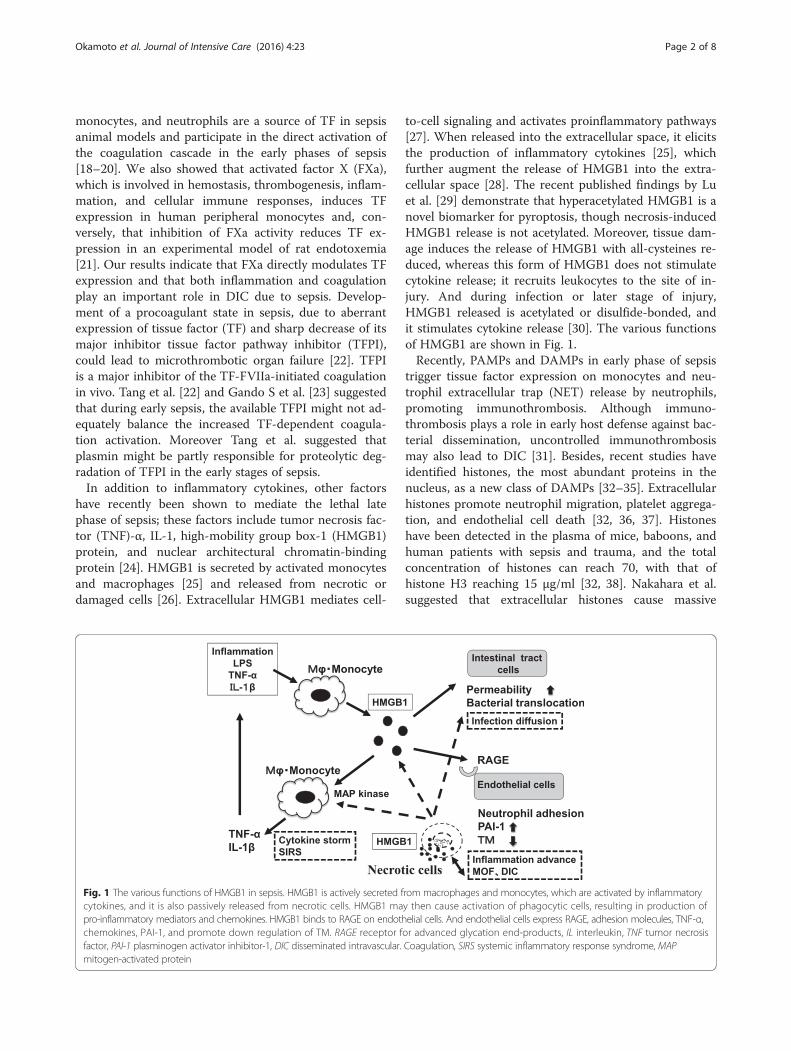
have recently been shown to mediate the lethal latephase of sepsis; these factors include tumor necrosis fac-tor (TNF)-α, IL-1, high-mobility group box-1 (HMGB1)protein, and nuclear architectural chromatin-bindingprotein [24]. HMGB1 is secreted by activated monocytesand macrophages [25] and released from necrotic ordamaged cells [26]. Extracellular HMGB1 mediates cell-
to-cell signaling and activates proinflammatory pathways[27]. When released into the extracellular space, it elicitsthe production of inflammatory cytokines [25], whichfurther augment the release of HMGB1 into the extra-cellular space [28]. The recent published findings by Luet al. [29] demonstrate that hyperacetylated HMGB1 is anovel biomarker for pyroptosis, though necrosis-inducedHMGB1 release is not acetylated. Moreover, tissue dam-age induces the release of HMGB1 with all-cysteines re-duced, whereas this form of HMGB1 does not stimulatecytokine release; it recruits leukocytes to the site of in-jury. And during infection or later stage of injury,HMGB1 released is acetylated or disulfide-bonded, andit stimulates cytokine release [30]. The various functionsof HMGB1 are shown in Fig. 1.Recently, PAMPs and DAMPs in early phase of sepsis
trigger tissue factor expression on monocytes and neu-trophil extracellular trap (NET) release by neutrophils,promoting immunothrombosis. Although immuno-thrombosis plays a role in early host defense against bac-terial dissemination, uncontrolled immunothrombosismay also lead to DIC [31]. Besides, recent studies haveidentified histones, the most abundant proteins in thenucleus, as a new class of DAMPs [32–35]. Extracellularhistones promote neutrophil migration, platelet aggrega-tion, and endothelial cell death [32, 36, 37]. Histoneshave been detected in the plasma of mice, baboons, andhuman patients with sepsis and trauma, and the totalconcentration of histones can reach 70, with that ofhistone H3 reaching 15 μg/ml [32, 38]. Nakahara et al.suggested that extracellular histones cause massive
Fig. 1 The various functions of HMGB1 in sepsis. HMGB1 is actively secreted from macrophages and monocytes, which are activated by inflammatorycytokines, and it is also passively released from necrotic cells. HMGB1 may then cause activation of phagocytic cells, resulting in production ofpro-inflammatory mediators and chemokines. HMGB1 binds to RAGE on endothelial cells. And endothelial cells express RAGE, adhesion molecules, TNF-α,chemokines, PAI-1, and promote down regulation of TM. RAGE receptor for advanced glycation end-products, IL interleukin, TNF tumor necrosisfactor, PAI-1 plasminogen activator inhibitor-1, DIC disseminated intravascular. Coagulation, SIRS systemic inflammatory response syndrome, MAPmitogen-activated protein
Okamoto et al. Journal of Intensive Care (2016) 4:23 Page 2 of 8

thromboembolism associated with consumptive coagu-lopathy, which is diagnostically indistinguishable fromDIC and that rTM binds to histones and neutralizes theprothrombotic action of histones [39]. A mechanism ofDIC and MOF due to sepsis are shown in Fig. 2.Moreover, if the severity of the infectious disease is the
same, coagulopathy of infectious disease in surgically pa-tients is increased by addition of the coagulation dis-order due to surgical stress (Fig.3). In treatment of basicdisease, the surgeons and intensivists must take that co-agulopathy of the surgical stress deteriorates DIC tem-porarily into consideration.
Diagnostic criteria of septic DICDifferent diagnostic criteria of septic DIC have beenestablished by the International Society on Thrombosisand Haemostasis [40], the Japanese Ministry of Health,Labor and Welfare (JMHLW) [41], and the JapaneseAssociation of Acute Medicine (JAAM) [42].Although the criteria of the JAAM are the most spe-
cific for septic DIC [42, 43], a prospective study in Japanfound no significant differences in the odds ratios forprediction of DIC outcomes calculated on the basis ofthese three diagnostic criteria [44]. As the mortality rateof DIC is still high, early diagnosis and treatment arerequired.
Laboratory testsScreening assays (global coagulation tests) using scoringparameters, such as prothrombin time, fibrinogen level,
platelet count, and levels of fibrin-related markers, pro-vide important information about the degree of coagula-tion factor activation and consumption.Examination of DIC scores (based on the JMHLW cri-
teria) at the beginning of DIC treatment showed thatgreater treatment efficacy was achieved in pre-DIC thanin DIC patients [45]. Outcome worsened as the DICscore increased, thus suggesting that both early diagnosisand early treatment of DIC are important. To define thepre-DIC state, we prospectively evaluated global coagu-lation tests, hemostatic molecular markers, and the on-set of DIC within a week after registration [46]. Thelevels of D-dimer and FMC were significantly lower inpatients with pre-DIC than in those without DIC,whereas there were no significant differences in thelevels of thrombin-antithrombin complex (TAT),plasmin-α2plasmin inhibitor complex (PIC), antithrom-bin (AT), and thrombomodulin (TM). However, nomarkers that provided an appropriate cutoff value fordifferentiating between “pre-DIC” and “without DIC” (asdo DIC scores) were identified.
Treatment of septic DICCommon sense dictates that administration of an anti-biotic that specifically targets the infection is the mostimportant therapy in septic DIC. After administering an-tibiotics, surgical drainage at the infection site should beperformed as soon as possible. Physicians should firstadminister treatment for the underlying disease whensepsis is diagnosed [4, 8].
Fig. 2 A mechanism of DIC and MOF due to sepsis. When the pathogen-associated molecular patterns (PAMPs) (for example, endotoxin) anddamage-associated molecular patterns (DAMPs) act on monocytes via TLR and on neutrophils, a reactivated monocyte produce TF, various inflammatorycytokines, and HMGB1, and moreover, detection of PAMPs and DAMPs trigger neutrophil extracellular traps (NETs) release by neutrophils, promotingimmunothrombosis. The uncontrolled immunothrombosis may lead to disseminated intravascular coagulation. And HMGB1 acts on EC and promotesupregulation of TF and downregulation of TM from EC, resulting endothelial cell injury, and microcirculation disorder develops DIC and MOF. TF tissuefactor, TM thrombomodulin, TLR Toll-like receptor, IL-1β interleukin-1β, TNF-α tumor necrosis factor-α, EC endothelial cell, HMGB1 high-mobility groupbox protein 1, PAI plasminogen activator inhibitor, MOF multiple organ failure, NETs neutrophil extracellular traps
Okamoto et al. Journal of Intensive Care (2016) 4:23 Page 3 of 8

AntithrombinAT is a single-stranded glycoprotein with a molecularweight of ca. 59,000. It is synthesized in the liver and in-hibits the activity of thrombin and activated factors X,IX, VII, XI, and XII [47]. Extensive clinical studies havebeen performed in patients with severe sepsis [48–53] todetermine the appropriate dose of AT. Twenty-eightdays of AT treatment did not improve the survival ratein the KyberSept trial [48], which was a multicenter,double-blind phase III study that included 2314 patientswith severe sepsis (a total of 30,000 IU of AT was ad-ministered over 4 days). However, in a subgroup ana-lysis, an improvement in the survival rate on day 90 wasobserved in patients not receiving concomitant heparintreatment; this finding agrees with the results of previousphase II studies supporting the efficacy of AT [54–58]. Arecent Japanese study by Iba et al. [59] used a nonrando-mized, multi-institutional, post-marketing survey to deter-mine the optimal AT dose for treating septic DIC. Theyreported survival rates of 65.2 % in patients receiving1500 IU/day and 74.7 % in patients receiving 3000 IU/day.A logistic regression analysis showed that the higher dose(3000 IU/day) was associated with a better survival out-come [59]. A second survey, in which the baseline ATlevels in patients with septic DIC were less than 40 %,showed a significantly higher rate of DIC resolution and abetter survival outcome in patients receiving 3000 IU/daycompared with those receiving 1500 IU/day [60]. The ratioof bleeding events in the two groups was not significantlydifferent.We conducted a prospective, randomized, controlled
multicenter trial for DIC patients with sepsis and ATlevels of 50 to 80 % to test the hypothesis that concen-trated administration of AT improves DIC, resulting infaster recoveries and better outcomes [61]. Patients re-ceiving AT for 3 days had significantly lower DIC scoresand higher recovery rates than did those who did not
receive AT. This finding suggests that moderate doses ofAT (30 IU/kg per day) improve DIC scores, thereby in-creasing the recovery rate without any risk of bleedingin patients with septic DIC.Tagami et al. [62] performed an analysis using infor-
mation collected from a nationwide administrative data-base in Japan. Patients with severe pneumonia and DIC(n=9075) were divided into an AT group (n=2663) and acontrol (no AT) group (n=6412). Propensity scorematching created a matched cohort of 2194 paired pa-tients who did or not receive AT treatment. The 28-daymortality rate was 9.9 % lower in the AT group than inthe control group. Multiple logistic regression analysesshowed an association between AT use and the 28-daymortality rate (adjusted odds ratio, 0.85).
HeparinThe British guidelines recommend the use of unfractio-nated heparin (UFH) because of its short half-life andavailability of antagonists, especially in patients at a highrisk of bleeding. Japanese guidelines indicate a prefer-ence for low molecular weight heparin because it provedsuperior in improving coagulation abnormalities andcaused fewer hemorrhagic adverse events in a random-ized controlled trial (RCT) conducted in DIC [63]. Inthe HETRASE (A Randomized Clinical Trial of Unfrac-tioned Heparin for Treatment of Sepsis) study [64], theresults of which were reported after publication of theguidelines, and the efficacy of UFH for sepsis was de-nied. Zarychanski R et al. [65] reported that the risk haz-ard ratio for death associated with the use of heparin inseptic patients was 0.88 (95 % confidence interval (CI),0.77–1.00; I2 = 0 %). In addition, Wang et al. [66] also re-ported a decreased mortality associated with heparin use(odds ratio = 0.656, 95 % CI = 0.562–0.765, P < 0.0001).Moreover, Iba et al. [67] reported that both UFH andLMWH attenuated the toxicity of histone H3, in vivo as
Fig. 3 Effect of surgical stress for coagulopathy (DIC) due to infection. If the severity of the infectious disease is the same, coagulopathy ofinfectious disease in surgically patients is increased by addition of the coagulation disorder due to surgical stress. In the treatment of infectioncontrol, the surgeons and intensivists must take that coagulopathy of the surgical stress deteriorates DIC temporarily into consideration
Okamoto et al. Journal of Intensive Care (2016) 4:23 Page 4 of 8

well as in vitro, and that the effects of heparins shown inex vivo study were independent of their anticoagulant ef-fect. They suggested that the administration of heparincould become a treatment of choice for patients suffer-ing from severe sepsis.
ThrombomodulinTM is an endothelial anticoagulant cofactor that playsan important role in the regulation of intravascular co-agulation [68]. It accelerates the thrombin-catalyzedconversion of protein C to activated protein C, which in-hibits monocyte and macrophage activation [69, 70] andconsequently suppresses the production of inflammatorycytokines such as TNF-α and IL-1β [70]. In addition, re-cent studies have shown that TM binds to HMGB1 toprevent its interaction with the receptors for advancedglycation end-products [71]. We reported that TM notonly binds HMGB1 but also aids the proteolytic cleavageof HMGB1 by thrombin [72]. These findings highlightthe novel anti-inflammatory actions of TM.We investigated the effects of soluble recombinant hu-
man TM on the production of inflammatory cytokinesand the plasma level of HMGB1 in an experimentalendotoxemia model [73]. Endotoxemia was induced inrats via a bolus intravenous injection of 4 mg/kg lipo-polysaccharide (LPS). Recombinant TM (1 mg/kg) wasadministered as a bolus injection 30 min before or 4 hafter LPS. LPS increased the plasma levels of TNF-α andIL-1β, which peaked at 1 and 3 h, respectively, and overtime, the plasma levels of HMGB1. Even when its ad-ministration was delayed, recombinant TM markedlyinhibited the LPS-induced increase in plasma levels ofHMGB1 (Fig. 4) and the thrombin-AT complex, as wellas the increase in liver dysfunction and mortality. The
use of recombinant TM may therefore be beneficial fortreatment of septic patients.In a Japanese phase III randomized control trial (RCT)
in which 227 DIC patients with 125 hematological ma-lignancies and 102 infections (sepsis) received recombin-ant TM or unfractionated heparin (UFH), the rate ofresolution of DIC was 66.1 and 49.9 %, respectively [74].The rate of disappearance of bleeding was 35.2 % in therecombinant TM group and 20.9 % in the UFH group,and the 28-day mortality rate was 28.0 and 34.6 %, re-spectively. In an analysis of 80 patients with infectiousDIC, the rate of resolution of DIC was 63.2 % in theUFH group and 73.2 % in the recombinant TM group[75]. In an international phase II RCT of 750 septic pa-tients with suspected DIC, the 28-day mortality ratewas 17.8 % in the recombinant TM group and 21.6 %in the placebo group [76]; there was a tendency towarda low rate in the TM group, although the differencewas not significant (P = 0.273). An international phaseIII clinical trial evaluating the efficacy of TM in pa-tients with severe sepsis and coagulopathy is ongoing inthe USA, South America, Asia, Australia, the EuropeanUnion, and other countries (https://clinicaltrials.gov/ct2/show/NCT01598831?term=ART-123&rank=2).On the other hand, Tagami et al. [77] found that re-
combinant TM was not an effective treatment forsepsis-associated DIC following severe pneumonia. Thisconclusion was based on propensity scores and an in-strumental variable analysis of information obtainedfrom the Japanese Diagnosis Procedure Combination(JDPC) inpatient database, a nationwide administrativedatabase. No significant difference in the 28-day mortal-ity rate was documented between the two groups in apropensity-matched analysis.
Fig. 4 Effect of rTM on the plasma levels of HMGB1. Temporal changes in plasma HMGB1 concentrations after injection of lipopolysaccharide(LPS). Rats were given saline plus LPS (closed squares); pretreatment of recombinant human soluble thrombomodulin (rTM), LPS plus saline (closedcircles); or saline, LPS plus delayed treatment of rTM (closed triangles). All data represent the mean and SEM (n = 6 per group). [73] *P < 0.05 (vs.the LPS group). #P < 0.01 (vs. the LPS group). rTM recombinant thrombomodulin
Okamoto et al. Journal of Intensive Care (2016) 4:23 Page 5 of 8

We also evaluated the efficacy of recombinant TM forDIC using the JDPC database [78–80]. We found thatthe frequency of use of AT, heparin, and protease inhibi-tors decreased from 2010 to 2012 in Japan, while that ofrecombinant TM significantly increased (25.1, 43.1, and56.8 % in 2010, 2011, and 2012, respectively; P < 0.001).Logistic regression analysis showed that the study periodwas associated with the use of recombinant TM in pa-tients with DIC. The odds ratio (OR) was 2.34 (95 %confidence interval [CI], 2.12–2 to 58; P < 0.001) in 2011compared with 4.34 (95 % CI, 3.94–4.79; P < 0.001) in2012. Large hospital size was the most significant factorassociated with the use of recombinant TM in patientswith DIC (OR, 3.14; 95 % CI, 2.68–3.66; P < 0.001). Theuse of recombinant TM has dramatically increased, anda large hospital size was significantly associated with in-creased use from 2010 to 2012 in Japan. We found nosignificant difference in the in-hospital mortality rate be-tween patients receiving AT and recombinant TM. How-ever, the administration of recombinant TM wassignificantly associated with lower hospitalization timesand medical costs during hospitalization.
ConclusionsThis review discussed the mechanisms that underlie theinteraction between sepsis and DIC and the effects of sep-sis on the coagulation system, as highlighted by our data.Further investigation of the efficacy of recombinant TMand AT in countries other than Japan, as well as the moni-toring of medical costs incurred during hospitalization,will help validate the use of TM and AT for treatment ofseptic DIC.
AbbreviationsAT: antithrombin; CI: confidence interval; DAMPs: damage-associatedmolecular patterns; DIC: disseminated intravascular coagulation;FXa: activated factor X; HMGB1: high-mobility group box-1; IL: interleukin;JAAM: Japanese Association of Acute Medicine; JDPC: Japanese DiagnosisProcedure Combination; JMHLW: Japanese Ministry of Health, Labor andWelfare; LPS: lipopolysaccharide; OR: odds ratio; PAMPs: pathogen-associatedmolecular patterns; PAR: protease-activated receptor; PIC: plasmin-α2plasmininhibitor complex; RCT: randomized control trial; TAT: thrombin-antithrombincomplex; TF: tissue factor; TM: thrombomodulin; TNF-α: tumor necrosisfactor; UHF: unfractionated heparin.
Competing interestsNone of the authors disclose any financial or personal relationships withother people or organizations that could inappropriately influence (bias)their work. Examples of potential conflicts of interest include employment,consultancies, stock ownership, honoraria, paid expert testimony, patentapplications/registrations, and grants or other funding.
Authors’ contributionsKO mainly contributed to write this paper. TT and YS mainly contributed toreview references. All of authors discussed for this review. All authors readand approved the final manuscript.
AcknowledgementsThis study was supported in part by research grants from the JapaneseMinistry of Health, Labour and Welfare and the Japanese Ministry ofEducation, Science, Sports and Culture.
Received: 23 November 2015 Accepted: 4 March 2016
References1. Kinasewitz GT, Yan SB, Basson B, Comp P, Russell JA, Cariou A, Um SL,
Utterback B, Laterre PF, Dhainaut JF, PROWESS Sepsis Study Group.Universal changes in biomarkers of coagulation and inflammation occur inpatients with severe sepsis, regardless of causative micro-organism[ISRCTN74215569]. Crit Care. 2004;8:R82–90.
2. Levi M, ten Cate H. Disseminated intravascular coagulation. N Engl J Med.1999;341:586–92.
3. Bakhtiari K, Meijers JC, de Jonge E, Levi M. Prospective validation of theInternational Society of Thrombosis and Haemostasis scoring system fordisseminated intravascular coagulation. Crit Care Med. 2004;32:2416–21.
4. Dhainaut JF, Yan SB, Joyce DE, Pettilä V, Basson B, Brandt JT, Sundin DP, LeviM. Treatment effects of drotrecogin alfa (activated) in patients with severesepsis with or without overt disseminated intravascular coagulation. JThromb Haemost. 2004;2:1924–33.
5. Dellinger RP, Levy MM, Rhodes A, Annane D, Gerlach H, Opal SM, SevranskyJE, Sprung CL, Douglas IS, Jaeschke R, Osborn TM, Nunnally ME, TownsendSR, Reinhart K, Kleinpell RM, Angus DC, Deutschman CS, Machado FR,Rubenfeld GD, Webb S, Beale RJ, Vincent JL, Moreno R. Surviving SepsisCampaign Guidelines Committee including The Pediatric Subgroup.Surviving Sepsis Campaign: international guidelines for management ofsevere sepsis and septic shock, 2012. Intensive Care Med. 2013;39(2):165–228.
6. Dellinger RP, Levy MM, Rhodes A, Annane D, Gerlach H, Opal SM, SevranskyJE, Sprung CL, Douglas IS, Jaeschke R, Osborn TM, Nunnally ME, TownsendSR, Reinhart K, Kleinpell RM, Angus DC, Deutschman CS, Machado FR,Rubenfeld GD, Webb SA, Beale RJ, Vincent JL, Moreno R, Surviving SepsisCampaign Guidelines Committee including the Pediatric Subgroup.Surviving sepsis campaign: international guidelines for management ofsevere sepsis and septic shock: 2012. Crit Care Med. 2013;41(2):580–637.
7. Oda S, Aibiki M, Ikeda T, Imaizumi H, Endo S, Ochiai R, Kotani J, Shime N,Nishida O, Noguchi T, Matsuda N, Hirasawa H. Sepsis Registry Committee ofThe Japanese Society of Intensive Care Medicine. The Japanese guidelinesfor the management of sepsis. J Intensive Care.2014;2(1):55. doi:10.1186/s40560-014-0055-2. eCollection 2014.
8. Wada H, Asakura H, Okamoto K, Iba T, Uchiyama T, Kawasugi K, Koga S,Mayumi T, Koike K, Gando S, Kushimoto S, Seki Y, Madoiwa S, Maruyama I,Yoshioka A. Japanese Society of Thrombosis Hemostasis/DIC subcommittee:expert consensus for the treatment of disseminated intravascularcoagulation in Japan. Thromb Res. 2010;125:6–11.
9. Wada H, Okamoto K, Iba T, Kushimoto S, Kawasugi K, Gando S, Madoiwa S,Uchiyama T, Mayumi T, Seki Y, Japanese Society of Thrombosis Hemostasis/DIC subcommittee. Addition of recommendations for the use ofrecombinant human thrombomodulin to the “Expert consensus for thetreatment of disseminated intravascular coagulation in Japan”. Thromb Res.2014;134(4):924–5.
10. Levi M, van der Poll T. Inflammation and coagulation. Crit Care Med.2010;38:S26–34.
11. O’Brien M. The reciprocal relationship between inflammation and coagulation.Top Companion Anim Med. 2012;27:46–52.
12. Levi M, van der Poll T, ten Cate H, van Deventer SJ. The cytokine-mediatedimbalance between coagulant and anticoagulant mechanisms in sepsis andendotoxemia. Eur J Clin Invest. 1997;27:3–9.
13. Itoh H, Okamoto K. The mechanism behind coagulation disorders andorgan dysfunction due to abdominal sepsis. J Abdomin Emerg Med.2002;22(5):729–37 (in Japanese).
14. Johnson K, Choi Y, DeGroot E, Samuels I, Creasey A, Aardwn I. Potentialmechanism for a proinflammatory vascular cytokine response tocoagulation activation. J Immunol. 1998;160:5130–5.
15. Johnson K, Aarden L, Choi Y, De Groot E, Creasey A. The proinflammatorycytokine response to coagulation and endotoxin in whole blood. Blood.1996;87:5051–60.
16. Esmon CT. Does inflammation contribute to thrombotic events?Haemostasis. 2000;30 Suppl 2:34–40.
17. Camerer E, Kolsto AB, Prydz H. Cell biology of tissue factor, the principalinitiator of blood coagulation. Thromb Res. 1996;81:1–41.
18. Higure A, Okamoto K, Hirata K, Todoroki H, Nagafuchi Y, Takeda S, Katoh H,Itoh H, Ohsato K, Nakamura S. Macrophages and neutrophils infiltrating into
Okamoto et al. Journal of Intensive Care (2016) 4:23 Page 6 of 8

the liver are responsible for tissue factor expression in a rabbit model ofacute obstructive cholangitis. Thromb Haemost. 1996;75(5):791–5.
19. Todoroki H, Nakamura S, Higure A, Okamoto K, Takeda S, Nagata N, Itoh H,Ohsato K. Neutrophils express tissue factor in a monkey model of sepsis.Surgery. 2000;127(2):209–16.
20. Nakamura S, Imamura T, Okamoto K. Tissue factor in neutrophils: yes.J Thromb Haemost. 2004;2(2):214–7.
21. Akahane K, Okamoto K, Kikuchi M, Todoroki H, Higure A, Sugiyama T,Kitahara K, Takeda S, Itoh H, Ohsato K. Inhibition of factor Xa suppresses theexpression of tissue factor in human monocytes and lipopolysaccharideinduced endotoxemia in rats. Surgery. 2001;130(5):809–18.
22. Tang H, Ivanciu L, Popescu N, Peer G, Hack E, Lupu C, Taylor FB Jr.Lupu F. Sepsis-induced coagulation in the baboon lung isassociated with decreased tissue factor pathway inhibitor. Am JPathol. 2007;171:1066–77.
23. Gando S, Kameue T, Morimoto Y, Matsuda N, Hayakawa M, Kemmotsu O.Tissue factor production not balanced by tissue factor pathway inhibitor insepsis promotes poor prognosis. Crit Care Med. 2002;30:1729–34.
24. Wang H, Yang H, Czura CJ, Sama AE, Tracey KJ. HMGB1 as a late mediator oflethal systemic inflammation. Am J Respir Crit Care Med. 2001;164:1768–73.
25. Wang H, Bloom O, Zhang M, Vishnubhakat JM, Ombrellino M, Che J, Frazier A,Yang H, Ivanova S, Borovikova L, Manogue KR, Faist E, Abraham E, Andersson J,Andersson U, Molina PE, Abumrad NN, Sama A, Tracey KJ. HMG-1 as a latemediator of endotoxin lethality in mice. Science. 1999;285:248–51.
26. Scaffidi P, Misteli T, Bianchi ME. Release of chromatin protein HMGB1 bynecrotic cells triggers inflammation. Nature. 2002;418:191–5.
27. Schmidt AM, Yan SD, Yan SF, Stern DM. The multiligand receptor RAGE as aprogression factor amplifying immune and inflammatory responses. J ClinInvest. 2001;108:949–55.
28. Wang H, Vishnubhakat JM, Bloom O, Zhang M, Ombrellino M, Sama A,Tracey KJ. Proinflammatory cytokines (tumor necrosis factor and interleukin1) stimulate release of high mobility group protein-1 by pituicytes. Surgery.1999;126:389–92.
29. Lu B, Nakamura T, Inouye K, Li J, Tang Y, Lundbäck P, Valdes-Ferrer SI,Olofsson P, Kalb T, Roth J, Zou Y, Erlandsson-Harris H, Yang H, Ting JP,Wang H, Andersson U, Antoine DJ, Chavan SS, Hotamisligil GS, Tracey KJ.Novel role of PKR in inflammasome activation and HMGB1 release. Nature.2012;488:670–4.
30. Yang H, Antoine DJ, Andersson U, Tracey KJ. The many faces of HMGB1:molecular structure-functional activity in inflammation, apoptosis, andchemotaxis. J Leukoc Biol. 2013;93:865–73.
31. Ito T. PAMPs and DAMPs as triggers for DIC. J Intensive Care. 2014;2(1):67.32. Xu J, Zhang X, Pelayo R, Monestier M, Ammollo CT, Semerano F, Taylor FB,
Esmon NL, Lupu F, Esmon CT. Extracellular histones are major mediators ofdeath in sepsis. Nat Med. 2009;15:1318–21.
33. Chaput C, Zychlinsky A. Sepsis: the dark side of histones. Nat Med.2009;15:1245–6.
34. Huang H, Evankovich J, Yan W, Nace G, Zhang L, Ross M, Liao X, Billiar T, Xu J,Esmon CT, Tsung A. Endogenous histones function as alarmins in sterileinflammatory liver injury through Toll-like receptor 9 in mice. Hepatology.2011;54:999–1008.
35. Abrams ST, Zhang N, Manson J, Liu T, Dart C, Baluwa F, Wang SS, Brohl K,Kipar A, Yu W, Wang G, Toh CH. Circulating histones are mediators oftrauma-associated lung injury. Am J Respir Crit Care Med. 2013;187:160–9.
36. Fuchs TA, Brill A, Duerschmied D, Schatzberg D, Monestier M, Myers Jr DD,Wrobleski SK, Wakefield TW, Hartwig JH, Wagner DD. Extracellular DNA trapspromote thrombosis. Proc Natl Acad Sci U S A. 2010;107:15880–5.
37. Fuchs TA, Bhandari AA, Wagner DD. Histones induce rapid and profoundthrombocytopenia in mice. Blood. 2011;118:3708–14.
38. Xu J, Lupu F, Esmon CT. Inflammation, innate immunity and bloodcoagulation. Hamostaseologie. 2010;30:5–9.
39. Nakahara M, Ito T, Kawahara K, Yamamoto M, Nagasato T, Shrestha B,Yamada S, Miyauchi T, Higuchi K, Takenaka T, Yasuda T, Matsunaga A,Kakihana Y, Hashiguchi T, Kanmura Y, Maruyama I. Recombinantthrombomodulin protects mice against histone-induced lethalthromboembolism. PLoS One. 2013;8(9):e75961.
40. Taylor FB, Toh CH, Hoots WK, Wada H, Levi M. Towards definition, clinicaland laboratory criteria, and a scoring system for disseminated intravascularcoagulation. Thromb Haemost. 2001;86:1327–30.
41. Kobayashi N, Maegawa T, Takada M, Tanaka H, Gonmori H. Criteria fordiagnosis of DIC based on the analysis of clinical and laboratory findings in
345 DIC patients collected by the Research Committee on DIC in Japan. BiblHaematol. 1983;49:265–75.
42. Gando S, Iba T, Eguchi Y, Ohtomo Y, Okamoto K, Koseki K, Mayumi T,Murata A, Ikeda T, Ishikura H, Ueyama M, Ogura H, Kushimoto S, Saitoh D,Endo S, Shimazaki S, Japanese Association for Acute Medicine DisseminatedIntravascular Coagulation (JAAM DIC) Study Group. A multicenter, prospectivevalidation of disseminated intravascular coagulation diagnostic criteria forcritically ill patients: comparing current criteria. Crit Care Med. 2006;34:625–31.
43. Ogura H, Gando S, Iba T, Eguchi Y, Ohtomo Y, Okamoto K, Koseki K, Mayumi T,Murata A, Ikeda T, Ishikura H, Ueyama M, Kushimoto S, Saitoh D, Endo S,Shimazaki S. Japanese Association for Acute Medicine DisseminatedIntravascular Coagulation (JAAM DIC) Study Group. SIRS-associatedcoagulopathy and organ dysfunction in critically ill patients withthrombocytopenia. Shock. 2007;28:411–7.
44. Takemitsu T, Wada H, Hatada T, Ohmori Y, Ishikura K, Takeda T, Sugiyama T,Yamada N, Maruyama K, Katayama N, Isaji S, Shimpo H, Kusunoki M, NoboriT. Prospective evaluation of three different diagnostic criteria fordisseminated intravascular coagulation. Thromb Haemost. 2011;105:40–4.
45. Wada H, Wakita Y, Nakase T, Shimura M, Hiyoyama K, Nagaya S, Mori Y,Shiku H. Outcome of disseminated intravascular coagulation in relation tothe score when treatment was begun. Thromb Haemost. 1995;74:848–52.
46. Okamoto K, Wada H, Hatada T, Uchiyama T, Kawasugi K, Mayumi T, Gando S,Kushimoto S, Seki Y, Madoiwa S, Asakura H, Koga S, Iba T, Maruyama I,Japanese Society of Thrombosis Hemostasis/DIC subcommittee. Frequencyand hemostatic abnormalities in pre-DIC patients. Thromb Res. 2010;126:74–8.
47. Rosenberg RD, Damus PS. The purification and mechanism of action ofhuman antithrombin-heparin cofactor. J Biol Chem. 1973;248:6490–505.
48. Warren BL, Eid A, Singer P, Pillay SS, Carl P, Novak I, Chalupa P, Atherstone A,Pénzes I, Kübler A, Knaub S, Keinecke HO, Heinrichs H, Schindel F, Juers M,Bone RC, Opal SM; KyberSept Trial Study Group. High-dose antithrombin insevere sepsis. A randomized controlled trial. JAMA. 2001;286:1869–78.
49. Vinazzer H. Therapeutic use of antithrombin III in shock and disseminatedintravascular coagulation. Semin Thromb Hemost. 1989;15:347–52.
50. Dzinic L, Marenovic T, Lakic-Trajkovic Z. Clinical study of the therapeutic value ofKybernin in the treatment of antithrombin III deficiency. Med Pregl. 1991;44:245–8.
51. Albert J, Blomqvist H, Gardlund B, Jakobsson J, Svensson J, Blombäck M.Effect of antithrombin concentrate on haemostatic variables in critically illpatients. Acta Anaesthesiol Scand. 1992;36:745–52.
52. Fourrier F, Chopin C, Huart JJ, Runge I, Caron C, Goudemand J. Double-blind,placebocontrolled trial of antithrombin III concentrates in septic shock withdisseminated intravascular coagulation. Chest. 1993;104:882–8.
53. Waydhas C, Nast-Kolb D, Gippner-Steppert C, Trupka A, Pfundstein C,Schweiberer L, Jochum M. High-dose antithrombin III treatment of severelyinjured patients: results of a prospective study. J Trauma. 1998;45:931–40.
54. Baudo F, Caimi TM, de Cataldo F, Ravizza A, Arlati S, Casella G, Carugo D,Palareti G, Legnani C, Ridolfi L, Rossi R, D'Angelo A, Crippa L, Giudici D,Gallioli G, Wolfler A, Calori G. Antithrombin III (ATIII) replacement therapy inpatients with sepsis and/or postsurgical complications: a controlled double-blind, randomized, multicenter study. Intensive Care Med. 1998;24:336–42.
55. Eisele B, Lamy M, Thijs LG, Keinecke HO, Schuster HP, Matthias FR, Fourrier F,Heinrichs H, Delvos U. Antithrombin III in patients with severe sepsis. Arandomized, placebo-controlled, double-blind multicenter trial plus a meta-analysis on all randomized, placebo-controlled, double blind trials withantithrombin III in severe sepsis. Intensive CareMed. 1998;24:663–72.
56. Hoffmann JN, Muhlbayer D, Jochum M, Inthorn D. Effect of long-term andhigh-dose antithrombin supplementation on coagulation and fibrinolysis inpatients with severe sepsis. Crit Care Med. 2004;32:1851–9.
57. Kienast J, Juers M, Wiedermann CJ, Hoffmann JN, Ostermann H, Strauss R,Keinecke HO, Warren BL, Opal SM, KyberSept investigators. Treatmenteffects of high-dose antithrombin without concomitant heparin in patientswith severe sepsis with or without disseminated intravascular coagulation. JThromb Haemost. 2006;4:90–7.
58. Wiedermann CJ, Kaneider N. A systematic review of antithrombinconcentrate use in patients with disseminated intravascular coagulation ofsevere sepsis. Blood Coagul Fibrinolysis. 2006;17:521–6.
59. Iba T, Saito D, Wada H, Asakura H. Efficacy and bleeding risk of antithrombinsupplementation in septic disseminated intravascular coagulation: aprospective multicenter survey. Thromb Res. 2012;130:e129–33.
60. Iba T, Saitoh D, Wada H, Asakura H. Efficacy and bleeding risk ofantithrombin supplementation in septic disseminated intravascularcoagulation: a secondary survey. Crit Care. 2014;18:497.
Okamoto et al. Journal of Intensive Care (2016) 4:23 Page 7 of 8

61. Gando S, Saitoh D, Ishikura H, Ueyama M, Otomo Y, Oda S, Kushimoto S,Tanjoh K, Mayumi T, Ikeda T, Iba T, Eguchi Y, Okamoto K, Ogura H, Koseki K,Sakamoto Y, Takayama Y, Shirai K, Takasu O, Inoue Y, Mashiko K, Tsubota T,Endo S. A randomized, controlled, multicenter trial of the effects ofantithrombin on disseminated intravascular coagulation in patients withsepsis. Crit Care. 2013;17:R297.
62. Tagami T, Matsui H, Horiguchi H, Fushimi K, Yasunaga H. Antithrombin andmortality in severe pneumonia patients with sepsis-associated disseminatedintravascular coagulation: an observational nationwide study. J ThrombHaemost. 2014;12:1470–9.
63. Sakuragawa N, Hasegawa H, Maki M, Nakagawa M, Nakashima M. Clinicalevaluation of low-molecular-weight heparin (FR-860) on disseminatedintravascular coagulation (DIC)—a multicenter co-operative double-blindtrial in comparison with heparin. Thromb Res. 1993;72:475–500.
64. Jaimes F, De La Rosa G, Morales C, Fortich F, Arango C, Aguirre D, Mun˜ozA. Unfractioned heparin for treatment of sepsis: a randomized clinical trial(The HETRASE study). Crit Care Med. 2009;37:1185–96.
65. Zarychanski R, Abou-Setta AM, Kanji S, Turgeon AF, Kumar A, Houston DS,Rimmer E, Houston BL, McIntyre L, Fox-Robichaud AE, Hébert P, Cook DJ,Fergusson DA, Canadian Critical Care Trials Group. The efficacy and safety ofheparin in patients with sepsis: a systematic review and metaanalysis. CritCare Med. 2015;43:511–28.
66. Wang C, Chi C, Guo L, Wang X, Guo L, Sun J, Sun B, Liu S, Chang X, Li E.Heparin therapy reduces 28-day mortality in adult severe sepsis patients: asystematic review and meta-analysis. Crit Care. 2014;18:563.
67. Iba T, Hashiguchi N, Nagaoka I, Tabe Y, Kadota K, Sato K. Heparinsattenuated histone-mediated cytotoxicity in vitro and improved the survivalin a rat model of histone-induced organ dysfunction. Intensive Care MedExp. 2015;3(1):36. doi:10.1186/s40635-015-0072-z. Epub 2015 Dec 29.
68. Esmon CT. The interactions between inflammation and coagulation.Br J Haematol. 2005;131:417–30.
69. Esmon CT. The regulation of natural anticoagulant pathways. Science.1987;235:1348–52.
70. Grey ST, Tsuchida A, Hau H, Orthner CL, Salem HH, Hancock WW. Selectiveinhibitory effects of the anticoagulant activated protein C on the responsesof human mononuclear phagocytes to LPS, IFN gamma, or phorbol ester.J Immunol. 1994;153:3664–72.
71. Abeyama K, Stern DM, Ito Y, Kawahara K, Yoshimoto Y, Tanaka M,Uchimura T, Ida N, Yamazaki Y, Yamada S, Yamamoto Y, Yamamoto H,Iino S, Taniguchi N, Maruyama I. The N-terminal domain ofthrombomodulin sequesters high-mobility group-B1 protein, a novelantiinflammatory mechanism. J Clin Invest. 2005;115:1267–74.
72. Ito T, Kawahara K, Okamoto K, Yamada S, Yasuda M, Imaizumi H, Nawa Y,Meug X, Shrestha B, Hashiguchi T, Maruyama I. Proteolytic cleavage of highmobility group box 1 protein by thrombin-thrombomodulin complexes.Arterioscler Thromb Biol. 2008;28:1825–30.
73. Nagato M, Okamoto K, Abe Y, Higure A, Yamaguchi K. Recombinant humansoluble thrombomodulin (ART-123) decreases the plasma HMGB1 levels,while improving the acute liver injury and survival rates in experimentalendotoxemia. Crit Care Med. 2009;37:2181–6.
74. Saito H, Maruyama I, Shimazaki S, Yamamoto Y, Aikawa N, Ohno R,Hirayama A, Matsuda T, Asakura H, Nakashima M, Aoki N. Efficacy and safetyof recombinant human soluble thrombomodulin(ART-123) in disseminated intravascular coagulation: results of a phase III,randomized, double-blind clinical trial. J Thromb Haemost. 2007;5:31–41.
75. Aikawa N, Shimazaki S, Yamamoto Y, Saito H, Maruyama I, Ohno R,Hirayama A, Aoki Y, Aoki N. Thrombomodulin alfa in the treatment ofinfectious patients complicated by disseminated intravascular coagulation:subanalysis from the phase 3 trial. Shock. 2011;35:349–54.
76. Vincent JL, Ramesh MK, Ernest D, Larosa SP, Pach J, Aikawa N, Hoste E, LevyH, Hirman J, Levi M, Daga M, Kutsogiannis DJ, Crowther M, Bernard GR,Devriendt J, Puigserver JV, Blanzaco DU, Esmon CT, Parrillo JE, Guzzi L,Henderson SJ, Pothirat C, Mehta P, Fareed J, Talwar D, Tsuruta K, Gorelick KJ,Osawa Y, Kaul I. A randomized, double-blind, placebo-controlled, phase 2bstudy to evaluate the safety and efficacy of recombinant human solublethrombomodulin, ART-123, in patients with sepsis and suspecteddisseminated intravascular coagulation. Crit Care Med. 2013;41:2069–79.
77. Tagami T, Matsui H, Horiguchi H, Fushimmi K, Yasunaga H. Recombinanthuman soluble thrombomodulin and mortality in severe pneumoniapatients with sepsis-associated disseminated intravascular coagulation: anobservational nationwide study. J Thromb Haemost. 2015;13:31–40.
78. Murata A, Okamoto K, Mayumi T, Muramatsu K, Matsuda S. The recent timetrend of outcomes of disseminated intravascular coagulation in Japan: anobservational study based on a national administrative database.J Thrombosis Thrombolysis. 2014;38(3):364–71.
79. Murata A, Okamoto K, Mayumi T, Muramatsu K, Matsuda S. Observationalstudy to compare antithrombin and thrombomodulin for disseminatedintravascular coagulation. Int J Clin Pharm. 2015;37(1):139–47.
80. Murata A, Okamoto K, Mayumi T, Muramatsu K, Matsuda S. Recent changein treatment of disseminated intravascular coagulation in Japan: anepidemiological study based on a National Administrative Database.Clin Appl Thromb Hemost. 2016;22:21-27.
• We accept pre-submission inquiries
• Our selector tool helps you to find the most relevant journal
• We provide round the clock customer support
• Convenient online submission
• Thorough peer review
• Inclusion in PubMed and all major indexing services
• Maximum visibility for your research
Submit your manuscript atwww.biomedcentral.com/submit
Submit your next manuscript to BioMed Central and we will help you at every step:
Okamoto et al. Journal of Intensive Care (2016) 4:23 Page 8 of 8


![Disseminated Intravascular Dic Auto Saved]](https://static.fdocuments.net/doc/165x107/577d229f1a28ab4e1e97d81f/disseminated-intravascular-dic-auto-saved.jpg)


![Successful anticoagulant therapy for disseminated ......including sepsis, disseminated intravascular coagulation (DIC), massive hemorrhage, and uterine infection [13, 14]. We report](https://static.fdocuments.net/doc/165x107/60f79fd72dadc86c41591dfc/successful-anticoagulant-therapy-for-disseminated-including-sepsis-disseminated.jpg)