Coagulation Disorders and Disseminated Intravascular Coagulation (DIC)
Clinical Expert Series
Disseminated Intravascular CoagulationSyndromes in Obstetrics
F. Gary Cunningham, MD, and David B. Nelson, MD
Disseminated intravascular coagulation (DIC) is a syndrome that can be initiated by a myriad ofmedical, surgical, and obstetric disorders. Also known as consumptive coagulopathy, DIC isa common contributor to maternal morbidity and mortality and is associated with up to 25% ofmaternal deaths. The etiopathogenesis of DIC is complex and currently thought to be initiated bytissue factor or thromboplastin, which is released from trophoblastic or fetal tissue, or maternaldecidua or endothelium. Tissue factor activates the coagulation sequence to cause fibrin clottingand its dissolution by the fibrinolysin system. The result of this process can range from mild,clinically insignificant laboratory derangements to marked coagulopathy with bleeding at sitesof minimal trauma. Although clinical recognition varies by disease severity, several organizationshave attempted to standardize the diagnosis through development of scoring systems. Severalimportant—albeit not necessarily common—obstetric disorders associated with DIC include pla-cental abruption, amniotic fluid embolism, sepsis syndrome, and acute fatty liver of pregnancy.More common disorders include severe preeclampsia, hemolysis, elevated liver enzymes, and lowplatelet count syndrome, and massive obstetric hemorrhage. Importantly, many of these disorderseither cause or are associated with substantive obstetric hemorrhage. Treatment of DIC is centeredon two principles. The first is identification and treatment of the underlying disorder. Becausemany women with consumptive coagulopathy also have massive hemorrhage, the second tenet oftreatment is that obstetric complications such as uterine atony or lacerations must be controlledsimultaneously with prompt blood and component replacement for a salutary outcome.
(Obstet Gynecol 2015;126:999–1011)
DOI: 10.1097/AOG.0000000000001110
D isseminated intravascular coagulation (DIC)—also known variably as consumptive coagulop-
athy, defibrination syndrome, and generalized intra-vascular coagulation—is not a disease per se, but rathera clinicopathologic syndrome that can be initiated bya myriad of underlying diseases, conditions, or disor-ders. Specific to the obstetric arena, some form of
consumptive coagulopathy continues to be a concernafter more than a century after DeLee’s description of“temporary hemophilia” that developed in womenwith a placental abruption or a long-dead fetus in1901.1 Resultant hemostatic abnormalities with amni-otic fluid embolism were later described by Ratnoffand colleagues.2–4 Ultimately, this clinicopathologicphenomenon culminates in a systemic intravascularactivation of coagulation that completely disrupts nat-ural hemostasis.5 In severe cases, this ineffective bal-ance of natural anticoagulant mechanisms can resultin widespread fibrin deposition leading to multiorganfailure.
SIGNIFICANCE
The reported incidence of DIC varies because ofdiffering definitions and diagnostic criteria. To putthis condition into the context of obstetric care in theUnited States, Callaghan et al6 reviewed delivery andpostpartum hospitalizations in 2-year increments over
From the Division of Maternal-Fetal Medicine, Department of Obstetrics andGynecology, University of Texas Southwestern Medical Center at Dallas,Dallas, Texas.
Continuing medical education for this article is available at http://links.lww.com/AOG/A704.
Corresponding author: David B. Nelson, MD, Dedman Scholar in Clinical Care,Division of Maternal-Fetal Medicine, Department of Obstetrics and Gynecology,University of Texas Southwestern Medical Center at Dallas, 5323 Harry HinesBoulevard, Dallas, TX 75390-9032; email: [email protected].
Financial DisclosureThe authors did not report any potential conflicts of interest.
© 2015 by The American College of Obstetricians and Gynecologists. Publishedby Wolters Kluwer Health, Inc. All rights reserved.ISSN: 0029-7844/15
VOL. 126, NO. 5, NOVEMBER 2015 OBSTETRICS & GYNECOLOGY 999
Copyright ª by The American College of Obstetriciansand Gynecologists. Published by Wolters Kluwer Health, Inc.
Unauthorized reproduction of this article is prohibited.
a decade from the Nationwide Inpatient Sample.From 1998 to 2009, they found that the prevalenceof DIC had significantly increased from 9.2 to 12.5per 10,000 delivery hospitalizations—a 35% increase.Moreover, DIC complicating postpartum hospitaliza-tions increased 83% over the same time period—1.2–2.2 per 10,000 delivery hospitalizations. For the mostrecent 2-year period of 2010–2011, DIC was reportedto be the second most common severe maternal mor-bidity indicator—32 per 10,000 delivery hospitaliza-tions.7 Even more importantly, DIC was associatedwith nearly one fourth of maternal deaths during thisstudy period.6 Even so, DIC as the sole cause ofmaternal death is relatively uncommon and accountsfor only 0.2% of pregnancy-related deaths in thiscountry.8 Put another way, DIC is rarely the causeof death alone, but it is commonly associated withconditions leading to death. In this context, DIC isone of the most important clinical conditions in termsof maternal morbidity and mortality in obstetrics.
PHYSIOLOGY AND PATHOGENESIS
During pregnancy, a substantive increase in plasmavolume is concomitantly augmented by production ofmost procoagulants.9 Importantly, fibrinogen (factor I)concentration increases approximately 50% above non-pregnant values, and during late pregnancy, it rangesfrom approximately 375 to 620 mg/dL.10 Thus, virtu-ally all clotting factors increase and some of these areshown in Table 1. At the same time, there is a reductionin levels of natural anticoagulants protein C and S andtissue factor pathway inhibitor-1 as well as an acquiredresistance to protein C.11 In addition, profibrinolysin or
plasminogen levels increase but there is also increasedinhibition of fibrinolysis.9 As a result of all of thesealterations, the net result is that pregnancy is a procoa-gulant state.
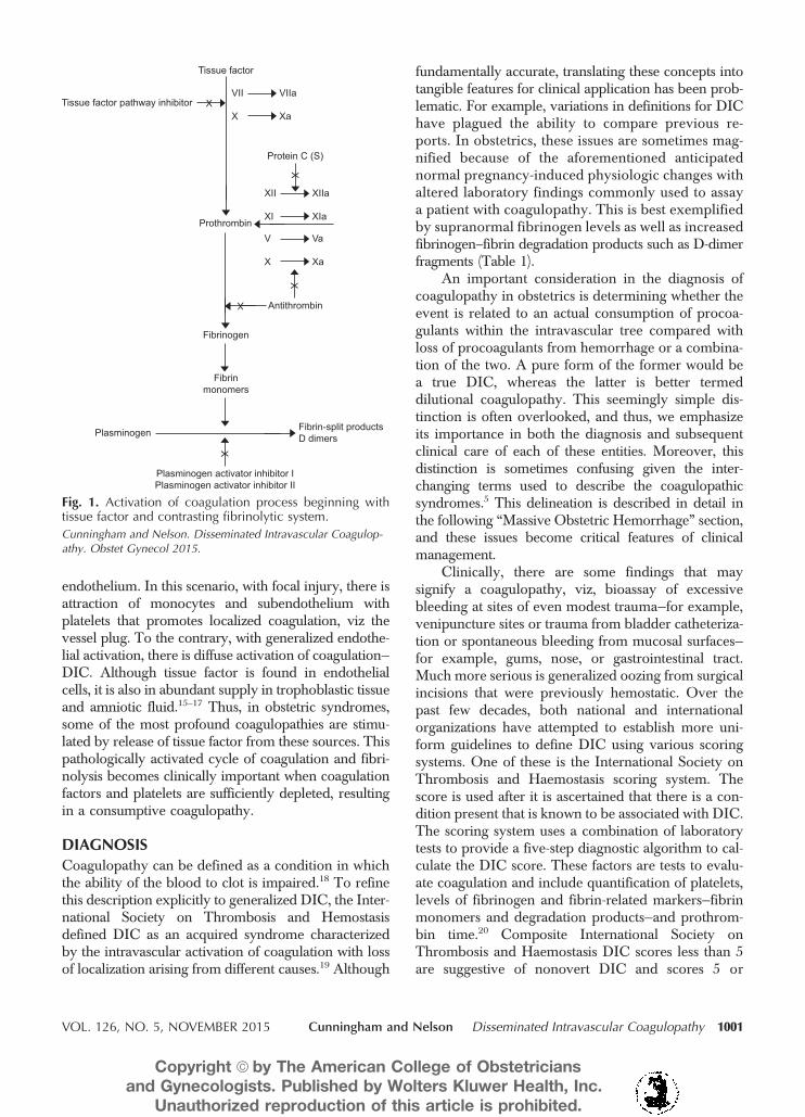
The literature describing the physiologic process ofcoagulation continues to evolve. For many years it wasproposed that there was a coagulation “cascade” or“waterfall.”12 Instead, the current theory is that coagu-lation is primarily initiated by tissue factor, or throm-boplastin, that forms complexes with factors VII andVIIa.11,13 Tissue factor is an integral membrane glyco-protein that is found in highly vascularized organs suchas the brain, lungs, and placenta, and it also can beexpressed constitutively by certain cell types.14 In brief,the development of tissue factor FVIIa complexes ulti-mately generates activated factor X to initiate clotting,whereas the previously labeled “intrinsic” pathway isresponsible for the amplification of this process. Thismain role of tissue factor FVIIa complex in coagulationis depicted in Figure 1. The end result of this coagula-tion process is fibrin formation, which is then counter-balanced by the fibrinolytic system—dedicated to theremoval of excess fibrin. Also shown in the schematicis the fibrinolytic system with plasminogen activated bytissue factor, and this is augmented by thrombin toproduce plasmin, which lyses fibrin and fibrinogen.The end result is production of fibrinogen–fibrin splitproducts, which include D-dimers.
The initiation of DIC begins with the release oftissue factor by any number of pathologic conditions.In most cases, tissue factor is released by damagedsubendothelial tissue and stimulated monocytes,which in turn provoke release of cytokines from the
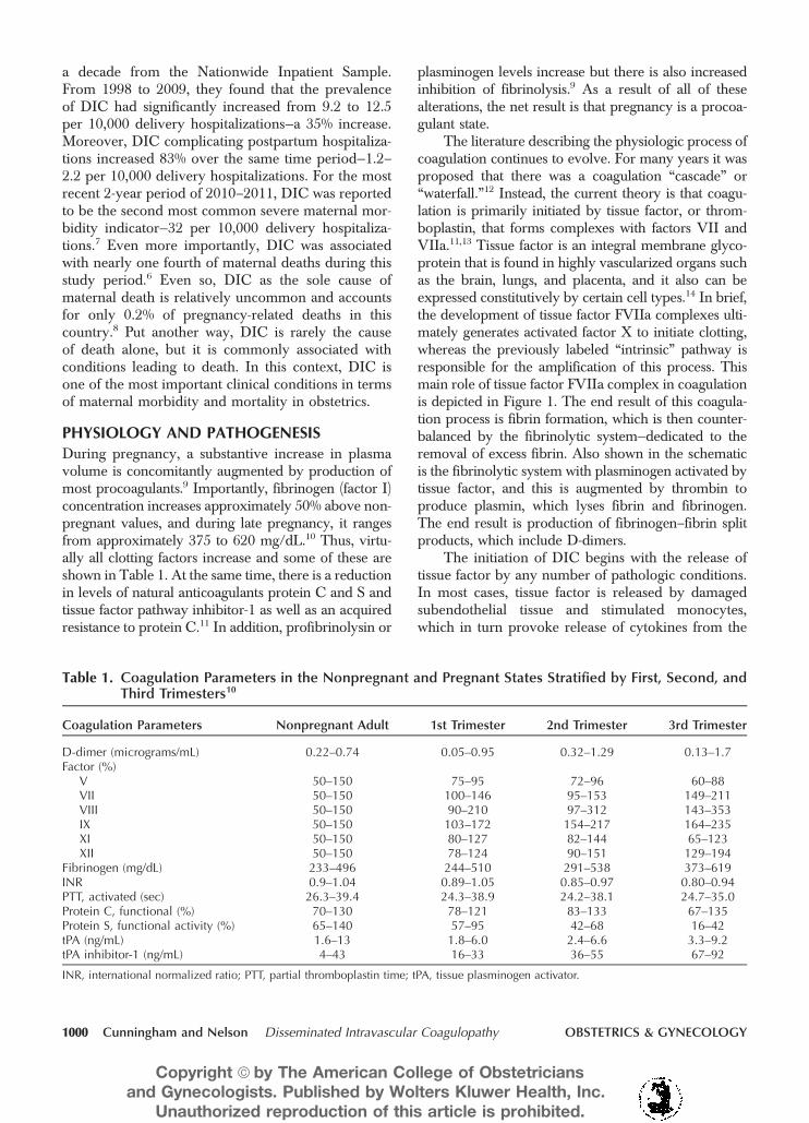
Table 1. Coagulation Parameters in the Nonpregnant and Pregnant States Stratified by First, Second, andThird Trimesters10
Coagulation Parameters Nonpregnant Adult 1st Trimester 2nd Trimester 3rd Trimester
D-dimer (micrograms/mL) 0.22–0.74 0.05–0.95 0.32–1.29 0.13–1.7Factor (%)
V 50–150 75–95 72–96 60–88VII 50–150 100–146 95–153 149–211VIII 50–150 90–210 97–312 143–353IX 50–150 103–172 154–217 164–235XI 50–150 80–127 82–144 65–123XII 50–150 78–124 90–151 129–194
Fibrinogen (mg/dL) 233–496 244–510 291–538 373–619INR 0.9–1.04 0.89–1.05 0.85–0.97 0.80–0.94PTT, activated (sec) 26.3–39.4 24.3–38.9 24.2–38.1 24.7–35.0Protein C, functional (%) 70–130 78–121 83–133 67–135Protein S, functional activity (%) 65–140 57–95 42–68 16–42tPA (ng/mL) 1.6–13 1.8–6.0 2.4–6.6 3.3–9.2tPA inhibitor-1 (ng/mL) 4–43 16–33 36–55 67–92
INR, international normalized ratio; PTT, partial thromboplastin time; tPA, tissue plasminogen activator.
1000 Cunningham and Nelson Disseminated Intravascular Coagulopathy OBSTETRICS & GYNECOLOGY
Copyright ª by The American College of Obstetriciansand Gynecologists. Published by Wolters Kluwer Health, Inc.
Unauthorized reproduction of this article is prohibited.
endothelium. In this scenario, with focal injury, there isattraction of monocytes and subendothelium withplatelets that promotes localized coagulation, viz thevessel plug. To the contrary, with generalized endothe-lial activation, there is diffuse activation of coagulation—DIC. Although tissue factor is found in endothelialcells, it is also in abundant supply in trophoblastic tissueand amniotic fluid.15–17 Thus, in obstetric syndromes,some of the most profound coagulopathies are stimu-lated by release of tissue factor from these sources. Thispathologically activated cycle of coagulation and fibri-nolysis becomes clinically important when coagulationfactors and platelets are sufficiently depleted, resultingin a consumptive coagulopathy.
DIAGNOSIS
Coagulopathy can be defined as a condition in whichthe ability of the blood to clot is impaired.18 To refinethis description explicitly to generalized DIC, the Inter-national Society on Thrombosis and Hemostasisdefined DIC as an acquired syndrome characterizedby the intravascular activation of coagulation with lossof localization arising from different causes.19 Although
fundamentally accurate, translating these concepts intotangible features for clinical application has been prob-lematic. For example, variations in definitions for DIChave plagued the ability to compare previous re-ports. In obstetrics, these issues are sometimes mag-nified because of the aforementioned anticipatednormal pregnancy-induced physiologic changes withaltered laboratory findings commonly used to assaya patient with coagulopathy. This is best exemplifiedby supranormal fibrinogen levels as well as increasedfibrinogen–fibrin degradation products such as D-dimerfragments (Table 1).
An important consideration in the diagnosis ofcoagulopathy in obstetrics is determining whether theevent is related to an actual consumption of procoa-gulants within the intravascular tree compared withloss of procoagulants from hemorrhage or a combina-tion of the two. A pure form of the former would bea true DIC, whereas the latter is better termeddilutional coagulopathy. This seemingly simple dis-tinction is often overlooked, and thus, we emphasizeits importance in both the diagnosis and subsequentclinical care of each of these entities. Moreover, thisdistinction is sometimes confusing given the inter-changing terms used to describe the coagulopathicsyndromes.5 This delineation is described in detail inthe following “Massive Obstetric Hemorrhage” section,and these issues become critical features of clinicalmanagement.
Clinically, there are some findings that maysignify a coagulopathy, viz, bioassay of excessivebleeding at sites of even modest trauma—for example,venipuncture sites or trauma from bladder catheteriza-tion or spontaneous bleeding from mucosal surfaces—for example, gums, nose, or gastrointestinal tract.Much more serious is generalized oozing from surgicalincisions that were previously hemostatic. Over thepast few decades, both national and internationalorganizations have attempted to establish more uni-form guidelines to define DIC using various scoringsystems. One of these is the International Society onThrombosis and Haemostasis scoring system. Thescore is used after it is ascertained that there is a con-dition present that is known to be associated with DIC.The scoring system uses a combination of laboratorytests to provide a five-step diagnostic algorithm to cal-culate the DIC score. These factors are tests to evalu-ate coagulation and include quantification of platelets,levels of fibrinogen and fibrin-related markers—fibrinmonomers and degradation products—and prothrom-bin time.20 Composite International Society onThrombosis and Haemostasis DIC scores less than 5are suggestive of nonovert DIC and scores 5 or
Fig. 1. Activation of coagulation process beginning withtissue factor and contrasting fibrinolytic system.
Cunningham and Nelson. Disseminated Intravascular Coagulop-athy. Obstet Gynecol 2015.
VOL. 126, NO. 5, NOVEMBER 2015 Cunningham and Nelson Disseminated Intravascular Coagulopathy 1001
Copyright ª by The American College of Obstetriciansand Gynecologists. Published by Wolters Kluwer Health, Inc.
Unauthorized reproduction of this article is prohibited.
greater are considered to be compatible with overtDIC. Other than one report of acute fatty liver ofpregnancy,21 this algorithm has not been appliedspecifically to other obstetric conditions that causeDIC.
Although a number of DIC scoring systems havebeen developed with the aim of improving out-comes,22–25 none has been proven more effective thanassessing pertinent laboratory values in the context ofthe clinical situation.
OBSTETRIC CAUSES OF DISSEMINATEDINTRAVASCULAR COAGULATION
One of the central features in the management of DICis recognizing the concomitant, underlying disorder.Although intuitive, identifying these clinical conditionsis paramount in correcting the underlying disorder toreconcile the coagulopathy. For this reason, we havechosen to outline seven of the most common incitingobstetric events that may result in DIC. Examples ofsome parameters used to assess DIC for some of theseconditions are shown in Table 2.
Placental Abruption
The reported incidence of placental abruption variesbut averages approximately 0.5% or one in 200births.26 It is a common cause of perinatal mortalityand approximately 10% of third-trimester stillborn neo-nates are attributed to abruption. According to the Cen-ters for Disease Control and Prevention, placentalabruption was the direct cause of maternal mortalityin 1.1% of pregnancy-related deaths in the UnitedStates from 2006 to 2010.8 Extensive placental abrup-tion causes immediate and frequently profound DIC.This is initiated by large amounts of decidual andplacental-derived tissue factor that rapidly enters the
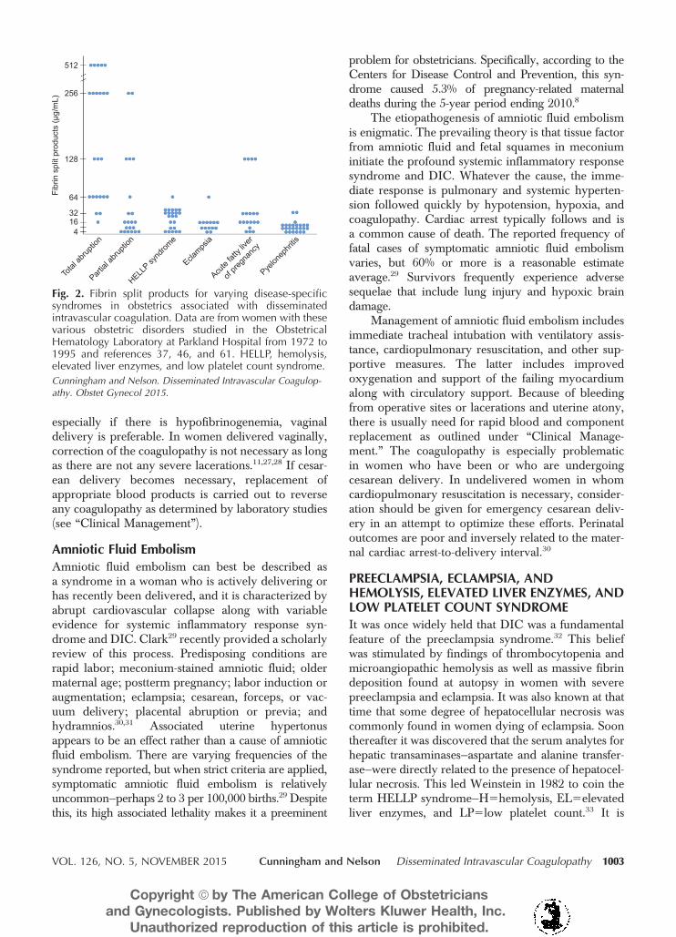
maternal circulation to activate widespread coagulationwith depletion of procoagulants.17 Clotting intensityand plasma fibrinogen depletion are related to severalimportant factors. The first of these is the amount ofplacental tissue involved, and thus total abruptions typ-ically cause more intense DIC than partial ones(Table 2; Fig. 2). Specifically, one third of women withan abruption severe enough to kill the fetus will havea plasma fibrinogen less than 150 mg/dL.27 Second,a woman with a concealed abruption—partial or com-plete—more likely will exhibit DIC because the intra-uterine pressure is higher than in those patients withexternal vaginal bleeding. The third important factor isthe baseline fibrinogen level—recall that plasma fibrin-ogen levels are elevated substantively in late pregnancyand range from approximately 400 to 650 mg/dL.10
Thus, a woman with a fibrinogen level of 600 mg/dLmight have a level of 300 mg/dL postabruption, whichsignifies massive intravascular utilization of fibrinogen,but at the same time, plasma fibrinogen concentrationis sufficient to maintain hemostasis. Lastly, the durationof ongoing DIC caused by an abruption appears to beself-limited. Although the plasma fibrinogen nadir willusually be manifest by 8 hours, continuing blood lossfrom the implantation site will result in procoagulantdeficiency if only packed red cells are transfused.27
Based on the studies of placental abruption byPritchard and Brekken,27 the mainstay of managementincludes immediate resuscitation of hypovolemia as dis-cussed under “Clinical Management.” Adequate intra-venous access is obtained and laboratory studies aresent to assess the degree of coagulopathy. If there isaccompanying severe preeclampsia, magnesium sulfateis given for seizure prevention. The decision concern-ing delivery is based on gestational age and whether thefetus is dead or alive. When there is fetal death, and
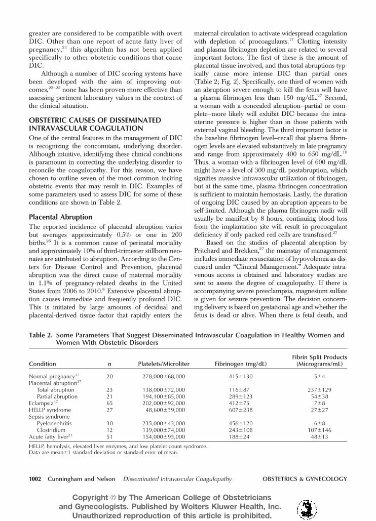
Table 2. Some Parameters That Suggest Disseminated Intravascular Coagulation in Healthy Women andWomen With Obstetric Disorders
Condition n Platelets/Microliter Fibrinogen (mg/dL)Fibrin Split Products(Micrograms/mL)
Normal pregnancy37 20 278,000668,000 4156130 564Placental abruption37
Total abruption 23 138,000672,000 116687 2376129Partial abruption 21 194,100685,000 2896123 54638
Eclampsia37 65 202,000692,000 412675 768HELLP syndrome 27 48,600639,000 6076238 27627Sepsis syndrome
Pyelonephritis 30 235,000643,000 4566120 668Clostridium 12 139,000674,000 2436108 1076146
Acute fatty liver21 51 154,000695,000 188624 48613
HELLP, hemolysis, elevated liver enzymes, and low platelet count syndrome.Data are mean61 standard deviation or standard error of mean.
1002 Cunningham and Nelson Disseminated Intravascular Coagulopathy OBSTETRICS & GYNECOLOGY
Copyright ª by The American College of Obstetriciansand Gynecologists. Published by Wolters Kluwer Health, Inc.
Unauthorized reproduction of this article is prohibited.
especially if there is hypofibrinogenemia, vaginaldelivery is preferable. In women delivered vaginally,correction of the coagulopathy is not necessary as longas there are not any severe lacerations.11,27,28 If cesar-ean delivery becomes necessary, replacement ofappropriate blood products is carried out to reverseany coagulopathy as determined by laboratory studies(see “Clinical Management”).
Amniotic Fluid Embolism
Amniotic fluid embolism can best be described asa syndrome in a woman who is actively delivering orhas recently been delivered, and it is characterized byabrupt cardiovascular collapse along with variableevidence for systemic inflammatory response syn-drome and DIC. Clark29 recently provided a scholarlyreview of this process. Predisposing conditions arerapid labor; meconium-stained amniotic fluid; oldermaternal age; postterm pregnancy; labor induction oraugmentation; eclampsia; cesarean, forceps, or vac-uum delivery; placental abruption or previa; andhydramnios.30,31 Associated uterine hypertonusappears to be an effect rather than a cause of amnioticfluid embolism. There are varying frequencies of thesyndrome reported, but when strict criteria are applied,symptomatic amniotic fluid embolism is relativelyuncommon—perhaps 2 to 3 per 100,000 births.29 Despitethis, its high associated lethality makes it a preeminent
problem for obstetricians. Specifically, according to theCenters for Disease Control and Prevention, this syn-drome caused 5.3% of pregnancy-related maternaldeaths during the 5-year period ending 2010.8
The etiopathogenesis of amniotic fluid embolismis enigmatic. The prevailing theory is that tissue factorfrom amniotic fluid and fetal squames in meconiuminitiate the profound systemic inflammatory responsesyndrome and DIC. Whatever the cause, the imme-diate response is pulmonary and systemic hyperten-sion followed quickly by hypotension, hypoxia, andcoagulopathy. Cardiac arrest typically follows and isa common cause of death. The reported frequency offatal cases of symptomatic amniotic fluid embolismvaries, but 60% or more is a reasonable estimateaverage.29 Survivors frequently experience adversesequelae that include lung injury and hypoxic braindamage.
Management of amniotic fluid embolism includesimmediate tracheal intubation with ventilatory assis-tance, cardiopulmonary resuscitation, and other sup-portive measures. The latter includes improvedoxygenation and support of the failing myocardiumalong with circulatory support. Because of bleedingfrom operative sites or lacerations and uterine atony,there is usually need for rapid blood and componentreplacement as outlined under “Clinical Manage-ment.” The coagulopathy is especially problematicin women who have been or who are undergoingcesarean delivery. In undelivered women in whomcardiopulmonary resuscitation is necessary, consider-ation should be given for emergency cesarean deliv-ery in an attempt to optimize these efforts. Perinataloutcomes are poor and inversely related to the mater-nal cardiac arrest-to-delivery interval.30
PREECLAMPSIA, ECLAMPSIA, ANDHEMOLYSIS, ELEVATED LIVER ENZYMES, ANDLOW PLATELET COUNT SYNDROME
It was once widely held that DIC was a fundamentalfeature of the preeclampsia syndrome.32 This beliefwas stimulated by findings of thrombocytopenia andmicroangiopathic hemolysis as well as massive fibrindeposition found at autopsy in women with severepreeclampsia and eclampsia. It was also known at thattime that some degree of hepatocellular necrosis wascommonly found in women dying of eclampsia. Soonthereafter it was discovered that the serum analytes forhepatic transaminases—aspartate and alanine transfer-ase—were directly related to the presence of hepatocel-lular necrosis. This led Weinstein in 1982 to coin theterm HELLP syndrome—H5hemolysis, EL5elevatedliver enzymes, and LP5low platelet count.33 It is
Fig. 2. Fibrin split products for varying disease-specificsyndromes in obstetrics associated with disseminatedintravascular coagulation. Data are from women with thesevarious obstetric disorders studied in the ObstetricalHematology Laboratory at Parkland Hospital from 1972 to1995 and references 37, 46, and 61. HELLP, hemolysis,elevated liver enzymes, and low platelet count syndrome.
Cunningham and Nelson. Disseminated Intravascular Coagulop-athy. Obstet Gynecol 2015.
VOL. 126, NO. 5, NOVEMBER 2015 Cunningham and Nelson Disseminated Intravascular Coagulopathy 1003
Copyright ª by The American College of Obstetriciansand Gynecologists. Published by Wolters Kluwer Health, Inc.
Unauthorized reproduction of this article is prohibited.
generally agreed that HELLP syndrome is usuallydiagnosed in preeclamptic women by the triad ofthrombocytopenia, elevated hepatic transaminases—aspartate transferase and alanine transferase, and micro-angiopathic hemolysis detected either by abnormallyhigh lactate dehydrogenase levels or abnormally lowhaptoglobin levels.34,35 Generally speaking, sicker pa-tients have progressively more abnormal laboratoryfindings. Even so, the principal differentiating factor isthat the vast majority of women with preeclampsia orHELLP syndromes do not have evidence of liver dys-function. Appreciable hepatic dysfunction characterizedby low cholesterol and high bilirubin levels, low levelsof fibrinogen and other procoagulants, and a prolongedprothrombin time are characteristic of acute fatty liverof pregnancy36 (see “Acute Fatty Liver of Pregnancy”).
Thus, the preeclampsia syndrome includes plate-let activation, dysfunction, and increased adherencealong with microangiopathic hemolysis.9 At the sametime, however, preeclampsia, eclampsia, and HELLPsyndrome are usually not associated with clinicallyrelevant DIC.37–39 Shown in Table 2 are mean valuesfor platelets, fibrinogen, and fibrin split products inwomen with eclampsia or HELLP syndrome. Exceptfor platelets, the values are similar to those levels mea-sured in late normal pregnancy. The modest degree ofelevation of fibrin split products is also compared withother conditions in Figure 2. The mild-to-moderatelyincreased levels in some of these women, along withslightly elevated levels of thrombin–antithrombincomplexes are indicative of some increased intravas-cular coagulation.38,40 It follows that treatment is notnecessary for the mild DIC associated with pre-eclampsia, eclampsia, or HELLP syndrome. Oneexception is the occasional woman who will needplatelet transfusions for troublesome bleeding result-ing from thrombocytopenia and platelet dysfunctionat the time of cesarean delivery. Other exceptions arewomen who have a concomitant placental abruption,acute fatty liver disease, or dilutional coagulopathyfrom major hemorrhage.
Sepsis Syndrome
The sepsis syndrome is induced by a systemic inflam-matory response to bacteria or viruses or theirbyproducts such as endotoxins or exotoxins. CD4 Tcells and leukocytes are stimulated to produce proin-flammatory compounds that include tumor necrosisfactor-a, several interleukins, other cytokines, pro-teases, oxidants, and bradykinin that result in a “cyto-kine storm.”41 Many other cellular reactions thenfollow that include stimulation of proinflammatoryand anti-inflammatory compounds, procoagulant
activity, gene activation, receptor regulation, andimmune suppression.42 At the same time, endotoxinstimulates endothelial cells to upregulate tissue factorand thus procoagulant production while it decreasesthe anticoagulant action of activated protein C.
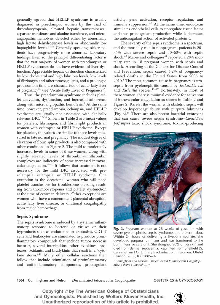
The severity of the sepsis syndrome is a spectrum,and the mortality rate in nonpregnant patients is 20–35% with severe sepsis and 40–60% with septicshock.43 Mabie and colleagues44 reported a 28% mor-tality rate in 18 pregnant women with sepsis andshock. According to the Centers for Disease Controland Prevention, sepsis caused 4.2% of pregnancy-related deaths in the United States from 2006 to2010.8 The most common cause in pregnancy is uro-sepsis from pyelonephritis caused by Escherichia coliand Klebsiella species.45–47 Fortunately, in most ofthese women, there is minimal evidence for activationof intravascular coagulation as shown in Table 2 andFigure 2. Rarely, the woman with obstetric sepsis willdevelop hypercoagulability with purpura fulminans(Fig. 3).18 There are also potent bacterial exotoxinsthat can cause severe sepsis syndrome—Clostridiumperfringens toxic shock syndrome, toxin-1-producing
Fig. 3. Pregnant woman at 28 weeks of gestation withsevere pyelonephritis, sepsis syndrome, and preterm labor.Within 24 hours of delivering a liveborn neonate, shedeveloped purpura fulminans and was transferred to theburn intensive care unit. She sloughed 90% of her skin anddied from dermal septicemia. Reprinted from Sheffield JS,Cunningham FG. Urinary tract infection in women. ObstetGynecol 2005;106:1085–92.
Cunningham and Nelson. Disseminated Intravascular Coagulop-athy. Obstet Gynecol 2015.
1004 Cunningham and Nelson Disseminated Intravascular Coagulopathy OBSTETRICS & GYNECOLOGY
Copyright ª by The American College of Obstetriciansand Gynecologists. Published by Wolters Kluwer Health, Inc.
Unauthorized reproduction of this article is prohibited.
Staphylococcus aureus, and toxic shock-like exotoxinfrom group A b-hemolytic streptococci.48,49 Theseexotoxins cause rapid and extensive tissue necrosisand gangrene, especially of the postpartum or posta-bortal uterus, and they may cause profound cardio-vascular collapse. Exotoxins are also potent inducersof DIC, and some of the effects of clostridial sepsis onplatelets, fibrinogen, and fibrin split products areshown in Table 2 and Figure 2.
The pathophysiologic response to this cascade isselective vasodilation with maldistribution of bloodflow. Leukocyte and platelet aggregation cause capil-lary plugging. Worsening endothelial injury causesprofound permeability capillary leakage and intersti-tial fluid accumulation. Depending on the degree ofinjury and inflammatory response, there is a patho-physiologic and clinical continuum. The sepsis syn-drome has a myriad of clinical manifestations that, atleast in part, are dependent on the specific invadingmicroorganism and its particular endo- or exotoxins.These were discussed in detail by Barton and Sibai.46
Because of the high associated mortality rate, in2004, a consensus effort was launched as the Surviv-ing Sepsis Campaign.50 The cornerstone of manage-ment is early goal-directed management and promptrecognition of serious bacterial infection and closemonitoring of vital signs and urine flow. The cam-paign calls for three basic steps to be performed assimultaneously as possible: 1) evaluation of the sepsissource and its sequelae; 2) assessment of cardiopulmo-nary function; and 3) immediate management. Themost important step in sepsis management is rapidinfusion of 2 L and sometimes as many as 4–6 L ofcrystalloid fluids to restore renal perfusion in severelyaffected women. Simultaneously, appropriately chosenbroad-spectrum antimicrobials are begun. If anemiacoexists with severe sepsis, blood is given along withcrystalloid to maintain the hematocrit at approximately30%.43,51 The use of colloid solutions is controver-sial.52,53 With severe sepsis, damage to pulmonary cap-illary endothelium and alveolar epithelium causesalveolar flooding and pulmonary edema—acute respira-tory distress syndrome. At the same time, determinantsof the DIC syndrome may be detected by laboratorystudies. Treatment is outlined under “Clinical Manage-ment.” In general, with sepsis, correction of all facets ofDIC is not necessary as long as the patient is not bleed-ing. Because of its putative central role, there have beenseveral agents developed to block coagulation; how-ever, none improved outcomes.54,55 Because continuingsepsis may prove fatal, débridement of necrotic tissue ordrainage of purulent material is crucial.46 Some exam-ples are uterine curettage for septic abortion, hysterec-
tomy for a necrotic uterus, ureteral catheterization forobstructive pyelonephritis, and débridement for necro-tizing fasciitis.46,49
Acute Fatty Liver of Pregnancy
Also known as acute fatty metamorphosis, acute fattyliver of pregnancy is a unique syndrome with anapproximate incidence of 1 per 10,000 births. Itusually develops in late pregnancy and is character-ized by varying degrees of hepatic failure, acutekidney injury, and moderate-to-profound consump-tive coagulopathy.11,21,36,39 Hepatic cytoplasm is re-placed with microvesicular fat—usually triglycerides—and thus the disorder has also been termed acute yellowatrophy. For many years the associated coagulopathywas attributed solely to hepatic failure and diminishedprocoagulant production.56,57 Since then, however, ithas convincingly been shown that there is also ongo-ing robust DIC as well as brisk hemolysis.21,39,58 Spe-cific factors that initiate the coagulopathy areunknown but may be related to maternal acidosisfrom liver failure or from endothelial injury.
Clinically, acute fatty liver of pregnancy is easilyconfused with preeclampsia and especially HELLPsyndrome. Although there are many similarities betweenthese—hypertension, thrombocytopenia, transaminitis,and hemolysis— acute fatty liver of pregnancy is furthercharacterized by liver dysfunction that can be profound.Also with acute fatty liver of pregnancy, there is usuallymoderate-to-severe acute kidney injury that is worse thanthat usually seen with HELLP syndrome. Shown inTable 2 are comparative mean values of some coagu-lation studies done in women with acute fatty liver ofpregnancy, placental abruption, eclampsia, and HELLPsyndrome. Of the analytes associated with obstetriccoagulopathy, fibrinogen levels with acute fatty liverof pregnancy were depressed second only those seenin women with placental abruption. Importantly, halfof women with acute fatty liver of pregnancy hada plasma fibrinogen less than 150 mg/dL at the timeof delivery. Fibrin split products are also significantlyelevated (Fig. 2). Postpartum, evidence for ongoingliver injury and prolonged dysfunction with stiltedrecovery comes from depressed fibrinogen levels alongwith increasing bilirubin levels.36 Continuing DIC ischaracterized by International Society on Thrombosisand Haemostasis DIC scores that are persistentlyabnormal for several days as well as continued eleva-tion of fibrinogen–fibrin split products.21
Acute fatty liver is one of the most seriouscoagulopathies in obstetrics. Associated maternalmortality from liver failure even with state-of-the-artcare approaches 10–15%.36,39,59,60 Management
VOL. 126, NO. 5, NOVEMBER 2015 Cunningham and Nelson Disseminated Intravascular Coagulopathy 1005
Copyright ª by The American College of Obstetriciansand Gynecologists. Published by Wolters Kluwer Health, Inc.
Unauthorized reproduction of this article is prohibited.
concerns for severely affected women are dictated bythe fact that the coagulopathy can be as profound asthat seen in some women with a total placental abrup-tion. This is particularly problematic in these womenwith acute fatty liver of pregnancy, because theirreported cesarean delivery rate with a live fetus is80–90%.36,39,59,60 Because of this, many of thesewomen require large amounts of blood and procoa-gulant replacement to combat hemorrhage and toachieve surgical hemostasis as described under “Clin-ical Management.” After delivery, and after hemosta-sis is secured, because of brisk ongoing hemolysis withanemia, one fourth of women with acute fatty liver ofpregnancy will require continuing red cell transfu-sions for several days postpartum.21
Fetal Death and Delayed Delivery
Disseminated intravascular coagulation associatedwith prolonged retention of a dead fetus is unusualtoday because fetal demise is easily confirmed byultrasonography, and there are highly effective meth-ods for pregnancy termination on its discovery. Thepathogenesis of this coagulopathy is thought to bemediated by the slow release of tissue factor orthromboplastin from the dead fetus and the pla-centa.61,62 Currently, the syndrome is only occasion-ally encountered in twin or triplet pregnancy in whichthere is one dead co-fetus with one or two survivingfetuses in an intact and ongoing pregnancy.63,64 It isalso encountered in women with a missed abortion ofseveral weeks’ duration. If the surviving fetus is deliv-ered during this time when there is a clinical coagul-opathy, treatment of the coagulopathy as describedunder “Clinical Management” may be necessary ifcesarean delivery is undertaken or if severe lacera-tions are incurred.
Massive Obstetric Hemorrhage
Although listed last in order, massive obstetric hem-orrhage represents one of the most commonlyencountered disease-specific conditions that resultsin consumptive coagulopathy. Hemorrhage is a majorcause of maternal morbidity and mortality.8,65,66
Importantly, the underlying obstetric conditions thatcause DIC are also frequently associated with thepotential for worsening of hemorrhage, viz, placentalabruption, sepsis syndrome, amniotic fluid embolism,and acute fatty liver of pregnancy. Two of the mostcommon causes of obstetric hemorrhage include uter-ine atony and genital tract lacerations either of theperineum or encountered at cesarean delivery. Giventhe breadth of the topic of hemorrhage, we refer to theoften-cited American College of Obstetricians and
Gynecologists Practice Bulletin No. 76 for a detailedreview of risk factors, battery of medical measures,and surgical techniques deployed to arrest activebleeding.67
For the purposes of the current article, ouremphasis is on two important considerations whenfaced with torrential obstetric hemorrhage. The first isthe development of a “dilutional coagulopathy” associ-ated with massive red blood cell transfusions withoutreplacement of clotting factors. The second is that DICcan develop early with massive obstetric hemorrhagewithout other underlying causes—in fact, “pure” obstet-ric hemorrhage is reportedly the cause of DIC in 25–35% of observational series.68–70 In a recent Israelistudy, in one third of 87 women with DIC, the coagul-opathy was attributed to uterine atony or genital tractlacerations.25 Similar observations were reported froma Canadian study.71 In these patients, blood loss is a con-tinuum and clotting dysfunction is aggravated and per-petuated because initial blood loss is replaced withpacked red blood cells and crystalloid, both essentiallydevoid of clotting factors. This common form of con-sumptive coagulopathy has been variably termed bloodbank coagulopathy, exsanguination coagulopathy, dilu-tional coagulopathy, and washout phenomenon.68,72,73
It was this recognition that stimulated development ofmassive transfusion protocols for patients still activelybleeding.73 These issues become critical features of clin-ical management, and as discussed subsequently, thetreatment of dilutional coagulopathy and DIC in thesewomen is the same.
CLINICAL MANAGEMENT
There are two important tenets to be considered in theclinical management of pregnant women with DIC.The first is that regardless of the underlying cause,identification and treatment of the specific underlyingdisorder are imperative in achieving a salutary out-come. The second is that the majority of cases ofsevere pregnancy-associated DIC are accompaniedby massive obstetric hemorrhage. As discussed, a gen-eralized coagulopathy can be manifest by bleeding orhypercoagulability—the former is usually in the realmof the surgeon and the latter of concern to the inter-nist.18 For the obstetrician, it can be both. Thus, itfollows that any treatment algorithm for obstetricDIC must take into consideration simultaneous andprompt replacement of blood loss in addition to treat-ment of the accompanying DIC and its cause. Impor-tantly, some of the aforementioned causes of DIC havespecific treatments for the underlying disorder—treat-ment of sepsis with drainage, débridement, and antimi-crobial therapy being an obvious example. In other
1006 Cunningham and Nelson Disseminated Intravascular Coagulopathy OBSTETRICS & GYNECOLOGY
Copyright ª by The American College of Obstetriciansand Gynecologists. Published by Wolters Kluwer Health, Inc.
Unauthorized reproduction of this article is prohibited.
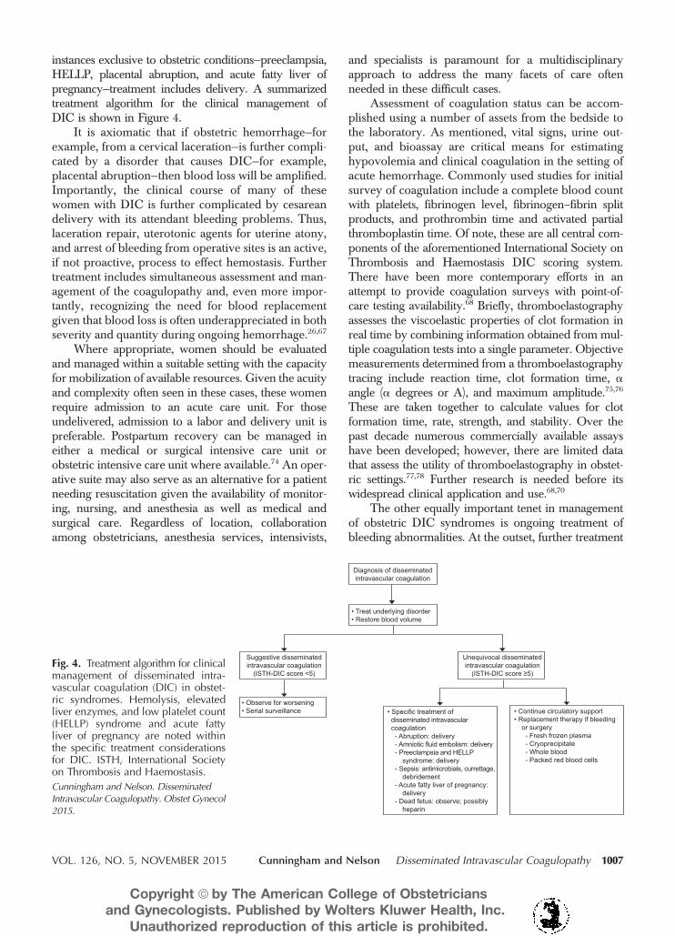
instances exclusive to obstetric conditions—preeclampsia,HELLP, placental abruption, and acute fatty liver ofpregnancy—treatment includes delivery. A summarizedtreatment algorithm for the clinical management ofDIC is shown in Figure 4.
It is axiomatic that if obstetric hemorrhage—forexample, from a cervical laceration—is further compli-cated by a disorder that causes DIC—for example,placental abruption—then blood loss will be amplified.Importantly, the clinical course of many of thesewomen with DIC is further complicated by cesareandelivery with its attendant bleeding problems. Thus,laceration repair, uterotonic agents for uterine atony,and arrest of bleeding from operative sites is an active,if not proactive, process to effect hemostasis. Furthertreatment includes simultaneous assessment and man-agement of the coagulopathy and, even more impor-tantly, recognizing the need for blood replacementgiven that blood loss is often underappreciated in bothseverity and quantity during ongoing hemorrhage.26,67
Where appropriate, women should be evaluatedand managed within a suitable setting with the capacityfor mobilization of available resources. Given the acuityand complexity often seen in these cases, these womenrequire admission to an acute care unit. For thoseundelivered, admission to a labor and delivery unit ispreferable. Postpartum recovery can be managed ineither a medical or surgical intensive care unit orobstetric intensive care unit where available.74 An oper-ative suite may also serve as an alternative for a patientneeding resuscitation given the availability of monitor-ing, nursing, and anesthesia as well as medical andsurgical care. Regardless of location, collaborationamong obstetricians, anesthesia services, intensivists,
and specialists is paramount for a multidisciplinaryapproach to address the many facets of care oftenneeded in these difficult cases.
Assessment of coagulation status can be accom-plished using a number of assets from the bedside tothe laboratory. As mentioned, vital signs, urine out-put, and bioassay are critical means for estimatinghypovolemia and clinical coagulation in the setting ofacute hemorrhage. Commonly used studies for initialsurvey of coagulation include a complete blood countwith platelets, fibrinogen level, fibrinogen–fibrin splitproducts, and prothrombin time and activated partialthromboplastin time. Of note, these are all central com-ponents of the aforementioned International Society onThrombosis and Haemostasis DIC scoring system.There have been more contemporary efforts in anattempt to provide coagulation surveys with point-of-care testing availability.68 Briefly, thromboelastographyassesses the viscoelastic properties of clot formation inreal time by combining information obtained from mul-tiple coagulation tests into a single parameter. Objectivemeasurements determined from a thromboelastographytracing include reaction time, clot formation time, aangle (a degrees or A), and maximum amplitude.75,76
These are taken together to calculate values for clotformation time, rate, strength, and stability. Over thepast decade numerous commercially available assayshave been developed; however, there are limited datathat assess the utility of thromboelastography in obstet-ric settings.77,78 Further research is needed before itswidespread clinical application and use.68,70
The other equally important tenet in managementof obstetric DIC syndromes is ongoing treatment ofbleeding abnormalities. At the outset, further treatment
Fig. 4. Treatment algorithm for clinicalmanagement of disseminated intra-vascular coagulation (DIC) in obstet-ric syndromes. Hemolysis, elevatedliver enzymes, and low platelet count(HELLP) syndrome and acute fattyliver of pregnancy are noted withinthe specific treatment considerationsfor DIC. ISTH, International Societyon Thrombosis and Haemostasis.
Cunningham and Nelson. DisseminatedIntravascular Coagulopathy. Obstet Gynecol2015.
VOL. 126, NO. 5, NOVEMBER 2015 Cunningham and Nelson Disseminated Intravascular Coagulopathy 1007
Copyright ª by The American College of Obstetriciansand Gynecologists. Published by Wolters Kluwer Health, Inc.
Unauthorized reproduction of this article is prohibited.
may not be required in women with mild coagulationabnormalities and who have no evidence of ongoingbleeding.18 To the contrary, however, in the majority ofobstetric disorders, bleeding has a prominent role inclinical management. Globally, guidelines for manage-ment in women with coagulopathy and bleeding arebased mainly on expert opinion that recommendsreplacement of red blood cells, procoagulant proteins,and platelets.18 Given that hemorrhage has sucha prominent role in disease-specific conditions associ-ated with DIC in obstetrics, basic measures for resusci-tation are paramount. For example, when blood loss isexcessive, a survey of indices of anemia with hemato-crit is determined and physiologic monitoring for dete-rioration is undertaken. Urine output as a surrogate forrenal blood flow is especially sensitive to changes inblood volume, and urine flow of at least 30 mL andpreferably 60 mL per hour or more should be main-tained as measured by an indwelling bladder catheter.26
The use of colloid solutions in lieu of crystalloid iscontroversial, and in view of their equality, most preferthe less-expensive crystalloid.79,80
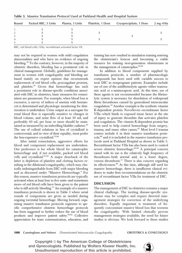
After initial resuscitation, aggressive efforts forblood and component replacement are undertaken.Our preference is for whole blood for catastrophichemorrhage and, if not available, packed red bloodcells and crystalloid.81,82 A major drawback of thelatter is depletion of platelets and clotting factors re-sulting in the dilutional coagulopathy, which may clin-ically indistinguishable from DIC with major bleedingand as discussed under “Massive Hemorrhage.” Forthis reason, massive transfusion protocols are typicallyactivated when at least four to five units—and sometimesmore—of red blood cells have been given to the patientwho is still actively bleeding.73 An example of a massivetransfusion protocols is shown in Table 3 where serialrounds of components are given as needed duringongoing torrential hemorrhage. Moving forward, orga-nizing massive transfusion protocols regimens to spe-cific comprehensive obstetric hemorrhage protocolshas been suggested to further reduce the use of bloodproducts and improve patient safety.83,84 Collectiveappreciation for team communication, education, and
training has now resulted in simulation training enteringthe obstetrician’s lexicon and becoming a viableresource for training next-generation obstetricians inthe management of catastrophes.85,86
In addition to blood components supplied bytransfusion protocols, a number of pharmacologiccompounds has been used with variable success totreat DIC in nonpregnant patients. Examples includeuse of one of the antifibrinolytic agents—either tranexa-mic acid or e-aminocaproic acid. At this time, use ofthese agents is not recommended because the fibrino-lytic system is necessary for dissolution of widespreadfibrin thromboses caused by generalized intravascularcoagulation.18 Another example is the synthetic vitaminK-dependent protein NovoSeven—recombinant factorVIIa—which binds to exposed tissue factor at the siteof injury to generate thrombin that activates plateletsand coagulation. The vitamin K-dependent protein hasbeen used to help control hemorrhage from surgery,trauma, and many other causes.87 Most level I traumacenters include it in their massive transfusion proto-cols,88 and it is included in the massive transfusion pro-tocols used at Parkland Hospital as shown in Table 3.Recombinant factor VIIa has also been used to controlsevere obstetric hemorrhage.89,90 A principal concernraised with its use is the relatively high frequency ofthromboses—both arterial and, to a lesser degree,venous thrombosis.11 There is also concern regardingits effectiveness.88 At this time, although still used formassive hemorrhage, there is insufficient clinical evi-dence to make firm recommendations on the obstetricuse of recombinant factor VIIa for treatment of DIC.
DISCUSSION
The management of DIC in obstetrics remains a majorclinical challenge. The inciting disease-specific syn-drome may be complex and require directed man-agement strategies for correction of the underlyingdisorders. Equally important is treatment of fre-quently concomitant massive blood loss that worsensthe coagulopathy. With limited clinically provenmanagement strategies available, the need for futurestudies is obvious. We look forward to these studies
Table 3. Massive Transfusion Protocol Used at Parkland Health and Hospital System
Round Packed RBC, 5 Units Plasma, 3 Units Platelets, 1 Dose Cryoprecipitate, 1 Dose 2 mg rVIIa
1 X X2 X X X X3 X X X4 X X X X5 X X
RBC, red blood cells; rVIIa, recombinant activated factor VII.
1008 Cunningham and Nelson Disseminated Intravascular Coagulopathy OBSTETRICS & GYNECOLOGY
Copyright ª by The American College of Obstetriciansand Gynecologists. Published by Wolters Kluwer Health, Inc.
Unauthorized reproduction of this article is prohibited.
designed to address our numerous evidence-baseddeficits, especially regarding management of obstetricDIC syndromes.
REFERENCES1. DeLee JB. A case of fatal hemorrhagic diathesis, with premature
detachment of the placenta. Am J Obstet Gynecol 1901;44:785.
2. Ratnoff OD, Colopy JE, Pritchard JA. The blood-clotting mecha-nism during normal parturition. J Lab Clin Med 1954;44:408–15.
3. Ratnoff OD, Pritchard JA, Colopy JE. Hemorrhagic states duringpregnancy. N Engl J Med 1955;253:63–9.
4. Ratnoff OD, Pritchard JA, Colopy JE. Hemorrhagic states duringpregnancy. N Engl J Med 1955;253:97–102.
5. Levi M, van der Poll T. A short contemporary history of dis-seminated intravascular coagulation. Semin Thromb Hemost2014;40:874–80.
6. Callaghan WM, Creanga AA, Kuklina EV. Severe maternalmorbidity among delivery, postpartum hospitalizations in theUnited States. Obstet Gynecol 2012;120:1029–36.
7. Creanga AA, Berg CJ, Ko JY, Farr SL, Tong VT, Bruce FC,et al. Maternal mortality and morbidity in the United States:where are we now? J Womens Health (Larchmt) 2014;23:3–9.
8. Creanga AA, Berg CJ, Syverson C, Seed K, Bruce FC,Callaghan WM. Pregnancy-related mortality in the UnitedStates, 2006-2010. Obstet Gynecol 2015;125:5–12.
9. Kenny L, McCrae K, Cunningham FG. Platelets, coagulation,and the liver. In: Taylor R, Roberts JM, Cunningham FG, edi-tors. Chesley’s hypertension in pregnancy. 4th ed. Amsterdam(the Netherlands): Academic Press; 2014.
10. Abbassi-Ghanavati M, Greer LG, Cunningham FG. Pregnancy andlaboratory studies: a reference table for clinicians [published erra-tum appears in Obstet Gynecol 2010;115:387]. Obstet Gynecol2009;114:1326–31.
11. Levi M, Seligsohn U. Disseminated intravascular coagulation.In: Kaushansky K, Lichtman M, Beutler K, et al, editors. Wil-liams hematology. 8th ed. New York (NY): McGraw-Hill; 2010.p. 2101.
12. Davie EW, Ratnoff OD. Waterfall sequence for intrinsic bloodclotting. Science 1964;145:1310–2.
13. Rapaport SI, Rao LV. The tissue factor pathway: how it hasbecome a “prima ballerina”. Thromb Haemost 1995;74:7–17.
14. Østerud B, Bjørklid E. Sources of tissue factor. Semin ThrombHemost 2006;32:11–23.
15. Uszy�nski M, Zekanowska E, Uszy�nski W, Kuczy�nski J. Tissuefactor (TF) and tissue factor pathway inhibitor (TFPI) in amni-otic fluid and blood plasma: implications for the mechanism ofamniotic fluid embolism. Eur J Obstet Gynecol Reprod Biol2001;95:163–6.
16. Boer K, den Hollander IA, Meijers JC, Levi M. Tissue factor-dependent blood coagulation is enhanced following deliveryirrespective of the mode of delivery. J Thromb Haemost2007;5:2415–20.
17. Kuczy�nski J, Uszy�nski W, Zekanowska E, Soszka T,Uszy�nski M. Tissue factor (TF) and tissue factor pathway inhib-itor (TFPI) in the placenta and myometrium. Eur J Obstet Gy-necol Reprod Biol 2002;105:15–9.
18. Hunt BJ. Bleeding and coagulopathies in critical care. N Engl JMed 2014;370:847–59.
19. Toh CH, Hoots WK; SSC on Disseminated IntravascularCoagulation of the ISTH. The scoring system of the Scientific
and Standardisation Committee on Disseminated IntravascularCoagulation of the International Society on Thrombosis andHaemostasis: a 5-year overview. J Thromb Haemost 2007;5:604–6.
20. Taylor FB Jr, Toh CH, Hoots WK, Wada H, Levi M; ScientificSubcommittee on Disseminated Intravascular Coagulation(DIC) of the International Society on Thrombosis and Haemo-stasis (ISTH). Towards definition, clinical and laboratory crite-ria, and a scoring system for disseminated intravascularcoagulation. Thromb Haemost 2001;86:1327–30.
21. Nelson DB, Yost NP, Cunningham FG. Hemostatic dysfunctionwith acute fatty liver of pregnancy. Obstet Gynecol 2014;124:40–6.
22. Gando S, Saitoh D, Ogura H, Fujishima S, Mayumi T, Araki T,et al. A multicenter, prospective validation study of the Japa-nese Association for Acute Medicine disseminated intravascularcoagulation scoring system in patients with severe sepsis. CritCare 2013;17:R111.
23. Terao T, Maki M, Ikenoue T. A prospective study in 38 pa-tients with abruptio placentae of 70 cases complicated by DIC.Asia Oceania J Obstet Gynaecol 1987;13:1–13.
24. Kobayashi T. Obstetrical disseminated intravascular coagula-tion score. J Obstet Gynaecol Res 2014;40:1500–6.
25. Erez O, Novack L, Beer-Weisel R, Dukler D, Press F,Zlotnik A, et al. DIC score in pregnant women—a populationbased modification of the International Society on Thrombosisand Hemostasis score. PLoS One 2014;9:e93240.
26. Cunningham F, Leveno KJ, Bloom SL, Spong CY, Dashe JS,Hoffman BL, et al. Obstetrical hemorrhage. In: Cunningham F,Leveno KJ, Bloom SL, Spong CY, Dashe JS, Hoffman BL, et al,editors. Williams obstetrics. 24th ed. New York (NY): McGraw-Hill; 2013.
27. Pritchard JA, Brekken AL. Clinical and laboratory studieson severe abruptio placentae. Am J Obstet Gynecol 1967;97:681–700.
28. Oyelese Y, Ananth CV. Placental abruption. Obstet Gynecol2006;108:1005–16.
29. Clark SL. Amniotic fluid embolism. Obstet Gynecol 2014;123:337–48.
30. Kramer MS, Rouleau J, Liu S, Bartholomew S, Joseph KS; Mater-nal Health Study Group of the Canadian Perinatal SurveillanceSystem. Amniotic fluid embolism: incidence, risk factors, andimpact on perinatal outcome. BJOG 2012;119:874–9.
31. Knight M, Berg C, Brocklehurst P, Kramer M, Lewis G, Oats J,et al. Amniotic fluid embolism incidence, risk factors and out-comes: a review and recommendations. BMC Pregnancy Child-birth 2012;12:7.
32. Chesley LC, editor. Hypertensive disorders in pregnancy. NewYork (NY): Appleton-Century-Crofts; 1978.
33. Weinstein L. Syndrome of hemolysis, elevated liver enzymesand low platelet count: a severe consequence of hypertension inpregnancy. Am J Obstet Gynecol 1982;142:159–67.
34. Sibai BM. Diagnosis, controversies, and management of thesyndrome of hemolysis, elevated liver enzymes, and low plate-let count. Obstet Gynecol 2004;103:981–91.
35. Martin JN Jr, Rinehart BK, May WL, Magann EF, Terrone DA,Blake PG. The spectrum of severe preeclampsia: comparativeanalysis by HELLP (hemolysis, elevated liver enzyme levels,and low platelet count) syndrome classification. Am J ObstetGynecol 1999;180:1373–84.
VOL. 126, NO. 5, NOVEMBER 2015 Cunningham and Nelson Disseminated Intravascular Coagulopathy 1009
Copyright ª by The American College of Obstetriciansand Gynecologists. Published by Wolters Kluwer Health, Inc.
Unauthorized reproduction of this article is prohibited.
36. Nelson DB, Yost NP, Cunningham FG. Acute fatty liver ofpregnancy: clinical outcomes and expected duration of recov-ery. Am J Obstet Gynecol 2013;209:456.e1–7.
37. Pritchard JA, Cunningham FG, Mason RA. Coagulationchanges in eclampsia: their frequency and pathogenesis. Am JObstet Gynecol 1976;124:855–64.
38. McCrae KR. Thrombocytopenia in pregnancy: differentialdiagnosis, pathogenesis and management. Blood Rev 2003;17:7–14.
39. Sibai BM. Imitators of severe preeclampsia. Obstet Gynecol2007;109:956–66.
40. Cunningham FG, Pritchard JA. Hematologic considerations ofpregnancy-induced hypertension. Semin Perinatol 1978;2:29–38.
41. Russell JA. Management of sepsis [published erratum appearsin N Engl J Med 2006;355:2267]. N Engl J Med 2006;355:1699–713.
42. Moellering RC Jr, Abbott GF, Ferraro MJ. Case records of theMassachusetts General Hospital Case 2–2011. A 30-year-oldwoman with shock after treatment for a furuncle. N Engl JMed 2011;364:266–75.
43. Munford RS. Severe sepsis and septic shock: introduction. In:Longo DL, Fauci AS, Kasper DL, et al, editors. Harrison’sprinciples of internal medicine. 18th ed. New York (NY):McGraw Hill; 2012.p. 2223.
44. Mabie WC, Barton JR, Sibai BM. Septic shock in pregnancy.Obstet Gynecol 1997;90:553–61.
45. Cunningham FG, Lucas MJ, Hankins GD. Pulmonary injurycomplicating antepartum pyelonephritis. Am J Obstet Gynecol1987;156:797–807.
46. Barton JR, Sibai BM. Severe sepsis and septic shock in preg-nancy. Obstet Gynecol 2012;120:689–706.
47. Sheffield JS, Cunningham FG. Urinary tract infection inwomen. Obstet Gynecol 2005;106:1085–92.
48. Daif JL, Levie M, Chudnoff S, Kaiser B, Shahabi S. Group AStreptococcus causing necrotizing fasciitis and toxic shock syn-drome after medical termination of pregnancy. Obstet Gynecol2009;113:504–6.
49. Soper DE, Lee SI, Kim JY, McDonald AG. Case records of theMassachusetts General Hospital. Case 35–2011: A 33-year-oldwoman with postpartum leukocytosis and Gram-positive bac-teremia. N Engl J Med 2011;365:1916–24.
50. Dellinger RP, Levy MM, Carlet JM, Bion J, Parker MM,Jaeschke R, et al. Surviving Sepsis Campaign: internationalguidelines for management of severe sepsis and septic shock:2008. Crit Care Med 2008;36:296–327.
51. Rivers E, Nguyen B, Havstad S, Ressler J, Muzzin A,Knoblich B, et al. Early goal-directed therapy in the treatmentof severe sepsis and septic shock. N Engl J Med 2001;345:1368–77.
52. Myburgh JA, Finfer S, Bellomo R, Billot L, Cass A, Gattas D,et al. Hydroxyethyl starch or saline for fluid resuscitation inintensive care. N Engl J Med 2012;367:1901–11.
53. Perner A, Naase N, Guttormsen AB, Tenhunen J, Klemenzson G,Åneman A, et al. Hydroxyethyl starch 130/0.4 versus Ringer’sacetate in severe sepsis. N Engl J Med 2012;367:124–34.
54. Suffredini AF, Munford RS. Novel therapies for septic shockover the past 4 decades. JAMA 2011;306:194–9.
55. Wenzel RP, Edmond MB. Septic shock—evaluating anotherfailed treatment. N Engl J Med 2012;366:2122–4.
56. Sherlock S. Acute fatty liver of pregnancy and the microvesic-ular fat diseases. Gut 1983;24:265–9.
57. Burroughs AK, Seong NG, Dojcinov DM, Scheuer PJ,Sherlock SV. Idiopathic acute fatty liver of pregnancy in 12patients. Q J Med 1982;51:481–97.
58. Castro MA, Fasset MJ, Reynolds TB, Shaw KJ, Goodwin TM.Reversible peripartum liver failure: a new perspective on thediagnosis, treatment, and cause of acute fatty liver of pregnancybased on 28 cases. Am J Obstet Gynecol 1999;181:389–95.
59. Knight M, Nelson-Piercy C, Kurinczuk JJ, Spark P, Brocklehurst P;UK Obstetric Surveillance System. A prospective national study ofacute fatty liver of pregnancy in the UK. Gut 2008;57:951–6.
60. Vigil-de Gracia P, Montufar-Rueda C. Acute fatty liver of preg-nancy: diagnosis, treatment, and outcome based on 35 consec-utive cases. J Matern Fetal Neonatal Med 2011;24:1143–6.
61. Jimenez JM, Pritchard JA. Pathogenesis and treatment of coag-ulation defects resulting from fetal death. Obstet Gynecol 1968;32:449–59.
62. Lerner R, Margolin M, Slate WG, Rosenfeld H. Heparin in thetreatment of hypofibrinogenemia complicating fetal death inutero. Am J Obstet Gynecol 1967;97:373–8.
63. Chescheir NC, Seeds JW. Spontaneous resolution of hypofibri-nogenemia associated with death of a twin in utero: a casereport. Am J Obstet Gynecol 1988;159:1183–4.
64. Eddib A, Rodgers B, Lawler J, Yeh J. Monochorionic pseudo-monoamniotic twin pregnancy with fetal demise of one twinand development of maternal consumptive coagulopathy.Ultrasound Obstet Gynecol 2006;28:736–7.
65. Khan KS, Wojdyla D, Say L, Gülmezoglu AM, Van Look PF.WHO analysis of causes of maternal death: a systematic review.Lancet 2006;367:1066–74.
66. Kramer MS, Berg C, Abenhaim H, Dahhou M, Rouleau J,Mehrabadi A, et al. Incidence, risk factors, and temporal trendsin severe postpartum hemorrhage. Am J Obstet Gynecol 2013;209:449.e1–7.
67. Postpartum hemorrhage. ACOG Practice Bulletin No. 76.American College of obstetricians and Gynecologists. ObstetGynecol 2006;108:1039–47.
68. Abdul-Kadir R, McLintock C, Ducloy AS, El-Refaey H,England A, Federici AB, et al. Evaluation and management ofpostpartum hemorrhage: consensus from an internationalexpert panel. Transfusion 2014;54:1756–68.
69. Hossain N, Paidas MJ. Disseminated intravascular coagulation.Semin Perinatol 2013;37:257–66.
70. James AH, McLintock C, Lockhart E. Postpartum hemorrhage:when uterotonics and sutures fail. Am J Hematol 2012;87(suppl 1):S16–22.
71. Rattray DD, O’Connell CM, Baskett TF. Acute disseminatedintravascular coagulation in obstetrics: a tertiary centre popula-tion review (1980 to 2009). J Obstet Gynaecol Can 2012;34:341–7.
72. Su LL, Chong YS. Massive obstetric haemorrhage with dissem-inated intravascular coagulopathy. Best Pract Res Clin ObstetGynaecol 2012;26:77–90.
73. Waters JH. Role of the massive transfusion protocol in themanagement of haemorrhagic shock. Br J Anaesth 2014;113(suppl 2):ii3–8.
74. Zeeman GG, Wendel GD Jr, Cunningham FG. A blueprint forobstetric critical care. Am J Obstet Gynecol 2003;188:532–6.
75. Thrombelastograph coagulation analyzer. Skokie (IL): Haemo-scope Corporation; 1991.
76. Sharma P, Saxena R. A novel thromboelastographic score toidentify overt disseminated intravascular coagulation resultingin a hypocoagulable state. Am J Clin Pathol 2010;134:97–102.
1010 Cunningham and Nelson Disseminated Intravascular Coagulopathy OBSTETRICS & GYNECOLOGY
Copyright ª by The American College of Obstetriciansand Gynecologists. Published by Wolters Kluwer Health, Inc.
Unauthorized reproduction of this article is prohibited.
77. Sharma SK, Vera RL, Stegall WC, Whitten CW. Managementof a postpartum coagulopathy using thrombelastography. J ClinAnesth 1997;9:243–7.
78. Sharma SK, Philip J, Whitten CW, Padakandla UB,Landers DF. Assessment of changes in coagulation in parturi-ents with preeclampsia using thromboelastography. Anesthesi-ology 1999;90:385–90.
79. Perel P, Roberts I. Colloids versus crystalloids for fluid resus-citation in critically ill patients. The Cochrane Database of Sys-tematic Reviews 2007:CD000567. DOI: 10.1002/14651858.CD000567.pub3.
80. Finfer S, Bellomo R, Boyce N, French J, Myburgh J, Norton R;SAFE Study Investigators. A comparison of albumin and salinefor fluid resuscitation in the intensive care unit. N Engl J Med2004;350:2247–56.
81. Alexander JM, Sarode R, McIntire DD, Burner JD, Leveno KJ.Use of whole blood in the management of hypovolemia due toobstetric hemorrhage. Obstet Gynecol 2009;113:1320–6.
82. Hernandez JS, Alexander JM, Sarode R, McIntire DD,Leveno KJ. Calculated blood loss in severe obstetric hemor-rhage and its relation to body mass index. Am J Perinatol2012;29:557–60.
83. Shields LE, Wiesner S, Fulton J, Pelletreau B. Comprehensivematernal hemorrhage protocols reduce the use of blood prod-
ucts and improve patient safety. Am J Obstet Gynecol 2015;212:272–80.
84. Einerson BD, Miller ES, Grobman WA. Does a postpartumhemorrhage patient safety program result in sustained changesin management and outcomes? Am J Obstet Gynecol 2015;212:140–4.e1.
85. D’Alton ME, Bonanno CA, Berkowitz RL, Brown HL,Copel JA, Cunningham FG, et al. Putting the “M” back inmaternal-fetal medicine. Am J Obstet Gynecol 2013;208:442–8.
86. Postpartum hemorrhage from vaginal delivery. Patient SafetyChecklist No. 10. American College of Obstetricians and Gy-necologists. Obstet Gynecol 2013;121:1151–2.
87. Mannucci PM, Levi M. Prevention and treatment of majorblood loss. N Engl J Med 2007;356:2301–11.
88. Pacheco LD, Saade GR, Gei AF, Hankins GD. Cutting-edgeadvances in the medical management of obstetrical hemor-rhage. Am J Obstet Gynecol 2011;205:526–32.
89. Alfirevic Z, Elbourne D, Pavord S, Bolte A, Van Geijn H,Mercier F, et al. Use of recombinant activated factor VII inprimary postpartum hemorrhage: the North European Registry2000–2004. Obstet Gynecol 2007;110:1270–8.
90. Franchini M, Lippi G, Franchi M. The use of recombinantactivated factor VII in obstetric and gynaecological haemor-rhage. BJOG 2007;114:8–15.
Artículos de las Series de Especialidad Clínica ¡Ahora en Español!
La traducciones de los artículos de las Series de Especialidad Clínica publicados a partir de abril de 2010 están disponibles en línea solamente en http://www.greenjournal.org. Para ver la colección entera de artículos traducidos, haga click en “Collections” y luego seleccione “Translations (Español).”
rev 12/2014
VOL. 126, NO. 5, NOVEMBER 2015 Cunningham and Nelson Disseminated Intravascular Coagulopathy 1011
Copyright ª by The American College of Obstetriciansand Gynecologists. Published by Wolters Kluwer Health, Inc.
Unauthorized reproduction of this article is prohibited.