HISTOPATHOLOGICAL SURVEY OF PROTOZOA, HELMINTHS ...
15
Instructions for use Title HISTOPATHOLOGICAL SURVEY OF PROTOZOA, HELMINTHS AND ACARIDS OF IMPORTED AND LOCAL PSITTACINE AND PASSERINE BIRDS IN JAPAN Author(s) TSAI, Shinn-Shyong; HIRAI, Katsuya; ITAKURA, Chitoshi Citation Japanese Journal of Veterinary Research, 40(4): 161-174 Issue Date 1992-12-28 DOI 10.14943/jjvr.40.4.161 Doc URL http://hdl.handle.net/2115/2407 Type bulletin File Information KJ00002377611.pdf Hokkaido University Collection of Scholarly and Academic Papers : HUSCAP
Transcript of HISTOPATHOLOGICAL SURVEY OF PROTOZOA, HELMINTHS ...
Instructions for use
Title HISTOPATHOLOGICAL SURVEY OF PROTOZOA, HELMINTHS AND ACARIDS OFIMPORTED AND LOCAL PSITTACINE AND PASSERINE BIRDS IN JAPAN
Author(s) TSAI, Shinn-Shyong; HIRAI, Katsuya; ITAKURA, Chitoshi
Citation Japanese Journal of Veterinary Research, 40(4): 161-174
Issue Date 1992-12-28
DOI 10.14943/jjvr.40.4.161
Doc URL http://hdl.handle.net/2115/2407
Type bulletin
File Information KJ00002377611.pdf
Hokkaido University Collection of Scholarly and Academic Papers : HUSCAP
lPn. l. Vet. Res., 40, 161-174 (1992)
HISTOPATHOLOGICAL SURVEY OF HELMINTHS AND ACARIDS OF PROTOZOA,
IMPORTED AND PASSERINE
LOCAL PSITTACINE AND BIRDS IN JAPAN
Shinn-Shyong TSAI1, Katsuya HIRAI2 and Chitoshi ITAKURA 1
(Accepted for pUblication: Nov. 5, 1992)
ABSTRACT
A total of 534 psittacine and passerine birds consisting of 241 imported arid
293 local birds were examined histologically. As a result, the following parasites were found: Giardia (86 cases), Knemido-coptes (26 cases), coccidia (10 cases),
Ascaridia (6 cases), Cryptosporidium (5 cases), Sarcocystis (5 cases), tapeworm
(4 cases), microfilaria (2 cases), Hexamita (1 case), and Spiroptera (1 case). High incidences of giardiasis and knemido-coptic infestation were detected in the
local birds, but rarely in the imported birds. Giardial trophozoites were
observed mainly in the duodenum of budgerigars (Melopsittacus undulatus). Knemidocoptic mites burrowed i~to the epidermis producing proliferative dermati
tis in 25 budgerigars and 1 African Grey Parrot (Psittacus erithacus erithacus).
This ectoparasite often infested the skin around the cloaca. Coccidiosis was seen only in the small intestines of the finch (Poephila gouldiae gouldiae), African
Grey Parrot, Rainbow lory (Trichoglossus haematodus) , Indian Ring-necked para
keet (Psittacula kramer; manillensis) and peach-faced lovebird (Agapornis
roseicollis). Two parrots (Amazona aestiva aestiva and Psittacus erithacus erithacus) and two budgerigars had intestinal cryptosporidiosis. Conjunctivitis associ
ated with cryptosporidial infection was seen in a lovebird. Sarcocystis cysts
containing crescent-shaped bradyzoites were found not only in the thigh and breast but also in the heart and cloacal muscles. Other organisms such as
Ascaridia, tapeworm, microfilaria, Hexamita, and Spiroptera were clinically less
significant. However, infections such as Giardia and Cryptosporidim might have zQonotic implications.
Key words: pet bird, protozoan, helminth, acarid, histopathology.
L Department of Comparative Pathology, Faculty of Veterinary Medicine, Hokkaido University, Sapporo 060, Japan
2. Department of Veterinary Microbiology, Faculty of Agriculture, Gifu University, Gifu 501-11, Japan

162 TSAI, HIRAI, and ITAKURA
INTRODUCTION
Parasitism is often overlooked in the companion birds because it does not seem to cause obvious clinical disorders. In addition, most of the pet birds kept in indoor cages or aviaries have little chance of contact with parasites. Contamination of food and water with infective materials either from humans, newcomers or wild birds may be a source of infection for captive birds9 , 17, 20, 21). Therefore, helminthic and
protozoal diseases may be more frequent than previously recognized. Various parasites can cause high mortality and also have a significant adverse effect on the outcome of other diseases26
).
Helminthic and protozoal diseases of pet birds such as toxoplasmosis 13),
schistosomiasis8), giardiasis28), and cryptosporidiosis2) have been implicated in parasitic zoonoses. As far as we know, only a few studies have been carried out on protozoal and helminthic diseases in caged birds in Japan32
, 39). This paper is a retrospective histopathological study on parasitic infections in both imported and local pet birds.
MATERIALS AND METHODS
A total of 534 pet birds comprising 241 imported birds and 293 local budgerigars were examined. The imported birds consisted of 67 parakeets (Psittacula krameri manillensis) from India, 52 cockatiels (Nymphicus hollandicus) from Taiwan, 29 parrots (19 were Amazona aestiva aestiva from Argentina and 10 were Psitlacus erithacus erithacus from Ghana), 18 lories (Trichoglossus haematodus) from Indonesia, 8 love
birds (Agapornis roseicollis) from Taiwan, 2 rosellas (Platycercus emimius) from Holland, 47 budgerigars (Melopsittacus undulatus) from the philippines and 18 finches (Poephila gouldiae gouldiae) from Taiwan. These imported birds died during the two-week quarantine period in a bird shop, and the local birds were obtained from bird
dealers. Tissue samples for histopathological examination consisted of the brain, heart,
respiratory tract, digestive tract, kidney, spleen, thymus, bursa of Fabricius, parathyroid gland, thyroid gland, adrenal gland, bone marrow, genital organs, eye, conjuctivum, skeletal muscles and skin. The samples were fixed in 10% buffered formalin, embedded in paraffin, sectioned, and stained with either hematoxylin and eosin (HE),
Gram, or periodic acid-Schiff (PAS) methods. The parasites were identified on the basis of their morphological features in the tissue sections.
RESULTS
Among the protozoal infections, giardiasis, found in the small intestine, especially in the duodenum (Table 1), was the most common disorder. Of the 86 budgerigars infected with Giardia, 85 were local and only one was imported. Histologically, large numbers of trophozoites admixed with mucus in the lumen were observed to adhere to
Histopathological Survey of Parasitic Infestation in Pet Birds
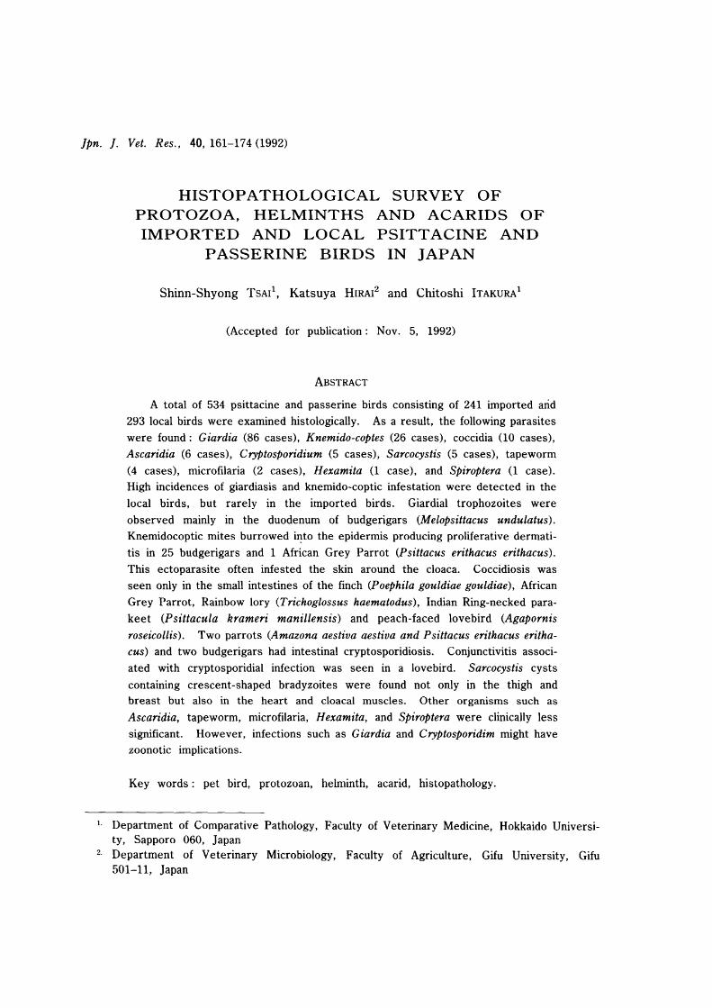
Table 1. Protozoal infection in imported and local birds
Protozoan No. of cases
LCb
Giardia 1 85
Coccidia 10 o
Sarcocystis 4 1
Cryptosporidium 3 2
Hexamita 1 o
a Imported birds. b Local birds.
Bird host
(No. infected)
Budgerigar (86)
Finch (4), African
Grey (2), parakeet (2)
lory (11, lovebird (1)
African Grey (2'1,
Amazon parrot (2),
budgerigar (1)
Amazon parrot (1 ),
African Grey (l),
budgerigar (2)
Lovebird (1)
Finch (1)
Site of infection
Small intestine
Small intestine
Striated muscle
Small intestine
Conjunctivum
Small intestine
Cecum
163
the brush borders of the intestinal epithelium. They appeared as pear-shaped or small sickle-shaped bodies which were dorsally convex and had no undulating membrane (Fig. 1). It was difficult to demonstrate their nuclei and flagella. The epithelium was intact in the intestinal villi, but the lamina propria was often filled with chronic inflammatory cells, consisting mainly of lymphocytes and plasma cells. Some of the plasma cells had Russell bodies in their cytoplasm.
Coccidia were found in the small intestines in 4 finches, 2 parrots, 1 lory and 1 lovebird. Macrogametocytes, microgametocytes and immature oocysts were observed
only in ,the villar epithelium and lamina propria of the small intestine (Fig. 2). No other visceral lesion was induced by these coccidia.
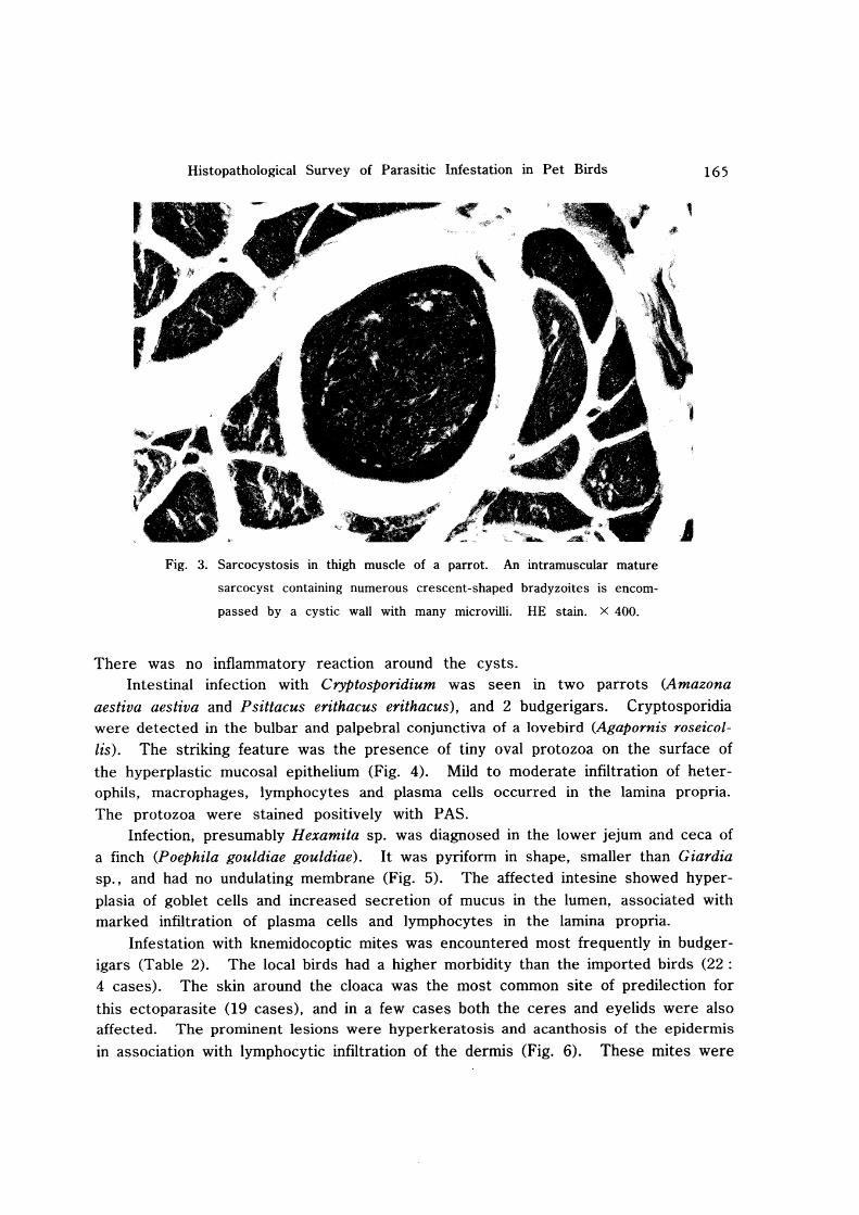
Sarcocystis cysts were observed in 4 parrots (2 Amazona aestiva aestiva and 2 Psittacus erithacus) and 1 budgerigar. All the birds had mature cysts in their breast and thigh muscles. In an Amazona parrot, the cysts were also observed in the heart and cloacal muscles. These cysts which contained crescent-shaped bradyzoites were elongated, and had a thick-wall with many villous protrusions on its internal surface (Fig. 3). The bradyzoites were stained positively with PAS, but not the cyst wall.

164 TSAI, HIRAI, and ITAKURA
Fig. 1. Giardiasis in duodenum of a parakeet. Numerous trophozoites of Giar
dia species with sharp borders ends, frequently found between the
intestinal villi. Gram stain. X 600.
Fig. 2. Coccidiosis in small intestine of a finch. Many immature oocysts are
seen in the intestinal epithelium. HE stain. X 440.
Histopathological Survey of Parasitic Infestation in Pet Birds
Fig. 3. Sarcocystosis in thigh muscle of a parrot. An intramuscular mature
sarcocyst containing numerous crescent-shaped bradyzoites is encom
passed by a cystic wall with many microvilli. HE stain. X 400.
There was no inflammatory reaction around the cysts.
165
Intestinal infection with Cryptosporidium was seen in two parrots (Amazona
aestiva aestiva and Psittacus erithacus erithacus), and 2 budgerigars. Cryptosporidia were detected in the bulbar and palpebral conjunctiva of a lovebird (Agapornis roseicollis). The striking feature was the presence of tiny oval protozoa on the surface of
the hyperplastic mucosal epithelium (Fig. 4). Mild to moderate infiltration of heterophils, macrophages, lymphocytes and plasma cells occurred in the lamina propria. The protozoa were stained positively with PAS.
Infection, presumably Hexamita sp. was diagnosed in the lower jejum and ceca of a finch (Poephila gouldiae gouldiae). It was pyriform in shape, smaller than Giardia sp., and had no undulating membrane (Fig. 5). The affected intesine showed hyperplasia of goblet cells and increased secretion of mucus in the lumen, associated with marked infiltration of plasma cells and lymphocytes in the lamina propria.
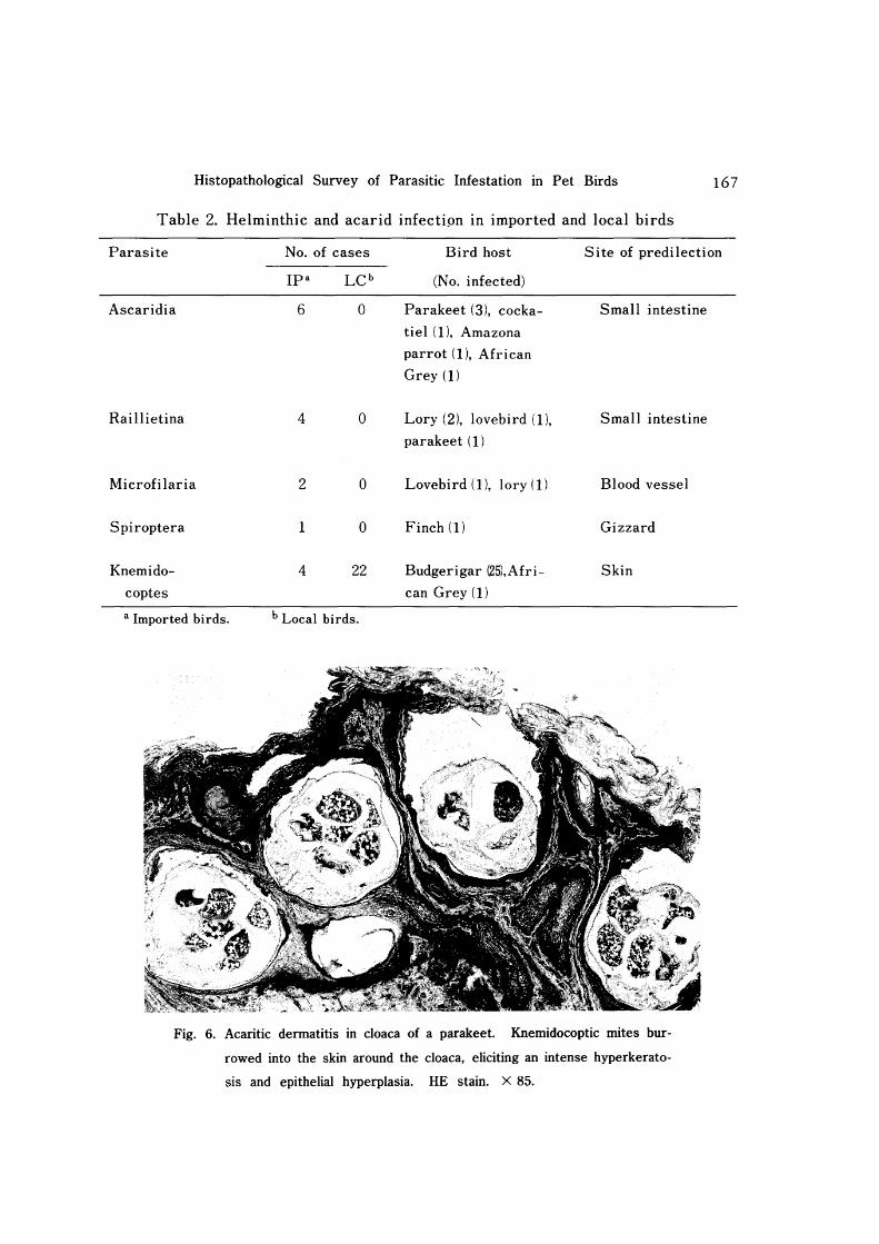
Infestation with knemidocoptic mites was encountered most frequently in budgerigars (Table 2). The local birds had a higher morbidity than the imported birds (22 : 4 cases). The skin around the cloaca was the most common site of predilection for
this ectoparasite (19 cases), and in a few cases both the ceres and eyelids were also affected. The prominent lesions were hyperkeratosis and acanthosis of the epidermis
in association with lymphocytic infiltration of the dermis (Fig. 6). These mites were

166 TSAI, HIRAI, and ITAKURA
Fig. 4. Cryptosporidiosis in conjunctiva of a lovebird. Tiny oval cryptosporidial
organisms (arrowheads) are attached to the hyperplastic epithelium in
bulbar and palpebral conjunctiva. Note the infiltration of macrophages,
heterophils, lymphocytes and plasma cells in the lamina propria. HE
stain. X 900.
Fig. 5. Large numbers of pyriform-shaped Hexamita sp. trophozoites (arrow
heads) are seen in the jejunum of a finch, associated with catarrhal
inflammation. HE stain. X 400.
Histopathological Survey of Parasitic Infestation in Pet Birds 167
Table 2. Helminthic and acarid infectipn in imported and local birds
Parasite No. of cases Bird host Site of predilection
Ipa LC b (No. infected)
Ascaridia 6 0 Parakeet (3), cocka- Small intestine
tiel (I), Amazona
parrot(l~ African
Grey (1)
Raillietina 4 0 Lory (2), lovebird (I), Small intestine
parakeet (1)
Microfilaria 2 0 Lovebird (I), lory (I) Blood vessel
Spiroptera 1 0 Finch (I) Gizzard
Knemido- 4 22 Budgerigar (25),Afri- Skin
coptes can Grey (1)
a Imported birds. b Local birds.
Fig. 6. Acaritic dermatitis in cloaca of a parakeet. Knemidocoptic mites bur
rowed into the skin around the cloaca, eliciting an intense hyperkerato
sis and epithelial hyperplasia. HE stain. X 85.
168 TSAI, HIRAI, and ITAKURA
often embedded in the thickened keratin layer. Some burrowed into the epidermis, but not beyond the basal layer.
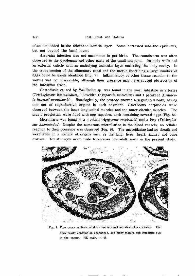
Ascaridia infection was not uncommon in pet birds. The roundworm was often observed in the duodenum and other parts of the small intestine. Its body walls had an external cuticle with an underlying muscular layer encircling the body cavity. In
the cross-section of the alimentary canal and the uterus containing a large number of eggs could be easily identified (Fig. 7). Inflammatory or other tissue reaction to the worms was not discernible, although their presence may have caused obstruction of the intestinal tract.
Cestodiasis caused by Raillietina sp. was found in the small intestine in 2 lories (Trichoglossus haematodus) , 1 lovebird (Agapornis roseicollis) and 1 parakeet (Psittacula krameri manillensis). Histologically, the cestode showed a segmented body, having one set of reproductive organs in each segment. Calcareous corpuscles were observed between the inner longitudinal muscles and the outer circular muscles. The gravid proglottids were filled with egg capsules, each containing several eggs (Fig. 8).
Microfilaria was found in a lovebird (Agapornis roseicollis) and a lory (Trichoglossus haematodus). Despite the numerous microfilariae in the blood vessels, no cellular reaction to their presence was observed (Fig. 9). The microfilariae had no sheath and were seen in a variety of organs such as the lung, liver, heart, kidney and bone marrow. No attempts were made to recover the adult worm in the present study.
Fig. 7. Four cross sections of Ascaridia in small intestine of a cockatiel. The
body cavity contains an esophagus, and many mature and immature ova
in the uterus. HE stain. X 45.

Histopathological Survey of Parasitic Infestation III Pet Birds
.~ ... ~ Fig. 8. Raillietina sp. in small intestine of a lory. The longitudinal section of
the strobila reveals three segments, which contain many egg capsules
filled with immature ova. HE stain. X 35.
Fig. 9. Microfilariae in heart of a lovebird. Numerous microfilariae are seen in
the lumen of the left atrium. HE stain. X 400.
169
170 TSAI, HIRAI, and ITAKURA
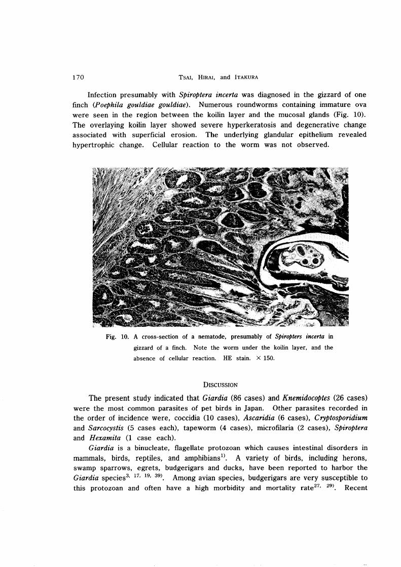
Infection presumably with Spiroptera incerla was diagnosed in the gizzard of one finch (Poephila gouldiae gouldiae). Numerous roundworms containing immature ova were seen in the region between the koilin layer and the mucosal glands (Fig. 10). The overlaying koilin layer showed severe hyperkeratosis and degenerative change associated with superficial erosion. The underlying glandular epithelium revealed hypertrophic change. Cellular reaction to the worm was not observed.
Fig. 10. A cross-section of a nematode, presumably of Spiropters incerta in
gizzard of a finch. Note the worm under the koilin layer, and the
absence of cellular reaction. HE stain. X 150.
DISCUSSION
The present study indicated that Giardia (86 cases) and Knemidocoptes (26 cases) were the most common parasites of pet birds in Japan. Other parasites recorded in the order of incidence were, coccidia (10 cases), Ascaridia (6 cases), Cryptosporidium and Sarcocystis (5 cases each), tapeworm (4 cases), microfilaria (2 cases), Spiroptera and Hexamita (1 case each).
Giardia is a binucleate, flagellate protozoan which causes intestinal disorders in mammals, birds, reptiles, and amphibians 1). A variety of birds, including herons, swamp sparrows, egrets, budgerigars and ducks, have been reported to harbor the Giardia species3, 17, 19, 39) Among avian species, budgerigars are very susceptible to this protozoan and often have a high morbidity and mortality rate27
, 29) Recent

Histopathological Survey of Parasitic Infestation in Pet Birds 171
studies have shown that surface and filtered drinking water supplies could be contaminated with high levels of Giardia cysts and Cryptosporidium spp. oocysts20
, 21).
This has been attributed to the high resistance of Giardia cysts to the routine chlorination recommended for drinking water9
).
We found that Giardia species were more prevalent in the local budgerigars (85/293) than in the imported birds (11241). Waterborne and contact infections might be the possible routes of transmission for this protozoan. Since all the infected budgerigars had large numbers of trophozoites in the upper small intestine, equally large numbers of cysts could be expected to be excreted in their feces. These cysts may contaminate the environment and present a possible source of infection to other birds.
Coccidiosis occurred primarily in companion birds that were given feed on the ground. Eimeria spp. have been reported to parasitize the small intestine, being confined mainly to the duodenum, and are host specific26
, 33). Although all cases of coccidiosis in this study showed sexual stages of their life cycle in the duodenum, it was difficult to identify their genera in tissue sections.
Sarcocystis falcatula has been reported to have a wide range of intermediate hosts, including psittaciforme, passeriforme, and columbiforme birds6
• 7, 36). It may
cause debilitation and sometimes mortality in susceptible birds, due to obstruction of the pulmonary blood vessels36
). In chronic infection, cysts are formed in the skeletal muscles and, to a lesser extent, in myocardial fibers6
). The parasites are characterized microscopically by the presence of metrocytes encompassed by the cyst wall with microvilli, and the compartmentalization of the cyst5
, 14). We observed 4 cases with Sarcocystis cysts in the muscles of the breast and thigh, including an Amazona parrot with cysts in the heart, breast and cloacal muscles.
Cryptosporidiosis has been recorded in various mammals, birds and fishes24, 35).
Among the caged birds, this protozoan has observed mainly in the digestive tract, kidney and respiratory tract of parrots 12), canaries38), finches I6), lovebirds4) and budgerigars25). Conjunctival cryptosporidiosis is occasionally found in chickens 18),
peacocks23), pheasants30
) and ducks22). This protozoan induces conjunctivitis and
excess mucus in the nasal cleft. We found intestinal cryptosporidiosis in two parrots and two budgerigars, and conjunctival cryptosporidiosis in a lovebird. The organism has been proven to be monoxenous and interhost species transmission has been reportedlO
). This may have public health implications2).
Knemidocoptes pilae commonly infests the skin of budgerigars, and feeds on keratin40
). This mite produces pruritic dermatitis with scaly epithelial proliferation which is characterized by the formation of chalk-like, honeycombed encrustations in the affected areas26
• 37). We found a higher incidence in local birds than in imported ones, and the skin around the cloaca was the most common site of predilection. This
contradicts previous reports, which stated that the lesions were predominantly seen on

172 TSAI, HIRAI, and ITAKURA
the beak, cere, eyelids and legs, thus leading to the name "scaly-leg and face mite" for this ectoparasite15, 31). This discrepancy may be due to the culling of the birds
with gross lesions on easily noticeable sites other than the cloaca by owners. Moreover, acaritic dermatitis around the cloaca can easily be overlooked. The higher morbidity in the local birds may be attributable to overcrowding or huddling together on a perch due to low room temperature.
Ascaridia and tapeworms were detected only in the imported birds in the present study. Unless in debilitated birds or those with heavy parasite burdens, they do not
seem to present any problems in practice26). Microfilariasis has been found in a wide
variety of birds, including the orders Passeriformes, Psittaciformes and
Falconiformes26). In Japan, of 17 captured jungle-crows 7 were observed to harbor
microfilariae32). Our study showed that the imported lovebird and lory were also
infected with microfilariae. These parasites were seen in the blood vessels of blood-rich organs such as the lung, liver, heart, kidney and bone marrow. Microscopically, they seemed to be nonpathogenic, because no lesion or cellular reaction was observed. However, parasitic emboli in the tiny blood vessels may lead to circulatory disturbances 11).
The gizzard lesions presumably caused by Spiroptera incerta in the finch in the present study were similar to those reported for the Bluefaced fmch attributed to
Cheilospirura infection34). Cellular reaction to the infection with Cheilospirura sp. was
rarely noticed, except for the marked degenertion of the koilin layer and the accumulation of necrotic debris in the mucosal glands.
We have previously encountered two parasites affecting the proventriculus and gizzard, including Tetrameres jissispina in ducks, and Dispharynx nasuta in guinea fowls (unpublished data.). Both the parasities induced chronic proliferative inflammatory reactions. However, the definite diagnosis of Spiroptera incerta in this study should be based on morphological examination of the intact worms.
REFERENCES
1) ADAM, R. D. (1991): The biology of Giardia spp. Microbiol. Rev., 55, 706-732
2) ANDERSON, B. C., DONNDELING, T., WILKINGS, R. M. & SMITHE, J. (1982): Cryptosporidiosis in a veterinary student. J. Am. Vet. Med. Assoc., 180, 408-409
3) ANSARI, M. A. R. (1951): Contribution a {'etude du gene Giardia Kunstler, 1882 (Mastigophora, Octomitidae). Ann. Parasitol. Hum. Comp., 26, 421-490
4) BELTON, D. J. & POWELL, I. B. (1987): Cryptosporidiosis in lovebirds (Agapornis
sp.). New Zealand Vet. J., 35, 15
5) BOLON, B., GREINER, E. C. & CALDERWOOD MAYS, M. B. (1989): Microscopic
features of Sarcocystis falcatula in skeletal muscle from a Patagonian conure. Vet.
Pathol. , 26, 282-284
6) Box, E. D., MEIER, J. L. & SMITH, J; H. (1984): Description of Sarcocystis /alcatula Stiles, 1983, a parasite of birds and opossums. J. Protozool., 31, 521-524
Histopathological Survey of Parasitic Infestation in Pet Birds
7) CLUBB, S. L., FRENKEL, 1. K., GARDINER, C. H. & GRAHAM, D. L. (1986): An acute
fatal illness in old world psittacine birds associated with Sarcocystis falcatula of
opossums. Proc. Am. Avian Vet., pp. 139-149. (Madison, Omnipress)
8) CORT, W. W. (1950): Studies on schistosome dermatitis. Xl. Status of knowledge
after more than twenty years. Am J. Hyg., 52, 251-307
9) CRAUN, G. F. (1986): Waterborne giardiasis in the United States 1965-1984, Lancet
ii, 513-514
10) CURRENT, W; L. (1986): Cryptosporidium: its biology and potential for environmen
tal transmission. CRC Critical Reviews in Environmental control, 17, 21-31
173
11) DHARMA, D. N., DARMADI, P., PURNOMO & ARSANA, 1. B. (1985): Filariasis and
microfilaria sis in parrots in the Eastern Islands of Indonesia. Avian Dis., 29,
881-885
12) DOSTER, A. R., MAHAFFEY, E. A. & MCCLEAREN, j. R. (1979): Cryptosporidia in the cloacal coprodeum of red-Iored parrots (Amazona autumnalis). Avian Dis., 23, 654-661
13) DUBEY, J. P. & BEATTIE, c. P. (1988): Toxoplasmosis of animals and man. CRC
Press, Boca Raton, Fla. pp. 1-220
14) DUBEY, J. P., SPEER, C. A. & FAYER, R. (1989): Sarcocystosis of animals and man.
CRC Press, Boca Raton, Fla. pp. 1-215
15) FROST, C. (1961): Experiences with pet budgerigars. Vet. Rec., 73, 621-626
16) GARDINER, C. H. & IMES, G. D. Jr. (1984): Crsyptosporidium sp. in the kidneys of a
black-throated finch. J. Am. Vet. Med. Assoc., 185, 1401-1402 17) GEORGI, M. E., CARLISLE, M. S. & SMILEY, L. E. (1986): Giardiasis in a great blue
heron (Ardea herodias) in New York State: Another potential source of waterborne
giardiasis. Am. J. Epidemiol., 123, 916-917
18) GOODWIN, M. A. (1989): Cryptosporidiosis in birds-A review. Avian Pathol., 18, 365-384
19) KULDA, J. & NOHYNKOVA, E. (1978): Flagellates of the human intestine and of intestines of other species. In: ]. Krier (Ed) Parasitic Protozoa, pp. 1-138, New
York, Academic Press
20) LECHEVALLIER, M. W., NORTON, W. D. & LEE, R. G. (1991a): Occurrence of
Giardia and Cryptosporidium spp. in surface water supplies. Appl. Environ. Micro
bioI., 57, 2610-2616
21) LECHEVALLIER, M. W., NORTON, W. D. & LEE, R. G. (1991b): Giardia and
Cryptosporidium spp. in filtered drinking water supplies. Appl. Environ. Microbiol.,
57, 2617-2621
22) MASON, R. W. (1986): Conjunctival cryptosporidiosis in a duck. Avian Dis., 30,
598-600 23) MASON, R. W. & HARTLEY, J. W. (1980): Respiratory cryptosporidiosis in a peacock
chick Avian Dis., 24, 771-776 24) O'DONGHUE, P. J. (1985): Cryptosporidium infection in man, animal, birds and fish.
Aust. Vet. J. , 62, 453-458 25) O'DONGHUE, P. ]., THAM, V. L., de SARAM, W. G., PAULL, K. L. & McDERMOTT, S.

174 TSAI, HIRAI, and ITAKURA
(1987): Cryptosporidium infections in birds and mammals and attempted crosstransmisson studies. Vet. Parasitol., 26, 1-11
26) OLSEN, D. E. & DOLPHIN, R. E. (1978): Parasitism in the companion bird. Vet. Med. ISmail Anim. Clinic. , 73, 640-644
27) PANIGRAPHY, B., ELISSALDE, G., GRUMBLES, L. C. & Hall, C. F. (1978): Giardiasis
infection in parakeets. Avian Dis., 22, 815-818
28) PANIGRAPHY, B., GRIMES, J. E., RIDEOUT, M. I., SIMPSON, R. B. & GRUMBLES, L. C. (1979): Zoonotic diseases in psittacine birds: Apparent increased occurrence of
chlamydiosis (psittacosis), salmonellosis, and giardiasis. J. Am. Vet. Med. Assoc., 175, 359-361
29) PANIGRAPHY, B., MATHEWSON, 1. J., HALL, C. F. & GRUMBLES, L. C. (1981): Unusual disease conditions in pet and aviary birds. J. Am. Vet. Med. Assoc., 178,
394-395 30) RANDALL, C. J. (1986): Conjunctivitis in pheasants associated with cryptosporidial
infection. Vet. Rec., 118, 211-212
31) RICHARD, D. A. (1975): Cnemidocoptic mange in parakeets. Vet. Med.lSmall Anim. Clinic., 70, 729-731
32) SAKAMOTO, T., KONO, 1., YASUDA, N., SAKOH, T. & KAWABATA, S. (1981): Studies
on parasites of corvus 1. Parasites of Corvus macrorynchos in Kagoshima District. Bull. Fac. Agr., Kagoshima University, 31, 83-93 (in Japanese)
33) SCHOCK, R. C. & COOPER, R. (1978): Internal parasitism in captive birds. Mod. Vet. Prac., 59, 439-443
34) SHANTHIKUMAR, S. R. (1987): Helminthology, In: E. W. Burr (Ed) Companion Bird Medicine, 1st Edn, Ames, Iowa State University Press, pp. 135-146
35) SIRONI, G., RAMPIN, T. & BURZONI, G. (1991): Cryptosporidiosis in game birds. Vet.
Rec., 129, 337-338
36) SMITH, 1. H., MEIER, J. L., NEILL, P. J. G. & Box, E. D. (1987): Pathogenesis of Sarcocystis falcatula in the budgerigar: I. Early pulmonary schizogony. II. Pulmon
ary Pathology. Lab. Invest., 56, 60-84
37) STEINER, C. V. & DAVIS, R. B. (1979): Scaly-leg and face-mite infestation in a parakeet. Vet. Med.lSmall Anim. Clinic., 74, 965-968
38) TSAI, S. S., Ho, L. F., CHANG, C. F. & CHU, R. M. (1983): Cryptosporidiosis in
domestic birds. Chin. J. Microbiol. Immunol., 16, 307-313 39) YAMASHITA, T., HIRAI, K., SHIMAKURA, S., ITOH, K., HIRATA, A. & HASHIMOTO, A.
(1981): Recent occurrence of chlamydiosis and giardiasis in budgerigars (Melopsittacus undulatus) in Japan. Jpn. J. Vet. ScL, 43, 963-965
40) YUNKER, C. E. & ISHAK, K. G. (1957): Histopathological observations on the
sequence of infection in knemidokoptic mange of budgerigars (Melopsittacus undulatus). J. Parasitol., 43, 664-669


![Effects of Hygiene and Defecation Behavior on Helminths ... · [23]. To our knowledge, the effect of CLTS on re-infection patterns with helminths and intestinal protozoa infections](https://static.fdocuments.net/doc/165x107/5ecdb4b071fb394e4f7767d0/effects-of-hygiene-and-defecation-behavior-on-helminths-23-to-our-knowledge.jpg)

