MEDICAL PARASITOLOGY Protozoa and Helminths. INFORMATION EMPHASIS Agent ID and general importance...
70
MEDICAL PARASITOLOGY Protozoa and Helminths
-
Upload
elaine-webster -
Category
Documents
-
view
218 -
download
1
Transcript of MEDICAL PARASITOLOGY Protozoa and Helminths. INFORMATION EMPHASIS Agent ID and general importance...

MEDICAL PARASITOLOGY
Protozoa and Helminths

INFORMATION EMPHASIS
• Agent ID and general importance
• Epidemiology (transmission, distribution, etc)
• Agent damage capability
• Diagnostics
• Control

BASIC TERMINOLOGY AND PRINCIPLES
• Symbiosis: Living together
• Commensalism: One symbiont benefits, other unaffected
• Mutualism: Both symbionts benefit
• Parasitism: One symbiont benefits, other is damaged

COMMON TERMS
• Obligate/Facultative Parasites
• Endo/Ecto Parasites
• Pseudo/Spurious Parasites
• Zoonotic Parasites
• Host-specific/Non-specific Parasites
• Definitive/Intermediate Hosts
• Paratenic/Transfer Hosts
• Vector Hosts
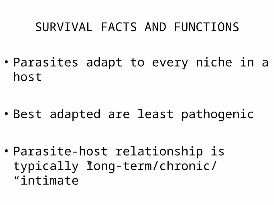
SURVIVAL FACTS AND FUNCTIONS
• Parasites adapt to every niche in a host
• Best adapted are least pathogenic
• Parasite-host relationship is typically long-term/chronic/ “intimate”

CONDITIONS REQUIRED FOR ENDEMIC PARASITISM
• Reservoir of Infection
• Means of Transmission to Susceptible Hosts
• Ability to Invade and Establish in New Hosts
• Ability to Reproduce
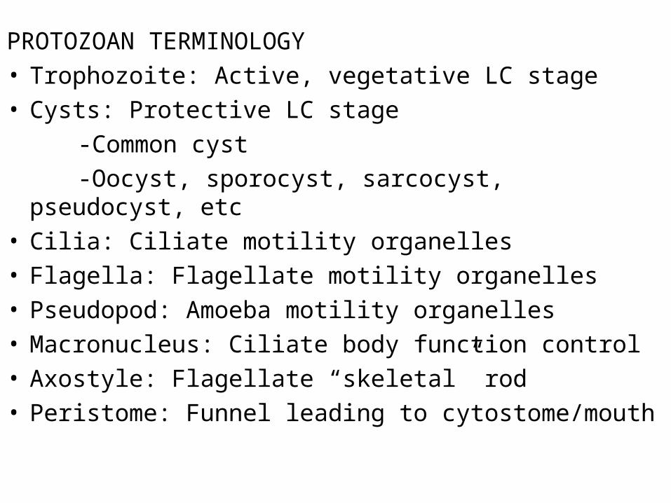
PROTOZOAN TERMINOLOGY• Trophozoite: Active, vegetative LC stage• Cysts: Protective LC stage
-Common cyst
-Oocyst, sporocyst, sarcocyst, pseudocyst, etc• Cilia: Ciliate motility organelles• Flagella: Flagellate motility organelles• Pseudopod: Amoeba motility organelles• Macronucleus: Ciliate body function control• Axostyle: Flagellate “skeletal” rod• Peristome: Funnel leading to cytostome/mouth

CILIATE PARASITEBalantidium coli
Trophozoite Cyst
Cytostome Macronucleus
Macronucleus
Cilia
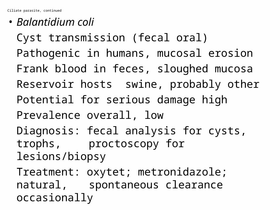
Ciliate parasite, continued
• Balantidium coli
Cyst transmission (fecal oral)
Pathogenic in humans, mucosal erosion
Frank blood in feces, sloughed mucosa
Reservoir hosts swine, probably other
Potential for serious damage high
Prevalence overall, low
Diagnosis: fecal analysis for cysts, trophs, proctoscopy for lesions/biopsy
Treatment: oxytet; metronidazole; natural, spontaneous clearance occasionally

Balantidium coli Life Cycle

FLAGELLATE PARASITES AND COMMENSALS
Trichomonads and Dientamoeba fragilis
Trophozoites only
Flagella
Nucleus/nuclei
Body shape & size

Flagellates, continued
Trichomonas tenax
Trophozoite transmission-direct oral
Mouth inhabitant, oral hygiene factor
Nonpathogenic, thrives in bad conditions
Reservoir unknown, probably wide
Considered classically commensalistic
Prevalence data spotty
Diagnosis by culture, microscopic exam of oral fluids/scrapings
Eliminated by good oral hygiene

Pentatrichomonas hominis, Dientamoeba fragilis
Transmission direct-oral, no cysts (you tell me)Colon/caecum inhabitants Non-pathogenicReservoir unknown, probably wideConsidered commensalistic (D. fragilis ???)Prevalence unknownDiagnosis usually incidental-fecal smear-stain,
wet mounts + microscopyTreatment: incidental elimination-Flagyl, et.al.

Dientamoeba fragilis Life Cycle

Flagellate, continued
Trichomonas vaginalis
Transmitted by sexual intercourseInduces vaginal pH change, erosion of
normal mucosa in womenVaginal itching, burning, yellow discharge
in women, occasional urethritis, prostate swelling in men
Human reservoir, zoonotic potential ??Prevalence varies with population &
cultureDiagnosis by visual features, microscopyTreatment usually Flagyl
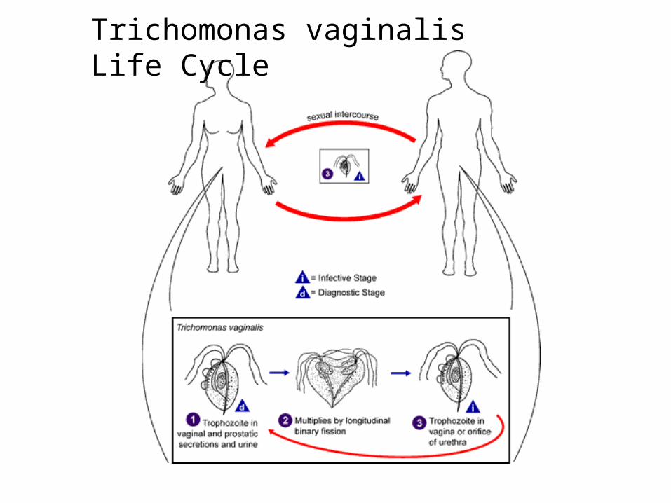
Trichomonas vaginalis Life Cycle

Flagellate Parasites and Commensals
Chilomastix mesnili Enteromonas hominis, Retortamonas intestinalis, Giardia lamblia, et.al.
Trophozoites Cysts
Nuclei Nuclei
Flagella Size & shape
Size & shape

Flagellates, continued
Chilomastix mesnili, Enteromonas sp., Retortamonas sp.,, others
Caecum/colon inhabitantsTransmission by cyst or trophozoiteNonpathogenic, commensalisticThrive in most diarrheic conditionsReservoir pool (probably) wide, unknownWidespread, sanitation dependentDiagnosis: microscopic fecal examTreatment: unnecessary in most cases,
Flagyl will work

Flagellates, continued
Giardia lamblia, etcCyst transmissionPathogenic potential individually inconsistentClinical signs variable
Diarrhea/dysentery, periodic or steadyGas productionBorborygmusAnorexiaSkin rashFibromyalgiaSpontaneous lactose intoleranceFatigue, mild/severeOther

Flagellates, continued
Giardia, continued
Reservoir hosts: almost any mammal
Damage potential: individual factors
Immunocompetence of host
Natural, undefined host tolerance level
Other (fuzzy factors)
Worldwide distribution, sanitation dependent
Diagnosis: fecal ELISA, direct microscopic exam for cysts/trophs
Treatment: Atabrine, Flagyl, other

Giardia Life Cycle

Flagellates, continued
HAEMOFLAGELLATES
Trypanosomes/ Leishmanias/
trypomastigote forms amastigote forms


Haemoflagellates, continued
Trypanosoma brucei complex, T.b. gambiense, T.b. rhodesiense, others
Vector transmission, Tse tse flies
Pathogenic, terminal ‘sleeping sickness’, East African SS less acute than West African SS
Signs: swollen cervical lymph nodes, fever,
rashes, headache, malaise, nausea, eventually coma
Various wild/domestic animal reservoirs
West African much more acute and severe than East African SS.

Haemoflagellates, continued
Trypanosoma brucei complex, continued
T.b. gambiense in West Africa, overlaps with endemic East African T.b. rhodesiense in center of continent
Microscopy of concentrated or cultured blood or fluid aspirates, RES biopsy
normal diagnostic methods
Treatment: melarsoprol complex, suramin
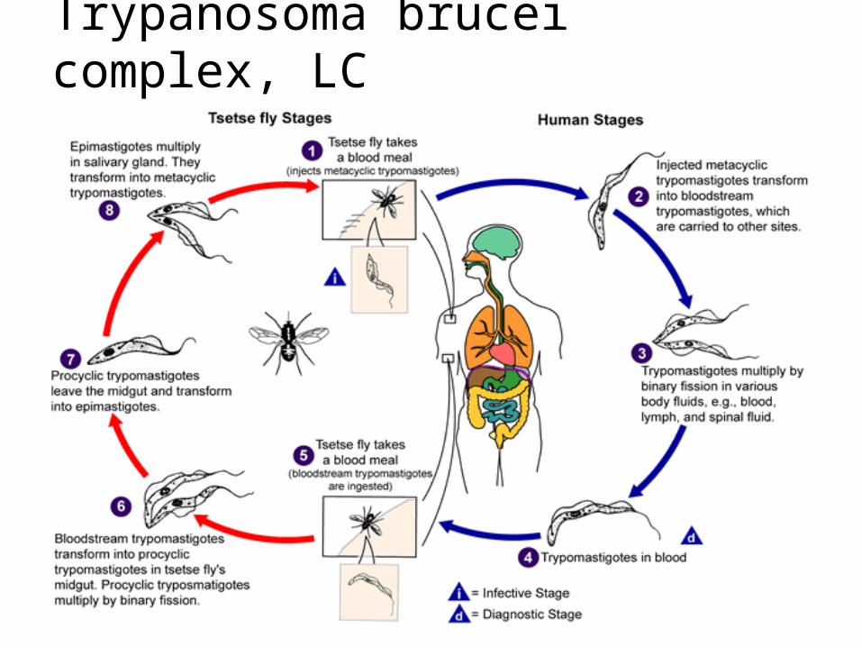
Trypanosoma brucei complex, LC
Haemoflagellates, continued
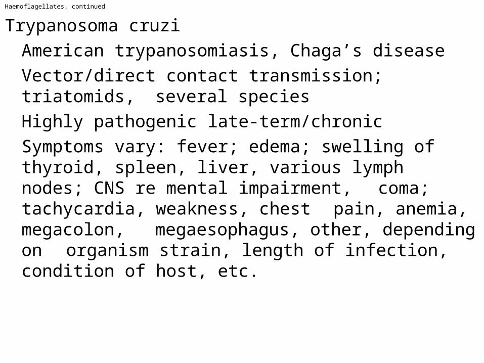
Trypanosoma cruzi
American trypanosomiasis, Chaga’s disease
Vector/direct contact transmission; triatomids, several species
Highly pathogenic late-term/chronic
Symptoms vary: fever; edema; swelling of thyroid, spleen, liver, various lymph nodes; CNS re mental impairment, coma; tachycardia, weakness, chest pain, anemia, megacolon, megaesophagus, other, depending on organism strain, length of infection, condition of host, etc.

Haemoflagellates, continue
T. cruzi, continuedReservoir large, many carnivore, omnivore
& herbivore speciesDamage severe, early (fulminating) or late
(chronic), depends on various factorsPrevalence < 3% to > 50% in endemic
areasfrom southcentral USA to southern SA
Diagnosis: cell/fluid culture, xenodiagnosis, direct microscopy
Treatment: no reliable/curative; nifurtimox, primaquine & related drugs reduce but do not eliminate blood stage, nothing effective X cellular stage

T.cruzi, continued
Trypomastigote/Trypanosome
Triatomid Vector

Haemoflagellates, continued
T. Cruzi life cycle

Haemoflagellates, continued
Leishmania topica complex, L.t. mexicana complex, L.t. braziliense complex, et.al.
Vector trans. by sand flies Superficial to extensive, shallow to deep Cutaneous lesions, vary by strain/species
Oriental sore: limited, wet ulcerChiclero ulcer: ear ‘notches’Diffuse cutaneous: dry, diffuseMucocutaneous: cartilage erosion
Reservoir: large; many native carnivore, omnivore, herbivore vertebrates

Haemolagellates, continued
Leishmania tropica complex, continued
Lesion severity varies with species/strain, simple limited wet/dry to severe
erosion
Widespread in tropical, subtropical & warm temperate regions worldwide
Lesion appearance is diagnostic, agents can be cultured or viewed microscopically
Pentavalent antimony compound treatment, with/without amphotericin B

Haemoflagellates, continued
Leishmania donovoni complex
Vector transmission, sandflies
Visceral, reticulo-endothelial system inhabitation, often lethal
Fevers (variable), anemia, hepatomegaly
splenomegaly, ascites, Kala-azar (blackening of facial skin), et.al.
Reservoir: domestic & wild vetebrates
Damage potential varies with species/strains
Distributed widely, tropics, subtropics, warm temperate and cool temperate regions
Diagnosis by serology, culture of blood or biopsy
Antimony, amphotericin-B, allopurinol treatments
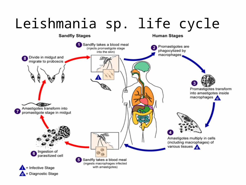
Leishmania sp. life cycle
Sarcodina
AMOEBIC PARASITES AND COMMENSALS
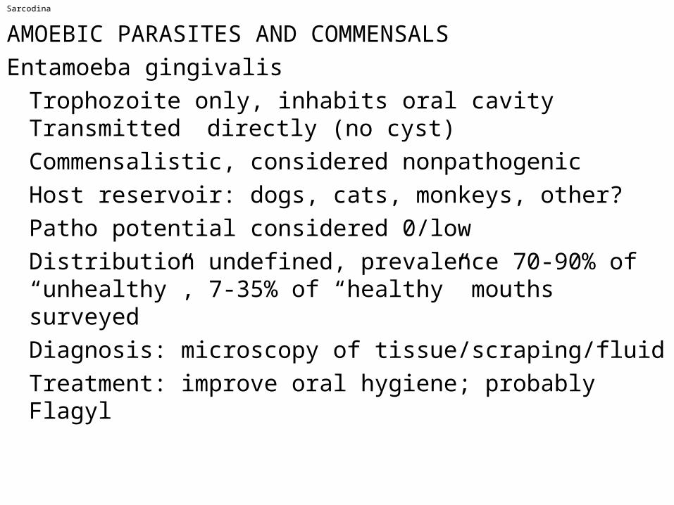
Entamoeba gingivalis
Trophozoite only, inhabits oral cavity Transmitted directly (no cyst)
Commensalistic, considered nonpathogenic
Host reservoir: dogs, cats, monkeys, other?
Patho potential considered 0/low
Distribution undefined, prevalence 70-90% of “unhealthy”, 7-35% of “healthy” mouths surveyed
Diagnosis: microscopy of tissue/scraping/fluid
Treatment: improve oral hygiene; probably Flagyl

Sarcodines, continued
Amebic parasites and commensals, continued
Entamoeba histolytica
Trophozoite in caecum/colon, if invasive may inhabit liver, lungs, other tissues;
Cysts (infective stage) form in normal stools
Pathology variable: noninvasive; if invasive, ulcerates colonic mucosa, spreads to liver, lung, et.al., produces abcesses;
path potential indicated by colony site
Reservoir includes monkeys, dogs, pigs, et.al.
Distribution worldwide: tropical, subtropical, warm temperate areas; sanitation
dependent

Sarcondines, continued
E. histolytica, continued
Prevalence rated second to Giardia worldly
Diagnosis by microscopic ID of trophs, cysts in feces, trophs in tissue-based
abcesses
Treated with Flagyl (metronidazol), various Emetine formulations, Diiodohydroxyquin, et.al.
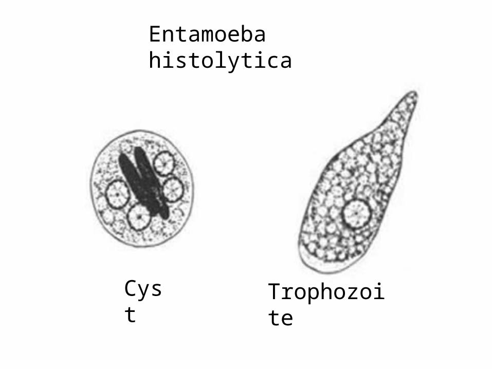
Entamoeba histolytica
Cyst Trophozoite
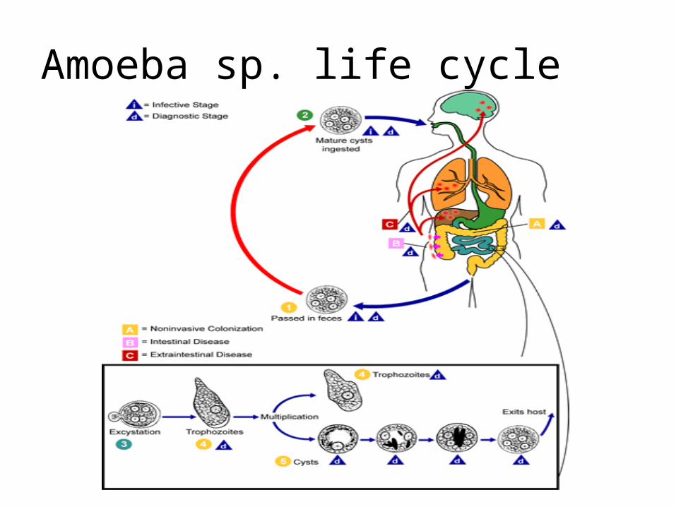
Amoeba sp. life cycle

Sarcodines, continued
Entamoeba coli, E. hartmanni, E. dispar, E. sp.(unnamed), Endolimax nana, Iodamoeba butschlii, a few others
Caecum/colon inhabitants, transmitted by cysts,
All commensals (with rare exceptions)Diarrhea enhances production of trophsReservoir: various vertebrate animalsDamage potential 0/low (some
exceptions?) Prevalence high, world-wide warm areas
Diagnosis: microscopic ID in fecesTreatment considered unnecessary
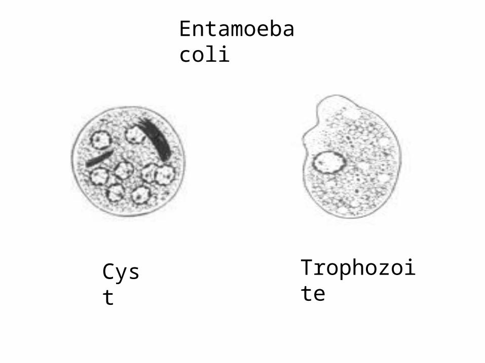
Entamoeba coli
Cyst Trophozoite

Sporozoa/apicomplexa
SPOROZOA/APICOMPLEXA TERMINOLOGY
Sporogony: basic life cycle stage; sporozoite generation
Schizogony/merogony: basic life cycle stage; (asexual repro) merozoite generation
Gametogony/gamogony: basic life cycle stage; (sexual repro) gametocyte generation
Oocyst: cyst produced in sporogony
Sporocyst: cyst within oocyst, produced in sporogony
Sporozoite: basic infective unit in oocysts/sporocysts

Sporozoa, continued
Sporozoan terminology, continued
Trophozoite: transitional zoite, between sporozoite and schizont/merozoite
Merozoite: basic zoite product of schizogony
Tachyzoite: rapidly replicating merozoite
Bradyzoite: slowly replicating merozoite
Sarcocyst: end-stage schizont in intermediate host with Sarcocystis sp. infection
Pseudocyst: end-stage schizont in intermediate host with Toxoplasma gondii infection

Sporozoa, continued
Sporozoa, continued
Basic Life Cycle Stages
Sporogony: formation of sporocysts and sporozoites
Schizogony/merogony: formation of merozoites/tachyzoites/bradyzoites
Gamogony/gametogony: formation of gametocytes and gametes

Sporozoa, continued
Isospora belliTransmission direct, fecal oral, via oocystsPathogenic potential low, non-bloody diarrhea common in immunodeficient hosts, uncommon in others Clinical signs absent, except in rare casesReservoir limited to humans, other anthropoids,strongly host-specificDamage low, destroys superficial mucosal cellsPrevalence world-wide, sanitation dependentDiagnosis by microscopic ID of oocysts in fecal flotationTreatment usually unnecessary, pyrimethamine + a sulfa, trimethoprim, when needed
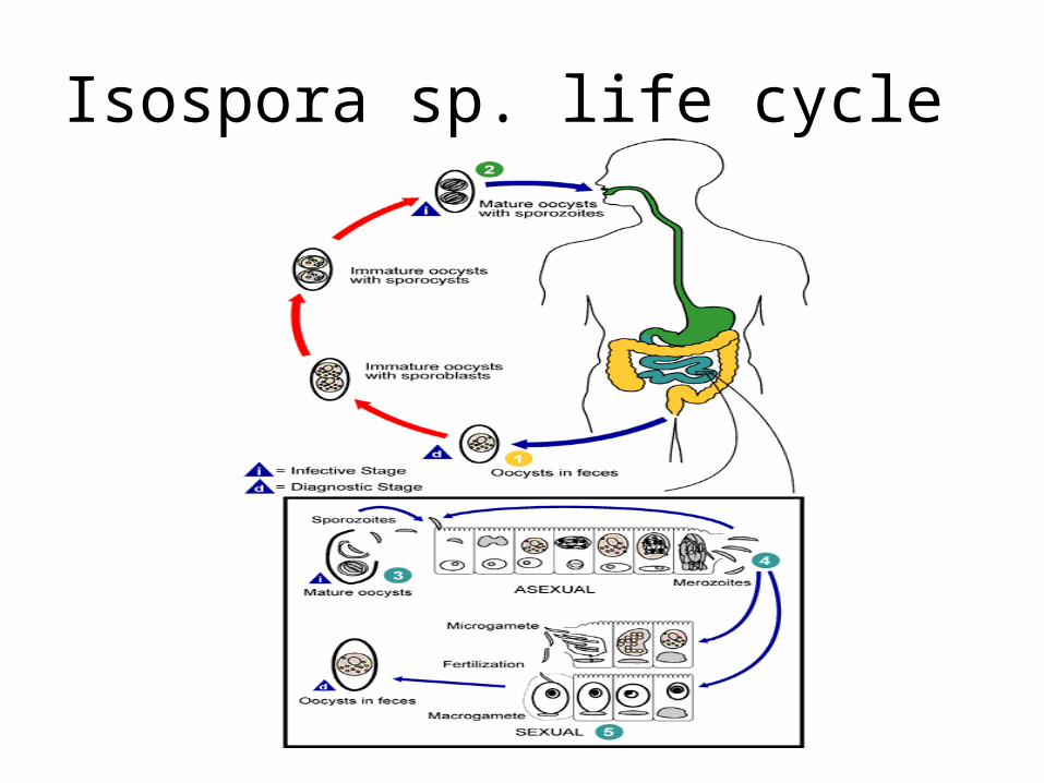
Isospora sp. life cycle

Sporozoa, continued
Cyclospora cayetanensis
Transmission direct fecal-oral, via oocysts
Pathogenic potential low/moderate, non-bloody diarrhea in sporadic cases, most severe in immunodeficient individuals
Diarrhea ~3 weeks in “healthy” hosts, longer/much longer in immonodeficient; can be cyclic, recurrent; long-term may + anorexia, fatigue, weight loss, fever
Reservoir hosts: reptiles, rodents, insectivores, probably other domestic & wild animals;
species ID is incomplete in host animals

Sporzoa, continued
C. cayetanensis, continued
Damage: jujunal villous atrophy, crypt hyperplasia, inflammation
Prevalence spotty, outbreaks in New Guinea, Nepal, Peru, Chicago, Canada, other
Diagnosis: microscopic ID of oocysts from fresh feces, acid-fast-stained smears,
fluorescent Ab-stain preps
Treatment: trimethoprim + sulfamethoxazole
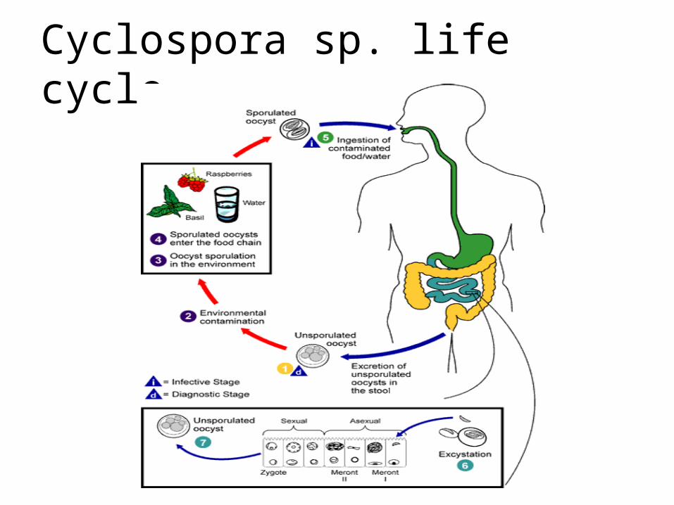
Cyclospora sp. life cycle

Sporozoa, continued
Cryptosporidium parvum
Transmission direct, fecal-oral, via oocysts
Pathogenic potential variable: low in “healthy”, moderate/high in “deficient” hosts,
depending on immunocompetence level
Clinical signs: non-bloody diarrhea/dysentery, mild/short-term (~2 weeks) to severe/long-term (steady or recurrent)
Reservoir: complete spectrum unknown, but many domestic animals are known
Damage potential and mechanisms vary with hosts & species, poorly understood

Sporozoa, continued
C. parvum, continued
Prevalence world-wide, sanitation dependent,
Diagnosis: microscopic ID of oocysts in feces by flotation, acid-fast or
immunofluorescent staining; histologic or immunohistologic exam of biopsy of intestinal mucosa
Treatment: paramomycin may be suppressive in specific cases, not curative (no curative medication known)

Cryptosporidium sp. life cycle

Sporozoa, continued
Sarcocystis bovihominis, S. suihominis, probably others
Transmission: ingestion of beef or pork (or other), uncooked/undercooked, containing sarcocysts in muscle fibers
Pathogenic potential low in human DH
Clinical signs absent except in rare cases
Reservoir limited to human DH (+ possibly other anthropoids), and bovine/porcine IHs
Damage low in human DH, inconsequential erosion of intestinal mucosa
Sporozoa, continued
S. bovihominis, S. suihominis, etc. continued
Prevalence world-wide, determined by cultural food prep and consumption factors
Diagnosis: microscopic ID of oocysts and/or sporocysts in feces
No treatment identified: trimethoprim + sulfamethoxazol probably suppressive

Sporozoa, continued
Sarcocystis lindemanni
Transmission by ingestion of sporocysts from unknown DHs in fecal contamination
Pathogenic potential unknown
Clinical signs unknown
Reservoir unknown
Damage potential unknown
Prevalence unknown, probably sanitation dependent
Diagnosis: histologic examination of muscle
Treatment unknown
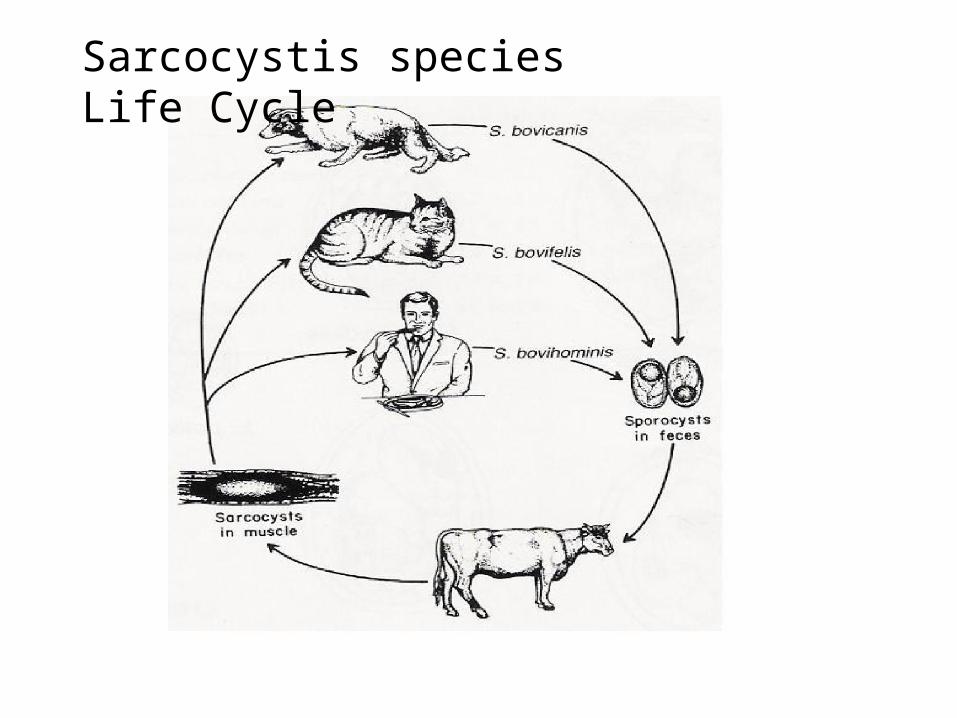
Sarcocystis species Life Cycle

Sporozoa, continued
Toxoplasma gondii
Transmission direct via oocysts (fecal-oral), ingestion of infected meat, transplacental, nursing, organ transplantation, et.al.
Pathogenicity moderate to high, depending on strain, host “health” factors
Clinical signs: Acute infection; range from unnoticeable to severe flu-like (chills, fever, headache, fatigue, lymphoid pain & swelling)
Transplacental; death & abortion, various encephalomyelitis, megacephaly,
microcephaly, blindness, deafness
Sporozoa, continued
T. gondii, continuedReservoir enormous: nearly all warm-blooded
vertebrates including birds, suitability variedDamage potential dependent on strain, host susceptibility, host “health” conditionPrevalence variable, depending on association
with feline DHs, and food (meat) preference & preparationDiagnosis: Indirect; fluorescent Ab, latex aggl.,
serum ELISA, other serotests. Direct; culture of body fluids & tissue samples, immunohistochemistry, histopathologyTreatment: Pyrimethamine + a pyrimidine

Toxoplasma gondii life cycle

Sporozoa, continued
Plasmodium vivax
Transmission: female Anopheles mosquito vector, blood transfusion
Pathogenicity high, especially in 1st infections, moderate/high in subsequent infections and relapses, depending on host condition
Symptomatics: ~12-20 day prepatency (no signs); prodroma (influenza-like; headache, nausea,
vomiting, anorexia, muscle aches); sudden,severe shock-like chill (paroxysm), fever cycle quickly stabilizing at ~48 hr, continuous for 3-10 weeks; recrudescences/relapses for 5-8 years
Reservoir: humans, monkeys, apes, Anopheles vector

Sporozoa,continued
P. vivax, continued
Damage: extensive hemolysis, production of toxic parasite metabolites
Prevalence: world-wide tropical and sub-tropical less common in warm temperate regions
Diagnosis: microscopic ID/differentiation of species by microscopic exam of stained smears of blood properly collected and prepared
Treatment: Quinine & related alkaloids, at least 15 additional, used or in trial, singly or in combination, efficacy variable

Plasmodium sp. life cycle

Sporozoa, continued
Plasmodium ovale
All factors involving this species are nearly identical to those listed for P. vivax, except for severity of damage, symptoms, prevalence and duration of infection.
Damage potential: low/moderate, primary erythrocytic cycle ~2-3 weeks, total duration of infection ~1-2 years
Symptoms: similar to P. vivax but less severe, same fever cycle periodicity
Prevalence: widespread tropical & subtropical
Treatment: same as for P. vivax, et.al.

Sporozoa, continued
Plasmodium malariae
Transmission as described for P. vivax
Pathogenic potential high re: hemolysis and CNS involvement late in infection
Symptoms similar to P. vivax & P ovale, with longer fever cycle periodicity (72 hr), 3-24 weeks primary duration, 20-50 years duration of untreated infection with probability of recrudescence
Reservoir as described for P. vivax, P. ovale
Damage high; anemia, CNS & kidney syndrome

Sporozoa, continued
P. malariae, continued
Prevalence more common in subtropical and warm temperate regions than tropical, but endemic where other species occur
Diagnosis as described for P. vivax & P. ovale
Treatment as described for P. vivax & P. ovale

Sporozoa, continued
Plasmodium falciparum
Transmission as described for P. vivax, et.al.
Pathogenic potential highest of all Plasmodium species, most likely of all to kill IH (human)
Clinical signs similar to those described: shorter (8-11 days) incubation period, prodroma similar but mild, cycle periodicity ~ 48hr,
initial paroxysm severe & long (16-36hr), 2-3 week duration of primary attack, 6-17 months duration of untreated infection
Reservoir: humans, monkeys, apes, Anopheles mosquito vector

Sporozoa, continued
P. falciparum, continued
Damage as described: hemolysis, etc., but also causes cytoadherence to endothelium of damaged and intact parasitized cells and cellular debris; all organs (brain, kidneys,liver, etc.) are affected
Prevalence world-wide, but confined to tropics and subtropics
Treatment as described for other species

Spoorozoa, continued
Babesia spp. (B. microti, B. divergens, B. gibsoni)
Transmission by vector ixodid tick DHs
Pathogenic potential high in splenectomized and other immunocompromised humans, may be mild or serious in intact hosts, species/strain differences are known
Clinical signs: malaise, headache, fever, chills, swetting, fatigue, weakness, anemia, jaundice, renal failure
Reservoir: rodents, livestock, other “natural” hosts, humans appear to be accidentals
Damage high in immunodeficient, moderate in most others; much depends on species/strain of agent involved

Sporozoa,continued
Babesia spp., continued
Prevalence widespread in “natural” reservoir hosts, spotty in humans: Europe, NE USA, Texas, Mexico, NC USA, et.al.
Diagnosis: microscopic ID and differentiation from malarial (Plasmodium sp.) agents
Treatment: oral quinine + IV clindamycin, a few others, less efficacious

Babesia sp. life cycle


![Prevalence of intestinal parasitic infections and ... · intestinal parasitic infections caused by helminths and intestinal protozoa [1, 11–15]. In Burkina Faso, where polyparasitism](https://static.fdocuments.net/doc/165x107/5ecdb4a171fb394e4f7767a3/prevalence-of-intestinal-parasitic-infections-and-intestinal-parasitic-infections.jpg)