
High diversity and morphological convergence among ...
18
Persoonia 21, 2008: 93–110 www.persoonia.org doi:10.3767/003158508X371379 © 2008 Nationaal Herbarium Nederland Centraalbureau voor Schimmelcultures RESEARCH ARTICLE INTRODUCTION Rock substrata have been explored and analysed with in- creased microbiological attention because of their extreme variations in environmental factors that select strongly for colonisation by stress-tolerant fungi (Staley et al. 1982, Urzí et al. 1993, Sterflinger & Krumbein 1995, Wollenzien et al. 1995, Sterflinger & Krumbein 1997, Sterflinger 2000, Sterflin- ger & Prillinger 2001, Bogomolova & Minter 2003, de Leo et al. 2003). Rock surfaces have been recognised as a reservoir for highly melanised, slow-growing filamentous and yeast-like fungi of ascomycetous affinities. Darkly pigmented fungi from geological materials have been named by a variety of terms, e.g., black fungi, black yeasts, or microcolonial fungi (Staley et al. 1982), in reference to their prominent shared morphological characteristics. The interests in melanised fungi are multiple and varied. From a clinical point of view, the anamorphs of Capronia (Herpotri- chiellaceae), and many species belonging to the Exophiala- Ramichloridium-Rhinocladiella-Cladophialophora complex, constitute a group of medically significant opportunists (de Hoog et al. 1994, 1998, 2000b). Another recent trend has been to focus on these organisms as a model for astrobiological stud- ies of eukaryotes (Gorbushina 2003). Finally, the life histories of melanised fungi from rock surfaces may intersect with other saprobic, lichenicolous, epiphytic or even phytopathogenic fungi (Untereiner & Malloch 1999, Crous et al. 2000, 2007a). Observations of novel melanised fungi from man-made rock surfaces in arid and semi-arid regions, especially those of the Mediterranean basin, led us to test whether these fungi were a widespread component of natural rock surfaces. If such fungi were present, are they abundant and taxonomically distinct? Alternatively, rock surfaces may be primarily colonised by fungi deposited from soils, the phyllosphere, or from nascent lichen mycobionts. These questions were important for determining whether rock surfaces could harbour organisms producing secondary metabolites of relevance for the pharmaceutical industry. The fungi associated with rock surfaces present a series of characteristics that makes them a potentially attractive group for a microbial screening programme, e.g., their appar- ently ubiquitous habitats, their relative ease of collecting and cultivation in culture, and the fact that at least some species are related to other ascomycetous fungi known to possess multiple secondary metabolite biosynthetic pathways (Kroken et al. 2003). Dark pigmentation of the strains indicates that second- ary metabolite biosynthetic pathways, e.g., those involved in melanogenesis, are operational. High diversity and morphological convergence among melanised fungi from rock formations in the Central Mountain System of Spain C. Ruibal 1 , G. Platas 2 , G.F. Bills 2 1 Departamento de Ingeniería y Ciencia de los Materiales, Escuela Técnica Superior de Ingenieros Industriales, Universidad Politécnica de Madrid (UPM), José Gutiérrez Abascal 2, E-28006 Madrid, Spain; corresponding author e-mail: [email protected]. 2 Centro de Investigación Básica, Merck, Sharp & Dohme de España, S.A., Josefa Valcárcel 38, E-28027 Madrid, Spain. Key words biodiversity black fungi Capnodiales Chaetothyriales Dothideomycetes extremotolerance Abstract Melanised fungi were isolated from rock surfaces in the Central Mountain System of Spain. Two hundred sixty six isolates were recovered from four geologically and topographically distinct sites. Microsatellite-primed PCR techniques were used to group isolates into genotypes assumed to represent species. One hundred and sixty three genotypes were characterised from the four sites. Only five genotypes were common to two or more sites. Morphological and molecular data were used to characterise and identify representative strains, but morphology rarely provided a definitive identification due to the scarce differentiation of the fungal structures or the apparent novelty of the isolates. Vegetative states of fungi prevailed in culture and in many cases could not be reliably dis- tinguished without sequence data. Morphological characters that were widespread among the isolates included scarce micronematous conidial states, endoconidia, mycelia with dark olive-green or black hyphae, and mycelia with torulose, isodiametric or moniliform hyphae whose cells develop one or more transverse and/or oblique septa. In many of the strains, mature hyphae disarticulated, suggesting asexual reproduction by a thallic micronematous conidiogenesis or by simple fragmentation. Sequencing of the internal transcribed spacers (ITS1, ITS2) and 5.8S rDNA gene were employed to investigate the phylogenetic affinities of the isolates. According to ITS sequence alignments, the majority of the isolates could be grouped among four main orders of Pezizomycotina: Pleosporales, Dothideales, Capnodiales, and Chaetothyriales. Ubiquitous known soil and epiphytic fungi species were generally absent from the rock surfaces. In part, the mycota of the rock surfaces shared similar elements with melanised fungi from plant surfaces and fungi described from rock formations in Europe and Antarctica. The possibility that some of the fungi were lichen mycobionts or lichen parasites could not be ruled out. Article info Received: 24 May 2008; Accepted: 9 September 2008; Published: 26 September 2008.
Transcript of High diversity and morphological convergence among ...

Persoonia 21, 2008: 93–110www.persoonia.org doi:10.3767/003158508X371379
© 2008 Nationaal Herbarium Nederland Centraalbureau voor Schimmelcultures
RESEARCH ARTICLE
INTRODUCTION
Rock substrata have been explored and analysed with in-creased microbiological attention because of their extreme variations in environmental factors that select strongly for colonisation by stress-tolerant fungi (Staley et al. 1982, Urzí et al. 1993, Sterflinger & Krumbein 1995, Wollenzien et al. 1995, Sterflinger & Krumbein 1997, Sterflinger 2000, Sterflin-ger & Prillinger 2001, Bogomolova & Minter 2003, de Leo et al. 2003). Rock surfaces have been recognised as a reservoir for highly melanised, slow-growing filamentous and yeast-like fungi of ascomycetous affinities. Darkly pigmented fungi from geological materials have been named by a variety of terms, e.g., black fungi, black yeasts, or microcolonial fungi (Staley et al. 1982), in reference to their prominent shared morphological characteristics.
The interests in melanised fungi are multiple and varied. From a clinical point of view, the anamorphs of Capronia (Herpotri-chiellaceae), and many species belonging to the Exophiala-Ramichloridium-Rhinocladiella-Cladophialophora complex, constitute a group of medically significant opportunists (de Hoog
et al. 1994, 1998, 2000b). Another recent trend has been to focus on these organisms as a model for astrobiological stud-ies of eukaryotes (Gorbushina 2003). Finally, the life histories of melanised fungi from rock surfaces may intersect with other saprobic, lichenicolous, epiphytic or even phytopathogenic fungi (Untereiner & Malloch 1999, Crous et al. 2000, 2007a).
Observations of novel melanised fungi from man-made rock surfaces in arid and semi-arid regions, especially those of the Mediterranean basin, led us to test whether these fungi were a widespread component of natural rock surfaces. If such fungi were present, are they abundant and taxonomically distinct? Alternatively, rock surfaces may be primarily colonised by fungi deposited from soils, the phyllosphere, or from nascent lichen mycobionts. These questions were important for determining whether rock surfaces could harbour organisms producing secondary metabolites of relevance for the pharmaceutical industry. The fungi associated with rock surfaces present a series of characteristics that makes them a potentially attractive group for a microbial screening programme, e.g., their appar-ently ubiquitous habitats, their relative ease of collecting and cultivation in culture, and the fact that at least some species are related to other ascomycetous fungi known to possess multiple secondary metabolite biosynthetic pathways (Kroken et al. 2003). Dark pigmentation of the strains indicates that second-ary metabolite biosynthetic pathways, e.g., those involved in melanogenesis, are operational.
High diversity and morphological convergence among melanised fungi from rock formations in the Central Mountain System of Spain
C. Ruibal1, G. Platas2, G.F. Bills2
1 Departamento de Ingeniería y Ciencia de los Materiales, Escuela Técnica Superior de Ingenieros Industriales, Universidad Politécnica de Madrid (UPM), José Gutiérrez Abascal 2, E-28006 Madrid, Spain;
corresponding author e-mail: [email protected] Centro de Investigación Básica, Merck, Sharp & Dohme de España, S.A.,
Josefa Valcárcel 38, E-28027 Madrid, Spain.
Key words
biodiversityblack fungiCapnodialesChaetothyrialesDothideomycetesextremotolerance
Abstract Melanised fungi were isolated from rock surfaces in the Central Mountain System of Spain. Two hundred sixty six isolates were recovered from four geologically and topographically distinct sites. Microsatellite-primed PCR techniques were used to group isolates into genotypes assumed to represent species. One hundred and sixty three genotypes were characterised from the four sites. Only five genotypes were common to two or more sites. Morphological and molecular data were used to characterise and identify representative strains, but morphology rarely provided a definitive identification due to the scarce differentiation of the fungal structures or the apparent novelty of the isolates. Vegetative states of fungi prevailed in culture and in many cases could not be reliably dis-tinguished without sequence data. Morphological characters that were widespread among the isolates included scarce micronematous conidial states, endoconidia, mycelia with dark olive-green or black hyphae, and mycelia with torulose, isodiametric or moniliform hyphae whose cells develop one or more transverse and/or oblique septa. In many of the strains, mature hyphae disarticulated, suggesting asexual reproduction by a thallic micronematous conidiogenesis or by simple fragmentation. Sequencing of the internal transcribed spacers (ITS1, ITS2) and 5.8S rDNA gene were employed to investigate the phylogenetic affinities of the isolates. According to ITS sequence alignments, the majority of the isolates could be grouped among four main orders of Pezizomycotina: Pleosporales, Dothideales, Capnodiales, and Chaetothyriales. Ubiquitous known soil and epiphytic fungi species were generally absent from the rock surfaces. In part, the mycota of the rock surfaces shared similar elements with melanised fungi from plant surfaces and fungi described from rock formations in Europe and Antarctica. The possibility that some of the fungi were lichen mycobionts or lichen parasites could not be ruled out.
Article info Received: 24 May 2008; Accepted: 9 September 2008; Published: 26 September 2008.

94 Persoonia – Volume 21, 2008
Fungi isolated from rocks share macro- and micromorphologi-cal characteristics, but their phylogenetic origins appear to be diverse (Sterflinger et al. 1999). A previous study demonstrated that limestone from Mallorca was colonised by a diverse and complex assemblage of Pezizomycotina, with many representa-tives among the classes Dothideomycetes and Chaetothyrio-mycetes (Ruibal et al. 2005). However, many more of the fungi belonged to undetermined orders and families, or corresponded poorly to known families of Pezizomycotina.
In order to confirm and expand our preliminary results (Ruibal et al. 2005), we analysed the number and kinds of melanised fungi associated with the surfaces of four different types of rock in the Central Mountain System of Spain. Our first step was to explore practical methods of providing large numbers of unique fungal isolates for use in natural products discovery and to measure the biodiversity of the fungal community in order to understand the logistics of handling large numbers of fungal strains. We enumerated the different melanised fungi that were recovered from granite, black slate, limestone, and quartzite formations using a combined macromorphology and microsatellite-primed PCR technique to select unique strains, grouping isolates into genotypes assumed to represent species. A preliminary evaluation of their phylogenetic diversity based on ITS1-5.8S-ITS2 rDNA sequences is presented. Phylogenetic re-lationships among the isolates and possible novel groups were explored by constructing a general sequence-based framework with known genera of similar fungi. The phylogenetic framework was correlated with cultural features, attempting to recognise morphologically defined genera and species.
MATERIALS AND METHODS
Study area, environmental and geological features
The Spanish Central System is an intraplate mountain range located in the central part of the Iberian Peninsula and consists, for the most part, of plutonic and metamorphic rocks belonging to the Hercynian basement and a locally preserved thin cover of Upper Cretaceous sedimentary rocks. Therefore, the area is lithologically and structurally complex and comprises a number of formations such as the Sierras of Guadarrama, Somosierra and Ayllón (García & Aparicio 1987).
The four sites were located in the province of Madrid, within an area of c. 200 km2, with distances between sites from 8 to 20 km. The zone receives c. 600–700 mm rainfall distributed irregularly during the year. Two main seasons predominate, a dry and hot summer, with temperatures fluctuating between 40 and 5 °C, and a more humid and cold winter, with temperatures ranging between 15 and -10 °C. The number of sunny days averages about 240/yr.
La Cabrera study site (40°52'30"N, 3°38'W) (Fig. 1) was located near the village of the same name, at c. 1600 m above sea level (asl). It is a pure granite formation of Plutonic origin, comprised of mixed fine to middle grained leucogranites of hernycic age (Bellido Mulas & Rodríguez Fernández 1991). The scarce vegetation consisted mostly of shrubs (dominated by Cistus ladanifer) and pasture. The site was sampled in August 2001. The Atazar site (40°57'N, 3°25'W), sampled in October 2001, was located c. 7 km northeast of the village of the same name, at c. 1300 m asl. The zone consisted of metamorphic, black slate from the Silurian period (Pérez González & Portero 1990).
Fig. 1 General view of La Cabrera study site.

95C. Ruibal et al. Melanised fungi from Spain
The vegetation was sparse, consisting of shrubs (dominated by Cistus species) and pasture. The Patones site (40°53'N, 3°27'W) was located c. 5 km to the east of the village of the same name along the road M-102, at c. 800 m asl, on a ridge crest that delimits the valley of the Jarama River. The area was at the southern border of the Central Mountain System and was formed of cretaceous limestone and dolomites (Pérez González & Portero 1990). The site was sampled in May 2003. The vegetation was chaparral-like and dominated by Quercus ilex, Retama sphaerocarpa, and Juniperus oxycedrus. The Puebla de la Sierra site (41°4'N, 3°29'W), sampled in October 2003, was located c. 2 km on the trail towards La Peña de Cabra at km 9 of the road M-130, between the villages of Prádena del Rincón and Puebla de la Sierra at an elevation of c. 1700 m asl. The zone was formed mainly of quartzite, with mixed small quantities of sandstone and conglomerates, from the Ordovi-cian period (García & Aparicio 1987). The site was the only one that was tree-covered, mainly with planted Scots pine (Pinus sylvestris) and Spanish fir (Abies pinsapo).
Isolation of fungi from rock surfaces
Sample selection was determined by locating a clean and easily collectible sample. Rock samples were taken from clean, verti-cal, lichen-free surfaces. At each site, 2–5 rock faces separated by 20–200 m and in different orientations were sampled. A cold chisel was inserted into an existing crack, and the pieces of superficial rock were chipped off the surface with a hammer. Rock fragments were packed in sterile, clean paper bags and transported to the laboratory at ambient temperature. Samples were processed for isolation within 1 wk.
The rock surfaces were rinsed with 96 % ethanol and allowed to dry to reduce the influence of dust and airborne spores. Sam-ples for isolation plates were prepared by chipping or scraping surfaces with a sterile knife and chisel blade to remove small superficial fragments from 10–20 cm2 area from the outer layer of the field-collected rock. Penetration into the rock was c. 0.5–3 mm, depending on the rock texture. The fragments were further pulverised in a sterile mortar. About 2–3 cm3 of pulverised rock were added to a sterile 50 mL centrifuge tube and washed once with a weak sterile detergent solution (aque-ous Tween 20, Sigma -Aldrich, Inc., St. Louis, MO, USA), fol-lowed by five washes, three with distilled water and two more with sterile water. Washed samples were resuspended in 40 mL of sterile water and aliquots were spread homogenously onto isolation media plates. For each rock sample, aliquots of 500, 300, 200, 100, and 50 µL, were spread onto three plates
of each of dichloran-rose bengal agar base (DRBC) (Oxoid Ltd., Basingstoke, Hampshire, GB); and dichloran-glycerol agar base (DG18) (Oxoid Ltd.). Media were supplemented with streptomycin sulfate and terramycin (50 µg/mL) to suppress bacterial growth.
Plates were incubated for 2–3 wk with 12 h fluorescent light at 22 °C. Within a few days of the first appearance of colonies on the plates, all colonies that had dark hyphae were isolated, while faster-growing hyaline mycelia were eliminated by dis-section. Continual removal of rapidly extending unpigmented colonies permitted more time and space for development of slower growing pigmented colonies. Plates were examined for up to 1 mo until no more new colonies appeared.
Strain characterisation
The darkly pigmented isolates obtained from isolation plates were grown in duplicate on potato-dextrose agar (PDA, Difco Laboratories) plates at 22 °C for 2 wk. Grouping the isolates into gross morphological types by colony colour, texture, margin type, and radial extension was attempted. However, the crude grouping was inadequate to discriminate numbers of different species. To recognise numbers of unique genotypes and which isolates were genotypically identical, we used a microsatellite-primed PCR (MP-PCR) fingerprinting technique (Longato & Bonfante 1997). Band patterns of PCR products in agarose gels were used to group isolates. The groupings based on MP-PCR were further corroborated by the macroscopic morphology of isolates in PDA plates.
Once the isolates were grouped into genotypes, at least one representative member of each group was processed and analysed. Our underlying assumption was that this isolate was representative of a single species and that the rest of the isolates of the group were the same species, although it was recognised that genetically distinct individuals of the same population could belong to the same species. At least one, and sometimes more than one, isolate of each group were preserved when morphological deviants were observed, or when multiple strains were sequenced. Isolates (Table 1) were frozen as vegetative mycelia in 10 % glycerol at -80 °C at Merck Sharp & Dohme de España, S.A.
The representative isolates were cultured in triplicate in five different media to study their macro- and microscopic charac-ters and to attempt identification. The set of media included: potato-carrot agar (PCA) (Gams et al. 2007), malt extract agar (MEA, Difco Laboratories), Czapek-Dox agar (CZA, Difco Laboratories), oatmeal agar (OA, Difco Laboratories), and
Sampling sites Isolates Rock type Genotypes Isolates ITS Preserved sequenced representative isolates
La Cabrera 69 Granite 33 39 63
Atazar 85 Slate 57 57 81
Patones 47 Limestone 34 35 41
Puebla de la Sierra 65 Quartzite 39 40 57
Totals 266 163 171 242
Table 1 Numbers of melanised fungal isolates and genotypes isolated, sequenced and preserved from four stone formations in the Central Mountain System.

96 Persoonia – Volume 21, 2008S
trai
n(s)
S
ite
Nº
of
ITS
reg
ion
ITS
reg
ion
Id
entif
icat
ion
(s
ampl
e no
.)
isol
ates
pr
imer
s ac
cess
ion2
TR
N1
La C
abre
ra (
1)
1 IT
S1F
/ITS
1-3
AY
8430
39
Ste
rile,
‘Sar
cino
myc
es p
etric
ola’
cla
de, C
haet
othy
riale
sT
RN
3, T
RN
31
La C
abre
ra (
1)
2 IT
S5/
ITS
4A
AY
8430
40, A
Y61
6206
H
orm
onem
a ca
rpet
anum
, Dot
hide
ales
(co
re),
Dot
hide
ales
TR
N4
La C
abre
ra (
3)
1 IT
S5/
ITS
4A
AY
8430
41
Pha
eoco
ccom
yces
cat
enat
us, S
acin
omyc
es p
etric
ola
clad
e,
Cha
etot
hyria
les
TR
N5
La C
abre
ra (
2)
1 IT
S5/
ITS
4A
AY
8430
42
Ste
rile,
Myc
osph
aere
llace
aeT
RN
6 La
Cab
rera
(1)
3
ITS
1F/IT
S1-
3 A
Y84
3043
To
rula
-like
, Sac
inom
yces
pet
ricol
a cl
ade,
Cha
etot
hyria
les
TR
N7
La C
abre
ra (
1)
1 IT
S1F
/ITS
1-3
AY
8430
44
Toru
la-li
ke, S
acin
omyc
es p
etric
ola
clad
e, C
haet
othy
riale
sT
RN
9 La
Cab
rera
(1)
1
ITS
1F/IT
S1-
3 A
Y84
3045
‘ S
tigm
ina’
sp.
, Dot
hide
ales
(co
re),
Dot
hide
ales
TR
N13
v La
Cab
rera
(1)
1
ITS
5/IT
S4A
A
Y84
3048
S
teril
e, M
ycos
phae
rella
ceae
TR
N14
La
Cab
rera
(4)
1
ITS
1F/IT
S4
AY
8430
49
Exo
phia
la s
p., C
apro
nia
clad
e, C
haet
othy
riale
sT
RN
24, T
RN
40
La C
abre
ra (
1)
2 IT
S5/
ITS
4A
AY
6162
02, A
Y61
6201
H
orm
onem
a ca
rpet
anum
, Dot
hide
ales
(co
re),
Dot
hide
ales
TR
N25
La
Cab
rera
(1)
5
ITS
5/IT
S4A
A
Y61
6205
H
orm
onem
a ca
rpet
anum
, Dot
hide
ales
(co
re),
Dot
hide
ales
TR
N30
La
Cab
rera
(2)
1
ITS
5/IT
S4A
A
Y84
3051
E
xoph
iala
sp.
, Cap
roni
a cl
ade,
Cha
etot
hyria
les
TR
N35
La
Cab
rera
(2)
1
ITS
5/IT
S4A
A
Y84
3052
S
teril
e, ‘ S
arci
nom
yces
pet
ricol
a’ c
lade
, Cha
etot
hyria
les
TR
N37
La
Cab
rera
(1)
3
ITS
5/IT
S4A
A
Y84
3054
A
ureo
basi
dium
pul
lula
ns, D
othi
deal
es (
core
), D
othi
deal
esT
RN
41
La C
abre
ra (
1)
1 IT
S5/
ITS
4A
AY
8430
55
Ste
rile,
‘ Cry
omyc
es’ g
roup
TR
N42
La
Cab
rera
(1)
1
ITS
1F/IT
S1-
3 A
Y84
3056
F
usic
ladi
um-li
ke, M
ycos
phae
rella
cla
de, M
ycos
phae
rella
ceae
TR
N43
La
Cab
rera
(1)
1
ITS
5/IT
S4A
A
Y84
3057
F
usic
ladi
um-li
ke, M
ycos
phae
rella
cla
de, M
ycos
phae
rella
ceae
TR
N44
La
Cab
rera
(1)
1
ITS
5/IT
S4A
A
Y84
3058
S
teril
e, M
ycos
phae
rella
cla
de, M
ycos
phae
rella
ceae
TR
N45
La
Cab
rera
(3)
1
ITS
1F/IT
S1-
3 A
Y84
3059
Le
cyth
opho
ra s
p., ‘
Cry
omyc
es’ g
roup
TR
N47
La
Cab
rera
(1)
2
ITS
5/IT
S4A
A
Y84
3060
P
leos
pora
les
TR
N49
La
Cab
rera
(1)
1
ITS
5/IT
S4A
A
Y84
3061
P
leos
pora
les
TR
N50
, TR
N15
La
Cab
rera
(1)
18
IT
S5/
ITS
4A, I
TS
1F/IT
S1-
3 A
Y84
3062
, AY
8430
50
Hor
mon
ema
sp.,
Dot
hide
ales
(co
re),
Dot
hide
ales
TR
N15
7 La
Cab
rera
(1)
1
ITS
5/IT
S4A
A
Y84
3064
F
usic
ladi
um-li
ke, T
erat
osph
aeria
ceae
cla
de, C
apno
dial
esT
RN
158
La C
abre
ra (
1)
1 IT
S5/
ITS
4A
AY
8430
65
Cla
doph
ialo
phor
a sp
., un
know
n zo
ne 3
, Cha
etot
hyria
les
TR
N15
9 La
Cab
rera
(1)
1
ITS
5/IT
S4A
A
Y84
3066
S
teril
e, M
ycos
phae
rella
ceae
TR
N16
2 La
Cab
rera
(1)
1
ITS
5/IT
S4A
A
Y84
3068
E
xoph
iala
sp.
?T
RN
165,
TR
N11
, TR
N13
n La
Cab
rera
(1)
3
ITS
5/IT
S4A
, IT
S1F
/NL1
R (
TR
N13
n)
AY
8430
69, A
Y84
3046
, AY
8430
47
Ste
rile,
Dot
hide
ales
(co
re),
Dot
hide
ales
TR
N17
0, T
RN
172
La C
abre
ra (
3)
2 IT
S5/
ITS
4A
AY
8430
70, A
Y84
3071
S
cler
ococ
cum
sp.
, ‘C
ryom
yces
’ gro
upT
RN
173
La C
abre
ra (
1)
1 IT
S5/
ITS
4A
AY
8430
72
Ste
rile,
Myc
osph
aere
llace
aeT
RN
174
La
Cab
rera
(1)
3
ITS
5/IT
S4A
A
Y84
3073
U
nkno
wn
zone
1, C
apro
nia
clad
e, C
haet
othy
riale
sT
RN
206
Ata
zar
(1)
3 IT
S1F
/NL1
R
AY
8430
74
Pha
eoth
eca
sp.,
Pha
eoth
eca
clad
e, M
ycos
phae
rella
ceae
TR
N20
9 A
taza
r (3
) 1
ITS
1F/N
L1R
A
Y84
3075
P
haeo
thec
a sp
., P
haeo
thec
a cl
ade,
Myc
osph
aere
llace
aeT
RN
210
Ata
zar
(1)
1 IT
S5/
ITS
4A
AY
8430
76
Cla
doph
ialo
phor
a sp
., un
know
n zo
ne 3
, Cha
etot
hyria
les
TR
N21
1 A
taza
r (1
) 1
ITS
5/N
L1R
A
Y84
3077
P
hom
a-lik
e, M
ycos
phae
rella
ceae
TR
N21
2 A
taza
r (2
) 1
ITS
1F/N
L1R
A
Y84
3078
P
haeo
thec
a sp
., P
haeo
thec
a cl
ade,
Myc
osph
aere
llace
aeT
RN
213
Ata
zar
(1)
1 IT
S1F
/NL1
R
AY
8430
79
Pha
eoco
ccom
yces
sp.
TR
N21
4 A
taza
r (1
) 1
18S
-1/IT
S4A
A
Y84
3080
C
lado
phia
loph
ora
sp.?
, Cha
etot
hyria
les
TR
N21
5 A
taza
r (1
) 1
ITS
5/IT
S4A
A
Y84
3081
S
teril
e, P
leos
pora
les
TR
N21
6 A
taza
r (2
) 3
ITS
5/N
L1R
A
Y84
3082
S
teril
e, P
leos
pora
les
TR
N21
9 A
taza
r (1
) 1
ITS
5/N
L1R
A
Y84
3083
S
teril
e, P
leos
pora
les
TR
N22
0 A
taza
r (1
) 1
ITS
5/N
L1R
A
Y84
3084
S
teril
e, ‘ C
ryom
yces
’ cla
deT
RN
221
Ata
zar
(3)
1 IT
S5/
NL1
R
AY
8430
85
Ste
rile,
Ple
ospo
rale
sT
RN
222
Ata
zar
(1)
2 IT
S5/
NL1
R
AY
8430
86
Ste
rile,
Ple
ospo
rale
sT
RN
225
Ata
zar
(2)
3 IT
S5/
NL1
R
AY
8430
87
Unk
now
n zo
ne 1
, Cap
roni
a cl
ade,
Cha
etot
hyria
les
TR
N22
7 A
taza
r (1
) 2
ITS
5/IT
S4A
A
Y84
3088
U
nkno
wn
zone
1, C
apro
nia
clad
e, C
haet
othy
riale
sT
RN
230
Ata
zar
(2)
2 IT
S5/
ITS
4A
AY
8430
89
Ste
rile,
‘ Sar
cino
myc
es p
etric
ola’
cla
de, C
haet
othy
riale
sT
RN
231
Ata
zar
(1)
1 IT
S5/
NL1
R
AY
8430
90
Ste
rile,
Myc
osph
aere
llace
aeT
RN
232
Ata
zar
(1)
2 IT
S5/
NL1
R
AY
8430
91
Ste
rile,
Ter
atos
phae
riace
ae c
lade
, Cap
nodi
ales
TR
N23
5 A
taza
r (1
) 1
18S
-3/N
L1R
A
Y84
3092
P
haeo
scle
ra s
p., D
othi
deal
es (
Pha
eosc
lera
cla
de),
Dot
hide
ales
Tab
le 2
M
elan
ised
fun
gal
isol
ates
fro
m r
ock
of t
he C
entr
al M
ount
ain
Sys
tem
. O
rigin
, pr
imer
s us
ed i
n th
e am
plifi
catio
n of
the
IT
S r
egio
n1 an
d G
enB
ank
acce
ssio
n nu
mbe
rs o
f th
e se
quen
ces
obta
ined
.

97C. Ruibal et al. Melanised fungi from Spain
TR
N23
6 A
taza
r (2
) 1
18S
-3/IT
S4A
A
Y84
3093
P
haeo
scle
ra s
p., D
othi
deal
es (
Pha
eosc
lera
cla
de),
Dot
hide
ales
TR
N23
7 A
taza
r (3
) 3
ITS
5/IT
S4A
A
Y84
3094
U
nkno
wn
zone
1, C
apro
nia
clad
e, C
haet
othy
riale
sT
RN
240
Ata
zar
(2)
1 IT
S5/
ITS
4A
AY
8430
95
Unk
now
n zo
ne 1
, Cap
roni
a cl
ade,
Cha
etot
hyria
les
TR
N24
2 A
taza
r (1
) 1
ITS
5/IT
S4A
A
Y84
3097
S
teril
e, C
haet
othy
riale
sT
RN
244
Ata
zar
(2)
2 IT
S5/
NL1
R
AY
8430
98
Ste
rile,
Myc
osph
aere
llace
aeT
RN
245
Ata
zar
(1)
1 IT
S5/
NL1
R
AY
8430
99
Ste
rile,
Myc
osph
aere
llace
aeT
RN
246
Ata
zar
(2)
1 IT
S5/
NL1
R
AY
8431
00
Unk
now
n zo
ne 1
, Cap
roni
a cl
ade,
Cha
etot
hyria
les
TR
N24
7 A
taza
r (1
) 1
ITS
5/N
L1R
A
Y84
3101
U
nkno
wn
zone
1, C
apro
nia
clad
e, C
haet
othy
riale
sT
RN
248
Ata
zar
(1)
3 IT
S5/
NL1
R
AY
8431
02
Unk
now
n zo
ne 1
, Cap
roni
a cl
ade,
Cha
etot
hyria
les
TR
N25
2 A
taza
r (3
) 2
ITS
5/N
L1R
A
Y84
3103
U
nkno
wn
zone
1, C
apro
nia
clad
e, C
haet
othy
riale
sT
RN
254
Ata
zar
(3)
2 18
S-3
/ITS
4A
AY
8431
04
Pha
eosc
lera
sp.
, Dot
hide
ales
(P
haeo
scle
ra c
lade
), D
othi
deal
esT
RN
256
Ata
zar
(1)
2 18
S-3
/ITS
4A
AY
8431
05
Pha
eosc
lera
sp.
, Dot
hide
ales
(P
haeo
scle
ra c
lade
), D
othi
deal
esT
RN
258
Ata
zar
(3)
2 IT
S1F
/NL1
R
AY
8431
06
Ste
rile,
Dot
hide
ales
(co
re),
Dot
hide
ales
TR
N26
0 A
taza
r (1
) 1
ITS
1F/N
L1R
A
Y84
3107
U
nkno
wn
zone
1, C
apro
nia
clad
e, C
haet
othy
riale
sT
RN
261
Ata
zar
(1)
1 18
S-1
/ITS
4A
AY
8431
08
Pha
eosc
lera
sp.
, Dot
hide
ales
(P
haeo
scle
ra c
lade
), D
othi
deal
esT
RN
262
Ata
zar
(3)
1 IT
S1F
/NL1
R
AY
8431
09
Ste
rile,
Myc
osph
aere
llace
aeT
RN
263
Ata
zar
(3)
1 IT
S1F
/NL1
R
AY
8431
10
Pha
eoth
eca
sp.,
Pha
eoth
eca
clad
e, M
ycos
phae
rella
ceae
TR
N26
4 A
taza
r (1
) 1
18S
-3/N
L1R
A
Y84
3111
E
xoph
iala
sp.
, Cha
etot
hyia
les
TR
N26
7 A
taza
r (3
) 2
ITS
1F/N
L1R
A
Y84
3112
A
crod
ontiu
m c
rate
rifor
me,
Myc
osph
aere
llace
aeT
RN
268
Ata
zar
(3)
1 IT
S1F
/NL1
R
AY
8431
13
Hor
mon
ema
sp.,
Dot
hide
ales
(co
re),
Dot
hide
ales
TR
N26
9 A
taza
r (3
) 1
ITS
1F/N
L1R
A
Y84
3114
A
ureo
basi
dium
pul
lula
ns, D
othi
deal
es (
core
), D
othi
deal
esT
RN
270
Ata
zar
(1)
2 IT
S1F
/NL1
R
AY
8431
15
Pha
eoco
ccom
yces
sp.
, ‘bl
ack
yeas
ts’ g
roup
TR
N27
2 A
taza
r (3
) 3
ITS
1F/N
L1R
A
Y84
3116
E
ndoc
onid
iom
a sp
., D
othi
deal
es (
core
), D
othi
deal
esT
RN
275
Ata
zar
(1)
3 IT
S1F
/NL1
R
AY
8431
17
End
ocon
idio
ma
sp.,
Dot
hide
ales
(co
re),
Dot
hide
ales
TR
N27
8 A
taza
r (2
) 1
ITS
1F/N
L1R
A
Y61
6199
H
orm
onem
a ca
rpet
anum
, Dot
hide
ales
(co
re),
Dot
hide
ales
TR
N27
9 A
taza
r (3
) 1
ITS
1F/N
L1R
A
Y84
3118
C
aten
ulos
trom
a sp
.?, T
erat
osph
aeria
ceae
cla
de, C
apno
dial
esT
RN
280
Ata
zar
(3)
1 IT
S1F
/NL1
R
AY
8431
19
Cat
enul
ostr
oma
sp.?
, Ter
atos
phae
riace
ae c
lade
, Cap
nodi
ales
TR
N28
1 A
taza
r (1
) 1
18S
-3/N
L1R
A
Y84
3120
S
teril
e, ‘C
ryom
yces
’ gro
upT
RN
282
Ata
zar
(1)
1 18
S-3
/NL1
R
AY
8431
21
Ste
rile,
Myc
osph
aere
llace
aeT
RN
283
Ata
zar
(1)
1 IT
S1F
/NL1
R
AY
8431
22
Ste
rile,
Myc
osph
aere
llace
aeT
RN
284
Ata
zar
(1)
1 IT
S1F
/NL1
R
AY
8431
23
Ste
rile,
Myc
osph
aere
llace
aeT
RN
286
Ata
zar
(3)
1 IT
S1F
/NL1
R
AY
8431
24
Ste
rile,
Myc
osph
aere
llace
aeT
RN
287
Ata
zar
(3)
1 IT
S5/
ITS
4A
AY
8431
25
Ste
rile,
Myc
osph
aere
llace
ae
TR
N28
8 A
taza
r (1
) 1
ITS
1F/N
L1R
A
Y84
3126
S
teril
e, T
erat
osph
aeria
ceae
cla
de, C
apno
dial
esT
RN
289
Ata
zar
(1)
1 IT
S5/
ITS
4A
AY
8431
27
Cat
enul
ostr
oma
sp.?
, Ter
atos
phae
riace
ae c
lade
, Cap
nodi
ales
TR
N29
0 A
taza
r (1
) 1
18S
-1/N
L1R
A
Y84
3128
C
aten
ulos
trom
a sp
.?, T
erat
osph
aeria
ceae
cla
de, C
apno
dial
esT
RN
431
Pat
ones
(4)
1
18S
-3/IT
S4A
A
Y84
3129
S
teril
e, M
ycos
phae
rella
ceae
TR
N43
2 P
aton
es (
1)
1 18
S-3
/ITS
4A
AY
8431
30
Con
iosp
oriu
m a
polli
nis,
‘Con
iosp
oriu
m a
polli
nis’
cla
deT
RN
433
Pat
ones
(4)
1
18S
-3/IT
S4A
A
Y84
3131
S
teril
e, M
ycos
phae
rella
ceae
TR
N43
4 P
aton
es (
3)
1 18
S-3
/ITS
4A
AY
8431
32
Ple
ospo
rale
sT
RN
436
Pat
ones
(2)
1
18S
-3/IT
S4A
A
Y84
3134
W
ardo
myc
es s
p.?,
Cha
etot
hyria
les
TR
N43
7 P
aton
es (
4)
1 18
S-3
/ITS
4A
AY
8431
35
Ste
rile,
Myc
osph
aere
llace
aeT
RN
438
Pat
ones
(4)
1
18S
-3/IT
S4A
A
Y84
3136
S
teril
e, M
ycos
phae
rella
ceae
TR
N43
9 P
aton
es (
1)
1 18
S-3
/ITS
4A
AY
8431
37
Cla
dosp
oriu
m s
p., M
ycos
phae
rella
ceae
TR
N44
0 P
aton
es (
4)
1 18
S-3
/ITS
4A
AY
8431
38
Ste
rile,
Myc
osph
aere
llace
aeT
RN
441
Pat
ones
(1)
1
18S
-1/IT
S4A
A
Y84
3139
S
teril
e, T
rimm
atos
trom
a cl
ade,
Myc
osph
aere
llace
aeT
RN
442
Pat
ones
(4)
1
18S
-1/IT
S4A
A
Y84
3140
S
teril
e, M
ycos
phae
rella
ceae
TR
N44
3 P
aton
es (
4)
1 18
S-3
/ITS
4A
AY
8431
41
Ste
rile,
Myc
osph
aere
llace
aeT
RN
444
Pat
ones
(3)
1
18S
-3/IT
S4A
A
Y84
3142
S
teril
e, D
othi
deal
esT
RN
445
Pat
ones
(4)
1
18S
-3/IT
S4A
A
Y84
3143
C
aten
ulos
trom
a sp
.?, T
erat
osph
aeria
ceae
cla
de, C
apno
dial
esT
RN
446
Pat
ones
(1)
1
18S
-3/IT
S4A
A
Y84
3144
C
aten
ulos
trom
a sp
.?, T
erat
osph
aeria
ceae
cla
de, C
apno
dial
esT
RN
447
Pat
ones
(3)
1
18S
-3/IT
S4A
A
Y84
3145
P
haeo
scle
ra s
p., D
othi
deal
es (
Pha
eosc
lera
cla
de),
Dot
hide
ales
TR
N44
8 P
aton
es (
1)
1 18
S-1
/ITS
4A
AY
8431
46
Pha
eosc
lera
sp.
, Dot
hide
ales
(P
haeo
scle
ra c
lade
), D
othi
deal
esT
RN
449
Pat
ones
(2)
1
18S
-3/IT
S4A
A
Y84
3147
P
haeo
cocc
omyc
es s
p., ‘
blac
k ye
asts
’ gro
upT
RN
450
Pat
ones
(2)
1
18S
-3/IT
S4A
A
Y84
3148
C
ylin
drox
yphi
um s
p., C
apno
dial
es s
.str.
, Myc
osph
aere
llace
ae

98 Persoonia – Volume 21, 2008
Str
ain(
s)
Site
N
º of
IT
S r
egio
n IT
S r
egio
n
Iden
tific
atio
n
(sam
ple
no.)
is
olat
es
prim
ers
acce
ssio
n2
TR
N45
1 P
aton
es (
2)
1 18
S-3
/ITS
4A
AY
8431
49
Pha
eoco
ccom
yces
cat
enat
us, S
acin
omyc
es p
etric
ola
clad
e,
Cha
etot
hyria
les
TR
N45
2 P
aton
es (
1)
1 18
S-3
/ITS
4A
AY
8431
50
Pha
eoco
ccom
yces
sp.
, ‘bl
ack
yeas
ts’ g
roup
TR
N45
3 P
aton
es (
1)
1 18
S-3
/ITS
4A
AY
8431
51
Pha
eoth
eca
sp.,
Pha
eoth
eca
clad
e, M
ycos
phae
rella
ceae
TR
N45
4 P
aton
es (
3)
1 18
S-3
/ITS
4A
AY
8431
52
Pha
eoth
eca
sp.,
Pha
eoth
eca
clad
e, M
ycos
phae
rella
ceae
TR
N45
5 P
aton
es (
1)
1 18
S-3
/ITS
4A
AY
8431
53
Pha
eoco
ccom
yces
sp.
, ‘bl
ack
yeas
ts’ g
roup
TR
N45
6 P
aton
es (
4)
1 18
S-3
/ITS
4A
AY
8431
54
Pha
eoco
ccom
yces
nig
rican
s, ‘b
lack
yea
sts’
gro
upT
RN
458
Pat
ones
(2)
1
18S
-3/IT
S4A
A
Y84
3156
P
haeo
cocc
omyc
es s
p., ‘
blac
k ye
asts
’ gro
upT
RN
461
Pat
ones
(4)
4
18S
-3/IT
S4A
A
Y84
3158
S
teril
e, M
ycos
phae
rella
ceae
TR
N46
5 P
aton
es (
1)
1 18
S-3
/ITS
4A
AY
8431
60
Con
iosp
oriu
m s
p., ‘
Con
iosp
oriu
m a
polli
nis’
cla
deT
RN
467
Pat
ones
(4)
2
18S
-3/IT
S4A
A
Y84
3161
S
teril
e, M
ycos
phae
rella
ceae
TR
N46
8 P
aton
es (
4)
1 18
S-3
/ITS
4A
AY
8431
62
Ste
rile,
Myc
osph
aere
llace
aeT
RN
472
Pue
bla
de la
Sie
rra
3 18
S-3
/ITS
4A
AY
8431
64
Ste
rile,
Unk
now
n zo
ne 2
, Cha
etot
hyria
les
TR
N47
3 P
uebl
a de
la S
ierr
a 3
18S
-3/IT
S4A
A
Y84
3165
S
teril
e, U
nkno
wn
zone
2, C
haet
othy
riale
sT
RN
475
Pue
bla
de la
Sie
rra
3 18
S-3
/ITS
4A
AY
8431
66
Ste
rile,
Unk
now
n zo
ne 2
, Cha
etot
hyria
les
TR
N48
1 P
uebl
a de
la S
ierr
a 1
18S
-3/IT
S4A
A
Y84
3167
S
teril
e, U
nkno
wn
zone
2, C
haet
othy
riale
sT
RN
482
Pue
bla
de la
Sie
rra
2 18
S-3
/ITS
4A
AY
8431
68
Ste
rile,
Unk
now
n zo
ne 2
, Cha
etot
hyria
les
TR
N48
4 P
uebl
a de
la S
ierr
a 1
18S
-1/IT
S1-
3 A
Y84
3169
S
teril
e, V
entu
riace
aeT
RN
485
Pue
bla
de la
Sie
rra
1 18
S-1
/ITS
1-3
AY
8431
70
Unk
now
n zo
ne 2
, Cha
etot
hyria
les
TR
N48
6 P
uebl
a de
la S
ierr
a 1
18S
-3/N
L1R
A
Y84
3171
S
teril
e, C
apro
nia
clad
e, C
haet
othy
riale
sT
RN
487
Pue
bla
de la
Sie
rra
1 IT
S5/
ITS
4A
AY
8431
72
Ste
rile,
Cap
roni
a cl
ade,
Cha
etot
hyria
les
TR
N48
8 P
uebl
a de
la S
ierr
a 1
ITS
5/IT
S4A
A
Y84
3173
C
lado
phia
loph
ora
sp.,
Cha
etot
hyria
les
TR
N48
9 P
uebl
a de
la S
ierr
a 1
ITS
5/IT
S4A
A
Y84
3174
C
lado
phia
loph
ora
sp.,
Cha
etot
hyria
les
TR
N49
1 P
uebl
a de
la S
ierr
a 1
ITS
5/IT
S4A
A
Y84
3175
S
teril
e, M
ycos
phae
rella
ceae
TR
N49
2 P
uebl
a de
la S
ierr
a 1
ITS
5/IT
S4A
A
Y84
3176
P
haeo
cocc
omyc
es s
p., ‘
blac
k ye
asts
’ gro
upT
RN
493
Pue
bla
de la
Sie
rra
3 18
S-3
/ITS
4A
AY
8431
77
Exo
phia
la s
p., C
apro
nia
clad
e, C
haet
othy
riale
sT
RN
495
Pue
bla
de la
Sie
rra
1 18
S-3
/ITS
4A
AY
8431
78
Cap
roni
a cl
ade,
Cha
etot
hyria
les
TR
N49
7 P
uebl
a de
la S
ierr
a 1
18S
-3/IT
S4A
A
Y84
3179
E
xoph
iala
sp.
, Cap
roni
a cl
ade,
Cha
etot
hyria
les
TR
N49
8 P
uebl
a de
la S
ierr
a 1
18S
-3/IT
S4A
A
Y84
3180
E
xoph
iala
sp.
, Cap
roni
a cl
ade,
Cha
etot
hyria
les
TR
N49
9 P
uebl
a de
la S
ierr
a 2
18S
-3/IT
S4A
A
Y84
3181
S
teril
e, P
leos
pora
les
TR
N50
0 P
uebl
a de
la S
ierr
a 1
18S
-3/IT
S4A
A
Y84
3182
S
teril
e, P
haeo
thec
a cl
ade,
Myc
osph
aere
llace
aeT
RN
502,
TR
N50
4 P
uebl
a de
la S
ierr
a 4
18S
-3/IT
S4A
A
Y84
3183
, AY
8431
84
Cla
doph
ialo
phor
a sp
., un
know
n zo
ne 3
, Cha
etot
hyria
les
TR
N50
6 P
uebl
a de
la S
ierr
a 1
18S
-3/IT
S4A
A
Y84
3185
C
lado
phia
loph
ora
sp.,
unkn
own
zone
3, C
haet
othy
riale
sT
RN
507
Pue
bla
de la
Sie
rra
1 18
S-3
/ITS
4A
AY
8431
86
Cla
doph
ialo
phor
a sp
., un
know
n zo
ne 3
, Cha
etot
hyria
les
TR
N50
8 P
uebl
a de
la S
ierr
a 3
ITS
5/IT
S4A
A
Y84
3187
C
lado
phia
loph
ora
sp.,
unkn
own
zone
3, C
haet
othy
riale
sT
RN
509
Pue
bla
de la
Sie
rra
3 IT
S5/
ITS
4A
AY
8431
88
Cla
doph
ialo
phor
a sp
., un
know
n zo
ne 3
, Cha
etot
hyria
les
TR
N51
0 P
uebl
a de
la S
ierr
a 1
ITS
5/IT
S4A
A
Y84
3189
C
lado
phia
loph
ora
sp.,
unkn
own
zone
3, C
haet
othy
riale
sT
RN
515
Pue
bla
de la
Sie
rra
5 18
S-3
/ITS
4A
AY
8431
90
Cla
doph
ialo
phor
a sp
., un
know
n zo
ne 3
, Cha
etot
hyria
les
TR
N52
0 P
uebl
a de
la S
ierr
a 1
18S
-3/IT
S4A
A
Y84
3191
P
haeo
cocc
omyc
es-li
ke, ‘
blac
k ye
asts
’ gro
upT
RN
521
Pue
bla
de la
Sie
rra
1 18
S-3
/ITS
4A
AY
8431
92
Pha
eosc
lera
sp.
, Dot
hide
ales
(P
haeo
scle
ra c
lade
), D
othi
deal
esT
RN
522
Pue
bla
de la
Sie
rra
1 18
S-3
/ITS
4A
AY
8431
93
Cla
doph
ialo
phor
a sp
., un
know
n zo
ne 3
, Cha
etot
hyria
les
TR
N52
3 P
uebl
a de
la S
ierr
a 1
18S
-3/IT
S4A
A
Y84
3194
P
haeo
scle
ra s
p., D
othi
deal
es (
Pha
eosc
lera
cla
de),
Dot
hide
ales
TR
N52
4 P
uebl
a de
la S
ierr
a 1
18S
-1/IT
S1-
3 A
Y84
3195
P
haeo
scle
ra s
p., D
othi
deal
es (
Pha
eosc
lera
cla
de),
Dot
hide
ales
TR
N52
5 P
uebl
a de
la S
ierr
a 1
ITS
1f/N
L1R
A
Y84
3196
P
haeo
scle
ra s
p., D
othi
deal
es (
Pha
eosc
lera
cla
de),
Dot
hide
ales
TR
N52
6 P
uebl
a de
la S
ierr
a 1
18S
-3/IT
S4A
A
Y84
3197
S
teril
e, D
othi
deal
es (
core
), D
othi
deal
esT
RN
527
Pue
bla
de la
Sie
rra
1 18
S-3
/ITS
4A
AY
8431
98
Ste
rile,
unk
now
n zo
ne 2
, Cha
etot
hyria
les
TR
N52
8 P
uebl
a de
la S
ierr
a 3
18S
-3/IT
S4A
A
Y84
3199
C
lado
phia
loph
ora
sp.,
unkn
own
zone
3, C
haet
othy
riale
sT
RN
529
Pue
bla
de la
Sie
rra
1 18
S-3
/ITS
4A
AY
8432
00
phae
ococ
com
yces
-like
, ‘bl
ack
yeas
ts’ g
roup
TR
N53
1 P
uebl
a de
la S
ierr
a 1
18S
-3/IT
S4A
A
Y84
3201
S
teril
e, C
apro
nia
clad
e, C
haet
othy
riale
sT
RN
533
Pue
bla
de la
Sie
rra
1 IT
S5/
ITS
4A
AY
8432
02
Ste
rile,
Dot
hide
ales
(co
re),
Dot
hide
ales
1 S
eque
nces
leng
th fr
om 4
55 to
568
bp.
2 IT
S r
egio
n ac
cess
ion
num
bers
are
in th
e sa
me
orde
r as
str
ain
num
bers
, unl
ess
indi
cate
d ot
herw
ise.
Tab
le 2
(c
ont.)

99C. Ruibal et al. Melanised fungi from Spain
water agar supplemented with 0.2 % yeast extract (AI, Difco Laboratories). Colony diameter, texture, pigmentation, margin appearance, exudates, and colours in the descriptions were recorded after 2 wk at 22 °C.
Microscopic features were evaluated by 1) observing hyphae and other structures mounted in 5 % KOH; 2) growing fungal colonies on cover glasses immersed in cornmeal agar (Difco Laboratories) and malt agar (0.5 % malt extract, 1.5 % agar) and supported on 2 % water agar. Three-week-old colonies on cover glasses were fixed in 95 % ethanol, mounted in cotton blue-lactophenol, and photographed.
DNA extraction, PCR amplification, and DNA sequencing
DNA extraction was performed following previously described methods (Peláez et al. 1996). The MP-PCR was performed using (GTG)5 primer (Longato & Bonfante 1997). Reactions (25 µL) were carried out in the following conditions: 0.2 mM of each dNTPs (geneAmp dNTPs Perkin Elmer, Norwalk, CT, USA), primer at a final concentration of 0.4 µM, 5 µL of fungal DNA solution (1/100 dilution of the extraction solution with a concentration between 0.1 and 0.01 µg/mL) and 0.2 U of Taq polymerase (QBiogene, Inc., Carlsbad, CA, USA) in a reac-tion buffer. The steps of the PCR reactions were: 40 cycles of 30 s at 95 °C, 30 s at 53 °C and 2 min at 72 °C and the process was completed with a final step of 10 min at 72 °C for the polymerisation of incomplete fragments. Reactions were performed following manufacturer’s recommendations. The amplification products were checked for appropriate size by electrophoresis in 1.2 % agarose gels (Hispanlab, S.A., Alcobendas, Madrid, Spain) in a Pharmacia LKB-GPS 200 electrophoresis apparatus at 120 V (Amersham Biosciences Ltd., Buckinghamshire, UK).
To analyse and compare the band patterns from MP-PCR, fragments were separated electrophoretically in prefabricated polyacrylamide (12.5 %) gels (GeneCel Excel 12.5/24 Kit, Amersham Biosciences Ltd.) in a GenePhor unit (Amersham Biosciences Ltd.) at 600 V and visualised by silver staining (PLUS ONE DNA Silver Staining Kit, 17-6000-36, Amersham Biosciences Ltd.) following the manufacturer’s procedures. Gel band patterns were scanned and used to produce phenetic groupings with Gel Compar 4.1 software package (Applied Bio-maths). The combined data of all fingerprints for each isolate were compared in a pairwise manner using Jaccard’s index (Sneath & Sokal 1973) to generate a matrix of similarities. A dendrogram showing the relative similarity among the different isolates was calculated by UPGMA (Sneath & Sokal 1973). Isolates with 97 % or greater homology were considered to be the same genotype.
The ITS1-5.8S-ITS2 fragment of the isolates was amplified with the following primers: ITS1F (Gardes & Bruns 1993), ITS4A (Larena et al. 1999), ITS5 and ITS2 (White et al. 1990), NL1R (reverse of NL1) (O’Donnell 1993), 18S-1 (Platas et al. 2004), ITS1-3: 5’-AGTTCAGCGGGTAGTCC-3’ (designed in the laboratory) and 18S-3: 5’-GATGCCCTTAGATGTTCT-GGGG-3’ (designed in-house). The specific primers applied for amplification of each isolate are listed in Table 2. Some additional sequencing of the rDNA region containing the initial
sequence of the 18S rDNA gene of some of the isolates was attempted to correlate isolates with previous studies (Ruibal et al. 2005). The amplifications were performed using primers NS1 and NS2 (White et al. 1990).
PCR amplifications were performed with Taq DNA polymerase (QBiogene, Inc.) following the procedures recommended by the manufacturer (5 min at 93 °C followed by 40 cycles of 30 s at 93 °C, 30 s at 53 °C and 2 min at 72 °C). The amplification products (0.1 µg/mL) were sequenced using the Bigdye Ter-minator Cycle Sequencing Ready Reaction Kit with an ABI PRISM 3700 DNA Analyzer (Applied Biosystems, Foster City, CA, USA) following the manufacturer’s recommendations. Each strand of the amplification products was sequenced with the same primers used for the initial amplification.
Sequence analyses
Partial sequences obtained in sequencing reactions were as-sembled using GCG software (GCG Wisconsin Package, v. 10.3- Unix, Accelrys Inc., San Diego, CA) generating a consensus sequence for each fungal strain. Consensus sequences were aligned by CLUSTAL X (Thompson et al. 1997) and the pro-gram-generated multiple alignments were visually adjusted with GeneDoc v. 2.5 software (Nicholas & Nicholas 1997). Maximum-parsimony and neighbour-joining analyses were performed with PAUP v. 4 using the default settings (Swofford 1998). Data were resampled with 1 000 bootstrap replicates (Felsenstein 1985) by using the heuristic search option of PAUP. The percentage of bootstrap replicates that yielded greater than 50 % for each group was used as a measure of statistical confidence. Sequence matching with public or proprietary data-bases was performed with BLAST2N and FastA applications (GCG Wisconsin Package).
RESULTS
Number of isolates and efficiency of the isolation method
A total of 266 strains were isolated from the four locations of the Central Mountain System (Table 1). Sixty nine isolates were obtained from La Cabrera, representing 33 genotypes; from Atazar, 85 isolates were recovered, representing 57 genotypes; from Patones, 47 isolates were obtained that accounted for 34 genotypes; and finally, 65 isolates were recovered from Puebla de la Sierra, representing 39 genotypes. From the total set, only five genotypes were found to be common to two or more sites: 1) TRN156, TRN492, and TRN270; 2) TRN165 and TRN258; 3) TRN280/TRN289, and TRN173; 4) TRN4 and TRN451; and 5) TRN1, TRN230, and TRN35. Seventy five percent (122) of the genotypes established by MP-PCR were isolated only once; the rest varied in abundance (Fig. 2). Only one genotype (TRN50 group) was significantly abundant; it was isolated 18 times from granite of La Cabrera. These results were consistent with our previous studies of limestone in Mallorca, where infre-quent and unique genotypes predominated (Ruibal et al. 2005). The even distribution of predominantly single and infrequent genotypes indicated that the isolation method was adequate to recover large number of genetically different strains and

100 Persoonia – Volume 21, 2008
suggested an undersampling of high species diversity (Gotelli & Colwell 2001). Because of the high number of single genotypes and scant overlap among the four samples, no obvious pattern with respect to site or rock type was evident. The absence of cosmopolitan, airborne fungi, e.g., Penicillium and Alternaria species, among the set of isolates indicated that the whole isolation procedure, including both rock surface sterilisation and dissection of fast, hyaline mycelia, was effective.
Morphology, grouping of taxonomic units and identification
The set of isolates generally exhibited a limited spectrum of morphological features. Colonies were strongly pigmented, ranging across a gradient of different tones and intensities from grey, dark-olive, olive-brown to black. Dark pigmentation was the main criterion for colony selection from isolation plates and therefore its predominance limited reliable discrimination among isolates.
Another macromorphological trend among our isolates was their slow radial growth. The mean diameter of a set of isolates selected based on an ITS-sequence similarity grouping (n = 70) on the five characterisation media was 14.1 mm, with the larg-est average radial growth in OTM medium (17.1 mm) and the smallest in AI medium (11 mm). The mycelia of a significant pro-portion of isolates (10 %) exhibited scarce or nearly impercep-tible radial growth from the inoculation point. Also a significant percentage of the plates (10 %) exhibited a pattern of forming tiny punctuate colonies, probably arising from fragmented cell masses, or by forming colonies from cells accumulated in dense, irregular masses via meristematic growth.
Among the array of colony textures observed, filamentous textures with poorly developed aerial mycelia were the most common, although compact and dense colonies with a dry, granulose-crustose texture were evident in a high percent-age of the isolates (mainly in CZA medium). Black yeast-like colonies in the classical sense (de Hoog & Hermanides-Njihof 1977) were infrequent among our set of isolates (see below). Mycelial exudates or soluble pigments in the agar were largely absent among the isolates.
The observation and identification of melanised rock fungi in culture was problematic due to their strong pigmentation and their often hard or granular texture which often made prepara-
tion of isolates for microscopy difficult. The microscopic mor-phology of the majority of the isolates revealed sterile mycelia with poorly differentiated, few or no specialised structures and tissues. Asexual sporulating states were infrequent and sexual states absent. Common microscopic features observed among isolates included hyphae with densely melanised, firm or thick-walled cells, vegetative hyphae of nearly rectangular cells that develop into torulose/moniliform hyphal pattern, cells that occasionally develop transverse and oblique septa, and that sometimes develop muriform or multicellular structures.
A hundred and fifty three isolates were selected (Table 2) based in their position in the ITS tree and examined microscopically; 64 of them (41.8 %) did not exhibit any conidial or sexual state on the media set and were considered sterile mycelia. The rest exhibited diverse modes of conidiogenesis and, in a few cases, isolates developed pycnidial conidiomata, or apparently multi-cellular structures that may have been primordial or aborted conidiomata. Unequivocal identification of isolates with conidial states was only possible in the cases of TRN3, TRN31, TRN25, TRN278, TRN40, TRN24 (Hormonema carpetanum), TRN37, TRN 269 (Aureobasidium pullulans), TRN432 (Coniosporium apollinis), TRN456 (Phaeococcomyces nigricans), TRN497, TRN498 (Exophiala spinifera), TRN4, TRN451 (Phaeococcomyces catenatus), and TRN267 (Acrodontium crateriforme). In other cases, we classified the isolates into anamorph form genera, such as Endoconidioma, ‘Stigmina’ (see Crous et al. 2006a), Phaeosclera, Coniosporium, Phaeococcomyces, Sclerococcum, Exophiala, Cladophialophora, Wardomyces, Catenulostroma, Trimmatostroma, Fusicladium, Phaeotheca and Cylindroxyphium (Table 2). Lastly, other isolates were assigned to morphological types coinciding with classical, polyphyletic genera, e.g., Cladosporium (Table 2). In most cases, however, unequivocal identifications were not possible; therefore more extensive studies will be necessary to determine whether the fungi could be new species or if they were vegeta-tive states of known ascomycetous fungi.
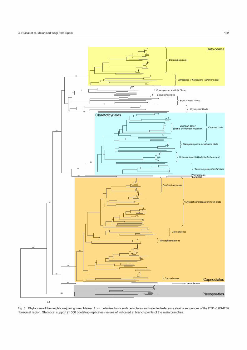
Strain grouping based on position in the ITS tree attempted to reveal correlations between morphological and molecular traits (Table 2). When morphological trends were superimposed onto a sequenced-based framework some molecular groups showed strong morphological cohesion. Clades of fungi corresponding to Catenulostroma, Cladophialophora, Exophiala, Hormonema, Phaeococcomyces, Phaeosclera, and Phaeotheca, were ap-parent (Fig. 3–5).
Recognition of genotypes by microsatellite-primed PCR
Cluster analysis of MP-PCR products of the 266 initial isolates identified 163 different genotypes (analysis not shown). The ITS region from at least one example of each genotype was sequenced. In several cases, the ITS region from more than one representative of each genotype was sequenced to verify the assumption that a group of isolates from a common MP-PCR pattern represented a single species (Table 1). Therefore, ITS sequences were obtained from a total of 171 isolates (Table 2). Phenetically similar isolates from different sites were also se- quenced to determine if they shared common genotypes leading to the conclusion that only five genotypes occurred at multiple collection sites.
Fig. 2 Isolation frequencies of 162 different fungal genotypes isolated from stone. Genotypes were established based on a grouping by microsatellite-primed PCR products.
101C. Ruibal et al. Melanised fungi from Spain
CapnodialesVenturiaceae
Pleosporales
0.1
65
Verrucariales
Unknown zone 3 (Cladophialophora spp.)
Cladophialophora minutissima clade
Unknown zone 1(Sterile or stromatic mycelium)
99
97
94
97
96
92
Dothideales (core)
Dothideales (Phaeosclera -Sarcinomyces)
Dothideales
‘Black Yeasts’ Group
‘Sarcinomyces petricola’ clade
Eurotiales
Capronia clade
Chaetothyriales
Teratosphaeriaceae
Davidiellaceae
Mycosphaerellaceae
Capnodiaceae
70
100
100
97
100
100
100
‘Coniosporium apollinis’ Clade
‘Cryomyces’ Clade
Mycosphaerellaceae unknown clade
Botryosphaeriales
Fig. 3 Phylogram of the neighbour-joining tree obtained from melanised rock surface isolates and selected reference strains sequences of the ITS1-5.8S-ITS2 ribosomal region. Statistical support (1 000 bootstrap replicates) values of indicated at branch points of the main branches.

102 Persoonia – Volume 21, 2008
99
97
Doth
ideale
s (co
re)
Doth
ideale
s (Ph
aeos
clera
-Sar
cinom
yces
)
Doth
ideale
s
‘Blac
k Yea
sts’ G
roup
100
"Con
iospo
rium
apo
llinis"
Clad
e
‘Cry
omyc
es’ C
lade
Horm
onem
a pr
unor
um C
BS 9
33.7
2
Horm
onem
a ca
rpet
anum
TRN
3Ho
rmon
ema
carp
etan
um T
RN31
Horm
onem
a ca
rpet
anum
TRN
25Ho
rmon
ema
carp
etan
um T
RN27
8Ho
rmon
ema
carp
etan
um T
RN40
Horm
onem
a ca
rpet
anum
TRN
24Ho
rmon
ema
carp
etan
um A
TCC
7436
0Ho
rmon
ema
sp. T
RN15
Horm
onem
a sp
. TRN
50Ho
rmon
ema
sp. T
RN26
8Ka
batin
a th
ujae
CBS
238.
66Ka
batin
a jun
iperi
AF26
0224
Doth
idea
berb
eridi
s CBS
186.
58Do
thide
a ins
cupt
a CB
S 18
9.58
Scler
ocon
idiom
a sp
hagn
icola
UAM
H 97
31Sc
leroc
onidi
oma
spha
gnico
la UA
MH
9731
TRN1
1TR
N258
TRN1
3nTR
N165
TRN5
26TR
N533
Endo
conid
ioma
sp. T
RN27
2En
doco
nidiom
a sp
. TRN
275
Horm
onem
a sp
. GCA
200
4Au
reob
asidi
um p
ullula
ns T
RN37
Aure
obas
idium
pull
ulans
TRN
269
Aure
obas
idium
pull
ulans
TRN
135
Aure
obas
idium
pull
ulans
AF0
1322
9Au
reob
asidi
um p
ullula
ns M
Z58
Stigm
ina sp
. TRN
9TR
N444
Phae
oscle
ra sp
. TRN
261
Phae
oscle
ra sp
. TRN
236
Phae
oscle
ra sp
. TRN
525
Phae
oscle
ra sp
. TRN
524
Phae
oscle
ra sp
. TRN
235
Phae
oscle
ra sp
. TRN
523
Phae
oscle
ra sp
. TRN
448
Phae
oscle
ra sp
. TRN
447
Phae
oscle
ra sp
. TRN
256
Phae
oscle
ra d
emat
ioide
s CBS
157
.81
Phae
oscle
ra sp
. TRN
521
Sarc
inom
yces
crus
tace
us C
BS 1
56.8
9Ph
aeos
clera
sp. T
RN25
4TR
N71
TRN5
6TR
N57
TRN7
3Co
niosp
orium
sp. T
RN74
Conio
spor
ium sp
. TRN
465
Conio
spor
ium a
pollin
is TR
N432
Conio
spor
ium a
pollin
is CB
S 10
0213
Conio
spor
ium a
pollin
is CB
S 10
0216
Conio
spor
ium a
pollin
is CB
S 10
0218
Lasio
diplod
ia th
eobr
omae
CM
W 9
074
Botry
osph
aeria
dot
hidea
CM
W 9
075
TRN4
92Ph
aeoc
occo
myc
es sp
. TRN
270
Phae
ococ
com
yces
sp. T
RN52
0Ph
aeoc
occo
myc
es sp
. TRN
529
Phae
ococ
com
yces
sp. T
RN21
3Ph
aeoc
occo
myc
es sp
.TRN
458
Phae
ococ
com
yces
sp.T
RN44
9Ph
aeoc
occo
myc
es sp
.TRN
455
Phae
ococ
com
yces
sp.T
RN45
2Ph
aeoc
occo
myc
es cf
. nigr
icans
TRN
456
Phae
ococ
com
yces
nigr
icans
CBS
652
.76
Scler
ococ
cum
sp. T
RN17
0TR
N172
Cryo
myc
es m
inter
i CBS
116
302
Cryo
myc
es a
ntar
cticu
s CBS
116
301
TRN4
1TR
N281
Conio
spor
ium u
ncina
tum
CBS
100
219
TRN4
5TR
N220
100
100
100
100
100
100 100
100
100 100
100
100
100
100
100
100
100
100
91
98 94
90
93
99
91
9997
96
97
96
9394
99
Botry
osph
aeria
les
‘Con
iospo
rium
apo
llinis’
Clad
e
Fig
. 4
Par
tial p
hylo
gram
of t
he n
eigh
bour
-join
ing
tree
obt
aine
d fr
om m
elan
ised
roc
k su
rfac
e is
olat
es a
nd r
efer
ence
str
ains
seq
uenc
es o
f the
ITS
1-5.
8S-I
TS
2 rib
osom
al r
egio
n (F
ig. 3
). B
ranc
hes
cor-
resp
ondi
ng to
the
Dot
hide
ales
cla
de, ‘
Con
iosp
oriu
m a
polli
nis’
cla
de, ‘
blac
k ye
asts
’ gro
up, a
nd ‘C
ryom
yces
’ cla
de.
Dot
hide
ales
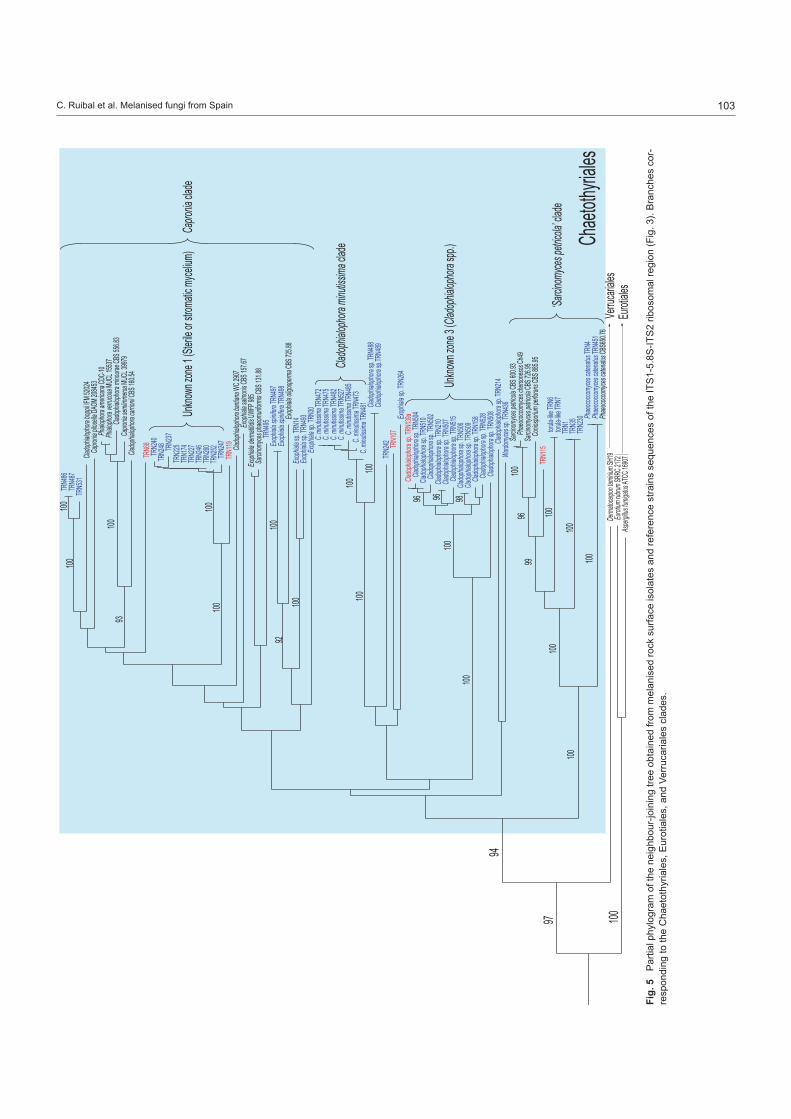
103C. Ruibal et al. Melanised fungi from Spain
Verru
cariale
s
Unkno
wn zo
ne 3 (
Clado
phialo
phora
spp.)
Clado
phialo
phora
minu
tissima
clade
Unkno
wn zo
ne 1 (
Sterile
or str
omatic
mycel
ium)
94
97‘Sa
rcinom
yces p
etrico
la’ cla
de
Eurot
iales
Capro
niacla
de
Chaet
othyria
les100
TRN4
86TR
N487
TRN5
31Cla
doph
ialoph
ora bo
ppii IF
M 5202
4Ca
pronia
pilose
lla DA
OM 20
8453
Phialo
phora
ameri
cana C
DC-10
Phialo
phora
verru
cosa M
UCL 1
5537
Clado
phialo
phora
mino
urae C
BS 55
6.83
Capro
nia se
miimm
ersa M
UCL 3
9979
Clado
phialo
phora
carrio
nii CB
S 160.
54
TRN6
8TR
N240
TRN2
48 TRN2
37TR
N225
TRN1
74TR
N227
TRN2
46TR
N260
TRN2
52TR
N247
TRN1
19 Clado
phialo
phora
bantia
na W
C 2907
Exop
hiala s
almon
is CBS
157.6
7Ex
ophia
la derm
atitidis
UWFP
985
Sarcin
omyce
s pha
eomu
riform
is CBS
131.8
8TR
N495
Exop
hiala s
pinifer
a TRN
497Ex
ophia
la spin
ifera T
RN498
Exop
hiala o
ligospe
rma C
BS 72
5.88
Exop
hialas
p.TRN
14Ex
ophia
lasp.T
RN493
Exop
hialas
p.TRN
30C.
minutis
sima T
RN472
C. min
utissim
a TRN
475C.
minutis
sima T
RN482
C. min
utissim
a TRN
527C.
minutis
sima T
RN485
C. min
utissim
a TRN
473C.
minutis
sima T
RN481 Cla
doph
ialoph
orasp.
TRN4
88Cla
doph
ialoph
orasp.
TRN4
89TR
N242 TR
N107
Exop
hialas
p.TRN
264Cla
doph
ialoph
orasp.
TRN1
39aCla
doph
ialoph
orasp.
TRN5
04Cla
doph
ialoph
orasp.
TRN5
10Cla
doph
ialoph
orasp.
TRN5
02Cla
doph
ialoph
orasp.
TRN2
10Cla
doph
ialoph
orasp.
TRN5
07Cla
doph
ialoph
orasp.
TRN5
15Cla
doph
ialoph
orasp.
TRN5
06Cla
doph
ialoph
orasp.
TRN5
09Cla
doph
ialoph
orasp.
TRN1
58Cla
doph
ialoph
orasp.
TRN5
28Cla
doph
ialoph
orasp.
TRN5
08Cla
doph
ialoph
orasp.
TRN2
14Wa
rdomy
cessp.
TRN4
36Sa
rcinom
yces p
etrico
la CBS
600.9
3Ph
aeoco
ccomy
ces ch
erson
esos C
h49Sa
rcinom
yces p
etrico
la CBS
726.9
5Co
niospo
rium
perfo
rans C
BS 88
5.95
TRN1
15tor
ula-lik
e TRN
6tor
ula-lik
e TRN
7TR
N1TR
N35
TRN2
30 Phae
ococco
myces
caten
atus T
RN4
Phae
ococco
myces
caten
atus T
RN451
Phae
ococco
myces
caten
atus C
BS650
.76De
rmato
carpo
n tam
inium S
H19
Eurot
ium ru
brum S
RRC 2
172As
pergi
llus fu
migatu
s ATC
C 1690
7
100
100
100
100100100
100100
100100
100
100
100
100
100 100
100
100
9696 98
92
93
9699
Fig
. 5
Par
tial p
hylo
gram
of t
he n
eigh
bour
-join
ing
tree
obt
aine
d fr
om m
elan
ised
roc
k su
rfac
e is
olat
es a
nd r
efer
ence
str
ains
seq
uenc
es o
f the
ITS
1-5.
8S-I
TS
2 rib
osom
al r
egio
n (F
ig. 3
). B
ranc
hes
cor-
resp
ondi
ng to
the
Cha
etot
hyria
les,
Eur
otia
les,
and
Ver
ruca
riale
s cl
ades
.

104 Persoonia – Volume 21, 2008
Capn
odial
esVe
ntur
iacea
e
Pleo
spor
ales
65
96
92
Tera
tosp
haer
iacea
e
David
iellac
eae
Myc
osph
aere
llace
ae
Capn
odiac
eae
100
97
100
100
Myc
osph
aere
llace
ae u
nkno
wn cl
ade
TRN1
40
Cate
nulos
trom
a sp
. TRN
280
Cate
nulos
trom
a sp
. TRN
446
Cate
nulos
trom
a sp
. TRN
445
Cate
nulos
trom
a sp
. TRN
290
Cate
nulos
trom
a sp
. TRN
289
TRN1
73 fusic
ladium
-like
TRN1
57TR
N13v
TRN2
88Ca
tenu
lostro
ma
sp.
TRN2
79TR
N232
TRN4
41Ca
tenu
lostro
ma
abiet
is CB
S 61
8.84
Cate
nulos
trom
a ab
ietis
TRN1
27Ca
tenu
lostro
ma
salic
is CB
S 30
0.81
Tera
tosp
haer
ia m
icros
pora
STE
-U 1
848
TRN1
28Te
rato
spha
eria
micr
ospo
ra S
TE-U
183
2Ph
aeot
heca
sp. T
RN20
6Ph
aeot
heca
sp. T
RN20
9Ph
aeot
heca
sp. T
RN21
2Ph
aeot
heca
sp. T
RN26
3Ph
aeot
heca
sp. T
RN45
3Ph
aeot
heca
sp. T
RN45
4Ph
aeot
heca
sp. T
RN12
6TR
N500
TRN4
91Ph
oma-
like
TRN2
11Ps
eudo
taen
iolina
glob
osa
CBS
303.
84Ps
eudo
taen
iolina
glob
osa
CBS
1098
89Ho
rtaea
wer
neck
ii CBS
117
.90
Horta
ea w
erne
ckii C
BS 3
59.6
6Ho
rtaea
wer
neck
ii TRN
122
Siro
spor
ium sp
. TRN
129
Acro
dont
ium cr
ater
iform
e TR
N267
TRN2
84TR
N283 TR
N286
TRN2
62TR
N282
TRN2
31 TRN4
38TR
N442 TR
N431
Myc
ocali
cium
victo
riae
AF24
3135
Myc
ocali
cium
victo
riae
AJ31
2123
TRN4
40Ca
tenu
lostro
ma
elgine
nse
STE-
U 19
58TR
N132
TRN7
8TR
N79
Fried
man
niom
yces
end
olith
icus C
CFEE
522
Fried
man
niom
yces
sim
plex C
BS 1
1677
5TR
N5TR
N159 TR
N245
Cate
nulos
trom
a ch
rom
oblas
tom
ycos
um C
BS 5
97.9
7TR
N123
Cate
nulos
trom
a m
acow
anii S
TE-U
187
2Cl
ados
poriu
m m
acro
carp
um C
BS 1
75.6
2Da
vidiel
la ta
ssian
a AT
CC 2
0109
0Cl
ados
poriu
m sp
. TRN
439
Clad
ospo
rium
tenu
issim
um A
TCC
3802
7Cl
ados
poriu
m o
xysp
orium
CBS
125
.80
Trim
mat
ostro
ma
salin
um M
ZKI B
-963
fusic
ladium
-like
TR43
TR44
fusic
ladium
-like
TR42
Myc
osph
aere
lla ir
idis C
BS28
2.49
TRN4
37TR
N443
Phae
otec
a tri
angu
laris
CBS
471.
90Pa
ssalo
ra fu
lva A
TCC
4496
0Pa
ssalo
ra fu
lva S
TE-U
368
8Pa
ssalo
ra fu
lva L
2543
0Pa
ssalo
ra d
issilie
ns A
F222
835
Pass
alora
bell
ynck
ii AF2
2283
1Ca
pnob
otry
ella
renis
pora
CBS
214
.90
TRN1
13TR
N96
TRN7
6TR
N88
TRN1
03TR
N112
TRN1
08TR
N86
TRN1
37TR
N61
TRN1
31TR
N60
TRN4
33 TRN8
0TR
N244
TRN6
6TR
N136 TR
N70
TRN6
5De
vries
ia am
erica
na C
PC 5
121
TRN9
1TR
N111
TRN1
47TR
N138 TR
N62
TRN7
7TR
N139
bTR
N152
TRN8
1TR
N153
TRN8
7TR
N142
TRN1
24TR
N145
TRN1
04TR
N114
TRN4
61TR
N468
TRN4
67Cy
lindr
oxyp
hium
sp. T
RN45
0Ca
pnod
ium co
ffeae
CBS
147
.52
Lept
oxyp
hium
sp. T
RN12
5Ca
pnod
ium sa
licinu
m C
BS 1
31.3
4TR
N287
Phae
otec
a fis
sure
lla C
BS 5
20.8
9Ve
ntur
ia ce
rasi
AF06
5847
Fusic
ladium
effu
sum
JDTR
N484
TRN2
16TR
N222
TRN2
19TR
N215
TRN2
21TR
N47
TRN4
9TR
N434
Para
phae
osph
aeria
cong
lomer
ata
AF25
0824
Phae
osph
aeria
nod
orum
N15
7Ph
aeos
phae
ria a
vena
ria S
AT00
4C n
d-96
Stag
onos
pora
are
naria
144
4TR
N162
TRN4
99
Rhizo
ctonia
sp. E
ab-S
4
100
100
100
100
100
62
100
100 10
010
0
100
100
100
100
100
100
100
100
100
100
100
100
100
100
90
100
100
100
100
100
100
98
9992
97 9299
96
98
65
9599
90
96
9998
98
99
98
9493
97
89
100
61
72
71
61
Fig
. 6
Par
tial p
hylo
gram
of t
he n
eigh
bour
-join
ing
tree
obt
aine
d fr
om m
elan
ised
roc
k su
rfac
e is
olat
es a
nd r
efer
ence
str
ains
seq
uenc
es o
f the
ITS
1-5.
8S-I
TS
2 rib
osom
al r
egio
n (F
ig. 3
). B
ranc
hes
cor-
resp
ondi
ng to
the
Myc
osph
aere
llace
ae, V
entu
riace
ae, a
nd P
leos
pora
les
clad
es.

105C. Ruibal et al. Melanised fungi from Spain
Amplification and sequencing of the ITS region of rDNA
To estimate the relative phylogenetic relationships among the isolates and the reference strains, the ITS sequences of the 159 isolates were aligned with reference sequences from GenBank that were selected based on sequence similarity, or because they were potentially related taxa. A sequence alignment was used to generate a tree by the neighbour-joining method (Fig. 3). A basidiomycete was chosen as outgroup (Rhizoctonia sp. Eab-S4) because FASTA analyses indicated the isolates spanned a broad spectrum of the Pezizomycotina. We did not intend to recreate the taxonomic frame of the phylum, but sought to quickly estimate the relative phylogenetic position and if possible arrive at an identification. Results were similar to more specific analyses of other authors in similar groups, and several main clades received strong bootstrap support, so the consistency of the tree seemed to be remarkably high considering that we had attempted to compare sequences of such different fungi. Isolate TRN140 yielded the most disparate ITS sequence and was not clearly associated with any other group of strains (Fig. 3).
ITS-tree, Dothideomycetes
The tree was divided in four main branches (Fig. 3). The first of these branches grouped together (with no relevant support), the order Dothideales and other clades of uncertain taxonomic position. The first branch in the Dothideales (Fig. 4) was the only which received strong bootstrap support (97 %). It was divided into two main sub-branches; the first comprised the reference strains of the Dothideales s.str. and a group of isolates identified as Hormonema carpetanum (Bills et al. 2004b), and other hor-monema-like isolates. The second branch was referable to the reference strains Sarcinomyces crustaceus (CBS 156.89) and Phaeosclera dematioides (CBS 157.81), both ascomycetous anamorphs of uncertain affinity. All the isolates included in this branch were identified as Phaeosclera sp. Although these fungi are of unknown affinities, the branch was monophyletic with that of the Dothideales s.str., so a close relationship could be inferred among the branches.
ITS-tree, unidentified branches associated with the Dothideomycetes
In a major branch parallel to the Dothideales clade appeared a series of sub-branches with no relevant statistical support (Fig. 4). The lack of statistical confidence and the heterogene-ous nature of the isolates casted doubts on the phylogenetic significance of this clade. The first among the branches was designated as the ‘Coniosporium apollinis’ clade (Fig. 4), and included two reference sequences of C. apollinis, a rock-inhabit-ing fungus from Greece (Sterflinger et al. 1997). This branch, strongly supported by a bootstrap value of 99 %, also contained our isolates; TRN432, identified as C. apollinis based on its sequence and its morphology, and TRN465, TRN74, designated as Coniosporium sp. The rest of the isolates (TRN71, TRN56, TRN57, and TRN73) may be congeneric with C. apollinis. Also two species of the Botryosphaeriaceae, Lasiodiplodia theobromae (Alves et al. 2008) and B. dothidea (Crous et al. 2006b), appeared associated with the ‘Coniosporium apollinis’ clade.
The next unsupported clade was designated as the ‘black yeasts’ group (Fig. 4) because all strains formed colonies con-sisting largely of melanised unicellular aggregates. Yeast-like forms were infrequently found among other clades of the tree. However, in this branch, the nearly exclusive yeast-like growth was distinctive and diagnostic. The only reference sequence that grouped within this clade was Phaeococcomyces nigricans (CBS 652.76). Due to their distinctive morphological charac-teristics and their position in the tree, the other isolates in this group were identified as Phaeococcomyces spp. The last of the unsupported clades related to the Dothideales was designated as the ‘Cryomyces’ clade, because the isolates grouped with reference sequences of Cryomyces antarcticus and C. minteri (Selbmann et al. 2005) (Fig. 4).
ITS-tree, Eurotiomycetes
Although the second main clade corresponded with the orders Chaetothyriales, Verrucariales, and Eurotiales (Fig. 3), it lacked significant support. These three orders have been proposed to belong to the class Eurotiomycetes (Lutzoni et al. 2004, Miadlikowska & Lutzoni 2004, Reeb et al. 2004). The Euro-tiales branch (100 % bootstrap value) was formed only by the reference sequences and included no rock isolates (Fig. 5). Chaetothyriales and Verrucariales appeared grouped with a high bootstrap value (97 %). The order Chaetothyriales was amply represented among our isolates and received a bootstrap value of 94 % (Fig. 5). The clade was divided into a number of sub-branches. The only one with a strong support (bootstrap value of 100 %), contained several previously characterised melanised fungi found in rocks such as Sarcinomyces petricola and Coniosporium perforans, and sequences of the anamorph species Phaeococcomyces catenatus. The remaining branches of the Chaetothyriales (Capronia clade, Cladophialophora minutissima clade) did not receive relevant support and seemed to correspond to the Chaetothyriales family Herpotrichiellaceae, which includes the genus Capronia and some of its recognised or suspected anamorphs, e.g., Cladophialophora, Exophiala, and Phialophora. Also within this region appeared groups of closely related sequences (unknown zones 1 and 2) with no correspondence with any reference sequence (Fig. 5). The last branch of the Eurotiomycetes clade was formed solely by the reference sequence of the lichenised fungus Dermatocarpon taminium SH19 (Fig. 5), a member of the order Verrucariales (Heiðmarsson 2003).
ITS-tree, Capnodiales
The third main branch grouped with relatively high bootstrapping (bootstrap value 96 %) both the order Capnodiales (bootstrap support 100 %) and the family Venturiaceae of the order Pleo-sporales (97 % bootstrap value), (Fig. 3). The Capnodiales clade comprised a number of sub-branches grouped with a bootstrap value of 65 %, and a Capnodiaceae clade. Among the other sub-branches were a Davidiellaceae clade (Crous et al. 2007a, b) and a Mycosphaerellaceae clade, both with high bootstrap values (Schoch et al. 2006) (Fig. 6). A clade (bootstrap value of 89 %) named as ‘Mycosphaerellaceae unknown clade’, included a number of miscellaneous anamorphic ascomycetes, including the Teratosphaeriaceae (72 % bootstrap value) (Crous

106 Persoonia – Volume 21, 2008
et al. 2007a). Finally, a clade (65 % bootstrap value) containing a number of isolates and Devriesia americana (STE-U 5121 = CBS 117726) (Crous et al. 2007c) as unique reference se-quence. Finally, the Venturiaceae was represented by isolate TRN484, grouped with the reference strains Venturia cerasi and Fusicladium effusum (Fig. 6). For the most part, isolates in the Capnodiales formed only vegetative mycelia in culture. The major exceptions were fungi with endoconidia that we tentatively called Phaeosclera (Sigler et al. 1981), cladosporium-like, and catenulostroma-like fungi.
ITS-tree, Pleosporales
The last of these branches was identified with the order Pleo-sporales (bootstrap value of 100 %) and included reference sequences belonging to the family Phaeosphaeriaceae (Pleo-sporales) (Fig. 3). All the isolates in the clade only formed vegetative mycelia and could not be directly related with any known reference sequences, although the high bootstrap values of all the branches show the high affinity of the isolates with the Phaeosphaeriaceae (Fig. 6).
DISCUSSION
Biogeography and diversity
The abundance and variety of isolates encountered in a previ-ous study of fungi in Mallorcan limestone (Ruibal et al. 2005) was surprisingly high, therefore we wanted to confirm whether this pattern of taxonomic diversity was repeatable in the conti-nental environment of central Spain. In Mallorca, limestone from two sites yielded 117 melanised isolates that were determined to represent 39 different genotypes. Anticipating that levels of diversity could be comparable, we were cautious not to sample widely or extensively until the levels of abundance of fungi in continental rock samples could be evaluated. We started with four different rock types (granite, slate, limestone, and quartzite) at four different sites. The only criteria used to select different kinds of rock samples were that rocks would be relatively clean and free from influence of soil and lichens and could be easily lifted off the rock faces with a chisel. No attempt was made to enrich the samples by choosing stained or biologically dete-riorated surfaces or surfaces that were obviously colonised by black fungal structures.
Rock surfaces harboured an easily accessible reservoir of melanised ascomycetes. Even with arbitrary selection of rock substrata, a set of isolates was obtained that exhibited a combination of cryptic morphology and phylogenetic affinities agreeing with, or even surpassing, previous studies on rock-inhabiting fungi in the Mediterranean and other locations. The assemblage of isolates from central Spain resembled that recovered from limestone in the Balearic Islands (Ruibal et al. 2005) and included some genera, species or species as-semblages detected by other investigators (Staley et al. 1982, de Hoog et al. 1997, Sterflinger et al. 1997, Wollenzien et al. 1997, de Leo et al. 1999, 2003, Onofri et al. 1999, Bogomolova & Minter 2003). The phylogenetic bias towards the Dothideales, Capnodiales, and Chaetothyriales was especially evident (Fig. 3–5). The high diversity observed in two different regions in
Spain confirms that our isolation technique was an appropriate method for the isolation of melanised fungi on rock surfaces and that the patterns of diversity are probably common for rock sur-faces in a semi-arid environment. The patterns of diversity and species assemblages of the rock surfaces contrasted sharply and had little in common with the species assemblages that would be expected from an adjacent community of soil fungi (Bills et al. 2004a).
Genotypic diversity
Because many of the isolates were macroscopically similar, even on sets of media that normally would exaggerate differ-ences in other fungi, we could not confidently use traits such as mycelial growth rate and pigmentation or texture to group them into operational taxonomic units (Arnold et al. 2000, Bills et al. 2004a). We based our comparison and grouping strategy on MP-PCR complemented by the macromorphological appear-ance of the isolates. The method allowed us to more efficiently analyse fewer isolates without losing diversity information. We were able to check the usefulness of this method by verify-ing that sequenced isolates with the same MP-PCR pattern grouped together in the ITS tree (Table 2, Fig. 3). However, we found isolates with virtually identical ITS sequences but with different MP-PCR patterns, implying that different individuals of the same species were grouped together. On the other hand, when different strains with identical MP-PCR patterns were sequenced, they consistently had identical ITS sequences. Therefore, the strategy avoided loss of diversity when choos-ing isolates for sequencing. The assumption that one MP-PCR pattern was equivalent to one species was questionable in many cases. Hormonema carpetanum strains with discrete MP-PCR patterns also varied by a few base pairs in their ITS2 sequences, yet upon detailed morphological and fine-scale phylogenetic analyses strains from rocks were considered to be conspecific with plant-associated isolates from the same region (Bills et al. 2004b). Terminal branches consisting of essen- tially one ITS type (Fig. 2–5) likely represented multiple iso- lates of single species, such as in the case of TRN261, TRN236, TRN525, TRN524, and TRN235 (Fig. 4), unknown zones 1 and 2 (Fig. 5), or TRN280, TRN446, TRN445, TRN290, TRN289 (Fig. 6).
Morphological data
The convergent morphological features of these fungi are thought to be related to adaptations to environmental condi-tions. The most obvious was the melanin pigment production, necessary for protection against desiccation and ultraviolet radiation, and possibly the binding of certain metallic ions, which, depending on their nature, may stimulate the growth and survival of melanised fungi or protect them from metal toxicity (Gorbushina 2003). It has been shown that certain metabolites, such as mycosporines, apart from their ultra- violet-protective properties, possess germination self-inhibitory functions, useful for the survival, persistence and longevity of these organisms. The slow and granular meristematic growth is thought to be related with energy conservation, necessary for survival on substrates with scarce nutrient input (Gorbushina 2003, Gorbushina et al. 2003).

107C. Ruibal et al. Melanised fungi from Spain
Microscopically, sterile mycelia predominated among the iso-lates (Table 2). Lack of specialised reproductive structures could be an artefact caused by the cultural conditions; however, unspecialised reproduction would be consistent with a hypoth-esis of specialised organisms living in an oligotrophic, extreme environment. The lack of reproductive structures could also be considered an adaptation for energy conservation (Gorbushina 2003, Gorbushina et al. 2003). In a regime of scarce nutrients and water that exist on rock surfaces, the high energetic costs required for the growth of complex reproductive structures would select for fungi with less elaborate reproduction sys-tems, such as simple modes of conidiogenesis, or melanised, desiccation-resistant, hyphal structures that fragment and act as propagules.
Among the isolates that formed conidial states, varied types of conidiogenesis were observed (Table 2), which was reflected in the diverse taxonomic origins of the isolates. In the rest of the isolates, conidial states were not observed, although in some, hyphal disarticulation could free cells or cells aggregates that could act as propagules. This disarticulation of melanised, toru-lose hyphae, whose cells could occasionally develop transverse septa, suggested a thallic micronematous conidiogenesis or simple reproduction by propagative fragmentation. Other char-acters observed in the sterile mycelia, such as torulose and/or moniliform hyphae, chlamydospore-like structures, hypho- podia-like branching, and muriform or meristematic develop-ment, even though relatively common, were of limited diagnostic value. Correspondence among morphological types and the groups established by the phylogenetic analysis of the ITS sequences of the isolates was evident only in some cases. Although meristematic strains with black yeast-like growth pat-terns have been documented in culture (de Hoog et al. 1999, Sterflinger et al. 1999), yeast-like growth forms were infrequent among the isolates.
Diagnostic value of the molecular data: ITS rDNA phylogram
Most isolates could be loosely grouped in two main classes of ascomycetes: Dothideomycetes and Eurotiomycetes, and among four orders: Pleosporales, Dothideales, Capnodiales, and Chaetothyriales. Our analyses were not able to recover monophyly in the Dothideomycetes, as in Schoch et al. (2006), but our data sampling and analyses were inappropriate for this type of resolution.
Dothideomycetes
The order Dothideales (Fig. 4) was amply represented. A di-verse set of isolates within this cluster were identified as Phaeo sclera spp. based primarily on their hard, granular to cerebriform colonies originating from meristematic cell division (Sigler et al. 1981), and forming cell masses disarticulating into single cells or cell aggregates.
Ambiguous clades adjacent to the Dothideales
The taxonomic affiliations of the fungi belonging to the group of ITS clades adjacent to the Dothideales clade (the ‘Coniosporium apollinis’ clade, Botryosphaeriales, the ‘black yeast’ group, and the ‘Cryomyces’ clade) were difficult to discern. In the first,
‘Coniosporium apollinis’ clade (Fig. 4), the majority of the refer-ence sequences belonged to strains of the genus Coniosporium. This is a heterogeneous form-genus applied to some melanised meristematic fungi that cluster close to the Chaetothyriales (de Leo et al. 1999). The Coniosporium species referenced in this tree were all isolated from stone (C. apollinis in the ‘Coniosporium apollinis’ clade, C. uncinatum in the ‘Cryomyces’ clade, and C. petricola in the Chaetothyriales clade). The polyphyly of Coniosporium species in the ITS tree is an example of an artificial genus based on convergent morphologies possibly related to similar adaptations to extreme environments. Two reference strains belonging to the Botryosphaeriaceae corresponded to the recently described Botryosphaeriales (Crous et al. 2006b, Schoch et al. 2006).
The second clade of uncertain affinities, labelled ‘black yeast group’ (Fig. 4), included isolates with entirely yeast-like growth, and as the only reference strain Phaeococcomyces nigricans CBS 652.76. The genus Phaeococcomyces is employed for melanised fungi that lack filamentous growth and has been considered a synanamorph of certain species of Exophiala (Haase et al. 1995, de Hoog et al. 2000a). The clade perhaps could represent an unrecognised new genus related to the Chaetothyriales.
The last unsupported clade, ‘Cryomyces’ clade (Fig. 4), in-cluded reference sequences of Cryomyces antarcticus and C. minteri (Selbmann et al. 2005), both meristematic, melanised endolithic fungi isolated from Antarctic. The result pointed to a possible cosmopolitan distribution of melanised, meristematic, rock-inhabiting fungi, enabled by their exceptional tolerance to extreme environmental conditions, and emphasised the great unexplored diversity of this ecological group. Reference se-quence Coniosporium uncinatum CBS 100219 appeared in this group, along with isolate TRN174, identified as the form-genus Sclerococcum based on its sporodochia-like colonies, consist-ing of masses of melanised cells organised in chains and arising from a meristematic tissue (Hawksworth 1979).
Chaetothyriales
This was the best represented order among our isolates. The clade was subdivided in three subclades (Fig. 5). In the first one, identified as the ‘S. petricola’ clade, series of isolates were grouped together with reference sequences of uncertain affiliation, e.g., Coniosporium perforans, Phaeococcomyces chersonesos, and Sarcinomyces petricola. All are species of melanised fungi isolated from rock surfaces (Sterflinger et al. 1997, Wollenzien et al. 1997, Bogomolova & Minter 2003) and were assigned to form-genera solely based on their morpholo-gies. Placement of these heterogeneous groups in a particular order was ambiguous based only on ITS sequences analysis. For example, Sarcinomyces is a controversial form-genus almost entirely defined by being meristematic growth of cells to form asexual propagules, which may be an inadequate definition for organisms with high morphological plasticity. Note as an example the polyphyly represented in the ITS analysis by Sarcinomyces petricola, Sarcinomyces phaeomuriformis (in the Capronia clade), and Sarcinomyces crustaceus (in the PhaeoscleraSarcinomyces branch of the Dothideales clade). These species have been associated with Phaeococcomyces

108 Persoonia – Volume 21, 2008
catenatus in a separate clade adjacent to the Herpotrichiella-ceae (Chaetothyriales) (Sterflinger et al. 1999). The strong statistical support of these associations suggested that these fungi belong to the Chaetothyriales.
The second subclade in the Chaetothyriales, identified as the Capronia clade, included reference sequences of the Herpotri-chiellaceae. The subclade also included a group of very similar vegetative isolates with a high statistical support and no related reference strains. This distinctive clade structure (marked as ‘unknown zone 1’, Fig. 5) might represent individuals of a new species, or members of a species complex.
The third subclade of the Chaetothyriales group comprised two similarly structured groups of isolates. A group appar-ently conspecific with Cladophialophora minutissima (Davey & Currah 2007), a recently described new species from boreal bryophytes, and the unknown zone 3 (Fig. 5).
Capnodiales
The ITS Capnodiales clade (Fig. 6) was composed of five main subclades, three of them, Davidiellaceae, Mycosphaerel-laceae, and Capnodiaceae, comprising the Capnodiales as defined by Schoch et al. (2006). The two remaining subclades shared some of the characteristics of other groups of uncertain affiliation that were previously mentioned. The ‘Mycosphaerel-laceae unknown clade’ contained reference strains considered synonyms of Mycosphaerella (Teratosphaeria microspora) or its anamorphs, some isolated as leaf spot pathogens on Pro-teaceae (Taylor et al. 2003). These strains, identified initially as Trimmatostroma and Stenella, have been later renamed as Catenulostroma (Crous et al. 2007a). In particular, the Teratosphaeriaceae clade (Fig. 6) showed some morphologi-cal resemblance to Trimmatostroma species characteristic of decayed wood surfaces (Ellis 1971, 1976). Trimmatostroma, with the generic type species T. salicis, was recently treated as a genus of Leotiales, while the capnodialean species were reclassified in Catenulostroma (Crous et al. 2007a). Teleomorph connections of Catenulostroma are in Teratosphaeria.
The last subclade in the Capnodiales included several vegeta-tive mycelial strains, mostly isolated from Mallorca. The nearest reference sequence to these series of isolates was Devriesia americana (STE-U 5121) (Crous et al. 2000, 2007b). Recently, the phylogeny of the Capnodiales has been extensively treated by Schoch et al. (2006). The resulting phylogeny differentiates two subclasses in the Dothideomycetes, Pleosporomicetidae (Pleosporales) and Dothideomycetidae (Dothideales, Myriangi-ales, and Capnodiales), Capnodiaceae, Mycosphaerellaceae, Davidiellaceae, and Piedraiaceae are introduced or placed within the Capnodiales. Crous et al. (2007a) have also included Teratosphaeriaceae in the Capnodiales. In our analysis, the Capnodiales was poorly delineated, especially regarding rock isolates as in previous studies (Ruibal et al. 2005).
Pleosporales
Isolates and reference sequences belonging to the order Pleo-sporales, were separated into two groups in the analysis. First, the Pleosporales clade comprised the majority of the isolates and reference sequences belonging to the family Phaeosphaer-iaceae. The second was comprised of the Venturiaceae
clade, with only one isolate from rock, TRN484 (Fig. 6). The polyphyletic character of the Pleosporales clade, supported by high bootstrappings, is in agreement with the results of other authors (Braun et al. 2003). Devriesia americana and other isolates sequences morphologically identified as fusicladium-like anamorph appeared divergent to the reference sequences of the Venturiaceae (Venturia cerasi, Fusicladium effusum). These contradictory analyses are an example of the inability of ribosomal rDNA sequences to resolve many relationships among the euascomycetes (Berbee et al. 2000), but also ac-count for the unclear identity and classification of some of the 18S and ITS sequences deposited in GenBank.
Conclusions
The analysis, and that of Ruibal et al. (2005), revealed that a remarkable diversity of ascomycetous fungi can be found in a relatively small surface area of natural rock. Our results confirm those conclusions over a larger geographical scale. Because most isolates were rare, the question as to whether they were restricted to a particular location or rock type was not evident. Our analysis did not reveal any genotypic specificity for the types of rock or sites sampled, as evidenced by few number MP-PCR patterns common to at least two isolation sites and the apparent random distribution of isolates from different sites throughout the ITS tree. A more extensive and intensive microscale analysis emphasizing aspect, shade, waterflow, and other rock patterns possibly could have detected prefer-ential colonisation patterns. On the other hand, rock surfaces may simply provide mechanical support, where organisms disperse at random and can compete in an ecological space with a relatively low population density, and where competi-tiveness for resources is much lower than in other niches. The mineral composition of rocks may only have minimal effects on fungal nutritional metabolism. Nutrient sources are likely to be exogenous and very transient. Perhaps water from rain, fog, and surface run off carry dissolved or suspended organic and inorganic nutrients to the vicinity of fungal cells. Such an oligotrophic pattern along with the extreme conditions would prevent colonisation by high nutrient-dependent, cosmopolitan organisms, while favouring the settlement and specialisation of a wide range of organisms that share a common set of morpho-logical and physiological features. The characteristics exhibited by these organisms would not necessarily be exclusive to rock surfaces, they might also allow them to colonise other living or inert surfaces in the same ecosystems, such as bark and leaf epiphytes, plant pathogens (Mycosphaerella-related strains) or even animal pathogens (human-pathogenic strains in the Chaetothyriales). The physiological and morphological spe-cialisation of these fungi would give them an advantage in such environments that might be also considered ‘extreme’. If this hypothesis was true, rock surfaces, with their very oligotrophic environment, could be considered the origin and reservoir of these pathogenic organisms.
Acknowledgements The expert and steadfast technical assistance of Mrs Ana Pérez (Merck, Spain) was greatly appreciated. Portions of this work were derived from a doctoral dissertation submitted by the first author to the Faculty of Sciences of the Universidad Autónoma de Madrid.

109C. Ruibal et al. Melanised fungi from Spain
REFERENCES
Alves A, Crous PW, Correia A, Phillips AJL. 2008. Morphological and mo-lecular data reveal cryptic speciation in Lasiodiplodia theobromae. Fungal Diversity 28: 1–13.
Arnold AE, Maynard Z, Gilbert G, Coley PD, Kursar TA. 2000. Are tropical fungal endophytes hyperdiverse? Ecology Letters 3: 267–274.
Bellido Mulas F, Rodríguez Fernández LR. 1991. Mapa Geológico de Es-paña. Instituto Tecnológico Geominero de España. Buitrago de Lozoya, Madrid, Spain.
Berbee ML, Carmean DA, Winka K. 2000. Ribosomal DNA and resolution of branching order among the Ascomycota: How many nucleotides are enough? Molecular Phylogenetics and Evolution 17: 337–344.
Bills GF, Christensen M, Powell M, Thorn G. 2004a. Saprobic soil fungi. In: Mueller GM, Bills GF, Foster MS (eds), Biodiversity of fungi. Inventory and monitoring methods: 271–302. Elsevier Academic Press, Burlington, MA, USA.
Bills GF, Collado J, Ruibal C, Peláez F, Platas G. 2004b. Hormonema carpe-tanum sp. nov., a new lineage of dothideaceous black yeasts from Spain. Studies in Mycology 50: 14–157.
Bogomolova EV, Minter DW. 2003. A new microcolonial rock-inhabiting fungus from marble in Chersonesos (Crimea, Ukraine). Mycotaxon 86: 195–204.
Braun U, Crous PW, Dugan F, Groenewald JZ, Hoog GS de. 2003. Phylogeny and taxonomy of Cladosporium-like hyphomycetes, including Davidiella gen. nov., the teleomorph of Cladosporium s.str. Mycological Progress 2: 3–18.
Crous PW, Aptroot A, Kang JC, Braun U, Wingfield MJ. 2000. The genus Mycosphaerella and its anamorphs. Studies in Mycology 45: 107–121.
Crous PW, Braun U, Groenewald JZ. 2007a. Mycosphaerella is polyphyletic. Studies in Mycology 58: 1–32.
Crous PW, Braun U, Schubert K, Groenewald JZ. 2007b. Delimiting cladospo-rium from morphologically similar genera. Studies in Mycology 58: 33–56.
Crous PW, Liebenberg MM, Braun U, Groenewald JZ. 2006a. Re-evalua-ting the taxonomic status of Phaeoisariopsis griseola, the causal agent of angular leaf spot of bean. Studies in Mycology 55: 163–173.
Crous PW, Schubert K, Braun U, Hoog GS de, Hocking AD, Shin H-D, Groenewald JZ. 2007c. Opportunistic, human-pathogenic species in the Herpotrichiellaceae are phenotypically similar to saprobic or phytopatho-genic species in the Venturiaceae. Studies in Mycology 58: 185–217.
Crous PW, Slippers B, Wingfield MJ, Rheeder J, Marasas WFO, Philips AJL, Alves A, Burgess T, Barber P, Groenewald JZ. 2006b. Phylogenetic lineages in the Botryosphaeriaceae. Studies in Mycology 55: 235–253.
Davey ML, Currah RS. 2007. A new species of Cladophialophora (hypho-mycetes) from boreal and montane bryophytes. Mycological Research 111: 106–116.
Ellis MB. 1971. Dematiaceous hyphomycetes. Commonwealth Mycological Institute, Kew, United Kingdom.
Ellis MB. 1976. More dematiaceous hyphomycetes. Commonwealth Myco-logical Institute, Kew, United Kingdom.
Felsenstein J. 1985. Confidence intervals on phylogenies: an approach using bootstrap. Evolution 39: 783–791.
Gams W, Verkley GJM, Crous PW. 2007. CBS course of mycology, 5th ed. Centraalbureau voor Schimmelcultures, Utrecht, the Netherlands.
García L, Aparicio A. 1987. Geología del Sistema Central Español. Comuni-dad de Madrid, Madrid, Spain.
Gardes M, Bruns TD. 1993. ITS primers with enhanced specificity for basi-diomycetes - Application to the identification of mycorrhizae and rust. Molecular Ecology 2: 113–118.
Gorbushina AA. 2003. Methodologies and techniques for detecting extrater-restrial (microbial) life. Microcolonial fungi: survival potential of terrestrial vegetative structures. Astrobiology 3: 543–554.
Gorbushina AA, Whitehead K, Dornieden T, Niesse A, Schulte A, Hedges JI. 2003. Black fungal colonies as unit of survival: hyphal mycosporines synthesized by rock dwelling microcolonial fungi. Canadian Journal of Botany 81: 131–138.
Gotelli NJ, Colwell RK. 2001. Quantifying biodiversity: procedures and pitfalls in the measurement and comparison of species richness. Ecology Letters 4: 379–391.
Haase G, Sonntag L, Peer Y van der, Uijthof JMJ, Podbielski A, Melzerkrick M. 1995. Phylogenetic analysis of ten black yeast species using nuclear small subunit rRNA gene sequences. Antonie van Leeuwenhoek 68: 19–33.
Hawkworth DL. 1979. The lichenicolous hyphomycetes. Bulletin of the British Museum of Natural History (Botany) 6: 183–300.
Heiðmarsson S. 2003. Molecular study of Dermatocarpon miniatum (Ver-rucariales) and allied taxa. Mycological Research 107: 459–468.
Hoog GS de, Beguin H, Batenburg-van de Vegte WH. 1997. Phaeotheca triangularis, a new meristematic black yeast from a humidifier. Antonie van Leeuwenhoek 71: 289–295.
Hoog GS de, Bowman B, Graser Y, Haase G, El Fari M, Ende AGGH van der, Melzer-Krick B, Untereiner WA. 1998. Molecular phylogeny and taxonomy of medically important fungi. Medical Mycology 36: 52–56.
Hoog GS de, Guarro J, Gené J, Figueras MJ. 2000a. Atlas of clinical fungi. 2nd ed. Centraalbureau voor Schimmelcultures/Universitat Rovira i Virgili. Utrecht, The Netherlands/Reus, Spain.
Hoog GS de, Hermanides-Nijhof EJ. 1977. The black yeasts and allied hyphomycetes. Studies in Mycology 15: 1–222.
Hoog GS de, Queiroz-Telles F, Haase G, Fernandez-Zeppenfeldt G, Attili Angelis D, Gerrits van der Ende AHG, Matos T, Peltroche-Llacsahuanga H, Pizzirani-Kleiner AA, Rainer J, Richard-Yegres N, Vicente V, Yegres F. 2000b. Black fungi: clinical and pathogenic approaches. Medical Mycol-ogy 38: 243–250.
Hoog GS de, Sigler L, Untereiner WA, Kwon-Chung KJ, Guého E, Uijthof JMJ. 1994. Changing taxonomic concepts and their impact on nomenclatural stability. Journal of Medical and Veterinary Mycology 32: 113–122.
Hoog GS de, Zalar P, Urzí C, Leo F de, Yurlova NA, Sterflinger K. 1999. Relationships of dothideaceous black yeasts and meristematic fungi based on 5.8S and ITS2 rDNA sequence comparison. Studies in Mycology 43: 31–37.
Kroken S, Glass NL, Taylor J, Yoder OC, Turgeon B. 2003. Phylogenomic analysis of type I polyketide synthase genes in pathogenic and saprobic ascomycetes. Proceedings of the National Academy of Sciences of the United States of America 100: 15670–15675.
Larena I, Salazar O, González V, Julian MC, Rubio V. 1999. Design of a primer for ribosomal DNA internal transcribed spacer with enhanced specificity for ascomycetes. Journal of Biotechnology 75: 187–194.
Leo F de, Urzí C, Hoog GS de. 1999. Two coniosporium species from rock surfaces. Studies in Mycology 43: 70–79.
Leo F de, Urzí C, Hoog GS de. 2003. A new meristematic fungus, Pseudo-taeniolina globosa. Antonie van Leeuwenhoek 83: 351–360.
Longato S, Bonfante P. 1997. Molecular identification of mycorrhizal fungi by direct amplification of microsatellite regions. Mycological Research 101: 425–432.
Lutzoni F, Kauff F, Cox CJ, McLaughlin D, Celio G, Dentinger B, Padamsee M, Hibbett D, James TY, Baloch E et al. 2004. Assembling the fungal tree of life: progress, classification, and evolution of subcellular traits. American Journal of Botany 91: 1446–1480.
Miadlikowska J, Lutzoni F. 2004. Phylogenetic classification of peltigeralean fungi (Peltigerales, Ascomycota) based on ribosomal RNA small and large subunits. American Journal of Botany 91: 449–464.
Nicholas KB, Nicholas HB. 1997. GeneDoc, a tool for editing and annotating multiple sequence alignments. (Software distributed by the author.)
O’Donnell K. 1993. Fusarium and its near relatives. In: Reynolds D, Taylor J (eds), The fungal holomorph: mitotic, meiotic and pleomorphic speciation in fungal systematics: 225–233. C.A.B. International Wallingford.
Onofri S, Pagano S, Zucconi L, Tosi S. 1999. Friedmanniomyces endolithicus (Fungi, Hyphomyces), anam.-gen. and sp. nov., from continental Antarctica. Nova Hedwigia 68: 175–181.
Peláez F, Platas G, Collado J, Díez MT. 1996. Infraspecific variation in two species of aquatic hyphomycetes, assessed by RAPD analysis. Mycological Research 100: 831–837.
Pérez González A, Portero JM. 1990. Mapa Geológico de España. Instituto Tecnológico Geominero de España. Valdepeñas de la Sierra, Madrid, Spain.

110 Persoonia – Volume 21, 2008
Platas G, Ruibal C, Collado J. 2004. Size and sequence heterogeneity in the ITS1 of Xylaria hypoxylon isolates. Mycological Research 108: 71–75.
Reeb V, Lutzoni F, Roux C. 2004. Contribution of RPB2 to multilocus phylo-genetic studies of the euascomycetes (Pezizomycotina, Fungi) with special emphasis on the lichen-forming Acarosporaceae and evolution of polyspory. Molecular Phylogenetics and Evolution 32: 1036–1060.
Ruibal C, Platas G, Bills GF. 2005. Isolation and characterization of mela-nised fungi from limestone formations in Mallorca. Mycological Progress 4: 23–38.
Schoch C, Shoemaker RA, Seifert KA, Hambleton S, Spatafora JW, Crous PW. 2006. A multigene phylogeny of the Dothideomycetes using four nuclear loci. Mycologia 98: 1041–1052.
Selbmann L, Hoog GS de, Mazzaglia A, Friedmann EI, Onofri S. 2005. Fungi at the edge of life: cryptoendolithic black fungi from Antarctic desert. Stud-ies in Mycology 51: 1–32.
Sigler L, Tsuneda A, Carmichael JW. 1981. Phaeotheca and Phaeosclera, two new genera of dematiaceous hyphomycetes and a redescription of Sarcinomyces Linder. Mycotaxon 12: 449–467.
Sneath PHA, Sokal RR. 1973. Numerical taxonomy. The principles and prac-tice of numerical classification. Freeman & Co. San Francisco, USA.
Staley JT, Palmer FE, Adams JB. 1982. Microcolonial fungi: Common inhabit-ants on desert rocks? Science 215: 1093–1095.
Sterflinger K. 2000. Fungi as geologic agents. Geomicrobiology Journal 17: 97–124.
Sterflinger K, Baere R de, Hoog GS de, Wachter R de, Krumbein WE, Haase G. 1997. Coniosporium perforans and C. apollinis, two new rock-inhabiting fungi isolated from marble in the Sanctuary of Delos (Cyclades, Greece). Antonie van Leeuwenhoek 72: 349–363.
Sterflinger K, Hoog GS de, Haase G. 1999. Phylogeny and ecology of meri-stematic ascomycetes. Studies in Mycology 43: 5–22.
Sterflinger K, Krumbein WE. 1995. Multiple stress factors affecting growth of rock-inhabiting fungi. Botanica Acta 108: 490–496.
Sterflinger K, Krumbein WE. 1997. Dematiaceous fungi as a major agent for biopitting on Mediterranean marbles and limestones. Geomicrobiology Journal 14: 219–230.
Sterflinger K, Prillinger H. 2001. Molecular taxonomy and biodiversity of rock fungal communities in an urban environment. Antonie van Leeuwenhoek 80: 275–286.
Swofford DL 1998. PAUP*. Phylogenetic Analysis Using Parsimony and Other Methods. Version 4. Sinauer Associates, Sunderland, Massachu-setts, USA.
Taylor JE, Groenewald JZ, Crous PW. 2003. A phylogenetic analysis of Mycosphaerellaceae leaf spot pathogens of Proteaceae. Mycological Research 107: 653–658.
Thompson JD, Gibson TJ, Plewniak F, Jeanmougin F, Higgins DG. 1997. The ClustalX windows interface: flexible strategies for multiple sequence alignment aided by quality analysis tools. Nucleid Acids Research 24: 4876–4882.
Untereiner WA, Malloch D. 1999. Patterns of substrate utilization in species of Capronia and allied black yeasts: ecological and taxonomic implications. Mycologia 91: 417–427.
Urzí C, Krumbein WE, Criseo G, Gorbushina AA, Wollenzien U. 1993. Are colour changes of rocks caused by climate, pollution, biological growth or by interactions of the three? 21 Proceedings of the International RILEM/UNESCO. Congress on the Conservation of Stone and other Materials 1: 279–286. M.J. Thiel, E&FN Spon, Chapman & Hall, London & Paris.
White TJ, Bruns T, Lee J, Taylor J. 1990. Amplification and direct sequencing of fungal ribosomal RNA genes for phylogenetics. In: Innis MA, Gelfand DH, Sninsky JJ, White TJ (eds), PCR protocols: a guide to methods and applications: 315–322. Academic Press, San Diego, California, USA.
Wollenzien U, Hoog GS de, Krumbein WE, Urzí C. 1995. On the isolation of microcolonial fungi occurring on and in marble and other calcareous rocks. Science of the Total Environment 167: 287–294.
Wollenzien U, Hoog GS de, Krumbein WE, Uijthof JMJ. 1997. Sarcinomyces petricola, a new microcolonial fungus from marble in the Mediterranean basin. Antonie van Leeuwenhoek 71: 281–288.


