Morphological and Genetic Diversity of the Wood-Boring ...
20
Morphological and Genetic Diversity of the Wood-Boring Xylophaga (Mollusca, Bivalvia): New Species and Records from Deep-Sea Iberian Canyons Chiara Romano 1 *, Janet Ruth Voight 2 , Rocı´o Pe ´ rez-Portela 1 , Daniel Martin 1 1 Centre d’Estudis Avancats de Blanes (CEAB - CSIC), Blanes (Girona), Catalunya, Spain, 2 Integrative Research Center, The Field Museum, Chicago, Illinois, United States of America Abstract Deep-sea bivalves of the Xylophagaidae, a poorly known group, are obligate wood-borers. Deployment of wood in three submarine canyons off the Iberian coast, the Blanes and La Fonera Canyons (Mediterranean Sea) and the Avile ´ s Canyon (Cantabric Sea, Bay of Biscay), lead to the discovery of four xylophagaid species in our samples. Xylophaga dorsalis (the dominant species), X. atlantica, X. cf. anselli and the new species X. brava, were identified on the basis of morphological data, and supported by a phylogenetic reconstruction based on the nuclear genes 18S rDNA and 28S rDNA and including several genus of Xylophagaidae. Genetic divergence between species of Xylophaga varied between genes, ranging from 0.5 to 4.0% for the 18SrDNA and from 4.1 to 16.6% for the 28SrDNA. Xylophaga brava sp. nov. appeared to be restricted to the Mediterranean and morphologically resembled the closely related X. cf. anselli from the Cantabrian Sea. However, they clearly diverged in two well-supported clades. Low levels of intraspecific variability and higher interspecific divergence between species also supported the existence of these two different species. Morphologically they differ in the number of cirri at the siphon openings, in the shape of the posterior shell and in the size of prodissoconch II. The new species is characterized by having weak, poorly mineralized mesoplax and siphons united throughout, covered by a periostracal, non- calcified tube; distinct proximal and distal siphons, the former translucent and soft, the latter muscular, with concentric rings. Xylophaga atlantica, previously known only from the western Atlantic, is reported for the first time in the Mediterranean Sea. Whether its presence in the Mediterranean indicates its natural distribution or reflects its recent introduction is unknown. Although xylophagaids have been previously reported to recruit heavily to wood deposited on the seabed, these four species colonized wood suspended 30 m above the seafloor. Citation: Romano C, Voight JR, Pe ´ rez-Portela R, Martin D (2014) Morphological and Genetic Diversity of the Wood-Boring Xylophaga (Mollusca, Bivalvia): New Species and Records from Deep-Sea Iberian Canyons. PLoS ONE 9(7): e102887. doi:10.1371/journal.pone.0102887 Editor: Hector Escriva, Laboratoire Arago, France Received April 14, 2014; Accepted June 20, 2014; Published July 25, 2014 Copyright: ß 2014 Romano et al. This is an open-access article distributed under the terms of the Creative Commons Attribution License, which permits unrestricted use, distribution, and reproduction in any medium, provided the original author and source are credited. Data Availability: The authors confirm that all data underlying the findings are fully available without restriction. All the sequences used in this study were deposited and are available from GenBank. The type specimens were deposited in two museums. All the reference numbers are provided in the text of the paper. Funding: The study was carried out within the framework of the research projects PROMETEO (CTM2007-66316-C02-01/MAR), DOSMARES (CTM2010-21810-C03- 03), and BENTOMICS (CTM2010-22218-C02-01), funded by the Spanish Plan Nacional de Investigacio ´ n Cientı ´fica, Desarrollo e Innovacio ´ n Tecnolo ´ gica. This paper is a contribution to the Consolidated Research Group 2009SRG665 of the Generalitat of Catalunya. CR and RPP were supported by a JAE and a Juan de la Cierva posdoctoral fellowship respectively. The Malacological Society of London partially financed JV’s travel and stay in London and in Blanes. The funders had no role in study design, data collection and analysis, decision to publish, or preparation of the manuscript. Competing Interests: The authors have declared that no competing interests exist. * Email: [email protected] Introduction Bivalves in the genus Xylophaga Turton, 1922 are obligate wood-borers in the deep sea. Their specialized shell carries denticles on their anterior beak to scrape wood, boring a hole in which they live, and their large, U-shaped diverticulum ofthe stomach accumulates wood shavings [1]. Symbiotic bacteria had been observed in their gills [2] but not yet cultivated and characterized. With limited sampling, the level of species diversity of xylophagaids is surprisingly high [3]. However, most species have been collected only once [4,5] and very little is known of their distribution, dispersal potential and other biological features. The Mediterranean Sea is among the world’s best known marine areas [6], however its deeper water fauna is largely unexplored. No species of Xylophaga have been formally described from Mediterranean specimens, although their presence had previously been reported without comment (e.g.[7,8]). In total, only five xylophagaid species are known from the entire eastern Atlantic Ocean, including the Mediterranean Sea [4]. Submarine canyons are widely distributed along the ocean margin and more closely spaced in the Mediterranean than in other areas [9].They contribute substantially to channelling matter from the shore to deep basins [10,11] and often sustain high biodiversity and biomass of many benthic faunal groups [12–15]. However, few studies on macrobenthic fauna have focused in Mediterranean submarine canyons [15,16]. The organic material concentrated on the submarine canyon floors may include sunken wood and other vegetation. Consequently canyons may offer ideal habitats for wood-dependent xylophagous bivalves, as suggested by the higher abundance of Xylophaga specimens inside a NW Mediterranean canyon than on the adjacent open slope [17]. Nevertheless, the taxonomic identification of the wood-boring PLOS ONE | www.plosone.org 1 July 2014 | Volume 9 | Issue 7 | e102887
Transcript of Morphological and Genetic Diversity of the Wood-Boring ...

Morphological and Genetic Diversity of the Wood-BoringXylophaga (Mollusca, Bivalvia): New Species and Recordsfrom Deep-Sea Iberian CanyonsChiara Romano1*, Janet Ruth Voight2, Rocıo Perez-Portela1, Daniel Martin1
1 Centre d’Estudis Avancats de Blanes (CEAB - CSIC), Blanes (Girona), Catalunya, Spain, 2 Integrative Research Center, The Field Museum, Chicago, Illinois, United States of
America
Abstract
Deep-sea bivalves of the Xylophagaidae, a poorly known group, are obligate wood-borers. Deployment of wood in threesubmarine canyons off the Iberian coast, the Blanes and La Fonera Canyons (Mediterranean Sea) and the Aviles Canyon(Cantabric Sea, Bay of Biscay), lead to the discovery of four xylophagaid species in our samples. Xylophaga dorsalis (thedominant species), X. atlantica, X. cf. anselli and the new species X. brava, were identified on the basis of morphological data,and supported by a phylogenetic reconstruction based on the nuclear genes 18S rDNA and 28S rDNA and including severalgenus of Xylophagaidae. Genetic divergence between species of Xylophaga varied between genes, ranging from 0.5 to 4.0%for the 18SrDNA and from 4.1 to 16.6% for the 28SrDNA. Xylophaga brava sp. nov. appeared to be restricted to theMediterranean and morphologically resembled the closely related X. cf. anselli from the Cantabrian Sea. However, theyclearly diverged in two well-supported clades. Low levels of intraspecific variability and higher interspecific divergencebetween species also supported the existence of these two different species. Morphologically they differ in the number ofcirri at the siphon openings, in the shape of the posterior shell and in the size of prodissoconch II. The new species ischaracterized by having weak, poorly mineralized mesoplax and siphons united throughout, covered by a periostracal, non-calcified tube; distinct proximal and distal siphons, the former translucent and soft, the latter muscular, with concentricrings. Xylophaga atlantica, previously known only from the western Atlantic, is reported for the first time in theMediterranean Sea. Whether its presence in the Mediterranean indicates its natural distribution or reflects its recentintroduction is unknown. Although xylophagaids have been previously reported to recruit heavily to wood deposited onthe seabed, these four species colonized wood suspended 30 m above the seafloor.
Citation: Romano C, Voight JR, Perez-Portela R, Martin D (2014) Morphological and Genetic Diversity of the Wood-Boring Xylophaga (Mollusca, Bivalvia): NewSpecies and Records from Deep-Sea Iberian Canyons. PLoS ONE 9(7): e102887. doi:10.1371/journal.pone.0102887
Editor: Hector Escriva, Laboratoire Arago, France
Received April 14, 2014; Accepted June 20, 2014; Published July 25, 2014
Copyright: � 2014 Romano et al. This is an open-access article distributed under the terms of the Creative Commons Attribution License, which permitsunrestricted use, distribution, and reproduction in any medium, provided the original author and source are credited.
Data Availability: The authors confirm that all data underlying the findings are fully available without restriction. All the sequences used in this study weredeposited and are available from GenBank. The type specimens were deposited in two museums. All the reference numbers are provided in the text of the paper.
Funding: The study was carried out within the framework of the research projects PROMETEO (CTM2007-66316-C02-01/MAR), DOSMARES (CTM2010-21810-C03-03), and BENTOMICS (CTM2010-22218-C02-01), funded by the Spanish Plan Nacional de Investigacion Cientıfica, Desarrollo e Innovacion Tecnologica. This paper isa contribution to the Consolidated Research Group 2009SRG665 of the Generalitat of Catalunya. CR and RPP were supported by a JAE and a Juan de la Ciervaposdoctoral fellowship respectively. The Malacological Society of London partially financed JV’s travel and stay in London and in Blanes. The funders had no rolein study design, data collection and analysis, decision to publish, or preparation of the manuscript.
Competing Interests: The authors have declared that no competing interests exist.
* Email: [email protected]
Introduction
Bivalves in the genus Xylophaga Turton, 1922 are obligate
wood-borers in the deep sea. Their specialized shell carries
denticles on their anterior beak to scrape wood, boring a hole in
which they live, and their large, U-shaped diverticulum ofthe
stomach accumulates wood shavings [1]. Symbiotic bacteria had
been observed in their gills [2] but not yet cultivated and
characterized. With limited sampling, the level of species diversity
of xylophagaids is surprisingly high [3]. However, most species
have been collected only once [4,5] and very little is known of their
distribution, dispersal potential and other biological features.
The Mediterranean Sea is among the world’s best known
marine areas [6], however its deeper water fauna is largely
unexplored. No species of Xylophaga have been formally described
from Mediterranean specimens, although their presence had
previously been reported without comment (e.g.[7,8]). In total,
only five xylophagaid species are known from the entire eastern
Atlantic Ocean, including the Mediterranean Sea [4].
Submarine canyons are widely distributed along the ocean
margin and more closely spaced in the Mediterranean than in
other areas [9].They contribute substantially to channelling matter
from the shore to deep basins [10,11] and often sustain high
biodiversity and biomass of many benthic faunal groups [12–15].
However, few studies on macrobenthic fauna have focused in
Mediterranean submarine canyons [15,16]. The organic material
concentrated on the submarine canyon floors may include sunken
wood and other vegetation. Consequently canyons may offer ideal
habitats for wood-dependent xylophagous bivalves, as suggested
by the higher abundance of Xylophaga specimens inside a NW
Mediterranean canyon than on the adjacent open slope [17].
Nevertheless, the taxonomic identification of the wood-boring
PLOS ONE | www.plosone.org 1 July 2014 | Volume 9 | Issue 7 | e102887

organisms has not been pursued, despite recent studies on sunken
wood in the Mediterranean [17–21].
Given the lack of available information about members of
Xylophaga along the Iberian Peninsula, and in general in the
Mediterranean Sea, the present study intends to: i) increase our
knowledge of the diversity and distribution of the xylophagaids in
three submarine canyons of the Iberian Peninsula (Blanes and La
Fonera at the NW Mediterranean Sea and Aviles at the
Cantabrian Sea in the Atlantic Ocean, Fig. 1), ii) formally describe
a new species of Xylophaga from the Mediterranean deep sea, and
iii) improve taxonomic resolution for Xylophaga species combin-
ing, for the first time for this genus, morphological with molecular
methods. The application of molecular tools complements
morphological analyses, and clarifies species that are difficult to
distinguish based only on morphology [22]. Molecular analyses
may be especially useful for wood-boring bivalves because little
information about within-species phenotypic plasticity is available,
and specimens of this group are often extracted incomplete from
the wood, making species identification from morphological
features difficult.
Material and Methods
Sampling areasBlanes (BC) and La Fonera (LFC) Canyons are shelf-incised,
long canyons at the Catalan margin of the Northwest Mediter-
ranean Sea, along the ‘‘Costa Brava’’ shoreline (Fig.1). Their
heads are separated by almost 38 km of relatively shallow shelf
(Fig. 1). BC is north–south oriented; it extends in total 184 km to
the Channel of Valencia at approximately 2600 m depth [23].
LFC, oriented almost NW–SE, extends 110 km long to a
maximum depth of approximately 2550 m [24,25]. Heads of
both canyons are about 4 km from the coast, near the mouths of
the Tordera and Ter Rivers, respectively. BC and LFC are key
areas for the recruitment and maintenance of economically
relevant biological resources, such as the rose shrimp Aristeusantennatus (Risso, 1816), [26] and are hotspots for biomass
concentration and elevated biodiversity [17,27–29].
Aviles Canyon (AC) is one of the world’s deepest canyons.
Located on the Bay of Biscay (commonly named Cantabric Sea)
platform (NE Atlantic, Spain, Fig. 1), it is about 2,400 km from
BC. Its head (140 m deep) is 13 km from the coast and its
maximum depth is 4750 m [30]. AC is an essential habitat for a
highly diverse benthic community [30] including reproductive
stages of commercially important species such as the sardine,
Sardina pilchardus (Walbaum, 1792), [31], and for many different
marine predators such as seabirds, cetaceans and the giant squid,
Architeuthis dux Steenstrup, 1857 [32].
Figure 1. Map showing the Blanes Canyon (BC), La Fonera Canyon (LFC) and Aviles Canyon (AC) and location of deployments alongthe Iberian Peninsula. Map was modified from Romano et al. 2013.doi:10.1371/journal.pone.0102887.g001
Diversity of the Wood-Boring Xylophaga from Deep-Sea Iberian Canyons
PLOS ONE | www.plosone.org 2 July 2014 | Volume 9 | Issue 7 | e102887

Experimental deploymentsWood deployments were submerged in the three canyons
described above (Fig. 1). Each deployment consisted of three
replicates cubes (8 cm on a side) or pieces (1361363 cm) of each
of the most common wood in adjacent watersheds, pine and oak.
Each wood piece was enclosed by plastic net bag with a 0.5 cm
mesh. This allowed juveniles to settle but minimized losses during
recovery. Each deployment was fixed to mooring lines (see[33] for
full description), 20–40 m above the anchor weights; floats kept
them above the substrate. Except for the shallowest, deployments
were dropped from the surface and recovered when an acoustic
release freed the mooring lines, deployment, and floats from the
anchor weight. The shallowest deployment (LFC 130 m deep)
rested on the sediment and was recovered after 10 months by an
ROV (Remotely Operated Vehicle).
Inside BC, a deployment was made at intervals of 300 m depth
from 900 to 1500 m; a similar series of deployments was made on
the canyon’s adjacent open slope (BC-OS) from 1200 to 1800 m
depth, 30 km to the west. In LFC, deployments were made at
130 m and 1100 m depth in the main axis of the canyon. In AC, a
deployment was made at 2000 and 4700 m depth inside the
canyon and one was made at 1200 m depth on its western slope
(AC-WS). In BC and BC-OS at 1200 m depth we collected two
deployments (a, b), after 3 and 9 months of immersion respectively.
In the other sites, deployments remained in place from 7 to 12
months (Table 1).
No specific permissions were required for deploying submerged
moorings in Blanes, La Foneara and Aviles Canyons, as they are
not protected areas, moreover this study did not involve
endangered or protected species.
Sample treatment and morphological observationsImmediately after recovery, two blocks of oak and of pine from
each deployment were fixed in 4% buffered formaldehyde-
seawater solution; the others were covered in absolute ethanol
until laboratory analysis. Two blocks from two Mediterranean
deployments (LFC 130 and 1100 m depth) were immediately
immersed in seawater in refrigerated tanks (13uC) to observe the
behaviour of Xylophaga spp. (Fig. 2). In the laboratory, the
formalin-preserved blocks were washed with distilled water. All
wood blocks were photographed, sectioned by hand, and then
carefully dissected with the aid of a magnifier (2 X) or a dissecting
microscope to count all xylophagaids. The specimens obtained
were either stored in absolute ethanol or, if fixed in formalin, in
70% ethanol for further morphological observations. However, we
do not expect preservation artifact to impact our results because
different recoveries were treated in the same manner. Measure-
ments were made to the nearest 0.1 mm using electronic calipers.
Specimens were grouped by morphotype into 5 groups (A to E
following Romano et. al. [17]), plus morphotype F that
corresponds to some xylophagaid specimens collected in AC.
Light microscope micrographs were made with a CT5 digital
camera (Jenoptics, Jena) attached to a SMZ1000 Nikon stereomi-
croscope using the software Capture Pro 2.8.8.1 � by Jenoptics
2011. Illustrations were made with a Wacom digitizing tablet,
using C5 Adobe Illustrator (15.0.2) and Photoshop (version 12.0.4)
� by Adobe Systems Inc. 2010. Scanning Electron Microscope
(SEM) micrographs were made with a Hitachi TM 3000 Tabletop
electron microscope, at the SEM service of the Centre d’Estudis
Avancats de Blanes (CEAB), based on ethanol-preserved speci-
mens dried at 50 to 60uC.
Specimens of Xylophaga dorsalis (Turton, 1819) from Natural
History Museum, London (BMNH), the Plymouth City Museum
and Art Gallery (PCMAG), the Musee Oceanographique de
Monaco (MOM), the Landeshauptstadt Dusseldorf (LHD), the
Marine Biological Association (MBA) were used for comparison.
Specimens of X. anselli from The Field Museum of Chicago
(FMNH) were also analysed. Type specimens and additional
material from the present study were deposited at FMNH and at
the Museo Nacional de Ciencias Naturales de Madrid (MNCN).
DNA extraction, amplification, and sequencingXylophagaid specimens of four different morphotypes were
randomly selected for genetic analyses (Table 2) from ethanol-
preserved samples. Only one individual of morphotype D was
available for genetic analyses, and no suitably ethanol- preserved
material of morphotype E was available. Analyses were also
conducted on specimens of X. dorsalis (morphotype A): three from
PCMAG (ID 2000.1.-2921.2;.3161.1; 3168.2), one specimen from
the LHD, and two from MBA collected near the species type
locality.
DNA was extracted from muscle (siphon or foot) using the
DNeasy Mini Kit (Qiagen, www.qiagen.com). Fragments of the
nuclear 28S rDNA and 18S rDNA genes (henceforth 28S and 18S,
respectively) were amplified and sequenced by polymerase chain
reaction with the primers used by Distel [41]: 18S-EukF, 18S-
EukR, 28S-NLF184-21 and 28S-1600R as listed in Table 3. PCR
amplification reactions were performed in a 50 ml of total reaction
volume with 1 ml of each primer (10 mM), 1.5 ml dNTPs (10 mM),
5 ml buffer, 0.75 ml MgCl2, 1 ml Bovine Serum Albumin, 0.7 Taq
polymersase (Genotek), 37.05-38.05 ml milliQ water, and 1-
2 mlDNA template. PCR program consisted in a single step at
94uC for 5 min followed by 35 cycles (denaturation at 94uC for
1 min, annealing at 50–53uC for 1 min and extension at 72uC for
1 min) and a final extension at 72uC for 5 min on a thermal cycler
(BioRad MyCycler). PCR products were purified and sequenced
in both directions with the forward and reverse primers, with an
ABI Bigdye v3.1 Cycle Sequencing Kit on a 3730XL DNA
Analyzer at Macrogen (http://www.macrogen.com/eng/). All
sequences generated are deposited in Genbank (Table 2).
Molecular analysesConsensus sequences for each individual and gene were
obtained from forward and reverse sequences, following basic
editing using Bioedit Sequence Alignment Editor v. 7.0.9.1 [34].
All sequences (28S and 18S) were submitted to BLAST searches in
the GenBank database to ensure that symbiont bacteria and other
potential contaminants had not been erroneously co-amplified. To
the dataset of sequences of Xylophaga spp. that we generated, we
added sequences of the 28S and 18S from Genbank (Table 2) for
other Xylophaga species, for the related genera Xyloredo and
Xylopholas, and Barnea, Bankia, and Mya species as outgroups.
Sequences were aligned using MUSCLE as implemented in
SEAVIEW 4.4.2 [35] and confirmed by eye. Final sequence
lengths after trimming and alignment were 1107 bp and 1403 bp
for 28S and 18S, respectively.
For analysis, the individual data sets of 28S and 18S were
separately run under the BI (Bayesian Inference) method. Both
markers were then concatenated and BI and Maximum Likeli-
hood (ML) methods were applied for phylogenetic reconstruction.
The best-fit models of nucleotide substitution were selected by
statistical comparison of 812 different models of evolution with the
program jmodeltest2 [36] under the Akaike Information Criterion
(AIC). For BI analyses we used MrBayes 3.1.2 [37].Values of the
evolution models selected were input in the software, and two
million generations were executed with a sample frequency of 100
(20,000 final trees). After verifying that stationarity had been
reached, both in terms of likelihood scores and parameter
Diversity of the Wood-Boring Xylophaga from Deep-Sea Iberian Canyons
PLOS ONE | www.plosone.org 3 July 2014 | Volume 9 | Issue 7 | e102887

Ta
ble
1.
Loca
tio
nan
dd
ep
loym
en
tti
me
of
the
wo
od
trap
sin
the
Bla
ne
sC
anyo
n(B
C),
inth
ead
jace
nt
op
en
slo
pe
(BC
-OS)
,in
the
LaFo
ne
raC
anyo
n(L
FC),
inth
eA
vile
sC
anyo
n(A
C)
and
its
adja
cen
tw
est
ern
slo
pe
(AC
-WS)
.
Tra
pS
ea
flo
or
de
pth
(m)
La
titu
de
Lo
ng
itu
de
Dro
pd
ate
Sa
mp
lin
gd
ate
Du
rati
on
(mo
nth
s)X
ylo
ph
aga
spe
cie
s
BC
90
08
94
41u
34
’1
2.7
2’’
N2u
50
’1
9.1
4’’
EN
ove
mb
er
20
08
No
vem
be
r2
00
91
2X
at-
Xb
r-
Xd
o
BC
12
00
a1
19
04
1u
31
’1
5’’
N2u
50
’5
0’’
EN
ove
mb
er
20
08
Feb
ruar
y2
00
93
Xd
o
BC
12
00
b1
19
54
1u
31
’1
5.0
6’’
N2u
50
’4
9.2
6’’
EFe
bru
ary
20
09
No
vem
be
r2
00
99
Xat
-X
br
-X
do
BC
15
00
14
68
41u
27
’2
8.8
0’’
N2u
52
’5
8’’
EN
ove
mb
er
20
08
No
vem
be
r2
00
91
2X
at-
Xb
r-
Xd
o
BC
-OS
12
00
a1
18
04
1u
13
’9
.2’’
N2u
48
’5
5’’
EN
ove
mb
er
20
08
Feb
ruar
y2
00
93
Xd
o
BC
-OS
12
00
b1
18
44
1u
13
’8
,99
’’N
2u
48
’5
4.6
’’E
Feb
ruar
y2
00
9N
ove
mb
er
20
09
9X
at-
Xd
o
BC
-OS
15
00
14
97
41u
09
’0
.59
’’N
2u
53
’4
8’’
EN
ove
mb
er
20
08
No
vem
be
r2
00
91
2X
at-
Xb
r-
Xd
o
BC
-OS
18
00
18
06
41u
04
’5
2.1
9’’
N2u
58
’9
’’E
No
vem
be
r2
00
8N
ove
mb
er
20
09
12
Xat
-X
br
-X
do
LFC
13
01
35
41u
53
’2
7.2
5"
N3u
14
’4
5.2
"E
Jun
e2
01
0A
pri
l2
01
11
0X
do
LFC
11
00
11
50
41u
51
’5
1"
N3u
20
’1
0.2
"E
Mar
ch2
01
2O
cto
be
r2
01
27
Xat
-X
br
-X
do
AC
20
00
21
00
43u
54
’3
7.3
"N
6u
21
’0
.1"
WM
arch
20
12
Sep
tem
be
r2
01
27
Xat
-Xd
o-
Xan
s
AC
47
00
47
26
44u
07
’4
7.9
"N
6u
14
’2
2.3
2"
WM
arch
20
12
Sep
tem
be
r2
01
27
-
AC
-WS
12
00
12
57
43u
58
’0
6.9
"N
6u
30
’5
0.9
"W
Mar
ch2
01
2Se
pte
mb
er
20
12
7X
at-
Xd
o-
Xan
s
Xyl
op
ha
ga
spe
cie
sin
eac
hd
ep
loym
en
tar
eal
sore
po
rte
d.
Xat
=X
.a
tla
nti
ca,
Xb
r=X
.b
rava
n.s
p,
Xd
o=
X.d
ors
alis
,X
an
s=
X.
cf.
an
selli
.d
oi:1
0.1
37
1/j
ou
rnal
.po
ne
.01
02
88
7.t
00
1
Diversity of the Wood-Boring Xylophaga from Deep-Sea Iberian Canyons
PLOS ONE | www.plosone.org 4 July 2014 | Volume 9 | Issue 7 | e102887

estimation, the first 5,000 trees were discarded, and independent
majority-rule consensus trees were generated from the remaining
(15,000) trees. For ML the models selected were input into
RAxML [38] and nodal supports were assessed by 500 bootstrap
replicates. The appropriate partitions were defined for the
concatenated analyses, and the different substitution models
applied.
For species delimitation we used a Poisson Tree Processes model
(PTP) on the rooted phylogenetic trees of the 28S and 18S, and as
implemented in the PTP web server. Genetic divergences between
and within species were calculated as p-distances with the software
MEGA v 5.1 [39].
Nomenclatural ActsThe electronic edition of this article conforms to the requirements
of the amended International Code of Zoological Nomenclature,
and hence the new names contained herein are available under that
Code from the electronic edition of this article. This published work
and the nomenclatural acts it contains have been registered in
ZooBank, the online registration system for the ICZN. The
ZooBank LSIDs (Life Science Identifiers) can be resolved and the
associated information viewed through any standard web browser
by appending the LSID to the prefix "http://zoobank.org/". The
LSID for this publication is: urn:lsid:zoobank.org:pub:390E5C26-
92C2-4EA4-A39C-2D244829AFAF. The electronic edition of this
work was published in a journal with an ISSN, and has been
archived and is available from the following digital repositories:
PubMed Central, LOCKSS and Digital.CSIC.
Results
All the deployed wood pieces were colonized by Xylophagaspecimens (Fig. 2), except for those collected at the deepest AC
station (AC 4700) that appeared to be intact and were not
colonized by any macroinvertebrate.
The six morphotypes (A to F) identified are described in the
morphological section below. The shallowest deployment (LFC
130) and the three-month samples (BC and BC-OS 1200) were
colonized only by morphotype A; more than one species co-
occurred in all other deployments (Table 1). A total of 3502
specimens of Xylophaga were examined. Morphotype A was
dominant (83%), followed by morphotype B (14%); the other
morphotypes were recovered at dramatically lower frequencies
(each one at ,1%).
Within a few days of being placed in the aquarium, faecal
chimneys surrounding and covering the siphons of the living
Xylophaga spp. were seen outside the wood (Fig. 2E).
Molecular analysesTwenty-two specimens from five morphotypes from different
canyons were successfully sequenced (Table 2). Attempts to obtain
sequences from museum specimens of Xylophaga dorsalis from
LHD, PCMAG and MBA were unsuccessful.
In total, 29 (for 28S) and 33 (for 18S) sequences (Tab. 2) were
analysed to reconstruct phylogenetic trees for both genes (data not
shown). Nevertheless, the final tree from the concatenated dataset
included 2510 bp from both genes and 26 specimens. In the
Modeltest procedure, the AIC indicated that the GRT + G + I was
Figure 2. Experimental wood samples: A) on the seafloor at LFC at 130 m depth before recovery (artificially lit by ROV); B), C)Individuals of X. dorsalis in the aquarium in wood that was recovered after 10 months from 130 m deep in LFC; D) Xylophaga spp.the day after recovery from La Fonera Canyon (1100 m deep) in wood after 6 months immersion; E) oak from La Fonera Canyon1100 m one week after recovery. Note the development of fecal chimneys around the siphons.doi:10.1371/journal.pone.0102887.g002
Diversity of the Wood-Boring Xylophaga from Deep-Sea Iberian Canyons
PLOS ONE | www.plosone.org 5 July 2014 | Volume 9 | Issue 7 | e102887

the best-fit model for 28S sequences, and the TrN + G + I was the
best-fit model among those evaluated for 18S sequences.
The phylogenetic trees obtained by BI and ML of the 28S and
18S concatenated sequences (Fig. 3) showed basically the same
topology, and the monophyly of the Xylophagaidae, as major and
well-supported clade (posterior probabilities of 0.99 and 87% of
bootstrap support).
Moreover the phylogenetic tree recovered our specimens of
Xylophaga as forming four well-supported clades (posterior
probabilities of 1.00 and 100% of bootstraps), namely Clade 1,
Clade 2, and Clade 3 and Clade 4. However, species of Xylophagaappeared separated in two main groups: one clade included X.washingtona and Clade 1, and the second that grouped four
species of Xylophaga. This second group also separated two
subclades; Xylophaga sp. and Clade 2 as sister species together
Table 2. Geographical location and GEnBank accession numbers of taxa and sequences used for this study.
Species ID Morphotype Location, Depth (m)GenBankaccession 18S
GenBankaccession 28S
Xylophaga dorsalis 4 A BC, 1200 KJ946323 KJ946340
Xylophaga dorsalis 29 A LFC, 130 KJ946324 KJ946341
Xylophaga dorsalis 30 A LFC, 130 KJ946349
Xylophaga dorsalis 31 A AC-WS, 1200 KJ946321 KJ946346
Xylophaga dorsalis 32 A AC, 1200 KJ946322
Xylophaga brava n. sp. 7 B BC, 1200 KJ946325 KJ946335
Xylophaga brava n. sp. 8 B BC, 1200 KJ946326 KJ946336
Xylophaga brava n. sp. 9 B BC, 1200 KJ946327 KJ946337
Xylophaga brava n. sp. 18 B BC, 1200 KJ946328
Xylophaga brava n. sp. 36 B LFC, 1100 KJ946329
Xylophaga brava n. sp. 37 B LFC, 1100 KJ946330
Xylophaga atlantica 19 C BC, 1200 KJ946316 KJ946339
Xylophaga atlantica 26 C BC, 1200 KJ946317 KJ946342
Xylophaga atlantica 38 C LFC, 1100 KJ946318 KJ946338
Xylophaga atlantica 39 C LFC, 1100 KJ946319 KJ946344
Xylophaga atlantica 41 C LFC, 1100 KJ946315 KJ946345
Xylophaga atlantica 40 D LFC, 1100 KJ946320 KJ946343
Xylophaga cf. anselli 86 F AC 2000 KJ946332 KJ946350
Xylophaga cf. anselli 89 F AC, 2000 KJ946331 KJ946347
Xylophaga cf. anselli 95 F AC, 2000 KJ946333
Xylophaga cf. anselli 98 F AC, 2000 KJ946334 KJ946348
Xylophaga atlantica 12 miles east of Southwest Harbor, ME, USA 100 AY070123 AY070132
Xylophaga sp. SE of Port Dundorf, South Africa (29u02.2’S, 32u19.6’E) 800 m JF899226 JF899198
Xylophaga washingtona Dredged wood, Friday Harbor, WA, USA JF899227 JF899199
Xylopholas sp. Gulf of Mexico (27u44.75’N, 91u13.31’W) 540 m JF899228 JF899200
Xyloredo sp. Gulf of Mexico (27u44.75’N, 91u13.31’W) 540 m JF899229 JF899201
Barnea candida Whitstable, UK AM774541 AM779715
Barnea truncata Wachapreague, VA USA JF899206 JF899178
Mya arenaria Pavillion Beach, Ipswich, MA, USA AF120560 FM999792
Sequences of Xylophaga spp. determined in this investigation are indicated with an individual code (ID). Samples from this study are from NW Mediterranean canyons(Blanes Canyon, BC; La Fonera Canyon, LFC) except 31-32, which are from Cantabric Sea canyon (Aviles Canyon AC) (NE Atlantic Ocean).doi:10.1371/journal.pone.0102887.t002
Table 3. Sequences of the primers used for amplification and sequencing in this study.
Target-Primer Name Primer Sequence (59 R 39) Sense References
18S-EukF WAYCTGGTTGATCCTGCCAGT Forward Medlin et al., 1988
18S-EukR TGATCCTTCYGCAGGTTCACCTAC Reverse Medlin et al., 1988
28S-NLF184-21 ACCCGCTGAAYTTAAGCATAT Forward www.psb.urgent.be/rRNA
28S-1600R AGCGCCATCCATTTTCAGG Reverse Distel et al., 2011
doi:10.1371/journal.pone.0102887.t003
Diversity of the Wood-Boring Xylophaga from Deep-Sea Iberian Canyons
PLOS ONE | www.plosone.org 6 July 2014 | Volume 9 | Issue 7 | e102887

with a sequences of Xylopholas sp. from Genbank, and Clade 3
and Clade 4 as closely related species. The species Xyloredo sp.
obtained from Genbank also appeared included within this second
group but separated from the Xylophaga species (Fig. 3).
Clade 1 corresponds to morphotype A from all three canyons;
specimens from the two geographically close canyons and the third
distant canyon were functionally inseparable based on the
sequenced genes (Fig. 3). These are identified as X. dorsalis in
the systematic account. Clade 2 corresponds to morphotype C
from BC and LFC and to a small specimen of morphotype D from
LFC, which were genetically indistinguishable from each other
and from the sequence of X. atlantica Richards, 1942 deposited in
GenBank [40,41]. Clade 3 corresponds to morphotype B from BC
and LFC, which is formally described as a new species, X. brava,in the systematic account of this paper. Finally, Clade 4 appears to
be a different and well-supported cluster, more closely related to
Clade 3 than other clades, and including morphotype F from AC,
which is identified as X. cf. anselli in the systematic account.
Genetic divergence among the four clades was congruent with
the observed morphological differences. Levels of divergence were
higher in the 28S sequences than in 18S (Table 4). Sequences of X.washingtona, obtained from Genbank, showed the greatest genetic
distances with the other species for both genes.
For the 28S gene, Xylophaga brava sp. nov. displayed genetic
distances of 9%,14.4% and 4% with X. atlantica, X. dorsalis and X
cf. anselli, respectively. The intraspecific variation for 28S ranged
between 0.00% in X. brava and 0.98% in X cf. anselli (Table 4).
The 18S gene was characterized by low interspecific divergenc-
es and close to zero intraspecific variations. Genetic distances
between species were comparable among X. dorsalis, X. brava sp.
nov. and X. atlantica (about 1.6–1.7%). The minimum genetic
distance in 18S was found between X. brava sp. nov. and X. cf.
anselli (0.5%). Although within-species, some nucleotides differed
between individuals, intraspecific variability was always ,0.001%
(Table 4).
Xylophaga brava sp. nov. and X. cf. anselli seemed to be closely
related but are distinct and separated in two different clades. The
PTP method recovered six species of Xylophaga, with a moderate
model fitting (Dtest: 1.182, P-value = 0.000), and supported the
hypothesis that X. brava sp. nov. and X. cf. anselli are two
different species. This method however failed when applied to the
18S due to very poor model fitting (P-value.0.05) and all species,
including the outgroups, were considered within a unique clade.
Systematic AccountPHYLUM MOLLUSCA
CLASS BIVALVIA
SUBCLASS HETERODONTA
ORDER MYOIDA
FAMILY XYLOPHAGAIDAE [42]
GENUS XYLOPHAGA TURTON, 1822
Figure 3. Bayesian Inference (BI) tree, including Maximum Likelihood (ML) bootstrap supports on the nodes, generated fromsequences of the18SrDNA and 28S rDNA genes. Values of support below 0.5 and 50% are not shown. The scale bar represents the number ofnucleotide substitutions per site. Letters (A, B, C and D) after the genus name correspond to the ‘‘morphotype’’ of Xylophaga, and numbers inbrackets to the individual codes (Table 2).doi:10.1371/journal.pone.0102887.g003
Diversity of the Wood-Boring Xylophaga from Deep-Sea Iberian Canyons
PLOS ONE | www.plosone.org 7 July 2014 | Volume 9 | Issue 7 | e102887

Type species. Teredo dorsalis Turton, 1819 by original
designation.
Diagnosis. Teredo-like shell lacking apophyses; with chon-
drophore and internal ligament. Body contained within the shell,
siphons often able to retract between valves. Mesoplax divided,
shape and size variable. Siphons entirely or partly fused, otherwise
variable; excurrent siphon may be truncated. No calcareous tube
[4].
Xylophaga dorsalis (Turton, 1819)Teredo dorsalis Turton, 1819.
Xylophaga A in Romano et al. [17].
Material examined. BC 900, 1200 and 1500 m depth: 1450
specimens; BC-OS 1200 m, 1500, 1800 m depth: 91 specimens;
LFC 130 m depth: 845 specimens; LFC 1100 m: 473 specimens;
AC 2000 m depth: 3 specimens; AC-WS 1200 m depth: 1
specimen. Additional material from the present study was
deposited at MNCN (15.07/12214-12216) and at FMNH
(342501-342507).
Additional Material Examined. BMNH: 4 lots of dry
specimens only; PCMAG: 6 specimens, Eddystone and Plymouth
Sound (UK) in 1908 and 1910 (2000.1.2921.1-4 and 3168.1-2);
MOM: 25 specimens Azores, the Bay of Biscay and Tenerife in
1887-1888; LHD: 1 specimen, unknown locality; MBA: 2
specimens, Tamar estuary (UK) in 2009.
Extended Diagnosis. Shell: rounded from 1.5 to 15.1 mm
long, lacking posterior extension (Fig. 4); ear-shaped mesoplax
highly variable in shape and size, with a sharp lateral edge (Fig. 5);
beak non-projecting; umbonal-ventral sulcus with prominent
posterior ridge, extending ventrally beyond shell margin. Inner
shell: Shiny, poorly marked, oval posterior adductor scar (Fig. 4D).
Faecal chimney present.
Excurrent siphon truncated near shell. Incurrent siphon with
fringed lateral lobes on dorsal side and white spots set on line on
lateral side (Fig. 4). Incurrent siphon 1.4 to 4.3 times excurrent
siphon length (n = 60).
Siphons: openings lack cirri, proper; typically with conspicuous
dorsal fringed lappets on incurrent siphon distal to excurrent
siphon opening; lateral and ventral parts of incurrent siphon
usually with dense finger-like extensions of tissue, becoming more
evident distally; white protruding spots at base of lateral siphon
form a line extending distally beyond the excurrent siphon
opening. These spots are hard in texture and they may fall off
(Fig. 4 A, B). Two specimens from 1100 m depth at LFC with
two-three tiny individuals (about 300 mm in diameter) attached to
siphon near excurrent siphon opening (Fig. 6). These specimens
were not included in the molecular analysis.
Remarks. A complete redescription of X. dorsalis is needed.
We assigned these specimens to X. dorsalis based on their
similarity to illustrations in Purchon [1] and Turner [43] and to
specimens we examined from near the type locality. Turton’s
(1819) original description proved to be inadequate for a definitive
identification. Turner [43] stated that the types of X. dorsalis were
probably in the British Museum of London, but they are not.
According to Waren (1993) Turton’s collection was acquired by
Jeffreys and later sold to the Smithsonian Institution (USNM); he
recommended that any type material from Jeffrey’s collection not
present in the USNM should be considered to be lost. Despite this
advice, we requested several museums check their holdings for
potential types, to no avail.
The specimens we refer to X. dorsalis differ slightly from those
illustrated in having a rounder, rather than a globular-oval shell,
an umbonal-ventral sulcus with a prominent posterior ridge that
extends ventrally beyond the margin of the shell and in lacking
proper cirri at the siphonal openings (see below). Also, the white
spots on the lateral siphon that were described as mucous glands
forming a wedge-shaped mass by Purchon [1] are rigid and form
in our specimens a line extending distally (Fig. 4 A, B).The
mesoplax, once considered a conservative species-level character
(Knudsen, 1961) in Xylophaga species, varies considerably in our
specimens (Fig. 5), as had previously been reported among
apparently conspecific Norwegian specimens [44,45] and in X.depalmai Turner, 2002. The dense finger-like extensions of tissue
on lateral and ventral incurrent siphon agree with those illustrated
by Purchon (1941 Fig. 6b); these are likely transient structures
similar to those in X. multichela Voight, 2008 illustrated by Reft &
Voight (2009). In some views of some specimens, these can mimic
cirri at the siphonal opening, but we do not consider them to form
cirri proper.
Without geographical data, separating species that share an
incomplete siphon, ear-shaped mesoplax, variable dorsal lappets
on the incurrent siphon, a papillose incurrent siphon (features that
we suggest are retractile and thus variable), and white spots on the
lateral siphon (that can fall off) appears to be very difficult.
Seemingly subtle characters of the mesoplax such as the sharp keel
Turner [4] reported in X. tipperi or the triangle of hooks found by
SEM in X. multichela may be important. Species that share these
general characters in addition to X. dorsalis are those of Turner’s
(2002) Group 5: X. depalmai, X. guineensis Knudsen, 1961, X.mexicana Dall, 1908 (sensu Turner 2002), X. tipperi Turner, 2002,
X. bayeri Turner, 2002, X. globosa Sowerby I, 1835, X. japonica
Table 4. Pairwise genetic divergence for Xylophaga species.
X. washingtona n = 1 X. dorsalis n = 5X. brava sp. nov.n = 6 X. atlantica n = 6 X. cf. anselli n = 4
Within species(18SrDNA)
X. washingtona n = 1 - 0.03660.005 0.03360.005 0.04060.005 0.03160.004 n.a.
X. dorsalis n = 4 0.13760.01 - 0.01660.003 0.01660.003 0.01260.003 0.0000
X. brava sp. nov. n = 3 0.14560.01 0.13460.01 - 0.01760.004 0.0060.002 0.0000
X. atlantica n = 6 0.12760.01 0.14260.01 0.09560.009 - 0.01260.003 0.0000
X. cf. anselli n = 3 0.13660.01 0.12660.01 0.04160.006 0.08360.008 - 0.0000
Within species(28SrDNA)
n.a. 0.00260.001 0.00060.000 0.003660.001 0.009860.003 -
p-values and standard deviation for 28SrDNA and 18rDNA are represented below and above the diagonal, respectively. n.a. = not applicable, group with only onespecimen.doi:10.1371/journal.pone.0102887.t004
Diversity of the Wood-Boring Xylophaga from Deep-Sea Iberian Canyons
PLOS ONE | www.plosone.org 8 July 2014 | Volume 9 | Issue 7 | e102887

Figure 4. Xylophaga dorsalis: A) Dorsal view of shell with soft parts. B) Lateral view of shell with soft parts. C) Frontal view. D) Inner shell. g =white spots; PAS = posterior adductor scar; SR = siphonal retractor scar.doi:10.1371/journal.pone.0102887.g004
Diversity of the Wood-Boring Xylophaga from Deep-Sea Iberian Canyons
PLOS ONE | www.plosone.org 9 July 2014 | Volume 9 | Issue 7 | e102887

Taki & Habe, 1950, X. indica Smith, 1904, and X. multichela, the
latter having dwarf males attached to autonomously boring large
ones, as do some of our specimens from LFC. The presence of
dwarf males has been traditionally considered a species-specific
character [46], further analyses are required to test this
assumption.
Distribution. NW Mediterranean (BC, BC-OS, LFC), from
130 to 2000 m depth (Table 1), E Mediterranean at Nile Fan,
Figure 5. Variation of mesoplax among Mediterranean specimens of Xylophaga dorsalis.doi:10.1371/journal.pone.0102887.g005
Diversity of the Wood-Boring Xylophaga from Deep-Sea Iberian Canyons
PLOS ONE | www.plosone.org 10 July 2014 | Volume 9 | Issue 7 | e102887

1690 m depth [20]; widely distributed in the Atlantic Ocean
around the British Isles, from northern Norway (in the Norwegian
Sea) to the Mediterranean [47,48]. Literature reports of the
species in the Western Atlantic from the Gulf of St. Lawrence to
Cape Cod [47–49] are considered to be suspect, and may be better
attributed to X. depalmai which is reported from as far north as
Massachusetts, USA[4].
Xylophaga cf. atlantica Richards, 1942Xylophaga C in Romano et al. 2013.
Material examined. BC 900, 1200 and 1500 m depth: 20
specimens; BC-OS 1200, 1500 and 1800 m depth: 11 specimens;
LFC 1100 m depth: 5 specimens; AC 2000 m depth: 3 specimens,
AC-WS 1200: 3 specimens. Additional material from the present
study was deposited at MNCN (15.07/12217 -12219) and at
FMNH (342508-342510).
Extended Diagnosis. Shell ventrally bent toward inside at
umbonal-ventral sulcus, subtly inflated posterior to sulcus. Shell
with smooth ridge on either side of umbonal-ventral sulcus and
length from 2.5 to 10.8 mm. Parallel bands of small spines on the
concentric growth rings, visible on the posterior disk, may be
present. Inner shell: posterior adductor scar with linear elements;
pedal retractor scar evident (Fig. 7H); umbonal-ventral ridge mid-
dorsally pronounced and thicker, narrows ventrally, with semi-
circular condyle.
Mesoplax small with two plates set an acute angle to each other,
ranging from trapezoid flat -vertically elongated plates (morpho-
type C Fig. 7A, D) to small triangular-projecting forward plates
with long extensions forming a folded (morphotype D, Fig. 7B, E,
G) or vertical support (morphotype E, Fig. 7C, F).
Siphons closely associated, generally shorter than shell length;
excurrent siphon subterminal, opening dorsal to incurrent siphon,
with a single outer band of 12-22 long and short cirri; incurrent
opening with 2-3 rings of cirri. In morphotype D and E, incurrent
siphon opens inside two rings of cirri: outer ring carries .20
stouter and big cirri, inner ring with 7–10 relatively long cirri
(some cirri when viewed very closely appear to be bicuspidate) and
a few smaller ones (Fig. 7J). In morphotype C, incurrent siphon
opens inside three rings of cirri: outer ring with .20 very small,
short papillae-like cirri, median ring with .10 small and thin cirri,
inner ring with 6–8 relatively long, thin cirri.
Distribution. BC, LFC and AC from 900 to 1800 m depth
(Table 4). Western Atlantic: Newfoundland, Canada south to
Cape Henry, Virginia, USA from 15 to 1242 m depth (Turner,
2002). Species has also been reported from the Scotia Sea [50].
Remarks. Morphotypes C, D and E are each distinguishable
based on the characters above However, the molecular results
indicate that the one individual of type D sequenced cannot be
distinguished from Morphotype C and they group together with X.atlantica from GenBank. Unfortunately, we had no available
material of morphotype E to carry out molecular analyses. All
these morphotypes share the mesoplax characters and subequal
siphons with cirri at both openings with species of Turner’s Group
4 [4]. The variation here is perplexing, but overall the specimens
are consistent with X. atlantica Richards, 1942. Morphological
variation has not been discussed within the xylophagaids, however,
Turner [4] admitted to having confused X. atlantica with her X.clenchi Turner, 2002 in discussing the occurrence of what she
termed brooded young, now hypothesized to be dwarf males. She
may also have done so in reporting the number of papillae at the
excurrent opening in 1955 [43] which differed from the value she
reported in 2002. No dwarf males were found on individuals of
morphotypes C, D or E.
Designating these specimens as members of X. atlantica is
conservative as it is in accordance with GenBank; it is the senior
name, if X. atlantica and X. clenchi are eventually found to be
synonyms. Given that the holotype of X. atlantica is a dry shell
(Richards, 1942) and [51] reported that the best way to distinguish
the species was by the siphons, the matter will not be readily
resolved.
Analyses including other genes and additional specimens of
each morphotype are needed to confirm that they are conspecific.
Figure 6. Xylophaga cf. dorsalis with small individuals attached to the siphons indicated by arrows. es = excurrent siphon opening In B,note the white spots on the lateral siphon.doi:10.1371/journal.pone.0102887.g006
Diversity of the Wood-Boring Xylophaga from Deep-Sea Iberian Canyons
PLOS ONE | www.plosone.org 11 July 2014 | Volume 9 | Issue 7 | e102887

Among the other species of Turner’s (2002) Group 4, Xylophagaabyssorum Dall, 1886 is unique in having a prominent ridge
posterior to the umbonal-ventral sulcus. Xylophaga muraokaiTurner, 2002 has an excurrent siphon opening with cirri differing
in size from mid-dorsal to ventral, and an incurrent siphonal
opening with cirri of three types. In our specimens, the excurrent
cirri are mixed (long and short) and the incurrent ones are of two
types. Xylophaga foliata Knudsen, 1961 and X. duplicataKnudsen, 1961 are characterized by having a mesoplax with
longitudinal folds and being erect (standing off the surface of the
valves), respectively. X. duplicata also has a smooth posterior
adductor scar, and only 6-8 cirri at each siphon opening. Both
species can be easily distinguished from our specimens, which have
a smaller mesoplax, a marked posterior adductor scar, and many
more cirri. The siphons of X. grevei Knudsen, 1961 are similar to
those of our specimens, but that species has an elongated shell and
incurrent and excurrent siphon openings with 35 similarly-sized
cirri and only 6 cirri, respectively.
Xylophaga brava sp. nov. Romano, Perez-Portela andMartin
urn:lsid:zoobank.org:act:742686D2-B4AD-431F-BA73-1A26713CBE07
Xylophaga B in Romano et al. (2013).
Material examined. BC 900 m, 1200 m, 1500 m depth:
261 specimens; BC-OS 1500 m, 1800 m depth, 12-month: 23
specimens; LFC 1100 m depth: 47 specimens.
Type material. Holotype: Museo Nacional de Ciencias
Naturales de Madrid (MNCN 15.07/15003); Blanes Canyon,
Spain 41u 27’ 29’’ N 2u 52’ 58’’ E, 1500 m depth in pine 12-month
deployments. Measurements of holotype: Shell length 7.1 mm,
height 7.0 mm, width 6.9 mm, siphons length 12.1 mm, proximal
Figure 7. Xylophaga cf. atlantica. A-C: Dorsal views. D-F. Enlarged view of the mesoplax. G: Lateral view of shell with soft parts. H: internal shell. J:siphon openings. A,D,H: Morphotype C. B,E,G,J: morphotype D. C,F: morphotype E. Scale bar in A,B,C,G,H = 5 mm; in D,E,F = 2 mm; in J = 1 mm.PAS = posterior adductor scar; PR = pedal retractor scar; SR = siphonal retractor scar.doi:10.1371/journal.pone.0102887.g007
Diversity of the Wood-Boring Xylophaga from Deep-Sea Iberian Canyons
PLOS ONE | www.plosone.org 12 July 2014 | Volume 9 | Issue 7 | e102887
part length 6.5 mm. Paratypes: 4 specimens(FMNH 328599-
328600) and 7 specimens (MNCN 15.07/15004-15005), Blanes
Canyon, 900–1500 m in pine deployments. Other voucher
material was deposited at Centre d’Estudis Avancats de Blanes
(CEAB).
Etymology. Species name referring to ‘‘Costa Brava’’ ( =
‘‘wild coast’’ in Catalan), the NW Mediterranean Iberian coast
along which the specimens were first found.
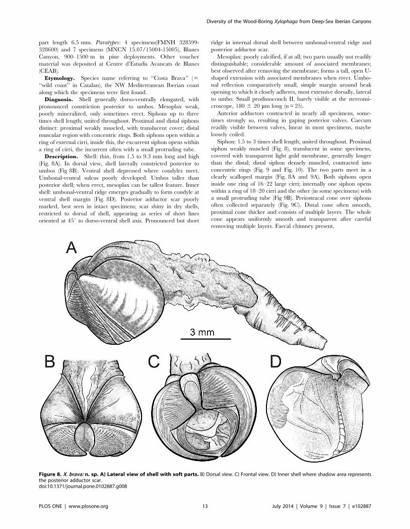
Diagnosis. Shell generally dorso-ventrally elongated, with
pronounced constriction posterior to umbos. Mesoplax weak,
poorly mineralized, only sometimes erect. Siphons up to three
times shell length; united throughout. Proximal and distal siphons
distinct: proximal weakly muscled, with translucent cover; distal
muscular region with concentric rings. Both siphons open within a
ring of external cirri, inside this, the excurrent siphon opens within
a ring of cirri, the incurrent often with a small protruding tube.
Description. Shell: thin, from 1.5 to 9.3 mm long and high
(Fig. 8A). In dorsal view, shell laterally constricted posterior to
umbos (Fig 8B). Ventral shell depressed where condyles meet.
Umbonal-ventral sulcus poorly developed. Umbos taller than
posterior shell; when erect, mesoplax can be tallest feature. Inner
shell: umbonal-ventral ridge emerges gradually to form condyle at
ventral shell margin (Fig. 8D). Posterior adductor scar poorly
marked, best seen in intact specimens; scar shiny in dry shells,
restricted to dorsal of shell, appearing as series of short lines
oriented at 45u to dorso-ventral shell axis. Pronounced but short
ridge in internal dorsal shell between umbonal-ventral ridge and
posterior adductor scar.
Mesoplax: poorly calcified, if at all; two parts usually not readily
distinguishable; considerable amount of associated membranes;
best observed after removing the membrane; forms a tall, open U-
shaped extension with associated membranes when erect. Umbo-
nal reflection comparatively small, simple margin around beak
opening to which it closely adheres, most extensive dorsally, lateral
to umbo. Small prodissoconch II, barely visible at the stereomi-
croscope, 180 6 20 mm long (n = 25).
Anterior adductors contracted in nearly all specimens, some-
times strongly so, resulting in gaping posterior valves. Caecum
readily visible between valves, linear in most specimens, maybe
loosely coiled.
Siphon: 1.5 to 3 times shell length; united throughout. Proximal
siphon weakly muscled (Fig. 8), translucent in some specimens,
covered with transparent light gold membrane, generally longer
than the distal; distal siphon densely muscled, contracted into
concentric rings (Fig. 9 and Fig. 10). The two parts meet in a
clearly scalloped margin (Fig. 8A and 9A). Both siphons open
inside one ring of 16–22 large cirri; internally one siphon opens
within a ring of 18–20 cirri and the other (in some specimens) with
a small protruding tube (Fig 9B). Periostracal cone over siphons
often collected separately (Fig. 9C). Distal cone often smooth,
proximal cone thicker and consists of multiple layers. The whole
cone appears uniformly smooth and transparent after careful
removing multiple layers. Faecal chimney present.
Figure 8. X. brava: n. sp. A) Lateral view of shell with soft parts. B) Dorsal view. C) Frontal view. D) Inner shell where shadow area representsthe posterior adductor scar.doi:10.1371/journal.pone.0102887.g008
Diversity of the Wood-Boring Xylophaga from Deep-Sea Iberian Canyons
PLOS ONE | www.plosone.org 13 July 2014 | Volume 9 | Issue 7 | e102887

Distribution. NW Mediterranean at BC, BC-OS, and LFC,
from 900 to 1800 m depth (Table 1).
Remarks. The two-parted siphon may be somewhat sugges-
tive of species in the genus Xyloredo Turner, 1972, recently
redescribed by Haga & Kase [52], which also have a strongly
reduced mesoplax. However, our specimens do not have a
calcareous tube lining their burrows. This, together with all
remaining morphological characters, defines our species as
belonging to Xylophaga.
Xylophaga brava sp. nov. resembles Xylophaga heterosiphonVoight, 2007 in having distinctly two-parted siphons, poorly
calcified mesoplax, siphonal openings within a common ring of
cirri, thin shell, and periostracal cones. The two species differ in: 1)
the muscle texture of the proximal siphon is notably weaker, even
transparent in X. brava sp. nov. and comparatively denser in X.heterosiphon; 2) the posterior adductor muscle scar is clearer in X.heterosiphon than in the new species; 3) a ridge in the inner shell
extends from near the umbo to the posterior-ventral margin in X.heterosiphon, in X. brava sp. nov. the strongly-defined ridge is
short and 4) the anterior umbonal reflection in X. heterosiphon is
much reduced.
X. brava sp. nov. resembles X. gerda Turner, 2002 from 284 m
depth off Grand Bahama Island, in having the shell with a concave
posterior slope, tiny mesoplax, equal length siphons with a
periostracal sheath, generally smooth and glisteny inner shell, a
weakly marked posterior adductor scar, and a low umbonal-
ventral ridge. However, X. brava sp. nov. can be distinguished
from X. gerda by its distinctly different proximal and distal
siphons, its mesoplax that can extend beyond the umbos when
erect, its siphons that are united through their length and that both
siphonal apertures open within a single ring of cirri.
X. brava sp. nov resembles X. concava Knudsen, 1961: notably
shell shape, erectable mesoplax, and long siphons that open close
to each other. However, X. concava differs in having apparently a
periostracal cover over the entire siphon, as opposed to a cone, an
essentially semicircular mesoplax, and attached juveniles [53].
Xylophaga brava sp. nov. also resembles X. anselli Harvey, 1996,
a species described from Atlantic Scottish waters based on a few,
very small individuals (,1 mm). However, the former differs in
having often weakly muscled and transparent proximal siphon, a
smaller prodissoconch II (see the description of X. cf. anselli), more
cirri at the siphon openings, and a rounder posterior shell margin.
The specimens of morphotype F from AC in the Cantabrian Sea
are geographically and morphologically closer to X. anselli (see the
description of X. cf. anselli). Moreover, our molecular data show
the respective specimens of both species grouped in two different,
Figure 9. X. brava n. sp: A.)Enlarged view of the distal siphons. B) Common siphon opening. C) cones which cover the siphons.doi:10.1371/journal.pone.0102887.g009
Diversity of the Wood-Boring Xylophaga from Deep-Sea Iberian Canyons
PLOS ONE | www.plosone.org 14 July 2014 | Volume 9 | Issue 7 | e102887

well-supported clusters, thus confirming the genetic isolation
between them.
Xylophaga cf. anselliMaterial examined. AC-WS 1200 m depth: 2 specimens;
AC 2000 m depth: 16 specimens. FMNH312286.
Diagnosis. Shell length in our specimens from 1.8 to
8.5 mm. Shell thin, anterior beak narrow. Umbonal-ventral sulcus
only slightly depressed. Posterior shell margin vertical, dorsally and
ventrally equally developed. On the inner shell, umbonal-ventral
ridge emerges gradually at ventral shell margin (Fig. 11). Posterior
adductor scar large (Fig. 11C).
Siphons: 1 to 1.7 times shell length; united throughout.
Proximal and distal siphons distinct: proximal opaque and
wrinkled; distal densely muscled, contracted into concentric rings.
The two parts meet in a linear margin. Both siphons open in one
ring of up to 15 cirri. Periostracal cone that covers at least the
distal siphons may be present.
Mesoplax: poorly calcified, if at all; two plates not seen. Amber
brown prodissoconch II always visible at the stereomicroscope,
240 6 15 mm long (n = 6) (Fig. 12) in agreement with Harvey
Figure 10. X. brava n. sp: A) SEM micrograph of the whole animal from left side. B) enlargement of the distal siphons.doi:10.1371/journal.pone.0102887.g010
Diversity of the Wood-Boring Xylophaga from Deep-Sea Iberian Canyons
PLOS ONE | www.plosone.org 15 July 2014 | Volume 9 | Issue 7 | e102887

(1996) and with observation of the FMNH specimen that had a
visible prodissoconch, 200 mm long.
Remarks. We assigned these specimens to X. cf. anselli based
on their morphological similarity to the original description by
Harvey (1996) and comparison to FMNH 312286, which was
collected at the type locality.. However, all Harvey’s specimens
were less than 1 mm in shell length, making it impossible to
compare the adult characters to those in the juveniles. The larger
specimen in FMNH 312286 shows a trace of a periostracal cover
on the proximal siphon. Harvey [54] did not distinguish two
separate siphon parts, perhaps because of their size, the separation
was not apparent; he described only one siphon opening and did
not discuss whether both siphons could open in a single ring of
cirri. He also reported up to 12 cirri in the siphon opening, while
our specimens show up to 15 cirri. However, we suggest that cirri
number may increase with increasing size. A complete redescrip-
tion of X. anselli based on specimens of a wide series from 1 mm
to full adult from the type locality is thus required.
Xylophaga cf. anselli from the Cantabrian Sea, Atlantic Ocean
differed from X. brava sp. nov., Mediterranean Sea, in having a
proximal siphon which is denser and more compact, never
transparent; fewer cirri at the external siphon opening (up to 15 vs.
Figure 11. X. cf. anselli: A) Lateral view of the whole animal. B) Dorsal view. C) Internal shell. PAS = posterior adductor scar. Scale bar in A= 2 mm, in B and C = 3 mm.doi:10.1371/journal.pone.0102887.g011
Figure 12. SEM micrographs of umbonal areas of specimens of X. cf. anselli (left) and X. brava (right) Arrows measure prodissoconchdiameter. Scale bars = 250 mmdoi:10.1371/journal.pone.0102887.g012
Diversity of the Wood-Boring Xylophaga from Deep-Sea Iberian Canyons
PLOS ONE | www.plosone.org 16 July 2014 | Volume 9 | Issue 7 | e102887

22 in X. brava sp. nov. in adults of similar size) and a larger
prodissoconch (240 6 15 mm vs 180 6 20 mm in X. brava sp.
nov., Fig. 12).
X. anselli differs from X. heterosiphon in frontal view by the
larger umbonal reflection that extends more ventrally in the
former species than in the latter. A limited number of poorly
preserved specimens in the collections of the United States
National Museum (USNM 757359; 757342) indicate that this or
an overtly similar species occurs in the western Atlantic Ocean at
depths of 2915-3090 m off Norfolk, VA USA.
Discussion
Few studies investigated the evolutionary relationship among
members of Pholadoidea [40,55,56] and fewer still have tried to
integrate morphological and molecular studies in the taxonomy of
marine wood borers of the Teredinidaes [57,58]. However, as far
as we are aware, molecular tools have never been used in
taxonomic studies to assist in species delimitation among
xylophagaids.
Our combined morphological/molecular study demonstrated
the existence of several species of Xylophaga in Iberian Peninsula
canyons and increased significantly our knowledge about the
distribution and morphological plasticity of some of the species
found. Our study also recorded various degrees of separation
among the Atlantic and the Mediterranean forms of Xylophagaspp.
SystematicsRegarding Xylophaga. dorsalis, we observed subtle morpholog-
ical differences between our morphotype A and previous
descriptions of the species based on specimens from the SW
British coasts. Comparison of molecular data with voucher
specimens was not possible because attempts to sequence material
from museums were unsuccessful due to degraded DNA and the
poor quality of the extractions. However, we considered these
differences insufficient to assign species-level status. Accordingly,
the respective AC (Atlantic) and BC and LFC (Mediterranean)
specimens were found to be genetically inseparable. Considering
that the type material is missing, there remains the concern that a
complete redescription of X. dorsalis is required.
Similar results were found for X. atlantica. Although slight
morphological variation occurred among our specimens (morpho-
types C-E), the respective sequences of morphotype C and D
grouped all together and formed a single clade with X. atlanticafrom the West Atlantic. No clustering of sequences from different
canyons was seen (Fig. 3) and intraspecific variation was low
(Table 4). Unfortunately we had no sequences of morphotype E
for comparison but we have no evidences to demonstrate they
belong to a different species from the other morphotypes of X.atlantica.
In contrast, X. brava sp. nov. and X. cf. anselli, arose as two
sibling species with few morphological differences, inhabiting
respectively Mediterranean and Atlantic submarine canyons and
clearly representing different taxonomic units. These two species
are morphologically similar, deriving from a recent ancestor, but
genetically isolated in two different and vicariant evolutionary
units. Additionally, interspecific divergences for both genes, the
28S and 18S, were significantly higher than the intraspecific
variability observed. Our results confirm that the 18S and 28S
genes are highly conserved (the former more than the latter),
making our conclusions about X. brava sp. nov. and X. cf ansellimore robust. The variability levels among Xylophaga spp. are
comparable to those reported for other bivalves, which also lack
18S intraspecific variation [59], and support the hypothesis that
small distances (around 0.3%) among allopatric specimens are
evolutionary meaningful [57].
Therefore, the divergence between X. brava sp. nov. and X. cf.
anselli and their grouping in two well-supported clades show the
absence of interbreeding between the Mediterranean and the
Atlantic evolutionary lineages, suggesting that this situation has
been stable over time leading them to split into different species.
The Atlanto-Mediterranean littoral region experienced an intri-
cate geological and climatological history [60–62]. The Gibraltar
Strait, the only connection between the Atlantic and the
Mediterranean, has been one of the most important potential
biogeographical breakpoints in the world’s oceans due to its strong
current regimes [63,64], and repeated disconnections and
reconnections over geological timescales. As a consequence a
number of speciation processes and genetic divergence between
the basins was promoted [64–67]. Hence nowadays the Cantab-
rian and Mediterranean Seas, where X. cf. anselli and X. brava sp.
nov. were found, belong to different biogeographical Ecoregions,
the Lusitanian South-European and Mediterranean regions [68].
From a morphological point of view, we found some differences
between X. brava sp. nov. and X. cf. anselli, but to some extent,
differences in some life strategy may have contributed to promote
evolutionary differentiation of the two species. For instance, the
smaller prodissoconch in X. brava sp. nov. suggests the existence of
larvae with a shorter planktonic stage and, consequently, reduced
dispersal potential. Moreover, speciation is not always accompa-
nied by large morphological divergence [69–71], particularly in
recent episodes or under very extreme environmental forcing [22],
which exert similar pressures to the isolated populations.
The original description of Xylophaga anselli was based on
small specimens (juveniles) from the North-East Atlantic coast of
Scotland [54]. Lacking adequate material of this species to allow a
complete description based on adult specimens from the type
locality, as well as molecular analyses, we decided to report the
Cantabrian specimens as X. cf. anselli in accordance to their
morphological similarity and geographical proximity, while we
describe the isolated Mediterranean species as X. brava sp. nov.
The combined results of our morphological and molecular data
also suggested that further studies including more species of
Xylophaga (borehole lining absent, well-developed, calcified
mesoplax) and Xyloredo (calcified borehole lining, reduced
mesoplax), should be addressed to resolve the relationship among
these two genera, particularly taking into account that species such
as X anselli, X. heterosiphon and X. brava sp. nov., showed
intermediate morphological characters (i.e. organic borehole
lining, small uncalcified mesoplax) and turned to be clustered
within Xyloredo and Xylopholas in our molecular analyses.
Diversity of Xylophaga species in Iberian canyonsThe presence of three species of Xylophaga in two NW
Mediterranean deep-sea submarine canyons increases the recog-
nized diversity of the genus in the Mediterranean by a 200%. Only
X. dorsalis had been previously reported from that sea, initially
from the Iberian coasts (Jeffrey, 1865) and, more recently, from the
Nile Fan (Eastern Mediterranean) at 1640 m depth [19,20].
Among the species with Atlantic distribution, X. anselli was
heretofore known only from the Northeast Atlantic type localities
(Hebridean slope and Anton Dohrn Seamount, 1370–2195 m
depth) [54]. Thus, our discovery of the species in the Cantabric
Sea would extend its geographical distribution to the south.
Xylophaga dorsalis was by far the most abundant species, based
both on the colonized deployments (nearly all) and the number of
individuals in each deployment, this abundance being consistent
Diversity of the Wood-Boring Xylophaga from Deep-Sea Iberian Canyons
PLOS ONE | www.plosone.org 17 July 2014 | Volume 9 | Issue 7 | e102887

with the previous reports from Mediterranean locations [17].
Xylophaga brava sp. nov., the second species in abundance, was
much more common in pine than in oak and the available data
suggested it is restricted to the Western Mediterranean, more
studies are needed to better define its distribution. Specimens
attributed to X. atlantica were comparatively rare, but they
occurred in all canyons (Fig. 1). The species had previously been
reported only from the West Atlantic [4]. The shallow-water
wood-borer, Teredothyra dominicensis, previously considered to be
a Caribbean endemism, has also been reported from the
Mediterranean [57]. Whether the presence of these species
indicates recent invasions or simply reflects an increasing number
of available samples or of the taxonomic expertise, both leading to
a better knowledge of the species distribution, cannot be currently
assessed.
Our results confirm that deep-sea environments still host a large
amount of undiscovered fauna. Particularly, submarine canyons,
where sedimentation and accumulation of organic debris are
enhanced, may represent hot spot of biomass and endemism
[17,72,73] and therefore should be recognized as potential high
priority areas for the conservation of the deep sea. The 4700 m
depth site from AC appears to be exceptional in that none of the
wood cubes deployed there were colonized by Xylopaga, or any
other macroinvertebrate. This may be due to unfavorable local
circulation patterns, extreme environmental factors, or the lack
wood as potential food source to maintain a stable population of
wood borers.
Our results refute the long-held view that larvae of Xylophagado not colonize wood that is not in contact with, or in close
proximity to, the seafloor [74], only Turner [4] previously
reported that few specimens of X. depalmai were recovered up
to 90 m from the bottom but she considered the event very
unusual. Xylophaga species seem to disperse higher in the water
column than do other deep-sea benthic species associated with
ephemeral, fragmented habitats such as organic falls. Specimens of
the bone-eating Osedax worms, for example, decreased drastically
one meter above the sediment floor [75]. Moreover specimens of
Idas Jeffreys, 1876, a deep-sea bivalve generally associated with
wood falls, were found in wood deployments at the sediment
surface but were never recovered in suspended ones at the same
site [17].The ability of larval Xylophaga to disperse higher in the
water column, and therefore to be subject by transport of stronger
currents, may be a key factor in allowing the persistence of the
adult populations despite living in patchy wood fall habitats.
Future studies focusing on ecology and populations’ connectiv-
ity inside and outside canyons, as well as between other deep-sea
environments, are required to assess the real larval dispersal and
colonizing abilities of these highly specialized organisms. To assess
populations’ connectivity among hot spots of biomass and
endemisms such as submarine canyons will be fundamental for
the development of a sustainable management strategy of the
deep-sea ecosystems and its associated resources [76] and will
certainly aid in the scientific design of marine protected areas.
Acknowledgments
We thank Gemma Agell, Daphne Cortese, and Gustavo Carreras for their
valuable support and help during the laboratory and molecular analyses
carried out at CEAB. We are grateful to Angel Luque, Elena Aloisio, Elena
Lloret, Andrea Caputo and Sven Laming for help in extracting specimens
from wood and to Francesco Pititto for preparing the map. We thank the
other teams of the projects, especially that of Prof. A. Calafat for allowing
us to deploy the wood traps with their mooring equipment. Special thanks
to the crew of R/V Garcia del Cid, Sara Roman and Marta Segura for
support during the cruises.
We are very grateful to Jan Freedman from the Plymouth City Museum
and Art Gallery, Michele Bruni from The Oceanographic Museum of
Monaco, Dr Jack Sewell from Marine Biological Association (UK) and Dr.
Silke Stoll from Landeshauptstadt Dusseldorf for sending us some material
of Xylophaga dorsalis.We also thank the museums that replied to our request of type material:
the Naturmuseum Senckenberg of Frankfurt, the Lobbecke Museum, The
Natural History Museum in Munchen, The University of Bristol, the
Bristol Museum & Art Gallery, The National Museum of Cardiff, the
National History Museum in London and the Smithsonian National
Museum of Natural History.
We are grateful to the ArgoMaris Foudation and the crew of the Pigallvessel and the pilot of the ROV, Seaeye Linx 1500 for help during the
deployments of the trap at the head of the Blanes Canyon. Special thanks
to D. Reid at BMNH, and the Malacological Society of London to partially
finance JV travel and stay in London and in Blanes. We are grateful to
Prof. Nadine Le Bris and her team for letting us to analyse some samples
from Lacaze Duthiers Canyon and to join the DIWOOD network. This
paper is a contribution to the Consolidated Research Group 2009SRG665
of the Generalitat of Catalunya. We are grateful to two anonymous
reviewers for their comments that helped to improve the manuscript.
Author Contributions
Conceived and designed the experiments: DM CR. Performed the
experiments: CR. Analyzed the data: CR JRV RPP. Contributed
reagents/materials/analysis tools: CR DM. Contributed to the writing of
the manuscript: CR JRV RPP DM.
References
1. Purchon (1941) On the Biology and relationships of the Lamellibrach Xylophagadorsalis (Turton). Journal of the Marine Biological Association of the United
Kingdom 25: 1–39.
2. Distel DL, Roberts SJ (1997) Bacterial endosymbionts in the gills of the deep-sea
wood-boring bivalves Xylophaga atlantica and Xylophaga washingtona.
Biological Bulletin (Woods Hole) 192: 253–261.
3. Voight JR (2007) Experimental deep-sea deployments reveal diverse Northeast
Pacific wood-boring bivalves of Xylophagainae (Myoida: Pholadidae). J Mol-
luscan Stud 73: 377–391.
4. Turner RD (2002) On the subfamily Xylophagainae (family Pholadidae,
Bivalvia, Mollusca). Bulletin of the Museum of Comparative Zoology. 157:223–
307.
5. Voight JR, Segonzac M (2012) At the bottom of the deep blue sea: a new wood-
boring bivalve (Mollusca, Pholadidae, Xylophaga) from the Cape Verde Abyssal
Plain (subtropical Atlantic). Zoosystema 34: 171–180.
6. Coll M, Piroddi C, Steenbeek J, Kaschner K, Ben Rais Lasram F, et al. (2010)
The biodiversity of the Mediterranean Sea: Estimates, patterns and treats PLoS
ONE 5: e11842.
7. Zenetos A (1996) Fauna graeciae VII: The Marine Bivalvia (Mollusca) of
Greece. Hellenic Zoological Society and NCMR.
8. Albayrak S, Balkis H, Balkis N (2004) Bivalvia (Mollusca) fauna of the Sea of
Marmara. Acta Adriat 45: 9–26.
9. Harris PT, Whiteway T (2011) Global distribution of large submarine canyons:
Geomorphic differences between active and passive continental margins. Mar
Geol 285: 69–86.
10. Canals M, Danovaro R, Heussner S, Lykousis V, Puig P, et al. (2010) Cascades
in mediterranean submarine grand canyons. Oceanography 22: 26–43.
11. Garcia R, Thomsen L, de Stigter HC, Epping E, Soetaert K, et al. (2010)
Sediment bioavailable organic matter, deposition rates and mixing intensity in
the Setubal-Lisbon canyon and adjacent slope (Western Iberian Margin). Deep-
Sea Research Part I: Oceanographic Research Papers 57: 1012–1026.
12. Vetter EW, Dayton PK (1998) Macrofaunal communities within and adjacent to
a detritus-rich submarine canyon system. Deep Sea Research Part II: Topical
Studies in Oceanography 45: 25–54.
13. De Leo FC, Smith CR, Rowden AA, Bowden DA, Clark MR (2010) Submarine
canyons: Hotspots of benthic biomass and productivity in the deep sea.
Proceedings of the Royal Society B: Biological Sciences 277: 2783–2792.
14. Orejas C, Gori A, Lo Iacono C, Puig P, Gili JM, et al. (2009) Cold-water corals
in the Cap de Creus canyon, northwestern Mediterranean: spatial distribution,
density and anthropogenic impact. Mar Ecol Prog Ser 51: 37–51.
15. Cartes JE, Maynou F, Fanelli E, Romano C, Mamouridis V, et al. (2009) The
distribution of megabenthic, invertebrate epifauna in the Balearic Basin (western
Mediterranean) between 400 and 2300 m: Environmental gradients influencing
assemblages composition and biomass trends. J Sea Res 61: 244–257.
Diversity of the Wood-Boring Xylophaga from Deep-Sea Iberian Canyons
PLOS ONE | www.plosone.org 18 July 2014 | Volume 9 | Issue 7 | e102887

16. Stora G, Bourcier M, Arnoux A, Gerino M, Le Campion J, et al. (1999) The
deep-sea macrobenthos on the continental slope of the northwestern
Mediterranean Sea: a quantitative approach. Deep-Sea Research Part I-
Oceanographic Research Papers 46: 1339–1368.
17. Romano C, Voight JR, Company JB, Plyuscheva M, Martin D (2013)
Submarine canyons as the preferred habitat for wood-boring species of
Xylophaga (Mollusca, Bivalvia). Progress in Oceanography 118: 175–187.
18. Fagervold SK, Bessette S, Romano C, Martin D, Plyuscheva M, et al. (2013)
Microbial communities associated with the degradation of oak wood in the
Blanes submarine canyon and its adjacent open slope (NW Mediterranean).
Progress in Oceanography 118: 137–143.
19. Bienhold C, Pop Ristova P, Wenzhofer F, Dittmar T, Boetius A (2013) How
Deep-Sea Wood Falls Sustain Chemosynthetic Life. PLoS ONE 8.
20. Gaudron SM, Pradillon F, Pailleret M, Duperron S, Le Bris N, et al. (2010)
Colonization of organic substrates deployed in deep-sea reducing habitats by
symbiotic species and associated fauna. Mar Envir Res 70: 1–12.
21. Yucel M, Galand PE, Fagervold SK, Contreira-Pereira L, Le Bris N (2013)
Sulfide production and consumption in degrading wood in the marine
environment. Chemosphere 90: 403–409.
22. Bickford D, Lohman DJ, Sodhi NS, Ng PKL, Meier R, et al. (2007) Cryptic
species as a window on diversity and conservation. Trends in Ecology &
Evolution 22: 148–155.
23. Amblas D, Canals M, Urgeles R, Lastras G, Liquete C, et al. (2006)
Morphogenetic mesoscale analysis of the northeastern Iberian margin, NW
Mediterranean Basin. Mar Geol 234: 3–20.
24. Palanques A, Martın J, Puig P, Guillen J, Company JB, et al. (2006) Evidence of
sediment gravity flows induced by trawling in the Palamos (Fonera) submarine
canyon (northwestern Mediterranean). Deep Sea Research Part I: Oceano-
graphic Research Papers 53: 201–214.
25. Lastras G, Canals M, Amblas D, Lavoie C, Church I, et al. (2011)
Understanding sediment dynamics of two large submarine valleys from seafloor
data: Blanes and La Fonera canyons, northwestern Mediterranean Sea. Mar
Geol 280: 20–39.
26. Sarda F, Company JB, Bahamon N, Rotllant G, Flexas MM, et al. (2009)
Relationship between environment and the occurrence of the deep-water rose
shrimp Aristeus antennatus (Risso, 1816) in the Blanes submarine canyon (NW
Mediterranean). Progress In Oceanography 82: 227–238
27. Ingels J, Vanreusel A, Romano C, Coenjaerts J, Mar Flexas M, et al. (2013)
Spatial and temporal infaunal dynamics of the Blanes submarine canyon-slope
system (NW Mediterranean); changes in nematode standing stocks, feeding types
and gender-life stage ratios. Progress in Oceanography 118: 159–174.
28. Tecchio S, Ramırez-Llodra E, Aguzzi J, Sanchez-Vidal A, Flexas MM, et al.
(2013) Seasonal fluctuations of deep megabenthos: Finding evidence of standing
stock accumulation in a flux-rich continental slope. Progress in Oceanography
118: 188–198.
29. Romano C, Coenjaerts J, Flexas MM, Zuniga D, Vanreusel A, et al. (2013)
Spatial and temporal variability of meiobenthic density in the Blanes submarine
canyon (NW Mediterranean). Progress in Oceanography 118: 144–158.
30. Louzao M, Anadon N, Arrontes J, Alvarez-Claudio C, Fuente DM, et al. (2010)
Historical macrobenthic community assemblages in the Aviles Canyon, N
Iberian Shelf: Baseline biodiversity information for a marine protected area.
J Marine Syst 80: 47–56.
31. Gonzalez-Quiros R, Cabal J, Alvarez-Marques F, Isla A (2003) Ichthyoplankton
distribution and plankton production related to the shelf break front at the Aviles
Canyon. ICES J Mar Sci 60: 198–210.
32. Ruano A, Silva P, Solano S, Naves J (2007) Cetaceos del litoral asturiano: areas
de interes para la conservacion. Consejerıa de Medio Ambiente, Ordenacion del
Territorio e Infraestructuras y Obra Social La Caixa.
33. Lopez-Fernandez P, Calafat A, Sanchez-Vidal A, Canals M, Mar Flexas M, et
al. (2013) Multiple drivers of particle fluxes in the Blanes submarine canyon and
southern open slope: Results of a year round experiment. Progress in
Oceanography 118: 95–107.
34. Hall TA (1999) BioEdit: a user-friendly biological sequence alignment editor and
analysis program for Windows 95/98/NT. Nucl Acids Symp Ser 41: 95–98.
35. Galtier N, Gouy M, Gautier C (1996) SEAVIEW and PHYLO_WIN: two
graphic tools for sequence alignment and molecular phylogeny. Bioinformatics
12: 543–548.
36. Darriba D, Taboada G, Doallo R, Posada D (2012) jModelTest 2: more models,
new heuristics and parallel computing. Nature Methods 9: 772.
37. Huelsenbeck JP, Ronquist F (2001) MRBAYES: Bayesian inference of
phylogenetic trees. Bioinformatics 17: 754–755.
38. Stamatakis A, Hoover P, Rougemont J (2008) A Rapid Bootstrap Algorithm for
the RAxML Web-Servers. Syst Biol 75: 758–771.
39. Tamura K, Peterson D, Peterson N, Stecher G, Nei M, et al. (2011) MEGA5:
Molecular Evolutionary Genetics Analysis using Maximum Likelihood,
Evolutionary Distance, and Maximum Parsimony Methods. Mol Biol Evol 28:
2731–2739.
40. Distel DL, Mehwish A, Adam B, Linton E, Gustaf M, et al. (2011) Molecular
phylogeny of Pholadoidea Lamarck, 1809 supports a single origin for xylotrophy
(wood feeding) and xylotrophic bacterial endosymbiosis in Bivalvia.
41. Giribet G, Distel D (2003) Bivalve Phylogeny and Molecular data. In: Lydeard
C& Lindberg DR, editors. Molecular systematics and phylogeography of
mollusks. Washington, DC. pp. 45–90.
42. Haga T, Kase T (2013) Progenetic dwarf males in the deep-sea wood-boring
genus Xylophaga (Bivalvia: Pholadoidea). J Molluscan Stud 79: 90–94.
43. Turner RD (1955) The fanily Pholadidae in the western Atlantic and the eastern
Pacific. Part II - Martesiinae, Jouannetiinae and Xylopagainae. Johnsonia 3: 65–
160.
44. Dons C (1929) Zoologiske Notiser V. Xylophaga dorsalis I Norge. Det Kongelige
Norsske Videnskabers Selskab Forhandlinger 65: 196–199.
45. Santhakumaran LN, Sneli JA (1984) Studies on the marine fouling and wood-
boring organisms of the Trondheimsfjord (western Norway). Gunneria: 1–36.
46. Voight JR (2008) Deep-sea wood-boring bivalves of Xylophaga (Myoida:
Pholadidae) on the Continental Shelf: a new species described. J Mar Biol
Assoc UK 88: 1459–1464.
47. Parenzan P (1974) Carta d’identita delle conchiglie del Mediterraneo. Bivalvi.:
Bios-Taras. Taranto.
48. Jeffreys (1865) British Conchology. London: Van Voorst.
49. Tebble N (1976) British bivalve seashells: A handbook for identification.
Published for the Royal Scottish Museum by H.M. Stationery Off. (Edinburgh)
50. Kudinova-Pasternak RK (1975) Xylophaga Mollusks (Bivalvia, Pholadidae)
found in a Scotia Sea shipwreck. Institute of Oceanology Research Publication
103: 179–182.
51. Turner RD, Culliney J (1971) Some anatomical and life history studies of wood
boring bivalve systematics. Ann Rept American Malacological Union for 1970:
65–66.
52. Haga T, Kase T (2008) Redescription of the Deep-sea Wood Borer
Neoxylophaga teramachii Taki & Habe, 1950 and its Assignment to the Genus
Xyloredo (Bivalvia: Myoida: Pholadoidea) with Comments on Fossil Pholadoi-
dae. Veliger 50: 107–119.
53. Knudsen J (1961) The bathyal and abissal Xylophaga (Pholadidae, Bivalvia).
Galathea Reports 5: 163–209.
54. Harvey R (1996) Deep water Xylophagaidae (Pelecypoda: Pholadacea) from the
North Atlantic with descriptions of three new species. Journal of Conchology 35:
473–481.
55. Monari S (2009) Phylogeny and biogeography of pholadid bivalve Barnea
(Anchomasa) with considerations on the phylogeny of Pholadoidea. Acta
Palaeontol Pol 54: 315–335.
56. Santos SML, Tagliaro CH, Beasley CR, Schneider H, Sampaio I, et al. (2005)
Taxonomic implications of molecular studies on northern Brazilian Teredinidae
(Mollusca, Bivalvia) specimens. Genetics and Molecular Biology 28: 175–179.
57. Borges LMS, Sivrikaya H, le Roux A, Shipway JR, Cragg SM, et al. (2012)
Investigating the taxonomy and systematics of marine wood borers (Bivalvia:
Teredinidae) combining evidence from morphology, DNA barcodes and nuclear
locus sequences. Invertebrate Systematics 26: 572–582.
58. Shipway RJ, Borges LS, Muller J, Cragg S (2014) The broadcast spawning
Caribbean shipworm, Teredothyra dominicensis (Bivalvia, Teredinidae), has
invaded and become established in the eastern Mediterranean Sea. Biological
Invasions: 1–12.
59. Espineira M, Gonzalez-Lavin N, Vieites JM, Santaclara FJ (2009) Development
of a Method for the Genetic Identification of Commercial Bivalve Species Based
on Mitochondrial 18S rRNA Sequences. J Agric Food Chem 57: 495–502.
60. Duggen K, Hoernle PVD, Boogard L, Morgan JP (2003) Deep roots of the
Messinian salinity crisis. Nature 422: 602–606.
61. Brault N, Bourquin S, Guillocheau F, Dabard MP, Bonnet S, et al. (2004) Mio–
Pliocene to Pleistocene paleotopographic evolution of Britanny (France) from a
sequence stratigraphic analysis: relative influence of tectonics and climate. Sedim
Geol 163: 175–210.
62. Maggs CA, Castilho R, Foltz D, Henzler C, Jolly MT, et al. (2008) Evaluating
signatures of the glacial refugia for North Atlantic benthic marine taxa. Ecology
89: 108–122.
63. Tintore’ J, Laviolette PE, Blade I, Cruzado A (1988) A study of an intense
density front in the Eastern Alboran-Sea– the Almeria-Oran Front. Journal of
Physical Oceanography 18: 1384–1397.
64. Patarnello T, Volckaert F, Castilho R (2007) Pillars of Hercules: Is the Atlantic-
Mediterranean transition a phylogeographical break? Molecular Ecology 16:
4426–4444.
65. Borsa P, Naciri M, Bahri L, Chikhi L, Garcia de Leon FJ, et al. (1997)
Infraspecific zoogeography of the Mediterranean: population genetic analysis on
sixteen Atlanto-Mediterranean species (fishes and invertebrates). Zoogeographie
infra-specifique de la Mer Mediterranee: analyse des donnees genetiques
populationnelles sur seize especes atlanto-mediterraneennes (poissons et
invertebres) 47: 295–305.
66. Perez-Portela R, Turon X (2008) Cryptic divergence and strong population
structure in the colonial invertebrate Pycnoclavella communis (Ascidiacea)
inferred from molecular data. Zoology 111: 163–178.
67. Perez-Portela R, Villamor A, Almada V (2010) Phylogeography of the sea star
Marthasterias glacialis (Asteroidea, Echinodermata): deep genetic divergence
between mitochondrial lineages in the north-western mediterranean. Mar Biol
157: 2015–2028.
68. Spalding MD, Fox HE, Allen GR, Davidson N, Ferdan, a ZA, et al. (2007)
Marine ecoregions of the world: a bioregionalization of coastal and shelf areas.
Bioscience 57: 573–583.
69. Knowlton N (2000) Molecular genetic analyses of species boundaries in the sea.
Hydrobiologia 420: 73–90.
Diversity of the Wood-Boring Xylophaga from Deep-Sea Iberian Canyons
PLOS ONE | www.plosone.org 19 July 2014 | Volume 9 | Issue 7 | e102887

70. Perez-Portela R, Duran S, Palacın C, Turon X (2007) The genus Pycnoclavella
(Ascidiacea) in the Atlanto-Mediterranean region: a combined molecular and
morphological approach. Invertebrate Systematics 21: 187–205.
71. Perez-Portela R, Almada V, Turon X (2013) Cryptic speciation and genetic
structure of widely distributed brittle stars (Ophiuroidea) in Europe. Zool Scr 42:
151–169.
72. Gili JM, Pages F, Bouillon J, Palanques A, Puig P, et al. (2000) A
multidisciplinary approach to the understanding of hydromedusan populations
inhabiting Mediterranean submarine canyons. Deep-Sea Research Part I-
Oceanographic Research Papers 47: 1513–1533.
73. Mecho A, Aguzzi J, Company JB, Canals M, Lastras G, et al. (2014) First in situ
observations of the deep-sea carnivorous ascidian Dicopia antirrhinum MonniotC., 1972 in the Western Mediterranean Sea. Deep Sea Research Part I:
Oceanographic Research Papers 83: 51–56.
74. Tipper RC (1968) Ecological aspects of two wood-boring molluscs from thecontinental terrace off Oregon.: Oregon State University.
75. Jones WJ, Johnson SB, Rouse GW, Vrijenhoek RC (2008) Marine worms (genusOsedax) colonize cow bones. Proceedings of the Royal Society B: Biological
Sciences 275: 387–391.
76. Van Dover CL, Aronson J, Pendleton L, Smith S, Arnaud-Haond S, et al. (2014)Ecological restoration in the deep sea: Desiderata. Marine Policy 44: 98–106.
Diversity of the Wood-Boring Xylophaga from Deep-Sea Iberian Canyons
PLOS ONE | www.plosone.org 20 July 2014 | Volume 9 | Issue 7 | e102887