Hemophagocytic Lymphohistiocytosis in A Newborn Infant...
4
241 KUWJ\ITMFDTCAI JOURNJ\l September 2016 Case Report Hemophagocytic Lymphohistiocytosis in A Newborn Infant Presenting with Nonimmune Hydrops Fetalis: A Case Report Badr Hasan Sobaih', Shihab Ahmed Almashhadanf 'King Saud University, College of Medicine, Department of Pediatrics, King Khalid University Hospital, Riyadh, Kingdom of Saudi Arabia 2KingSaud University, College of Medicine, Pathology Department, Hematology Division, King Khalid University Hospital, Riyadh, Saudi Arabia Kuwait Medical Journal 2016; 48 (3): 241 - 244 ABSTRACT------------------------------------------------------------ Nonimmune hydrops fetalis is a condition that may result as a cause of many underlying conditions and in around 15% of cases no cause could be identified. Hemophagocytic lymphohistiocytosis had been identified as a rare cause of nonimmune hydrops fetalis in the newborn period in few cases around the world. Here, we reported one case of nonimmune hydrops fetalis born for Saudi parents associated with Hemophagocytic Iymphohistiocytosis presented at birth and was born at 32 weeks of gestation. Diagnosis was confirmed by bone marrow aspiration. She was treated with chemotherapy but did not respond to therapy and died. We suggest that Hemophagocytic lymphohistiocytosis should be seriously considered as a cause of nonimmune hydrops fetalis, if the later condition is associated with unexplained cytopenias, high ferritin level, and splenomegaly. KEY WORDS: cytopenias, hemophagocytic Iymphohistiocytosis, high ferritin, nonirnmune hydrops fetalis INTRODUCTION Hemophagocytic Lyrnphohistiocytosis (HLH), also known as hemophagocytic syndrome, is a rare hematologic disorder which affects the immune system characterized by abnormal proliferation of macrophages in various tissues and organs causing multi organ failure and often results in deathl'J. HLH encompasses several entities, including a primary form that may be familial HLH, with an estimated incidence of one in 50,000 births'", and a secondary form associated with infections, malignancies, and rheumatologic disordersl". Hydrops fetalis is a condition with many underlying causes. Despite advances in ante-natal and post-mortem diagnostic techniques, the cause remains unknown in up to 15% of cases!". Hemophagocytic lymphohistiocytosis had been reported as one of the rare causes of non immune hydrops fetalis in few case reports [5- 10 1. To our knowledge, this is the first reported case in Saudi Arabia. CASE REPORT A newborn girl was admitted to our Neonatal Intensive Care Unit (NICU) as a case of non-immune hydrops fetalis which was diagnosed by ante-natal ultrasound. She was delivered by emergency cesarean section at 32 weeks of gestation for a healthy 25 years old, booked Saudi primigravida due to fetal distress. Apgar score was 2, 5, and 7 at 1, 5, and 10 minutes respectively, with birth weight of 2.8 kg. Delivery was attended by three senior neonatologists who conducted full resuscitation for this hydropic baby. Pleural effusion was drained through bilateral thoracocentesis, while normal saline boluses were infused via an emergency umbilical venous catheter (UVC). She needed high frequency oscillatory ventilation (HFOV). She developed pneumothorax which was drained via Address correspondence to: Badr Hasan Sobaih, MBBS, ABP', Department of Pediatrics, King Saud University, College of Medicine, King Khalid University Hospital, Riyadh, Kingdom of Saudi Arabia, Mobile: 966505453580, Fax: 96614672395, PO Box 2925, Riyadh, 11461, Email: [email protected], drbsobaih@ yahoo com, [email protected];
Transcript of Hemophagocytic Lymphohistiocytosis in A Newborn Infant...
241 KUWJ\ITMFDTCAI JOURNJ\l September 2016
Case Report
Hemophagocytic Lymphohistiocytosis inA Newborn Infant Presenting with Nonimmune
Hydrops Fetalis: A Case ReportBadr Hasan Sobaih', Shihab Ahmed Almashhadanf
'King Saud University, College of Medicine, Department of Pediatrics, King Khalid University Hospital, Riyadh, Kingdomof Saudi Arabia
2KingSaud University, College of Medicine, Pathology Department, Hematology Division, King Khalid UniversityHospital, Riyadh, Saudi Arabia
Kuwait Medical Journal 2016; 48 (3): 241 - 244
ABSTRACT------------------------------------------------------------
Nonimmune hydrops fetalis is a condition that may resultas a cause of many underlying conditions and in around15% of cases no cause could be identified. Hemophagocyticlymphohistiocytosis had been identified as a rare causeof nonimmune hydrops fetalis in the newborn period infew cases around the world. Here, we reported one caseof nonimmune hydrops fetalis born for Saudi parentsassociated with Hemophagocytic Iymphohistiocytosis
presented at birth and was born at 32 weeks of gestation.Diagnosis was confirmed by bone marrow aspiration.She was treated with chemotherapy but did not respondto therapy and died. We suggest that Hemophagocyticlymphohistiocytosis should be seriously considered as acause of nonimmune hydrops fetalis, if the later conditionis associated with unexplained cytopenias, high ferritinlevel, and splenomegaly.
KEY WORDS: cytopenias, hemophagocytic Iymphohistiocytosis, high ferritin, nonirnmune hydrops fetalis
INTRODUCTIONHemophagocytic Lyrnphohistiocytosis (HLH),
also known as hemophagocytic syndrome, is a rarehematologic disorder which affects the immunesystem characterized by abnormal proliferation ofmacrophages in various tissues and organs causingmulti organ failure and often results in deathl'J. HLHencompasses several entities, including a primaryform that may be familial HLH, with an estimatedincidence of one in 50,000 births'", and a secondaryform associated with infections, malignancies, andrheumatologic disordersl".
Hydrops fetalis is a condition with manyunderlying causes. Despite advances in ante-natal andpost-mortem diagnostic techniques, the cause remainsunknown in up to 15% of cases!".
Hemophagocytic lymphohistiocytosis had beenreported as one of the rare causes of non immunehydrops fetalis in few case reports [5-101. To our
knowledge, this is the first reported case in SaudiArabia.
CASE REPORTA newborn girl was admitted to our Neonatal
Intensive Care Unit (NICU) as a case of non-immunehydrops fetalis which was diagnosed by ante-natalultrasound. She was delivered by emergency cesareansection at 32 weeks of gestation for a healthy 25 yearsold, booked Saudi primigravida due to fetal distress.Apgar score was 2, 5, and 7 at 1, 5, and 10 minutesrespectively, with birth weight of 2.8 kg. Delivery wasattended by three senior neonatologists who conductedfull resuscitation for this hydropic baby. Pleuraleffusion was drained through bilateral thoracocentesis,while normal saline boluses were infused via anemergency umbilical venous catheter (UVC). Sheneeded high frequency oscillatory ventilation (HFOV).She developed pneumothorax which was drained via
Address correspondence to:Badr Hasan Sobaih, MBBS, ABP', Department of Pediatrics, King Saud University, College of Medicine, King Khalid University Hospital, Riyadh,Kingdom of Saudi Arabia, Mobile: 966505453580, Fax: 96614672395, PO Box 2925, Riyadh, 11461, Email: [email protected], drbsobaih@yahoo com, [email protected];

September 2016 242KUWAIT MEDICAL JOURNAL
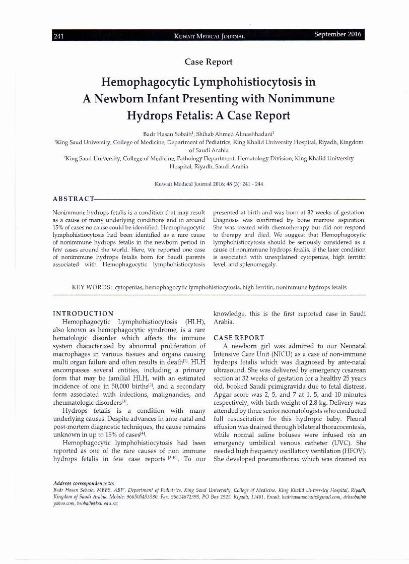
Fig. 1:Bonemarrow slide showing single hemophagocytic histiocytecontaining depris in its cytoplasm.
chest tube. Her pleural effusion and pnuemothoraxeventually resolved within four days. Echocardiogramwas done as part of evaluation for hydrops fetalis andshowed partial atrio-ventricular septal defect, largeatrial septal defect (ASD) secundum, small muscularventricular septal defect (VSD), and large patentductus arteriosus (PDA). She also developed severesupra systemic persistent pulmonary hypertension(PPHN). The impression of our cardiologist is thatthese findings cannot explain the etiology of hydropsfetalis for this baby. Renal ultrasound was normal.Generalized edema resolved gradually with the use ofdiuretics and fluid management. Physical examinationrevealed Splenomegaly which was persistentthroughout her hospital course.
Initial CBC showed WBC count of 35.65 x 10⁹/1,Hemoglobin was 124 g/l, and low platelet count of19 xl09/1. Subsequent CBC results showed persistentanemia with marked thrombocytopenia for whichmultiple transfusions of platelets and packed redcells were given, but failed to show acceptableimprovement. Intravenous immunoglobulin anddexamethasone also failed to improve the condition.She was empirically covered with broad spectrumantibiotics after obtaining blood culture. Hemoglobinlevel remained below 90 g/l and platelets count neverexceeds 30 x 109/1.
Other investigations for the etiology of hyd ropsfetal is such as TORCH screen, Parvovirus antibodies,Hepatitis markers, and HIV antibodies all came tobe negative. Pediatrics hematology/oncology servicewas involved in the investigation and management ofthis case. Serum Ferritin level was 1926.00 I-lglL thenincreased to 6844.00 I-lglL two weeks later. Peripheralblood film demonstrated leuko-erythroblastic bloodpicture. Neutrophilia with left shift and some toxicchanges were evident. RBC morphology reveals some
Fig. 2: Bone marrow slide showing many hemophagocytichistiocytes in clusters containing cellular depris in their cytoplasm.
degree of anisopoikilocytosis and polychromasia.Severe thrombocytopenia was confirmed. BandT lymphocytes subsets were low. Bone marrowaspiration was done and revealed hypercellularparticles with hypercellular trails that show adequatemegakaryocytes with occasional coarse basophilicgranulations. Erythropoiesis looks essentiallynormoblastic and shows progressive maturationalsequences. Granulopoiesis looks adequate and revealsslight left shift. M: E ratio 3.9:1. Blasts account for 3% ofall nucleated cells. Macrophages were prominent withobvious hemophagocytosis involving mostly erythroidcells and platelets. Some hemophagocytic cells were inclusters and some in single form all over the marrowaspirate smears (Fig. I, 2). Iron stain: Stainable ironstores. Iron was markedly increased with adequateerythron iron and no ring sideroblasts. With thesefindings, it was concluded that this hypercellularbone marrow aspirate showed adequate lineagehematopoietic elements with prominent phagocyticactivity. These findings were compatible with thediagnoses of hemophagocytic Lymphohistiocytosis(HLH).
Henceforth, she was started on triple drug regimenCyclosporin, IV Immunoglobulin and Dexamethasoneand showed progressive but transient response.Baby's general condition progressively deteriorateddespite full and aggressive management until she hadfulminant gram negative septicemia which eventuallyled to her death at the age of 44 days.
DISCUSSIONWe described a newborn baby who presented
to us as a case of non-immune hydrops fetalis andwas treated accordingly. This is the first time in ourpractice to see unusual course for such neonates as shepersistently had bicytopenia despite our aggressive

243 September 2016Hemophagocytic Lymphohistiocytosis in A Newborn Infant Presenting ...
intervention, although there was great improvementin pleural effusion and ascitis as well as generalizededema. The presence of persistent bicytopenia raisedour suspicion of dealing with a rare cause of hydropsfetalis that could be related to a disease affectingcell proliferation. After seeing the results of bonemarrow aspiration we searched the literature to findif there is association between hydrops fetal is andhemophagocytic Lymphohistiocytosis and we foundfew reported cases [5.101.
According to HLH-2004 protocol for diagnosticguidelines [111, diagnosis of HLH can be establishedafter fulfillment of either one or two of the followingcriteria: A molecular diagnosis consistent with HLH,and or Fulfillment of five out of eight of the followingdiagnostic criteria [ Fever, Splenomegaly, Cytopeniasaffecting ~ 2 of 3 lineages in the peripheral bloodHypertriglyceridemia and/ or hypofibrinogenemia,Hemophagocytosis in bone marrow or spleen orlymph nodes, Low or absent NK-ce\l activity, Highferritin, High CD 25 (soluble IL-2 receptor)].
Our patient fulfilled the criteria for the diagnosis ofHLH as she presented with cytopenias, splenomegaly,low B and T lymphocytes subsets, high ferritin level,and hemophagocytosis in bone marrow.
Around 10% of cases with HLH present withinthe first month of life, often after a symptom-free period. However, presentation in the firstday of life seems to be rare [121. Natural killer cellactivity, T-Iymphocyte mitogen response, antibody-dependent cellular cytotoxicity, and interleukin-land interferon production are all diminished [131. Thelymphoproliferative disorder of HLH can be dividedinto two categories: primary (familial) hemophagocyticIymphohistiocytosis (FHL) with an incidence of1:50,000 births and equal gender distribution, andsecondary HLH which may affect any age and mayresolve spontaneously. FHL is seen primarily inchildren and is fatal if untreated, with most casesdiagnosed before two years of age, and the diseasemay present in the newborn period [141. HLH may bedue to an immunoregulatory defect that predisposesto an uncontrolled production of activated histiocytesin response to a stimulus such as a viral infection [151.
The aim of current treatment protocols is to achieveremission with immune-chemotherapy, followed by acure with bone marrow transplantation [161.
CONCLUSIONWe conclude from this case that hemophagocytic
lymphohistiocytosis (HLH) should be consideredas a possible cause of nonimmune hydrops fetalisin hydropic newborns presenting with persistentunexplained bicytopenia, splenomegaly, and high
serum ferritin especially when no metabolic orinfectious cause is found. Diagnosis of HLH purelyby clinical presentation in newborn period is difficult.Establishment of the diagnosis by appropriateinvestigation tools such as bone marrow aspiration ismandatory for parent's counseling and initiation oftherapy.
REFERENCES
1. Ishii E, Ohga S, Tanimura M, et ai. Clinical andepidemiological studies of familial hemophagocyticlymphohitiocytosis in Japan. Japan LCH studygroup. Med Pediatr Onco11998; 30:276-83.
2. Henter JI, Elinder G, Soder 0, et al. Incidencein Sweden and clinical features of familialhemophagocytic Lymphohistiocytosis. ActaPaediatr Scand 1991; 80:428-35.
3. Janka G, Imashuku S, Elinder G, et al. Infection andmalignancy associated hemophaocytic syndromes.Secondary hemophagocytic lymphohitiocytosis.Hematol Oncol Clin North Am 1998; 12:435 - 44.
4. Huang F, Areci R. The histiocytoses of infancy.Semin Perinatol1999; 23: 319 - 331.
5. Marijn J Vermeulen, Valerie de Haas, Margot F.Mulder, Claudie Flohil, Willem P.F. Fetter, JiddekeM. Van de Kamp. Hydrops fetalis and early neonatalmultiple organ failure in familial hemophagocyticlymphohistiocytosis. Eur J Med Genet 2009; 52: 417- 420.
6. Balta G, Topcuoglu, Guarsoy T,Gurgey A, OvaliF. Association of nonimmune hydrop fetalis withfamilial hemophagocytic lymphohistiocytosis inidentical twin with perforin His222Arg (c665A>G)mutation. J Pediatr Hematol Oncol 2013; 35:e332-334.
7. Bechara E, Oijoud F, de Saint Basile G, Bertrand Y,Ponarre C. Hemophagocytic lymphohistiocytosiswith Munc 13-4 mutation: a cause of recurrent fatalhydrops fetalis. Pediatrics 2011; 128: e251 - 254.
8. Yetgin S, Aytac S, Gurakan F, Yurdakok M.Nonimmune hydrops fetalis in two cases ofconsanguineous parents and associated withhereditary spheocytosis and hemophagocytichystiocytosis. J Perinatol 2007; 27: 452 - 454.
9. Stapp J, Wilkerson S, Stewart 0, Coventry S, MoJQ Bove KE. Fulminant neonatal liver failure insiblings: probable congenital hemophagocyticlymphohistiocytosis. Pediatr Oev Patho12006; 9:239-244.
10. Malloy CA, Polinski C,AlkanS, Manera R,ChallapalliM. Hemophagocytic lymphohistiocytosis presentingwith nonimmune hydrops fetalis. J Perinatol 2004;24:458 - 460.
11. JI Henter, A. Horne, M. Arico, et al. HLH-2004: diagnosic and therapeutic guidelines forhemophagocytic lym phohistiocytosis. Pediatr BloodCancer 2007; 48:124-131.

September 2016 244KUWAIT MEDICAL JOURNAL
12. G.E Janka. Familial and acquired hemophagocyticlymphohistiocytosis. Eur J Pediatr 2007; 166:95-109.
13. Kataoka Y, Todo 5, Morioka y, el nl. Impairednatural killer activity and expression of iterleukin-2receptor antigen in familial erythrophagocyticlymphohistiocytosis. Cancer 1990;65:1937-1941.
14. Henter JI, Arico M, Elinder G, et nl. Familialhemophagocytic lymphohistiocytosis. Primaryhemophagocytic Iymphohistiocytosis. Hematol OncolClin North Am 1998;12:417-33.
15. McClain K, Gehrz R, Grierson H, et al. Virus associatedhistiocytic prolifera tions in children. Frequentassociation with Ebstein-Barr virus and congenital oracquired immunodeficiencies. Am J Pediatr HematolOnco11998; 10:196-205.
16. Henter JI, Samuelsson-Home A, Arico M, el nl.Treatment of hemophagocytic Iymphohistiocytosiswith HLH-94 immunochemotherapy and bone marrowtransplantation. Blood 2002;100:2367-2373.