Case Report Hemophagocytic Lymphohistiocytosis...
4
Hindawi Publishing Corporation Case Reports in Hematology Volume 2013, Article ID 581073, 3 pages http://dx.doi.org/10.1155/2013/581073 Case Report Hemophagocytic Lymphohistiocytosis Complicating Erythroleukemia in a Child with Monosomy 7: A Case Report and Review of the Literature Samin Alavi, 1 Maryam Ebadi, 2 Alireza Jenabzadeh, 1 M. T. Arzanian, 1 and Sh. Shamsian 1 1 Pediatric Congenital Hematologic Disorders Research Center, Shahid Beheshti University of Medical Sciences, Mofid Children’s Hospital, Tehran 15468-15514, Iran 2 Tehran University of Medical Sciences, Tehran, Iran Correspondence should be addressed to Samin Alavi; [email protected] Received 13 September 2013; Accepted 25 November 2013 Academic Editors: U. Dasgupta and N. Hamerschlak Copyright © 2013 Samin Alavi et al. is is an open access article distributed under the Creative Commons Attribution License, which permits unrestricted use, distribution, and reproduction in any medium, provided the original work is properly cited. Herein, the first case of childhood erythrophagocytosis following chemotherapy for erythroleukemia in a child with monosomy 7 is reported. A 5-year-old boy presented with anemia, thrombocytopenia, and hepatosplenomegaly in whom erythroleukemia was diagnosed. Prolonged pancytopenia accompanied by persistent fever and huge splenomegaly and hepatomegaly became evident aſter 2 courses of chemotherapy. On bone marrow aspiration, macrophages phagocytosing erythroid precursors were observed and the diagnosis of HLH was established; additionally, monosomy 7 was detected on bone marrow cytogenetic examination. In conclusion, monosomy 7 can lead to erythrophagocytosis associated with erythroid leukemia and should be considered among the chromosomal abnormalities contributing to the association. 1. Introduction Hemophagocytic lymphohistiocytosis (HLH), a critical and severe disorder, is characterized by severe hyperinflammation resulting from proliferation of reactive lymphohistiocytes on the basis of various inherited or acquired immune deficien- cies [1]. e entity is further classified into two subgroups: familial (primary) HLH and acquired (secondary) HLH [1]. Malignant neoplasm-associated HLH, a subtype of secondary HLH, is mainly accompanied by malignant lymphoma and, less frequently, by other hematological malignancies and carcinomas. HLH is known as a rare and adverse compli- cation of childhood malignancies including acute myeloid leukemia (AML) [2]. ere are only four reports available in the literature on the association of AML (M6) and HLH, all of which are described in adult patients [1, 3](Table 1). A variety of chromosomal abnormalities are observed in these associations; however, monosomy 7 has not been previously reported in these cases. Herein, the first case of childhood HLH complicating erythroleukemia in a 5-year- old boy with monosomy 7 is reported. 2. Case Report A 5-year-old boy was admitted to pediatric oncology department with anemia, thrombocytopenia, and hep- atosplenomegaly. Bone marrow aspiration displayed ery- throid hyperplasia with erythroid precursors forming more than 50% of the cell population. Megakaryocytes were decreased in number. On repeat bone marrow aspiration performed two weeks later, more than 50% of nucleated cells were erythroblasts accompanied by myeloblast population more than 20%, compatible with AML6a. Chemotherapy with DAT protocol (daunorubicin, cytarabine, and thiogua- nine) was initiated. Following 2 courses of DAT therapy, pro- longed pancytopenia lasting for more than 2 months with no evidence of hematologic recovery was manifested. Persistent fever and huge splenomegaly and hepatomegaly (3 cm below costal margin) were new findings on physical examination. Bone marrow aspiration was performed which proved to be severely hypocellular. A noticeable finding at this point was macrophages phagocytosing other blood cells mainly erythroid precursors; however, no evidence of increased
Transcript of Case Report Hemophagocytic Lymphohistiocytosis...

Hindawi Publishing CorporationCase Reports in HematologyVolume 2013, Article ID 581073, 3 pageshttp://dx.doi.org/10.1155/2013/581073
Case ReportHemophagocytic Lymphohistiocytosis ComplicatingErythroleukemia in a Child with Monosomy 7: A Case Reportand Review of the Literature
Samin Alavi,1 Maryam Ebadi,2 Alireza Jenabzadeh,1 M. T. Arzanian,1 and Sh. Shamsian1
1 Pediatric Congenital Hematologic Disorders Research Center, Shahid Beheshti University of Medical Sciences,Mofid Children’s Hospital, Tehran 15468-15514, Iran
2 Tehran University of Medical Sciences, Tehran, Iran
Correspondence should be addressed to Samin Alavi; [email protected]
Received 13 September 2013; Accepted 25 November 2013
Academic Editors: U. Dasgupta and N. Hamerschlak
Copyright © 2013 Samin Alavi et al. This is an open access article distributed under the Creative Commons Attribution License,which permits unrestricted use, distribution, and reproduction in any medium, provided the original work is properly cited.
Herein, the first case of childhood erythrophagocytosis following chemotherapy for erythroleukemia in a child with monosomy 7is reported. A 5-year-old boy presented with anemia, thrombocytopenia, and hepatosplenomegaly in whom erythroleukemia wasdiagnosed. Prolonged pancytopenia accompanied by persistent fever and huge splenomegaly and hepatomegaly became evidentafter 2 courses of chemotherapy. On bone marrow aspiration, macrophages phagocytosing erythroid precursors were observedand the diagnosis of HLH was established; additionally, monosomy 7 was detected on bone marrow cytogenetic examination. Inconclusion, monosomy 7 can lead to erythrophagocytosis associated with erythroid leukemia and should be considered among thechromosomal abnormalities contributing to the association.
1. Introduction
Hemophagocytic lymphohistiocytosis (HLH), a critical andsevere disorder, is characterized by severe hyperinflammationresulting from proliferation of reactive lymphohistiocytes onthe basis of various inherited or acquired immune deficien-cies [1]. The entity is further classified into two subgroups:familial (primary) HLH and acquired (secondary) HLH [1].Malignant neoplasm-associatedHLH, a subtype of secondaryHLH, is mainly accompanied by malignant lymphoma and,less frequently, by other hematological malignancies andcarcinomas. HLH is known as a rare and adverse compli-cation of childhood malignancies including acute myeloidleukemia (AML) [2]. There are only four reports availablein the literature on the association of AML (M6) and HLH,all of which are described in adult patients [1, 3] (Table 1).A variety of chromosomal abnormalities are observed inthese associations; however, monosomy 7 has not beenpreviously reported in these cases. Herein, the first case ofchildhood HLH complicating erythroleukemia in a 5-year-old boy with monosomy 7 is reported.
2. Case Report
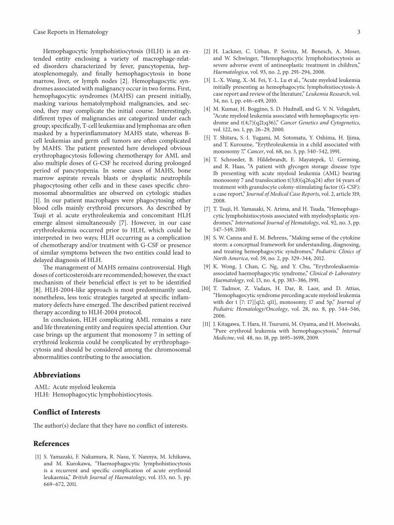
A 5-year-old boy was admitted to pediatric oncologydepartment with anemia, thrombocytopenia, and hep-atosplenomegaly. Bone marrow aspiration displayed ery-throid hyperplasia with erythroid precursors forming morethan 50% of the cell population. Megakaryocytes weredecreased in number. On repeat bone marrow aspirationperformed two weeks later, more than 50% of nucleated cellswere erythroblasts accompanied by myeloblast populationmore than 20%, compatible with AML6a. Chemotherapywith DAT protocol (daunorubicin, cytarabine, and thiogua-nine) was initiated. Following 2 courses of DAT therapy, pro-longed pancytopenia lasting for more than 2 months with noevidence of hematologic recovery was manifested. Persistentfever and huge splenomegaly and hepatomegaly (3 cm belowcostal margin) were new findings on physical examination.Bone marrow aspiration was performed which proved tobe severely hypocellular. A noticeable finding at this pointwas macrophages phagocytosing other blood cells mainlyerythroid precursors; however, no evidence of increased
2 Case Reports in Hematology
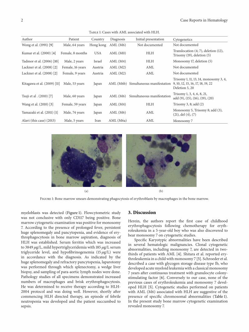
Table 1: Cases with AML associated with HLH.
Author Patient Country Diagnosis Initial presentation CytogeneticsWong et al. (1991) [9] Male, 64 years Hong kong AML (M6) Not documented Not documented
Kumar et al. (2000) [4] Female, 8 months USA AML (M0) HLH Translocation (4; 7), deletion (12),Trisomy (19), deletion (5)
Tadmor et al. (2006) [10] Male, 2 years Israel AML (M4) HLH Monosomy 17, deletion (5)Lackner et al. (2008) [2] Female, 16 years Austria AML (M2) AML Not documentedLackner et al. (2008) [2] Female, 9 years Austria AML (M2) AML Not documented
Kitagawa et al. (2009) [11] Male, 53 years Japan AML (M6b) Simultaneous manifestationTrisomy 1, 11, 13, 14, monosomy 3, 4,9, 10, 12, 15, 16, 17, 18, 19, 22Deletion 5, 20
Tsuji et al . (2010) [7] Male, 60 years Japan AML (M6) Simultaneous manifestation Trisomy 1, 3, 4, 6, 8, 21,add (9), (15), (16), (19), (20)
Wang et al. (2010) [3] Female, 59 years Japan AML (M4) HLH Trisomy 3, 8; add (2)
Yamazaki et al. (2011) [1] Male, 74 years Japan AML (M6) AML Monosomy 5, Trisomy 8; add (3),(21), del (4), (7)
Alavi (this case) (2013) Male, 5 years Iran AML (M6a) AML Monosomy 7
(a) (b)
Figure 1: Bone marrow smears demonstrating phagocytosis of erythroblasts by macrophages in the bone marrow.
myeloblasts was detected (Figure 1). Flowcytometric studywas not conclusive with only CD117 being positive. Bonemarrow cytogenetic examination was positive for monosomy7. According to the presence of prolonged fever, persistenthuge splenomegaly and pancytopenia, and evidence of ery-throphagocytosis in bone marrow aspiration, diagnosis ofHLH was established. Serum ferritin which was increasedto 3849 𝜇g/L, mild hypertriglyceridemia with 185𝜇g/L serumtriglyceride level, and hypofibrinogenemia (15𝜇g/L) werein accordance with the diagnosis. As indicated by thehuge splenomegaly and refractory pancytopenia, laparotomywas performed through which splenectomy, a wedge liverbiopsy, and sampling of para-aortic lymph nodes were done.Pathology studies of all specimens demonstrated increasednumbers of macrophages and brisk erythrophagocytosis.He was determined to receive therapy according to HLH-2004 protocol and was doing well. However, shortly aftercommencing HLH directed therapy, an episode of febrileneutropenia was developed and the patient succumbed tosepsis.
3. Discussion
Herein, the authors report the first case of childhooderythrophagocytosis following chemotherapy for eryth-roleukemia in a 5-year-old boy who was also discovered tobear monosomy 7 on cytogenetic studies.
Specific Karyotypic abnormalities have been describedin several hematologic malignancies. Clonal cytogeneticabnormalities, including monosomy 7, are detected in two-thirds of patients with AML [4]. Shitara et al. reported ery-throleukemia in a childwithmonosomy 7 [5]. Schroeder et al.described a case with glycogen storage disease type Ib, whodeveloped acutemyeloid leukemiawith a classicalmonosomy7 years after continuous treatment with granulocyte colony-stimulating factor [6]. Conversely to our case, none of theprevious cases of erythroleukemia and monosomy 7 devel-oped HLH [5]. Cytogenetic studies performed on patientswith AML (M6) associated with HLH are suggestive of thepresence of specific chromosomal abnormalities (Table 1).In the present study bone marrow cytogenetic examinationrevealed monosomy 7.
Case Reports in Hematology 3
Hemophagocytic lymphohistiocytosis (HLH) is an ex-tended entity enclosing a variety of macrophage-relat-ed disorders characterized by fever, pancytopenia, hep-atosplenomegaly, and finally hemophagocytosis in bonemarrow, liver, or lymph nodes [2]. Hemophagocytic syn-dromes associated withmalignancy occur in two forms. First,hemophagocytic syndromes (MAHS) can present initially,masking various hematolymphoid malignancies, and sec-ond, they may complicate the initial course. Interestingly,different types of malignancies are categorized under eachgroup; specifically, T-cell leukemias and lymphomas are oftenmasked by a hyperinflammatory MAHS state, whereas B-cell leukemias and germ cell tumors are often complicatedby MAHS. The patient presented here developed obviouserythrophagocytosis following chemotherapy for AML andalso multiple doses of G-CSF he received during prolongedperiod of pancytopenia. In some cases of MAHS, bonemarrow aspirate reveals blasts or dysplastic neutrophilsphagocytosing other cells and in these cases specific chro-mosomal abnormalities are observed on cytologic studies[1]. In our patient macrophages were phagocytosing otherblood cells mainly erythroid precursors. As described byTsuji et al. acute erythroleukemia and concomitant HLHemerge almost simultaneously [7]. However, in our caseerythroleukemia occurred prior to HLH, which could beinterpreted in two ways; HLH occurring as a complicationof chemotherapy and/or treatment with G-CSF or presenceof similar symptoms between the two entities could lead todelayed diagnosis of HLH.
The management of MAHS remains controversial. Highdoses of corticosteroids are recommended; however, the exactmechanism of their beneficial effect is yet to be identified[8]. HLH-2004-like approach is most predominantly used,nonetheless, less toxic strategies targeted at specific inflam-matory defects have emerged.The described patient receivedtherapy according to HLH-2004 protocol.
In conclusion, HLH complicating AML remains a rareand life threatening entity and requires special attention. Ourcase brings up the argument that monosomy 7 in setting oferythroid leukemia could be complicated by erythrophago-cytosis and should be considered among the chromosomalabnormalities contributing to the association.
Abbreviations
AML: Acute myeloid leukemiaHLH: Hemophagocytic lymphohistiocytosis.
Conflict of Interests
The author(s) declare that they have no conflict of interests.
References
[1] S. Yamazaki, F. Nakamura, R. Nasu, Y. Nannya, M. Ichikawa,and M. Kurokawa, “Haemophagocytic lymphohistiocytosisis a recurrent and specific complication of acute erythroidleukaemia,” British Journal of Haematology, vol. 153, no. 5, pp.669–672, 2011.
[2] H. Lackner, C. Urban, P. Sovinz, M. Benesch, A. Moser,and W. Schwinger, “Hemophagocytic lymphohistiocytosis assevere adverse event of antineoplastic treatment in children,”Haematologica, vol. 93, no. 2, pp. 291–294, 2008.
[3] L.-X. Wang, X.-M. Fei, Y.-L. Lu et al., “Acute myeloid leukemiainitially presenting as hemophagocytic lymphohistiocytosis-Acase report and review of the literature,” Leukemia Research, vol.34, no. 1, pp. e46–e49, 2010.
[4] M. Kumar, H. Boggino, S. D. Hudnall, and G. V. N. Velagaleti,“Acute myeloid leukemia associated with hemophagocytic syn-drome and t(4;7)(q21;q36),” Cancer Genetics and Cytogenetics,vol. 122, no. 1, pp. 26–29, 2000.
[5] T. Shitara, S.-I. Yugami, M. Sotomatu, Y. Oshima, H. Ijima,and T. Kuroume, “Erythroleukemia in a child associated withmonosomy 7,” Cancer, vol. 68, no. 3, pp. 540–542, 1991.
[6] T. Schroeder, B. Hildebrandt, E. Mayatepek, U. Germing,and R. Haas, “A patient with glycogen storage disease typeIb presenting with acute myeloid leukemia (AML) bearingmonosomy 7 and translocation t(3;8)(q26;q24) after 14 years oftreatment with granulocyte colony-stimulating factor (G-CSF):a case report,” Journal of Medical Case Reports, vol. 2, article 319,2008.
[7] T. Tsuji, H. Yamasaki, N. Arima, and H. Tsuda, “Hemophago-cytic lymphohistiocytosis associated with myelodysplastic syn-dromes,” International Journal of Hematology, vol. 92, no. 3, pp.547–549, 2010.
[8] S. W. Canna and E. M. Behrens, “Making sense of the cytokinestorm: a conceptual framework for understanding, diagnosing,and treating hemophagocytic syndromes,” Pediatric Clinics ofNorth America, vol. 59, no. 2, pp. 329–344, 2012.
[9] K. Wong, J. Chan, C. Ng, and Y. Chu, “Erythroleukaemia-associated haemophagocytic syndrome,” Clinical & LaboratoryHaematology, vol. 13, no. 4, pp. 383–386, 1991.
[10] T. Tadmor, Z. Vadazs, H. Dar, R. Laor, and D. Attias,“Hemophagocytic syndrome preceding acutemyeloid leukemiawith der t [7: 17][q12; q11], monosomy, 17 and 5p,” Journal ofPediatric Hematology/Oncology, vol. 28, no. 8, pp. 544–546,2006.
[11] J. Kitagawa, T. Hara, H. Tsurumi, M. Oyama, and H. Moriwaki,“Pure erythroid leukemia with hemophagocytosis,” InternalMedicine, vol. 48, no. 18, pp. 1695–1698, 2009.

Submit your manuscripts athttp://www.hindawi.com
Stem CellsInternational
Hindawi Publishing Corporationhttp://www.hindawi.com Volume 2014
Hindawi Publishing Corporationhttp://www.hindawi.com Volume 2014
MEDIATORSINFLAMMATION
of
Hindawi Publishing Corporationhttp://www.hindawi.com Volume 2014
Behavioural Neurology
EndocrinologyInternational Journal of
Hindawi Publishing Corporationhttp://www.hindawi.com Volume 2014
Hindawi Publishing Corporationhttp://www.hindawi.com Volume 2014
Disease Markers
Hindawi Publishing Corporationhttp://www.hindawi.com Volume 2014
BioMed Research International
OncologyJournal of
Hindawi Publishing Corporationhttp://www.hindawi.com Volume 2014
Hindawi Publishing Corporationhttp://www.hindawi.com Volume 2014
Oxidative Medicine and Cellular Longevity
Hindawi Publishing Corporationhttp://www.hindawi.com Volume 2014
PPAR Research
The Scientific World JournalHindawi Publishing Corporation http://www.hindawi.com Volume 2014
Immunology ResearchHindawi Publishing Corporationhttp://www.hindawi.com Volume 2014
Journal of
ObesityJournal of
Hindawi Publishing Corporationhttp://www.hindawi.com Volume 2014
Hindawi Publishing Corporationhttp://www.hindawi.com Volume 2014
Computational and Mathematical Methods in Medicine
OphthalmologyJournal of
Hindawi Publishing Corporationhttp://www.hindawi.com Volume 2014
Diabetes ResearchJournal of
Hindawi Publishing Corporationhttp://www.hindawi.com Volume 2014
Hindawi Publishing Corporationhttp://www.hindawi.com Volume 2014
Research and TreatmentAIDS
Hindawi Publishing Corporationhttp://www.hindawi.com Volume 2014
Gastroenterology Research and Practice
Hindawi Publishing Corporationhttp://www.hindawi.com Volume 2014
Parkinson’s Disease
Evidence-Based Complementary and Alternative Medicine
Volume 2014Hindawi Publishing Corporationhttp://www.hindawi.com