How I treat hemophagocytic lymphohistiocytosis - University of
Case ReportHemophagocytic Lymphohistiocytosis and GastrointestinalBleeding: What a Surgeon Should Know
S. Popeskou,1 M. Gavillet,2 N. Demartines,3 and D. Christoforidis1,4
1Department of Surgery, Lugano Regional Hospital, Switzerland2Department of Hematology, University Hospital of Lausanne, Switzerland3Department of Surgery, University Hospital of Lausanne, Switzerland4University Hospital of Lausanne, Switzerland
Correspondence should be addressed to S. Popeskou; [email protected]
Received 6 April 2015; Accepted 25 May 2015
Academic Editor: Fernando Turegano
Copyright © 2015 S. Popeskou et al. This is an open access article distributed under the Creative Commons Attribution License,which permits unrestricted use, distribution, and reproduction in any medium, provided the original work is properly cited.
This paper presents to the surgical community an unusual and often ignored cause of gastrointestinal bleeding. Hemophagocyticsyndrome or hemophagocytic lymphohistiocytosis (HLH) is a rare medical entity characterized by phagocytosis of red blood cells,leucocytes, platelets, and their precursors in the bone marrow by activated macrophages. When intestinal bleeding is present, themanagement is very challenging with extremely high mortality rates. Early diagnosis and treatment seem to be the most importantfactors for a successful outcome. We present two cases and review another 18 from the literature.
1. Introduction
Gastrointestinal (GI) bleeding is a common cause for hospitaladmission to surgical wards. In westernized countries, theannual incidence of upper GI bleeding is approximately 100−200/100,000 and for lower GI bleeding 20–30/100,000. Theestimated mortality ranges from 6% to 10% for upper GIbleeding and from2% to 4% for lowerGI bleeding [1–4].Mostepisodes of lower GI bleeding resolve spontaneously, whileothers can be treated endoscopically or by interventionalradiology. A minority of patients become hemodynamicallyunstable and require emergency surgery. In such cases, thesource of bleeding is not always clear and the surgeonmay face the difficult decision to perform extensive colonicresections, at a price of significant mortality rates (up to25%), without the certitude of controlling the source ofbleeding [5–7].Themost frequent causes of lowerGI bleedingare diverticulosis and arterial-venous malformations. Otherfrequent causes are colitis, neoplasms, inflammatory boweldisease, and hemorrhoids [8, 9].
In this paper, we report the dramatic cases of two patientswho presented massive lower GI bleeding originating fromdiffuse GI mucosal ulcerations secondary to acute Epstein
Barr virus (EBV) proliferation and hemophagocytic lympho-histiocytosis (HLH) and review the literature for GI bleedingassociated with this rare syndrome.
2. Case 1
A 26-year-old male patient, treated for Crohn’s disease withazathioprine (150mg/day) for 3 years, was admitted to theENT department of a community hospital for treatment of afebrile pharyngitis with 10-day cefuroxime. Lab tests revealedleucopenia (2.7 g/L, 89% neutrophils, 8% monocytes) and amild elevation of the liver enzymes. Clinically and radiolog-ically, splenomegaly was present. An active EBV infection(IgG and IgM positive) was diagnosed on hospital day 7and treatment with valacyclovir (1 gr × 3/day) was started.At the same day, the patient presented massive lower GIbleeding requiring admission to the ICU. The initial emer-gency colonoscopy revealed severe pancolitis with multipleulcerations which was attributed to a flare of Crohn’s colitis.Three days later (day 10), despite multiple transfusions, hebecame hemodynamically unstable due to continuous bleed-ing (CRP 121mg/L, procalcitonin 1.21mcg/L). The patientwas therefore transferred to our University Hospital. Upon
Hindawi Publishing CorporationCase Reports in SurgeryVolume 2015, Article ID 745848, 6 pageshttp://dx.doi.org/10.1155/2015/745848

2 Case Reports in Surgery
arrival, he developed a hypovolemic shock requiring massiveblood transfusions and an emergent total colectomy withileostomy was performed. Broad-spectrum antibiotherapywith piperacillin/tazobactam (4.5 gr × 4/day) and prednisone50mg/d was initiated in the immediate postoperative period.A thoracic CT-scan showed bilateral pulmonary infiltrates,which were attributed to transfusion related acute lung injury(TRALI). Blood tests revealed pancytopenia and increasinglevels of ferritin (2600mg/L) thought to be due to thecombination of inflammation and azathioprine treatment.Three days later (day 13), the patient was transferred back tothe ICU of the first hospital.
There, severe mononucleosis with persistent high EBVviremia despite antiviral treatment was diagnosed. Thepatient presented multiple episodes of upper GI bleedingthat required further repeated transfusions.Upper endoscopyrevealed hemorrhagic gastroduodenitis with multiple bleed-ing ulcers. Blood tests revealed a further increase of ferritin(up to 15,000𝜇g/L) and LDH (up to 4000U/L) togetherwith worsening pancytopenia and finally agranulocytosis.Repeated bone morrow biopsies finally revealed HLH withmacrophage activation syndrome and hemophagocytosis.The patient was treated with high doses of steroids alone andexhibited transient improvement but eventually expired fol-lowing multiple organ failure 12 days later. The biopsies fromgastric, duodenal, and colonic tissues all revealed an EBV-associated lymphoid proliferation. The autopsy concluded toa generalized EBV infection with lymphoid proliferation ofthe entire digestive tube and multiple organs, with associatedsecondary macrophage activation syndrome.
3. Case 2
A 61-year-old male patient in good general health presentedwith an episode of significant lower GI bleeding. The patienthad had a minor similar episode 4 months ago. One monthago, he had felt fatigue and repeated episodes of fever withoutany other specific symptoms.
Upon primary admission to another hospital, the patientwas tachycardic and slightly hypotensive but improved aftermassive hydration and was transferred to our center. Anupper endoscopy revealed only a small Mallory-Weiss typeulcer, which seemedunlikely to have been the source of bleed-ing. An angio-CT-scan of the abdomen revealed extensivediverticulosis with signs of transverse colitis but without aprecise source of bleeding. The patient was treated conserva-tively but, in the following days, he developed fever and animportant inflammatory syndrome (CRP 253mg/L, normalleucocyte count) with rhabdomyolysis, acute renal failure,and high levels of LDH (1451U/L) and ferritin (1200mg/L)along with anemia and progressive thrombocytopenia.Threedays after his admission, he once again became hemodynam-ically unstable due to recurrent massive low GI bleeding.From the time of patient’s entry to that point, he had receiveda total of 10 units of RBC. Emergency laparotomy withintraoperative colonoscopy and enteroscopy was performed.Findings were an important inflammatory status around thetransverse colon, a small intestinal perforation at the distalileum, and 10–20 ulcerated inflammatory lesions in the ileum,
one of which was actively bleeding. A resection of 60 cm ofileumwas carried out and the patientwas admitted to the ICUfor postoperative surveillance. Histopathology of the ileumrevealed a high grade NK-T lymphoma Epstein Barr virus(EBV) positive.
Two days after surgery, the patient became septic. A tho-racoabdominal CT-scan showed bilateral pleural effusionsbut no specific septic source. In the absence of radiolog-ical signs that would explain the patient’s septic status, a“second look” laparotomy was performed but no source ofsepsis was identified. Subsequently, the patient’s conditiondeteriorated rapidly. He developed disseminated intravascu-lar coagulation and finally a new episode of GI bleeding.Based on the above listed observations together with ferritinlevels at 15000mcg/L, HLH was suspected and a treatmentwith CHOP regimen (cyclophosphamide 750mg/m2 day1, doxorubicin 50mg/m2/day 1, Oncovin 1.4mg/m2/day 1,and prednisone 60mg/m2/day 1 to 5) plus etoposide wasinitiated. No bone marrow biopsies were performed asdeath due to hemodynamic instability followed the next day.Autopsy revealed a NK-T extranasal EBV + lymphoma withintestinal localization. Bone marrow presented with massivehemophagocytosis without any evidence of tumor infiltration(see Figure 1).
4. Discussion
Hemophagocytic lymphohistiocytosis (HLH) or hemopha-gocytic syndrome represents a rare pathophysiologic entitycharacterized by a hyperinflammatory state, cytokine dereg-ulation, impaired function of cytotoxic T-cells (CTL), andnatural killer (NK) cells in addition to hemophagocytosis(i.e., phagocytosis by activated macrophages of red bloodcells, leucocytes, platelets and their precursors in the bloodmarrow, spleen, or other lymphoid organs) [23, 24].
There is an inherited or familial form (FHLH) and anacquired or secondary HLH [25]. The FHLH incidence isestimated to be 0.12/100.000 children per year and has itsonset under the age of 1 year in the majority (70%–80%)of patients [26]. Patients with inherited immune deficiencieslike the Chediak-Higashi syndrome as well as the X-linkedproliferative syndrome are also at high risk to develop HLH.
Secondary HLH may occur at all ages [24] and is usuallydiagnosed in association with malignant (mostly with cuta-neous or anaplastic lymphomas), autoimmune, or infectiousdiseases. Infection associated with HLH is mostly associatedwith viral infections of the herpes group, in particular EBVand cytomegalovirus (CMV) [23, 25]. EBV induced HLHmay mimic or favor T-cell lymphoma and other EBV-linkedlymphoproliferative disorders [23]. Other nonviral agentssuch as bacteria, mycobacteria, protozoa, and fungi canmorerarely trigger HLH [24]. Cases of HLH after immunizationwith measles vaccine, probably in predisposed patients, havealso been documented [27].
The dominant clinical characteristics of HLH are persis-tent fever, usually unresponsive to antibiotics, splenomegalyand/or hepatomegaly, lymphadenopathies, icterus, rash,anorexia, and GI bleeding. Neurological symptoms, such as
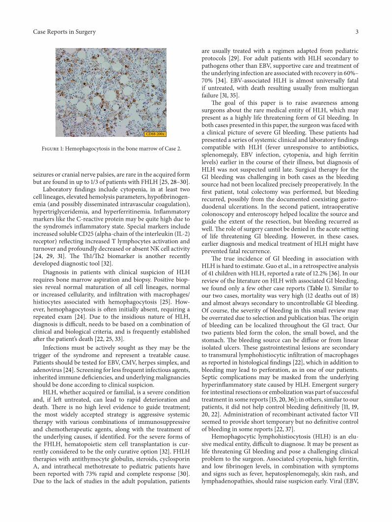
Case Reports in Surgery 3
CD68-200x
Figure 1: Hemophagocytosis in the bone marrow of Case 2.
seizures or cranial nerve palsies, are rare in the acquired formbut are found in up to 1/3 of patients with FHLH [25, 28–30].
Laboratory findings include cytopenia, in at least twocell lineages, elevated hemolysis parameters, hypofibrinogen-emia (and possibly disseminated intravascular coagulation),hypertriglyceridemia, and hyperferritinemia. Inflammatorymarkers like the C-reactive protein may be quite high due tothe syndrome’s inflammatory state. Special markers includeincreased soluble CD25 (alpha-chain of the interleukin (IL-2)receptor) reflecting increased T lymphocytes activation andturnover and profoundly decreased or absent NK cell activity[24, 29, 31]. The Th1/Th2 biomarker is another recentlydeveloped diagnostic tool [32].
Diagnosis in patients with clinical suspicion of HLHrequires bone marrow aspiration and biopsy. Positive biop-sies reveal normal maturation of all cell lineages, normalor increased cellularity, and infiltration with macrophages/histiocytes associated with hemophagocytosis [25]. How-ever, hemophagocytosis is often initially absent, requiring arepeated exam [24]. Due to the insidious nature of HLH,diagnosis is difficult, needs to be based on a combination ofclinical and biological criteria, and is frequently establishedafter the patient’s death [22, 25, 33].
Infections must be actively sought as they may be thetrigger of the syndrome and represent a treatable cause.Patients should be tested for EBV, CMV, herpes simplex, andadenovirus [24]. Screening for less frequent infectious agents,inherited immune deficiencies, and underlying malignanciesshould be done according to clinical suspicion.
HLH, whether acquired or familial, is a severe conditionand, if left untreated, can lead to rapid deterioration anddeath. There is no high level evidence to guide treatment;the most widely accepted strategy is aggressive systemictherapy with various combinations of immunosuppressiveand chemotherapeutic agents, along with the treatment ofthe underlying causes, if identified. For the severe forms ofthe FHLH, hematopoietic stem cell transplantation is cur-rently considered to be the only curative option [32]. FHLHtherapies with antithymocyte globulin, steroids, cyclosporinA, and intrathecal methotrexate to pediatric patients havebeen reported with 73% rapid and complete response [30].Due to the lack of studies in the adult population, patients
are usually treated with a regimen adapted from pediatricprotocols [29]. For adult patients with HLH secondary topathogens other than EBV, supportive care and treatment ofthe underlying infection are associatedwith recovery in 60%–70% [34]. EBV-associated HLH is almost universally fatalif untreated, with death resulting usually from multiorganfailure [31, 35].
The goal of this paper is to raise awareness amongsurgeons about the rare medical entity of HLH, which maypresent as a highly life threatening form of GI bleeding. Inboth cases presented in this paper, the surgeonwas faced witha clinical picture of severe GI bleeding. These patients hadpresented a series of systemic clinical and laboratory findingscompatible with HLH (fever unresponsive to antibiotics,splenomegaly, EBV infection, cytopenia, and high ferritinlevels) earlier in the course of their illness, but diagnosis ofHLH was not suspected until late. Surgical therapy for theGI bleeding was challenging in both cases as the bleedingsource had not been localized precisely preoperatively. In thefirst patient, total colectomy was performed, but bleedingrecurred, possibly from the documented coexisting gastro-duodenal ulcerations. In the second patient, intraoperativecolonoscopy and enteroscopy helped localize the source andguide the extent of the resection, but bleeding recurred aswell.The role of surgery cannot be denied in the acute settingof life threatening GI bleeding. However, in these cases,earlier diagnosis and medical treatment of HLH might haveprevented fatal recurrence.
The true incidence of GI bleeding in association withHLH is hard to estimate. Guo et al., in a retrospective analysisof 41 children with HLH, reported a rate of 12.2% [36]. In ourreview of the literature on HLH with associated GI bleeding,we found only a few other case reports (Table 1). Similar toour two cases, mortality was very high (12 deaths out of 18)and almost always secondary to uncontrollable GI bleeding.Of course, the severity of bleeding in this small review maybe overrated due to selection and publication bias.The originof bleeding can be localized throughout the GI tract. Ourtwo patients bled form the colon, the small bowel, and thestomach. The bleeding source can be diffuse or from linearisolated ulcers. These gastrointestinal lesions are secondaryto transmural lymphohistiocytic infiltration of macrophagesas reported in histological findings [22], which in addition tobleeding may lead to perforation, as in one of our patients.Septic complications may be masked from the underlyinghyperinflammatory state caused by HLH. Emergent surgeryfor intestinal resections or embolizationwas part of successfultreatment in some reports [15, 20, 36]; in others, similar to ourpatients, it did not help control bleeding definitively [11, 19,20, 22]. Administration of recombinant activated factor VIIseemed to provide short temporary but no definitive controlof bleeding in some reports [22, 37].
Hemophagocytic lymphohistiocytosis (HLH) is an elu-sive medical entity, difficult to diagnose. It may be present aslife threatening GI bleeding and pose a challenging clinicalproblem to the surgeon. Associated cytopenia, high ferritin,and low fibrinogen levels, in combination with symptomsand signs such as fever, hepatosplenomegaly, skin rash, andlymphadenopathies, should raise suspicion early. Viral (EBV,
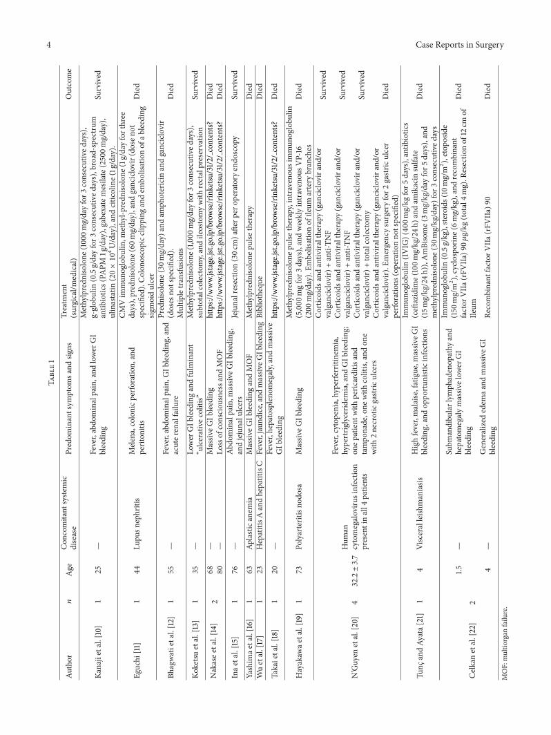
4 Case Reports in SurgeryTa
ble1
Author
𝑛Age
Con
comitant
syste
mic
disease
Predom
inantsym
ptom
sand
signs
Treatm
ent
(surgical/m
edical)
Outcome
Kanajietal.[10]
125
—Fever,abdo
minalpain,and
lower
GI
bleeding
Methylpredn
isolone
(100
0mg/dayfor3
consecutived
ays),
g-glob
ulin
(0.5g/dayfor3
consecutived
ays),broad-spectrum
antib
iotic
s(PA
PM1g/day),gabexatemesilate(2500m
g/day),
ulinastatin
(20×10
4U/day),andcitic
oline(1g/day).
Survived
Eguchi
[11]
144
Lupu
snephritis
Mele
na,colon
icperfo
ratio
n,and
periton
itis
CMVim
mun
oglobu
lin,m
ethyl-p
redn
isolone
(1g/dayforthree
days),prednisolone
(60m
g/day),and
ganciclovir(do
seno
tspecified).Colon
oscopicc
lipping
andem
bolisationof
ableeding
sigmoidulcer
Died
Bhagwatietal.[12]
155
—Fever,abdo
minalpain,G
Ibleeding,and
acuter
enalfailu
re
Prednisolone
(30m
g/day)
andam
photericin
andganciclovir
(doses
notspecified).
Multip
letransfu
sions
Died
Koketsu
etal.[13]
135
—Lo
wer
GIb
leedingandfulm
inant
“ulcerativec
olitis”
Methylpredn
isolone
(1,000
mg/dayfor3
consecutived
ays),
subtotalcolectom
y,andileostomywith
rectalpreservatio
nSurvived
Nakasee
tal.[14
]2
68—
Massiv
eGIb
leeding
https://w
ww.jstage.jst.go.jp/browse/rinketsu
/31/2
/contents?
Died
80—
Lossof
consciou
snessa
ndMOF
https://w
ww.jstage.jst.go.jp/browse/rinketsu
/31/2
/contents?
Died
Inae
tal.[15]
176
—Ab
dominalpain,m
assiv
eGIb
leeding,
andjejunalu
lcers
Jejunalresectio
n(30c
m)a
fterp
erop
eratoryendo
scop
ySurvived
Yashim
aetal.[16]
163
Aplasticanem
iaMassiv
eGIb
leedingandMOF
Methylpredn
isolone
pulse
therapy
Died
Wuetal.[17]
123
HepatitisA
andhepatitisC
Fever,jaun
dice,and
massiv
eGIb
leedingBibliotheque
Died
Takaietal.[18]
120
—Fever,hepatosplen
omegaly,andmassiv
eGIb
leeding
https://w
ww.jstage.jst.go.jp/browse/rinketsu
/31/2
/contents?
Died
Hayakaw
aetal.[19
]1
73Po
lyarteritisno
dosa
Massiv
eGIb
leeding
Methylpredn
isolone
pulse
therapy,intravenou
simmun
oglobu
lin(5,000
mgfor3
days),andweeklyintravenou
sVP-16
(200
mg/day).E
mbo
lisationof
ileum
artery
branches
Died
N’Guyen
etal.[20]
432.2±3.7
Hum
ancytomegaloviru
sinfectio
npresentinall4
patie
nts
Fever,cytopenia,hyperfe
rritinemia,
hypertrig
lycerid
emia,and
GIb
leeding;
onep
atient
with
peric
arditis
and
tampo
nade,one
with
colitis,
andon
ewith
2necroticgastric
ulcers
Cortic
oids
andantiv
iraltherapy
(ganciclo
vira
nd/or
valganciclo
vir)+anti-TN
FSurvived
Cortic
oids
andantiv
iraltherapy
(ganciclo
vira
nd/or
valganciclo
vir)+anti-TN
FSurvived
Cortic
oids
andantiv
iraltherapy
(ganciclo
vira
nd/or
valganciclo
vir)+totalcolectomy
Survived
Cortic
oids
andantiv
iraltherapy
(ganciclo
vira
nd/or
valganciclo
vir).E
mergencysurgeryfor2
gastric
ulcer
perfo
ratio
ns(operatio
nno
tspecified)
Died
Tunc
andAy
ata[
21]
14
Visceralleish
maniasis
Highfever,malaise,fatigue,m
assiv
eGI
bleeding
,and
oppo
rtun
isticinfections
Immun
oglobu
lin(IVIG
)(40
0mg/kg
for5
days),antib
iotic
s(ceft
azidim
e(100m
g/kg/24h
)and
amikacin
sulfate
(15m
g/kg/24h
)),A
mBisome(3m
g/kg/day
for5
days),and
methylpredn
isolone
(30m
g/kg/day)for
3consecutived
ays
Died
Celkanetal.[22]
21.5
—Subm
andibu
larlym
phadenop
athy
and
hepatomegalymassiv
elow
erGI
bleeding
Immun
oglobu
lin(0.5g/kg),ste
roids(10mg/m
2 ),etopo
side
(150
mg/m
2 ),cyclosporine(6m
g/kg),andrecombinant
factor
VIIa
(rFV
IIa)9
0𝜇g/kg
(total4
mg).R
esectio
nof
12cm
ofIle
um
Died
4—
Generalized
edem
aand
massiv
eGI
bleeding
Recombinant
factor
VIIa
(rFV
IIa)9
0Died
MOF:multio
rgan
failu
re.

Case Reports in Surgery 5
CMV, and herpes simplex virus) and other (Mycobacteriumtuberculosis and Mycoplasma pneumonia) infections shouldbe looked for as they may represent treatable causes. Treat-ment requires a multidisciplinary approach and surgery mayprovide a temporary solution for GI bleeding or perforation,but early diagnosis and systemic therapy are the only hope forcure.
Conflict of Interests
The authors declare that there is no conflict of interestsregarding the publication of this paper.
References
[1] C.M.Wilcox, B. L. Cryer, H. J. Henk et al., “Mortality associatedwith gastrointestinal bleeding events: comparing short-termclinical outcomes of patients hospitalized for upper GI bleedingand acute myocardial infarction in a US managed care setting,”Clinical and Experimental Gastroenterology, vol. 2, pp. 21–30,2009.
[2] G. F. Longstreth, “Epidemiology of hospitalization for acuteupper gastrointestinal hemorrhage: a population-based study,”The American Journal of Gastroenterology, vol. 90, no. 2, pp.206–210, 1995.
[3] R. T. Yavorski, R. K. H. Wong, C. Maydonovitch, L. S. Battin,A. Furnia, and D. E. Amundson, “Analysis of 3,294 cases ofupper gastrointestinal bleeding in military medical facilities,”The American Journal of Gastroenterology, vol. 90, no. 4, pp.568–573, 1995.
[4] D. R. Parker, X. Luo, J. J. Jalbert, and A. R. Assaf, “Impactof upper and lower gastrointestinal blood loss on healthcareutilization and costs: a systematic review,” Journal of MedicalEconomics, vol. 14, no. 3, pp. 279–287, 2011.
[5] W. Browder, E. J. Cerise, andM. S. Litwin, “Impact of emergencyangiography inmassive lower gastrointestinal bleeding,”Annalsof Surgery, vol. 204, no. 5, pp. 530–536, 1986.
[6] I. M. Leitman, D. E. Paull, and G. T. Shires III, “Evaluation andmanagement of massive lower gastrointestinal hemorrhage,”Annals of Surgery, vol. 209, no. 2, pp. 175–180, 1989.
[7] J. Lee, T. W. Costantini, and R. Coimbra, “Acute lower GIbleeding for the acute care surgeon: current diagnosis andmanagement,” Scandinavian Journal of Surgery, vol. 98, no. 3,pp. 135–142, 2009.
[8] C. Gayer, A. Chino, C. Lucas et al., “Acute lower gastrointestinalbleeding in 1,112 patients admitted to an urban emergencymedical center,” Surgery, vol. 146, no. 4, pp. 600–607, 2009.
[9] A. M. Vernava III, W. E. Longo, K. S. Virgo, and F. E. Johnson,“A nationwide study of the incidence and etiology of lowergastrointestinal bleeding,” Surgical Research Communications,vol. 18, no. 2, pp. 113–120, 1996.
[10] S. Kanaji, K.Okuma, Y. Tokumitsu, S. Yoshizawa,M.Nakamura,and Y. Niho, “Hemophagocytic syndrome associated withfulminant ulcerative colitis and presumed acute pancreatitis,”The American Journal of Gastroenterology, vol. 93, no. 10, pp.1956–1959, 1998.
[11] K. Eguchi, “Systemic lupus erythematosus complicated bycytomegalovirus-induced hemophagocytic syndrome and col-itis,” Internal Medicine, vol. 41, no. 2, pp. 77–78, 2002.
[12] N. S. Bhagwati, S. J. Oiseth, L. S. Abebe, and P. H. Wiernik,“Intravascular lymphoma associatedwith hemophagocytic syn-drome: a rare but aggressive clinical entity,” Annals of Hematol-ogy, vol. 83, no. 4, pp. 247–250, 2004.
[13] S.-I. Koketsu, T. Watanabe, N. Hori, N. Umetani, Y. Takazawa,and H. Nagawa, “Hemophagocytic syndrome caused by fulmi-nant ulcerative colitis and cytomegalovirus infection: report ofa case,” Diseases of the Colon & Rectum, vol. 47, no. 7, pp. 1250–1253, 2004.
[14] T. Nakase, K. Morita, M. Tomeoku, and M. Katou, “Hemopha-gocytic syndrome in two elderly men,” Rinsho Ketsueki, vol. 31,no. 2, pp. 258–259, 1990.
[15] S. Ina, M. Tani, K. Takifuji, S. Yamazoe, Y. Nakatani, andH. Yamaue, “Virus-associated hemophagocytic syndrome andhemorrhagic jejunal ulcer caused by cytomegalovirus infectionin a non-compromised host; a case report of unusual entity,”Hepato-Gastroenterology, vol. 51, no. 56, pp. 491–493, 2004.
[16] A. Yashima, Y. Narigasawa, Y. Ishida et al., “Hemophagocyticsyndrome due to miliary tuberculosis in the course of aplasticanemia,” Rinsho Ketsueki, vol. 39, no. 5, pp. 392–397, 1998.
[17] C.-S.Wu,K.-Y. Chang, P.Dunn, andT.-H. Lo, “Acute hepatitis Awith coexistent hepatitis C virus infection presenting as a virus-associated hemophagocytic syndrome: a case report,”AmericanJournal of Gastroenterology, vol. 90, no. 6, pp. 1002–1005, 1995.
[18] K. Takai, M. Sanada, and H. Shibuya, “Epstein-Barr virusassociated natural killer cell leukemia: report of an autopsycase,” Rinsho Ketsueki, vol. 36, no. 5, pp. 500–505, 1995.
[19] I. Hayakawa, F. Shirasaki, H. Ikeda et al., “Reactive hemophago-cytic syndrome in a patient with polyarteritis nodosa associatedwith Epstein-Barr virus reactivation,” Rheumatology Interna-tional, vol. 26, no. 6, pp. 573–576, 2006.
[20] Y. N’Guyen, S. Baumard, J. H. Salmon et al., “Cytomegalovirusassociated hemophagocytic lymphohistiocytosis in patients suf-fering from crohn’s disease treated by azathioprine: a series offour cases,” Inflammatory Bowel Diseases, vol. 17, no. 9, pp. E116–E118, 2011.
[21] B. Tunc and A. Ayata, “Hemophagocytic syndrome: a rare life-threatening complication of visceral leishmaniasis in a youngboy,” Pediatric Hematology and Oncology, vol. 18, no. 8, pp. 531–536, 2001.
[22] T. Celkan, S. Alhaj, M. Civilibal, and M. Elicevik, “Control ofbleeding associated with hemophagocytic syndrome in chil-dren: an audit of the clinical use of recombinant activated factorVII,” Pediatric Hematology and Oncology, vol. 24, no. 2, pp. 117–121, 2007.
[23] D. N. Fisman, “Hemophagocytic syndromes and infection,”Emerging Infectious Diseases, vol. 6, no. 6, pp. 601–608, 2000.
[24] G. E. Janka, “Hemophagocytic syndromes,” Blood Reviews, vol.21, no. 5, pp. 245–253, 2007.
[25] R. J. Arceci, “When T cells and macrophages do not talk: thehemophagocytic syndromes,” Current Opinion in Hematology,vol. 15, no. 4, pp. 359–367, 2008.
[26] J.-I. Henter, G. Elinder, O. Soder, and A. Ost, “Incidencein Sweden and clinical features of familial hemophagocyticlymphohistiocytosis,” Acta Paediatrica, vol. 80, no. 4, pp. 428–435, 1991.
[27] T. Otagiri, T. Mitsui, T. Kawakami et al., “Haemophagocyticlymphohistiocytosis following measles vaccination,” EuropeanJournal of Pediatrics, vol. 161, no. 9, pp. 494–496, 2002.
6 Case Reports in Surgery
[28] J. I. Henter, M. Arico, G. Elinder, S. Imashuku, and G. Janka,“FHLH. Primary hemophagocytic lymphohistiocytosis,”Hema-tology/Oncology Clinics of North America, vol. 12, no. 2, pp. 417–433, 1998.
[29] J.-I. Henter, A. Horne, M. Arico et al., “HLH-2004: diagnosticand therapeutic guidelines for hemophagocytic lymphohistio-cytosis,” Pediatric Blood and Cancer, vol. 48, no. 2, pp. 124–131,2007.
[30] N. Mahlaoui, M. Ouachee-Chardin, G. D. S. Basile et al.,“Immunotherapy of familial hemophagocytic lymphohistiocy-tosis with antithymocyte globulins: a single-center retrospectivereport of 38 patients,” Pediatrics, vol. 120, no. 3, pp. e622–e628,2007.
[31] J. Chen, G. Li, J. Lu et al., “A novel type of PTD, common helix-loop-helix motif, could efficiently mediate protein transductioninto mammalian cells,” Biochemical and Biophysical ResearchCommunications, vol. 347, no. 4, pp. 931–940, 2006.
[32] Y.-M.Tang andX.-J. Xu, “Advances in hemophagocytic lympho-histiocytosis: pathogenesis, early diagnosis/differential diagno-sis, and treatment,”The ScientificWorld Journal, vol. 11, pp. 697–708, 2011.
[33] G. E. Janka, “Familial and acquired hemophagocytic lympho-histiocytosis,” European Journal of Pediatrics, vol. 166, no. 2, pp.95–109, 2007.
[34] A. P. Reiner and J. L. Spivak, “Hematophagic histiocytosis.A report of 23 new patients and a review of the literature,”Medicine, vol. 67, no. 6, pp. 369–388, 1988.
[35] C. Cerboni, A. Zingoni,M. Cippitelli,M. Piccoli, L. Frati, andA.Santoni, “Antigen-activated human T lymphocytes express cell-surface NKG2D ligands via an ATM/ATR-dependent mech-anism and become susceptible to autologous NK-cell lysis,”Blood, vol. 110, no. 2, pp. 606–615, 2007.
[36] X. Guo, Q. Li, and C.-Y. Zhou, “Retrospective analysis of 41childhood hemophagocytic syndrome,” Zhonghua Xue Ye XueZa Zhi, vol. 28, no. 7, pp. 449–453, 2007.
[37] C. G. Millar, M. D. Stringer, I. Sugarman, and M. Richards,“The use of recombinant factor VIIa for bleeding in paediatricpractice,” Haemophilia, vol. 11, no. 2, pp. 171–174, 2005.

Submit your manuscripts athttp://www.hindawi.com
Stem CellsInternational
Hindawi Publishing Corporationhttp://www.hindawi.com Volume 2014
Hindawi Publishing Corporationhttp://www.hindawi.com Volume 2014
MEDIATORSINFLAMMATION
of
Hindawi Publishing Corporationhttp://www.hindawi.com Volume 2014
Behavioural Neurology
EndocrinologyInternational Journal of
Hindawi Publishing Corporationhttp://www.hindawi.com Volume 2014
Hindawi Publishing Corporationhttp://www.hindawi.com Volume 2014
Disease Markers
Hindawi Publishing Corporationhttp://www.hindawi.com Volume 2014
BioMed Research International
OncologyJournal of
Hindawi Publishing Corporationhttp://www.hindawi.com Volume 2014
Hindawi Publishing Corporationhttp://www.hindawi.com Volume 2014
Oxidative Medicine and Cellular Longevity
Hindawi Publishing Corporationhttp://www.hindawi.com Volume 2014
PPAR Research
The Scientific World JournalHindawi Publishing Corporation http://www.hindawi.com Volume 2014
Immunology ResearchHindawi Publishing Corporationhttp://www.hindawi.com Volume 2014
Journal of
ObesityJournal of
Hindawi Publishing Corporationhttp://www.hindawi.com Volume 2014
Hindawi Publishing Corporationhttp://www.hindawi.com Volume 2014
Computational and Mathematical Methods in Medicine
OphthalmologyJournal of
Hindawi Publishing Corporationhttp://www.hindawi.com Volume 2014
Diabetes ResearchJournal of
Hindawi Publishing Corporationhttp://www.hindawi.com Volume 2014
Hindawi Publishing Corporationhttp://www.hindawi.com Volume 2014
Research and TreatmentAIDS
Hindawi Publishing Corporationhttp://www.hindawi.com Volume 2014
Gastroenterology Research and Practice
Hindawi Publishing Corporationhttp://www.hindawi.com Volume 2014
Parkinson’s Disease
Evidence-Based Complementary and Alternative Medicine
Volume 2014Hindawi Publishing Corporationhttp://www.hindawi.com