FUJIFILM SonoSite, Inc. 0U 0DUN -RE Regulatory … · Table 1.3-1: Diagnostic Ultrasound...
26
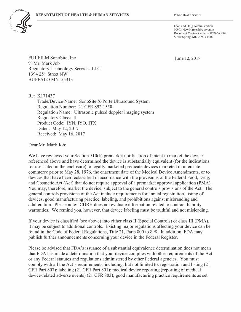
DEPARTMENT OF HEALTH & HUMAN SERVICES Public Health Service __________________________________________________________________________________________________________________________ Food and Drug Administration 10903 New Hampshire Avenue Document Control Center – WO66-G609 Silver Spring, MD 20993-0002 FUJIFILM SonoSite, Inc. Regulatory Technology Services LLC 1394 25 th Street NW BUFFALO MN 55313 Re: K171437 Trade/Device Name: SonoSite X-Porte Ultrasound System Regulation Number: 21 CFR 892.1550 Regulation Name: Ultrasonic pulsed doppler imaging system Regulatory Class: II Product Code: IYN, IYO, ITX Dated: May 12, 2017 Received: May 16, 2017 Dear Mr. Mark Job: We have reviewed your Section 510(k) premarket notification of intent to market the device referenced above and have determined the device is substantially equivalent (for the indications for use stated in the enclosure) to legally marketed predicate devices marketed in interstate commerce prior to May 28, 1976, the enactment date of the Medical Device Amendments, or to devices that have been reclassified in accordance with the provisions of the Federal Food, Drug, and Cosmetic Act (Act) that do not require approval of a premarket approval application (PMA). You may, therefore, market the device, subject to the general controls provisions of the Act. The general controls provisions of the Act include requirements for annual registration, listing of devices, good manufacturing practice, labeling, and prohibitions against misbranding and adulteration. Please note: CDRH does not evaluate information related to contract liability warranties. We remind you, however, that device labeling must be truthful and not misleading. If your device is classified (see above) into either class II (Special Controls) or class III (PMA), it may be subject to additional controls. Existing major regulations affecting your device can be found in the Code of Federal Regulations, Title 21, Parts 800 to 898. In addition, FDA may publish further announcements concerning your device in the Federal Register. Please be advised that FDA’s issuance of a substantial equivalence determination does not mean that FDA has made a determination that your device complies with other requirements of the Act or any Federal statutes and regulations administered by other Federal agencies. You must comply with all the Act’s requirements, including, but not limited to: registration and listing (21 CFR Part 807); labeling (21 CFR Part 801); medical device reporting (reporting of medical device-related adverse events) (21 CFR 803); good manufacturing practice requirements as set
Transcript of FUJIFILM SonoSite, Inc. 0U 0DUN -RE Regulatory … · Table 1.3-1: Diagnostic Ultrasound...
DEPARTMENT OF HEALTH & HUMAN SERVICES Public Health Service__________________________________________________________________________________________________________________________
Food and Drug Administration10903 New Hampshire AvenueDocument Control Center – WO66-G609Silver Spring, MD 20993-0002
FUJIFILM SonoSite, Inc.
Regulatory Technology Services LLC1394 25th Street NWBUFFALO MN 55313
Re: K171437 Trade/Device Name: SonoSite X-Porte Ultrasound System Regulation Number: 21 CFR 892.1550 Regulation Name: Ultrasonic pulsed doppler imaging systemRegulatory Class: IIProduct Code: IYN, IYO, ITXDated: May 12, 2017 Received: May 16, 2017
Dear Mr. Mark Job:
We have reviewed your Section 510(k) premarket notification of intent to market the device referenced above and have determined the device is substantially equivalent (for the indicationsfor use stated in the enclosure) to legally marketed predicate devices marketed in interstate commerce prior to May 28, 1976, the enactment date of the Medical Device Amendments, or to devices that have been reclassified in accordance with the provisions of the Federal Food, Drug, and Cosmetic Act (Act) that do not require approval of a premarket approval application (PMA). You may, therefore, market the device, subject to the general controls provisions of the Act. The general controls provisions of the Act include requirements for annual registration, listing of devices, good manufacturing practice, labeling, and prohibitions against misbranding and adulteration. Please note: CDRH does not evaluate information related to contract liability warranties. We remind you, however, that device labeling must be truthful and not misleading.
If your device is classified (see above) into either class II (Special Controls) or class III (PMA), it may be subject to additional controls. Existing major regulations affecting your device can be found in the Code of Federal Regulations, Title 21, Parts 800 to 898. In addition, FDA may publish further announcements concerning your device in the Federal Register.
Please be advised that FDA’s issuance of a substantial equivalence determination does not mean that FDA has made a determination that your device complies with other requirements of the Act or any Federal statutes and regulations administered by other Federal agencies. You must comply with all the Act’s requirements, including, but not limited to: registration and listing (21 CFR Part 807); labeling (21 CFR Part 801); medical device reporting (reporting of medical device-related adverse events) (21 CFR 803); good manufacturing practice requirements as set

Page 2—Mr. Mark Job
forth in the quality systems (QS) regulation (21 CFR Part 820); and if applicable, the electronic product radiation control provisions (Sections 531-542 of the Act); 21 CFR 1000-1050. If you desire specific advice for your device on our labeling regulation (21 CFR Part 801), please contact the Division of Industry and Consumer Education at its toll-free number (800) 638 2041 or (301) 796-7100 or at its Internet address http://www.fda.gov/MedicalDevices/ResourcesforYou/Industry/default.htm. Also, please note the regulation entitled, “Misbranding by reference to premarket notification” (21 CFR Part 807.97). For questions regarding the reporting of adverse events under the MDR regulation (21 CFR Part 803), please go to http://www.fda.gov/MedicalDevices/Safety/ReportaProblem/default.htm for the CDRH’s Office of Surveillance and Biometrics/Division of Postmarket Surveillance.
You may obtain other general information on your responsibilities under the Act from the Division of Industry and Consumer Education at its toll-free number (800) 638-2041 or (301) 796-7100 or at its Internet address http://www.fda.gov/MedicalDevices/ResourcesforYou/Industry/default.htm.
Sincerely yours,
Robert Ochs, Ph.D. Director Division of Radiological HealthOffice of In Vitro Diagnosticsand Radiological Health
Center for Devices and Radiological Health
Enclosure
y y
FORM FDA 3881 (8/14) Page 1 of 1 PSC Publishing Services (301) 443-6740 EF
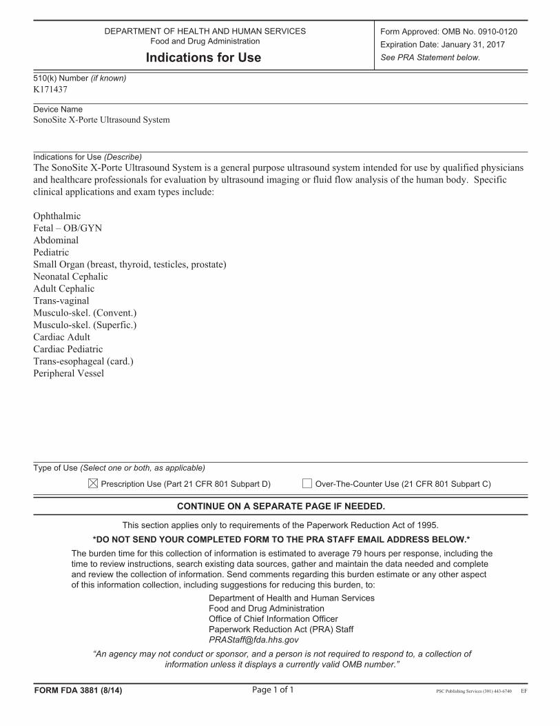
DEPARTMENT OF HEALTH AND HUMAN SERVICES Food and Drug Administration
Indications for Use
Form Approved: OMB No. 0910-0120Expiration Date: January 31, 2017See PRA Statement below.
510(k) Number (if known)K171437
Device NameSonoSite X-Porte Ultrasound System
Indications for Use (Describe)The SonoSite X-Porte Ultrasound System is a general purpose ultrasound system intended for use by qualified physicians and healthcare professionals for evaluation by ultrasound imaging or fluid flow analysis of the human body. Specific clinical applications and exam types include:
OphthalmicFetal – OB/GYN AbdominalPediatricSmall Organ (breast, thyroid, testicles, prostate) Neonatal Cephalic Adult Cephalic Trans-vaginalMusculo-skel. (Convent.) Musculo-skel. (Superfic.) Cardiac Adult Cardiac Pediatric Trans-esophageal (card.) Peripheral Vessel
Type of Use (Select one or both, as applicable)
Prescription Use (Part 21 CFR 801 Subpart D) Over-The-Counter Use (21 CFR 801 Subpart C)
CONTINUE ON A SEPARATE PAGE IF NEEDED.
This section applies only to requirements of the Paperwork Reduction Act of 1995.*DO NOT SEND YOUR COMPLETED FORM TO THE PRA STAFF EMAIL ADDRESS BELOW.*
The burden time for this collection of information is estimated to average 79 hours per response, including the time to review instructions, search existing data sources, gather and maintain the data needed and completeand review the collection of information. Send comments regarding this burden estimate or any other aspectof this information collection, including suggestions for reducing this burden, to:
Department of Health and Human ServicesFood and Drug AdministrationOffice of Chief Information OfficerPaperwork Reduction Act (PRA) [email protected]
“An agency may not conduct or sponsor, and a person is not required to respond to, a collection of information unless it displays a currently valid OMB number.”
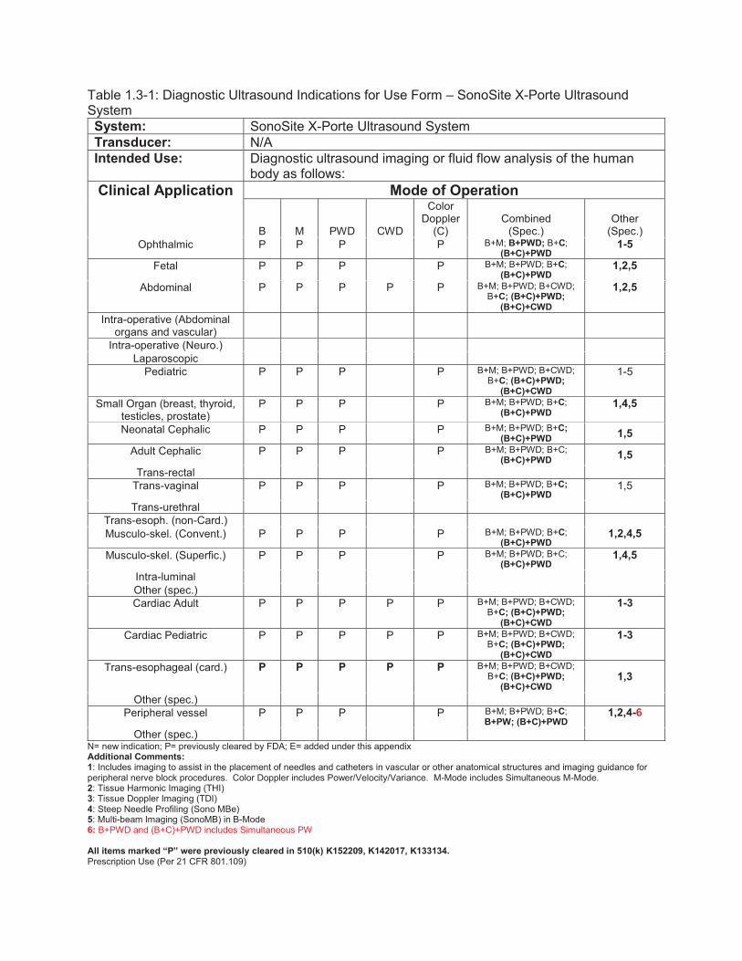
Table 1.3-1: Diagnostic Ultrasound Indications for Use Form – SonoSite X-Porte Ultrasound System System: SonoSite X-Porte Ultrasound System Transducer: N/A Intended Use: Diagnostic ultrasound imaging or fluid flow analysis of the human
body as follows: Clinical Application
Mode of Operation
B M PWD CWD
Color Doppler
(C) Combined
(Spec.) Other
(Spec.) Ophthalmic P P P P B+M; B+PWD; B+C;
(B+C)+PWD 1-5
Fetal P P P P B+M; B+PWD; B+C; (B+C)+PWD
1,2,5
Abdominal P P P P P B+M; B+PWD; B+CWD; B+C; (B+C)+PWD;
(B+C)+CWD
1,2,5
Intra-operative (Abdominal organs and vascular)
Intra-operative (Neuro.) Laparoscopic
Pediatric P P P P B+M; B+PWD; B+CWD; B+C; (B+C)+PWD;
(B+C)+CWD
1-5
Small Organ (breast, thyroid, testicles, prostate)
P P P P B+M; B+PWD; B+C; (B+C)+PWD
1,4,5
Neonatal Cephalic P P P P B+M; B+PWD; B+C; (B+C)+PWD 1,5
Adult Cephalic P P P P B+M; B+PWD; B+C; (B+C)+PWD 1,5
Trans-rectal Trans-vaginal P P P P B+M; B+PWD; B+C;
(B+C)+PWD 1,5
Trans-urethral Trans-esoph. (non-Card.) Musculo-skel. (Convent.) P P P P B+M; B+PWD; B+C;
(B+C)+PWD 1,2,4,5
Musculo-skel. (Superfic.) P P P P B+M; B+PWD; B+C; (B+C)+PWD
1,4,5
Intra-luminal Other (spec.) Cardiac Adult P P P P P B+M; B+PWD; B+CWD;
B+C; (B+C)+PWD; (B+C)+CWD
1-3
Cardiac Pediatric P P P P P B+M; B+PWD; B+CWD; B+C; (B+C)+PWD;
(B+C)+CWD
1-3
Trans-esophageal (card.) P P P P P B+M; B+PWD; B+CWD; B+C; (B+C)+PWD;
(B+C)+CWD 1,3
Other (spec.) Peripheral vessel P P P P B+M; B+PWD; B+C;
B+PW; (B+C)+PWD 1,2,4-6
Other (spec.) N= new indication; P= previously cleared by FDA; E= added under this appendix Additional Comments: 1: Includes imaging to assist in the placement of needles and catheters in vascular or other anatomical structures and imaging guidance for peripheral nerve block procedures. Color Doppler includes Power/Velocity/Variance. M-Mode includes Simultaneous M-Mode. 2: Tissue Harmonic Imaging (THI) 3: Tissue Doppler Imaging (TDI) 4: Steep Needle Profiling (Sono MBe) 5: Multi-beam Imaging (SonoMB) in B-Mode 6: B+PWD and (B+C)+PWD includes Simultaneous PW All items marked “P” were previously cleared in 510(k) K152209, K142017, K133134. Prescription Use (Per 21 CFR 801.109)
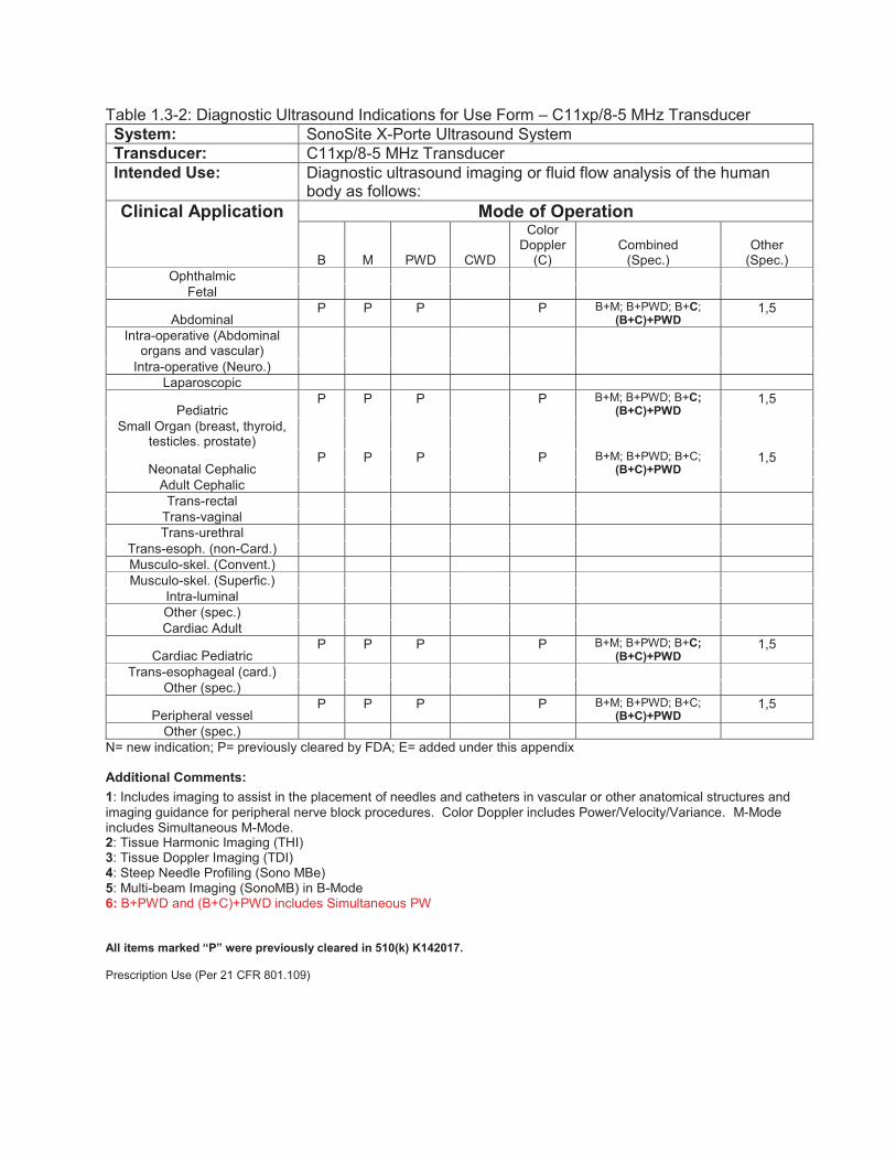
Table 1.3-2: Diagnostic Ultrasound Indications for Use Form – C11xp/8-5 MHz Transducer System: SonoSite X-Porte Ultrasound System Transducer: C11xp/8-5 MHz Transducer Intended Use: Diagnostic ultrasound imaging or fluid flow analysis of the human
body as follows: Clinical Application
Mode of Operation
B M PWD CWD
Color Doppler
(C) Combined
(Spec.) Other
(Spec.) Ophthalmic
Fetal
Abdominal P P P P B+M; B+PWD; B+C;
(B+C)+PWD 1,5
Intra-operative (Abdominal organs and vascular)
Intra-operative (Neuro.) Laparoscopic
Pediatric P P P P B+M; B+PWD; B+C;
(B+C)+PWD 1,5
Small Organ (breast, thyroid, testicles. prostate)
Neonatal Cephalic P P P P B+M; B+PWD; B+C;
(B+C)+PWD 1,5
Adult Cephalic Trans-rectal
Trans-vaginal Trans-urethral
Trans-esoph. (non-Card.) Musculo-skel. (Convent.) Musculo-skel. (Superfic.)
Intra-luminal Other (spec.) Cardiac Adult
Cardiac Pediatric P P P P B+M; B+PWD; B+C;
(B+C)+PWD 1,5
Trans-esophageal (card.) Other (spec.)
Peripheral vessel P P P P B+M; B+PWD; B+C;
(B+C)+PWD 1,5
Other (spec.) N= new indication; P= previously cleared by FDA; E= added under this appendix Additional Comments: 1: Includes imaging to assist in the placement of needles and catheters in vascular or other anatomical structures and imaging guidance for peripheral nerve block procedures. Color Doppler includes Power/Velocity/Variance. M-Mode includes Simultaneous M-Mode. 2: Tissue Harmonic Imaging (THI) 3: Tissue Doppler Imaging (TDI) 4: Steep Needle Profiling (Sono MBe) 5: Multi-beam Imaging (SonoMB) in B-Mode 6: B+PWD and (B+C)+PWD includes Simultaneous PW All items marked “P” were previously cleared in 510(k) K142017. Prescription Use (Per 21 CFR 801.109)

Table 1.3-3: Diagnostic Ultrasound Indications for Use Form – C35xp/8-3 MHz Transducer System: SonoSite X-Porte Ultrasound System Transducer: C35xp/8-3 MHz Transducer Intended Use: Diagnostic ultrasound imaging or fluid flow analysis of the human
body as follows: Clinical Application
Mode of Operation
B M PWD CWD
Color Doppler
(C) Combined
(Spec.) Other
(Spec.) Ophthalmic
Fetal
Abdominal P P P P B+M; B+PWD; B+C;
(B+C)+PWD 1,2,5 Intra-operative (Abdominal
organs and vascular)
Intra-operative (Neuro.) Laparoscopic
Pediatric P P P P B+M; B+PWD; B+C;
(B+C)+PWD 1,2,4,5 Small Organ (breast, thyroid,
testicles. prostate)
Neonatal Cephalic Adult Cephalic
Trans-rectal Trans-vaginal Trans-urethral
Trans-esoph. (non-Card.)
Musculo-skel. (Convent.) P P P P B+M; B+PWD; B+C;
(B+C)+PWD 1,2,4,5 Musculo-skel. (Superfic.)
Intra-luminal Other (spec.) Cardiac Adult
Cardiac Pediatric Trans-esophageal (card.)
Other (spec.)
Peripheral vessel P P P P B+M; B+PWD; B+C;
(B+C)+PWD 1,2,4,5 Other (spec.)
N= new indication; P= previously cleared by FDA; E= added under this appendix Additional Comments: 1: Includes imaging to assist in the placement of needles and catheters in vascular or other anatomical structures and imaging guidance for peripheral nerve block procedures. Color Doppler includes Power/Velocity/Variance. M-Mode includes Simultaneous M-Mode. 2: Tissue Harmonic Imaging (THI) 3: Tissue Doppler Imaging (TDI) 4: Steep Needle Profiling (Sono MBe) 5: Multi-beam Imaging (SonoMB) in B-Mode 6: B+PWD and (B+C)+PWD includes Simultaneous PW All items marked “P” were previously cleared in 510(k) K142017. Prescription Use (Per 21 CFR 801.109)
Table 1.3-4: Diagnostic Ultrasound Indications for Use Form – C60xp/5-2 MHz Transducer System: SonoSite X-Porte Ultrasound System Transducer: C60xp/5-2 MHz Transducer Intended Use: Diagnostic ultrasound imaging or fluid flow analysis of the human
body as follows: Clinical Application
Mode of Operation
B M PWD CWD
Color Doppler
(C) Combined
(Spec.) Other
(Spec.) Ophthalmic
Fetal P P P P B+M; B+PWD; B+C;
(B+C)+PWD 1,2,5
Abdominal P P P P B+M; B+PWD; B+C;
(B+C)+PWD 1,2,5 Intra-operative (Abdominal
organs and vascular)
Intra-operative (Neuro.) Laparoscopic
Pediatric P P P P B+M; B+PWD; B+C;
(B+C)+PWD 1,2,4,5 Small Organ (breast, thyroid,
testicles, prostate)
Neonatal Cephalic Adult Cephalic
Trans-rectal Trans-vaginal Trans-urethral
Trans-esoph. (non-Card.)
Musculo-skel. (Convent.) P P P P B+M; B+PWD; B+C;
(B+C)+PWD 1,2,4,5 Musculo-skel. (Superfic.)
Intra-luminal Other (spec.) Cardiac Adult
Cardiac Pediatric Trans-esophageal (card.)
Other (spec.)
Peripheral vessel P P P P B+M; B+PWD; B+C;
(B+C)+PWD 1,2,4,5 Other (spec.)
N= new indication; P= previously cleared by FDA; E= added under this appendix Additional Comments: 1: Includes imaging to assist in the placement of needles and catheters in vascular or other anatomical structures and imaging guidance for peripheral nerve block procedures. Color Doppler includes Power/Velocity/Variance. M-Mode includes Simultaneous M-Mode. 2: Tissue Harmonic Imaging (THI) 3: Tissue Doppler Imaging (TDI) 4: Steep Needle Profiling (Sono MBe) 5: Multi-beam Imaging (SonoMB) in B-Mode 6: B+PWD and (B+C)+PWD includes Simultaneous PW All items marked “P” were previously cleared in 510(k) K133134. Prescription Use (Per 21 CFR 801.109)
Table 1.3-5: Diagnostic Ultrasound Indications for Use Form – D2xp/2 MHz Transducer System: SonoSite X-Porte Ultrasound System Transducer: D2xp/2 MHz Transducer Intended Use: Diagnostic ultrasound imaging or fluid flow analysis of the human
body as follows: Clinical Application
Mode of Operation
B M PWD CWD
Color Doppler
(C) Combined
(Spec.) Other
(Spec.) Ophthalmic
Fetal Abdominal
Intra-operative (Abdominal organs and vascular)
Intra-operative (Neuro.) Laparoscopic
Pediatric Small Organ (breast, thyroid,
testicles, prostate)
Neonatal Cephalic Adult Cephalic
Trans-rectal Trans-vaginal Trans-urethral
Trans-esoph. (non-Card.) Musculo-skel. (Convent.) Musculo-skel. (Superfic.)
Intra-luminal Other (spec.) Cardiac Adult E
Cardiac Pediatric E Trans-esophageal (card.)
Other (spec.) Peripheral vessel
Other (spec.) N= new indication; P= previously cleared by FDA; E= added under this appendix
Additional Comments: 1: Includes imaging to assist in the placement of needles and catheters in vascular or other anatomical structures and imaging guidance for peripheral nerve block procedures. Color Doppler includes Power/Velocity/Variance. M-Mode includes Simultaneous M-Mode. 2: Tissue Harmonic Imaging (THI) 3: Tissue Doppler Imaging (TDI) 4: Steep Needle Profiling (Sono MBe) 5: Multi-beam Imaging (SonoMB) in B-Mode 6: B+PWD and (B+C)+PWD includes Simultaneous PW All items marked “E” were released via Letter to File/Non-filing Justification documentation since K152209. Prescription Use (Per 21 CFR 801.109)
Table 1.3-6: Diagnostic Ultrasound Indications for Use Form – HFL38xp/13-6 MHz Transducer System: SonoSite X-Porte Ultrasound System Transducer: HFL38xp/13-6 MHz Transducer Intended Use: Diagnostic ultrasound imaging or fluid flow analysis of the human
body as follows: Clinical Application
Mode of Operation
B M PWD CWD
Color Doppler
(C) Combined
(Spec.) Other
(Spec.) Ophthalmic
Fetal
Abdominal P P P
P B+M; B+PWD; B+C;
(B+C)+PWD 1,4,5
Intra-operative (Abdominal organs and vascular)
Intra-operative (Neuro.) Laparoscopic
Pediatric P P P
P B+M; B+PWD; B+C;
(B+C)+PWD 1,4,5
Small Organ (breast, thyroid, testicles. prostate)
P P P
P B+M; B+PWD; B+C; (B+C)+PWD
1,4,5
Neonatal Cephalic Adult Cephalic
Trans-rectal Trans-vaginal Trans-urethral
Trans-esoph. (non-Card.)
Musculo-skel. (Convent.) P P P
P B+M; B+PWD; B+C;
(B+C)+PWD 1,4,5
Musculo-skel. (Superfic.) P P P
P B+M; B+PWD; B+C;
(B+C)+PWD 1,4,5
Intra-luminal Other (spec.)
Cardiac Adult P P P
P B+M; B+PWD; B+C;
(B+C)+PWD 1,5
Cardiac Pediatric P P P
P B+M; B+PWD; B+C;
(B+C)+PWD 1,5 Trans-esophageal (card.)
Other (spec.)
Peripheral vessel P P P
P B+M; B+PWD; B+C;
B+PW; (B+C)+PWD 1,4,5,6
Other (spec.) N= new indication; P= previously cleared by FDA; E= added under this appendix Additional Comments: 1: Includes imaging to assist in the placement of needles and catheters in vascular or other anatomical structures and imaging guidance for peripheral nerve block procedures. Color Doppler includes Power/Velocity/Variance. M-Mode includes Simultaneous M-Mode. 2: Tissue Harmonic Imaging (THI) 3: Tissue Doppler Imaging (TDI) 4: Steep Needle Profiling (Sono MBe) 5: Multi-beam Imaging (SonoMB) in B-Mode 6: B+PWD and (B+C)+PWD includes Simultaneous PW All items marked “P” were previously cleared in 510(k) K152209 and K142017. Prescription Use (Per 21 CFR 801.109)
Table 1.3-7: Diagnostic Ultrasound Indications for Use Form – HFL50xp/15-6 MHz Transducer System: SonoSite X-Porte Ultrasound System Transducer: HFL50xp/15-6 MHz Transducer Intended Use: Diagnostic ultrasound imaging or fluid flow analysis of the human
body as follows: Clinical Application
Mode of Operation
B M PWD CWD
Color Doppler
(C) Combined
(Spec.) Other
(Spec.) Ophthalmic
Fetal
Abdominal P P P
P B+M; B+PWD; B+C;
(B+C)+PWD 1,4,5 Intra-operative (Abdominal
organs and vascular)
Intra-operative (Neuro.) Laparoscopic
Pediatric P P P
P B+M; B+PWD; B+C;
(B+C)+PWD 1,4,5 Small Organ (breast, thyroid,
testicles. prostate) P P P
P B+M; B+PWD; B+C;
(B+C)+PWD 1,4,5
Neonatal Cephalic Adult Cephalic
Trans-rectal Trans-vaginal Trans-urethral
Trans-esoph. (non-Card.)
Musculo-skel. (Convent.) P P P
P B+M; B+PWD; B+C;
(B+C)+PWD 1,4,5
Musculo-skel. (Superfic.) P P P
P B+M; B+PWD; B+C;
(B+C)+PWD 1,4,5 Intra-luminal Other (spec.) Cardiac Adult
Cardiac Pediatric Trans-esophageal (card.)
Other (spec.)
Peripheral vessel P P P
P B+M; B+PWD; B+C;
(B+C)+PWD 1,4,5 Other (spec.)
N= new indication; P= previously cleared by FDA; E= added under this appendix Additional Comments: 1: Includes imaging to assist in the placement of needles and catheters in vascular or other anatomical structures and imaging guidance for peripheral nerve block procedures. Color Doppler includes Power/Velocity/Variance. M-Mode includes Simultaneous M-Mode. 2: Tissue Harmonic Imaging (THI) 3: Tissue Doppler Imaging (TDI) 4: Steep Needle Profiling (Sono MBe) 5: Multi-beam Imaging (SonoMB) in B-Mode 6: B+PWD and (B+C)+PWD includes Simultaneous PW All items marked “P” were previously cleared in 510(k) K133134. Prescription Use (Per 21 CFR 801.109)

Table 1.3-8: Diagnostic Ultrasound Indications for Use Form – HSL25xp/13-6 MHz Transducer System: SonoSite X-Porte Ultrasound System Transducer: HSL25xp/13-6 MHz Transducer Intended Use: Diagnostic ultrasound imaging or fluid flow analysis of the human
body as follows: Clinical Application
Mode of Operation
B M PWD CWD
Color Doppler
(C) Combined
(Spec.) Other
(Spec.)
Ophthalmic P P P P B+M; B+PWD; B+C;
(B+C)+PWD 1,4,5
Fetal
Abdominal P P P P B+M; B+PWD; B+C;
(B+C)+PWD 1,4,5
Intra-operative (Abdominal organs and vascular)
Intra-operative (Neuro.) Laparoscopic
Pediatric P P P P B+M; B+PWD; B+C;
(B+C)+PWD 1,4,5
Small Organ (breast, thyroid, testicles. prostate)
P P P P B+M; B+PWD; B+C
1,4,5
Neonatal Cephalic Adult Cephalic
Trans-rectal Trans-vaginal Trans-urethral
Trans-esoph. (non-Card.)
Musculo-skel. (Convent.) P P P P B+M; B+PWD; B+C;
(B+C)+PWD 1,4,5
Musculo-skel. (Superfic.) P P P P B+M; B+PWD; B+C 1,4,5 Intra-luminal Other (spec.)
Cardiac Adult P P P P B+M; B+PWD; B+C;
(B+C)+PWD 1,5
Cardiac Pediatric P P P P B+M; B+PWD; B+C;
(B+C)+PWD 1,5
Trans-esophageal (card.) Other (spec.)
Peripheral vessel P P P P B+M; B+PWD; B+C;
(B+C)+PWD 1,4,5,6
Other (spec.) N= new indication; P= previously cleared by FDA; E= added under this appendix Additional Comments: 1: Includes imaging to assist in the placement of needles and catheters in vascular or other anatomical structures and imaging guidance for peripheral nerve block procedures. Color Doppler includes Power/Velocity/Variance. M-Mode includes Simultaneous M-Mode. 2: Tissue Harmonic Imaging (THI) 3: Tissue Doppler Imaging (TDI) 4: Steep Needle Profiling (Sono MBe) 5: Multi-beam Imaging (SonoMB) in B-Mode 6: B+PWD and (B+C)+PWD includes Simultaneous PW All items marked “P” were previously cleared in 510(k) K142017. Prescription Use (Per 21 CFR 801.109)
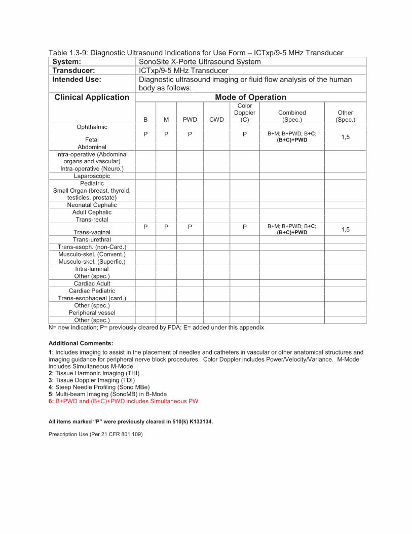
Table 1.3-9: Diagnostic Ultrasound Indications for Use Form – ICTxp/9-5 MHz Transducer System: SonoSite X-Porte Ultrasound System Transducer: ICTxp/9-5 MHz Transducer Intended Use: Diagnostic ultrasound imaging or fluid flow analysis of the human
body as follows: Clinical Application
Mode of Operation
B M PWD CWD
Color Doppler
(C) Combined
(Spec.) Other
(Spec.) Ophthalmic
Fetal P P P P B+M; B+PWD; B+C;
(B+C)+PWD 1,5 Abdominal
Intra-operative (Abdominal organs and vascular)
Intra-operative (Neuro.) Laparoscopic
Pediatric Small Organ (breast, thyroid,
testicles, prostate)
Neonatal Cephalic Adult Cephalic
Trans-rectal
Trans-vaginal P P P P B+M; B+PWD; B+C;
(B+C)+PWD 1,5 Trans-urethral
Trans-esoph. (non-Card.) Musculo-skel. (Convent.) Musculo-skel. (Superfic.)
Intra-luminal Other (spec.) Cardiac Adult
Cardiac Pediatric Trans-esophageal (card.)
Other (spec.) Peripheral vessel
Other (spec.) N= new indication; P= previously cleared by FDA; E= added under this appendix Additional Comments: 1: Includes imaging to assist in the placement of needles and catheters in vascular or other anatomical structures and imaging guidance for peripheral nerve block procedures. Color Doppler includes Power/Velocity/Variance. M-Mode includes Simultaneous M-Mode. 2: Tissue Harmonic Imaging (THI) 3: Tissue Doppler Imaging (TDI) 4: Steep Needle Profiling (Sono MBe) 5: Multi-beam Imaging (SonoMB) in B-Mode 6: B+PWD and (B+C)+PWD includes Simultaneous PW All items marked “P” were previously cleared in 510(k) K133134. Prescription Use (Per 21 CFR 801.109)
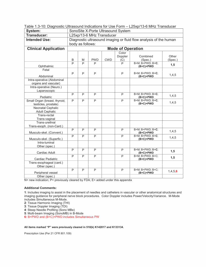
Table 1.3-10: Diagnostic Ultrasound Indications for Use Form – L25xp/13-6 MHz Transducer System: SonoSite X-Porte Ultrasound System Transducer: L25xp/13-6 MHz Transducer Intended Use: Diagnostic ultrasound imaging or fluid flow analysis of the human
body as follows: Clinical Application
Mode of Operation
B M PWD CWD
Color Doppler
(C) Combined
(Spec.) Other
(Spec.)
Ophthalmic P P P P B+M; B+PWD; B+C;
(B+C)+PWD 1,5 Fetal
Abdominal P P P P B+M; B+PWD; B+C;
(B+C)+PWD 1,4,5 Intra-operative (Abdominal
organs and vascular)
Intra-operative (Neuro.) Laparoscopic
Pediatric P P P P B+M; B+PWD; B+C;
(B+C)+PWD 1,4,5 Small Organ (breast, thyroid,
testicles, prostate) P P P P B+M; B+PWD; B+C;
(B+C)+PWD 1,4,5
Neonatal Cephalic Adult Cephalic
Trans-rectal Trans-vaginal Trans-urethral
Trans-esoph. (non-Card.)
Musculo-skel. (Convent.) P P P P B+M; B+PWD; B+C;
(B+C)+PWD 1,4,5
Musculo-skel. (Superfic.) P P P P B+M; B+PWD; B+C;
(B+C)+PWD 1,4,5 Intra-luminal Other (spec.)
Cardiac Adult P P P P B+M; B+PWD; B+C;
(B+C)+PWD 1,5
Cardiac Pediatric P P P P B+M; B+PWD; B+C;
(B+C)+PWD 1,5 Trans-esophageal (card.)
Other (spec.)
Peripheral vessel P P P P B+M; B+PWD; B+C;
(B+C)+PWD 1,4,5,6
Other (spec.) N= new indication; P= previously cleared by FDA; E= added under this appendix Additional Comments: 1: Includes imaging to assist in the placement of needles and catheters in vascular or other anatomical structures and imaging guidance for peripheral nerve block procedures. Color Doppler includes Power/Velocity/Variance. M-Mode includes Simultaneous M-Mode. 2: Tissue Harmonic Imaging (THI) 3: Tissue Doppler Imaging (TDI) 4: Steep Needle Profiling (Sono MBe) 5: Multi-beam Imaging (SonoMB) in B-Mode 6: B+PWD and (B+C)+PWD includes Simultaneous PW All items marked “P” were previously cleared in 510(k) K142017 and K133134. Prescription Use (Per 21 CFR 801.109)

Table 1.3-11: Diagnostic Ultrasound Indications for Use Form – L38xp/10-5 MHz Transducer System: SonoSite X-Porte Ultrasound System Transducer: L38xp/10-5 MHz Transducer Intended Use: Diagnostic ultrasound imaging or fluid flow analysis of the human
body as follows: Clinical Application
Mode of Operation
B M PWD CWD
Color Doppler
(C) Combined
(Spec.) Other
(Spec.) Ophthalmic
Fetal
Abdominal P P P
P B+M; B+PWD; B+C;
(B+C)+PWD 1,4,5 Intra-operative (Abdominal
organs and vascular)
Intra-operative (Neuro.) Laparoscopic
Pediatric P P P
P B+M; B+PWD; B+C;
(B+C)+PWD 1,4,5 Small Organ (breast, thyroid,
testicles. prostate) P P P
P B+M; B+PWD; B+C;
(B+C)+PWD 1,4,5
Neonatal Cephalic Adult Cephalic
Trans-rectal Trans-vaginal Trans-urethral
Trans-esoph. (non-Card.)
Musculo-skel. (Convent.) P P P
P B+M; B+PWD; B+C;
(B+C)+PWD 1,4,5
Musculo-skel. (Superfic.) P P P
P B+M; B+PWD; B+C;
(B+C)+PWD 1,4,5 Intra-luminal Other (spec.)
Cardiac Adult P P P P B+M; B+PWD; B+C;
(B+C)+PWD 1,5
Cardiac Pediatric P P P P B+M; B+PWD; B+C;
(B+C)+PWD 1,5 Trans-esophageal (card.)
Other (spec.)
Peripheral vessel P P P
P B+M; B+PWD; B+C;
(B+C)+PWD 1,4,5,6
Other (spec.) N= new indication; P= previously cleared by FDA; E= added under this appendix Additional Comments: 1: Includes imaging to assist in the placement of needles and catheters in vascular or other anatomical structures and imaging guidance for peripheral nerve block procedures. Color Doppler includes Power/Velocity/Variance. M-Mode includes Simultaneous M-Mode. 2: Tissue Harmonic Imaging (THI) 3: Tissue Doppler Imaging (TDI) 4: Steep Needle Profiling (Sono MBe) 5: Multi-beam Imaging (SonoMB) in B-Mode 6: B+PWD and (B+C)+PWD includes Simultaneous PW All items marked “P” were previously cleared in 510(k) K142017 and K133134. Prescription Use (Per 21 CFR 801.109)

Table 1.3-12: Diagnostic Ultrasound Indications for Use Form – P10xp/8-4 MHz Transducer System: SonoSite X-Porte Ultrasound System Transducer: P10xp/8-4 MHz Transducer Intended Use: Diagnostic ultrasound imaging or fluid flow analysis of the human
body as follows: Clinical Application
Mode of Operation
B M PWD CWD
Color Doppler
(C) Combined
(Spec.) Other
(Spec.) Ophthalmic
Fetal P P P P B+M; B+PWD; B+C;
(B+C)+PWD 1
Abdominal P P P P B+M; B+PWD; B+CWD;
B+C; (B+C)+PWD 1
Intra-operative (Abdominal organs and vascular)
Intra-operative (Neuro.) Laparoscopic
Pediatric P P P P B+M; B+PWD; B+C;
(B+C)+PWD 1
Small Organ (breast, thyroid, testicles. prostate)
P P P P B+M; B+PWD; B+C; (B+C)+PWD
1
Neonatal Cephalic P P P P B+M; B+PWD; B+C;
(B+C)+PWD 1
Adult Cephalic Trans-rectal
Trans-vaginal Trans-urethral
Trans-esoph. (non-Card.)
Musculo-skel. (Convent.) P P P P B+M; B+PWD; B+C;
(B+C)+PWD 1
Musculo-skel. (Superfic.) Intra-luminal Other (spec.)
Cardiac Adult
P P P P P B+M; B+PWD; B+CWD; B+C; (B+C)+PWD;
(B+C)+CWD
1,3
Cardiac Pediatric
P P P P P B+M; B+PWD; B+CWD; B+C; (B+C)+PWD;
(B+C)+CWD
1,3
Trans-esophageal (card.) Other (spec.)
Peripheral vessel P P P P B+M; B+PWD; B+C;
(B+C)+PWD 1
Other (spec.) N= new indication; P= previously cleared by FDA; E= added under this appendix Additional Comments: 1: Includes imaging to assist in the placement of needles and catheters in vascular or other anatomical structures and imaging guidance for peripheral nerve block procedures. Color Doppler includes Power/Velocity/Variance. M-Mode includes Simultaneous M-Mode. 2: Tissue Harmonic Imaging (THI) 3: Tissue Doppler Imaging (TDI) 4: Steep Needle Profiling (Sono MBe) 5: Multi-beam Imaging (SonoMB) in B-Mode 6: B+PWD and (B+C)+PWD includes Simultaneous PW All items marked “P” were previously cleared in 510(k) K142017. Prescription Use (Per 21 CFR 801.109)
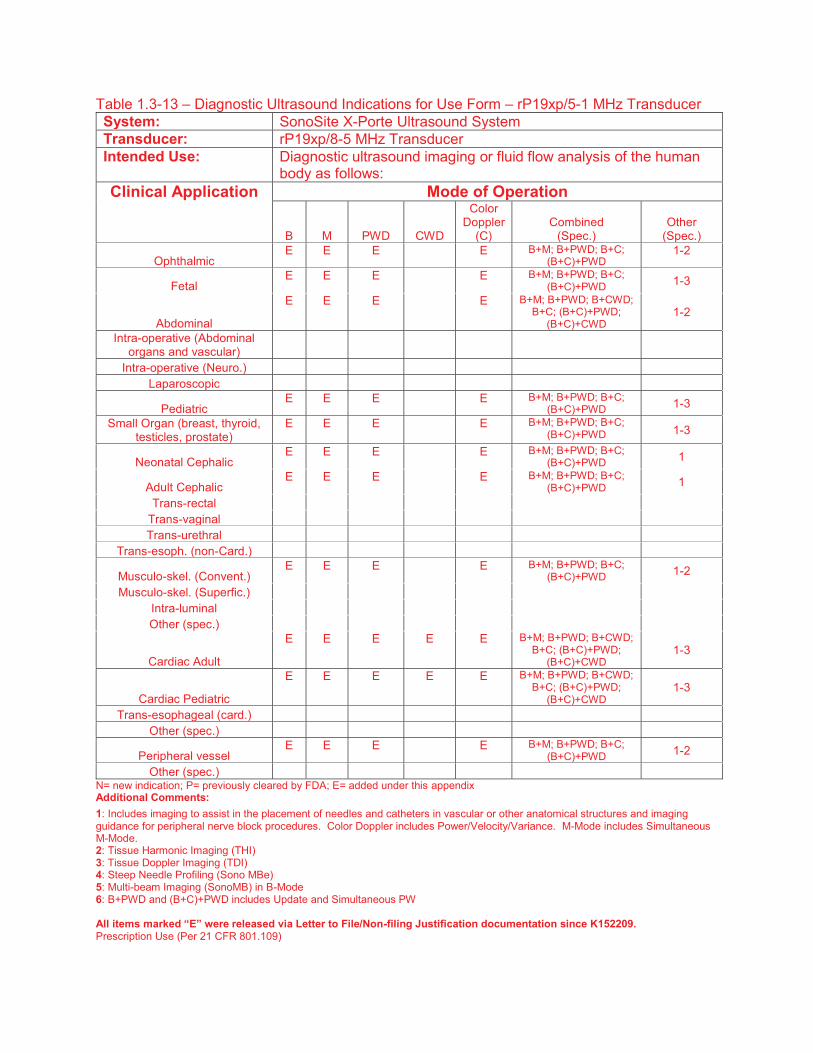
Table 1.3-13 – Diagnostic Ultrasound Indications for Use Form – rP19xp/5-1 MHz Transducer System: SonoSite X-Porte Ultrasound System Transducer: rP19xp/8-5 MHz Transducer Intended Use: Diagnostic ultrasound imaging or fluid flow analysis of the human
body as follows: Clinical Application
Mode of Operation
B M PWD CWD
Color Doppler
(C) Combined
(Spec.) Other
(Spec.)
Ophthalmic E E E E B+M; B+PWD; B+C;
(B+C)+PWD 1-2
Fetal E E E E B+M; B+PWD; B+C;
(B+C)+PWD 1-3
Abdominal
E E E E B+M; B+PWD; B+CWD; B+C; (B+C)+PWD;
(B+C)+CWD 1-2
Intra-operative (Abdominal organs and vascular)
Intra-operative (Neuro.) Laparoscopic
Pediatric E E E E B+M; B+PWD; B+C;
(B+C)+PWD 1-3 Small Organ (breast, thyroid,
testicles, prostate) E E E E B+M; B+PWD; B+C;
(B+C)+PWD 1-3
Neonatal Cephalic E E E E B+M; B+PWD; B+C;
(B+C)+PWD 1
Adult Cephalic E E E E B+M; B+PWD; B+C;
(B+C)+PWD 1 Trans-rectal
Trans-vaginal Trans-urethral
Trans-esoph. (non-Card.)
Musculo-skel. (Convent.) E E E E B+M; B+PWD; B+C;
(B+C)+PWD 1-2 Musculo-skel. (Superfic.)
Intra-luminal Other (spec.)
Cardiac Adult
E E E E E B+M; B+PWD; B+CWD; B+C; (B+C)+PWD;
(B+C)+CWD 1-3
Cardiac Pediatric
E E E E E B+M; B+PWD; B+CWD; B+C; (B+C)+PWD;
(B+C)+CWD 1-3
Trans-esophageal (card.) Other (spec.)
Peripheral vessel E E E E B+M; B+PWD; B+C;
(B+C)+PWD 1-2 Other (spec.)
N= new indication; P= previously cleared by FDA; E= added under this appendix Additional Comments: 1: Includes imaging to assist in the placement of needles and catheters in vascular or other anatomical structures and imaging guidance for peripheral nerve block procedures. Color Doppler includes Power/Velocity/Variance. M-Mode includes Simultaneous M-Mode. 2: Tissue Harmonic Imaging (THI) 3: Tissue Doppler Imaging (TDI) 4: Steep Needle Profiling (Sono MBe) 5: Multi-beam Imaging (SonoMB) in B-Mode 6: B+PWD and (B+C)+PWD includes Update and Simultaneous PW All items marked “E” were released via Letter to File/Non-filing Justification documentation since K152209. Prescription Use (Per 21 CFR 801.109)

Table 1.3-14: Diagnostic Ultrasound Indications for Use Form – P21xp/5-1 MHz Transducer System: SonoSite X-Porte Ultrasound System Transducer: P21xp/5-1 MHz Transducer Intended Use: Diagnostic ultrasound imaging or fluid flow analysis of the human
body as follows: Clinical Application
Mode of Operation
B M PWD CWD
Color Doppler
(C) Combined
(Spec.) Other
(Spec.)
Ophthalmic P P P P B+M; B+PWD; B+C;
(B+C)+PWD 1
Fetal P P P P B+M; B+PWD; B+C;
(B+C)+PWD 1-3
Abdominal
P P P P B+M; B+PWD; B+CWD; B+C; (B+C)+PWD;
(B+C)+CWD 1-3
Intra-operative (Abdominal organs and vascular)
Intra-operative (Neuro.) Laparoscopic
Pediatric P P P P B+M; B+PWD;
B+CWD; B+C 1-3 Small Organ (breast, thyroid,
testicles, prostate) P P P P B+M; B+PWD; B+C;
(B+C)+PWD 1-3
Neonatal Cephalic P P P P B+M; B+PWD; B+C;
(B+C)+PWD 1-3
Adult Cephalic P P P P B+M; B+PWD; B+C;
(B+C)+PWD 1-3 Trans-rectal
Trans-vaginal Trans-urethral
Trans-esoph. (non-Card.)
Musculo-skel. (Convent.) P P P P B+M; B+PWD; B+C;
(B+C)+PWD 1-3 Musculo-skel. (Superfic.)
Intra-luminal Other (spec.)
Cardiac Adult
P P P P P B+M; B+PWD; B+CWD; B+C; (B+C)+PWD;
(B+C)+CWD 1-3
Cardiac Pediatric
P P P P P B+M; B+PWD; B+CWD; B+C; (B+C)+PWD;
(B+C)+CWD 1-3
Trans-esophageal (card.) Other (spec.)
Peripheral vessel P P P P B+M; B+PWD; B+C;
(B+C)+PWD 1-3 Other (spec.)
N= new indication; P= previously cleared by FDA; E= added under this appendix Additional Comments: 1: Includes imaging to assist in the placement of needles and catheters in vascular or other anatomical structures and imaging guidance for peripheral nerve block procedures. Color Doppler includes Power/Velocity/Variance. M-Mode includes Simultaneous M-Mode. 2: Tissue Harmonic Imaging (THI) 3: Tissue Doppler Imaging (TDI) 4: Steep Needle Profiling (Sono MBe) 5: Multi-beam Imaging (SonoMB) in B-Mode 6: B+PWD and (B+C)+PWD includes Update and Simultaneous PW All items marked “P” were previously cleared in 510(k) K142017 and K133134. Prescription Use (Per 21 CFR 801.109)
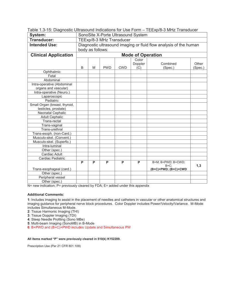
Table 1.3-15: Diagnostic Ultrasound Indications for Use Form – TEExp/8-3 MHz Transducer System: SonoSite X-Porte Ultrasound System Transducer: TEExp/8-3 MHz Transducer Intended Use: Diagnostic ultrasound imaging or fluid flow analysis of the human
body as follows: Clinical Application
Mode of Operation
B M PWD CWD
Color Doppler
(C) Combined
(Spec.) Other
(Spec.) Ophthalmic
Fetal Abdominal
Intra-operative (Abdominal organs and vascular)
Intra-operative (Neuro.) Laparoscopic
Pediatric Small Organ (breast, thyroid,
testicles, prostate)
Neonatal Cephalic Adult Cephalic
Trans-rectal Trans-vaginal Trans-urethral
Trans-esoph. (non-Card.) Musculo-skel. (Convent.) Musculo-skel. (Superfic.)
Intra-luminal Other (spec.) Cardiac Adult
Cardiac Pediatric
Trans-esophageal (card.)
P P P P P B+M; B+PWD; B+CWD; B+C;
(B+C)+PWD; (B+C)+CWD 1,3
Other (spec.) Peripheral vessel
Other (spec.) N= new indication; P= previously cleared by FDA; E= added under this appendix Additional Comments: 1: Includes imaging to assist in the placement of needles and catheters in vascular or other anatomical structures and imaging guidance for peripheral nerve block procedures. Color Doppler includes Power/Velocity/Variance. M-Mode includes Simultaneous M-Mode. 2: Tissue Harmonic Imaging (THI) 3: Tissue Doppler Imaging (TDI) 4: Steep Needle Profiling (Sono MBe) 5: Multi-beam Imaging (SonoMB) in B-Mode 6: B+PWD and (B+C)+PWD includes Update and Simultaneous PW All items marked “P” were previously cleared in 510(k) K152209. Prescription Use (Per 21 CFR 801.109)
510(K) Summary This summary of safety and effectiveness is provided as part of this Premarket Notification in compliance with 21 CFR, Part 807, Subpart E, Section 807.92.
1) Submitter’s name, address, telephone number, contact person:
FUJIFILM SonoSite, Inc.
21919 30th Drive SE
Bothell, WA 98021-3904
Corresponding Official: Jordan Lydia Grimmer
Sr. Regulatory Affairs Specialist
E-mail: [email protected]
Telephone: (425) 951-6984
Facsimile: (425) 951-1201
Date prepared: April 19, 2017
2) Name of the device, including the trade or proprietary name if applicable, the common or
usual name, and the classification name, if known: Common/ Usual Name Diagnostic Ultrasound System with Accessories Proprietary Name SonoSite X-Porte™ Ultrasound System Classification Names
Name FR Number Product Code
Ultrasonic Pulsed Doppler Imaging System 892.1550 90-IYN
Ultrasonic Pulsed Echo Imaging System 892.1560 90-IYO
Diagnostic Ultrasound Transducer 892.1570 90-ITX
3) Identification of the predicate or legally marketed device: SonoSite X-Porte Ultrasound System K152209 SonoSite Edge Ultrasound System K133454 SonoSite Edge II Ultrasound System K162045 4) Device Description: The SonoSite X-Porte Ultrasound System is a highly mobile, full featured, general purpose, diagnostic ultrasound system used to acquire and display high-resolution, real-time ultrasound data through multiple imaging modes. X-Porte is a custom fabricated digital electronic design that readily lends itself to be configured for specific ultrasound imaging applications through different system feature selections. The system interface can be customized for the user and controlled using a backlit touchscreen much like what is used in consumer tablet products. X-Porte can be operated in two different configurations, stand-based with AC power or battery, and desktop-based with AC power only. In desktop configuration the ultrasound engine can be removed from the stand and used by itself with a single transducer and external monitor.

5) Intended Use: The SonoSite X-Porte Ultrasound System is a general purpose ultrasound system intended for use by qualified physicians and healthcare professionals for evaluation by ultrasound imaging or fluid flow analysis of the human body. Specific clinical applications and exam types include:
Ophthalmic Fetal – OB/GYN
Abdominal Small Organ (breast, thyroid, testicles, prostate)
Neonatal Cephalic Adult Cephalic Trans-vaginal
Musculo-skel. (Convent.) Musculo-skel. (Superfic.)
Cardiac Adult Cardiac Pediatric
Trans-esophageal (card.) Peripheral Vessel
6) Technological Characteristics: SonoSite X-Porte, Edge, and Edge II Ultrasound Systems are both Track 3 devices that employ the same fundamental scientific technology. A comparison table is provided below.
Feature SonoSite X-Porte
Ultrasound System (This submission)
SonoSite X-Porte
Ultrasound System (K152209)
SonoSite Edge
Ultrasound System (K133454)
SonoSite Edge II
Ultrasound System (K162045)
Intended Use Diagnostic ultrasound
imaging or fluid flow
analysis of the human
body
Diagnostic ultrasound
imaging or fluid flow
analysis of the human
body
Diagnostic ultrasound
imaging or fluid flow
analysis of the human
body
Diagnostic ultrasound
imaging or fluid flow
analysis of the human
body
Indications for
Use
Ophthalmic
Fetal - OB/GYN
Abdominal
Pediatric
Small Organ (breast,
thyroid, testicle, prostate)
Neonatal Cephalic
Adult Cephalic
Trans-Vaginal
Musculo-skeletal
(Conventional)
Musculo-skeletal
(Superficial)
Cardiac Adult
Cardiac Pediatric
Trans-esophageal
(cardiac)
Peripheral Vessel
Ophthalmic
Fetal - OB/GYN
Abdominal
Intraoperative (abdominal
organs and vascular)
Intra-operative (Neuro.)
Pediatric
Small Organ (breast,
thyroid, testicle, prostate)
Neonatal Cephalic
Adult Cephalic
Trans-Rectal
Trans-Vaginal
Musculo-skeletal
(Conventional)
Musculo-skeletal
(Superficial)
Cardiac Adult
Cardiac Pediatric
Trans-esophageal
(cardiac)
Peripheral Vessel
Ophthalmic
Fetal – OB/GYN
Abdominal
Pediatric
Small Organ (breast,
thyroid, testicle, prostate)
Neonatal Cephalic
Adult Cephalic
Trans-Rectal
Trans-Vaginal
Musculo-skeletal
(Conventional)
Musculo-skeletal
(Superficial)
Cardiac Adult
Cardiac Pediatric
Trans-esophageal
(cardiac)
Peripheral Vessel
Feature SonoSite X-Porte
Ultrasound System (This submission)
SonoSite X-Porte
Ultrasound System (K152209)
SonoSite Edge
Ultrasound System (K133454)
SonoSite Edge II
Ultrasound System (K162045)
Needle
guidance
Needle
guidance
Needle
guidance
Needle
guidance
Transducer
Types
Linear Array
Curved Linear Array
Intracavitary
Phased Array
Static Probes
Trans-esophageal
Linear Array
Curved Linear Array
Intracavitary
Phased Array
Trans-esophageal
Linear Array
Curved Linear Array
Intracavitary
Phased Array
Static Probes
Trans-esophageal
Linear Array
Curved Linear Array
Intracavitary
Phased Array
Trans-esophageal
Transducer
Frequency
1.0 – 15.0 MHz 1.0 – 15.0 MHz 1.0 – 15.0 MHz 1.0 – 15.0 MHz
Global
Maximum
Outputs/Worst
Case Setting
Ispta.3: 629.3 (P21xp)
TI Type: TIB (P21xp)
TI Value: 4.0 (P21xp)
MI: 1.7 (P21xp)
Ipa.3@MI Max: 678 (L38xp)
Ispta.3: 629.3 (P21xp)
TI Type: TIB (P21xp)
TI Value: 4.0 (P21xp)
MI: 1.7 (P21xp)
Ipa.3@MI Max: 678 (L38xp)
Ispta.3: 709 (TEEx)
TI Type: TIB (P21x)
TI Value: 3.7 (P21x)
MI: 1.51 (P21x)
Ipa.3@MI Max: 776 (L38xi)
Ispta.3: 598.9 (HFL50x)
TI Type: TIB (rP19x)
TI Value: 4.98 (rP19x)
MI: 1.7 (rP19x)
Ipa.3@MI Max: 776 (L38xi)
Acoustic
Output Display
& FDA Limits
Display Feature for Higher
Outputs
MI Output Display
TI Output Display
Display Feature for Higher
Outputs
MI Output Display
TI Output Display
Display Feature for Higher
Outputs
MI Output Display
TI Output Display
Display Feature for Higher
Outputs
MI Output Display
TI Output Display
Feature SonoSite X-Porte
Ultrasound System (This submission)
SonoSite X-Porte
Ultrasound System (K152209)
SonoSite Edge
Ultrasound System (K133454)
SonoSite Edge II
Ultrasound System (K162045)
Modes of
Operation
B-mode Grayscale
Imaging
Tissue Harmonic Imaging
M-mode
Simultaneous M-Mode
Color Power Doppler
Zoom
Combination Modes
Simultaneous PW Imaging
Pulsed Wave (PW)
Doppler
Continuous Wave (CW)
Doppler
SonoHD2 Noise
Reduction
SonoMB/MBe Image
Compounding
Steered CW Doppler
Velocity Color Doppler
Tissue Doppler Imaging
(TDI)
B-mode Grayscale
Imaging
Tissue Harmonic Imaging
M-mode
Simultaneous M-Mode
Color Power Doppler
Zoom
Combination Modes
Pulsed Wave (PW)
Doppler
Continuous Wave (CW)
Doppler
SonoHD2 Noise
Reduction
SonoMB/MBe Image
Compounding
Steered CW Doppler
Velocity Color Doppler
Tissue Doppler Imaging
(TDI)
B-mode Grayscale
Imaging
Tissue Harmonic Imaging
M-mode
Color M-Mode
Color Power Doppler
Zoom
Combination Modes
Pulsed Wave (PW)
Doppler
Continuous Wave (CW)
Doppler
SonoHD2 Noise
Reduction
SonoMB/MBe Image
Compounding
Steered CW Doppler
Velocity Color Doppler
Tissue Doppler Imaging
(TDI)
B-mode Grayscale
Imaging
Tissue Harmonic Imaging
M-mode
Color M-Mode
Color Power Doppler
Zoom
Combination Modes
Pulsed Wave (PW)
Doppler
Continuous Wave (CW)
Doppler
SonoHD2 Noise
Reduction
SonoMB/MBe Image
Compounding
Steered CW Doppler
Velocity Color Doppler
Tissue Doppler Imaging
(TDI)
PW Doppler
Available on all imaging
transducers except D2xp.
Adjustable sample volume
size: 1.0 – 25 mm
Simultaneous or duplex
mode of operation
Simultaneous B-mode and
PW Doppler
High PRF capability
Available on all imaging
transducers.
Adjustable sample volume
size: 1.0 – 25 mm
Simultaneous or duplex
mode of operation
Simultaneous B-mode and
PW Doppler
High PRF capability
Available on all imaging
transducers except D2x/2
MHz.
Adjustable sample volume
size: 1.0 – 25 mm
Simultaneous or duplex
mode of operation
Simultaneous B-mode and
PW Doppler
High PRF capability
Available on all imaging
transducers except P11x.
Adjustable sample volume
size: 1.0 – 25 mm
Simultaneous or duplex
mode of operation
Simultaneous B-mode and
PW Doppler
High PRF capability
CW Doppler
Available on D2xp, P10xp,
rP19xp, P21xp, TEExp
Simultaneous or duplex
mode of operation
Simultaneous B-mode and
CW Doppler
Available on P10xp,
P21xp, TEExp
Simultaneous or duplex
mode of operation
Simultaneous B-mode and
CW Doppler
Available on C11x, D2x,
P10x, P21x, TEEx
Simultaneous or duplex
mode of operation
Simultaneous B-mode and
CW Doppler
Available on P10x, rP19x,
TEExi
Simultaneous or duplex
mode of operation
Simultaneous B-mode and
CW Doppler
Velocity Color
Doppler
Available on all
transducers except D2xp.
Available on all
transducers
Available on all
transducers except D2x
Available on all
transducers
Elastography
(Strain), and
Strain Rate
Imaging
Not available Not available Available on all
transducers except D2x
Not available
Feature SonoSite X-Porte
Ultrasound System (This submission)
SonoSite X-Porte
Ultrasound System (K152209)
SonoSite Edge
Ultrasound System (K133454)
SonoSite Edge II
Ultrasound System (K162045)
ECG Feature One 3-lead ECG input, or
One external ECG input,
or
ECG Slave Cable
One other physio input
One 3-lead ECG input, or
One external ECG input,
or
One other physio input
One 3-lead ECG input, or
One external ECG input,
or
One other physio input
3-lead ECG input, or
ECG Slave Cable
DICOM
DICOM 3.0 Store, Print,
Modality Worklist, Perform
Procedure Step (PPS),
Storage Commitment
DICOM 3.0 Store, Print,
Modality Worklist, Perform
Procedure Step (PPS),
Storage Commitment
DICOM 3.0 Store, Print,
and Modality Worklist
service class user features
DICOM 3.0 Store, Print,
Modality Worklist, Perform
Procedure Step (PPS),
Storage Commitment
IMT
Measurement
Not available Not available SonoCalc IMT provides
the capability for
automated measurement
of intima-media thickness
(IMT) of the carotid artery.
IMT functionality is
available both on the
ultrasound system and in
a stand alone software
program that runs on a
personal computer.
Not available
#Transmit
Channels
128 digital channels 128 digital channels 128 digital channels 128 digital channels
#Receive
Channels
64 digital channels
(128 digital channels using
Synthetic Aperture)
64 digital channels
(128 digital channels using
Synthetic Aperture)
64 digital channels
(128 digital channels using
Synthetic Aperture)
64 digital channels
(128 digital channels using
Synthetic Aperture)
Patient Contact
Materials
Transducers:
Acrylonitrile-butadien-
styrene (ABS)
Cycoloy
Dow Medical Adhesive,
Type A
Epoxy paste adhesive
Epoxy resin
Polyetherimide
Polyethylene (PE)
Ionomer
Polyetheretherketone
(PEEK)
Polysulfone UDEL P1700
Polyurethane
Poly-Vinyl-Chloride (PVC)
Silicone RTV Adhesive
Silicone Rubber
Urethane
Needle Guides:
Acetal copolymer
Acrylonitrile-butadien-
styrene (ABS)
Transducers:
Acrylonitrile-butadien-
styrene (ABS)
Cycoloy
Dow Medical Adhesive,
Type A
Epoxy paste adhesive
Epoxy resin
Polyetherimide
Polyethylene (PE)
Ionomer
Polyetheretherketone
(PEEK)
Polycarbonate
Polysulfone UDEL P1700
Polyurethane
Poly-Vinyl-Chloride (PVC)
Silicone RTV Adhesive
Silicone Rubber
Urethane
Needle Guides:
Acetal copolymer
Acrylonitrile-butadien-
styrene (ABS)
Transducers:
Acrylonitrile-butadien-
styrene (ABS)
Cycoloy
Epoxy paste adhesive
Polyethylene (PE)
Ionomer
Polyetheretherketone
(PEEK)
Polycarbonate
Polysulfone UDEL P1700
Polyurethane
Poly-Vinyl-Chloride (PVC)
Silicone RTV Adhesive
Silicone Rubber
Urethane
Needle Guides:
Acetal copolymer
Acrylonitrile-butadien-
styrene (ABS)
Feature SonoSite X-Porte
Ultrasound System (This submission)
SonoSite X-Porte
Ultrasound System (K152209)
SonoSite Edge
Ultrasound System (K133454)
SonoSite Edge II
Ultrasound System (K162045)
Product Safety
Certification
AAMI/ANSI ES60601-
1:2005 (R2012)
IEC 60601-2-37: 2007
CAN/CSA C22.2 No.
601.1
JIS T 0601-1, JIS T 1507
CEI/IEC 61157
ANSI/AAMI EC53
NEMA UD2-2004
IEC 62359:2010
AAMI/ANSI ES60601-
1:2005 (R2012)
IEC 60601-2-37: 2007
CAN/CSA C22.2 No.
601.1
JIS T 0601-1, JIS T 1507
CEI/IEC 61157
ANSI/AAMI EC53
NEMA UD2-2004
IEC 62359:2010
AAMI/ANSI ES60601-
1:2005 (R2012)
IEC 60601-2-37: 2007
CAN/CSA C22.2 No.
601.1
JIS T 0601-1, JIS T 1507
CEI/IEC 61157
ANSI/AAMI EC53
NEMA UD2-2004
AIUM RTD2-2004 (NEMA
UD3-2004 (R2009))
AAMI/ANSI ES60601-
1:2005 (R2012)
IEC 60601-2-37: 2007
CAN/CSA C22.2 No.
60601-1:08
NEMA UD2-2004
IEC 62359:2010
EMC
Compliance
IEC 60601-1-2:2007
CISPR 11
IEC 61000-4 pt 2-5
IEC 60601-1-2:2007
CISPR 11
IEC 61000-4 pt 2-5
IEC 60601-1-2:2007
CISPR 11
IEC 61000-4 pt 2-5
AAMI / ANSI / IEC 60601-
1-2:2007(R)2012
CISPR 11, Group 1, Class
A
DICOM NEMA PS3.15 2003 NEMA PS3.15 2003 NEMA PS3.15 2003 NEMA PS3.15 2003
Airborne
Equipment
Standards
RTCA/DO160D (section
21)
RTCA/DO160D (section
21)
RTCA/DO160D (section
21)
RTCA/DO160 (section 21)
System
Characteristics
X-Porte (stand
configuration):
Beamformer 128/128
using SA (configurable)
12.1” Capacitive touch
screen interface
19” LED LCD HD monitor
256 gray shades on LED
LCD
6 USB 2.0 ports
Stand Base Dimensions:
26.4” L x 21.2” W
Stand Height (max): 64”
(monitor up)
Stand Height (min): 42.2”
(monitor down)
Weight: 149.35 lbs (fully
configured w/ 3
transducers
System operates via
battery or AC power
Battery life: 1 hour
operational - 3 days idle
Input: 100 – 240 VAC,
50/60 Hz
Output 1: 24VDC output,
275 W max
Output 2: 100-240VAC,
50-60 Hz (AC Printer)
Edge:
Beamformer 128/128
using SA (configurable)
Hand held display and
control
Single 12.1” Liquid Crystal
Display (LCD)
256 gray shades on LCD
2 USB ports
Dimensions: 12.9"(W) x
12.4 (L) x 2.5"(H)
Weight: 8.5 lbs
Battery operated (1.5 - 4
hour operation per charge)
System operates via
battery or AC power
100 – 240V options, 50/60
Hz, 15VDC output
Edge II:
Beamformer 128/128
using SA (configurable)
Hand held display and
control
Single 12.1” Liquid Crystal
Display (LCD)
256 gray shades on LCD
2 USB ports
Dimensions: 12.8"(W) x
12.1” (L) x 2.5"(H)
Weight: 9.0 lbs
System operates via
battery or AC power
Battery life: 1.5 - 4 hour
operation per charge
100 – 240V options, 50/60
Hz, 15VDC output
Feature SonoSite X-Porte
Ultrasound System (This submission)
SonoSite X-Porte
Ultrasound System (K152209)
SonoSite Edge
Ultrasound System (K133454)
SonoSite Edge II
Ultrasound System (K162045)
Output 2: 100-240VAC,
50-60 Hz (AC Printer, DC
Printer)
Various obstetrical,
cardiac, volume, M-mode,
PW and CW Doppler
measurement and
calculation packages
ECG acquisition and
display capabilities
CW/PW Doppler Audio
Spectral Doppler Audio
and image storage on
removable media
Measurement on Recalled
Images.
Wireless 802.11 (a/b/g/n)
support for image transfer
X-Porte (desktop
configuration):
Same software
features/capabilities as the
stand configuration. Does
not have the stand, touch
panel interface, DVR, and
mobile power unit.
Weight: 32.80 lbs (w/ 1
transducer)
AC power only.
100 – 240V options, 50/60
Hz
Various obstetrical,
cardiac, volume, M-mode,
PW and CW Doppler
measurement and
calculation packages
ECG acquisition and
display capabilities
CW/PW Doppler Audio
Spectral Doppler Audio
and image storage on
removable media
Wireless 802.11 (a\b\g)
support for image transfer
Various obstetrical,
cardiac, volume, M-mode,
PW and CW Doppler
measurement and
calculation packages
ECG acquisition and
display capabilities
CW/PW Doppler Audio
Spectral Doppler Audio
and image storage on
removable media
Wireless 802.11 (b/g/n)
support for image transfer
510(k) Track Track 3 Track 3 Track 3 Track 3

7) Determination of Substantial Equivalence: Summary of Non-Clinical Tests: The X-Porte Ultrasound System has been evaluated for electrical, thermal, mechanical and EMC safety. Additionally, cleaning/disinfection, biocompatibility, and acoustic output have been evaluated, and the device has been found to conform to applicable mandatory medical device safety standards. Assurance of quality was established by employing the following elements of product development: Design Phase Reviews, Risk Assessment, Requirements Development, System and Software Verification, Hardware Verification, Safety Compliance Verification, Clinical Validation. All patient contact materials are biocompatible. Reports for these elements of product development are referenced in Attachment 6. The X-Porte Ultrasound System is designed to comply with the following voluntary standards.
Reference No. Title
AAMI / ANSI / ISO 10993-1
ISO 10993-1:2009/(R)2013, Biological evaluation of medical devices -- Part 1: Evaluation and testing within a risk management process
IEC 60601-1 AAMI / ANSI ES60601-1:2005/(R)2012 and A1:2012,, C1:2009/(R)2012 and A2:2010/(R)2012 (Consolidated Text) Medical electrical equipment - Part 1: General requirements for basic safety and essential performance (IEC 60601-1:2005, MOD)
IEC 60601-1-2 AAMI / ANSI / IEC 60601-1-2:2007(R)2012, Medical electrical equipment - Part 1-2: General requirements for basic safety and essential performance - Collateral standard: Electromagnetic compatibility - Requirements and tests (Edition 3)
IEC 60601-2-37 IEC 60601-2-37:2007, Particular Requirements for the basic safety and essential performance of ultrasonic medical diagnostic and monitoring equipment
IEC 62359 IEC 62359:2010, Ultrasonics – Field Characterization – Test Methods For The Determination Of Thermal And Mechanical Indices Related To Medical Diagnostic Ultrasonic Fields [Including: Technical Corrigendum 1 (2011)] (Edition 2)
ISO 14971 ISO 14971: 2007, Medical devices - Application of risk management to medical devices
NEMA UD 2-2004 Acoustic Output Measurement Standard for Diagnostic Ultrasound Equipment
NEMA PS 3.15 NEMA Ps 3.15:2011, Digital Imaging and Communications in Medicine (DICOM), Part 15: Security and System Management Profiles
Summary of Clinical Tests: The SonoSite X-Porte Ultrasound System and transducers, subject of this submission, did not require clinical studies to support the determination of substantial equivalence. 8) Conclusion: Intended uses and other key features are consistent with traditional clinical practice and FDA guidance. The X-Porte device and predicates conform to applicable electromedical device safety standards with compliance verified through independent evaluation. The X-Porte device and predicates meet FDA requirements for Track 3 devices, share indications for use, have biosafety equivalence and are manufactured using the same ISO 13485 quality system. FUJIFILM SonoSite, Inc. believes that the X-Porte Ultrasound System is substantially equivalent with regard to safety and effectiveness to the predicate devices.