
First posted online on 12 June 2018 as...
34
© 2018. Published by The Company of Biologists Ltd. This is an Open Access article distributed under the terms of the Creative Commons Attribution License (http://creativecommons.org/licenses/by/3.0), which permits unrestricted use, distribution and reproduction in any medium provided that the original work is properly attributed. Cerebellar synapse properties and cerebellum-dependent motor and non-motor performance in Dp71-null mice Romain Helleringer 1 , Delphine Le Verger, Xia Li, Charlotte Izabelle, Rémi Chaussenot, Mehdi Belmaati-Cherkaoui, Raoudha Dammak, Paulette Decottignies, Hervé Daniel, Micaela Galante*‡, Cyrille Vaillend*‡. Paris-Saclay Institute of Neuroscience (Neuro-PSI), UMR 9197, Université Paris Sud, CNRS, Université Paris Saclay, Orsay, France. ‡ The two authors equally contributed to supervision of experimental work. 1 present address: Institut de Biologie de l'Ecole Normale Supérieure (IBENS), Ecole Normale Supérieure, CNRS UMR 8197, INSERM U1024, 75005 Paris, France. *Corresponding authors at: Paris-Saclay Institute of Neuroscience (Neuro-PSI), CNRS UMR9197, 91405 Orsay cedex, France. Email addresses: [email protected] (M. Galante) and [email protected] (C. Vaillend). ABSTRACT A recent focus has been placed on the role that cerebellar dysfunctions could play in the genesis of cognitive deficits in Duchenne muscular dystrophy (DMD). However, relevant genotype-phenotype analyses are missing to define whether cerebellar defects underlie the severe cases of intellectual deficiency, which have been associated with genetic loss of the smallest product of the dmd gene, the Dp71 dystrophin. To determine for the first time whether Dp71 loss could affect cerebellar physiology and functions, we have used patch- clamp electrophysiological recordings in acute cerebellar slices and a cerebellum-dependent behavioral test battery addressing cerebellum-dependent motor and non-motor functions in Dp71-null transgenic mice. We found that Dp71 deficiency selectively enhances excitatory transmission at glutamatergic synapses formed by climbing fibers (CFs) on Purkinje neurons, but not at those formed by parallel fibers (PFs). Altered basal neurotransmission at CFs was associated with impairments in synaptic plasticity and clustering of the scaffolding post- synaptic density protein PSD-95. At the behavioral level, Dp71-null mice showed some improvements in motor coordination and were unimpaired for muscle force, static and dynamic equilibrium, motivation in high-motor demand and synchronization learning. However, Dp71-null mice displayed altered strategies in goal-oriented navigation tasks, suggesting a deficit in the cerebellum-dependent processing of the procedural components of spatial learning which could contribute to the visuo-spatial deficits identified in this model. In all, the observed deficits suggest that Dp71 loss alters cerebellar synapse function and cerebellum-dependent navigation strategies without being detrimental for motor functions. Keywords: Dystrophin Dp71, Purkinje neuron, glutamatergic transmission, motor coordination, cerebellum, cognitive deficit. Disease Models & Mechanisms • DMM • Accepted manuscript http://dmm.biologists.org/lookup/doi/10.1242/dmm.033258 Access the most recent version at First posted online on 12 June 2018 as 10.1242/dmm.033258
Transcript of First posted online on 12 June 2018 as...

© 2018. Published by The Company of Biologists Ltd.
This is an Open Access article distributed under the terms of the Creative Commons Attribution License
(http://creativecommons.org/licenses/by/3.0), which permits unrestricted use, distribution and reproduction in any medium provided that the original work is properly attributed.
Cerebellar synapse properties and cerebellum-dependent motor and non-motor performance
in Dp71-null mice
Romain Helleringer1, Delphine Le Verger, Xia Li, Charlotte Izabelle, Rémi Chaussenot,
Mehdi Belmaati-Cherkaoui, Raoudha Dammak, Paulette Decottignies, Hervé Daniel, Micaela
Galante*‡, Cyrille Vaillend*‡.
Paris-Saclay Institute of Neuroscience (Neuro-PSI), UMR 9197, Université Paris Sud, CNRS,
Université Paris Saclay, Orsay, France.
‡ The two authors equally contributed to supervision of experimental work.
1present address: Institut de Biologie de l'Ecole Normale Supérieure (IBENS), Ecole Normale
Supérieure, CNRS UMR 8197, INSERM U1024, 75005 Paris, France.
*Corresponding authors at: Paris-Saclay Institute of Neuroscience (Neuro-PSI), CNRS
UMR9197, 91405 Orsay cedex, France. Email addresses: [email protected] (M.
Galante) and [email protected] (C. Vaillend).
ABSTRACT
A recent focus has been placed on the role that cerebellar dysfunctions could play in the
genesis of cognitive deficits in Duchenne muscular dystrophy (DMD). However, relevant
genotype-phenotype analyses are missing to define whether cerebellar defects underlie the
severe cases of intellectual deficiency, which have been associated with genetic loss of the
smallest product of the dmd gene, the Dp71 dystrophin. To determine for the first time
whether Dp71 loss could affect cerebellar physiology and functions, we have used patch-
clamp electrophysiological recordings in acute cerebellar slices and a cerebellum-dependent
behavioral test battery addressing cerebellum-dependent motor and non-motor functions in
Dp71-null transgenic mice. We found that Dp71 deficiency selectively enhances excitatory
transmission at glutamatergic synapses formed by climbing fibers (CFs) on Purkinje neurons,
but not at those formed by parallel fibers (PFs). Altered basal neurotransmission at CFs was
associated with impairments in synaptic plasticity and clustering of the scaffolding post-
synaptic density protein PSD-95. At the behavioral level, Dp71-null mice showed some
improvements in motor coordination and were unimpaired for muscle force, static and
dynamic equilibrium, motivation in high-motor demand and synchronization learning.
However, Dp71-null mice displayed altered strategies in goal-oriented navigation tasks,
suggesting a deficit in the cerebellum-dependent processing of the procedural components of
spatial learning which could contribute to the visuo-spatial deficits identified in this model. In
all, the observed deficits suggest that Dp71 loss alters cerebellar synapse function and
cerebellum-dependent navigation strategies without being detrimental for motor functions.
Keywords: Dystrophin Dp71, Purkinje neuron, glutamatergic transmission, motor
coordination, cerebellum, cognitive deficit.
Dis
ease
Mo
dels
& M
echa
nism
s •
DM
M •
Acc
epte
d m
anus
crip
t
http://dmm.biologists.org/lookup/doi/10.1242/dmm.033258Access the most recent version at First posted online on 12 June 2018 as 10.1242/dmm.033258

INTRODUCTION
The Duchenne muscular dystrophy (dmd) gene has at least 8 promoters driving expression
of distinct dystrophin proteins which are components of different multiprotein complexes
linking cytoskeletal actin to cell membrane proteins in a variety of tissues. The first three
promoters consist of spliced unique first exons that control the synthesis of the 427kDa full-
length forms of the dystrophin protein (Dp427) respectively expressed in brain, muscle or in
cerebellar Purkinje neurons. Five downstream promoters give rise to shorter dystrophin
products (Dp260, Dp140, Dp116, Dp71) with cell-specific expression in various organs
including nervous system (Perronnet and Vaillend, 2010; Hendriksen et al., 2015). Mutations
in the dmd gene are responsible for the Duchenne muscular dystrophy (DMD) syndrome, in
which muscular degeneration is also associated with cognitive deficits presumably due to the
loss of the dystrophin-gene products that are normally expressed in brain (Kim et al., 1995;
Desguerre et al., 2009). The severity of the cognitive impairment in DMD is highly variable
depending on the mutation location. Mutations in the first exons that selectively affect
expression of Dp427 lead to moderate cognitive deficits, while in about one third of patients
downstream mutations inducing a cumulative loss of the shorter dystrophin products are
responsible for moderate to severe intellectual disability (Desguerre et al., 2009). Dp71 loss
appears to be a pivotal aggravating factor as patients with impaired expression of Dp71 are
most severely affected and systematically show intelligence quotients (IQs) below 70 (Daoud
et al., 2009a).
Dp71 is the most prominent dystrophin-gene product in the adult brain. The role of Dp71,
as well as the functional outcome of Dp71 deficiency appears to be quite complex due to its
expression in both neuronal and glial cells (Tadayoni et al., 2012; for a review). Dp71 was
detected in post-synaptic densities and its deficiency associated with altered hippocampal
glutamatergic synapse structure, synaptic transmission and plasticity (Blake et al., 1999;
Daoud et al., 2009b). Moreover, electron microscopy analyses revealed that Dp71-null mice
display significant changes in the density and size of presynaptic vesicles in hippocampal
glutamatergic synapses, suggesting synapse ultrastructural alterations (Miranda et al., 2011).
On the other hand, Dp71 is also highly expressed in glial cells where it contributes to the
membrane clustering of potassium (Kir4.1) and aquaporin (AQP4) channels in perivascular
domains, suggesting that its loss may affect extracellular K+ buffering and water balance,
which may also have an impact on neuronal functions (Dalloz et al., 2003; Amiry-
Mogghadam et al., 2004; Connors et al., 2004; Haenggi et al., 2006; Fort et al., 2008). Dp71
likely plays such a role in various glial cell types including cerebellar Bergmann glial cell
where it is required for proper localization of a pool of AQP4 water channels (Nicchia et al.,
2008). However, a functional evaluation of the role of Dp71 in this structure is still lacking.
Dis
ease
Mo
dels
& M
echa
nism
s •
DM
M •
Acc
epte
d m
anus
crip
t

In the present study, we have placed a focus on the functional impact of Dp71 loss in
cerebellar tissues, as a critical role for cerebellum dysfunction has been emphasized in clinical
studies of pediatric DMD patients (Cyrulnik and Hinton, 2008). It is now well admitted that
cerebellar neuronal networks, particularly the climbing and parallel-fiber inputs to Purkinje
neurons (PCs), contribute to navigation in rodents (Rondi-Reig et al., 2002; Rochefort et al.,
2011) and their dysfunction could therefore participate to the spatial learning deficits reported
in mice lacking Dp71 (Daoud et al., 2009b). Moreover, cerebellar alterations are commonly
associated with motor deficits that could contribute to the cardinal muscular symptoms in
DMD, as suggested by other mouse models with altered dystrophin-related mechanisms (e.g.,
Grady et al., 2006). To test these hypotheses, we first used whole-cell patch-clamp recordings
in acute cerebellar slices of Dp71-null and wild-type littermate mice to decipher the
electrophysiological properties of the two main glutamatergic inputs to cerebellar Purkinje
neurons, the parallel fibers (PFs) and the climbing fibers (CFs). This was complemented by a
quantitative immunofluorescence analysis of synaptic-density protein PSD-95 clusters, to
determine putative changes in synapse structural and molecular organization. A behavioral
study of these mice was then undertaken to characterize their performance in cerebellum-
dependent motor functions, including basal static versus dynamic coordination, during fine-
timing motor learning in the rotarod, as well as in a goal-oriented tasks in the water maze to
assess procedural learning performance and navigation strategies (Steinmayr et al., 1998;
Leggio et al., 1999; Rondi-Reig et al., 2002).
MATERIALS AND METHODS
Animals
Dp71-null mice were a kind gift from Prof. David Yaffe. They were originally generated in
his laboratory by homologous recombination, by replacing most of the first and unique exon
and a small part of the first intron of Dp71 by the promoter-less gene encoding a β-gal-
neomycin resistance chimeric protein, which specifically abolished the expression of Dp71
without interfering with the expression of other dmd-gene products (Sarig et al., 1999).
Transgenic mice were backcrossed for >10 generations to C57BL/6JRj mice (Janvier Labs,
France) in CDTA (Orléans, France) and breeders were kindly provided by Dr. Alvaro Rendon
(Institut de la Vision, Paris, France). Production and maintenance of the transgenic line was
undertaken in our animal facility by crossing heterozygous females with C57BL/6JRj mice to
generate Dp71-null and littermate controls (WT) males for experiments. Genotype was
determined by PCR analysis of tail DNA. Animals were kept under a 12-h light-dark cycle
(light on: 7.00 a.m.) with food and water ad libitum. Studies were conducted blind to the
Dis
ease
Mo
dels
& M
echa
nism
s •
DM
M •
Acc
epte
d m
anus
crip
t

genotype following guidelines of local mouse facility (agreement # D91-471-104) in
compliance with European Directive 2010/63/EU and French National Committee (87/848).
Experimental protocols were approved by the Animal welfare body of our Institution (Institut
des Neurosciences, Neuro-PSI) and Ethic committee #59.
Immunofluorescence and confocal image analyses
Sagittal cerebellar sections (30 µm thick) cut at −12 °C in a cryostat from fresh-frozen
dissected brains were collected on Superfrost+ glass slides (Roth, France) and stored at -80°C.
For immunochemistry, slides were thawed for 1 min at RT, immersed in acetone/methanol
(1:1) for 5 min at −20 °C, washed 3 times in 0.1 M phosphate-buffered saline (PBS),
incubated in a blocking solution for 45 min (10% normal goat serum, 0.3% Triton X-100, 1%
bovine serum albumin), then overnight at 4 °C with a primary polyclonal antibody directed
against the postsynaptic density protein PSD-95 (1:400, Invitrogen, #51-6900) followed by
washes and incubation with a goat anti-rabbit secondary antibody conjugated to Cy3 (1:500;
Jackson Immunoresearch, USA, # 111-165-003) in PBS 0.1 M with 5% NGS and 1% BSA
for 1h at RT. Slides were coverslipped using a mounting medium containing DAPI
(Fluoromount-G, Clinisciences, France). No staining was observed in sections processed from
control sections from both genotypes when primary antibody was omitted. A laser scanning
confocal microscope LSM 700 (Zeiss) was used to sequentially collect Cy3 immunoreactivity
at 555 nm and DAPI staining at 405 nm. Confocal images were imported using an EC Plan-
Neofluar 40x/1.30 Oil M27 at a resolution of 156 nm/pixel. All images were randomly taken
at same exposure times and equivalent stereotaxic coordinates. Punctate immunoreactivity
(IR) representing presumptive protein clusters was quantified using a threshold segmentation
algorithm in WCIF ImageJ for automatic detection of clusters (Vaillend et al., 2010). Minimal
cluster size was arbitrarily set to 3 adjacent pixels (0,05 µm2) and a maximal size of 2 μm2,
corresponding to more than 99% of clusters. The number and size of clusters were analyzed
within a total tissue surface of 13 165 µm2 per genotype (3 mice, >800 clusters per genotype).
Electrophysiology
Tissue slice preparation. Electrophysiological experiments were performed on C57Bl6 mice
and their Dp71-null littermates. Male, adult (2-4 months old) mice were anesthetized with 2-
bromo-2Cloro-1,1,1-trifluoroethane (Sigma-Aldrich, France), decapitated and the vermis
region of the cerebellum was isolated. Parasagittal slices (250m thick) were prepared in the
presence of a cold Buffered Bicarbonate Solution (BBS) containing (in mM): 124 NaCl, 3
KCl, 1.15 KH2PO4, 1.15 MgSO4, 24 NaHCO3, 10 Glucose, 2 CaCl2 (osmolarity: 330 mOsm
Dis
ease
Mo
dels
& M
echa
nism
s •
DM
M •
Acc
epte
d m
anus
crip
t

and pH 7.35). Then slices were then incubated at room temperature in (BBS) gassed with
95%O2-5%CO2 for at least 30min before recording.
Electrophysiological recordings. Patch-clamp whole-cell recordings in voltage clamp mode
were performed with an Axopatch 200 amplifier (Axon instruments). Pipettes were pulled
from borosilicate glass capillaries and had a resistance of 5.5-6.5M when filled with the
intracellular solution. For voltage clamp experiments, cells were maintained at a holding
potential of Vh=-70mV to record the response to PF stimulation (PF-EPSC), while to study
the climbing fiber-Purkinje cell synapse (CF-EPSC), the membrane potential was Vh=-20mV.
Series resistance was routinely checked delivering a negative (-10mV) voltage step. This
parameter was usually < 15MΩ, it was partially compensated (60-75%) and the recordings
were interrupted whenever the series resistance increased by more than 20-30% of its initial
value. During experiments, the recording chamber was continuously perfused at a rate of 2
ml/min with BBS supplemented with the GABAA receptor antagonist gabazine (5M, Sigma-
Aldrich) or bicuculline methiodide (10M, Sigma-Aldrich). All experiments were performed
at room temperature. EPSCs were evoked by 100s current pulses generated by a stimulus
isolator (Digitimer, Ltd, DS2) and delivered through a BBS-filled monopolar electrode placed
in the tissue slice.
Parallel fibers stimulation. Stimulating electrode was applied in the middle of the molecular
layer and two pulses were delivered separated by an inter-stimulus interval of 30ms except in
experiments examining the short-term plasticity at different inter-stimulus intervals. To record
PF-EPSCs the patch pipette was filled with an internal solution containing (mM): Cs-
gluconate 140, HEPES 10, EGTA 1, MgCl2 4.6, CaCl2 0.1, Na2-ATP 4, Na3-GTP 0.4,
tetraethylammonium (TEA) 20 (final pH 7.3 with CsOH and osmolarity 300 milliosmoles/l).
PF-EPSCs were recorded at a membrane holding potential of -70mV. Electrical stimulation
was delivered at a frequency of 0.2Hz.
Climbing fibers stimulation. To evoke CF-EPSC, the stimulation electrode was placed in the
granular layer. PC was first recorded at -70mV and then at -20mV to inactivate voltage-
dependent channels and to reduce CF-EPSC amplitude and consequent space clamp problems.
For this type of experiment, the patch pipette was filled with an internal solution containing
(mM): Cs-gluconate 140, HEPES 10, BAPTA 20, MgCl2 4.6, CaCl2 0.1, Na2-ATP 4, Na3-
GTP 0.4, tetraethylammonium (TEA) 20 (final pH 7.3 with CsOH and osmolarity 300
milliosmoles/l). To assess the presence of one or more CF innervating the patched Purkinje
neuron, several locations of the stimulating electrode in the granular layer and in the lower
half of the molecular layer were systematically tested for their ability to evoke potential multi-
step CF-EPSCs. In rare cases, two CFs were contacting Purkinje neurons and these recordings
were excluded from data analysis. In experiments studying CF-EPSC kinetics and TBOA
Dis
ease
Mo
dels
& M
echa
nism
s •
DM
M •
Acc
epte
d m
anus
crip
t

effects, low concentrations of NBQX (0.2-0.4µM) were added to BBS solution in order to
reduce voltage-clamp escape due to the large amplitude CF-EPSCs (holding potential=-
70mV). In voltage clamp experiments, CFs were stimulated at 0.2Hz.
Complex spikes recordings and synaptic plasticity induction. For current clamp experiments
and plasticity induction at CF to Purkinje cell synapse, patch pipettes were filled with an
internal solution containing (in mM): Kgluconate 137, KCl 6, MgCl2 3.5, HEPES 10mM,
NaCl 4, Na2-ATP 4, Na3-GTP 0.4 (pH adjusted to 7.3 with KOH; osmolarity: 295
milliOsmoles/l). The resting membrane potential of PCs was held to -65/-59 mV by injection
of small negative currents through the amplifier (Axopatch 200B, Axon Instruments) and
Series resistance were routinely controlled switching to voltage clamp mode. CFs were
stimulated at a frequency of 0.033Hz, CF-EPSPs with superposed complex spikes were
recorded for at least 5 minutes in the presence of gabazine before long-term plasticity
induction. For this purpose, CFs were stimulated at 5Hz for 30s as previously described
(Hansel and Linden; 2000). To monitor complex spike modifications, the amplitude of spike
components was measured for every experiment and then normalized to the mean amplitude
before plasticity induction. Normalized spike amplitudes were then averaged over several
cells. Statistics was performed on averaged amplitude values over a 5min period.
Paired-pulse ratio. The paired-pulse ratio was calculated for groups of 30 consecutive sweeps
as the ratio of the mean amplitude of the second EPSC to the mean of the first EPSC. Data
from different experiments were averaged to obtain the PPR mean values.
Data recording and analysis. Data were collected by Elphy software (G. Sadoc, CNRS,
UNIC, France). CF- and PF-mediated responses were analyzed off-line by using Clampfit
(Axon Instruments) and Igor (Wavemetrics) routines.
Behavioral phenotyping test battery
Exploration and motor coordination in a holeboard. The apparatus was derived from one
previously used with cerebellar mutant mice (Guastavino, 1984 ; Steinmayr et al., 1998 ;
Rondi-Reig et al., 1999) and was customized by Ugo Basile (Italy) following our
recommendations. It consists of an experimental box (35 x 35 x 25 cm) with a raised (2 cm)
platform, in which 36 holes were drilled (2 cm in diameter, spaced by about 4 cm) and
arranged in a 6 x 6 array. The mouse was placed in the center of the platform and
videotracked for 5 min using the ANY-maze software (Stoelting, USA) for automatic
quantification of distance travelled and travelling speed. The number of times the mouse
stumbled (one leg slipping into a hole) or performed hole nose pokes was manually recorded
Dis
ease
Mo
dels
& M
echa
nism
s •
DM
M •
Acc
epte
d m
anus
crip
t

using event-recorder keys. Stumble number was considered as a measure of motor
coordination and nose pokes as an estimation of exploratory stereotypies. A central square
containing a 4x4-hole array was used to assess avoidance of the central anxiogenic area.
Grip strength test. Strength was evaluated using a home-made traction apparatus equipped
with a horizontal grid to allow for gripping, connected to a digital dynamometer. Each mouse
was lifted by the tail, lowered over the top of the grid and allowed to grasp the grid with both
forepaws and hindpaws. While the torso of the animal was kept parallel to the grid, the mouse
was gently pulled back by the tail until it released its grip and the maximum tension (arbitrary
unit) was recorded. Three measures taken with a 5-min inter-trial interval (ITI) were
averaged.
Inverted screen test. Mice were placed individually on a cage wire screen about 35 cm above
a table. After slowly inverting the screen upside-down to 180° the ability to maintain a grip
was monitored (grip latency) and a maximum score of 120s given if the animal did not fall.
Testing was repeated three times with a 5-min ITI.
Wire suspension test. The mouse was hung by the forepaws on a 25-cm wire (3 mm in
diameter) resting on two vertical supports and elevated 35 cm above a flat surface. Three
trials spaced by a 5-min pause were performed with each trial limited to a 60s duration. The
amount of time spent by the mouse hanging onto the wire and the latency to touch the wire
with one hindpaw were recorded during each trial, and a mean score was then calculated.
Other qualitative parameters were recorded and a score was attributed corresponding to the
best performance achieved within the minute of testing according to the following scale
(Aruga et al., 2004): 0, fell off; 1, clung to the bar with two forepaws; 2, attempted to climb
on to the bar besides clinging to it with two forepaws; 3, hung on to the bar with two forepaws
and one or both hind paws; 4, hung on to the bar with all four paws with the tail additionally
wrapped around the bar; 5, escaped to one of the supports.
Static balance on an unstable platform. The apparatus consisted of a circular platform
(diameter 8.5 cm) made of grey perspex, fixed at its center on a vertical axis (1 m high) and
that could tilt by 30° in every direction. The mouse was placed on the middle of the platform
(horizontal situation) and the number of slips per min (at least one paw out of the platform
circumference) and the fall latency (cut-off: 180 s) were measured by direct observation
during three trials with an intertrial interval of 15 min. The day after, their ability to maintain
a balance using vestibular-associated self-motion cues was assessed by submitting the mice to
one trial in total darkness (visual cue removal) (Rochefort et al., 2011).
Dis
ease
Mo
dels
& M
echa
nism
s •
DM
M •
Acc
epte
d m
anus
crip
t

Forced swim test. Each mouse was lowered in an inescapable glass cylinder (Diameter: 11
cm; height: 23 cm) filled with 18 cm water at 25°C. Room temperature was 25°C. Behavior
was recorded on video for 5 min each day in two sessions separated by a 24h delay. Video
were analyzed offline using event-recorder keys in Any-maze to quantify the latency and
duration of three main parameters: climbing, staying afloat, and immobility (freezing).
Climbing was considered when mice had a vertical position of the spine with the forepaws
striking the glass walls while hind paws showed repetitive movement in water. Staying afloat
corresponded to movements simply performed to keep the head above water. Immobility was
defined by a complete immobilization of the body for at least 1s. The time not spent
performing any one of these activities represented either unspecific uncoordinated movements
or swimming activity involving horizontal spine position with legs treading water and
producing a clear displacement of body.
Tail suspension test. Each mouse was suspended by adhesive tape placed 2 cm from the tip of
the tail, 35 cm above a bench top during a 6 min period. Behavior was recorded on video
during two sessions separated by a 24h delay. The latency to the first bout of immobility
(freezing latency) and the duration of freezing were quantified offline using event-recorder
keys in Any-maze. Complete immobility for > 2s was regarded as freezing.
Rotarod test paradigms. Motor coordination and learning were evaluated by using several
paradigms in a mouse Rotarod with adjustable speed and accelerating mode (Cat.# 47600,
Ugo Basile, Italy). Four-five mice of the two genotypes were tested in parallel between 9 am
and 1 pm; the apparatus was cleaned with 100% ethanol after each trial. On the first day,
equilibrium was tested by placing each mouse on the unrotating rod, its body axis
perpendicular to the rod longitudinal axis. The time the animal stayed on the rod was recorded
and the trial stopped when the animal fell or after 180 s. On the next day, motor coordination
learning was evaluated by placing each mouse on the rod rotating at a constant speed (4 rpm),
its head directed against the direction of rotation so that it had to progress forward and
synchronize its walk with the speed of the rod to maintain balance. Training consisted of 5
successive trials with a 15-min ITI; fall latency was recorded during each trial with a 180 s
cut-off duration. Motor synchronization learning was further analyzed 72h later by placing
mice on the rotating rod which accelerated from 4 to 40 rpm in a 5-min trial. Training then
consisted of 5 successive trials per day with 15-min ITI, during 4 successive days. A distinct
group of naïve mice (both genotypes) was tested in a different protocol (Galante et al., 2009),
to assess motor synchronization learning without prior experience of neither motor tests nor
familiarization to rotarod. In this experiment, mice were submitted to 5-min trials during
which the rotating rod accelerated from 4 to 40 rpm. Mice underwent 4 trials per day with a
1h ITI during 3 days. On the next day (Day 4), the mice were submitted to 8 consecutive 2-
Dis
ease
Mo
dels
& M
echa
nism
s •
DM
M •
Acc
epte
d m
anus
crip
t

min trials at constant speeds of 40, 35, 30, 25, 20, 15, 10 and 5 rpm, respectively. This
protocol was repeated four times with a 1-h ITI.
Navigation tasks in a water maze. The maze was a circular water tank (1.5-m diameter)
placed in a well-lit room containing several extramaze cues on the walls and surmounted by a
video camera connected to a computer located in an adjacent room. It was filled with water
(22°C) to 15 cm below the edge of wall, which was made opaque by addition of a white paint
(Opacifier 631, Morton SA, France), and it contained a circular escape platform (10-cm
diameter) submerged 0.5 cm below water surface. Four positions (N, S, E and W)
equidistantly located around the maze divided it into four virtual quadrants. During
pretraining (Day 1), the platform was laid at the North (35 cm from the wall). The mouse was
released in the center of the maze and could swim for 60 s, after which it was gently guided
by hand to the platform and allowed to remain on it for 60 s. Two distinct groups of mice
(both genotypes) were then tested following two distinct protocols: the visible (cued) and
non-visible platform tasks. In the first protocol, a visible black tube (10 cm high) was placed
on top of the platform as a beacon enabling visual guidance strategies (proximal cue). In each
trial the platform was placed in the center of a different quadrant (35 cm from the wall) and
the mouse was introduced in the maze from different starting points (avoiding the quadrant
where the platform was located). Each mouse was submitted to four trials a day with a 10-min
ITI during 3 days, during which it was allowed to swim freely for 90 s to find the platform
and could stay on it for 60 s. In the non-visible platform task, single constant start point and
platform position were assigned to each mouse. Each mouse underwent four 90-s trials a day
with a 10-min ITI during 7 days. In both tasks, mice were videotracked using the ANY-maze
software (Stoelting, USA) for automatic quantification of averaged and maximum swim
speed, time needed to reach the platform (escape latency) and distance swum to the platform.
Other parameters were analyzed to detail animal’s navigation strategy: Entries in the quadrant
containing the platform, amount of circling behavior in a virtual corridor 19 cm in width set
along the wall (thigmotaxis), 360° rotations of animal’s body, absolute turn angle between
each movement vector of the mouse, cumulative product of the distance from platform and
time spent at this distance (cumulative distance to platform), meander (cumulative distance
divided by total distance) and angular velocity (cumulative distance divided by escape
latency). We also analyzed indexes reflecting deviation between optimal direction towards
platform and actual motion direction, such as angular deviation (heading error) from platform
position, corrected integrated path length, path efficiency and percent time spent and distance
swum in a 15 cm-wide virtual corridor that runs from animal’s start position to platform.
Dis
ease
Mo
dels
& M
echa
nism
s •
DM
M •
Acc
epte
d m
anus
crip
t

Statistics
Data are expressed as means ± SEM. Behavioral parameters were analyzed using two-way
ANOVAs with one between-subject factor (genotype: WT or Dp71-null) and one dependent
variable or within-subject factor when appropriate (training day, trial, rotating speed). For
electrophysiology data, genotype effects were evaluated with Mann-Whitney test and drug
effects within groups with Wilcoxon signed rank test. For western blot analyses, statistical
significance was evaluated by the Mann-Whitney non-parametric test. The p values < 0.05
were considered statistically significant. The Kolmogorov-Smirnov test (KS-Test) was used
to compare the distribution of protein cluster sizes (significance threshold: p<0.005).
RESULTS
Electrophysiological properties of the parallel fiber-Purkinje cell synapse
Purkinje neurons (PCs) receive glutamatergic inputs from both parallel fibers (PFs) and
climbing fibers (CFs). PFs are granule cell axons that run parallel to the Purkinje cell layer in
the coronal plane and perform several ‘en passant’ synapses on Purkinje cell dendrites. CF
fiber inputs originating from inferior olive are strikingly different: a Purkinje cell receives
several synaptic contacts from a single CF, and transmission in this pathway is one to one. To
assess the physiological consequences of Dp71 loss in cerebellar cortex, we therefore
investigated potential alterations in excitatory postsynaptic currents (EPSC) evoked by
electrical stimulation of PFs or CFs.
EPSCs were identified as PF-induced EPSCs when their amplitude increased in a graded
manner with stimulus intensity and when they displayed paired-pulse facilitation (PPF) (Fig.
1 A and 1 B, respectively). PF-EPSCs in WT (n=7, 5 mice) and in Dp71-null (n=9, =5 mice)
mice presented similar amplitudes as a function of stimulus intensity, as shown by the
superimposed input-output curves of the two genotypes (all p>0.14, Fig. 1 A). PF-EPSCs in
WT and Dp71-null mice presented similar kinetics as revealed by the mean rise time
(1.960.30 ms in WT and 1.670.14 ms in Dp71-null mice; P>0.5, n=13 cells, 10 mice) and
decay time constant (32.132.69 ms vs 27.833.94 ms, P>0.09, n=13 cells, 9 mice). PF-
EPSCs were completely blocked by the addition of NBQX (10µM), confirming that these
currents were entirely mediated by the activation of AMPA/Kainate receptors (not shown). To
investigate release probability at PF-PC synapses, two consecutive stimuli were delivered
with different inter-stimulus intervals (ISIs: 30, 50, 70, 90, 200 ms) and the ratio between the
amplitude of the second and the first EPSC was calculated (paired-pulse ratio). The paired-
Dis
ease
Mo
dels
& M
echa
nism
s •
DM
M •
Acc
epte
d m
anus
crip
t

pulse facilitation (PPF) observed at each ISI was not significantly different between WT and
Dp71-null mice (all p>0.07, n=9 cells, 6 WT mice vs n=8 cells, 5 Dp71-null mice; Fig. 1 B).
Electrophysiological properties of the climbing fiber-Purkinje cell synapse
To study CF-EPSCs, Purkinje cell membrane potential was held at -20mV (see Materials
and Methods) in order to reduce the driving force for glutamate currents and to limit the
voltage clamp escape generated by the large CF-EPSCs. In this condition, CF-EPSCs were
distinguished from PF-EPSCs based on their ‘all or none’ response (Fig. 2 A) and by the
occurrence of paired-pulse depression (PPD) when two stimuli were delivered (Fig. 2 E). CF-
EPSCs in WT animals were characterized by a mean peak amplitude of 1.350.13 nA (n=16
cells, 10 mice). Increasing stimulation intensity left the CF-EPSC amplitude unchanged, thus
indicating that Purkinje cells were contacted by only one CF (Fig. 2 A). We only observed a
double CF innervation in 1 out of 6 Purkinje neurons, confirming the low percentage of
multiple innervated PCs in adult mice (Nishiyama and Linden, 2004). In Dp71-null mice, CF-
EPSCs amplitudes were significantly larger than in WT Purkinje cells, with a mean peak
amplitude of 2.00.2 nA (n=16, 12 mice, p<0.02; Fig. 2 A and 2 B). This enhancement in
Dp71-null CF-EPSC peak amplitudes was not explained by an increase in the number of CFs
innervating a given PC, since cells from Dp71-null mice also presented the typical ‘all or
none’ response as in WT (Fig. 2 A) and only 2 Purkinje cells out of 7 presented a double CF
innervation. To investigate the nature of this enhanced CF-EPSC, we pharmacologically
blocked AMPA/Kainate receptors that mediate the majority of CF-response in Purkinje
neurons (Dzubay and Otis, 2002): As shown in Fig. 2 C (top traces), NBQX (10M)
drastically reduced CF-EPSC amplitude, leaving a residual current of only 19.74.2 pA (n=7,
5 mice). This small NBQX-resistant current had the same ‘all or none’ properties as the full
size CF-EPSC and it was partly suppressed by addition of 50µM APV, a selective NMDA-
receptor antagonist (57.0316.08% inhibition, n=3, 3 mice, Fig. 2 C, top traces). In Dp71-null
mice, the NMDA component of CF-EPSC was significantly enhanced when compared to WT
mice (6820pA, n=6; 4 mice, p<0.02; Fig. 2 C) and APV blocked that current by 55.918.2%
as in WT animals (n=5, 3 mice, p>0.05). These data show that in Dp71-null mice the
enhanced CF-EPSC amplitude could be observed for both AMPA/Kainate and NMDA
receptor components. However the inhibitory effect of NBQX on CF-EPSC amplitude was
similar in Dp71-null and WT mice (95.11.4%, n=6 and 97.90.6%, n=7, respectively,
p>0.09) suggesting that there was no imbalance in the AMPA/NMDA ratio in Dp71-null
mice. Finally, as glutamate uptake plays an important role in modulating extracellular
glutamate concentration, we analyzed the contribution of glutamate transporters to CF-EPSCs
(Fig. 2 D). The mean decay time constant of CF-EPSC was 9.171.9 ms in WT (n=8, 4 mice)
Dis
ease
Mo
dels
& M
echa
nism
s •
DM
M •
Acc
epte
d m
anus
crip
t

and 9.660.85 ms in Dp71-null mice (n=7, 3 mice) (p>0.7). Inhibition of glutamate
transporters by TBOA (100M) significantly increased this parameter in both WT (to
140.33.7 %, n=8, p<0.02, Fig. 2 D) and Dp71-null mice (137.76.1%, n=7, p<0.02, Fig. 2
D) in a similar manner (p>0.5). TBOA also increased slightly both the amplitude and rise
time of CF-EPSCs (not shown), but again no statistical difference was observed between the
two genotypes for these parameters (peak amplitude: p>0.2; rise time: p>0.5). Finally, we
measured the PPD, a presynaptic form of short-term plasticity that characterizes CF-EPSC.
As shown in Fig. 2 E, there was no significant difference between WT (n=9, 5 mice) and
Dp71-null mice (n=9, 6 mice) at any interstimulus intervals (All p>0.4).
Plasticity of Climbing fiber-Purkinje cell synapses.
In PCs, CF activation triggers the discharge of a multicomponent spike characterized by a
fast, classic Na+ action potential followed by smaller but broader spikes called spikelets,
which are due to the activation of voltage-dependent Ca2+ conductances (Fig. 3 A). This
complex spike waveform is the fingerprint of CF activation at Purkinje neurons; it generates
Ca2+ increases in dendrites that favor multiple types of long-term plasticity at PF synapses
(Ohtsuki et al., 2009; Daniel and Crepel, 2012; Hoxha et al., 2016). Moreover, homosynaptic
long-term depression (LTD) occurs at CF-Purkinje cell synapses (Hansel and Linden, 2000),
thus confirming the widespread notion that CF activity governs various types of plasticity that
can modulate Purkinje cell output (Ohtsuki et al., 2009). As these forms of synaptic plasticity
involve glutamatergic transmission, we hypothesized that the enhanced neurotransmission at
CF-PC synapses in Dp71-null mice could impact on long-term plasticity at these same
synapses. An initial set of experiments was conducted to determine whether the complex
spike waveform elicited by CF stimulation presented similar properties in WT and Dp71-null
mice. Although there was a certain degree of variability from cell to cell, the number of
spikelets (3.5 0.6, n=6 in WT vs 3.0 0.4 in Dp71-null mice, p>0.5, not shown), the plateau
potential and the latency were globally similar in WT (n=8, 3 mice) and mutant mice (n=6, 2
mice) (all p>0.5; Fig. 3 A), suggesting that the 50% increase in CF-EPSC amplitude found
in Dp71-null mice (see Fig. 2 A, 2 B) does not affect the production of multicomponent
spikes. We then induced synaptic plasticity at CF-PC synapses by stimulating CFs at 5 Hz
during 30s. Although this stimulation protocol was previously shown to reduce the amplitude
of CF-EPSC (in voltage clamp) and/or that of the first spikelet (in current clamp) in young
rats (Hansel and Linden, 2000; Weber et al., 2003; Schmolesky et al., 2007), in our hands
using adult WT mice, it elicited a progressive increase in the amplitude of the first spikelet
that stabilized 15 min after the stimulation train (Fig. 3 B). During the last 5 min of our
recordings, from 25 to 30 min following CF tetanization, the amplitude of the first slow
spikelet increased to 126.1 9.6% of baseline (n=7, p<0.05, Fig. 3 B). In marked contrast, CF
Dis
ease
Mo
dels
& M
echa
nism
s •
DM
M •
Acc
epte
d m
anus
crip
t

tetanization did not induce any noticeable change in spike amplitude in Dp71-null mice, since
amplitude of the first spikelet was 100.6 3.6% of baseline at 25-30 min post-tetanus (n=6,
p>0.5, Fig. 3 B). This indicates that in addition to the large enhancement of basal
neurotransmission, the absence of Dp71 in these mice also impaired synaptic plasticity at CF
synapse of cerebellar Purkinje cells.
Excitatory synapse organization
Synaptic expression of the scaffolding postsynaptic density protein PSD-95 was
characterized as punctate immunofluorescent dots (clusters) scattered in the dendritic areas of
Purkinje cells in the molecular layer (Fig. 4 A). As our electrophysiological results indicate
that the neurotransmission at CF-Purkinje cell synapses is altered in Dp71-null mice (Fig. 2
and 3), the density and size of PSD-95 clusters was analyzed in a proximal dendritic area
(<60µm from Purkinje-cell soma, Fig. 4 A), to specifically target the territory occupied by CF
synapses on Purkinje cell dendritic tree (Strata et al., 1998; Xu-Friedman et al., 2001;
Ichikawa et al., 2016). As shown in Fig. 4 B, the density of PSD-95-positive synapses was
comparable between the two genotypes (p>0.8). However, there was a significant leftward
shift of the distribution curves of PSD-95 cluster sizes in Dp71-null mice compared to WT
mice (KS-test, p < 0.005; Fig. 4 C), indicating a higher proportion of small clusters in Dp71-
null mice.
Motor abilities and exploratory behavior
All mice of both genotypes reached maximal criterion by maintaining a grip for 2 min in
three successive trials in the inverted screen test (not shown). Muscular force was also
comparable between genotypes in the grip strength test (p>0.5; Fig. 5 A). In the wire
suspension test, the latency to touch the wire with one hindpaw was shorter in Dp71-null
compared to WT mice (p<0.05; Fig. 5 B), suggesting facilitation of this reflex that involves
coordination of forepaw traction and hindpaw flexion. However, there was no genotype
difference in the latency to fall (p>0.1) or on the contrary, to escape to one of the wire support
in any trial (p>0.4), with no significant genotype x trial effect. Finally, mice of both
genotypes reached comparable best scores (Fig. 5 C) according to the motor performance
scale (Aruga et al., 2004). Static balance was evaluated by placing mice on an unstable
platform, to evaluate their capacity to maintain balance when displacements are limited. This
was first tested in light and then in complete darkness to force the mice to use vestibular-
associated self-motion cues in the absence of visual cues. Dp71-null and WT mice showed
Dis
ease
Mo
dels
& M
echa
nism
s •
DM
M •
Acc
epte
d m
anus
crip
t

comparable fall latencies in both conditions, suggesting unaltered vestibular functions (Fig. 5
D). The frequency of slips recorded in light condition were also comparable between
genotypes (Dp71-null : 0.059 ± 0.19 ; WT : 0.044 ± 0.011 ; p>0.4). During exploration of a
holeboard, mice of both genotypes made a comparable number of stumbles in holes during
walking (p>0.2; Fig. 5 E), total distance travelled (p>0.2, NS), average and maximal
travelling speed (p>0.1), suggesting unaltered dynamic balance and motor coordination in
Dp71-null mice while in motion. No specific stereotypies could be detected in the transgenic
mice in this test (number of hole nose pokes, genotype effect: p>0.8, Fig. 5 F). In the central
area of the apparatus, however, the Dp71-null mice displayed a reduced number of entries
(p<0.01, Fig. 5 G) and a shorter distance travelled (WT: 2.51 ±0.14m; Dp71-null: 1.56 ±
0.19m; p<0.001), suggesting enhanced anxiety.
Motivation in high motor-demand aversive situations
Motivation was assessed using the tail suspension (TST) and forced swim (FST) tests, two
paradigms in which mice initially engage vigorous movements to escape from an inescapable
stressful situation, and then show progressive increase in the frequency and duration of
immobility episodes that characterize behavioral despair. In the TST, latency of the first
immobility episode (freezing latency) was shorter during the second day of training (Fig. 5 H)
and immobility duration was conversely longer in all mice (not shown), with no significant
genotype differences (p>0.1, both parameters). Likewise, there was no genotype differences
in the FST (p>0.3, all parameters): Freezing latency (Fig. 5 I) and amount of climbing
behavior (Fig. 5 J) drastically decreased between the two days of testing in both genotypes,
while freezing and floating durations were increased during the second day (Fig. 5 J).
Motor synchronization learning
Motor synchronization learning was further detailed using the rotarod apparatus. In a first
set of experiments, mice were successively placed on the unrotating rod, on the rod rotating at
a constant low speed (4 rpm) and then during 4 consecutive days on a rotating rod that
accelerated from 4 to 40 rpm (Fig. 6 A). Mice showed normal static equilibrium as they did
not fall from the unrotating rod. The latency to fall from the rotating rod was also comparable
between genotypes, whether at low constant speed or during an accelerating speed ramp
(p>0.5). Dp71-null mice thus demonstrated normal ability to anticipate and prevent fall by
synchronizing walk with the speed of the rod to maintain balance. In a second experiment,
mice were first submitted to accelerating speed ramps (Fig. 6 B), during which mice displayed
Dis
ease
Mo
dels
& M
echa
nism
s •
DM
M •
Acc
epte
d m
anus
crip
t

increasing performance across 3 training days reflecting rapid learning of the coordination
motor task (p<0.01). No differences were found between genotypes during learning (genotype
and genotype x day interaction, p>0.5). On the fourth day, mice were tested 4 times in 8
consecutive trials at constant speeds of 40, 35, 30, 25, 20, 15, 10 and 5 rpm, respectively (Fig.
6 C). Performance of Dp71-null mice was globally comparable to that of the WT mice
(genotype effect: p>0.3), yet there was a significant genotype x speed interaction (p<0.05)
due to the better performance showed by Dp71-null mice at a 20 rpm speed (p<0.05).
Navigation performance and strategy
Goal-oriented navigation strategies were analyzed in two different paradigms and the main
genotype differences are summarized in Table 1. Visual guidance strategy was first assessed
in a water maze with a visible platform (proximal cue). In this test, mice were successively
released from different start points at the periphery of the maze. The time needed to find the
platform (Escape latency, Fig. 6 D) and the distance swum to platform (not shown) rapidly
decreased across trials in both genotypes. There was a trend in Dp71-null mice to display
shorter escape latencies compared to WT mice (p=0.054; Fig. 6 D), yet this was mostly
apparent on the first day of training (p=0.08). Interestingly, however, Dp71-null mice also had
a trend to spend less time circling along walls (WT: 7.78 ± 1.39s; Dp71-null: 5.12 ± 0.9s;
p=0.057), whereas they displayed significantly shorter cumulative distance to platform (WT:
9.97 ± 2.04m.s; Dp71-null: 6.71 ± 1.10m.s; p<0.05) and shorter corrected integrated path
length (WT: 8.29 ± 2m.s; Dp71-null: 5.19 ± 1.07m.s; p<0.05), two indexes of a better path
efficiency. This suggests that Dp71-null mice made more goal-oriented paths associated with
reduced exploration of the other parts of the maze that were distant from the visible platform.
This was confirmed by the analysis of the time spent and distance swum within a 15 cm-wide
Whishaw’s corridor, a virtual rectangular zone that runs from animal’s start position to the
platform. Indeed, the percent time spent and percent distance swum in this zone was
significantly larger in Dp71-null mice during the first training day (p<0.001 and p<0.01,
respectively) (Fig. 6 E).
When mice had to find a fixed hidden platform from fixed start points (Fig. 6 F-G; Table
1), their learning performance also rapidly reached asymptotic performance, expressed as a
drastic reduction in escape latency (not shown) and in the distance swum to reach the
platform (Fig. 6 F). However, in contrast with the visible-platform task, Dp71-null mice
showed a learning delay between training days 1 and 2 (Time to platform x genotype
interaction: p<0.05; distance swum x genotype: p<0.04; indicated by # in Fig. 6 F), an initial
period during which mice normally quickly acquire the procedural component of the task. To
detail the navigation strategy associated with this transient learning delay in Dp71-null mice,
we analyzed several parameters reflecting path tortuosity and path efficiency during the
Dis
ease
Mo
dels
& M
echa
nism
s •
DM
M •
Acc
epte
d m
anus
crip
t

second training day. During this session, there was no difference between genotypes
regarding the time spent and distance swum in the Whishaw’s corridor. However, Dp71-null
mice made more entries in the target quadrant (WT: 2.94 ± 0.26; Dp71-null: 3.92 ± 0.31;
p<0.04) and more exits from it (WT: 2 ± 0.27; Dp71-null: 3.27 ± 0.35; p<0.02) as compared
to WT mice, and thus they swam longer distances in the target quadrant before finding the
platform (p<0.01, Fig. 6 G). Interestingly, the path of Dp71-null mice was characterized by
larger absolute turn angle (between each movement vector of the mouse) (WT: 829 ± 76;
Dp71-null: 1142 ± 102; p<0.04) and slightly smaller meander (WT: 607 ± 37; Dp71-null: 502
± 22; p=0.054) as compared to WT mice, specifically when they were swimming within the
target quadrant, thus suggesting that path efficiency and tortuosity were altered in Dp71-null
mice.
DISCUSSION
Progressive muscle weakness is the most prominent, but not the only manifestation, in the
Duchenne muscular dystrophy (DMD) syndrome. Indeed, this dramatic pathology is also
associated with important deficits in high-order cognitive and executive functions, part of
which are thought to involve the cerebellar complex, such as sentence repetition, phonological
processing, verbal working memory and altered social behavior (Cyrulnik and Hinton, 2008).
This hypothesis is consistent with the extensive expression in the cerebellum of both the full-
length dystrophin, Dp427, and the short C-terminal product of this multi-promoter gene,
Dp71 (Knuesel et al., 1999; Blake and Kröger, 2000; Hendriksen et al., 2016). On a
mechanistic point of view, a possible role of Dp427 in controlling GABAA receptor clustering
(Kueh et al., 2008; 2011) and, thus, inhibitory synaptic transmission in Purkinje neurons
(Anderson et al., 2003) has already been identified. In contrast, no information is available on
the functions of Dp71 in cerebellar physiology. The loss of Dp71, which in DMD patients is
associated with moderate to severe intellectual disability, mainly induces spatial learning
(navigation) deficits in Dp71-null mice (Daoud et al., 2009b). As cerebellar neuronal
networks are involved in navigation strategies (Rochefort et al., 2013), we tested the
hypothesis that a cerebellar dysfunction contributes to the spatial-learning deficits in these
mice. As Dp71-null mice do not have myopathy, it also constitutes an interesting model to
determine whether central neuronal alterations could contribute to the motor deficits
associated with DMD.
Dis
ease
Mo
dels
& M
echa
nism
s •
DM
M •
Acc
epte
d m
anus
crip
t

We first examined the impact of Dp71 loss on glutamatergic synaptic transmission onto
Purkinje neurons. We identified an important contribution of Dp71 to the determination of
synaptic strength, selectively at the synapses formed by the climbing fibers (CF-PC), but not
at the vastly more numerous parallel fibers (PF-PC). Similarly to what was reported in
hippocampal CA1 glutamatergic synapses (Daoud et al., 2009b), our electrophysiological
experiments revealed that both the AMPA and NMDA receptor-mediated components of CF-
EPSCs were abnormally enhanced in Dp71-null mice compared to WT littermate control
mice. These alterations could in principle be attributed to diverse presynaptic and/or
postsynaptic factors. However, the lack of changes in paired-pulse synaptic depression
suggests that glutamate release probability is not modified in the absence of Dp71. Moreover,
the larger CF-EPSCs recorded in Dp71-null mice are likely not stemming from multiple CF
innervation of PCs, because the percentage of PCs contacted by at least two CFs was similarly
low in both WT and mutant mice. It therefore seems unlikely that major presynaptic
alterations could explain enhanced neurotransmission in Dp71-null mice, yet a putative
increase in the number of presynaptic boutons per fiber cannot be excluded. Importantly, our
data show that the increased synaptic strength at CF-PC synapses concerns both the AMPA
and NMDA-receptor components of EPSCs. It is important to mention that, in Purkinje cells,
functional NMDA receptors are expressed at CF-PC synapses, but not at PF-PC contacts.
NMDA receptors in PCs are mainly formed by NR2A and NR2B subunits, which provide
them with high sensitivity to extracellular Mg2+ block and a possible role in synaptic plasticity
at these synapses (Piochon et al., 2007; 2010). To further evaluate the possible consequence
of this stronger CF-PC response, we therefore analyzed the induction and expression of long-
term potentiation (LTP) and we found that this long-lasting form of synaptic plasticity is
impaired at these same CF-PC synapses in Dp71-null mice.
The specific neurophysiological modifications identified at CF-PC synapses in Dp71-null
mice are reminiscent of those found in hippocampus, where enhanced neurotransmission was
also associated with occlusion of LTP expression, yet the primary mechanisms linking these
alterations are uncertain (Daoud et al., 2009b). It is possible that Dp71 deficiency
independently alters glutamatergic synapse structure and function as well as glial-dependent
extracellular ion homeostasis, which both could contribute to modify synaptic transmission
and plasticity. The observed changes in CF-PC responses suggest that Dp71 loss altered
postsynaptic mechanisms needed for proper neurotransmission, which may include a putative
increase in the number of CF-PC contacts and/or glutamate receptors. To test this hypothesis,
we performed a quantitative analysis of the postsynaptic density proteins PSD-95 that is
involved in the postsynaptic clustering of NMDA and AMPA receptors (El-Husseini et al.,
2000; Xu, 2011) and which was found to interact with Dp71 in brain tissues (Daoud et al.,
2009b). We analyzed the number and size of PSD-95 immunoreactive clusters that represent
glutamatergic postsynaptic compartments, in the proximal dendritic region of Purkinje cells
Dis
ease
Mo
dels
& M
echa
nism
s •
DM
M •
Acc
epte
d m
anus
crip
t

corresponding to the main territory occupied by CF synapses (Xu-Friedman et al., 2001;
Ichikawa et al., 2016). We found that the density of PSD-95-positive synapses is comparable
in WT and Dp71-null mice and an additional evaluation of AMPAR subunits content by
western blot analysis revealed comparable levels of GluA1-3 subunits in cerebellum of the
two genotypes (p>0.1, n=4 mice per genotype, unpublished results). Overall, these data
mitigate the possibility that mutant mice may display an increased number of CF contacts or
higher level of expression of postsynaptic AMPARs. Accordingly, it is believed that Dp71 is
dispensable for postsynaptic targeting of glutamate receptor complexes, but may be a
modulator of their organized distribution at the postsynaptic membrane (Daoud et al., 2009b).
To address this latter hypothesis we analyzed the distribution of PSD-95 cluster size and
found that Dp71-null mice have a larger proportion of small clusters as compared to WT
mice. This indeed suggests that Dp71 is implicated in the molecular organization of
glutamatergic postsynaptic densities. Interestingly, quantitative changes in PSD-95 expression
may correlate with modifications in the length of the postsynaptic density and with the
number of postsynaptic glutamate receptors, which has been associated with the ability to
express synaptic plasticity (discussed in Ehrlich et al., 2007; Daoud et al., 2009b; Miranda et
al., 2009). This suggests a possible relationship between reduced PSD-95 cluster sizes and
impaired synaptic plasticity in Dp71-null mice, perhaps because lower levels of PDS-95 are
not sufficient to stabilize NMDA/AMPAR clusters. Nevertheless, other molecular
mechanisms may explain the changes in CF-PC synaptic strength and should be considered in
future studies. For instance, an option could involve transmembrane regulatory proteins
associated to AMPA receptors (briefly, TARPs; see Milstein and Nicoll, 2008). In particular,
it has been shown that TARP-2, which is expressed in the majority of neuronal types in the
cerebellum (Coombs and Cull-Candy, 2009), contributes to trafficking, subunit expression
and functional properties of AMPA receptors (Menuz et al., 2008; Menuz and Nicoll, 2008),
and, even more interestingly, to the amplitude of CF-EPCS in PCs (Yamazaki et al., 2010).
The observation that AMPA and NMDA receptor-mediated currents are affected in the
same way in hippocampus and cerebellum suggests that enhanced excitation and
compromised synaptic plasticity are generalized disturbances affecting brain structures
lacking Dp71. We previously showed that Dp71-null mice display deficits in spatial learning
and spatial position recognition memory, which have been attributed to hippocampal
dysfunction (Daoud et al., 2009b). However, the present alterations in cerebellar
neurophysiology prompted us to further characterize cerebellar functions at the behavioral
level. The cerebellum has a well-known role in the control of balance and posture, and in
learning of motor tasks. We therefore started by evaluating cerebellum integrity in a detailed
characterization of motor functions, including muscle strength, ambulation in novel
environment, basal static and dynamic coordination in rotarod, holeboard, unstable platform
and traction reflex tests, and fine-timing motor learning in the rotarod. In line with the lack of
Dis
ease
Mo
dels
& M
echa
nism
s •
DM
M •
Acc
epte
d m
anus
crip
t

function for Dp71 in skeletal muscles, Dp71-null mice showed no impairment in tests
evaluating sustained neuromuscular strength. More surprisingly, Dp71-null mice did not
display any motor impairment. There were no spontaneous dystonic postures, crawling or
motor stereotypies and the mice were not obviously ataxic during normal walking. This
excludes presence of a main cerebellar dysgenesis such as reported in spinocebellar mutant
lines or major dysregulation of muscle tone involving dysfunctional cerebellar anterior vermis
(Aruga et al., 2004). No deficits were found when mice were tested for equilibrium on a
static rod or in different paradigms requiring vestibular-dependent processing to maintain
equilibrium or more strict motor coordination during motion, such as during the wire-
suspension and rotarod synchronization learning tasks. Moreover, Dp71-null mice did not
express any disinhibitory tendencies such as those observed in mouse models of cerebellar
dysfunctions (Rondi-Reig et al., 1999; Lalonde and Strazielle, 2007), thus confirming that
mice lacking Dp71 do not phenocopy mouse models of cerebellar degeneration, which
commonly display major alterations in motor coordination tasks and disinhibitory tendencies.
Thus, despite the obvious presence of neurophysiological alterations in CF inputs onto
Purkinje cells, cerebellum-dependent motor functions appear to be largely preserved in mice
lacking Dp71, suggesting the putative involvement of compensatory mechanisms. Such a
possibility has been illustrated by studies showing for instance that changes at CF-PC
synapses can be compensated at the level of PF-PC synapses by « several types of plasticity
that do not require climbing fiber activity for induction » and are important for motor learning
(Ohtsuki et al., 2009).
Nevertheless, we also have identified selective behavioral alterations in Dp71-null mice
which suggest a contribution of Dp71 to cerebellar functions. First, Dp71-null mice
outperformed WT mice during coordination learning in the traction reflex and rotarod tests.
Since a reduction of CF-EPSC is accompanied by impaired motor performance in rotarod and
traction reflex in mouse models that selectively target Purlinje cells, , one may suggest that
the enhanced transmission at CF-PC synapses could explain these discrete but significant
facilitations of motor learning in Dp71-null mice (Yamazaki et al., 2015). In contrast, we also
show here that Dp71-null mice display learning delays and alterations of behavioral strategies
during non-spatial navigation tasks, suggesting that the synaptic alterations in this model
could alter selective cerebellum-dependent cognitive functions. Indeed, the past decades have
largely challenged the traditional view of the cerebellum as being exclusively devoted to
coordinate motor function. The role of cerebellar networks has been extended to the
modulation of cognitive, executive and affective processing, including in non-motor
conditions such as autism spectrum disorders (Reeber et al., 2013; for a review). A main
cognitive ability reported to be affected by cerebellar deficits is the capacity to acquire an
efficient strategy in a given context to solve a spatial problem. Cerebellar circuitry acts as an
adaptive filter of sensory information, linking the spatial context and the motor response
Dis
ease
Mo
dels
& M
echa
nism
s •
DM
M •
Acc
epte
d m
anus
crip
t

characterized by the animal’s trajectory to optimize motor response during navigation and in
self-motion–based hippocampal representation and path integration (Burguière et al., 2005;
2010). Accordingly, rodent models of cerebellar degeneration are greatly impaired in
processing the procedural components of spatial events, such as in goal-oriented tasks
involving navigation strategies in a water maze (Steinmayr et al., 1998; Leggio et al., 1999;
Rondi-Reig et al., 2005; Rochefort et al., 2011). To investigate this, we used two goal-
oriented tasks that minimize hippocampus-dependent spatial processing: (1) A navigation task
with a non-visible platform and a fixed start-fixed arrival procedure, in which mice learn to
orient their body directly to the platform and (2) a pure visuo-motor guidance task in which
they can use a single cue (visible platform) to reach the platform (Table 1). Both partial and
total lesion of CF and PF inputs of the cerebellum have been shown to induce deficits in the
non-visible platform task but not with a visible platform (Rondi-Reig et al., 2002). We
therefore hypothesized that the alterations of CF synapses in Dp71-null mice would
selectively alter their performance in the non-visible platform task. Indeed, Dp71-null mice
displayed a learning delay during the period normally needed to quickly acquire the
procedural component of the task. Moreover, they displayed a less precise motion direction
when engaged in a search strategy to find the hidden platform, which further suggests
difficulties to express optimal trajectories during path integration. This could not be attributed
to changes in their motivation to escape and/or ability to swim in high motor-demand aversive
situations, since performance of the two genotypes were comparable in behavioral-despair
tests. Despite their initial navigation deficit in the non-visible platform task, Dp71-null mice
had no impairment during the next days of testing and finally learnt the platform position,
thus showing a much less severe phenotype than mouse models with cerebellar degeneration
or impaired cerebellar connectivity. However, as expected, Dp71-null mice had deficits
selectively in the fixed start-fixed arrival procedure condition, not in the visible-platform
condition, which further supports evidence that changes in Purkinje cell physiology
specifically altered cerebellum-dependent navigation but not simple visuo-motor guidance.
In conclusion, our results demonstrate that the absence of Dp71 in mice is associated with
changes in the expression of scaffolding postsynaptic proteins, enhanced excitatory
neurotransmission and impaired synaptic plasticity at climbing-fiber synapses to Purkinje
neurons in the spinocerebellum. This is unequivocally consistent with what we observed in
other structures, such as in hippocampus (Daoud et al., 2009b) and cortex (C Vaillend,
personal observations) and it suggests that enhanced excitation and impaired plasticity likely
constitute a main and general feature of brain structures that lack Dp71. However, we also
provide evidence that cerebellar functions are mostly preserved at the behavioral level, the
main outcomes of Dp71 loss being selective changes in navigation strategies that may
contribute to spatial learning impairments and discrete improvements of performance in
motor learning. These results are particularly relevant regarding the debated emphasis placed
Dis
ease
Mo
dels
& M
echa
nism
s •
DM
M •
Acc
epte
d m
anus
crip
t

on cerebellar dysfunction in clinical studies of DMD patients, which either pointed to
cerebellar dysfunctions based on the comparison of patient’s cognitive profile with theoretical
anatomo-functional models (Cyrulnik and Hinton, 2008) or on the contrary did not detect any
deficit in specific cerebellum-dependent tasks (Schara et al., 2015). Moreover, these studies
were both performed in heterogeneous population of patients holding distinct mutations
without providing genotype-phenotype data to determine which dystrophin isoform was
involved. Hence the importance to decipher cerebellar functions in distinct mouse models
holding specific mutations in the dystrophin gene. A role for the full-length Dp427 dystrophin
in cerebellar functions has been suggested by studies of mdx mice (Grady et al., 2006;
Anderson et al., 2010), yet muscle wasting in this model lessens interpretations at the
behavioral level. Our study provides the first evaluation of the role of Dp71 in cerebellar
functions using a mouse model that display a selective loss of this protein but no myopathy.
Our results demonstrate the presence of cellular alterations in a critical pool of Purkinje-cell
synapses, which however result in selective and moderate behavioral modifications in mice.
The learning deficits of Dp71-null mice in navigation tasks are more important in conditions
that require hippocampal-dependent processing of spatial information (Daoud et al., 2009b),
suggesting a variability of the functional deficits induced by Dp71 deficiency depending on
the brain structure. Although the hippocampus is not required for the navigation tasks used
here, we could not totally exclude its putative contribution and we suggest that the cognitive
and behavioral dysfunctions due to Dp71 loss do not rely on a single brain structure and will
be better understood in terms of their interactions within larger-scale brain circuits.
Dis
ease
Mo
dels
& M
echa
nism
s •
DM
M •
Acc
epte
d m
anus
crip
t

Acknowledgments
The authors are grateful to the zootechnic platform of their institute for mouse breeding, care
and genotyping. The authors also thank L. Rondi-Reig, C. Rochefort and G. Dallerac for
advice and help to test vestibular-associated static coordination in mice.
Competing interests
No competing interests declared.
Funding
This work was supported by grants from the Association Française contre les Myopathies
(AFM, France ; #17117 GliaDYS), Agence Nationale de la Recherche (ANR, France ; #ANR-
14-CE13-0037-01 DYSther) and Institut fédératif de recherche Neurosud-Paris (IFR144 ;
#072000) to C.V. and M.G. and by a PhD fellowship from Région Île de France, (DIM
Cerveau et Pensée, France) to R.H.
Dis
ease
Mo
dels
& M
echa
nism
s •
DM
M •
Acc
epte
d m
anus
crip
t

REFERENCES
Amiry-Moghaddam M, Frydenlund DS, Ottersen OP (2004) Anchoring of aquaporin-4 in
brain: molecular mechanisms and implications for the physiology and pathophysiology of
water transport. Neuroscience 129(4): 999-1010.
Anderson JL, Morley JW, Head SI (2004) Enhanced homosynaptic LTD in cerebellar
Purkinje cells of the dystrophic MDX mouse. Muscle Nerve 41(3):329-34.
Aruga J, Ogura H, Shutoh F, Ogawa M, Franke B, Nagao S, Mikoshiba K (2004) Locomotor
and oculomotor impairment associated with cerebellar dysgenesis in Zic3-deficient (Bent
tail) mutant mice. Eur J Neurosci. 20(8):2159-67.
Blake DJ, Kröger S (2000) The neurobiology of duchenne muscular dystrophy: learning
lessons from muscle? Trends Neurosci. 23(3):92-9.
Burguière E, Arleo A, Hojjati Mr, Elgersma Y, De Zeeuw CI, Berthoz A, Rondi-Reig L
(2005) Spatial navigation impairment in mice lacking cerebellar LTD: a motor adaptation
deficit? Nat Neurosci. 8(10):1292-4.
Burguière E, Arabo A, Jarlier F, De Zeeuw CI, Rondi-Reig L (2010) Role of the cerebellar
cortex in conditioned goal-directed behavior. J Neurosci. 30(40):13265-71.
Cyrulnik SE, Hinton VJ. (2008) Duchenne muscular dystrophy: a cerebellar disorder?
Neurosci Biobehav Rev. 32(3):486-96.
Dalloz C, Sarig R, Fort P, Yaffe D, Bordais A, Pannicke T, Grosche J, Mornet D,
Reichenbach A, Sahel J, Nudel U, Rendon A (2003) Targeted inactivation of dystrophin
gene product Dp71: phenotypic impact in mouse retina. Hum Mol Genet 12(13): 1543-54.
Daniel H and Crepel F (2012) Handbook of the cerebellum and cerebellar disorders. Synaptic
processing and plasticity in the cerebellum Purkinje neurons. (Springer Ed.).
Daoud F, Angeard N, Demerre B et al (2009a) Analysis of Dp71 contribution in the severity
of mental retardation through comparison of Duchenne and Becker patients differing by
mutation consequences on Dp71 expression. Hum Mol Genet. 18:3779-3794.
Daoud F, Candelario-Martínez A, Billard JM, Avital A, Khelfaoui M, Rozenvald Y, Guegan
M, Mornet D, Jaillard D, Nudel U, Chelly J, Martínez-Rojas D, Laroche S, Yaffe D,
Vaillend C (2009b) The role of the dystrophin-gene product Dp71 in excitatory synapse
organization, glutamatergic transmission, synaptic plasticity, and selective behavioral
functions. PLoS One 4(8), e6574.
Desguerre I, Christov C, Mayer M, Zeller R, Becane HM, Bastuji-Garin S, Leturcq F, Chiron
C, Chelly J, Gherardi RK (2009) Clinical heterogeneity of duchenne muscular dystrophy
(DMD): definition of sub-phenotypes and predictive criteria by long-term follow-up. PLoS
One 4(2):e4347.
De Smet HJ, Paquier P, Verhoeven J, Mariën P (2013) The cerebellum: its role in language
and related cognitive and affective functions. Brain Lang.127(3):334-42.
Douyard J, Shen L, Huganir RL, Rubio ME (2007) Differential neuronal and glial expression
of GluR1 AMPA receptor subunit and the scaffolding proteins SAP97 and 4.1N during rat
cerebellar development. J Comp Neurol 502(1):141-56.
Dzubay JA, Otis TS (2002) Climbing fiber activation of metabotropic glutamate receptors on
cerebellar purkinje neurons. Neuron 36(6):1159-67.
Ehrlich I, Klein M, Rumpel S, Malinow R. (2007) PSD-95 is required for activity-driven
synapse stabilization. Proc Natl Acad Sci U S A. 104(10):4176-81.
El-Husseini AE, Schnell E, Chetkovich DM, Nicoll RA, Bredt DS (2000) PSD-95
involvement in maturation of excitatory synapses. Science 290:1364–1368.
Dis
ease
Mo
dels
& M
echa
nism
s •
DM
M •
Acc
epte
d m
anus
crip
t

Fort PE, Sene A, Pannicke T, Roux MJ, Forster V, Mornet D, Nudel U, Yaffe D, Reichenbach
A, Sahel JA, Rendon A (2008) Kir4.1 and AQP4 Associate with Dp71- and Utrophin-
DAPs Complexes in Specific and Defined Microdomains of Müller Retinal Glial Cell
Membrane. Glia 56(6):597-610.
Galante M, Jani H, Vanes L, Daniel H, Fisher EM, Tybulewicz VL, Bliss TV, Morice E
(2009) Impairments in motor coordination without major changes in cerebellar plasticity in
the Tc1 mouse model of Down syndrome. Hum Mol Genet. 18(8):1449-63.
Grady RM, Wozniak DF, Ohlemiller KK, Sanes JR (2006) Cerebellar synaptic defects and
abnormal motor behavior in mice lacking alpha- and beta-dystrobrevin. J Neurosci.
26(11):2841-51.
Guastavino JM (1984) Environmental features determining successful rearing in the mutant
mouse staggerer. Physiol Behav. 32(2):225-8.
Hansel C, Linden DJ 2000) Long-term depression of the cerebellar climbing fiber-Purkinje
neuron synapse. Neuron. 26(2): 473-82.
Hendriksen RG, Hoogland G, Schipper S, Hendriksen JG, Vles JS, Aalbers MW (2015) A
possible role of dystrophin in neuronal excitability: a review of the current literature.
Neurosci Biobehav Rev. 51:255-62.
Hendriksen RG, Schipper S, Hoogland G, Schijns OE, Dings JT, Aalbers MW, Vles JS
(2016) Dystrophin Distribution and Expression in Human and Experimental Temporal
Lobe Epilepsy. Front Cell Neurosci. 10:174.
Hoxha E, Tempia F, Lippiello P, Miniaci MC (2016) Modulation, Plasticity and
Pathophysiology of the Parallel Fiber-Purkinje Cell Synapse. Front Synaptic Neurosci.
8:35.
Ichikawa R, Hashimoto K, Miyazaki T, Uchigashima M, Yamasaki M, Aiba A, Kano M,
Watanabe M (2016) Territories of heterologous inputs onto Purkinje cell dendrites are
segregated by mGluR1-dependent parallel fiber synapse elimination. Proc Natl Acad Sci
U S A. 113(8):2282-7.
Kato AS, Zhou W, Milstein AD, Knierman MD, Siuda ER, Dotzlaf JE, Yu H, Hale JE,
Nisenbaum ES, Nicoll RA, Bredt DS (2007) New transmembrane AMPA receptor
regulatory protein isoform, gamma-7, differentially regulates AMPA receptors. J Neurosci
27(18):4969-77.
Kim TW, Wu K, Black IB (1995) Deficiency of brain synaptic dystrophin in human
Duchenne muscular dystrophy. Ann Neurol 38(3): 446-9.
Knuesel I, Mastrocola M, Zuellig RA, Bornhauser B, Schaub MC, Fritschy JM (1999) Short
communication: altered synaptic clustering of GABAA receptors in mice lacking
dystrophin (mdx mice). Eur J Neurosci. 11(12):4457-62.
Kueh SL, Head SI, Morley JW (2008) GABA(A) receptor expression and inhibitory post-
synaptic currents in cerebellar Purkinje cells in dystrophin-deficient mdx mice. Clin Exp
Pharmacol Physiol. 35(2):207-10.
Kueh SL, Dempster J, Head SI, Morley JW (2011) Reduced postsynaptic GABAA receptor
number and enhanced gaboxadol induced change in holding currents in Purkinje cells of
the dystrophin-deficient mdx mouse. Neurobiol Dis. 43(3):558-64.
Lalonde R and Strazielle C (2007) Spontaneous and induced mouse mutations with cerebellar
dysfunctions: behavior and neurochemistry. Brain Res. 1140:51-74.
Leggio MG, Neri P, Graziano A, Mandolesi L, Molinari M, Petrosini L (1999) Cerebellar
contribution to spatial event processing: characterization of procedural learning. Exp Brain
Res. 127(1):1-11.
Menuz K, Nicoll RA (2008) Loss of inhibitory neuron AMPA receptors contributes to ataxia
and epilepsy in stargazer mice. J Neurosci. 28(42):10599–10603.
Menuz K, O'Brien JL, Karmizadegan S, Bredt DS, Nicoll RA (2008) TARP redundancy is
Dis
ease
Mo
dels
& M
echa
nism
s •
DM
M •
Acc
epte
d m
anus
crip
t

critical for maintaining AMPA receptor function. J Neurosci. 28(35):8740-6.
Milstein AD, Nicoll RA (2008) Regulation of AMPA receptor gating and pharmacology by
TARP auxiliary subunits. Trends Pharmacol Sci. 29(7):333-9.
Miranda R, Sébrié C, Degrouard J, Gillet B, Jaillard D, Laroche S, Vaillend C (2009)
Reorganization of inhibitory synapses and increased PSD length of perforated excitatory
synapses in hippocampal area CA1 of dystrophin-deficient mdx mice. Cereb Cortex.
19(4):876-88.
Miranda R, Nudel U, Laroche S, Vaillend C (2011) Altered presynaptic ultrastructure in
excitatory hippocampal synapses of mice lacking dystrophins Dp427 or Dp71. Neurobiol
Dis. 43(1):134-41.
Moizard MP, Toutain A, Fournier D, Berret F, Raynaud M, Billard C, Andres C, Moraine C.
(2000) Severe cognitive impairment in DMD: obvious clinical indication for Dp71 isoform
point mutation screening. Eur J Hum Genet. 8(7): 552-6.
Nishiyama H, Linden DJ (2004) Differential maturation of climbing fiber innervation in
cerebellar vermis. J Neurosci. 24(16): 3926-32.
Nicchia GP, Rossi A, Nudel U, Svelto M, Frigeri A (2008) Dystrophin-Dependent and -
Independent AQP4 Pools Are Expressed in the Mouse Brain. Glia 56(8):869-76.
Ohtsuki G, Piochon C, Hansel C (2009) Climbing fiber signaling and cerebellar gain control.
Front Cell Neurosci. 3:4.
Perronnet C, Vaillend C (2010) Dystrophins, utrophins, and associated scaffolding
complexes: role in mammalian brain and implications for therapeutic strategies. J Biomed
Biotechnol. 2010:849426.
Piochon C, Irinopoulou T, Brusciano D, Bailly Y, Mariani J, Levenes C (2007) NMDA
receptor contribution to the climbing fiber response in the adult mouse Purkinje cell. J
Neurosci. 27(40):10797-809.
Piochon C, Levenes C, Ohtsuki G, Hansel C (2010) Purkinje cell NMDA receptors assume a
key role in synaptic gain control in the mature cerebellum. J Neurosci. 30(45):15330-5.
Reeber SL, Otis TS, Sillitoe RV (2013) New roles for the cerebellum in health and disease.
Front Syst Neurosci. 7:83.
Rochefort C, Arabo A, André M, Poucet B, Save E, Rondi-Reig L (2011) Cerebellum shapes
hippocampal spatial code. Science. 334(6054):385-9.
Rochefort C, Lefort JM, Rondi-Reig L (2013) The cerebellum: a new key structure in the
navigation system. Front Neural Circuits. 7:35.
Rondi-Reig L, Lohof A, Dubreuil YL, Delhaye-Bouchaud N, Martinou JC, Caston J, Mariani
J (1999) Hu-Bcl-2 transgenic mice with supernumerary neurons exhibit timing impairment
in a complex motor task. Eur J Neurosci. 11(7):2285-90.
Rondi-Reig L, Le Marec N, Caston J, Mariani J (2002) The role of climbing and parallel
fibers inputs to cerebellar cortex in navigation. Behav Brain Res. 132(1):11-8.
Rondi-Reig L, Burguière E (2005) Is the cerebellum ready for navigation? Prog Brain Res.
148:199-212.
Sarig R, Mezger-Lallemand V, Gitelman I, Davis C, Fuchs O, Yaffe D, Nudel U (1999)
Targeted inactivation of Dp71, the major non-muscle product of the DMD gene:
differential activity of the Dp71 promoter during development. Hum Mol Genet. 8(1):1-10.
Satake S, Song SY, Cao Q, Satoh H, Rusakov DA, Yanagawa Y, Ling EA, Imoto K, Konishi
S (2006) Characterization of AMPA receptors targeted by the climbing fiber transmitter
mediating presynaptic inhibition of GABAergic transmission at cerebellar interneuron-
Purkinje cell synapses. J Neurosci 26(8):2278-89.
Schara U, Busse M, Timmann D, Gerwig M (2015) Cerebellar-dependent associative learning
is preserved in Duchenne muscular dystrophy: a study using delay eyeblink conditioning.
PLoS One 10(5):e0126528.
Dis
ease
Mo
dels
& M
echa
nism
s •
DM
M •
Acc
epte
d m
anus
crip
t

Schmolesky MT, De Ruiter MM, De Zeeuw CI, Hansel C (2007) The neuropeptide
corticotropin-releasing factor regulates excitatory transmission and plasticity at the
climbing fibre-Purkinje cell synapse. Eur J Neurosci. 25(5):1460-6.
Steinmayr M, André E, Conquet F, Rondi-Reig L, Delhaye-Bouchaud N, Auclair N, Daniel
H, Crépel F, Mariani J, Sotelo C, Becker-André M (1998) staggerer phenotype in retinoid-
related orphan receptor alpha-deficient mice. Proc Natl Acad Sci U S A. 95(7):3960-5.
Strata P, Rossi F (1998) Plasticity of the olivocerebellar pathway. Trends Neurosci.
21(9):407-13.
Tadayoni R, Rendon A, Soria-Jasso LE, Cisneros B (2012) Dystrophin Dp71: the smallest but
multifunctional product of the Duchenne muscular dystrophy gene. Mol Neurobiol 45(1):
43-60.
Vaillend C, Perronnet C, Ros C, Gruszczynski C, Goyenvalle A, Laroche S, Danos O, Garcia
L, Peltekian E (2010) Rescue of a dystrophin-like protein by exon skipping in vivo restores
GABAA-receptor clustering in the hippocampus of the mdx mouse. Mol Ther 18:1683-
1688.
Weber JT, De Zeeuw CI, Linden DJ, Hansel C (2003) Long-term depression of climbing
fiber-evoked calcium transients in Purkinje cell dendrites. Proc Natl Acad Sci U S A.
100(5):2878-83.
Xu W (2011) PSD-95-like membrane associated guanylate kinases (PSD-MAGUKs) and
synaptic plasticity. Curr Opin Neurobiol. 21(2):306-12.
Xu-Friedman MA, Harris KM, Regehr WG (2001) Three-dimensional comparison of
ultrastructural characteristics at depressing and facilitating synapses onto cerebellar
Purkinje cells. J Neurosci. 21(17):6666-72.
Yamazaki M, Fukaya M, Hashimoto K, Yamasaki M, Tsujita M, Itakura M, Abe M, Natsume
R, Takahashi M, Kano M, Sakimura K, Watanabe M (2010) TARPs gamma-2 and gamma-
7 are essential for AMPA receptor expression in the cerebellum. Eur J Neurosci
31(12):2204–2220.
Yamazaki M, Le Pichon CE, Jackson AC, Cerpas M, Sakimura K, Scearce-Levie K, Nicoll
RA. (2015) Relative contribution of TARPs γ-2 and γ-7 to cerebellar excitatory synaptic
transmission and motor behavior. Proc Natl Acad Sci U S A. 112(4): E371-9.
Zhao HM, Wenthold RJ, Petralia RS (1998) Glutamate receptor targeting to synaptic
populations on Purkinje cells is developmentally regulated. J Neurosci. 18(14):5517-28.
Dis
ease
Mo
dels
& M
echa
nism
s •
DM
M •
Acc
epte
d m
anus
crip
t

Figures
Fig. 1.
Glutamate transmission at parallel fiber-Purkinje neuron synapses in WT and Dp71-
null mice. (A) Left: Representative superimposed EPSCs recorded in Purkinje neurons at
increasing electrical stimulation of parallel fibers (PF-EPSCs) in WT (black) and Dp71-null
mice (grey). Right: Input-output relations for WT (n=7, 5 mice) and Dp71-null mice (n=9, 5
mice); p>0.45. (B) Left: PF-EPSCs evoked by two stimuli separated by 30 ms induced paired-
pulse facilitation (PPF) in both WT (black) and Dp71-null mice (grey). Right: mean values of
PPF calculated at different interstimulus intervals (30, 50, 70, 90, 200 ms) for WT (n=9, 6
mice) and Dp71-null mice (n=8, 5 mice, p>0.08). Holding potential was -70mV with gabazine
5µM in the extracellular solution.
Dis
ease
Mo
dels
& M
echa
nism
s •
DM
M •
Acc
epte
d m
anus
crip
t

Fig. 2. Purkinje neurons from Dp71-null mice present larger climbing fiber (CF)-EPSCs.
(A). Representative ‘all or none’ CF-EPSCs from a Purkinje neuron in WT (black) and Dp71-
null mice (grey). The graph shows CF-EPSC mean amplitudes as a function of stimulus
intensity for n=16 cells in WT (11 mice) and n=15 cells in Dp71-null mice (11 mice). (B)
Distribution and mean peak values of CF-EPSCs from WT (n=16) and Dp71-null mice (n=15,
p=0.01). (C) Left: CF-EPSCs are mainly mediated by activation of AMPA/Kaïnate receptors
as they are strongly inhibited by NBQX (10µM). The remaining current is partially abolished
Dis
ease
Mo
dels
& M
echa
nism
s •
DM
M •
Acc
epte
d m
anus
crip
t

by further addition of APV (50µM) indicating an implication of NMDA receptors. Right:
Individual values of the NMDA receptor component of CF-EPSC in WT (n=7, 5 mice) and
Dp71-null mice (n= 6, 4 mice, p=0.029). (D) TBOA (100µM) significantly increases the
decay time of CF-EPSC in both WT (n=8, 4 mice, p=0.01) and in Dp71-null (n=7, 3 mice,
p=0.02) mice and no statistical difference was observed in the two populations (p=0.71). (E)
Top: CF-EPSCs evoked by two stimuli separated by 30 ms show a paired-pulse depression
(PPD) in both WT and Dp71-null mice. Bottom: Time course of recovery from PPD are
similar between WT and Dp71-null mice. Mean PPD values are from n=9 Purkinje cells in
both WT and Dp71-null mice. Interstimulus intervals were fixed at 30, 50, 70, 90, 120, 150,
180, 200, 500 ms (p>0.43 for each intervals). Holding potential was -20mV with gabazine
5µM in the extracellular solution.
Dis
ease
Mo
dels
& M
echa
nism
s •
DM
M •
Acc
epte
d m
anus
crip
t

Fig. 3. Tetanic stimulation of CF-Purkinje cell synapses reveals long-term plasticity
alterations in Dp71-null mice. (A) (top) Representative complex spikes recorded in a WT
(left) and a Dp71-null (right) Purkinje cell following CF stimulation at 0.033Hz. Note the
initial fast depolarizing component followed by smaller and slower depolarizing peaks
(spikelets). Dashed lines indicate the parameters characterizing the complex spike: the
depolarized plateau potential after the last spikelet (Vplateau) and the time interval between
the stimulus artifact and the peak of the first spikelet (latency). Single and averaged values
from WT (n=8, 3mice) and Dp71-null (n=6, 2 mice) mice are reported at the bottom. p>0.5
for all. (B) (top) CF-evoked complex spikes before (t=-5min) and after CF stimulation at 5 Hz
for 30s (t=25min) in WT and mutant mice. (bottom) Time course of the amplitude of the first
spikelet in WT (n=7, 3mice) and Dp71-null mice (n=6, 2 mice). The arrow shows the moment
of CF tetanic stimulation. Each data point represents the average of two successive responses
evoked at 0.033Hz.
Dis
ease
Mo
dels
& M
echa
nism
s •
DM
M •
Acc
epte
d m
anus
crip
t

Fig. 4. Excitatory synapse organization. (A) Sample confocal images taken in the cerebellar
molecular layer in WT and Dp71-null mice, showing immunoreactive clusters of PSD-95 in
the proximal dendritic area of Purkinje cells (≤ 60µm from neuron soma), as the majority of
CF synapses occupy this territory on Purkinje cell dendritic tree. Scale bar: 20 µm. (B)
Density of PSD-95 clusters in Purkinje-neuron proximal dendritic area, normalized to
10 000µm2 of tissue. (C) Cluster sizes were analyzed in a cumulative frequency curve. The
distribution of PSD-95 clusters (0.05 to 2 µm2) presented a leftward shift in Dp71-null mice
(arrow, p<0.005, n=3 mice per genotype). The lines show that 80% of clusters in Dp71-null
mice were ≤ 0,220 µm2 while 80% were ≤ 0,269 µm2 in WT mice.
Dis
ease
Mo
dels
& M
echa
nism
s •
DM
M •
Acc
epte
d m
anus
crip
t
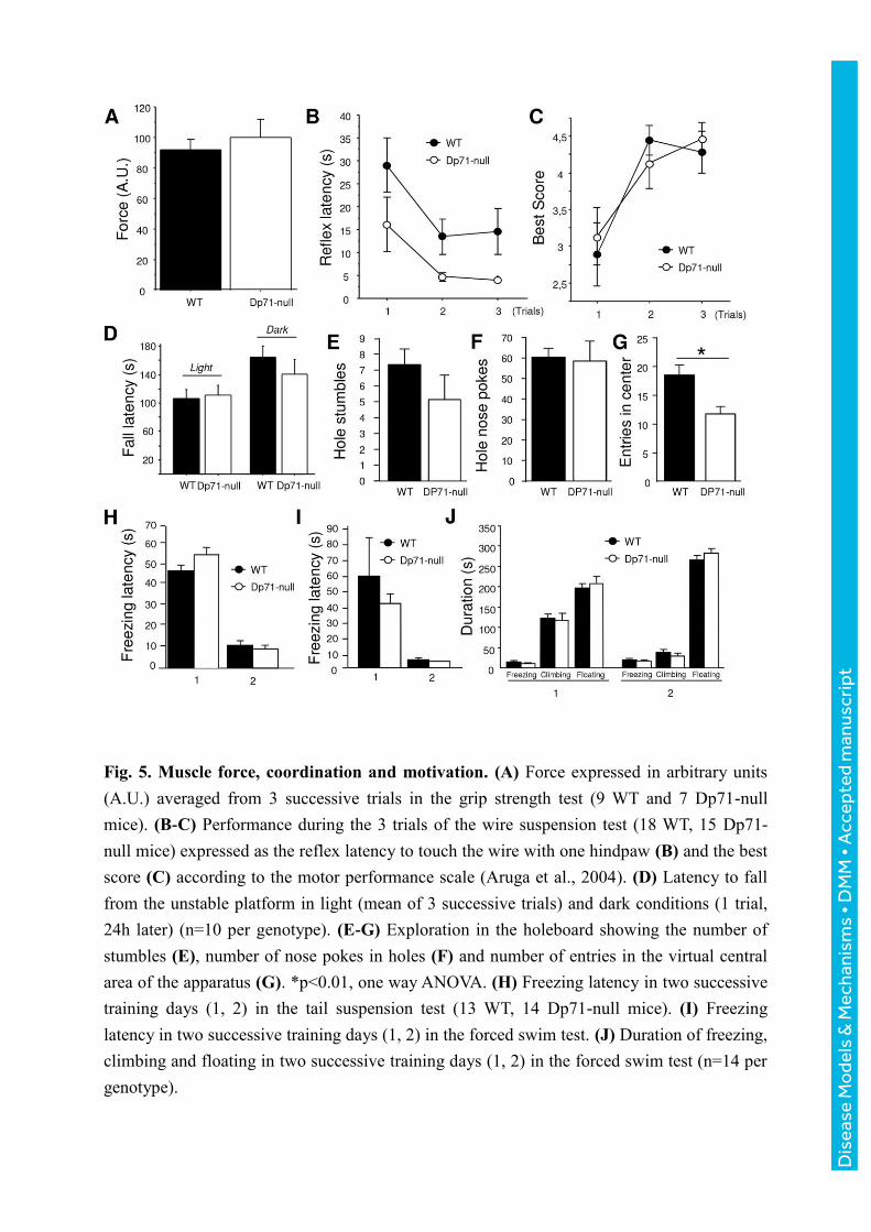
Fig. 5. Muscle force, coordination and motivation. (A) Force expressed in arbitrary units
(A.U.) averaged from 3 successive trials in the grip strength test (9 WT and 7 Dp71-null
mice). (B-C) Performance during the 3 trials of the wire suspension test (18 WT, 15 Dp71-
null mice) expressed as the reflex latency to touch the wire with one hindpaw (B) and the best
score (C) according to the motor performance scale (Aruga et al., 2004). (D) Latency to fall
from the unstable platform in light (mean of 3 successive trials) and dark conditions (1 trial,
24h later) (n=10 per genotype). (E-G) Exploration in the holeboard showing the number of
stumbles (E), number of nose pokes in holes (F) and number of entries in the virtual central
area of the apparatus (G). *p<0.01, one way ANOVA. (H) Freezing latency in two successive
training days (1, 2) in the tail suspension test (13 WT, 14 Dp71-null mice). (I) Freezing
latency in two successive training days (1, 2) in the forced swim test. (J) Duration of freezing,
climbing and floating in two successive training days (1, 2) in the forced swim test (n=14 per
genotype).
Dis
ease
Mo
dels
& M
echa
nism
s •
DM
M •
Acc
epte
d m
anus
crip
t

Fig. 6. Motor synchronization learning and navigation strategies. (A-B) Motor
synchronization learning in the rotarod test. In all experiments, performance was quantified as
the latency to fall from the rod. In a first protocol (A), mouse equilibrium was evaluated
following placement of the mouse on the unrotating rod (speed=0), and then on the rod
rotating at a constant low speed (4 rpm) and during accelerating speed ramps (4-40 rpm)
during 4 days (18 WT and 15 Dp71-null mice). In a second protocol (B-C), motor learning
was first assessed during accelerating speed ramps during 3 days (left curve, B), and then in 8
successive trials, each at a single constant speed as indicated on the x axis (right curve, C)
(n=10 per genotype). (D) Escape latency during 3-day training in the visible-platform
protocol in a water maze (10 WT, 11 Dp71-null mice). (E) Percent time spent in the
Whishaw’s corridor during the first training day in the visible-platform protocol. (F) Escape
latency during 7-day training in the nonvisible-platform protocol with fixed departure
locations in a water maze (n=12 per genotype). (G) Distance swum in the target quadrant
before finding the platform during day 2. Significant genotype effects: *p<0.05, **p<0.01,
***p<0.001; #p<0.05, significant genotype x day interaction.
Dis
ease
Mo
dels
& M
echa
nism
s •
DM
M •
Acc
epte
d m
anus
crip
t

Goal-oriented tasks in the water maze Genotype difference Significance
Visual guidance strategy (Visible platform)
Escape latency
Circling along walls
Cumulative distance to platform
Corrected integrated path length
% Time in Whishaw’s corridor (day 1)
% distance in Whishaw’s corridor (day 1)
Dp71-null < WT
Dp71-null < WT
Dp71-null < WT
Dp71-null < WT
Dp71-null > WT
Dp71-null > WT
p=0.054*
p=0.057*
p=0.045*
p=0.043*
p=0.0004
p=0.0019
Navigation to hidden platform (Fixed start/arrival)
Escape latency (Improvement from day 1 to day 2)
Distance swum (Improvement from day 1 to day 2)
Entries in target quadrant (day 2)
Exits from target quadrant (day 2)
Distance swum in target quadrant before escape (day 2)
Absolute turn angle in target quadrant (day 2)
Meander in target quadrant (day 2)
Dp71-null < WT
Dp71-null < WT
Dp71-null > WT
Dp71-null > WT
Dp71-null > WT
Dp71-null > WT
Dp71-null < WT
p=0.043*
p=0.033*
p=0.031
p=0.014
p=0.010
p=0.030
p=0.054
Table 1. Main genotype differences detected during testing in the water maze. (*) Statistics from
repeated-measure ANOVAs.
Dis
ease
Mo
dels
& M
echa
nism
s •
DM
M •
Acc
epte
d m
anus
crip
t