
Fetal Programming in Meat Production - INRA - Événements · Fetal Programming in Meat Production...
38
Fetal Programming in Meat Production Min Du Department of Animal Sciences Washington State University
Transcript of Fetal Programming in Meat Production - INRA - Événements · Fetal Programming in Meat Production...
Fetal Programming in Meat Production
Min Du
Department of Animal SciencesWashington State University
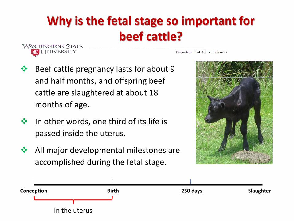
Beef cattle pregnancy lasts for about 9 and half months, and offspring beef cattle are slaughtered at about 18 months of age.
In other words, one third of its life is passed inside the uterus.
All major developmental milestones are accomplished during the fetal stage.
Why is the fetal stage so important for beef cattle?
Conception Birth 250 days Slaughter
In the uterus

Muscle development mainly occurs during the fetal stage.
There is no increase in the number of muscle fibers after birth.
Actually, for beef cattle, the formation of new muscle fibers largely stops after day 210 of gestation (Term around 283 days).
Afterwards, growth of skeletal muscle is mainly due to the increase in the diameter and the elongation of existing muscle fibers.
Fetal muscle development

Skeletal muscle development
First 3 months
Formation of new muscle fibers
3 to 7 months of pregnancy, Formation of new
muscle fibers Growth of muscle
fibers
7 months and after,
Growth of muscle fibers
Increase of muscle fiber formation during the fetal stage will increase later lean growth.

Besides formation of muscle fibers, fetal muscle development also involves formation of adipocytes and formation of fibrogenic cells (connective tissues).
Adipocytes formed during the fetal stage and neonatal stage accumulate lipids during fattening stage, forming marbling.
Excessive formation of connective tissue makes the meat tough.
Fetal muscle development

Mid to late fetal stageLate fetal stageMature muscle
Precursor cells
Myoblasts
Primary Muscle
fiber
Fat cells
Fibroblast
Pluripotentcells
MyoDMyf-5
Myogenin
SecondaryMuscle
fiber
Embryonic stage
Fetal muscle development
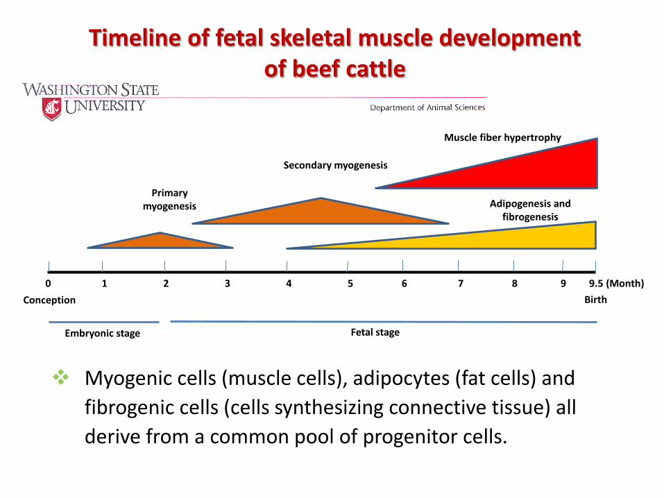
Conception
Embryonic stage
0 1 2 3 4 5 6 7 8 9 9.5 (Month)Birth
Fetal stage
Primary myogenesis
Secondary myogenesis
Muscle fiber hypertrophy
Adipogenesis and fibrogenesis
Timeline of fetal skeletal muscle development of beef cattle
Myogenic cells (muscle cells), adipocytes (fat cells) and fibrogenic cells (cells synthesizing connective tissue) all derive from a common pool of progenitor cells.

Maternal nutrient and marbling
Progenitor cells

Maternal physiological and nutritional status affects progenitor cell proliferation and differentiation into muscle, fat and fibrogenic cells, affecting the lean/fat ratio, beef production efficiency and quality.
Examples:
Nutrient deficiency during mid-gestation decreases the number of progenitor cells, forming less muscle fibers, decreasing muscle mass and lean/fat ratio.
Runt piglets always have a lower lean:fat ratio compared to their littermates.
Maternal nutrient restriction and fetal muscle development

Muscle growth and lean:fat ratio
By contrast, nutrient restriction during late gestation and neonatal stage only affects muscle fiber sizes, which are largely recoverable.
Late gestation is also critical for intramuscular adipogenesis, and nutrient deficiency reduces marbling and overall fatness of animals.

Due to frequent drought and other physiological stresses, beef cattle frequently experience nutrient deficiency during gestation.
Maternal nutrient supplementation is needed to improve production efficiency and quality of offspring.
Maternal nutrient restriction and fetal muscle development

Frequent drought reduces forage production, and cows in many areas experience nutrient deficiency during gestation.
What is the impact on fetal skeletal muscle development and the subsequent production efficiency and beef quality?

Nutrition during mid-gestation affects progeny performance
Animals
At 120 to 150 d of gestation, cows were allotted randomly to one of two dietary treatment, either native range (NR, n = 12) or improved pasture (IP, n = 14) with increased forage production, for 60 days.
Esophageal extrusa samples:
IP varied from 11.1% crude protein of organic matter early in the test period to 6.0% at the end of the grazing period.
NR ranged from 6.5% crude protein of organic matter during early grazing to 5.4 % at the end.

Treatment
Item Native range1 Improved pasture2 P-value
Birth weight, kg 38.7 ± 2.0 36.6 ± 1.9 0.46
Weaning weight, kg 242.1 ± 3.7 256.2 ± 3.5 0.02
Final body weight, kg 538.0 ± 8.3 560.2 ± 7.7 0.07
Average daily gain, kg/d 1.489 ± 0.067 1.656 ± 0.062 0.05
Total body weight gain, kg 180.2 ± 8.0 200.37 ± 7.5 0.05
Live weight at slaughter, kg 520.6 ± 7.7 543.9 ± 7.1 0.04
Effects of cows grazing either native range or improved pasture from 120 to 180 days of gestation on growth of steer progeny
Underwood et al., Meat Science, 86:588-593.

Treatment
Item Native range1 Improved pasture2 P-value
Kidney, Pelvic and Heart fat, % of HCW 3.96 ± 0.25 3.59 ± 0.24 0.32
HCW, kg 329.5 ± 4.8 348.2 ± 4.5 0.01
Yield grade 3.54 ± 0.18 3.84 ± 0.17 0.23
Marbling score3 420 ± 16 455 ± 15 0.12
Effects of cows grazing either native range or improved pasture from 120 to 180 days of gestation on carcass characteristics of
steer progeny
Underwood et al., Meat Science, 86:588-593.

Treatment
Item Native range1 Improved pasture2 P-value
Longissimus muscle area, cm2 75.4 ± 2.2 78.7 ± 2.0 0.26
Semitendinosus, % of HCW 1.16 ± 0.07 1.20 ± 0.07 0.19
Longissimus muscle WBSF, N 37.29 ± 1.28 31.00 ± 1.19 0.004
Collagen content, μg/mg of Ld muscle 19.2 ± 1.9 15.7 ± 1.9 0.08
Ether extract (fat, %) 4.82 ± 0.53 6.00 ± 0.49 0.06
Muscle characteristics of steers from cows grazing either native range or improved pasture from 120 to 180 days of gestation
Underwood et al., Meat Science, 86:588-593.
Likely, the difference in tenderness is due to the reduction in collagen content and increase in lipid content, associated fetal development ---- production and quality problems having a fetal origin.

Summary
Maternal nutrition alters fetal development which has long-term effect on the growth performance of offspring.
Grazing on improved pasture appears to enhance intramuscular adipogenesis and marbling, while reduces collagen content, resulting tender meat.
Poor maternal nutrition reduces growth potential and muscle development in offspring.
How could we solve this production problem?
If we supplement cows with proteins, would that increase muscle growth?

Maternal protein supplementation diverts adipogenesis to myogenesis in beef steers
Mid gestation is an important period for muscle and adipose tissue development.
Thirty six crossbred beef cows were randomly placed on a control diet (100% NRC requirements, n = 12, C), nutrient restricted (70% of requirements, n = 12, NR), or a nutrient restricted diet with protein supplement (NRP, n = 12) designed to equal flow of amino acids to the small intestine of C diet from d 45 to 185 of gestation.
Then, all groups of cows were placed together, managed to meet requirements and allowed to calve.
Steers were slaughtered at 405 days of age.

Treatment P-value
Item C1 NR2 NRP3 Treatment
Live BW, kg 567 ± 22a 588 ± 15a 615 ± 18a 0.240
HCW, kg 375.8 ± 13.8a 377.4 ± 9.6a 398.2 ± 11.2a 0.313
LM area, cm2 86.4 ± 4.2a 88.0 ± 3.0a 90.3 ± 3.4a 0.762
St muscle (kg) 2.44 ± 0.15b 2.55 ±0.10ab 2.87±0.12a 0.067
St muscle % HCW 1.25±0.05b 1.35±0.03ab 1.44±0.04ad 0.02
KPH, % HCW 3.05 ± 0.25a 2.88 ± 0.17a 2.30 ± 0.20b 0.050
Underwood et al., Unpublished data.
Maternal protein supplementation diverts adipogenesis to myogenesis in beef steers

Conclusion so far…
Fetal programming has a major role in determining the production efficiency of beef cattle, as well as beef quality.
Nutrition during pregnancy affects lean/fat ratio, feed efficiency and beef quality.
Through manipulation of nutrition, genetic and other environmental factors, we will be able to maximize the growth potential and meat quality of offspring.

Conception
Embryonic stage
0 1 2 3 4 5 6 7 8 9 9.5 (Month)Birth
Fetal stage
Primary myogenesis
Secondary myogenesis
Muscle fiber hypertrophy
Adipogenesis
Nutrient restriction reduces myogenesis, decreasing muscle fiber number and muscle mass in offspring
Nutrient restriction reduces adipogenesis, decreasing marbling in offspring
Nutrient restriction reduces muscle fiber hypertrophy, decreasing birth weight
Timeline of fetal muscle development in beef cattle

Marbling is critical for the eating quality of beef.
Increase in marbling (fat tissue) is due to: increase in both number and size of adipocytes.
Formation of marbling
Increases in cell sizes
Increases in cell number

Conception Adipogenesis Birth 250 days Slaughterinitiates
Density of multipotent cells
Adipocyte hyperplasia Adipocyte hypertrophy
Enhance marbling through genetic, nutrition and other strategies
Increases adipocyte number and marbling
Enhancing adipogenic differentiation increases fat cell number.
The number of multipotent cells decreases as animals become older.
Thus, to increase adipocyte number in beef cattle, fetal, neonatal and pre-weaning stages are the most effective stages.

Conception Adipogenesis Birth 250 days Slaughterinitiates
Density of multipotent cells
Adipocyte hyperplasia Adipocyte hypertrophy
Increase adipocyte number occurs between mid-gestation to about 250 days of age.
These adipocytes accumulate lipids during the later stage.
“Fattening” stage in feedlots: cattle is fed a grain-based diet.
Such high energy diets provide lipids, leading to increase in adipocyte size.

Conception Adipogenesis Birth 250 days Slaughterinitiates
Density of multipotent cells
Adipocyte hyperplasia Adipocyte hypertrophy
However, the increase in adipocyte size occurs in all fat depots.
The majority of lipids accumulates in visceral and subcutaneous fat, but only limited amount goes to intramuscular fat.
If we can specifically increase intramuscular adipocyte number, we will be able to increase marbling.

Conception Adipogenesis Birth 250 days Slaughterinitiates
Adipocyte hyperplasia Adipocyte hypertrophy
Challenges to specifically increase intramuscular adipogenesis?
Nutrient supplementation during “marbling window” specifically increases intramuscular adipocyte number.
Marbling window
Visceral fat
Subcutaneous fat
Intra and intermuscular fat

Increases intramuscular adipocytes without overall increase in obesity.
During fattening stage, lipid accumulation in intramuscular adipocytes forms abundant marbling.
Conception Adipogenesis Birth 250 days Slaughterinitiates
Marbling window
Visceral fat
Subcutaneous fat
Intra and intermuscular fat

Conception Adipogenesis Birth 250 days Slaughterinitiates
Density of multipotent cells
Adipocyte hyperplasia Adipocyte hypertrophy
Enhance marbling through genetic, nutrition and other strategies
Intramuscular adipogenesis and marbling
Marbling window: Early weaning to around 250 days
Marbling window


Fetal programming in chicken
Unlike mammals, birds are hatched from eggs.
Hatching temperature affects bird development.
Eggs challenged with 39.5 °C for 12 h/d between E7 to 16 compared to 37.8 °C reduced abdominal fat pad by 8% but increased breast muscle yield (Loyau e tal., 2013).
Why?

Future research focus
Continue to define mechanisms linking maternal nutrition to mesenchymal stem cell differentiation into myocytes, adipocytes and fibroblasts.
Many questions are unanswered and more questions are constantly generated.

The mechanisms regulating mesenchymal progenitor cell commitment to myogenesis, adipogenesis and fibrogenesis are poorly defined, especially in livestock.
Adipocytes
Fibroblasts
Satellite cells
Myocytes
Myogenic progenitor cells
Fibro/adipogenicprogenitor cells
Stem cells
1st ?
2nd ?
Resident fibro/adipogenicprogenitor cells

What controls mesenchymal stem cell differentiation?
A critical question.
If we know the answer,
In animal agriculture, we can: Improve lean/fat ratio and production
efficiency.
Enhance intramuscular adipogenesis and marbling.
Reduce fibrogenesis, thus less collagen accumulation and improves tenderness.

National Beef Quality Audits
Consistently identify two top beef quality problems,
Which are:
Insufficient marbling
Lack of tenderness.
Lack of intramuscular fat is also a problem for pork.

Multipotentcells
Muscle cells
Adipocytes
Fibroblasts
Epigenetic changes in progenitor
cells
cell signaling pathways
Genetic, nutritional and other factors
Mechanisms regulating mesenchymal stem cell differentiation

Yan Huang Qiyuan Yang Marcio Duarte Xing Fu Shawn Harris Junxing Zhao Bo Wang Das Arun David Hanna Pat Friel Junfang Liang
Acknowledgements
Lab members

Funding support:

Thanks, advices and questions?


![Ernährung im frühen Kindesalter.ppt [Kompatibilitätsmodus] · Bedeutung der Ernährung • Programmierung von Körperfunktionen: „fetal programming“/ „metabolic programming“](https://static.fdocuments.net/doc/165x107/5ca7191a88c993a22b8b7bf5/ernaehrung-im-fruehen-kompatibilitaetsmodus-bedeutung-der-ernaehrung-programmierung.jpg)
