Experiment Acknowledged the Watson-Crick Hypothesis: A ... · Watson-Crick ideal structure, is...
12
Gen. Physiol. Biophys. (1985), 4, 471 482 471 Experiment Acknowledged the Watson Crick Hypothesis: A Review of B DNA Double Helix Structural Features at Atomic Resolution J. KYPR and M. VORLÍČKOVÁ Institute of Biophysics, Czechoslovak Academy of Sciences, Královopolská 135, 612 65 Brno, Czechoslovakia Abstract. The dodecamer d(CGCGAATTCGCG) forms a right handed B DNA double helix of a Watson Crick type both in crystal and solution. It is the first piece of DNA longer than one helix turn whose molecular structure has become known at the atomic resolution. The article reviews qualitative aspects of its structure with a special emphasis on local variations in the disposition of base pairs in the double helix. Key words: DNA fragment crystals — B DNA double helix — DNA bending — Propeller twist of base pairs — Base stacking — Helical twist Introduction More than three decades ago, Watson and Crick (1953a) postulated the double helical nature of DNA structure. Their double helix designated B DNA was right handed, its interior contained hydrogen bonded pairs of complementary bases, and sugar phosphate chains run in antiparallel direction outside (Fig. 1). This view was actually not more than a hypothesis since the experimental data available could even a quarter of a century later be interpreted in other ways. Nonetheless, the double helix so naturally accounted for the existing knowledge concerning the gene that it was immediately accepted by the scientific community. The double helix not only showed what the central molecule of life looked like but it also suggested how it is copied (Watson and Crick 1953b). After the celebrated beginning of the double helix life it was soon realized that there was no direct evidence to substantiate the Watson Crick hypothesis. Wilkins and coworkers improved the quality of X ray diffraction pictures of DNA oriented fibres but they were fully aware (Fuller et al. 1965) not even to be able to demonstrate unambiguously the handedness of DNA structure. The resolution of the structural information was too low. The situation turned even more obscure in the mid seventies when two groups independently proposed stereochemically and energetically sound DNA structures in which the sugar phosphate chains did not
Transcript of Experiment Acknowledged the Watson-Crick Hypothesis: A ... · Watson-Crick ideal structure, is...
Gen. Physiol. Biophys. (1985), 4, 4 7 1 - 4 8 2 471
Experiment Acknowledged the Watson-Crick Hypothesis: A Review of B-DNA Double Helix Structural Features at Atomic Resolution
J. K Y P R a n d M . V O R L Í Č K O V Á
Institute of Biophysics, Czechoslovak Academy of Sciences, Královopolská 135, 612 65 Brno, Czechoslovakia
Abstract. The dodecamer d(CGCGAATTCGCG) forms a right-handed B-DNA double helix of a Watson-Crick type both in crystal and solution. It is the first piece of DNA longer than one helix turn whose molecular structure has become known at the atomic resolution. The article reviews qualitative aspects of its structure with a special emphasis on local variations in the disposition of base pairs in the double helix.
Key words: DNA fragment crystals — B - DNA double helix — DNA bending — Propeller twist of base pairs — Base stacking — Helical twist
Introduction
More than three decades ago, Watson and Crick (1953a) postulated the double-helical nature of DNA structure. Their double helix designated B-DNA was right-handed, its interior contained hydrogen-bonded pairs of complementary bases, and sugar-phosphate chains run in antiparallel direction outside (Fig. 1). This view was actually not more than a hypothesis since the experimental data available could even a quarter of a century later be interpreted in other ways. Nonetheless, the double helix so naturally accounted for the existing knowledge concerning the gene that it was immediately accepted by the scientific community. The double helix not only showed what the central molecule of life looked like but it also suggested how it is copied (Watson and Crick 1953b).
After the celebrated beginning of the double helix life it was soon realized that there was no direct evidence to substantiate the Watson-Crick hypothesis. Wilkins and coworkers improved the quality of X - ray diffraction pictures of DNA oriented fibres but they were fully aware (Fuller et al. 1965) not even to be able to demonstrate unambiguously the handedness of DNA structure. The resolution of the structural information was too low. The situation turned even more obscure in the mid-seventies when two groups independently proposed stereochemically and energetically sound DNA structures in which the sugar-phosphate chains did not

472 Kypr and Vorličková
Fig. 1. Schematic diagram showing DNA sugar-phosphate backbone and hydrogen bonding of complementary bases. Note the arrows indicating the conventional 5—»3' progression of the backbone.
intertwin. The structures were called SBS (side-by-side, Rodley et al. 1976) and RL (right-left, Sasisekharan and Pattabiraman 1976); they consisted of alternating right-handed and left-handed blocks and their calculated X-ray diffraction patterns were as consistent with the experimental data as the calculated pattern of the Watson-Crick double helix.
The period of diverse notions about the very nature of DNA structure and its handedness culminated by the totally unexpected discovery of the Z-DNA double helix (Wang et al. 1979; Drew et al. 1980; Pohl and Jovin 1972). Z-DNA is a zig-zag left-handed structure observed in the first single crystals containing double helices formed by DNA fragments whose structure has been successfully solved at atomic resolution. It is specific for the alternating purine-pyrimidine sequence of GC base pairs. The single crystal X-ray diffraction data are of sufficient resolution to avoid any reasonable doubt about their structural interpretation. However, fragments with sequences other than the strictly alternating GC were found to form the classical B-DNA and A - D N A double helices in crystals. Their basic structural properties are in perfect agreement with what Watson and Crick proposed thirty years ago. We review structural properties of
Molecular Structure of B-DNA 473
3*-<e)—c-
BASE STEP T-^T^C-^-@y^c-U®-*3'
pu - py steps
py - pu steps
£
+1
-u -3
-2 +2 +7
0.
+1
-4
-3
+2
Q.
+1
•1
-2
-2
+1
•1
+2
•2
+1
A -3
-2 +2 +2
•2
+i
4 -3
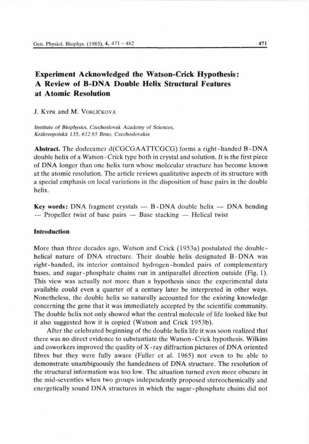
Fig. 2. Schematic drawing of the dodecamer d(CGCGAATTCGCG) duplex. Clashes between purines which are circled are indicated by twosided arrows. Br, P t l , Pt2 and Pt3 show the sites where the dodecamer was brominated or cisplatinated (the increasing number corresponds to a declining degree of modification). Calladine-Dickerson rules are used to predict local variations in the helical twist.
B-DNA in this article since it appears as the most frequent arrangement of genetic material in vivo.
Global features of B-DNA
The B-DNA double helix has been found to be formed by the dodecamer d(CGCGAATTCGCG). It has a selfcomplementary sequence (Fig. 2) and thus forms a double helix with itself. Its sequence has originally been chosen to incorporate the Eco R l restriction endonuclease target site, d(GAATTC). Since in the meantime the tetranucleotide d(CGCG) was discovered to adopt the left-handed Z-DNA helix (Drew et al. 1980) the dodecamer sequence was potentially even more interesting. It provided an example of two Z-compatible segments d(CGCG), bracketing a Z-incompatible d(AATT) core. The dodecamer crystals were grown from solutions as was d(CGCG). Consequently, if there had been a significant tendency for d(CGCGAATTCGCG) to adopt, say, a partial Z -DNA conformation, it should do so in the crystal. Instead, a right-handed B-DNA double helix was formed, with not even an indication of local structural irregularity in the d(CGCG) regions (Wing et al. 1980). Z-DNA thus seems rather scarce variant of the double helix.
The B-DNA molecule has an average rise of 0.34 nm per residue and 10.1 base pairs per turn. This is somewhat less than 10.4 base pairs per turn as measured in solution (Wang 1979) or as calculated from energy considerations (Levitt 1978). Even at the early stage of the structure analysis, two interesting departures from classical B - DNA geometry were noted with the dodecamer: each base pair had a propeller twist (the angle between base planes in the hydrogen - bonded pair,
474 Kypr and Vorlíčková
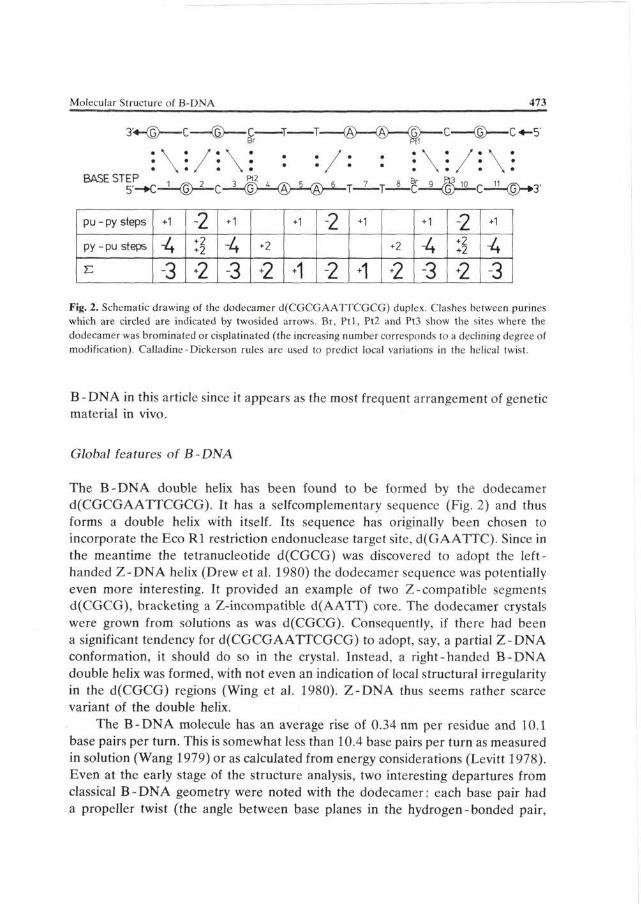
Fig. 3. Schematic drawing of the steric clash of purines caused by propeller twisting of base pairs. Note that the clash occurs in the minor groove for the pyrimidine-purine step.
Fig. 3) that increased the overlap between bases in the same chain, and the double helix was bent by 19° over the dodecamer molecule (Wing et al. 1980).
Helical twist and local helix types
Helical twist defined as the rotation of successive base pairs around the helix axis alternates between 27.4° (base step 3, i.e. C-G) and 40.3° (base step 8, i.e. T-C and in the opposite strand G-A) to give an average value of 35.8° consistent with that known from DNA fibre studies (Langridge et al. 1960). The base sequence variations are thus very marked (Dickerson and Drew 1981). There is a relatively high occurrence of C-G (1, 3, 9, 11) and G-C (2, 10) steps in the dodecamer. Helical rotations at C - G steps are systematically smaller than average, and those at G-C steps are larger. This suggests a form of molecular compensation, by which stacking in the pyrimidine-purine steps, where base overlap is small in the Watson-Crick ideal structure, is strengthened by a decreased helical twist. At the same time, the stronger stacking interactions in the purine-pyrimidine steps are weakened. The alternating sequence of AT base pairs, however, behaves quite differently. Klug et al. (1979) proposed on the basis of the crystal structure of the tetranucleotide d(ATAT) (Viswamitra et al. 1978) that the energy contributed by the T-A stacking was so small that little would be lost by eliminating it altogether with an increased twist. This would, on the other hand, permit unwinding and the resulting enhanced A-T stacking at adjacent positions. The dodecamer has only one A-T sequence at step 6 which has a small twist value (Dickerson and Drew 1981) consistent with the proposal. Consequently, Nature seems to have adopted
Molecular Structure of B-DNA 475
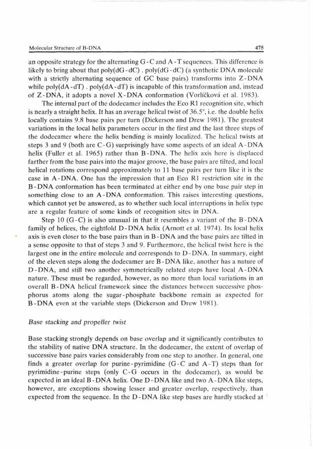
an opposite strategy for the alternating G - C and A - T sequences. This difference is likely to bring about that poly(dG-dC) . poly(dG-dC) (a synthetic DNA molecule with a strictly alternating sequence of GC base pairs) transforms into Z-DNA while poly(dA-dT) . poly(dA-dT) is incapable of this transformation and, instead of Z-DNA, it adopts a novel X-DNA conformation (Vorlíčková et al. 1983).
The internal part of the dodecamer includes the Eco R l recognition site, which is nearly a straight helix. It has an average helical twist of 36.5°, i.e. the double helix locally contains 9.8 base pairs per turn (Dickerson and Drew 1981). The greatest variations in the local helix parameters occur in the first and the last three steps of the dodecamer where the helix bending is mainly localized. The helical twists at steps 3 and 9 (both are C-G) surprisingly have some aspects of an ideal A-DNA helix (Fuller et al. 1965) rather than B-DNA. The helix axis here is displaced farther from the base pairs into the major groove, the base pairs are tilted, and local helical rotations correspond approximately to 11 base pairs per turn like it is the case in A-DNA. One has the impression that an Eco Rl restriction site in the B - DNA conformation has been terminated at either end by one base pair step in something close to an A-DNA conformation. This raises interesting questions, which cannot yet be answered, as to whether such local interruptions in helix type are a regular feature of some kinds of recognition sites in DNA.
Step 10 (G-C) is also unusual in that it resembles a variant of the B-DNA family of helices, the eightfold D - D N A helix (Arnott et al. 1974). Its local helix axis is even closer to the base pairs than in B-DNA and the base pairs are tilted in a sense opposite to that of steps 3 and 9. Furthermore, the helical twist here is the largest one in the entire molecule and corresponds to D-DNA. In summary, eight of the eleven steps along the dodecamer are B-DNA like, another has a nature of D - D N A , and still two another symmetrically related steps have local A-DNA nature. These must be regarded, however, as no more than local variations in an overall B-DNA helical framework since the distances between successive phosphorus atoms along the sugar-phosphate backbone remain as expected for B - D N A even at the variable steps (Dickerson and Drew 1981).
Base stacking and propeller twist
Base stacking strongly depends on base overlap and it significantly contributes to the stability of native DNA structure. In the dodecamer, the extent of overlap of successive base pairs varies considerably from one step to another. In general, one finds a greater overlap for purine - pyrimidine (G-C and A-T) steps than for pyrimidine-purine steps (only C - G occurs in the dodecamer), as would be expected in an ideal B-DNA helix. One D-DNA like and two A-DNA like steps, however, are exceptions showing lesser and greater overlap, respectively, than expected from the sequence. In the D - D N A like step bases are hardly stacked at

476 Kypr and Vorlíčková
all. On the other hand, the A-DNA like steps show a greater than expected overlap.
Propeller twist of base pairs was originally considered zero in DNA double helix modelling by Watson and Crick and other workers but crystallographic studies on the dodecamer and other DNA fragments convincingly showed that a substantial propeller twist was an inherent feature of DNA structure. It should be pointed out that its existence was proposed from energy calculations (Levitt 1978) and that the proposal was supported by electric dichroism measurements in solution (Hogan et al. 1978).
The presence of a propeller twist has non-trivial consequences for the architecture of DNA (Calladine 1982). Since purines are larger than pyrimidines, they extend across the helix axis. This does not matter at the purine-purine or pyrimidine-pyrimidine steps. However, if purines occur in next steps at the opposite strands then the propeller twist results in their steric clash in the helix centre (Fig. 3). The clash can be removed by several mechanisms (Calladine 1982). The base pairs can, for example, rotate as rigid units about their sugar-sugar virtual bonds. The rotation results in a compression of the double helix groove and bending of the double helix. Note that the double helix should bend toward the major groove in the pyrimidine-purine step (Fig. 3) but toward the minor groove in the purine-pyrimidine step, to avoid the steric clash. Another mechanism is a shift sideways of each base pair towards its purine end or a local double helix unwinding.
An inspection of the dodecamer crystal structure shows that propeller twist values for AT base pairs in the centre of the molecule have a mean of 17.3° and a standard deviation of 0.4°, whereas GC pairs on the ends of the molecule are less propeller twisted (11.5° in average) and the twists are much more variable (standard deviation 5.1°). This effect can be explained by the interactions between purine bases on the opposite strands which lead to a damping-down of the propeller twist in at least one of the two base pairs involved (Dickerson and Drew 1981). The damping is particularly strong with GC pairs at the molecule ends where purines and pyrimidines alternate on both strands. Here, the central GC pairs are constrained to a propeller twist of 5° by the flanking G's on the opposite strand. In contrast to the foregoing, no interstrand overlaps occur in the central portion of the dodecamer, where AT pairs have large and uniform propeller twists. AT pairs should have greater propeller twists than GC pairs in general, both because deletion of one hydrogen bond permits easier twisting, and because the absence of a side group on the C2 position of adenine decreases the likelihood of interstrand overlap. The distribution of propeller twists in the dodecamer suggests, contrary to expectations, that the interstrand base overlap is a more significant factor in lowering the propeller twist (Dickerson and Drew 1981).
It can be concluded that the propeller twist is a variable and significant

Molecular Structure of B-DNA 477
structural parameter of the double helix. It improves intrastrand base stacking but results in steric clashes at purine - pyrimidine and pyrimidine - purine steps that should be released by local helix adjustments. It will be shown in the following paragraph that it is the steric clash that may account for the sequence-dependent helical variations observed with the dodecamer.
Calladine - Dickerson rules
Calladine first noted the steric hindrance between purines at adjacent base pairs on opposite strands of the helix and he proposed mechanisms to relieve it (Calladine 1982). On the basis of Calladine's work, Dickerson defined simple sum functions by which local variations in several local helix parameters may be calculated from the base sequence (Dickerson 1983). The function to predict local helix variations takes into account the alternating purine-pyrimidine (R-Y) and pyrimidine-purine (Y - R) steps only and ignores R - R and Y - Y steps where no clash of purines can occur in the helix centre. When one predicts, for example, variations in helical twist then for the sequence x - R - Y - x , where x stands for any base, the values + 1 , —2, +1 are taken into the function for the x-R, R-Y and Y-x steps, respectively. On the other hand, +2, - 4 , +2 are included for sequences of the type x -Y-R-x . The function is built up by carrying up the assignment of the above values at every step of the sequence and summing up the particular contributions. An application of these rules to the dodecamer d(CGCGAATTCGCG) is given in Fig. 2. One unit in the function corresponds to 2.1° of the helix twist, centered around the function null value of 35.6° for a B-DNA helix.
The Calladine-Dickerson rules precisely predict the observed local variations in the helix twist of the dodecamer d(CGCGAATTCGCG) and other right-handed oligodeoxynucleotides (Dickerson 1983). They in fact predict what is recognized by certain deoxyribonucleases (Lomonossoff et al. 1981; Drew 1984). The correspondence between predicted and observed values is surprisingly excellent if we take into account the evident fact that the rules are still very approximative at the present state of art. It is naturally unacceptable to take, for example, G as identical with A, or C identical with T, but this only is a consequence of insufficient quantity of data. Despite the very approximative nature, the rules may be considered as the first step in establishing the DNA sequence - structure vocabulary. Even a brief inspection of the rules hints, and concrete calculations (Lennon and Nussinov 1984) have confirmed the view that the DNA sequence /structure vocabulary will contain many homonyms (similar sequences give different structures) and synonyms (different sequences give similar structures).
478 Kypr and Vorlíčková
Reversible bending in a B-DNA dodecamer
The dodecamer d(CGCGAATTCGCG) has been analysed by single crystal X-ray diffraction in several variants. Three different variants of the dodecamer were synthesized with 5-bromocytosine in the first, third, and ninth position, designated 1 -Br, 3-Br, and 9-Br, respectively. Crystals of the 1 -Br derivative were of poor quality and unsuitable for structural analysis. The 3-Br crystals were of no structural interest because of their excellent isomorphism with the parent dodecamer. The 9-Br compound, in contrast, proved to undergo a completely unexpected reversible bending-straightening phase transition within the crystal that has permitted to examine a straight, undistorted B-DNA double helix.
The 19° axial bend was one of the most striking features of the parent dodecamer structure. It was originally attributed to crystal-packing forces. The 9-Br helix also bends, but to a smaller extent (14°). However, soaking 9-Br crystals in alcohol induced a phase transition to an unbent helix (Fratini et al. 1982). Warming reversed the process and restored the 14° bend. Examination of the straight, 14° and 19° molecules enables to study bending of B-DNA and the structural effects of 5-bromo substitution at the cytosine ring which occurs in the major groove. Both these aspects are of a considerable biological interest. Bending closely relates to the mechanism by which DNA wraps into a supercoil of the radius of 4.2 nm about the histone octamer in chromatin. Cytosine methylation, on the other hand, regulates gene activity in higher organisms. These effects can, at least in part, be related to a change in the double helix structure as seen with the dodecamer. It should be pointed out that bromine and methyl are structurally homologous — their van der Waals radii are 0.2 nm.
Two alternative models have been suggested as to the basic principles controlling DNA bending: smooth, continuous or sharp, discontinuous. The bends one can see in the dodecamer d(CGCGAATTCGCG) are rather localized but the structural irregularity is propagated to either side of the bending point to avoid destacking of bases (Fratini et al. 1982). This has no effect on the helix twist but the major groove is compressed and the minor groove is extended at steps 4 (G-A) and 8 (T-C). At these steps the central A + T-rich region, where the propeller twist decreased from 22° to 17° during the unbent to bent helix transition, meets the bracketing regions, where, simultaneously, propeller twist increased from 5° to 11°. One can immediately see why the bromine atom, which drives the transition, prevents bending of the helix. It is located in the major groove at the step 8, where the helix bends, and sterically prevents its compression. In other words, it stiffens the helix and this may have far reaching biological consequences.

Molecular Structure of B-DNA 479 — — má
The primary mode of cisplatin binding to the dodecamer
Cisplatin (cis-Pt(NH3)2Cl2) is one of the most effective inorganic antitumour drugs. It is -believed to interfere with DNA replication and transcription in a manner similar to that of alkylating agents. In solution under normal physiological conditions, cisplatin binds most strongly to the N7 position of guanine. Visualization of its interaction with DNA at the atomic level might contribute to the understanding of its important biological function.
Wing et al. (1984) equilibrated native d(CGCGAATTCGCG) crystals in the solution of cisplatin to find its primary binding sites on the dodecamer. They obtained several crystals of the modified oligonucleotide, but only three of the eight guanines that could serve as binding sites for cisplatin were occupied even in the most highly substituted crystal. Moreover, the substitution was incomplete (61 % occupancy at the most reactive guanine, Fig. 2) and the occupancy was not equal (the other two reactive quanines were occupied in 30 % and 22 % cases, respectively). Attempts to obtain complete substitution or 100 % binding to the major site resulted in destruction of the crystal order.
An analysis of cisplatinated crystals indicated that the base pairs were shifted relative to their positions in the native structure. Guanines moved by almost 0.1 nm towards the platinum bound in the DNA major groove, bringing the hydrogen-bonded cytosines with them. The shift could be enough to destabilize the helix and to account for the crystal disruption at a high cisplatin content.
At the first sight, the radically different degrees of substitution at the eight guanine sites along the double helix are surprising; there however is a simple explanation in terms of the local guanine environment. The increased reactivity of guanines the farther they are from the ends of the helix probably is explained by intermolecular interactions in the crystal. The crystal contains columns of helices with overlapping ends whose minor grooves are interlocked by hydrogen bonding of the first two base pairs. Hence the first two bases from each helix end are immobilized on the minor groove side and cannot shift towards the major groove to accomodate cisplatin binding. The third base pair is somewhat less constrained and the fourth one is the most free of all. The reactivity of a particular guanine thus appears to be directly related to its ability to move towards the potential cisplatin site. Why, however, the extent of cisplatin binding differs between the two ends ?
It should be recalled that the dodecamer is bent with the sharpest bending asymmetrically located at the site where a spermine molecule bridges the major groove in the parent DNA structure (Wing et al. 1980). In the cisplatin complex, the spermine molecule is displaced by the diffused - in cisplatin, but the asymmetric bending remains. The bending constricts the major groove and hence decreases the affinity for cisplatin at one end of the dodecamer. It is thus evident that the study

480 Kypr and Vorlíčková
only shows primary binding modes of cisplatin binding to the DNA fragment whose structure was in several important aspects predetermined by crystal packing forces. One wonders what would the complex look like if the dodecamer were first platinated in solution and only then crystallized.
The dodecamer structure in solution
The dodecamer d(CGCGAATTCGCG) is the first piece of DNA for which results of both X-ray diffraction and NMR are available. This permits a detailed comparison of its structure in crystal and in solution. The 'H NMR spectrum of the dodecamer in solution contains six resonances of the imino hydrogens involved in the Watson-Crick base pairing, consistent with the formation of a 12 base pair self-complementary duplex with a twofold symmetry (Patel et al. 1982). In contrast, no exact twofold symmetry is observed in the dodecamer crystal, presumably due to contributions from packing forces in the solid state. Temperature-dependence of the six imino resonances shows striking differences between AT and GC base pairs (Patel et al. 1982). Increasing temperature may change winding of the duplex or the extent of propeller - twisting of the base pairs in the A + T-rich core of the duplex. On the other hand, GC base pairs at the ends of the dodecamer little change conformation.
'H NMR resonances of the C-H hydrogens of bases display characteristic upfield shifts upon duplex formation. Their magnitudes can be compared with theoretical calculations, which demonstrated that the overlap geometry of bases observed in the crystal structure of the dodecamer was retained in solution. 3 1 P NMR, furthermore, indicated sequence-dependent variations in the phos-phodiester backbone geometry. No sign of the presence of the left-handed Z-DNA conformation has been observed in dodecamer double helix (Patel et al. 1982).
The sequence-dependent variations in the dodecamer structure are recognized by DNAase I (Lomonossoff et al. 1981). This enzyme cuts the dodecamer at the individual steps with rates whose logarithms are proportional to the respective helical twists.
Conclusion
This review provides a picture about the internal disposition of base pairs in a right-handed B-DNA double helix of the dodecamer d(CGCGAATTCGCG), the first DNA fragment longer than one helical turn whose structure has been known at atomic resolution. In conclusion, we recall the words of Richard Dickerson, in whose laboratory an absolute majority of this really fundamental work has been done. He says: "We know much about one molecule, but are unsure
Molecular Structure of B-DNA 481
as to how this can safely be generalized to similar molecules with different sequences. All of the above principles must be tested against other DNA molecules, and if the history of protein crystallography is any guide, the present simplicity in DNA structure that comes mainly from ignorance will shortly be replaced by a bewildering complexity of new data, before it ultimately settles down again into the simplicity that means that we truly understood matters. The d(CGCGAATTCGCG) structure is a beginning."
Acknowledgement. We greatly appreciate the help of Mrs Marcela Pŕerovská with the manuscript preparation.
References
Arnott S., Chandrasekaran R., Hukins D W L., Smith P. J. C , Watts L. (1974): Structural details of a double helix observed for DNAs containing alternating purine and pyrimidine sequences. J. Mol. Biol. 88, 523 — 533
Calladine C. R (1982): Mechanics of sequence-dependent stacking of bases in B-DNA. J. Mol. Biol. 161, 343 — 352
Dickerson R. E. (1983): Base sequence and helix structure variation in B- and A-DNA. J. Mol. Biol. 166, 419—441
Dickerson R. E., Drew H. R. (1981): Structure of a B - DNA dodecamer. II. Influence of base sequence on helix structure. J. Mol. Biol. 149, 769—786
Drew H. R. (1984): Structural specificities of five commonly used DNA nucleases. J. Mol. Biol. 176, 535 — 557
Drew H. R., Takano T., Tanaka S., Itakura K., Dickerson R. E. (1980): High-salt d(CpGpCpG), a left-handed Z'DNA double helix. Nature 286, 567 — 573
Fratini A. V., Kôpka M. L., Drew H. R., Dickerson R. E. (1982): Reversible bending and helix geometry in a B-DNA dodecamer CGCGAATTB rCGCG. J. Biol. Chem. 257, 14686 — 14707
Fuller W., Wilkins M. H. F., Wilson H. R., Hamilton L. D. (1965): Molecular configuration of deoxyribonucleic acid. IV. X-ray diffraction study of the A-form. J. Mol. Biol. 12, 6 0 — 8 0
Hogan M., Dattagupta N., Crothers D. M. (1978): Transient electric dichroism of rod-like DNA molecules. Proc. Nat. Acad. Sci. USA 75, 195 — 199
Klug A., Jack A., Viswamitra M. A., Kennard O., Shakked Z., Steitz T. A. (1979): A hypothesis on a specific sequence-dependent variations in the conformation of DNA. J. Mol. Biol. 131, 669—680
Langridge R., Marvin D. A., Seeds W. E., Wilson H. R., Hooper C. W., Hamilton L. D. (1960): The molecular configuration of deoxyribonucleic acid. II. Molecular models and their Fourier transforms. J. Mol. Biol. 2, 38—64
Lennon G. G , Nussinov R. (1984): Homonyms, synonyms and mutations of the DNA sequence /structure vocabulary. J. Mol Biol. 175, 425—430
Levitt M (1978): How many base pairs per turn does DNA have in solution and in chromatin? Some theoretical calculations. Proc. Nat. Acad. Sci. USA 75, 640—644
Lomonossoff G. P., Butler P. J. G., Klug A. (1981): Sequence-dependent variations in the conformation of DNA. J. Mol. Biol. 149, 745 — 760
Patel D. J., Pardi A., Itakura K. (1982): DNA conformation, dynamics, and interactions in solution. Science 216, 581—590
482 Kypr and Vorlíčková
PohI F. M., Jovin T. M- (1972): Salt-induced cooperative conformational change of a synthetic DNA: Equilibrium and kinetic studies with poly(dG-dC). J. Mol. Biql. 67, 375 — 396
Rodley G. A., Scobie R. S., Bates R. H. T., Lewitt R. M. (1976): A possible conformation for double-stranded polynucleotides. Proc. Nat. Acad. Sci. USA 73, 2959—2963
Sasisekharan V., Pattabiraman N. (1976): Double-stranded polynucleotides: Two typical alternative conformations for nucleic acids. Current Science 20, 779—783
Viswamitra M. A., Kennard O., Jones P. G., Sheldrick G. M., Salisbury S., Falvello L., Shakked Z. (1978): DNA double helical fragment at atomic resolution. Nature 273, 687—688
Vorlíčková M., Sklenár V., Kypr J. (1983): Salt-induced conformational transition of poly(dA-dT). poly(dA-dT). J. Mol. Biol. 166, 85 — 92
Wang J. C. (1979): Helical repeat of DNA in solution. Proc. Nat. Acad. Sci. USA 76, 200—203 Wang A. H. J., Quigley G. J., Kolpak F. J., Crawford J. L., van Boom J. H-, van der Marel G-, Rich A.
(1979): Molecular structure of a left-handed double helical DNA fragment at atomic resolution. Nature 282, 6 8 0 ^ 6 8 6
Watson J ; D-, Crick F. H, C (1953a): Molecular structure of nuclejc acids. Nature 171, 737—740 Watson J. D-, Crick F. H. C. (1953b): Genetical implications of the structure oi deoxyribonucleic acid.
Nsture 171, 964—967 Wing R M., Drew H. R., Takano T., Broka C , Tanaka S., Itakura K., Dickersen R. E. (1980): Crystal
structure analysis of a complete turn of B-DNA. Nature 287, 755—758 Wing R- M., Pjura P., Drew H. R., Dickerson R. E. (1984): The primary mode of binding of cisplatin to
a ! = 0 N A dodecamer d(CGCGAATTCGCG). The EMBO J. 3, 1201 — 1206
Received January 13, 1984/Accepted November 1, 1984