Estudi de la neurotransmissió a l’esfínter esofàgic inferior · Especialista en Cirurgia de...
156
Estudi de la neurotransmissió a l’esfínter esofàgic inferior Ricard Farré i Martí Memòria presentada per optar al grau de Doctor en Biologia Fundació de Gastroenterologia Dr. Francisco Vilardell i Departament de Biologia Cel·lular, Fisiologia i Immunologia. Unitat de Fisiologia Animal Facultat de Veterinària Universitat Autònoma de Barcelona Bellaterra, desembre del 2005
Transcript of Estudi de la neurotransmissió a l’esfínter esofàgic inferior · Especialista en Cirurgia de...

Estudi de la neurotransmissió a
l’esfínter esofàgic inferior
Ricard Farré i Martí
Memòria presentada per optar al grau de Doctor en Biologia
Fundació de Gastroenterologia Dr. Francisco Vilardell i Departament de Biologia Cel·lular, Fisiologia i Immunologia. Unitat de Fisiologia Animal Facultat de Veterinària Universitat Autònoma de Barcelona
Bellaterra, desembre del 2005

Estudi de la neurotransmissió a
l’esfínter esofàgic inferior
Ricard Farré i Martí
Fundació de Gastroenterologia Dr. Francisco Vilardell i Departament de Biologia Cel·lular, Fisiologia i Immunologia Unitat de Fisiologia Animal Facultat de Veterinària Universitat Autònoma de Barcelona
Dr. Pere Clavé i Civit Doctor en Medicina i Cirurgia Especialista en Cirurgia de l’Aparell Digestiu Investigador Associat i Patró de la Fundació de Gastroenterologia Dr. F. Vilardell, Barcelona Metge adjunt del Servei de Cirurgia de l’Hospital de Mataró Dr. Marcel Jiménez i Farrerons Titular d’universitat Departament de Biologia Cel·lular, Fisiologia i Immunologia Facultat de Veterinària Fan constar que:
La tesi doctoral titulada: “Estudi de la neurotransmissió a l’esfínter esofàgic inferior”
presentada per Ricard Farré i Martí per optar al grau de doctor en Biologia, ha estat
realitzada en el marc del conveni del Grup de Recerca per el Estudi de la Motilitat
Gastrointestinal de la Universitat Autònoma de Barcelona amb la Fundació de
Gastroenterologia Dr. Francisco Vilardell; havent estat finalitzada i estant en condicions
per ser llegida pel candidat per obtenir el grau de Doctor en Biologia.
Bellaterra, octubre de 2005
Dr. Pere Clavé i Civit Dr. Marcel Jimenez i Farrerons Director de la tesi Tutor de la tesi

Dibuix de la coberta procedent del llibre Textura del sistema nervioso del hombre y de
los vertebrados de Santiago Ramón y Cajal (1904). Gangli del plexe d’Auerbach de
l’intestí del conill. Mètode de Ehrlich.

“Ensenya més la necessitat que la universitat”
Anònim
“A la Tresa, al Lluís i a la Fina, perquè sense ells no hagués arribat fins aquí”
“Al Pepito, per ensenyar-me tantes coses fins a l’últim moment”
“A tu, amor, per aguantar-me tots aquests anys”
“A la família Durà-Soler, per fer-me sentir com a casa”
“A Brikidiki, per tots els moments que em passat junts”
“I a tots aquells que encara tenen ganes de veure’m”

Agraïments Al Dr. Pere Clavé, per confiar en mi i donar-me l’oportunitat de poder fer
aquesta tesi.
A la Fundació de Gastroenterologia Dr. Francisco Vilardell, per cobrir gairebé
totes les despeses de la meva beca doctoral tot i la dificultat que entenc que això ha
comportat. Especialment a la Lupe, per la seva tasca administrativa i pels seus consells.
A l’Emma, per aguantar-me durant gairebé cinc anys en els millors i pitjors
moments, pel seu molt bon treball i per tots els cafès que em pres junts.
A la Begoña i a la Mariona per continuar els meus estudis i acabar els últims
experiments i pel temps que em estat junts.
A l’Aulí, per portar-se tan bé amb mi, per ajudar-me en tot i més i per tenir-te al
meu costat quan t’he necessitat.
Al la Patri i a la Maite pels la seva ajuda i consells.
Als becaris que estan ho han passat per la Unitat: Yolanda, Domènech, l’Anna
Alcántara, Mònica, Diana, Estefania, Esther, Carol, Joan i Hernan per la seva ajuda en
algun moment i per les bones estones que em passat.
A l’Antonio pel suport científic i per acompanyar-me a l’escorxador més d’un
cop, al Pepe pel seu suport administratiu.
Al Marcel, per ensenyar-me tantes coses a nivell científic i no científic, pels seus
consells i per cuidar-me tant.
A la Domènech, perquè sense ella no hagués pogut dipositar la tesi a temps, per
la seva ajuda i per fer-me companyia algun cap de setmana.
A les Mòniques i l’Aïda d’Anatomia Patològica, a l’Enric i a la Carme per la
seva ajuda amb les immunos.
A l’Escorxador Frigorífic Avinyó i a l’Escorxador de Sabadell per donar-nos la
major part de la matèria primera d’aquesta tesi.
A les persones de l’Hospital de Bellvitge, Hospital de Mataró, Hospital clínic i
Hospital Mútua de Terrassa que han fet possible el poder treballar amb teixit humà.
Als membres del tribunal per acceptar la invitació i pel gran esforç que els ha
suposat tot plegat.
A la Susanna Comellas, per acompanyar-me durant força temps a l’escorxador a
buscar teixit, a canvi de l’esmorzar: un croissant i un cuculat.

Al Jaume Gibert, per la seva acurada correcció del català i per ensenyar-me ja fa
uns anys, la frase més certa que mai he sentit.

Abreviacions AC: adenilat ciclasa
ACh : acetilcolina
AChT: colinacetiltransferasa
ADP: adenosina difosfat
AMP: adenosina monofosfat
ATP: adenosina 5-trifosfat
ATR: atropina
APA: apamina
cAMP: adenosina monofosfat cíclic
cGMP: guanosina monofosfat cíclic
CGRP: pèptid relacionat amb el gen de la calcitonina
CNS: sistema nerviós central
CO: monòxid de carboni
DAG: diacilglicerol
DMN: nucli motor dorsal
DRG: gangli de l’arrel dorsal
EB: cos esofàgic
EFS: estimulació elèctrica de camp
ENS: sistema nerviós entèric
GERD: malaltia per reflux gastroesofàgic
GI: gastrointestinal
HEX: hexametoni
HO-2: heme oxigenasa del tipus 2
IAS: esfínter anal intern
ICC: cèl·lules intersticials de Cajal
ICC-AP: ICC del plexe mientèric
ICC-IM: ICC intramuscular
ICC-DMP: ICC del plexe muscular profund
ICC-SMP: ICC del plexe submucós
IP3: inositol trifosfat
LES: esfínter esofàgic inferior
LMNs: neurones motores inferiors

L-NNA: N-nitro-L-arginina
L-NAME: N-nitro-L-arginina metil ester
nAChR: receptor nicotínic d’acetilcolina
mAChR: receptor muscarínic d’acetilcolina
NANC: no adrenèrgic no colinèrgic
MP: plexe mientèric
MRS2179: N6-methyl 2'-deoxyadenosine 3',5'-bisphosphate
NA: nucli ambigu
NF279: 8,8’-[Carbonylbis(imino-4,1-phenylenecarbonylimino-4,1-
phenylenecarbonylimino)]bis-1,3,5-naphthalenetrisulfonic acid hexasodium salt
NIC: nicotina
nNOS: sintetasa de l’òxid nítric neuronal
NO: òxid nítric
NOS: sintetasa de l’òxid nítric
ODQ: 1H-[1,2,4]oxadiazolo-[4,3-α]quinoxalin-1-one
PACAP: pèptid activador de l’adenil ciclasa pituïtària
sGC: guanilat ciclasa soluble
SKCa: canals de potassi de baixa conductància activats per calci
SNP: nitroprussiat de sodi
SP: substància P
tLESRs: relaxacions transitòries de l’esfínter esofàgic inferior
TTX: tetrodotoxina
VIP: pèptid intestinal vasoactiu

Llista de publicacions El treball d’aquesta tesi ha estat publicat o està pendent de publicar-se en
els següents articles: Gonzalez AA, Farre R, & Clave P (2004). Different responsiveness of excitatory and inhibitory enteric motor neurons in the human esophagus to electrical field stimulation and to nicotine. Am J Physiol Gastrointest Liver Physiol 287, G299-G306.
Farré R, Aulí M, Lecea B, Martínez E, & Clavé P (2005). Pharmacological characterization of intrinsic mechanisms controlling tone and relaxation of porcine lower esophageal sphincter. J Pharmacol Exp Ther. (Accepted)
Farré R, Martinez E, & Clave P. Asymmetrical mechanisms controlling resting tone, relaxation and contraction in clasp and sling regions of porcine LES. Am.J.Physiol Gastrointest.Liver Physiol . 2005. (In Press)
Farré R, Vidal E, Domenech A, Pumarola M, Clave P, & Jiménez M. Role of interstitial cells of Cajal (ICCs) in neuromuscular transmission in the rat lower esophageal sphincter (LES). Neurogastroenterol.Motil. 2005. (In Press)
El treball d’aquesta tesi ha estat publicat en forma d’abstract: Gonzalez AA, Farre R, Mones J, & Clave P. Different responsiveness to nicotine of postganglionic excitatory and inhibitory neurons in the human esophagus. Gastroenterology 118[4], A401. 2000.
Farré R, Martinez E, Auli M, Jimenez M, Suñol X, & Clave P. Nitric oxide, adenosine triphosphate and vasoactive intestinal peptide interactions in lower oesophageal sphincter relaxation. Neurogastroenterol. Motil. 14[5], 588. 2002.
Farré R, Martinez E, Sunyol X, & Clave P. Asymmetrical mechanisms controlling resting tone, relaxation and contraction in clasp and sling regions of porcine LES. Gastroenterology 126[4], A636-A637. 2004.
Farré R, Vidal E, Domenech A, Pumarola M, Clave P, & Jiménez M. Are ICCs involved in neurotransmission in the rat lower oesophageal sphincter (LES)? Neurogastroenterol.Motil. 16[6], 845. 2004.
Farré R, ., Martinez E, Lecea O, Estrada O, Auli M, & Clave P. Pharmacological characterization of intrinsic mechanisms controlling tone and relaxation of porcine lower esophageal sphincter. Neurogastroenterology and Motility 17[Supplement 2], 1-85. 2005.

Índex Introducció
1. Innervació del tracte gastrointestinal
1.1. General ..............................................................................................1
1.2. Innervació extrínseca del l’esòfag i de l’esfínter esofàgic inferior
1.2.1. Aferents sensitives ...............................................................2
1.2.2. Eferents motores vagals ......................................................3
1.2.3. Eferents motores simpàtiques .............................................6
1.3. Innervació intrínseca: plexe mientèric i plexe submucós
1.3.1. General ...............................................................................6
1.3.2. Glia......................................................................................8
1.3.3. Tipus de neurones entèriques .............................................8
1.3.4. Neurones motores i neurotransmissors ..............................8
1.3.5. Receptors dels diferents neurotransmissors
inhibidors .....................................................................................9
1.3.6. Interaccions entre els neurotransmissors
inhibidors ....................................................................................12
1.3.7. Cèl·lules intersticials de Cajal ..........................................12 2. Anatomia muscular i fisiologia de l’esòfag
2.1. Anatomia muscular de l’esfínter esofàgic inferior ..........................16
2.2. Fisiologia de l’esòfag i del LES. Control de l’activitat motora
2.2.1. Peristalsis secundaria en el múscul estriat .......................17
2.2.2. Peristalsis secundaria en el múscul llis ............................18
2.2.3. Peristalsis primària ...........................................................19
2.2.4. Esfínter esofàgic inferior ...................................................21
3. Trastorns de la motilitat esofàgica 3.1. Anomalies primàries de la motilitat esofàgica: acalàsia, espasme
esofàgic difús, esòfag hipercontràctil, esòfag hipocontràctil .................22
3.2. Anomalies secundàries de la motilitat esofàgica .............................24 3.3. Malaltia per reflux gastroesofàgic (GERD......................................25
4. Objectius..........................................................................................................29
Referències 31

Capítol 1. Different responsiveness of excitatory and inhibitory enteric motor
neurons in the human esophagus to electrical field stimulation and to nicotinic
1.1. Abstract……………………….…………………………………………...41
1.2. Introduction…………………………………………………..……….…...42
1.3. Methods
1.3.1. Preparations ……….……………………….………...…………43
1.3.2. Procedures …………………………………………...………….44
1.3.3. Experimental design …………….……………………...……..…44
1.3.4. Electrical field stimulation (EFS)………………………....……....44
1.3.5. Stimulation of nicotinic acetylcholine receptors (nAChRs)….…..45
1.3.6. Data analysis …………………………………………...…………...…..45
1.4. Results
1.4.1. Control of LES resting tone and effect of EFS
and nicotine on LES strips ……………………..…...…………………45
1.4.2. Effect of EFS and nicotine on circular and
longitudinal EB strips.………………………………..………..….…...49
1.5. Discussion……………………………………………………………..…..51
References 58
Capítol 2. Pharmacologic characterization of intrinsic mechanisms controlling tone and relaxation of porcine lower esophageal sphincter 2.1. Abstract……………………….…………………………………………...61
2.2. Introduction……………………………………………….…...…...….…..62
2.3. Methods
2.3.1. Preparations ………………………..………………………...…63
2.3.2. Procedures …………………………………………………...….64
2.3.3. Solutions and drugs …………………………………....…..……64
2.3.4. Experimental design
2.3.4.1. The effect of neurotransmitters on LES tone and the
selection of specific antagonists…………………………...……65
2.3.4.2. Control of active LES resting tone……………………..65
2.3.4.3. Stimulation of EMNs by EFS, the effects of
neurotransmitters released and the characterization of NO-
mediated effects …………………………………….………..…66
2.3.4.4. Stimulation of EMNs with nicotine, the neurotransmitters
released and the characterization of NO-mediated effects …….66
2.3.4.5. Data analysis ………………..………….……………...67
2.4. Results
2.4.1. Effect of neurotransmitters on LES tone and selection of specific
antagonists …….……………………………...……….……….....……67
2.4.2. Control of active LES resting tone …….……….………..….…...69
2.4.3. Inhibitory neurotransmitters in EFS-induced relaxation ………..70
2.4.4. Inhibitory neurotransmitters in nicotine-induced relaxation…….73
2.5. Discussion………………………………………………………………….75
References 80
Capítol 3. Asymmetrical mechanisms controlling resting tone, relaxation and
contraction in clasp and sling regions of porcine LES 3.1. Abstract……………………….…………………………………………...83
3.2. Introduction……………………………………………….…...…….….....84
3.3. Methods
3.3.1. Preparations ………………………..………………………...…85
3.3.2. Procedures …………………………………………………...….86
3.3.3. Experimental design
3.3.3.1. Origin and mechanisms controlling LES tone………....87
3.3.3.2. Effect of agonists for putative excitatory and inhibitory
neurotransmitters………………...……………………………..87
3.3.3.3. Characterization of motor responses following
stimulation of EMNs by EFS or through nAChRs….………...…87
3.3.4. Data analysis ……………………………………………….........88
3.3.5. Solution and drugs ………………………………………………88
3.4. Results
3.4.1. Control of LES tone, effect of antagonists………………………...88
3.4.2. Effects of stimulation of EMNs by EFS……………….…………..…90 3.4.3. Effects of stimulation of EMNs through nAChRs……………...…92
3.4.4. Effect of antagonists on putative excitatory and inhibitory
neurotransmitters……………………………………………………..…92
3.5. Discussion……………………………………………………………….....94
References 99

Capítol 4: Role of interstitial cells of Cajal (ICCs) in neuromuscular transmission
in the rat lower esophageal sphincter (LES)
4.1. Abstract……………………….…………………………………..……...102
4.2. Introduction ……………………………………………….…...………...103
4.3. Material and methods
4.3.1. Tissue preparation ………………………..………………...…104
4.3.2. c-kit immunohistochemistry ……………………………...…….105
4.3.3. Muscle bath studies………………………………………..……105
4.3.4. Experimental procedure to study
neuro-muscular transmission ………………………………….……..106
4.3.5. Solutions and drugs …………………………………………….106
4.3.6. Data analysis ……………………………………………….…..106
4.4. Results
4.4.1. c-kit immunohistochemistry …………...……….…..………..…107
4.4.2. Tension and spontaneous mechanical activity in SD, +/+ and
Ws/Ws mutants ………………………...…………….………..……....107
4.4.3. Neurotransmission in SD, +/+ and Ws/Ws rats …………...…..108
4.5. Discussion…………………………………………………………….…..112
References 116
Discussió general 118
Referències 137
Conclusions 141

Introducció
1. Innervació del tracte gastrointestinal 1.1 General El tracte gastrointestinal (GI) dels humans està format per un tub de 6-9 m que
comença a la boca i finalitza a l’anus, en el qual buiden el contingut una sèrie de
glàndules annexes. De més oral a més caudal, el tracte GI està format per: la boca,
l’esòfag, l’estómac, l’intestí prim (dividit en duodè, jejú i íleum), el còlon (dividit en
ascendent, transvers, descendent i sigma) i el recte. L’aliment entra per la boca, on se’n
redueix la grandària i on s’inicia la digestió abans d’anar a l’estómac, via esòfag. A
l’estómac s’emmagatzema l’aliment, es tritura i es barreja junt amb secrecions que
contenen pepsina, ions i aigua, fins obtenir partícules suficientment petites com per
passar pel pílor i entrar a l’intestí prim. A l’intestí prim, els enzims converteixen les
macromolècules en material absorbible durant el procés anomenat digestió; els
productes de la digestió s’absorbeixen a través de l’epiteli fins entrar als vasos sanguinis
i a la limfa. L’aigua dels aliments no digerits es reabsorbeix al còlon i la resta del
contingut s’evacua. Els processos motors són molt importants per assegurar la trituració,
la barreja, la propagació del contingut a través del tracte digestiu i, finalment, per a
l’eliminació dels aliments no digerits.
Encara que el tracte GI està format per diferents regions, la seva estructura
histològica és similar i consisteix en una sèrie de capes superposades, de l’interior a
l’exterior de la llum:
-Mucosa (especialitzada en cada tram del tracte GI).
-Submucosa: conté les glàndules de secreció i el plexe submucós o de Meissner
amb les neurones que controlen la secreció.
-Capes musculars circular i longitudinal: formades per múscul llis, encara que a
part l’esòfag són també de musculatura estriada. En algunes regions, com l’esfínter
esofàgic inferior (LES), les fibres musculars llises estan dividides per septes, teixit
conjuntiu que contenen vasos i axons que les embolcallen.
-Plexe mientèric (MP) o de Auerbach, situat entremig de les capes musculars,
format per estructures nervioses organitzades amb forma de ganglis i feixos nerviosos
interconnectats. És el lloc d’integració del control intrínsec de la motilitat.
-La serosa forma la capa més externa del tracte digestiu. L’esòfag no està

Introducció _____________________________________________________________
- 2 -
envoltat per serosa, la capa més externa s’anomena adventícia i consisteix en teixit
conjuntiu laxe que l’uneix als teixits del mediastí del voltant. Només petites àrees del
segment toràcic i un petit segment abdominal estan coberts per serosa.
Figura 1. Anatomia de les diferents capes que formen el tracte gastrointestinal (Christensen, 2001)
El control de la funció motora digestiva es produeix a dos nivells fonamentals:
un control intrínsec, que s’integra a nivell del sistema nerviós entèric (ENS) format pels
plexes submucós i mientèric, i una altre d’extrínsec constituït pel sistema nerviós
autònom (simpàtic i parasimpàtic). El sistema nerviós central (CNS) regula l’activitat de
l’autònom i, a més a més, participa en el control de les funcions digestives que
impliquen un control voluntari – la deglució i la defecació – (Aziz & Thompson,
1998;Bennett, 1997;Costa & Brookes, 1994;Furness et al., 1999;Goyal & Hirano,
1996;Kunze & Furness, 1999;Grundy, 2002).
1.2 Innervació extrínseca de l’esòfag i de l’esfínter esofàgic inferior Tant el sistema nerviós parasimpàtic com el simpàtic contenen fibres aferents
(branca sensitiva) i eferents (branca motora) que innerven l’esòfag i el LES.
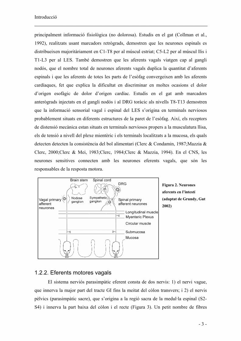
1.2.1. Aferents sensitives Les neurones aferents sensitives de l’esòfag i del LES viatgen amb els nervis
simpàtics, i s’anomenen aferents simpàtiques o aferents espinals, o amb els nervis
parasimpàtics vagals, i s’anomenen aferents vagals. Les primeres van cap al gangli de
l’arrel dorsal (DRG) i les segones cap al gangli nodós (Figura 2). Del total de les fibres
vagals, un 70-90% són fibres aferents. Les aferents espinals són neurones que
transmeten informació dolorosa mentre que les aferents vagals transmeten
Introducció _____________________________________________________________
- 3 -
principalment informació fisiològica (no dolorosa). Estudis en el gat (Collman et al.,
1992), realitzats usant marcadors retrògrads, demostren que les neurones espinals es
distribueixen majoritàriament en C1-T8 per al múscul estriat; C5-L2 per al múscul llis i
T1-L3 per al LES. També demostren que les aferents vagals viatgen cap al gangli
nodós, que el nombre total de neurones aferents vagals duplica la quantitat d’aferents
espinals i que les aferents de totes les parts de l’esòfag convergeixen amb les aferents
cardíaques, fet que explica la dificultat en discriminar en moltes ocasions el dolor
d’origen esofàgic de dolor d’origen cardíac. Estudis en el gat amb marcadors
anterògrads injectats en el gangli nodós i al DRG toràcic als nivells T8-T13 demostren
que la informació sensorial vagal i espinal del LES s’origina en terminals nerviosos
probablement situats en diferents estructures de la paret de l’esòfag. Així, els receptors
de distensió mecànica estan situats en terminals nerviosos propers a la musculatura llisa,
els de tensió a nivell del plexe mientèric i els terminals localitzats a la mucosa, els quals
detecten detecten la consistència del bol alimentari (Clerc & Condamin, 1987;Mazzia &
Clerc, 2000;Clerc & Mei, 1983;Clerc, 1984;Clerc & Mazzia, 1994). En el CNS, les
neurones sensitives connecten amb les neurones eferents vagals, que són les
responsables de la resposta motora.
Figura 2. Neurones
aferents en l’intestí
(adaptat de Grundy, Gut
2002)
1.2.2. Eferents motores vagals El sistema nerviós parasimpàtic eferent consta de dos nervis: 1) el nervi vague,
que innerva la major part del tracte GI fins la meitat del còlon transvers; i 2) el nervis
pèlvics (parasimpàtic sacre), que s’origina a la regió sacra de la medul·la espinal (S2-
S4) i innerva la part baixa del còlon i el recte (Figura 3). Un petit nombre de fibres

Introducció _____________________________________________________________
- 4 -
eferents vagals (10-20%) innerven un gran nombre de ganglis entèrics, fet que explica la
forta influència del vague sobre la motilitat (Wood J.D., 1989). Les fibres motores
vagals s’originen en nuclis diferents en funció del tipus de musculatura que innerven.
Figura 3. Innervació eferent extrínseca del tracte GI. A) Eferents motores vagals i B) Eferents motores
simpàtiques
Esòfag estriat En algunes espècies com el ratolí, la rata i el gos, l’esòfag sencer està format per
múscul estriat, excepte la part del LES, que està constituït per musculatura llisa. En
canvi, el gat, la fura, l’opòssum i el porc tenen múscul estriat en la regió més proximal
de l’esòfag, mentre que una porció de la part distal està constituïda per musculatura
llisa. En humans, la porció cervical de l’esòfag està formada per múscul estriat; mentre
que el segment toràcic està constituït per fibres llises. Tot el múscul estriat de l’esòfag
està innervat majoritàriament per neurones localitzades en el nucli ambigu (NA). Així
doncs, estudis amb rata, en què s’han fet servir traçadors retrògrads, demostren que la
zona compacta del NA conté totes les neurones motores inferiors (LMN) vagals que
controlen la funció motora de la faringe i de l’esòfag (Broussard & Altschuler, 2000).
Per altra banda, estudis amb gat en què s’ha injectat traçadors retrògrads fluorescents
han demostrat que principalment les fibres eferents que innerven la musculatura estriada
de l’esòfag es localitzen a la part rostral del NA, però que existeix un 8% de neurones
localitzades al nucli motor dorsal (DMN) del vague (Collman et al., 1993).
Mitjançant estudis immunohistoquímics amb rata, s’ha vist que aquestes
neurones contenen el transportador vesicular d’ACh, però també contenen CGRP (Lee
et al., 1992;Sang & Young, 1998), fet que suggereix que aquest últim actua com a co-

Introducció _____________________________________________________________
- 5 -
transmissor amb l’ACh. Els seus terminals nerviosos contacten les fibres musculars
estriades mitjançant plaques motores (Zhou et al., 1996). Estudis in vitro amb
estimulació vagal demostren que l’ACh alliberada a la placa motora activa receptors
colinèrgics nicotínics en el múscul estriat, per iniciar així la contracció muscular (Kerr
et al., 1995). La porció de múscul estriat de l’esòfag té un plexe mientèric poc dens i
irregular (Izumi et al., 2002), que per estudis immunohistològics s’ha vist que conté
neurones que expressen l’enzim òxid nítric sintetasa (NOS), galanina, CGRP
(Kuramoto et al., 1999) i VIP (Sang & Young, 1997), i que innerven el terminal nerviós
de la placa motora. Les eferents vagals colinèrgiques estan inhibides per aquestes
neurones nitrèrgiques, les quals modulen les contraccions peristàltiques a l’esòfag
estriat (Izumi et al., 2003). En canvi, les eferents vagals no semblen actuar sobre les
neurones inhibidores nitrèrgiques (Neuhuber et al., 1998).
Esòfag llis i LES Estudis amb gat, en què s’han injectat traçadors retrògrads a la regió toràcica de
múscul llis de l’esòfag i també del LES, han demostrat que principalment les fibres
eferents que innerven la musculatura llisa de l’esòfag es localitzen en el DMN en dos
grups, un de rostral i un de caudal. Un grup addicional es troba en el nucli retroambigu i
aproximadament un 8% són presents en el NA rostral, lateralment a les eferents del
múscul estriat. En el LES les aferents tenen una distribució similar que les aferents de
l’esòfag, però els cossos neuronals del DMN estan desplaçats més caudalment (Collman
et al., 1993). En un estudi similar en els que es va emprar la fura, es veu també una
distribució rostrocaudal dels cossos de les neurones eferents en el DMN (Hyland et al.,
2001).
Durant molt de temps es va creure que el nervi vague només tenia influències
excitadores en el tracte digestiu. Un dels majors avenços en el nostre coneixement de
com el nervi vague controla la motilitat gastrointestinal va ser demostrar que la via
motora vagal estava formada per vies paral·leles que provoquen accions inhibidores i
excitadores sobre el múscul. A diferència de la musculatura estriada, les neurones
vagals eferents no innerven directament la musculatura llisa, sinó que fan sinapsi amb
les motoneurones inhibidores i excitadores del plexe mientèric. En el LES existeix una
diferent distribució dels cossos de les neurones inhibidores i excitadores en el DMN, les
neurones de la via excitadora es localitzen en el DMN rostral, mentre les de la via

Introducció _____________________________________________________________
- 6 -
inhibidora estan presents en la part caudal del DMN (Rossiter et al., 1990). Estudis in
vivo amb opòssum (Goyal & Rattan, 1975) i estudis ex vivo amb fura (Smid &
Blackshaw, 2000) fent servir estimulació vagal han demostrat que la transmissió
sinàptica entre les neurones eferents i les neurones inhibidores del plexe està mitjançada
tant per receptors nicotínics com per muscarínics de tipus M1. Estudis en opòssum
suggereixen, a més, la participació de la 5-hidroxitriptamina (5-HT o serotonina) en la
transmissió sinàptica a aquest nivell (Rattan & Goyal, 1978;Paterson et al., 1992).
1.2.3. Eferents motores simpàtiques A l’esòfag, la innervació simpàtica parteix de cossos neuronals situats en els
segments espinals T5 i T6, envia els seus axons cap als ganglis cervicals i ganglis
simpàtics toràcics paravertebrals. En el LES, els cossos neuronals del component
simpàtic es troben en els segments espinals T6-T10, envien els axons cap al gangli
celíac a través del nervi esplàncnic major (Figura 3). Les neurones preganglionars són
colinèrgiques i activen els receptors nicotínics de les neurones noradrenèrgiques
postganglionars; aquestes últimes innerven l’esòfag o el LES acompanyant els vasos
sanguinis o, en menor quantitat, junt amb el nervi vague. Les fibres adrenèrgiques
postganglionars poden innervar tant les motoneurones entèriques com directament les
cèl·lules musculars del LES (Papasova M, 1989). La resposta simpàtica del LES
difereix en funció de l’espècie. Estudis d’estimulació del nervi esplàncnic en la fura
activa neurones adrenèrgiques i produeix relaxació a través de receptors de tipus β
(Blackshaw et al., 1997). En canvi, s’ha descrit en el gat (Gonella et al., 1979), que la
resposta simpàtica del LES és excitadora i és el resultat de l’estimulació tant de
receptors adrenèrgics situats en la musculatura llisa com de receptors situats en les
neurones del plexe que provoquen la posterior alliberació d’acetilcolina per part de les
motoneurones.
1.3 Innervació intrínseca: plexe mientèric i plexe submucós 1.3.1. General El sistema nerviós entèric està format per uns 100 milions de neurones i és
conegut també com “el cervell de l’intestí” perquè pot funcionar independentment del
CNS com van suggerir William M. Bayliss and Ernest H. Starling el 1899. Aquests dos
investigadors van demostrar, en gossos anestesiats, que l’aplicació d’una distensió

Introducció _____________________________________________________________
- 7 -
intraluminal produïa una contracció oral i una relaxació anal, seguida d’una ona
propulsiva de suficient magnitud com per propulsar l’aliment a través del tracte digestiu
(ells van anomenar aquest fenomen com a “llei de l’intestí” i equival actualment al
reflex peristàltic). Com que aquest reflex persistia sense la innervació extrínseca, van
deduir correctament que l’ENS pot operar independent dels inputs del CNS. Divuit anys
després, Paul Trendelenburg va confirmar aquest fet demostrant que el reflex peristàltic
es pot produir in vitro en l’intestí de conill porquí aïllat, sense participació del cervell,
de la medul·la espinal, de l’arrel de ganglis dorsal o de la cranials. Tot i això, com ja
s’ha vist, l’ENS comunica amb el CNS a través de fibres aferents i eferents simpàtiques
i parasimpàtiques.
La funció de l’ENS és controlar la motilitat, la secreció exocrina i l’endocrina i
la microcirculació del tracte digestiu, i està també implicat en la regulació immune i en
processos inflamatoris. Els cossos cel·lulars de les neurones s’agrupen en ganglis, que
estan connectats per processos intergangliònics formant dos plexes neuronals, el plexe
mientèric o de Auerbach i plexe submucós o de Meissner. El plexe submucós conté
menys neurones en termes globals, menys neurones per gangli i connexions
intergangliòniques més primes que el plexe mientèric. El plexe mientèric s’estén per tot
el tracte gastrointestinal, des de l’esòfag fins a l’esfínter anal intern (IAS). El plexe
submucós està molt desenvolupat a l’intestí prim, on juga un paper important en el
control de la secreció i l’absorció, encara que també innerva la muscularis mucosa,
algunes cèl·lules endocrines intestinals i vasos submucosos. A l’intestí també hi ha un
altre plexe, el plexe muscular profund, situat en la profunditat de la musculatura circular
(Furness et al., 1987) (Figura 4).
Figura 4. Representació
esquemàtica de les
diferents capes del tracte
gastrointestinal i de la
localització dels plexes
entèrics (adaptat de
Furness i Costa, 1987)

Introducció _____________________________________________________________
- 8 -
L’ENS és estructuralment i funcionalment molt semblant al cervell. Igual que a la resta
del sistema nerviós perifèric, els seus elements neuronals no estan suportats per
col·lagen ni per les cèl·lules de Schwann, però si per cèl·lules de la glia, que recorden
els astròcits del CNS.
1.3.2. Glia Existeixen evidències que fan pensar que la funció de les cèl·lules de la glia és
donar suport metabòlic a les neurones del MP, i que també poden jugar un paper en la
inflamació d’aquest (Cabarrocas et al., 2003;Ruhl et al., 2001). L’ENS es pot afectar en
malalties neurodegeneratives, apareixen en l’intestí de malalts de Parkinson cossos de
Lewy (Kupsky et al., 1987) i plaques amiloides en malalts d’Alzheimer (Joachim et al.,
1989).
1.3.3. Tipus de neurones entèriques S’han descrit quatre tipus funcionals de neurones entèriques: les neurones
secretores, les neurones motores, les neurones sensitives, també conegudes per IPAN
(intrinsic primary afferent neurons) i les interneurones. Les neurones motores, IPAN i
les interneurones estan implicades en els reflexos, inclosa la peristalsi intestinal. Els
seus cossos neuronals estan localitzats en el plexe mientèric i els terminals sensitius
estan situats a la mucosa; són sensibles a diferents estímuls com ara a tensió, a canvis en
el contingut luminal i a la serotonina. Algunes d’aquestes IPAN tenen els seus cossos
cel·lulars al plexe submucós; la seva estimulació pot controlar el transport d’aigua i
d’electròlits a través de la mucosa i el flux sanguini local (Kunze & Furness, 1999).
1.3.4. Neurones motores i neurotransmissors Estudis immunohistoquímics posen en evidència la presència de l’enzim de
síntesi de l’ACh (acetilcolintransferasa, AChT) en les motoneurones excitadores del
plexe mientèric de tot el tracte digestiu (Konomi et al., 2002;Porter et al., 2002;Ratcliffe
et al., 1998) i també de taquiquinines (SP, neurokinin A i B) (Sandler et al.,
1991;Shuttleworth et al., 1991;Yip et al., 2003;Yunker et al., 1999). Les motoneurones
inhibidores contenen els enzims de síntesi d’NO (sintetasa de l’òxid nítric, NOS) i del
monòxid de carboni (CO) (heme oxigenasa del tipus 2 o constitutiva, HO-2), els pèptids
VIP, PACAP, CGRP i també vesícules que contenen ATP (visualitzades usant la

Introducció _____________________________________________________________
- 9 -
tècnica fluorescent de quinacrina) (Belai & Burnstock, 1994;Clague et al., 1985;Miller
et al., 2001;Ny et al., 1997;Sann et al., 1998;Sundler et al., 1992). Per altra banda,
estudis mecànics i electrofisiològics realitzats usant antagonistes i inhibidors, han
evidenciat la implicació de l’NO com a principal neurotransmissor en l’esòfag
(Preiksaitis et al., 1994a), l’estómac (Todorov et al., 2003), l’intestí (Vanneste et al.,
2004), el còlon (Pluja et al., 1999) i l’IAS (Tomita et al., 2001). Existeix un paper
secundari per als altres neurotransmissors, les purines actuen en paral·lel amb l’NO a
l’estómac, a l’intestí i al còlon (de Man et al., 2003;Mule & Serio, 2003;Pluja et al.,
1999), de manera semblant a com ho fa el VIP també a l’estómac (Bayguinov et al.,
1999;Mule & Serio, 2003). Sembla que aquest pèptid no està implicat en la
neurotransmissió ni en l’intestí (Matsuda et al., 2004) ni en el còlon (Pluja et al., 2000).
Usant ratolins amb deleció de l’HO-2 (Xue et al., 2000a) s’ha vist que el CO podria
jugar un paper com a neurotransmissor en l’intestí, com també ho podria fer en l’IAS,
degut possiblement a la síntesi de CO induïda pel VIP (Watkins et al., 2004).
Pel que fa als neurotransmissors excitadors, s’ha vist que l’ACh és el principal
neurotransmissor en el tracte GI i que les taquiquinines poden tenir un paper més o
menys important en la contracció depenent de la porció del budell estudiada (Cao et al.,
2000;El-Mahmoudy et al., 2003;Krysiak & Preiksaitis, 2001).
1.3.5. Receptors dels diferents neurotransmissors inhibidors Guanilat ciclasa soluble L’NO i el CO són dos gasos que produeixen el seu efecte atesa l’activació de la
guanilat ciclasa soluble (sGC). Aquest enzim està format per una subunitat α i una
subunitat β, el seu acoblament és limitant perquè l’activació pels seus lligands produeixi
cGMP (Lucas et al., 2000). La producció de cGMP i la posterior fosforilació de
proteïnes tradueixen el senyal de l’NO per produir la relaxació del múscul llis (Lincoln
& Cornwell, 1991). Tot i això, s’ha vist en diferents teixits que l’NO pot tenir altres
dianes: l’NO pot actuar a través de canals iònics (Bolotina et al., 1994), incrementant la
conductància al K+ i que indueix així la hiperpolarització de la membrana, o bé activant
tirosina-cinases, fosfatases (Stamler, 1994), GTPases (Lander et al., 1995),
ADPribosiltransferases citosòliques (Brune & Lapetina, 1989) o factors de transcripció
(Lander et al., 1995). A diferència d’aquestes dos gasos, la resta de neurotransmissors
del tracte digestiu actuen sobre receptors.

Introducció _____________________________________________________________
- 10 -
Receptors purinèrgics Als anys 80, Burnstock (Burnstock, 1980) va proposar una classificació formal
dels receptors per l’adenosina i per l’ATP. Els receptors selectius per adenosina i
adenosina monofosfat (AMP) es van anomenar purinoceptors P1 i els selectius per ATP
i adenosina difosfat (ADP) es van anomenar purinoceptors P2. Aquesta classificació es
va ampliar en subdivisions addicionals, els receptors P1 es van dividir en A1, A2A, A2B i
A3 i els receptors P2 en les famílies P2X i P2Y. Actualment hi ha pocs agonistes i
antagonistes selectius per discriminar clarament entre receptors P2X i P2Y, o entre
diferents subtipus de receptors dins de cada família, encara que el 2-methylthio ADP (2-
MeSADP) i el MRS2179 sembla que siguin un agonista i un antagonista selectius,
respectivament, per als receptors P2Y1 (Camaioni et al., 1998).
Els receptors per l’adenosina com per l’ATP juguen un paper important en la
modulació de la motilitat en el tracte GI. L’adenosina pot activar directament receptors
localitzats en la musculatura llisa i produir tant relaxació com contracció (Kadowaki et
al., 2000;Nicholls et al., 1996) o actuar presinàpticament reduint l’alliberació de
neurotransmissors excitadors com l’ACh i la SP (Kadowaki et al., 2000;Moneta et al.,
1997). En moltes espècies, l’ATP activa receptors P2 en el múscul llis per produir
relaxació de la musculatura llisa en el tracte GI, encara que hi ha algunes evidències en
què l’ATP també produeix contracció del múscul llis en algunes regions. En un estudi
en el qual s’usen diferents parts del tracte digestiu del ratolí (fundus gàstric, duodè,
íleum i còlon) s’observa que receptors P2Y1 neurals estan implicats en la relaxació a
totes les regions del tracte digestiu, en gran part alliberant NO; la relaxació també és
deguda a receptors P2Y1, P2Y2 i P2Yn localitzats en la musculatura llisa. Els receptors
P2X2 estan implicats en la contracció només en el còlon (Giaroni et al., 2002). En
estudis més recents s’ha vist que l’estimulació elèctrica de camp (EFS) allibera NO i un
segon neurotransmissor sensible a l’MRS2179 en el fundus del ratolí (de Man et al.,
2003) i en el còlon humà (Auli et al., 2005;Gallego et al., 2005) fet que suggereix, la
participació d’una purina actuant a receptors P2Y1.
Els receptors P1 estan acoblats a la proteïna G, ja sigui disminuint-ne la
producció de cAMP o bé augmentant-la. També s’ha descrit que poden activar la via de
la fosfolipasa C (PLC) i produir la formació del trifosfat d’inositol (IP3) i diacilglicerol
(DAG). L’IP3 estimula l’alliberament de Ca++ intracel·lular per la interacció amb
receptors específics localitzats al reticle sarcoplasmàtic. L’augment del Ca++ citosòlic
Introducció _____________________________________________________________
- 11 -
per l’IP3 pot estimular diferents vies com la de la proteïna cinasa C (PKC), fosfolipasa
A2 (PLA2), canals de K+ dependents de Ca++ i òxid nítric sintetasa. Els receptors P2X
són canals iònics que uneixen ATP (ATP-gated), la seva activació produeix una ràpida
(entre 10 ms) i selectiva permeabilitat a cations (Na+, K+ i Ca++). Això contrasta amb la
resposta lenta (menys de 100 ms) de l’ATP quan actua sobre receptors metabotròpics
P2Y, que impliquen acoblament a proteïna G i sistemes de segons missatgers. La
majoria dels receptors P2Y actuen via proteïna G acoblada a l’activació de PLC, i
produeixen la formació d’IP3 i la mobilització de calci intracel·lular. S’ha descrit també
l’acoblament a l’AC en alguns receptors P2Y (Ralevic & Burnstock, 1998).
Receptors de VIP/PACAP Els receptors per VIP i per PACAP formen part d’una família de receptors
formada per set receptors transmembrana acoblats a la proteïna G, en la qual també
s’inclouen els receptors per CGRP. El PACAP té elevada afinitat per tres receptors
diferents (receptors PAC1, VPAC1 i VPAC2). Dos d’aquests, el VPAC1 i el VPAC2,
són també receptors d’alta afinitat pel VIP. Els tres receptors estan majoritàriament
acoblats a la proteïna Gs, que és la que activa l’adenilat ciclasa (AC); els PAC1 i
VPAC1 poden estar també acoblats a les proteïnes Gq i Go respectivament, les quals
són responsables de la mobilització de calci (Harmar et al., 1998).
Receptors de CGRP
Històricament, els receptors de CGRP han estat classificats en dues classes:
CGRP1 i CGRP2. Els receptors CGRP1 són més sensibles que els CGRP2 a
l’antagonista peptídic CGRP8-37. Per altra banda, els anàlegs lineals de CGRP,
[Cys(ACM)2,7]-CGRP i [Cys(Et)2,7]hαCGRP, són agonistes més potents per receptors
CGRP2 que per CGRP1. El CGRP augmenta el cAMP intracel·lular en molts sistemes
però no en tots, i pot activar també canals de K+ i de Ca++ via proteïna G (Poyner et al.,
2002).

Introducció _____________________________________________________________
- 12 -
Figura 5. Esquema resum dels principals neurotransmissors inhibidors amb els seus canals de K+ associats (Sanders, 2000)
1.3.6. Interaccions entre els neurotransmissors inhibidors
S’han descrit diferents mecanismes d’interacció entre els neurotransmissors
inhibidors. S’ha proposat que l’NO es pot alliberar en paral·lel i tenir accions
independents (cotransmissió) junt amb l’ATP (Soediono & Burnstock, 1994) i el VIP
(Bayguinov et al., 1999). L’NO també pot ser alliberat per altres neurotransmissors com
el VIP a nivell presinàptic (Kumano et al., 2001;Grider et al., 1992) i també és possible
que el VIP indueixi la síntesi d’NO a nivell postsinàptic (Ergun & Ogulener, 2001), de
la mateixa manera que ho poden fer l’ATP (Xue et al., 2000b), el PACAP (Zizzo et al.,
2004) i el CO (Lim et al., 2005). Per altra banda, també s’ha descrit que l’NO pot
alliberar VIP i PACAP a nivell del terminal nerviós (Grider et al., 1992;Zizzo et al.,
2004) i que el VIP pot induir la síntesi de CO (Watkins et al., 2004).
1.3.7. Cèl·lules intersticials de Cajal A més de les neurones i la glia de l’ENS, el tracte digestiu conté cèl·lules
intersticials de Cajal (ICC). Aquestes van ser descrites per primera vegada per Santiago
Ramón y Cajal cap a l’any 1890 usant tincions de blau de metilè. Les va descriure com:
“células fusiformes o triangulares de pequeña talla, pobres en protoplasma, del cual
parten varias expansiones varicosas, larguísimas y de ordinario ramificadas en ángulo
recto. Habitan especialmente entre los haces de fibro-células, adosándose a los
manojos del plexo intersticial o secundario; pero se las ve también en el contorno del
plexo de Auerbach y en la inmediación de los vasos. Algunas de sus expansiones
abandonan los hacecillos nerviosos y marchan independientemente sobre la trama
contráctil, siguiendo de preferencia a los intersticios musculares y constituyendo un
plexo de malla angosta, desigual y frecuentemente incompleta. Las últimas ramillas,

Introducció _____________________________________________________________
- 13 -
pálidas y granulosas, parecen conexionarse con las células musculares.” (Ramón y
Cajal, 1904). Cajal creia que aquestes cèl·lules eren neurones simpàtiques intersticials,
no vas ser fins als anys 90 que es va veure amb estudis immunohistoquímics que no
tenien marcadors clàssics de neurones mientèriques, ni de glia, ni de fibroblasts, ni de
múscul llis, però sí d’enolasa específica de neurones, resultats que suggerien una relació
amb algun tipus neuronal (Prosser et al., 1989). Posteriorment Huizinga et al. (Huizinga
et al., 1995), van demostrar que aquestes cèl·lules expressaven el receptor c-kit.
Actualment la presència d’aquest receptor és usada com a gold standard per marcar
aquestes cèl·lules.
Estudis morfològics on s’observa una disminució de les ICC, han portat a la
conclusió que aquestes cèl·lules poden tenir un paper important en la patofisiologia de
diferents malalties com ara l’estenosi pilòrica hipertròfica infantil, alguns síndromes de
pseudoobstrucció intestinal crònica (Kenny et al., 1998;Vanderwinden et al., 1996a), la
constipació de trànsit lent, la malaltia de Chagas, les alteracions de la motilitat
relacionades amb la diabetis , i l’hipoganglionosi (Vanderwinden et al., 1996b;Hagger
et al., 2000;He et al., 2000;Nakahara et al., 2002;Rolle et al., 2002;Tong et al., 2004;Yu
et al., 2002).
Localització de les ICC
Les ICC es localitzen en les diferents capes que formen el tracte digestiu
(Sanders et al., 2002).
• ICC del plexe mientèric (ICC-AP) En el plexe mientèric, les ICC estan estretament associades amb els ganglis i amb les
cèl·lules musculars de les capes circulars i longitudinals adjacents.
• ICC intramusculars (ICC-IM)
Amb tècniques d’immunohistoquímica, usant anticossos contra aquest receptor, s’ha
evidenciat la presència d’ICC-IM al llarg de tot el tracte digestiu, des de l’esòfag fins a
l’IAS (Hagger et al., 1998;Romert & Mikkelsen, 1998;Ward et al., 1998;Battish et al.,
2000;Burns et al., 1996). Usant tècniques de microscòpia electrònica, s’ha vist que a
l’esòfag, al LES, a l’estómac, a l’intestí prim i al còlon, aquestes cèl·lules estan en
contacte molt estret amb les varicositats terminals de les neurones motores entèriques i
formen unions gap amb les cèl·lules musculars llises (Daniel & Posey-Daniel,

Introducció _____________________________________________________________
- 14 -
1984;Faussone-Pellegrini & Cortesini, 1985;Faussone-Pellegrini, 1987) fet que fa
suggerir que aquestes cèl·lules puguin participar en la neurotransmissió (Figura 6).
Figura 6. Fotografia de microscòpia
electrònica en la musculatura circular
de l’estómac de la rata. Es Pot veure el
contacte estret entre el terminal nerviós
(NE) i una ICC-IM, aquesta està en
contacte amb una cèl·lula muscular llisa
a través d’una unió gap (Mitsui & Komuro, 2002) .
Altres estudis en microscòpia electrònica, en canvi, han descrit que la varicositat
terminal pot contactar amb el múscul directament, sense la presència de les ICC (Mitsui
& Komuro, 2002). Aquestes observacions suggereixen que hi ha dos tipus
d’interaccions varicositat-múscul: una innervació directa, on hi ha un contacte directe
entre el terminal i la cèl·lula muscular llisa; i una innervació indirecta, on el terminal
interacciona amb la cèl·lula muscular llisa a través d’una ICC (Figura 7).
Figura 7. Models d’interacció motoneurona-cèl·lula muscular llisa. Modificat de Sanders et al.
News Physiol. Sci. 2000
• ICC del plexe muscular profund (ICC-DMP)
A l’intestí, les cèl·lules semblants a les ICCs-IM estan restringides a la regió del plexe
muscular profund i reben el nom d’ICC-DMP (Wang et al., 2003).
• ICC del plexe submucós (ICC-SMP)
A l’estómac i al còlon, una població d’ICC també es troba a la musculatura circular
tocant el plexe submucós (Alberti et al., 2005;Mitsui & Komuro, 2003).

Introducció _____________________________________________________________
- 15 -
Funció de les ICCs
• ICC-AP i ICC-SMP
En estudis de microscòpi electrònic s’ha vist que les ICC formen unions gap
amb les cèl·lules musculars llises i amb altres ICC del seu voltant. En estudis amb
Figura. 8. Esquema
resum de la relació de
les cèl·lules intersticial
de Cajal amb altres
estructures del tracte
GI (Christensen, 2001)
ratolins mutants W/Wv (no tenen ICCs al plexe mientèric) i en estudis de patch-clamp
posaria: s’ha suggerit que les ICC-AP de l’estómac, de l’intestí prim i del còlon, i les
ICC-SMP en el còlon, poden tenir funcions duals com a marcapassos i com a vies de
conducció de les ones lentes (Huizinga et al., 1995;Langton et al., 1989;Ordog et al.,
1999).
• ICC-IM i ICC-DMP
En estudis funcionals in vitro, s’ha vist que en ratolins mutants Ws/Ws, que no
tenen ICC-IM, la innervació inhibidora nitrèrgica no és funcional ni en l’estómac
(Burns et al., 1996), ni en el LES ni en el pílor (Ward et al., 1998). En canvi, en estudis
in vivo en el LES (Sivarao et al., 2001) i en estudis in vivo i in vitro en l’IAS (Terauchi
et al., 2005;de Lorijn F. et al., 2005), s’ha vist que la neurotransmissió nitrèrgica
inhibidora està preservada en els animals mutants, cosa que suggereix que les ICC-IM
no tenen un paper important en aquest tipus de neurotransmissió. Pel que fa al
component excitador, s’ha descrit en el fundus gàstric del ratolí que aquestes cèl·lules
juguen un paper molt important en la neurotransmissió colinèrgica (Ward et al., 2000).
S’ha descrit que les ICC-DMP formen contactes semblants amb sinapsi amb
nervis nitrèrgics i colinèrgics i contactes amb cèl·lules musculars mitjançant unions gap,
fet que fa pensar que les ICC-DMP poden estar relacionades amb la neurotransmissió
del múscul llis en l’intestí (Wang et al., 2003). També s’ha descrit que les ICC-IM
poden funcionar com a sensors d’estirament (streach sensors) (de Lorijn F. et al., 2005).

Introducció _____________________________________________________________
- 16 -
2. Anatomia muscular i fisiologia de l’esòfag La funció de l’esòfag és transportar el material engolit des de la faringe fins a
l’estómac. L’esòfag es diferencia de la resta del tracte digestiu perquè no té funcions
secretores o d’absorció importants, i també pel tipus d’epiteli de la mucosa. Per altra
banda, la funció de l’esfínter esofàgic inferior és crear una zona d’alta pressió que
impedeixi el reflux del contingut gàstric cap a l’esòfag.
2.1 Anatomia muscular de l’esfínter esofàgic inferior En humans, l’esòfag està format per una porció de múscul estriat d’uns 12 cm
que comprèn la faringe i part de l’esòfag cervical, i, per una altra d’uns 15 cm formada
per múscul llis, que s’estén del terç del mig fins a la unió gastroesofàgica, localitzada al
nivell del diafragma. En aquest nivell, la musculatura de l’esòfag forma el LES. Les
principals funcions del LES són permetre el pas del bol durant la ingestió d’aliments de
l’esòfag fins a l’estómac i prevenir el reflux d’una quantitat significativa del contingut
gàstric cap a l’esòfag. La funció de la barrera antireflux depèn de la competència tant
del LES com del diafragma crural (Figura 9) (Hill et al., 1996;Pandolfino et al., 2003).
Estudis amb teixit fixat i usant tècniques d’imatge de manometria tridimensional in vivo
demostren que en humans el LES no és un anell muscular, sinó que està format per les
fibres semicirculars de clasp adjacents a la curvatura menor (que forma un anell
incomplet en forma de U) i per bandes musculars a la banda de la curvatura major,
formades per fibres gàstriques obliqües (fibres de sling) (Liebermann-Meffert et al.,
1979;Stein et al., 1995). En altres espècies, com en el gos (Friedland et al., 1971),
l’opòssum (Christensen & Torres, 1975) i el gat (Friedland et al., 1971;Preiksaitis et al.,
1994b) el múscul circular del LES forma un anell complet. Ambdós músculs mantenen
l’esfínter tancat i contribueixen a la pressió del LES. En l’única espècie on s’ha observat
que la distribució de les fibres de clasp i de sling és idèntica a la de l’home ha estat en el
porc (Vicente et al., 2001).

Introducció _____________________________________________________________
- 17 -
Figura 9. Anatomia de la unió gastroesofàgica en l’home. A) L’esfínter esofàgic inferior representa
la part interna (esfínter de múscul llis) mentre que el diafragma representa la part externa (esfínter
de múscul estriat). Podem veure també a la part dreta de la figura les fibres de sling. Adaptat de
(Mittal & Balaban, 1997). B) Esquema de la distribució de les fibres de clasp i sling en el LES humà
(Preiksaitis & Diamant, 1997) i C) en el porc (Vicente et al., 2001)
2.2 Fisiologia de l’esòfag i del LES. Control de l’activitat motora En la porció del múscul llis de l’esòfag, la deglució genera un reflex peristàltic
que és generat centralment i que està mediat per neurones eferents vagals procedents del
DMN. La peristalsi induïda per la deglució voluntària s’anomena peristalsi primària per
distingir-la de la peristalsi secundària, la qual és produïda per reflexes locals com la
distensió. La peristalsi primària és abolida per vagotomia o refredament vagal bilateral
(Roman & Gonella, 1981) (Ryan et al., 1977). Els eferents vagals estimulen la peristalsi
per activació i modificació del reflex peristàltic intrínsec. Per altra banda, la peristalsi
secundària en el múscul llis està generada per un reflex local i no es modifica per
vagotomia (Kravitz et al., 1978;Roman & Gonella, 1981); en canvi, la peristalsi
secundària en el múscul estriat està controlada centralment (Roman & Gonella, 1981).
2.2.1. Peristalsi secundària en el múscul estriat Contràriament al que ocorre en el múscul llis, en el múscul estriat, la peristalsi
secundària sí que s’aboleix per vagotomia. Aquest fet indica que el control de la
peristalsi és d’origen central (Roman & Gonella, 1981). L’estimulació elèctrica
d’aquestes neurones eferents generen només contraccions simultànies a tots els nivells
del múscul estriat, fet que fa pensar que es necessita aquesta integració central per
organitzar la natura seqüencial de les contraccions peristàltiques (Grundy, 1988). La
peristalsi en la porció de múscul estriat està generada pel nucli ambigu i mitjançada per

Introducció _____________________________________________________________
- 18 -
LMNs vagals. El nucli ambigu genera una activació seqüencial d’aquestes neurones que
produeixen contraccions successives en l’esòfag estriat (Figura 10).
Figura 10. Mecanisme de la peristalsi evocada
vagalment en el múscul estriat de l’esòfag. vs,
Estimulació vagal. (Chang et al., 2003)
2.2.2. Peristalsi secundària en el múscul llis La peristalsi esofàgica intrínseca està mitjançada per neurones mientèriques
inhibidores no adrenèrgiques no colinèrgiques (NANC) i motoneurones excitadores
actuant coordinadament. Encara que s’anomenen inhibidores, l’estimulació de les
motoneurones inhibidores en tires de múscul llis indueix una resposta bifàsica que
consisteix en una inhibició seguida d’una contracció rebot. En el múscul llis que no té to
basal, l’inhibició es veu només com un període de latència durant el període
d’estimulació nerviosa. La contracció del múscul llis després d’aquest període d’estímul
prolongat s’anomena contracció rebot o contracció en “off” (Knudsen et al., 1991) .
Weisbrodt et al. (Weisbrodt & Christensen, 1972) van descriure en tires de múscul
circular esofàgic d’opòssum que la duració del període de latència de la contracció en
“off” s’ incrementa progressivament en direcció craneocaudal. A partir d’aquests
resultats, aquests autors van proposar que el gradient de latència al llarg de l’esòfag està
generat per nervis NANC. Estudis posteriors van demostrar que l’NO era el
neurotransmissor NANC responsable d’aquesta resposta bifàsica (Preiksaitis et al.,
1994a;Murray et al., 1991;Knudsen et al., 1991). Estudis in vitro i in vivo amb
opòssums demostren que l’L-NAME (inhibidor NOS) aboleix la latència i el gradient de
latència de les contraccions esofàgiques produïdes per l’estimulació de nervis NANC a
diferents nivells de l’esòfag (Yamato et al., 1992b;Murray et al., 1991).

Introducció _____________________________________________________________
- 19 -
Les neurones excitadores del plexe mientèric esofàgic utilitzen majoritàriament
l’ACh com a neurotransmissor i freqüentment són anomenades colinèrgiques. In vitro,
l’estimulació nerviosa de curta durada, indueix una resposta inhibidora NANC que
apareix primer i que va seguida per una contracció colinèrgica que pot sobreposar-se
amb la contracció rebot NANC. Quan l’estímul nerviós és perllongat, la contracció
colinèrgica i la contracció rebot NANC poden aparèixer separades. Les contraccions
que ocorren durant l’estímul s’anomenen contraccions en “on”; les contraccions que
ocorren al finalitzar l’estímul s’anomenen contraccions en “off” (Crist et al., 1984a). De
manera similar a les neurones NANC, les neurones colinèrgiques formen un gradient
colinèrgic, però en aquest cas decreix distalment al llarg de l’esòfag (Crist et al., 1984b).
Aquest model incorpora el gradient d’influències NANC incrementades i un
d’influències colinèrgiques disminuïdes al llarg de l’esòfag per explicar l’amplitud i la
latència de la peristalsi esofàgica (Figura 11). Així es pot explicar tant els mecanismes
perifèrics pel comportament peristalsi-like de les tires circulars de l’esòfag in vitro, com
la peristalsi esofàgica provocada per estimulació vagal aferent in vivo.
Figura 11. Mecanisme perifèric
de la peristalsi. En aquest model
proposat per Crist et al., es
descriuen gradients de neurones
colinèrgiques i no colinèrgiques al
llarg de la porció de múscul llis de
l’esòfag. L’influència colinèrgica
predomina a l’esòfag proximal i
decreix distalment.
Contràriament, la influència no
colinèrgica és mínima
proximalment i incrementa en
l’esòfag distal. UES, esfínter
esofàgic superior (Chang et al.,
2003)
2.2.3. Peristalsi primària El reflex deglutori, que evoca peristalsi primària, està organitzat en el centre de
deglució (localitzat al tronc cerebral), aquest centre es comunica amb el plexe mientèric
esofàgic mitjançant neurones aferents vagals. Estudis amb opòssums anestesiats (Gidda
& Goyal, 1984) en els quals es van provocar deglucions i es van enregistrar potencials a

Introducció _____________________________________________________________
- 20 -
les fibres eferents, han suggerit que després de la deglució s’activen primer les neurones
NANC del plexe, i causen una ràpida inhibició del múscul llis de l’esòfag, fet que
ocorre en menys d’1 s. Aquest fenomen es coneix com a inhibició deglutòria. El grau i
la durada de la inhibició augmenta a mesura que ens apropem a la part distal de
l’esòfag, per raó de l’increment del gradient NANC, descrit anteriorment. La resposta
inhibidora està seguida per una contracció amb latències progressivament
incrementades al llarg de l’esòfag. La deglució també inicia l’activació seqüencial d’un
segon grup de fibres eferents amb períodes de latència compresos entre 1 i 5 s. Aquestes
fibres amb latències de major durada s’especula que projecten cap a les neurones
excitadores del plexe mientèric de l’esòfag. Durant la deglució la seqüència d’excitació
colinèrgica se superposa a la NANC modulant l’amplitud i el gradient de latència de la
contracció peristàltica primària (Chang et al., 2003) (Figura 12).
Figura 12. Paper dels nervis eferents vagals
en la peristalsi primària. L’activació
seqüencial de les neurones NANC i
colinèrgiques del plexe que innerven la
porció de múscul llis de l’esòfag explica la
inhibició deglutòria seguida de peristalsi.
Les fibres de latència curta activen la NANC
del plexe per produir inicialment una
inhibició del múscul llis, per altra banda, les
fibres de latència llarga estimulen
subseqüentment neurones colinèrgiques per
produir contraccions que estan
programades per coincidir amb la contracció NANC rebot (Chang et al., 2003).
En humans, la inhibició deglutòria que precedeix la peristalsi primària s’evidencia quan
deglucions repetides i successives es produeixen en intervals propers. Cada deglució
successiva inhibeix la contracció peristàltica de la deglució precedent, i només l’última
deglució està associada amb la contracció peristàltica (Sifrim et al., 1992).

Introducció _____________________________________________________________
- 21 -
2.2.4. Esfínter esofàgic inferior El LES està format per múscul llis amb propietats miogèniques intrínseques que
manté l’esfínter tancat en condicions basals. Aquesta contracció tònica fa que
manomètricament el LES s’evidenciï com una zona d’alta pressió a l’extrem inferior de
l’esòfag. Aquest to miogènic està modulat tant per les vies vagals inhibidores com per
les excitadores. En estudis in vivo s’ha vist que quan les influències excitadores i
inhibidores són iguals, ni la vagotomia bilateral (Rattan & Goyal, 1974) ni la
neurotoxina tetrodotoxina (TTX) canvien la pressió del LES (Goyal & Rattan, 1976).
Per altra banda, antagonistes selectius de només una via produeixen efectes oposats de
l’altra via. Això vol dir que el bloqueig de la via excitadora colinèrgica usant
anticolinèrgics (Dodds et al., 1981) o toxina botulínica (Pasricha et al., 1993)
disminueix la pressió del LES a causa d’una acció oposada dels nervis inhibidors, tot i
que en algunes espècies l’atropina no produeix cap canvi en la pressió del LES (Dodds
et al., 1981) . En canvi, el bloqueig de la via nitrèrgica inhibidora amb inhibidors de la
NOS produeix un increment de la pressió del LES (Xue et al., 1996;Yamato et al.,
1992a).
L’activació selectiva de la via vagal inhibidora pot generar la relaxació del LES.
La relaxació del LES és una part integral de la peristalsi primària induïda per la
deglució. Amb l’estímul de la deglució, el LES es relaxa durant 8-10 s i posteriorment
es produeix una contracció a la part proximal del LES. Aquesta sèrie d’esdeveniments
permeten al bol engolit passar a través de la zona d’alta pressió. La relaxació del LES és
el component més sensible de la peristalsi primària perquè la relaxació pot ocórrer sense
una peristalsi faríngia o esofàgica associada. La relaxació del LES produïda per
distensió de la porció llisa de l’esòfag, amb o sense peristalsi secundària associada, és
conseqüència de reflexos locals, i no s’afecta per vagotomia. Les vies vagals inhibidores
i excitadores s’activen selectivament en un nombre de reflexos motors que impliquen
l’esfínter esofàgic inferior. L’estimulació de les aferents vagals abdominals i la distensió
de l’estómac proximal disparen les vies inhibidores que produeixen la relaxació del
LES. Reflexos vagovagals com eructar i les relaxacions transitòries de l’esfínter
(tLESRs) també depenen de l’activació de la via inhibidora del LES (Chang et al.,
2003).

Introducció _____________________________________________________________
- 22 -
3. Trastorns de la motilitat esofàgica Els trastorns de la motilitat de l’esòfag comprenen diferents símptomes,
especialment disfàgia i dolor toràcic. Aquests trastorns es diagnostiquen per manometria
esofàgica, on es mesura la pressió i la relaxació del LES i la presència de peristalsi en el
cos esofàgic. Els paràmetres de les contraccions peristàltiques que es tenen en compte
per al diagnòstic són l’amplitud (alta o baixa), la duració, la naturalesa repetitiva i la
presencia d’ones no transmeses o parcialment transmeses. A continuació s’expliquen
breument les anomalies primàries com són l’acalàsia, l’espasme esofàgic difús, l’esòfag
hipercontràctil, l’hipocontràctil i també algunes anomalies secundàries, com els
trastorns de la motilitats produïts en l’escleroderma, malaltia de Chagas i diabetis
(Richter, 2001). Després es parla també de la malaltia per reflux gastroesofàgic
(GERD), que, com es veurà, es pot deure a una alteració de la motilitat i,
secundàriament, pot ocasionar alteracions en la motilitat esofàgica a causa de
l’exposició àcida (Castell et al., 2004).
3.1. Anomalies primàries de la motilitat esofàgica Acalàsia La simptomatologia es caracteritza per una obstrucció funcional amb disfàgia,
malestar toràcic i regurgitació. El símptoma més freqüent és la disfàgia a sòlids i
líquids, la gravetat de la qual fluctua, però que pot ser important fins a produir una
pèrdua de pes significativa.
Els estudis manomètrics es caracteritzen per (Clave & Castellvi, 2004):
- absència d’ones peristàltiques en l’esòfag llis (criteri diagnòstic obligat)
-absència o relaxació incompleta de l’esfínter durant la deglució. Aquest fet el
distingeix d’altres trastorns amb aperistalsi
-pot produir-se també un increment de la pressió del LES (>45 mmHg)
L’origen de l’acalàsia és desconeguda, s’han suggerit factors hereditaris,
degeneratius, autoimmunes i infecciosos com a possible causa. Aquests dos últims
factors són els dos més comunament acceptats (Castagliuolo et al., 2004;Ruiz-de-Leon
et al., 2002;Storch et al., 1995;Storch et al., 1995). S’han identificat canvis patològics
en espècimens procedents de necròpsies o d’intervencions quirúrgiques com són una
resposta inflamatòria prominent però irregular, consistent en l’aparició de limfòcits T i

Introducció _____________________________________________________________
- 23 -
un nombre variable d’eosinòfils i mastòcits, i algun grau de fibrosis neural mientèrica
(Raymond et al., 1999). El resultat final d’aquests canvis inflamatoris és una pèrdua
selectiva de motoneurones inhibidores nitrèrgiques (Lui et al., 1997;Mearin et al., 1993)
i neurones que contenen VIP (Aggestrup et al., 1983;Sigala et al., 1995). En estudis de
microscòpia electrònica, en teixit de pacients amb acalàsia, es mostra la presència de
vacuoles en les ICCs (Zarate et al., 2004) fet que fa pensar en una possible lesió també
d’aquestes cèl·lules. Aquesta pèrdua de neurones inhibidores provoca una relaxació
incomplerta del LES i una aperistalsi causada per la pèrdua del gradient de latència que
permet contraccions seqüencials al llarg de l’esòfag.
Espasme esofàgic difús L’espasme esofàgic difús es caracteritza per un peristalsi normal intermitentment
interrompuda per contraccions simultànies. Aquest malaltia és poc freqüent, ocorre al
voltant d’un 3-5% dels pacients estudiats per anomalies de la motilitat esofàgica. Es
desconeix l’etiopatologia d’aquest desordre. En estudis en què s’usen tests de
provocació amb metacolina, betanecol, carbacol i pentagastrina s’observen unes
contraccions augmentades en aquests malalts (Mellow, 1977). En un estudi on es
correlaciona el gruix de la musculatura en estat basal i l’amplitud de la contracció
peristàltica (Pehlivanov et al., 2002), s’observa que hi ha una associació que explicaria
per què aquests malalts tenen les contraccions augmentades i responen amb
contraccions majors a l’estimulació d’agonistes colinèrgics. També s’ha suggerit que
aquesta sensitivitat augmentada pot ser deguda a un defecte en la inhibició neural al
llarg el cos esofàgic, possiblement en relació amb una disfunció en la síntesi d’NO i/o
en la seva degradació (Konturek et al., 1995). L’espasme esofàgic difús es diagnostica
per manometria esofàgica, s’observen unes contraccions simultànies no peristàltiques
intermitents barrejades amb peristalsi normal. Altres troballes manomètriques menys
consistents inclouen contraccions de llarga durada, ones repetitives (tres pics o més),
contraccions espontànies no induïdes per la deglució, i anomalies de la pressió del LES
o de la seva relaxació. En estudis de pH-metria ambulatòria de 24 h s’ha vist que entre
un 20% i un 50% dels malalts tenen GERD.

Introducció _____________________________________________________________
- 24 -
Esòfag hipercontràctil Els pacients amb peristalsi esofàgica simptomàtica (esòfag en trencanous,
nutcracker esophagus) presenten contraccions no peristàltiques d’elevada amplitud i
poden presentar també contraccions peristaltiques de duració major. Existeix un
subgrup de pacients que presenta també un LES hipertens. Aquests dos resultats
manomètrics sovint coexisteixen, i suggereixen que poden representar una síndrome
d’un esòfag hipercontràctil. La causa de l’esòfag hipercontràctil és incerta; tot i això,
algunes contraccions d’elevada pressió poden ser secundàries a factors exògens com
reflux gastroesofàgic o l’estrès. El principal símptoma dels pacients amb esòfag
hipercontràctil és dolor toràcic.
Esòfag hipocontràctil La majoria del pacients que són diagnosticats, com que tenen una alteració de la
motilitat no especifica, tenen patrons de motilitat caracteritzats per qualsevol de les dues
alteracions següents: contraccions peristàltiques de baixa amplitud o contraccions
simultànies en l’esòfag distal, o una peristalsi fallida en la qual les ones no travessen per
complet tot l’esòfag distal (anomenada motilitat esofàgica inefectiva). El concepte
d’ones de baixa amplitud que són inefectives està suportat per estudis que usen
simultàniament manometria i videoradiografia de bari per mostrar que les ones
contràctils de menys de 30 mmHg d’amplitud, encara que són peristàltiques, no
transporten eficaçment i no netegen un bol de bari de l’esòfag. La majoria dels malalts
amb motilitat esofàgica inefectiva tenen GERD. Una pressió baixa del LES és vista
comunament amb pacients amb GERD, fet que suggereix que l’esòfag hipocontràctil
pot ser secundari a un dany àcid crònic de l’esòfag distal. El LES hipotònic pot no ser
sever; tot i això, hi ha estudis que suggereixen que una exposició àcida anormal en
aquests malalts es correlaciona millor amb una dèbil bomba esofàgica que amb la
pressió del LES disminuïda.
3.2. Anomalies secundàries de la motilitat esofàgica Els malalts amb alteracions secundàries de la motilitat esofàgica tenen patrons
de motilitat anormals secundaris a una malaltia sistèmica. En l’escleroderma, per
exemple, les anomalies de la motilitat es troben en un 80% dels malalts. El procés de la
malaltia subjacent està causat per l’obliteració vascular i la fibrosis secundària que

Introducció _____________________________________________________________
- 25 -
afecta el múscul llis esofàgic i la seva innervació. Aquest procés produeix una
disminució de la pressió en el LES (<10 mmHg) i una motilitat distal dèbil i inefectiva.
Aquests malalts normalment tenen GERD, fins un 60% tenen esofagitis, i alguns tenen
esòfag de Barrett.
La malaltia de Chagas, que està causada per la infecció amb al paràsit
Trypanosoma cruzi, és una malaltia endèmica del Brasil. En l’esòfag produeix una
síndrome molt semblant a l’acalàsia. Tot i això, a diferència dels malalts amb acalàsia
clàssica, els malalts amb Chagas tenen altres evidències de malaltia sistèmica que
inclouen cardiomiopatia, megacòlon i megaurèter.
La neuropatia autonòmica pot conduir a una motilitat anormal en la majoria de
pacients amb diabetis insulinodepenent, i algunes vegades pot assemblar-se a l’espasme
esofàgic difús. S’ha vist en malalts diabètics asimptomàtics, en què s’han usat estudis
radiogràfics, que mostren una reducció o una absència de contraccions peristàltiques.
D’una manera semblant, estudis manomètrics mostren una fallida de les seqüències de
contraccions i múltiples ones desorganitzades junt amb una descoordinació de la funció
del LES. L’amiloïdosi, l’alcoholisme i l’esclerosi múltiple poden estar també
acompanyades per hipocontraccions en l’esòfag distal. La pseudoobstrucció intestinal
idiopàtica crònica està gairebé sempre associada amb anomalies de la motilitat,
particularment pèrdua de peristalsi en l’esòfag distal, la qual de vegades pot mimetitzar
l’acalàsia (Richter, 2001).
3.3. Malaltia per reflux gastroesofàgic (GERD) El reflux gastroesofàgic és un fenomen tant fisiològic, que ocorre en la població
general, com patològic que pot produir símptomes des de suaus a severs. La pirosi, una
sensació de cremor o desconfort darrera de l’estèrnum, és el símptoma més comú del
GERD. La majoria de malalts amb GERD tenen un esòfag endoscòpicament normal.
Aquests malalts que tot i no tenir signes d’inflamació ni d’erosió, però que tenen els
símptomes de GERD així com sensacions de cremor darrera de l’estèrnum, s’inclouen
en el que s’anomena malaltia per reflux no erosiva (NERD). S’estima que un 10%
d’individus de la població general tenen episodis de pirosi un cop al dia i que més d’un
40% experimenten aquest símptoma al menys un cop al mes. S’utilitzen bàsicament dos
grups de fàrmacs per al tractament del reflux: els que redueixen la producció d’àcid o

Introducció _____________________________________________________________
- 26 -
inhibidors de la bomba de protons (PPIs) i els que protegeixen la mucosa (antiàcids),
que actuen neutralitzant l’àcid.
La patogènesi del GERD és multifactorial; implica l’aparició de diferents canvis
en la fisiologia normal que augmenten la presència del reflux àcid a l’esòfag. Aquests
canvis inclouen alteracions en les relaxacions transitòries de l’esfínter (tLESRs),
disminució de la pressió del LES, disminució del buidament gàstric, aclariment esofàgic
inefectiu, disminució de la salivació i alteracions en la resistència de la mucosa (Castell
et al., 2004).
Relaxacions transitòries de LES (tLESRs) Després de menjar, el LES està normalment tancat. Quan relaxa, pot permetre el
pas d’aliments i àcid cap a l’esòfag. En malalts amb GERD, la distensió gàstrica indueix
un augment del nombre de tLESRs, les quals són la principal causa dels episodis de
reflux. Com que el nombre de tLESRs augmenta, la freqüència dels episodis de reflux
augmenta, i llavors també augmenta el temps de pas d’àcid a l’esòfag.
Les tLESRs és el mecanisme pel qual ocorre el reflux en la població sana (reflux
fisiològic). La majoria dels malalts amb GERD tenen una pressió normal del LES, i són
les tLESRs la causa principal de reflux en aquests malalts (80% dels episodis de reflux).
Les tLESRs poden ser induïdes per estimulació gàstrica o faríngia, la qual inicia un
reflex inhibidor vagovagal (Figura 13). S’activen les vies sensitives que van cap al nucli
del tracte solitari, s’activen després les fibres motores eferents del nucli motor dorsal del
vague, que innerven el MP, on fan sinapsi amb les motoneurones inhibidores i
excitadores. Normalment les tLESRs duren menys de 30 s. S’ha vist en voluntaris sans
que l’hormona gastrointestinal colecistoquinina augmenta el reflux gastroesofàgic
després de la ingesta per raó de l’increment de l’índex de tLESRs i per reducció del to
del LES (Clave et al., 1998). Aquestes accions estan mitjançades per receptors CCK-A
situats fora del LES. L’intens efecte de l’activació d’aquests receptors extraesfintèrics
(probablement situats als aferents vagals) s’imposa a la resposta contràctil directa
induïda per la CCK sobre el múscul del LES, resposta mitjançada també per receptors
CCK-A (Gonzalez et al., 2000). Així doncs, les tLESRs poden ser també induïdes per
l’augment dels nivells de CCK postprandials. Actualment no hi ha fàrmacs al mercat
per tractar el GERD que disminueixin la freqüència de les tLESRs, encara que s’han

Introducció _____________________________________________________________
- 27 -
provat en animals i en humans fàrmacs com el baclofèn (agonista GABA B) amb un
bon resultat (Wise & Conklin, 2004).
Figura 13. Vies nervioses que coordinen les relaxacions transitòries de l’esfínter esofàgic inferior.
A) Adaptat de (Mittal & Balaban, 1997) i B) adaptat de (Chang et al., 2003)
Disminució del to del LES
El LES és la barrera primària antireflux. Una pressió disminuïda permet el reflux
cada cop que la pressió de l’estómac sobrepassa la del LES. Aquesta condició està
present en una minoria dels casos de GERD, i està normalment associada a esofagitis
severa.
Buidament gàstric endarrerit
Si el buidament gàstric està endarrerit, el volum del fluid gàstric està augmentat.
Es creu que un buidament gàstric endarrerit contribueix en una petita proporció de casos
de GERD per un increment del fluid disponible per refluir.
Aclariment esofàgic inefectiu
L’aclariment àcid de l’esòfag consta d’un procés de dos passos, anomenat
mecanisme antireflux. La peristalsi elimina el contingut gàstric de l’esòfag, i la saliva
engolida neutralitza l’àcid residual. S’ha vist en alguns pacients amb GERD una
disminució en l’amplitud de la peristalsi secundària i contraccions segmentàries.

Introducció _____________________________________________________________
- 28 -
Alteracions en l’aclariment esofàgic poden ser causades per un augment en el volum del
refluent i/o un efecte irritant durant un episodi de reflux precedent.
Disminució de la salivació
La saliva – que té un pH de 7,8-8,0 –, és rica en bicarbonat i pot neutralitzar
normalment l’àcid residual que recobreix l’esòfag després de la peristalsi secundària.
Així doncs, una disminució en la salivació pot contribuir a la duració de l’exposició
àcida a l’esòfag.
Alteracions en la resistència de la mucosa
La resistència tissular en l’esòfag consisteix en unes membranes de cèl·lules i
unes complexes unions intercel·lulars, que protegeixen contra l’àcid limitant que els
hidrogenions (H+) difonguin dins l’epiteli. L’esòfag també produeix bicarbonat, que
tampona l’àcid, i moc, que forma una barrera protectora en la superfície de l’epiteli. La
resistència de la mucosa esofàgica a l’àcid és menor que la de l’estómac. Quan ocorre
un dany esofàgic, hi ha massa àcid i massa pepsina presents perquè la mucosa estigui
protegida. Llavors la pepsina del refluent pot danyar l’esòfag digerint les proteïnes de
l’epiteli.

Introducció _____________________________________________________________
- 29 -
4. Objectius L’objectiu d’aquesta tesi ha estat estudiar in vitro, usant la tècnica de bany d’òrgans, els
neurotransmissors implicats en la relaxació i la contracció i en el control del to en
l’esfínter esofàgic inferior. Per això s’ha usat teixit procedent de tres espècies: l’home,
el porc i la rata. També s’ha estudiat el paper de les cèl·lules intersticials de Cajal
(ICCs) en la neurotransmissió excitadora i inhibidora, usant un model mutant en rata
(Ws/Ws) que no presenta algun dels tipus d’ICCs. Els objectius concrets de cada estudi
es descriuen a continuació.
Objectiu 1. Caracteritzar l’efecte de l’estimulació de les motoneurones excitadores i
inhibidores i les influències neurals sobre el to en el LES humà.
1.1. Caracteritzar els neurotransmissors alliberats per les motoneurones
excitadores i inhibidores
1.2. Comparar la resposta a l’estimulació de les motoneurones excitadores i
inhibidores usant nicotina i estimulació elèctrica de camp (EFS)
1.3. Avaluar les influències neurals sobre el to
Objectiu 2. Caracterització dels mecanismes neuromiogènics i dels neurotransmissors
que controlen el to i la relaxació de LES del porc.
2.1. Avaluar les influències neurals sobre el to
2.2. Caracteritzar la resposta dels possibles neurotransmissors
2.3. Caracteritzar farmacològicament els neurotransmissors alliberats per les
motoneurones usant EFS i estimulació dels receptors nicotínics
Objectiu 3. Avaluar les diferències existents entre les regions de clasp i sling en el LES
del porc pel que fa al control del to i a la contribució dels diferents components
implicats en la relaxació i en la contracció.
3.1. Avaluar les influències neurals sobre el to
3.2. Caracteritzar i avaluar la contribució dels components implicats en la
relaxació i en la contracció
3.3. Avaluar l’efecte de l’addició exògena del donador d’NO nitroprussiat de
sodi (SNP), de l’ATP i del carbacol

Introducció _____________________________________________________________
- 30 -
Objectiu 4. Caracterització i implicació de les cèl·lules intersticials de Cajal (ICCs) en
la neurotransmissió inhibidora i excitadora en el LES de la rata.
4.1. Estudiar de la distribució de les ICC en rates Sprague-Dawley, en rates
mutants Ws/Ws i en les seves germanes no mutants +/+
4.2. Caracterització dels neurotransmissors inhibidors i excitadors en rates
Sprague-Dawley, rates +/+ i rates mutants Ws/Ws

Introducció _____________________________________________________________
- 31 -
Referències
Aggestrup S, Uddman R, Sundler F, Fahrenkrug J, Hakanson R, Sorensen HR, & Hambraeus G (1983). Lack of vasoactive intestinal polypeptide nerves in esophageal achalasia. Gastroenterology 84, 924-927.
Alberti E, Mikkelsen HB, Larsen JO, & Jimenez M (2005). Motility patterns and distribution of interstitial cells of Cajal and nitrergic neurons in the proximal, mid- and distal-colon of the rat. Neurogastroenterol Motil 17, 133-147.
Auli M, Farre R, Hernandez P, Gallego D, Martinez E, Marti-Rague J, Suñol X, Jimenez M, & Clave P. Nitrergic and purinergic co-neurotransmission in human sigmoid colon. Neurogastroenterology and Motility 17[Supplement 2], 27. 2005. Ref Type: Abstract
Aziz Q & Thompson DG (1998). Brain-gut axis in health and disease. Gastroenterology 114, 559-578.
Battish R, Cao GY, Lynn RB, Chakder S, & Rattan S (2000). Heme oxygenase-2 distribution in anorectum: colocalization with neuronal nitric oxide synthase. Am J Physiol Gastrointest Liver Physiol 278, G148-G155.
Bayguinov O, Keef KD, Hagen B, & Sanders KM (1999). Parallel pathways mediate inhibitory effects of vasoactive intestinal polypeptide and nitric oxide in canine fundus. Br J Pharmacol 126, 1543-1552.
Belai A & Burnstock G (1994). Evidence for coexistence of ATP and nitric oxide in non-adrenergic, non-cholinergic (NANC) inhibitory neurones in the rat ileum, colon and anococcygeus muscle. Cell Tissue Res 278, 197-200.
Bennett MR (1997). Non-adrenergic non-cholinergic (NANC) transmission to smooth muscle: 35 years on. Prog Neurobiol 52, 159-195.
Blackshaw LA, Haupt JA, Omari T, & Dent J (1997). Vagal and sympathetic influences on the ferret lower oesophageal sphincter. J Auton Nerv Syst 66, 179-188.
Bolotina VM, Najibi S, Palacino JJ, Pagano PJ, & Cohen RA (1994). Nitric oxide directly activates calcium-dependent potassium channels in vascular smooth muscle. Nature 368, 850-853.
Broussard DL & Altschuler SM (2000). Brainstem viscerotopic organization of afferents and efferents involved in the control of swallowing. Am J Med 108 Suppl 4a, 79S-86S.
Brune B & Lapetina EG (1989). Activation of a cytosolic ADP-ribosyltransferase by nitric oxide-generating agents. J Biol Chem 264, 8455-8458.
Burns AJ, Lomax AE, Torihashi S, Sanders KM, & Ward SM (1996). Interstitial cells of Cajal mediate inhibitory neurotransmission in the stomach. Proc Natl Acad Sci U S A 93, 12008-12013.
Burnstock G (1980). Purinergic nerves and receptors. Prog Biochem Pharmacol 16, 141-154.
Cabarrocas J, Savidge TC, & Liblau RS (2003). Role of enteric glial cells in inflammatory bowel disease. Glia 41, 81-93.
Camaioni E, Boyer JL, Mohanram A, Harden TK, & Jacobson KA (1998). Deoxyadenosine bisphosphate derivatives as potent antagonists at P2Y1 receptors. J Med Chem 41, 183-190.
Cao W, Pricolo VE, Zhang L, Behar J, Biancani P, & Kirber MT (2000). Gq-linked NK(2) receptors mediate neurally induced contraction of human sigmoid circular smooth muscle. Gastroenterology 119, 51-61.

Introducció _____________________________________________________________
- 32 -
Castagliuolo I, Brun P, Costantini M, Rizzetto C, Palu G, Costantino M, Baldan N, & Zaninotto G (2004). Esophageal achalasia: is the herpes simplex virus really innocent? J Gastrointest Surg 8, 24-30.
Castell DO, Murray JA, Tutuian R, Orlando RC, & Arnold R (2004). Review article: the pathophysiology of gastro-oesophageal reflux disease - oesophageal manifestations. Aliment Pharmacol Ther 20 Suppl 9, 14-25.
Chang HY, Mashimo H, & Goyal RK (2003). Musings on the wanderer: what's new in our understanding of vago-vagal reflex? IV. Current concepts of vagal efferent projections to the gut. Am J Physiol Gastrointest Liver Physiol 284, G357-G366.
Christensen J (2001). Structure of the Motor Apparatus of the Gut. In The Motility of the Gastrointestinal Tract Peer Review Status.
Christensen J & Torres EI (1975). Three layers of the opossum stomach: responses to nerve stimulation. Gastroenterology 69, 641-648.
Clague JR, Sternini C, & Brecha NC (1985). Localization of calcitonin gene-related peptide-like immunoreactivity in neurons of the rat gastrointestinal tract. Neurosci Lett 56, 63-68.
Clave P & Castellvi JM (2004). Acalasia. Estrategia Diagnóstica. Gastroenterología y Hepatología Continuada 3, 52-58.
Clave P, Gonzalez A, Moreno A, Lopez R, Farre A, Cusso X, D'Amato M, Azpiroz F, & Lluis F (1998). Endogenous cholecystokinin enhances postprandial gastroesophageal reflux in humans through extrasphincteric receptors. Gastroenterology 115, 597-604.
Clerc N (1984). Afferent innervation of the lower esophageal sphincter of the cat. Pathways and functional characteristics. J Auton Nerv Syst 10, 213-216.
Clerc N & Condamin M (1987). Selective labeling of vagal sensory nerve fibers in the lower esophageal sphincter with anterogradely transported WGA-HRP. Brain Res 424, 216-224.
Clerc N & Mazzia C (1994). Morphological relationships of choleragenoid horseradish peroxidase-labeled spinal primary afferents with myenteric ganglia and mucosal associated lymphoid tissue in the cat esophagogastric junction. J Comp Neurol 347, 171-186.
Clerc N & Mei N (1983). Vagal mechanoreceptors located in the lower oesophageal sphincter of the cat. J Physiol 336, 487-498.
Collman PI, Tremblay L, & Diamant NE (1992). The distribution of spinal and vagal sensory neurons that innervate the esophagus of the cat. Gastroenterology 103, 817-822.
Collman PI, Tremblay L, & Diamant NE (1993). The central vagal efferent supply to the esophagus and lower esophageal sphincter of the cat. Gastroenterology 104, 1430-1438.
Costa M & Brookes SJ (1994). The enteric nervous system. Am J Gastroenterol 89, S129-S137.
Crist J, Gidda JS, & Goyal RK (1984a). Characteristics of "on" and "off" contractions in esophageal circular muscle in vitro. Am J Physiol 246, G137-G144.
Crist J, Gidda JS, & Goyal RK (1984b). Intramural mechanism of esophageal peristalsis: roles of cholinergic and noncholinergic nerves. Proc Natl Acad Sci U S A 81, 3595-3599.
Daniel EE & Posey-Daniel V (1984). Neuromuscular structures in opossum esophagus: role of interstitial cells of Cajal. Am J Physiol 246, G305-G315.

Introducció _____________________________________________________________
- 33 -
de Lorijn F., de Jonge WJ, Wedel T, Vanderwinden JM, Benninga MA, & Boeckxstaens GE (2005). Interstitial cells of Cajal are involved in the afferent limb of the rectoanal inhibitory reflex. Gut 54, 1107-1113.
de Man JG, de Winter BY, Seerden TC, De Schepper HU, Herman AG, & Pelckmans PA (2003). Functional evidence that ATP or a related purine is an inhibitory NANC neurotransmitter in the mouse jejunum: study on the identity of P2X and P2Y purinoceptors involved. Br J Pharmacol 140, 1108-1116.
Dodds WJ, Dent J, Hogan WJ, & Arndorfer RC (1981). Effect of atropine on esophageal motor function in humans. Am J Physiol 240, G290-G296.
El-Mahmoudy A, Matsuyama H, Khalifa M, Shimizu Y, & Takewaki T (2003). Tachykinins mediate non-adrenergic, non-cholinergic excitatory neurotransmission to the hamster ileum via NK1 and NK2 receptors. Life Sci 73, 1939-1951.
Ergun Y & Ogulener N (2001). Evidence for the interaction between nitric oxide and vasoactive intestinal polypeptide in the mouse gastric fundus. J Pharmacol Exp Ther 299, 945-950.
Faussone-Pellegrini MS (1987). Comparative study of interstitial cells of Cajal. Acta Anat (Basel) 130, 109-126.
Faussone-Pellegrini MS & Cortesini C (1985). Ultrastructural features and localization of the interstitial cells of Cajal in the smooth muscle coat of human esophagus. J Submicrosc Cytol 17, 187-197.
Friedland GW, Kohatsu S, & Lewin K (1971). Comparative anatomy of feline and canine gastric sling fibers. Analogy to human anatomy. Am J Dig Dis 16, 493-507.
Furness JB, Bornstein JC, Kunze WA, & Clerc N. (1999). The enteric nervous system and its extrinsic connections. In Textbook of Gastroenterology, ed. Yamada T, pp. 11-35. Lippincott-Raven, Philadelphia.
Furness JB, Costa M, & . (1987). The enteric nervous system, 3 ed., pp. 6-25. Churchil Living-stone, Edinburg,London, Melbourne, New YorK.
Gallego D, Hernandez P, Marti-Rague J, Clave P, & Jimenez M. P2YI receptor mediates inhibitory purinergic neuromuscular transmission in the human sigmoid colon. Gastroenterology 128[4], A13. 2005. Ref Type: Abstract
Giaroni C, Knight GE, Ruan HZ, Glass R, Bardini M, Lecchini S, Frigo G, & Burnstock G (2002). P2 receptors in the murine gastrointestinal tract. Neuropharmacology 43, 1313-1323.
Gidda JS & Goyal RK (1984). Swallow-evoked action potentials in vagal preganglionic efferents. J Neurophysiol 52, 1169-1180.
Gonella J, Niel JP, & Roman C (1979). Sympathetic control of lower oesophageal sphincter motility in the cat. J Physiol 287, 177-190.
Gonzalez AA, Farre R, Mones J, Capella G, & Clave P (2000). Pharmacological and molecular characterization of muscular cholecystokinin receptors in the human lower oesophageal sphincter. Neurogastroenterol Motil 12, 539-546.
Goyal RK & Hirano I (1996). The enteric nervous system. N Engl J Med 334, 1106-1115.
Goyal RK & Rattan S (1976). Genesis of basal sphincter pressure: effect of tetrodotoxin on lower esophageal sphincter pressure in opossum in vivo. Gastroenterology 71, 62-67.
Goyal RK & Rattan S (1975). Nature of the vagal inhibitory innervation to the lower esophageal sphincter. J Clin Invest 55, 1119-1126.

Introducció _____________________________________________________________
- 34 -
Grider JR, Murthy KS, Jin JG, & Makhlouf GM (1992). Stimulation of nitric oxide from muscle cells by VIP: prejunctional enhancement of VIP release. Am J Physiol 262, G774-G778.
Grundy D (2002). Neuroanatomy of visceral nociception: vagal and splanchnic afferent. Gut 51 Suppl 1, i2-i5.
Grundy D (1988). Vagal control of gastrointestinal function. Baillieres Clin Gastroenterol 2, 23-43.
Hagger R, Finlayson C, Kahn F, De OR, Chimelli L, & Kumar D (2000). A deficiency of interstitial cells of Cajal in Chagasic megacolon. J Auton Nerv Syst 80, 108-111.
Hagger R, Gharaie S, Finlayson C, & Kumar D (1998). Regional and transmural density of interstitial cells of Cajal in human colon and rectum. Am J Physiol 275, G1309-G1316.
Harmar AJ, Arimura A, Gozes I, Journot L, Laburthe M, Pisegna JR, Rawlings SR, Robberecht P, Said SI, Sreedharan SP, Wank SA, & Waschek JA (1998). International Union of Pharmacology. XVIII. Nomenclature of receptors for vasoactive intestinal peptide and pituitary adenylate cyclase-activating polypeptide. Pharmacol Rev 50, 265-270.
He CL, Burgart L, Wang L, Pemberton J, Young-Fadok T, Szurszewski J, & Farrugia G (2000). Decreased interstitial cell of cajal volume in patients with slow-transit constipation. Gastroenterology 118, 14-21.
Hill LD, Kozarek RA, Kraemer SJ, Aye RW, Mercer CD, Low DE, & Pope CE (1996). The gastroesophageal flap valve: in vitro and in vivo observations. Gastrointest Endosc 44, 541-547.
Huizinga JD, Thuneberg L, Kluppel M, Malysz J, Mikkelsen HB, & Bernstein A (1995). W/kit gene required for interstitial cells of Cajal and for intestinal pacemaker activity. Nature 373, 347-349.
Hyland NP, Abrahams TP, Fuchs K, Burmeister MA, & Hornby PJ (2001). Organization and neurochemistry of vagal preganglionic neurons innervating the lower esophageal sphincter in ferrets. J Comp Neurol 430, 222-234.
Izumi N, Matsuyama H, Ko M, Shimizu Y, & Takewaki T (2003). Role of intrinsic nitrergic neurones on vagally mediated striated muscle contractions in the hamster oesophagus. J Physiol 551, 287-294.
Izumi N, Matsuyama H, Yamamoto Y, Atoji Y, Suzuki Y, Unno T, & Takewaki T (2002). Morphological and morphometrical characteristics of the esophageal intrinsic nervous system in the golden hamster. Eur J Morphol 40, 137-144.
Joachim CL, Mori H, & Selkoe DJ (1989). Amyloid beta-protein deposition in tissues other than brain in Alzheimer's disease. Nature 341, 226-230.
Kadowaki M, Takeda M, Tokita K, Hanaoka K, & Tomoi M (2000). Molecular identification and pharmacological characterization of adenosine receptors in the guinea-pig colon. Br J Pharmacol 129, 871-876.
Kenny SE, Connell MG, Rintala RJ, Vaillant C, Edgar DH, & Lloyd DA (1998). Abnormal colonic interstitial cells of Cajal in children with anorectal malformations. J Pediatr Surg 33, 130-132.
Kerr KP, Stevenson JE, & Mitchelson F (1995). Simultaneous comparison of nicotinic receptor antagonists on three nicotinic acetylcholine receptors. J Pharm Pharmacol 47, 1002-1006.
Knudsen MA, Svane D, & Tottrup A (1991). Importance of the L-arginine-nitric oxide pathway in NANC nerve function of the opossum esophageal body. Dig Dis 9, 365-370.
Konomi H, Meedeniya AC, Simula ME, Toouli J, & Saccone GT (2002). Characterization of circular muscle motor neurons of the duodenum and distal colon in the Australian brush-tailed possum. J Comp Neurol 443, 15-26.

Introducció _____________________________________________________________
- 35 -
Konturek JW, Gillessen A, & Domschke W (1995). Diffuse esophageal spasm: a malfunction that involves nitric oxide? Scand J Gastroenterol 30, 1041-1045.
Kravitz JJ, Snape WJ, Jr., & Cohen S (1978). Effect of thoracic vagotomy and vagal stimulation on esophageal function. Am J Physiol 234, E359-E364.
Krysiak PS & Preiksaitis HG (2001). Tachykinins contribute to nerve-mediated contractions in the human esophagus. Gastroenterology 120, 39-48.
Kumano K, Fujimura M, Oshima S, Yamamoto H, Hayashi N, Nakamura T, & Fujimiya M (2001). Effects of VIP and NO on the motor activity of vascularly perfused rat proximal colon. Peptides 22, 91-98.
Kunze WA & Furness JB (1999). The enteric nervous system and regulation of intestinal motility. Annu Rev Physiol 61, 117-142.
Kupsky WJ, Grimes MM, Sweeting J, Bertsch R, & Cote LJ (1987). Parkinson's disease and megacolon: concentric hyaline inclusions (Lewy bodies) in enteric ganglion cells. Neurology 37, 1253-1255.
Kuramoto H, Kawano H, Sakamoto H, & Furness JB (1999). Motor innervation by enteric nerve fibers containing both nitric oxide synthase and galanin immunoreactivities in the striated muscle of the rat esophagus. Cell Tissue Res 295, 241-245.
Lander HM, Ogiste JS, Pearce SF, Levi R, & Novogrodsky A (1995). Nitric oxide-stimulated guanine nucleotide exchange on p21ras. J Biol Chem 270, 7017-7020.
Langton P, Ward SM, Carl A, Norell MA, & Sanders KM (1989). Spontaneous electrical activity of interstitial cells of Cajal isolated from canine proximal colon. Proc Natl Acad Sci U S A 86, 7280-7284.
Lee BH, Lynn RB, Lee HS, Miselis RR, & Altschuler SM (1992). Calcitonin gene-related peptide in nucleus ambiguus motoneurons in rat: viscerotopic organization. J Comp Neurol 320, 531-543.
Liebermann-Meffert D, Allgower M, Schmid P, & Blum AL (1979). Muscular equivalent of the lower esophageal sphincter. Gastroenterology 76, 31-38.
Lim I, Gibbons SJ, Lyford GL, Miller SM, Strege PR, Sarr MG, Chatterjee S, Szurszewski JH, Shah VH, & Farrugia G (2005). Carbon monoxide activates human intestinal smooth muscle L-type Ca2+ channels through a nitric oxide-dependent mechanism. Am J Physiol Gastrointest Liver Physiol 288, G7-14.
Lincoln TM & Cornwell TL (1991). Towards An Understanding of the Mechanism of Action of Cyclic-Amp and Cyclic-Gmp in Smooth-Muscle Relaxation. Blood Vessels 28, 129-137.
Lucas KA, Pitari GM, Kazerounian S, Ruiz-Stewart I, Park J, Schulz S, Chepenik KP, & Waldman SA (2000). Guanylyl cyclases and signaling by cyclic GMP. Pharmacol Rev 52, 375-414.
Lui H, Vanderwinden JM, Ji P, & De Laet MH (1997). Nitric oxide synthase distribution in the enteric nervous system of children with cardiac achalasia. Chin Med J (Engl ) 110, 358-361.
Matsuda NM, Miller SM, Sha L, Farrugia G, & Szurszewski JH (2004). Mediators of non-adrenergic non-cholinergic inhibitory neurotransmission in porcine jejunum. Neurogastroenterol Motil 16, 605-612.
Mazzia C & Clerc N (2000). Ultrastructural analysis of spinal primary afferent fibers within the circular muscle of the cat lower esophageal sphincter. Histochem Cell Biol 113, 235-239.
Mearin F, Mourelle M, Guarner F, Salas A, Riveros-Moreno V, Moncada S, & Malagelada JR (1993). Patients with achalasia lack nitric oxide synthase in the gastro-oesophageal junction. Eur J Clin Invest 23, 724-728.

Introducció _____________________________________________________________
- 36 -
Mellow M (1977). Symptomatic diffuse esophageal spasm. Manometric follow-up and response to cholinergic stimulation and cholinesterase inhibition. Gastroenterology 73, 237-240.
Miller SM, Reed D, Sarr MG, Farrugia G, & Szurszewski JH (2001). Haem oxygenase in enteric nervous system of human stomach and jejunum and co-localization with nitric oxide synthase. Neurogastroenterol Motil 13, 121-131.
Mitsui R & Komuro T (2003). Distribution and ultrastructure of interstitial cells of Cajal in the gastric antrum of wild-type and Ws/Ws rats. Anat Embryol (Berl) 206, 453-460.
Mitsui R & Komuro T (2002). Direct and indirect innervation of smooth muscle cells of rat stomach, with special reference to the interstitial cells of Cajal. Cell Tissue Res 309, 219-227.
Mittal RK & Balaban DH (1997). The esophagogastric junction. N Engl J Med 336, 924-932.
Moneta NA, McDonald TJ, & Cook MA (1997). Endogenous adenosine inhibits evoked substance P release from perifused networks of myenteric ganglia. Am J Physiol 272, G38-G45.
Mule F & Serio R (2003). NANC inhibitory neurotransmission in mouse isolated stomach: involvement of nitric oxide, ATP and vasoactive intestinal polypeptide. Br J Pharmacol 140, 431-437.
Murray J, Du C, Ledlow A, Bates JN, & Conklin JL (1991). Nitric oxide: mediator of nonadrenergic noncholinergic responses of opossum esophageal muscle. Am J Physiol 261, G401-G406.
Nakahara M, Isozaki K, Hirota S, Vanderwinden JM, Takakura R, Kinoshita K, Miyagawa J, Chen H, Miyazaki Y, Kiyohara T, Shinomura Y, & Matsuzawa Y (2002). Deficiency of KIT-positive cells in the colon of patients with diabetes mellitus. J Gastroenterol Hepatol 17, 666-670.
Neuhuber WL, Kressel M, Stark A, & Berthoud HR (1998). Vagal efferent and afferent innervation of the rat esophagus as demonstrated by anterograde DiI and DiA tracing: focus on myenteric ganglia. J Auton Nerv Syst 70, 92-102.
Nicholls J, Brownhill VR, & Hourani SM (1996). Characterization of P1-purinoceptors on rat isolated duodenum longitudinal muscle and muscularis mucosae. Br J Pharmacol 117, 170-174.
Ny L, Alm P, Larsson B, & Andersson KE (1997). Morphological relations between haem oxygenases, NO-synthase and VIP in the canine and feline gastrointestinal tracts. J Auton Nerv Syst 65, 49-56.
Ordog T, Ward SM, & Sanders KM (1999). Interstitial cells of cajal generate electrical slow waves in the murine stomach. J Physiol 518 ( Pt 1), 257-269.
Pandolfino JE, Shi G, Trueworthy B, & Kahrilas PJ (2003). Esophagogastric junction opening during relaxation distinguishes nonhernia reflux patients, hernia patients, and normal subjects. Gastroenterology 125, 1018-1024.
Papasova M (1989). Sphincteric function. In Handbook of physiology, The gastrointestinal system, ed. Wood J.D., pp. 987-2023. American Physiological Society, Washington, DC.
Pasricha PJ, Ravich WJ, & Kalloo AN (1993). Effects of intrasphincteric botulinum toxin on the lower esophageal sphincter in piglets. Gastroenterology 105, 1045-1049.
Paterson WG, Anderson MA, & Anand N (1992). Pharmacological characterization of lower esophageal sphincter relaxation induced by swallowing, vagal efferent nerve stimulation, and esophageal distention. Can J Physiol Pharmacol 70, 1011-1015.
Pehlivanov N, Liu J, Kassab GS, Beaumont C, & Mittal RK (2002). Relationship between esophageal muscle thickness and intraluminal pressure in patients with esophageal spasm. Am J Physiol Gastrointest Liver Physiol 282, G1016-G1023.

Introducció _____________________________________________________________
- 37 -
Pluja L, Fernandez E, & Jimenez M (1999). Neural modulation of the cyclic electrical and mechanical activity in the rat colonic circular muscle: putative role of ATP and NO. Br J Pharmacol 126, 883-892.
Pluja L, Fernandez E, & Jimenez M (2000). Electrical and mechanical effects of vasoactive intestinal peptide and pituitary adenylate cyclase-activating peptide in the rat colon involve different mechanisms. Eur J Pharmacol 389, 217-224.
Porter AJ, Wattchow DA, Brookes SJ, & Costa M (2002). Cholinergic and nitrergic interneurones in the myenteric plexus of the human colon. Gut 51, 70-75.
Poyner DR, Sexton PM, Marshall I, Smith DM, Quirion R, Born W, Muff R, Fischer JA, & Foord SM (2002). International Union of Pharmacology. XXXII. The mammalian calcitonin gene-related peptides, adrenomedullin, amylin, and calcitonin receptors. Pharmacol Rev 54, 233-246.
Preiksaitis HG & Diamant NE (1997). Regional differences in cholinergic activity of muscle fibers from the human gastroesophageal junction. Am J Physiol 272, G1321-G1327.
Preiksaitis HG, Tremblay L, & Diamant NE (1994b). Cholinergic responses in the cat lower esophageal sphincter show regional variation. Gastroenterology 106, 381-388.
Preiksaitis HG, Tremblay L, & Diamant NE (1994a). Nitric oxide mediates inhibitory nerve effects in human esophagus and lower esophageal sphincter. Dig Dis Sci 39, 770-775.
Prosser CL, Holzwarth MA, & Barr L (1989). Immunocytochemistry of the interstitial cells of Cajal in the rat intestine. J Auton Nerv Syst 27, 17-25.
Ralevic V & Burnstock G (1998). Receptors for purines and pyrimidines. Pharmacol Rev 50, 413-492.
Ramón y Cajal R (1904). Textura del sistema nervioso del hombre y de los vertebrados Madrid.
Ratcliffe EM, deSa DJ, Dixon MF, & Stead RH (1998). Choline acetyltransferase (ChAT) immunoreactivity in paraffin sections of normal and diseased intestines. J Histochem Cytochem 46, 1223-1231.
Rattan S & Goyal RK (1978). Evidence of 5-HT participation in vagal inhibitory pathway to opossum LES. Am J Physiol 234, E273-E276.
Rattan S & Goyal RK (1974). Neural control of the lower esophageal sphincter: influence of the vagus nerves. J Clin Invest 54, 899-906.
Raymond L, Lach B, & Shamji FM (1999). Inflammatory aetiology of primary oesophageal achalasia: an immunohistochemical and ultrastructural study of Auerbach's plexus. Histopathology 35, 445-453.
Richter JE (2001). Oesophageal motility disorders. Lancet 358, 823-828.
Rolle U, Yoneda A, Solari V, Nemeth L, & Puri P (2002). Abnormalities of C-Kit-positive cellular network in isolated hypoganglionosis. J Pediatr Surg 37, 709-714.
Roman C & Gonella J (1981). Extrinsec Control of Digestive Tract Motility. In Physiology of the Gastrointestinal Tract, ed. Johnson RL, pp. 289-333. Raven Press, New York.
Romert P & Mikkelsen HB (1998). c-kit immunoreactive interstitial cells of Cajal in the human small and large intestine. Histochem Cell Biol 109, 195-202.
Rossiter CD, Norman WP, Jain M, Hornby PJ, Benjamin S, & Gillis RA (1990). Control of lower esophageal sphincter pressure by two sites in dorsal motor nucleus of the vagus. Am J Physiol 259, G899-G906.

Introducció _____________________________________________________________
- 38 -
Ruhl A, Franzke S, Collins SM, & Stremmel W (2001). Interleukin-6 expression and regulation in rat enteric glial cells. Am J Physiol Gastrointest Liver Physiol 280, G1163-G1171.
Ruiz-de-Leon A, Mendoza J, Sevilla-Mantilla C, Fernandez AM, Perez-de-la-Serna J, Gonzalez VA, Rey E, Figueredo A, az-Rubio M, & De-la-Concha EG (2002). Myenteric antiplexus antibodies and class II HLA in achalasia. Dig Dis Sci 47, 15-19.
Ryan JP, Snape WJ, Jr., & Cohen S (1977). Influence of vagal cooling on esophageal function. Am J Physiol 232, E159-E164.
Sanders KM (2000). Postjunctional electrical mechanisms of enteric neurotransmission. Gut 47 Suppl 4, iv23-iv25.
Sanders KM, Ordog T, & Ward SM (2002). Physiology and pathophysiology of the interstitial cells of Cajal: from bench to bedside. IV. Genetic and animal models of GI motility disorders caused by loss of interstitial cells of Cajal. Am J Physiol Gastrointest Liver Physiol 282, G747-G756.
Sandler AD, Maher JW, Weinstock JV, Schmidt CD, Schlegel JF, Jew JY, & Williams TH (1991). Tachykinins in the canine gastroesophageal junction. Am J Surg 161, 165-170.
Sang Q & Young HM (1997). Development of nicotinic receptor clusters and innervation accompanying the change in muscle phenotype in the mouse esophagus. J Comp Neurol 386, 119-136.
Sang Q & Young HM (1998). The origin and development of the vagal and spinal innervation of the external muscle of the mouse esophagus. Brain Res 809, 253-268.
Sann H, Hoppe S, Baldwin L, Grundy D, & Schemann M (1998). Presence of putative neurotransmitters in the myenteric plexus of the gastrointestinal tract and in the musculature of the urinary bladder of the ferret. Neurogastroenterol Motil 10, 35-47.
Shuttleworth CW, Murphy R, Furness JB, & Pompolo S (1991). Comparison of the presence and actions of substance P and neurokinin A in guinea-pig taenia coli. Neuropeptides 19, 23-34.
Sifrim D, Janssens J, & Vantrappen G (1992). A wave of inhibition precedes primary peristaltic contractions in the human esophagus. Gastroenterology 103, 876-882.
Sigala S, Missale G, Missale C, Villanacci V, Cestari R, Grigolato PG, Lojacono L, & Spano PF (1995). Different neurotransmitter systems are involved in the development of esophageal achalasia. Life Sci 56, 1311-1320.
Sivarao DV, Mashimo HL, Thatte HS, & Goyal RK (2001). Lower esophageal sphincter is achalasic in nNOS(-/-) and hypotensive in W/W(v) mutant mice. Gastroenterology 121, 34-42.
Smid SD & Blackshaw LA (2000). Vagal ganglionic and nonadrenergic noncholinergic neurotransmission to the ferret lower oesophageal sphincter. Auton Neurosci 86, 30-36.
Soediono P & Burnstock G (1994). Contribution of ATP and nitric oxide to NANC inhibitory transmission in rat pyloric sphincter. Br J Pharmacol 113, 681-686.
Stamler JS (1994). Redox signaling: nitrosylation and related target interactions of nitric oxide. Cell 78, 931-936.
Stein HJ, Liebermann-Meffert D, DeMeester TR, & Siewert JR (1995). Three-dimensional pressure image and muscular structure of the human lower esophageal sphincter. Surgery 117, 692-698.
Storch WB, Eckardt VF, Wienbeck M, Eberl T, Auer PG, Hecker A, Junginger T, & Bosseckert H (1995). Autoantibodies to Auerbach's plexus in achalasia. Cell Mol Biol (Noisy -le-grand) 41, 1033-1038.

Introducció _____________________________________________________________
- 39 -
Sundler F, Ekblad E, Absood A, Hakanson R, Koves K, & Arimura A (1992). Pituitary adenylate cyclase activating peptide: a novel vasoactive intestinal peptide-like neuropeptide in the gut. Neuroscience 46, 439-454.
Terauchi A, Kobayashi D, & Mashimo H (2005). Distinct Roles of Nitric Oxide Synthases and Interstitial Cells of Cajal in Rectoanal Relaxation. Am J Physiol Gastrointest Liver Physiol.
Todorov S, Pozzoli C, Zamfirova R, & Poli E (2003). Prejunctional modulation of non-adrenergic non-cholinergic (NANC) inhibitory responses in the isolated guinea-pig gastric fundus. Neurogastroenterol Motil 15, 299-306.
Tomita R, Tanjoh K, Fujisaki S, & Fukuzawa M (2001). The role of nitric oxide (NO) in the human internal anal sphincter. J Gastroenterol 36, 386-391.
Tong WD, Liu BH, Zhang LY, Zhang SB, & Lei Y (2004). Decreased interstitial cells of Cajal in the sigmoid colon of patients with slow transit constipation. Int J Colorectal Dis 19, 467-473.
Vanderwinden JM, Liu H, De Laet MH, & Vanderhaeghen JJ (1996a). Study of the interstitial cells of Cajal in infantile hypertrophic pyloric stenosis. Gastroenterology 111, 279-288.
Vanderwinden JM, Rumessen JJ, Liu H, Descamps D, De Laet MH, & Vanderhaeghen JJ (1996b). Interstitial cells of Cajal in human colon and in Hirschsprung's disease. Gastroenterology 111, 901-910.
Vanneste G, Robberecht P, & Lefebvre RA (2004). Inhibitory pathways in the circular muscle of rat jejunum. Br J Pharmacol 143, 107-118.
Vicente Y, Da RC, Yu J, Hernandez-Peredo G, Martinez L, Perez-Mies B, & Tovar JA (2001). Architecture and function of the gastroesophageal barrier in the piglet. Dig Dis Sci 46, 1899-1908.
Wang XY, Paterson C, & Huizinga JD (2003). Cholinergic and nitrergic innervation of ICC-DMP and ICC-IM in the human small intestine. Neurogastroenterol Motil 15, 531-543.
Ward SM, Beckett EA, Wang X, Baker F, Khoyi M, & Sanders KM (2000). Interstitial cells of Cajal mediate cholinergic neurotransmission from enteric motor neurons. J Neurosci 20, 1393-1403.
Ward SM, Morris G, Reese L, Wang XY, & Sanders KM (1998). Interstitial cells of Cajal mediate enteric inhibitory neurotransmission in the lower esophageal and pyloric sphincters. Gastroenterology 115, 314-329.
Watkins CC, Boehning D, Kaplin AI, Rao M, Ferris CD, & Snyder SH (2004). Carbon monoxide mediates vasoactive intestinal polypeptide-associated nonadrenergic/noncholinergic neurotransmission. Proc Natl Acad Sci U S A 101, 2631-2635.
Weisbrodt NW & Christensen J (1972). Gradients of contractions in the opossum esophagus. Gastroenterology 62, 1159-1166.
Wise J & Conklin JL (2004). Gastroesophageal reflux disease and baclofen: is there a light at the end of the tunnel? Curr Gastroenterol Rep 6, 213-219.
Wood J.D. (1989). Electrical and synaptic behavior of enteric neurons. In Handbook of physiology, ed. Wood J.D., pp. 465-517. American Physiological Society, Bethesda.
Xue L, Farrugia G, Miller SM, Ferris CD, Snyder SH, & Szurszewski JH (2000a). Carbon monoxide and nitric oxide as coneurotransmitters in the enteric nervous system: evidence from genomic deletion of biosynthetic enzymes. Proc Natl Acad Sci U S A 97, 1851-1855.
Xue L, Farrugia G, & Szurszewski JH (2000b). Effect of exogenous ATP on canine jejunal smooth muscle. Am J Physiol Gastrointest Liver Physiol 278, G725-G733.

Introducció _____________________________________________________________
- 40 -
Xue S, Valdez D, Collman PI, & Diamant NE (1996). Effects of nitric oxide synthase blockade on esophageal peristalsis and the lower esophageal sphincter in the cat. Can J Physiol Pharmacol 74, 1249-1257.
Yamato S, Saha JK, & Goyal RK (1992a). Role of nitric oxide in lower esophageal sphincter relaxation to swallowing. Life Sci 50, 1263-1272.
Yamato S, Spechler SJ, & Goyal RK (1992b). Role of nitric oxide in esophageal peristalsis in the opossum. Gastroenterology 103, 197-204.
Yip L, Kwok YN, & Buchan AM (2003). Cellular localization and distribution of neurokinin-1 receptors in the rat stomach. Auton Neurosci 104, 95-108.
Yu CS, Kim HC, Hong HK, Chung DH, Kim HJ, Kang GH, & Kim JC (2002). Evaluation of myenteric ganglion cells and interstitial cells of Cajal in patients with chronic idiopathic constipation. Int J Colorectal Dis 17, 253-258.
Yunker AM, Krause JE, & Roth KA (1999). Neurokinin B- and substance P-like immunoreactivity are co-localized in enteric nerves of rat ileum. Regul Pept 80, 67-74.
Zarate N, Wang XY, & Huizinga JD. Ultrastructural injury to ICC is evident in the early stages of achalasia and is associated with mast cell infiltration. Gastroenterology 126[4], A111. 2004. Ref Type: Abstract
Zhou DS, Desaki J, & Komuro T (1996). Neuro-muscular junctions of longitudinal and circular muscle fibers of the guinea-pig esophagus and their relation to myenteric plexus. J Auton Nerv Syst 58, 63-68.
Zizzo MG, Mule F, & Serio R (2004). Interplay between PACAP and NO in mouse ileum. Neuropharmacology 46, 449-455.

Aquest capítol s’ha publicat a: Gonzalez AA, Farre R, Clave P. Different responsiveness of excitatory and inhibitory enteric motor neurons in the human esophagus to electrical field stimulation and to nicotine. Am J Physiol Gastrointest Liver Physiol 2004;287:G299-G306.
Capítol 1
Different responsiveness of excitatory and inhibitory enteric motor neurons in the human esophagus to electrical field stimulation and nicotine 1.1. Abstract To compare electrical field stimulation (EFS) with nicotine in the stimulation of
excitatory and inhibitory enteric motor neurones (EMN) in the human oesophagus.
Circular lower oesophageal sphincter (LES) and circular and longitudinal esophageal body
(EB) strips from 20 humans were studied in organ baths. Responses to EFS or nicotine 100
µM were compared in basal conditions, following L-NNA 100µM, and following L-NNA
and apamin 1 µM. LES strips developed myogenic tone enhanced by tetrodotoxin 5 µM
(TTX) or L-NNA. EFS-LES relaxation was abolished by TTX, unaffected by
hexamethonium 100 µM, and enhanced by atropine 3 µM. Nicotine-LES relaxation was
higher than EFS-relaxation, reduced by TTX or atropine, and blocked by hexamethonium.
Following L-NNA, EFS elicited a strong cholinergic contraction in circular LES and EB,
and nicotine a small relaxation in LES and no contractile effect in EB. Following L-NNA
and apamin, EFS elicited a strong cholinergic contraction in LES and EB, and nicotine a
weak contraction amounting to 6.64±3.19% and 9.20±5.51% of that induced by EFS. EFS
elicited a contraction in longitudinal strips; following L-NNA and apamin, nicotine did not
induce any response.
Inhibitory EMN tonically inhibit myogenic LES tone, and are efficiently stimulated
both by EFS and nicotinic acetylcholine receptors (nAChRs) located in somatodendritic
regions and nerve terminals, releasing NO and an apamin-sensitive neurotransmitter. In

Different responsiveness in human esophagus Capítol 1 _____________________________________________________________________________________
- 42 -
contrast, although oesophageal excitatory EMN are efficiently stimulated by EFS, their
stimulation through nAChRs is difficult and causes a weak response, suggesting the
participation of non-nicotinic mechanisms in neurotransmission to excitatory EMN in
human oesophagus.
1.2. Introduction
The two main physiologic motor events in the human oesophagus are the
swallowing-induced primary peristalsis and LES relaxation, and the transient LES
relaxation that causes physiological gastro-oesophageal reflux and allows belching.
Stimuli triggering both motor events are integrated in the CNS, conveyed to the dorsal
motor nucleus of the vagus which contains the preganglionic neurones, and travel along
a few vagal efferent fibres to a huge number of EMN located in the myenteric plexus
(MP) (Christensen J, 1994;Hornby & Abrahams, 2000). The oesophageal MP is a
network of interconnected ganglia that also have integrative functions independent of
the CNS that determine the peristaltic nature of the oesophageal body contraction and
LES relaxation, and modulate LES resting tone (Christensen J, 1994;Paterson et al.,
1992). The oesophageal EMN have their cell bodies in the myenteric ganglia, and their
motor axons travel in small nerve bundles, penetrating the longitudinal and circular
smooth muscle layers (Christensen J, 1994;Hornby & Abrahams, 2000). Efforts during
recent years have been directed at characterising the final neurotransmitters involved in
the terminal motor pathways of EMN to the oesophageal smooth muscle, but little is
known about the physiology of the neuronal circuitry that directly controls oesophageal
peristalsis, LES tone, and LES relaxation. In vitro studies on human oesophageal strips
using direct activation of EMN by electrical field stimulation (EFS) have shown that
LES relaxation is mainly caused by nitric oxide (NO) released from inhibitory EMN,
while oesophageal contraction is mainly caused by acetylcholine (ACh) released from
excitatory EMN acting on muscarinic receptors located in the smooth muscle (Krysiak
& Preiksaitis, 2001;Preiksaitis et al., 1994).
The vagal efferent fibres synapse with EMN in myenteric ganglia, and the
overall consensus is that vagal fibres can stimulate both inhibitory and excitatory EMN
(Hornby & Abrahams, 2000). Acetylcholine released by vagal fibres in the LES of
guinea pigs acts upon inhibitory motor neurones through nicotinic and muscarinic
receptors (Goyal & Rattan, 1975), although in most animals a serotoninergic antagonist
is also required to completely antagonise ganglionic transmission in this pathway

Different responsiveness in human esophagus Capítol 1 _____________________________________________________________________________________
- 43 -
(Rattan & Goyal, 1978). Nevertheless, there is some controversy over the existence and
nature of the vagal input to the excitatory EMN, as they appear to lack direct vagal
nicotinic inputs in the rat (Coruzzi et al., 1985) and guinea pig (Yuan et al., 1998) LES.
On the other hand, excitatory and inhibitory enteric motor reflexes caused by distention
or mucosal stimulation are also inhibited by nAChR antagonists (Schneider et al.,
2000). There is little data available on the receptors and interneurones involved in
neural transmission from vagal pathways and interneurones to EMN in the smooth
muscle of the human oesophagus.
The aim of the present study was to characterise, in vitro, the effect of
stimulation with nicotine upon intrinsic excitatory and inhibitory EMN in the smooth
muscle of the human oesophagus, and to compare it with the effect of direct stimulation
by EFS.
1.3. Methods 1.3.1. Preparations Twenty specimens including part of the gastric fundus, the gastro-oesophageal
junction (GOJ), and the EB were obtained from eight patients with oesophageal cancer and
twelve organ donors (female/male=1.2, mean age=56.3±4.5 years). The study protocol had
been previously approved by the Institutional Review Board of the Hospital de la Santa
Creu i Sant Pau, that of the Hospital de Mataró, and that of the Hospital Clínic i Provincial,
Barcelona, Spain. The specimens were opened following the lesser curvature of the
stomach, the transitional line was marked with sutures and the mucosa was resected.
Consecutive 3 mm-wide strips, including the circular muscle, the longitudinal muscle and
the MP, were cut parallel to the circular muscle fibres. Four to six consecutive strips were
obtained from each specimen, from both the right (clasp) and/or the left (sling) side of the
GOJ starting 3 mm distal to the zone of the transitional line (Preiksaitis & Diamant, 1997),
and four to six circular strips were obtained from the EB, 6-9 cm above the transitional
line. In five specimens, full-thickness longitudinal strips were also obtained from the EB,
6-9 cm above the transitional line. Strips were always obtained from macroscopically non-
invaded regions.

Different responsiveness in human esophagus Capítol 1 _____________________________________________________________________________________
- 44 -
1.3.2. Procedures Studies started within the 18 h-period following resection. Final strips measuring
10 mm in length were placed in 25 ml-organ baths containing Krebs solution constantly
bubbling with 5% CO2 in O2 (Clave et al., 1998;Tottrup et al., 1990a). Changes in
tension of the strips were measured using isometric force transducers and recorded on a
chart recorder (model 03 Force Transducer and model 7 Series Polygraph, Grass
Instruments Co, Quincy, MA). In each experiment, up to six strips from the same
specimen were simultaneously studied. After an equilibration period of one hour, strips
were stretched to 150% of their initial length and positioned between two parallel
platinum wire electrodes 10 mm apart (Tottrup et al., 1990a;Tottrup et al., 1990c).
Thereafter, the tension of most strips taken from the GOJ progressively increased over
the following 1-2 hours. This increase in tension was defined as the active resting tone
(Tottrup et al., 1990c).
1.3.3. Experimental design The influence of EMN on active LES resting tone was studied by measuring the
changes in tension of LES strips after exposure to tetrodotoxin 5 µM (TTX, Sigma, St.
Louis, MO, USA), to hexamethonium 100 µM (HEX, Sigma), to NG-nitro-L-arginine
100 µM (L-NNA, Sigma), to atropine 3 µM (ATR, Braun Medical S.A., Barcelona,
Spain); and the myogenic contribution was evaluated after 10 min of exposure to a
Krebs calcium-free buffer [14]. The concentrations of TTX and L-NNA had previously
been shown to abolish maximal EFS-induced relaxation [12-14]. The effect of these
antagonists was evaluated 30 minutes after their being added to the tissue bath.
Responses of circular LES and circular and longitudinal EB strips to EFS and nicotine
were assessed: a) at basal conditions, b) during nitrergic blockade by L-NNA 100 µM,
and c) during simultaneous blockade by L-NNA and apamin (APA, Sigma) 1 µM. The
concentration of apamin was the same as that which abolished the relaxation induced by
exogenous ATP 10 mM or PACAP 10µM in porcine LES in our previous studies (Farré
et al., 2002).
1.3.4 Electrical field stimulation (EFS) EFS was applied by means of an electrical stimulator (Model S88, Grass
Instruments Co) and a power booster (Stimu-Splitter II, Med-Lab Instruments,
Loveland, CO) in order to obtain four identical and undistorted signals. Pulses of 0.4 ms
duration and 0.3-70 Hz frequency were delivered in 5 s-trains using supramaximally

Different responsiveness in human esophagus Capítol 1 _____________________________________________________________________________________
- 45 -
effective voltage which had been previously set at 26 V (Clave et al., 1998;Tottrup et
al., 1990a). LES and EB responses to EFS were characterised using TTX 5 µM,
hexamethonium 100 µM, or atropine 3 µM. The concentration of atropine was the same
as that which had abolished the response of LES strips to carbachol 10 µM (Sigma) in
our previous studies on human LES (Gonzalez et al., 2000).
1.3.5. Stimulation of nicotinic acetylcholine receptors (nAChRs) The effect of nicotine (NIC, Sigma) was evaluated by exposing the strips to the
drug (2-100 µM) for 2 minutes. After washing the strips with 100 ml of fresh buffer
there was a 30-minute period before the next exposure. Repetitive additions of nicotine
(20 µM) did not result in desensitisation of nAChRs (not shown). LES and EB response
to nicotine (100 µM) were characterised by HEX, TTX 10 µM, or ATR. The effect of
nicotine was studied on circular LES and EB strips at rest, on circular EB strips
precontracted with carbachol 2 µM, and on longitudinal EB strips.
1.3.6. Data analysis The effect of EFS or pharmacological agents was determined in terms of changes
in tone. Relaxation was expressed in g and/or in the percentage of active LES resting tone
or carbachol-induced tone in EB strips. Contraction was expressed in g. The number of
experiments was represented by n (number of strips) and N (number of specimens). Values
were expressed as mean ± SEM. Student-t test was selected for comparisons, using the
paired mode when appropriate. Statistical significance was accepted if P<0.05.
1.4. Results 1.4.1. Control of LES resting tone and effect of EFS and nicotine on LES strips 75 strips taken from the GEJ of 17 specimens that developed active tone and
relaxed upon EFS were considered as pertaining to the LES. Mean resting tone was
3.6±0.36 g. Both TTX 5 µM and L-NNA 100 µM significantly increased (P<0.01) LES
resting tone by 13.43±2.47 (n=11, N=6) and 21.54±2.9% (n=20, N=9), respectively.
Neither hexamethonium 100 µM (n=6, N=4) nor atropine 3 µM (n=5, N=4) significantly
affected resting tone. Atropine also failed to significantly modify LES resting tone
following TTX (-2.20±0.35% n=4, N=2) or L-NNA, (-0.76±0.23%, n=4, N=2). Exposition

Different responsiveness in human esophagus Capítol 1 _____________________________________________________________________________________
- 46 -
for 10 min of the LES strips to a Krebs calcium-free buffer significantly decreased resting
tone by 47.5±4.8% (n=8, N=4).
At basal conditions, EFS induced an isolated “on” relaxation during stimulus in
35/75 LES strips, and an “on” relaxation followed by an “off” contraction at the end of the
stimulus in 40/75 (fig 1A).
Figure 1. Tracings showing the effect of
EFS (10 Hz, pulses indicated below
tracings) or nicotine (100 µM, dots above
tracings) on LES strips. A. EFS-induced
responses ("on" relaxation, "off"
contraction). L-NNA transformed this
response into an "on" contraction (central
panel) that was fully blocked by atropine.
Following L-NNA and atropine, a slight
"off" relaxation appeared. B. Nicotine-
induced LES relaxation (left panel) was
inhibited but not blocked by L-NNA. C.
EFS induced a strong cholinergic
contraction during simultaneous blockade
by L-NNA and APA; in this experimental
setting, NIC induced a slight cholinergic
contraction
When strips from the same specimen were compared, LES strips that showed biphasic
responses to EFS were always obtained from higher parts of the LES than the strips that
showed only an isolated “on” relaxation (n=16, N=8). The amplitude of both “on” and
“off” responses was frequency-dependent. Maximal relaxation in lower LES strips was
observed at 10 Hz (3.54±0.45 g). Maximal relaxation in upper LES strips was observed at
3 Hz (3.35±0.41 g, NS vs. lower, fig 2A), and maximal "off" contraction (1.60±0.25 g) at 3
Hz (fig. 2B, n=22, N=11). EFS-induced relaxation was unaffected by HEX 100 µM,
completely blocked by TTX 5 µM and significantly enhanced by ATR (fig.3). In all LES
strips, L-NNA 100 µM transformed the “on” relaxation into an “on” contraction during
EFS (fig 1A) which was also frequency-dependent and of higher amplitude than the “off”
contraction, (fig 2B), and fully abolished by atropine 3 µM (fig. 2B, n=7, N=3). A slight

Different responsiveness in human esophagus Capítol 1 _____________________________________________________________________________________
- 47 -
“off” relaxation following L-NNA and atropine appeared in 5/7 of these strips (fig 1A);
this “off” relaxation was fully blocked by TTX 5 µM.
Figure 2. A. Frequency-dependent relaxation curves induced by EFS in upper (n=12, N=4) and lower
(n=11, n=5) LES strips showing similar profiles. B. L-NNA changed the "on" relaxation to a
frequency-dependent "on" contraction during EFS which was fully blocked by atropine (n=12, N=6).
C. Nicotine induced a dose-dependent relaxation (n=5, N=3). Following NO blockade by L-NNA, a
residual but significant relaxation was still induced by 100 µM nicotine (n=4, N=3, * = p<0.05)
Nicotine relaxed LES strips in a concentration-dependent manner (fig 2C). Mean
relaxation induced by the highest concentration tested (100 µM), was 4.44±0.68 g (n=14,
N=7), significantly higher than that obtained by EFS 10 Hz (2.32±0.41, n=15, N=7,
p<0.01). HEX (100 µM) completely blocked the maximal response to nicotine. In contrast,
TTX (10µM) significantly decreased by 38±7.6%, but did not block, the relaxation
induced by nicotine, and ATR significantly inhibited the NIC induced relaxation by
88.45±11.55% (n=5, N=2, fig 3). L-NNA 100 µM abolished the relaxation induced by
nicotine at concentrations lower than 30 µM; at 100 µM, nicotine induced a slow
relaxation accounting for 0.54±0.19 g (p<0.05, fig. 1B and fig. 2C). Nicotine 100 µM did
not elicit any contraction in circular LES strips in the presence of L-NNA 100 µM.
Following simultaneous blockade with L-NNA and Apamin, EFS induced a strong
cholinergic “on” contraction (6.28±1.65 g at 10 Hz, n=8, N=3) in all LES strips; in
contrast, in the same experimental setting, NIC only elicited a slight contraction
(0.27±0.15 g, n=8, N=3) amounting to 6.64±3.19% of that induced by EFS on the same
strips (fig 4).

Different responsiveness in human esophagus Capítol 1 _____________________________________________________________________________________
- 48 -
Figure 3. Effect of the neurotoxin TTX,
the ganglionic blocker HEX, and ATR
on EFS (5 Hz) or nicotine-induced LES
relaxation (#,*,€ = p<0.05 vs control; * =
blockade, # = inhibition, € = increase).
HEX did not affect EFS-relaxation (n=5,
N=3) and fully blocked nicotine-induced
relaxation (n=3, N=2). TTX fully blocked
EFS-induced relaxation (n=5, N=3); in
contrast, relaxation induced by nicotine
was significantly reduced but not
abolished by TTX (n=4, N=2) suggesting the presence of somatodendritic as well as nerve terminal
nAChRs in nitrergic EMN. ATR significantly enhanced EFS-induced relaxation (10 Hz, n=5, N=3)
and, in contrast, significantly inhibited nicotine-induced relaxation (n=5, N=3)
Figure 4. Quantitative effects of stimulation
of LES EMN by EFS or nicotine. At basal
conditions (A and B left panels) inhibitory
LES EMN were efficiently stimulated by
both EFS and nAChRs (A: n=7, N=3; B:
n=8, N=3). A. Following Nitrergic blockade,
EFS induced a strong cholinergic
contraction showing intense stimulation of
excitatory EMN, and, in the same
experimental conditions, stimulation of
nAChRs still induced a slight relaxation. B.
During simultaneous blockade with L-NNA
and apamin, EFS also induced a strong
contraction and nicotine induced a slight
cholinergic contraction amounting to 6.64%
of that induced by EFS and showing less
efficient stimulation of excitatory LES EMN
through nAChRs. (* = p<0.05)
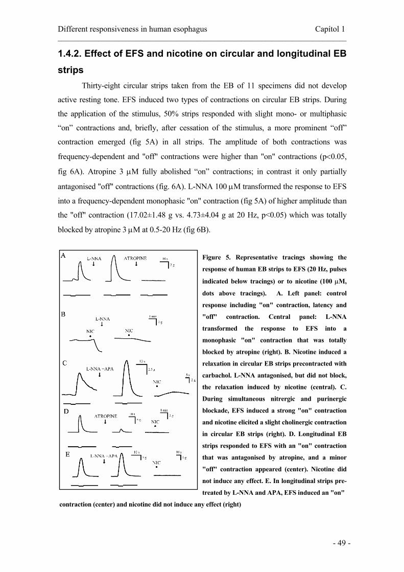
Different responsiveness in human esophagus Capítol 1 _____________________________________________________________________________________
- 49 -
1.4.2. Effect of EFS and nicotine on circular and longitudinal EB strips Thirty-eight circular strips taken from the EB of 11 specimens did not develop
active resting tone. EFS induced two types of contractions on circular EB strips. During
the application of the stimulus, 50% strips responded with slight mono- or multiphasic
“on” contractions and, briefly, after cessation of the stimulus, a more prominent “off”
contraction emerged (fig 5A) in all strips. The amplitude of both contractions was
frequency-dependent and "off" contractions were higher than "on" contractions (p<0.05,
fig 6A). Atropine 3 µM fully abolished “on” contractions; in contrast it only partially
antagonised "off" contractions (fig. 6A). L-NNA 100 µM transformed the response to EFS
into a frequency-dependent monophasic "on" contraction (fig 5A) of higher amplitude than
the "off" contraction (17.02±1.48 g vs. 4.73±4.04 g at 20 Hz, p<0.05) which was totally
blocked by atropine 3 µM at 0.5-20 Hz (fig 6B).
Figure 5. Representative tracings showing the
response of human EB strips to EFS (20 Hz, pulses
indicated below tracings) or to nicotine (100 µM,
dots above tracings). A. Left panel: control
response including "on" contraction, latency and
"off" contraction. Central panel: L-NNA
transformed the response to EFS into a
monophasic "on" contraction that was totally
blocked by atropine (right). B. Nicotine induced a
relaxation in circular EB strips precontracted with
carbachol. L-NNA antagonised, but did not block,
the relaxation induced by nicotine (central). C.
During simultaneous nitrergic and purinergic
blockade, EFS induced a strong "on" contraction
and nicotine elicited a slight cholinergic contraction
in circular EB strips (right). D. Longitudinal EB
strips responded to EFS with an "on" contraction
that was antagonised by atropine, and a minor
"off" contraction appeared (center). Nicotine did
not induce any effect. E. In longitudinal strips pre-
treated by L-NNA and APA, EFS induced an "on"
contraction (center) and nicotine did not induce any effect (right)

Different responsiveness in human esophagus Capítol 1 _____________________________________________________________________________________
- 50 -
Figure 6. Frequency-response curves of EB strips. Circular EB strips: A. EFS-induced "off"
contractions (n=24, N=9) were of higher amplitude than "on" contractions (n=19, N=9). "On"
contractions were fully cholinergic (n=5, N=4) whereas "off" contractions were only partly inhibited by
atropine (n=5, N=4). B. EFS induced only "on" contractions in the presence of L-NNA that were fully
blocked by atropine (n=6, N=4). Longitudinal EB strips: C. EFS-induced "on" contractions that were
antagonised but not blocked by atropine (n=6, N=3)
Nicotine (20-100 µM) did not elicit any mechanical response in EB circular strips
at rest (n=4, N=2). In EB strips precontracted at 5.3±0.6 g by 2 µM carbachol, nicotine 100
µM induced an immediate relaxation (fig 5B), decreasing the carbachol-induced tone by
82.2±5.8% (n=6, N=2). The relaxation induced by nicotine was completely blocked by
HEX 100 µM, and antagonised by pretreatment with 100 µM L-NNA, but a remaining
relaxation of 0.83±0.3 g (p<0.05) on the carbachol-induced tone was still elicited (fig. 5B).
Following simultaneous blockade with L-NNA and apamin, EFS induced a strong
cholinergic “on” contraction (16.56±1.57g at 20Hz) and NIC elicited a slight cholinergic
contraction (1.49±1.02 g, n=9, N=3), 9.20±5.51% of that induced by EFS (fig 7B).
Longitudinal EB strips did not develop active resting tone. EFS induced a
frequency-dependent “on” contraction on longitudinal EB strips (fig 5D) reaching
14.03±2.52 g at 70 Hz (fig 6C). Atropine 3 µM significantly reduced (from 90.2±9.8% at
10Hz to 94.39±3.21% at 70 Hz) the amplitude of longitudinal contractions (fig 7C) and
evoked the appearance of a residual low-amplitude "off" contraction following EFS (fig.
6C). Nicotine (20 µM -100 µM) did not elicit any mechanical response in longitudinal EB
strips at rest (n=5, N=3). Following L-NNA and apamin, EFS also induced a strong
cholinergic "on" contraction and nicotine did not induce any response (n=6, N=3, fig 7C).

Different responsiveness in human esophagus Capítol 1 _____________________________________________________________________________________
- 51 -
Figure 7. Quantitative effects of stimulation
of LES EMN by EFS or nicotine: At basal
conditions (A and B left panels) stimulation
of inhibitory LES by EFS caused an “off”
contraction, in contrast nicotine did not
induce any effect. A. Following Nitrergic
blockade, EFS induced a strong cholinergic
“on” contraction showing intense
stimulation of excitatory EMN, and, in the
same experimental conditions, stimulation
of nAChRs did not induce any effect. B.
During simultaneous blockade with L-NNA
and apamin, EFS also induced a strong
contraction and stimulation of nAChRs a
weak cholinergic contraction amounting to
9.20% of that induced by EFS and
suggesting a less efficient stimulation of
excitatory LES EMN. C. EFS also induced
an “on” contraction in longitudinal EB
strips at rest and also in those pre-treated by
L-NNA and apamin. Nicotine did not induce
any affect on longitudinal strips in both
experimental settings
1.5. Discussion We have found that inhibitory EMN in the human oesophagus are easily and
efficiently stimulated both by EFS and by nAChRs located in somatodendritic regions
as well as in nerve terminals. In contrast, although oesophageal excitatory EMN can be
efficiently stimulated by EFS even at basal conditions, its stimulation through nAChRs
is very difficult, requiring a full blockade of inhibitory motor pathways and causing a weak
response, suggesting the participation of non-nicotinic mechanisms in ganglionic
neurotransmission to excitatory oesophageal EMN in humans. We have also shown that
inhibitory EMN tonically inhibit LES resting tone, and that NO is the main but not the
only inhibitory neurotransmitter released by inhibitory EMN, suggesting a minor role
for an apamin-sensitive neurotransmitter in human oesophagus.

Different responsiveness in human esophagus Capítol 1 _____________________________________________________________________________________
- 52 -
The human LES is constituted by the sling muscle on the angle of Hiss and the
clasp component in lesser curvature, which develop greater active tone. This explains
the asymmetry of LES pressure in vivo (Preiksaitis & Diamant, 1997). We studied a
mixture of strips coming from both LES sides although, due to our method of
dissection, we probably obtained more sling strips than clasp-like fibres, thus
influencing our resting LES tone values. Our results show a sharp reduction in LES
resting tone following 10 min exposure to the clacium-free buffer, further confirming that
human LES tone is mainly myogenic and depends on extracellular calcium (Tottrup et al.,
1990c). There is controversy over the relative contribution of extra- and intracellular
calcium sources on myogenic LES tone (Biancani et al., 1987;Salapatek et al., 1998a),
and we cannot exclude a further reduction or even abolition of active tone by
prolongation of exposition of LES strips to a calcium-free medium (Biancani et al.,
1987). On the other hand, the relative neurogenic contribution to LES tone may vary
according to the species of animal. Previous studies on human LES strips failed to
demonstrate any effect of TTX (Preiksaitis & Diamant, 1997); however in vivo, NO
synthase inhibitors increased LES resting pressure in animal (Boulant et al., 1994;Xue et
al., 1996;Yamato et al., 1992), and human (Konturek et al., 1997) studies. A muscular as
well as a neural constitutive NO synthase could be responsible for this NO-mediated
inhibitory tonic influence on the LES muscle (Salapatek et al., 1998b). In our study, the
similarity to the increase in LES tone induced by TTX and L-NNA suggests that, in
humans, NO tonically released from inhibitory EMN continuously inhibits the myogenic
LES resting tone. The lack of effect of atropine suggests the absence of an intrinsic
cholinergic contribution to LES tone in vitro, agreeing with previous in vitro studies on the
human LES (Preiksaitis & Diamant, 1997). In vivo, atropine reduced LES pressure in
healthy humans (Dodds et al., 1981;Mittal et al., 1995), and hyoscine butylbromide, a
muscarinic antagonist which does not cross the blood-brain barrier, did not alter LES
pressure (Lidums et al., 1997). Altogether, these findings strongly suggest that the
atropine-induced fall in LES pressure in humans occurs at the CNS rather than at
peripheral sites.
In our study, EFS exclusively activates EMN during stimulus without direct
activation of muscle (Clave et al., 1998;Richards et al., 1995;Tottrup et al., 1990a;Tottrup
et al., 1990c) or preganglionic fibres because the neurotoxin TTX blocks and the
ganglionic blocker hexamethonium did not affect EFS-induced responses. The
simultaneous activation of both excitatory and inhibitory EMN by EFS is shown in our

Different responsiveness in human esophagus Capítol 1 _____________________________________________________________________________________
- 53 -
LES experiments by the selective blockade of inhibitory neurotransmitters (the relaxation
disappears and is replaced by a contraction induced by activation of excitatory EMN) or
blockade of the excitatory neurotransmitters (the relaxation increases). We used full
thickness oesophageal preparations that included the MP in order to compare these well-
characterised neural responses induced by EFS with those induced by nicotinic stimulation
of EMN. Most oesophageal EMN have single process Dogiel type I morphology and the
soma located in myenteric ganglia. In the LES of opossums, inhibitory EMN are larger and
with longer projections than the excitatory EMN, and the proportion of local excitatory and
inhibitory EMN is similar and about 50% (Brookes et al., 1996). In the human intestine,
circular inhibitory EMN also have larger cell bodies and larger projections (3.2 mm) than
the excitatory EMN (2 mm) (Porter et al., 1997), and EMN to longitudinal muscle were
smaller than those to the circular muscle (Porter et al., 1996;Wattchow et al., 1995). Based
on these morphological data, we inferred that our human oesophageal circular preparations
also contain a significant number and a similar proportion of both local excitatory and
inhibitory EMN. Immunohistochemical and functional studies have shown that nAChRs
are located in somatodendritic regions of EMN in myenteric ganglia (Schneider et al.,
2000), and nAChRs that cause transmitter release by action potential-independent
mechanisms have also recently been described in nerve terminals of excitatory EMN
(Galligan, 1999;Schneider et al., 2000). In our study, nicotine-induced LES relaxation was
partly antagonised by higher concentrations of TTX than those that fully blocked EFS-
relaxation and more than 30 times higher than those used to block nerve-mediated effects
in previous studies (Galligan, 1999;Schneider et al., 2000). Our data suggests the presence
of nAChRs in the soma as well as in the nerve terminals of human LES inhibitory EMN,
providing strong evidence for functional integrity of myenteric ganglia in our preparation.
It also allows an adequate experimental model for the comparison of the responses elicited
by stimulation of both intact and undamaged excitatory and inhibitory EMN. Our
preparation is transmural and contains the circular as well as the longitudinal muscular
layer and all the components of MP including myenteric ganglia, excitatory and inhibitory
EMN, and is not merely a muscle-axon preparation as nicotine did not induce any effect
on human muscle-axon LES preparations without MP (Tottrup et al., 1990a;Tottrup et al.,
1990c).
Integrated neuromuscular responses to EFS in our study were similar to those
previously reported in the human oesophagus (Burleigh, 1979;Preiksaitis & Diamant,
1997). Our results further confirm that NO is the main mediator for EFS-relaxation in the

Different responsiveness in human esophagus Capítol 1 _____________________________________________________________________________________
- 54 -
human LES (Preiksaitis et al., 1994) and suggest the release of one or more minor
inhibitory neurotransmitters, as an “off” relaxation sensitive to TTX appeared in our study
following nitrergic blockade (Uc et al., 1999). Our study also agrees with previous
studies that showed a relaxant effect through stimulation of nAChRs by nicotine or
DMPP in human LES strips (Burleigh, 1979;Smid & Blackshaw, 2000) also mainly
mediated by NO. Immunomorphological studies in the human oesophagus found that
most nitrergic EMN also contain VIP, galanin and PACAP (Ny et al., 1995;Singaram et
al., 1994;Uddman et al., 1991); and VIP, PACAP and adenosine relaxed human LES
strips in functional studies (Ny et al., 1995;Tottrup et al., 1990b). We recently found
nitrergic and apamin-sensitive inhibitory co-transmission on porcine LES (Farré et al.,
2002), very similar to that found by Yuan on the opossum (Yuan et al., 1998). In our
previous study on porcine LES, apamin inhibited the relaxation induced by ATP or
PACAP (Farré et al., 2002), and ATP has been proposed as the mediator for the apamin-
sensitive component in the opossum LES (Yuan et al., 1998). What we found in the
present study on human LES is indirect evidence for an apamin-sensitive inhibitory
neuromuscular co-neurotransmitter, as nicotine induced a relaxation following nitrergic
blockade and a small contraction following simultaneous blockade with L-NNA and
apamin. Further studies will ascertain the specific co-neurotransmitter involved in human
LES relaxation and explore its interaction with NO.
We compared the responses induced by direct stimulation of EMN by EFS with
those induced through stimulation of nAChRs. In our human study, LES stimulation by
nicotine in basal conditions caused a more intense relaxation than that induced by EFS,
both being LES inhibitory responses of comparable magnitude and suggesting high
efficiency of stimulation of inhibitory EMN. Simultaneous blockade of inhibitory
neurotransmitters by L-NNA and APA was required in opossum LES in order to show
focal EFS-induced stimulation of excitatory EMN (Yuan et al., 1998). This was not the
case in our study as a significant number of circular LES strips responded in basal
conditions to EFS with “off” contractions and circular EB strips responded with both
"on" and "off" frequency-dependent contractions (Richards et al., 1995;Tottrup et al.,
1990a;Tottrup et al., 1990c). The "on" contraction was abolished by atropine, clearly
showing its origin in cholinergic EMN. The latency depended on activation of inhibitory
EMN and it was fully blocked by NO synthesis inhibitors, as shown in other studies
(Christensen J, 1994;Preiksaitis et al., 1994). In contrast, the "off" contraction was only
partly antagonised by atropine (Christensen J, 1994;Krysiak & Preiksaitis, 2001;Richards

Different responsiveness in human esophagus Capítol 1 _____________________________________________________________________________________
- 55 -
et al., 1995;Tottrup et al., 1990a) suggesting the participation of a second non-
cholinergic mechanism such as rebound depolarisation (Paterson et al., 1991), or a second
excitatory neurotransmitter, such as tachykinins (Krysiak & Preiksaitis, 2001). We
explored the effect of nicotine on circular LES and EB circular strips at basal conditions
and following blockade of NO synthesis representing the majority of inhibitory
innervation. We expected nicotine would strongly stimulate nAChRs on cholinergic
EMN and make the strips contract, but it had no effect. However, EFS easily induced a
strong stimulation of excitatory EMN in both human LES and EB under the same
experimental conditions during nitrergic blockade. It could be argued that the
experiment must prove that inhibitory EMNs are completely suppressed prior to
stimulation of excitatory EMNs with nicotine in order to explain why nicotine does not
contract the muscle strips. Residual effect of the inhibitory EMNs after blockade with L-
NNA may be strong enough to negate the effects of nicotine on excitatory EMNs;
however this is not the case for EFS that acts as our “control” of a strong direct activation
of excitatory EMN in identical experimental conditions. In addition, our experimental
design included the comparison between EFS and nAChRs stimulation of EMN during
simultaneous blockade by L-NNA and Apamin in a third set of experiments. In order to
enhance our observations, the comparisons between EFS and nAChRs-mediated
responses were always performed in pairs, each strip acting as its own control. In this
experimental setting, stimulation of nAChRs induced a slight contraction in LES and EB
accounting for 6.64% and 9.20% respectively, of that obtained by EFS, and showing that
nAChRs stimulate excitatory EMN much less efficiently than EFS. Two additional
observations further confirmed the low sensitivity of excitatory oesophageal EMN to
nAChRs stimulation. First, as an isolated but significant observation, we found -as others
have (Misiewicz et al., 1969)- a strong direct stimulation of excitatory EMN by EFS and
no responses to nicotine in patients with achalasia and fully impaired inhibitory
neurotransmission (not shown). Second, that atropine inhibits nicotine-LES relaxation
(the opposite effect than on EFS relaxation) in our study, an effect very similar to that
caused by atropine during vagal efferent stimulation (Goyal & Rattan, 1975). As we
cannot attribute this effect to direct pharmacological antagonism (Heller J, 2001), we
speculate that the effect caused by atropine during LES stimulation by nAChRs could be
exerted on an interneuronal synapse, overriding the effect of atropine on excitatory EMN
at a neuromuscular level, and further showing a strong stimulation by nAChRs of a
polysynaptic inhibitory motor pathway and a weak stimulation of the excitatory one. In

Different responsiveness in human esophagus Capítol 1 _____________________________________________________________________________________
- 56 -
addition, the differences between the responsiveness of excitatory EMN to EFS and to
nicotine are also clearly observed in our experiments with longitudinal strips. As few
inhibitory EMN were found in the outer longitudinal smooth muscle layer of human
oesophagi in morphological studies (Singaram et al., 1994), no relaxant responses to EFS
could be evoked in longitudinal strips from human LES and EB strips (Tottrup et al.,
1990c), and EFS induced an “on” contraction in our basal studies. Stimulation of nAChRs
in these conditions did not induce any contraction, nor were any responses obtained by
nicotine following simultaneous blockade by L-NNA and apamin in longitudinal studies.
Thus, the low responsiveness of oesophageal excitatory EMN to stimulation through
nAChRs was observed in our study of circular LES and EB circular strips as well as in the
longitudinal EB layer.
Classical in vivo studies by Rattan and Goyal on the opossum suggested that the
vagi do not mediate any influence upon excitatory LES EMN and the vagal influence on
the sphincter is entirely inhibitory (Goyal & Rattan, 1975;Rattan & Goyal, 1974). These
authors also found that nicotine only stimulated inhibitory EMN (Rattan & Goyal, 1975)
and suggested that excitatory EMN may lie in an extravagal sympathetic pathway to the
LES (Rattan & Goyal, 1974). Recent studies on the same animal also found that vagal
stimulation did not evoke measurable excitatory responses even following complete
blockade of inhibitory LES pathways by L-NNA and apamin, suggesting that
preganglionic vagal efferent pathways probably activate LES inhibitory EMN more
efficiently than excitatory, or the excitatory lack direct vagal inputs (Yuan et al., 1998).
These results also agreed with those of our study but did not show how cholinergic EMN
in LES and EB were stimulated during primary and secondary peristalsis. In vivo studies
found hexamethonium-sensitive transmission to excitatory EMN in rat LES (Kawahara
et al., 1997), contrasting with in vitro studies also in the LES of the same animal
showing the absence of direct nicotinic inputs to excitatory EMN (Coruzzi et al., 1985),
and suggesting a polysynaptic pathway. In addition, recent studies in mice found
completely separate sets of preganglionic neurones located in different parts of dorsal
motor nucleus of the vagus projecting onto nitrergic or cholinergic LES EMN (Goyal et
al., 2001), providing an anatomical base for the physiological differences observed.
Otherwise, synaptic transmission in the ganglia of the autonomic nervous system is
classically described as mediated by acetylcholine acting on nAChRs; however,
ganglionic neurotransmission in the oesophagus can include important non-nicotinic
and also non-cholinergic synaptic responses. On the one hand, classical studies

Different responsiveness in human esophagus Capítol 1 _____________________________________________________________________________________
- 57 -
characterized the vagal efferent pathway to the opossum LES by means of both vagal
efferent stimulation and intramural EFS, and found that muscarinic (M1) and
serotoninergic (5HT3) receptors also mediate vagal inputs to inhibitory EMN (Gilbert et
al., 1984;Paterson et al., 1992) probably representing a mechanism of reserve or
redundancy of ganglionic transmission in this pathway [6]. We failed to show this
pathway in the present study, as the muscarinic ganglionic stimulant McN-A-343
(Gilbert et al., 1984) induced a contraction very similar to that induced by carbachol and
was also unaffected by TTX in our LES preparations (not shown) suggesting an effect
on muscular receptors. On the other hand, recent studies on animal models found up to
three subsets of EMNs in the MP that could be distinguished on the basis of the
neurotransmitters producing fast excitatory postsynaptic potentials (fEPSPs) in each
subset. In one subset (representing only 25%-36% of EMNs), fEPSPs was mediated
solely by Ach acting at nAChRs. In the other subsets, fEPSPs were mixed with a slight
contribution of Ach acting on nAChRs and a strong contribution of ATP acting on
purine P2X receptors, or 5-HT acting on 5-HT(3) receptors (Galligan et al.,
2000;Galligan, 2002). The relevance of each of these alternative mechanisms of
ganglionic neurotransmission can vary along the gastrointestinal tract and its role on
neurotransmission to excitatory oesophageal EMN needs further investigation.
Our results suggest that while vagal fibres or interneurones could easily and
efficiently stimulate inhibitory EMN through nAChRs in human LES and EB, full
stimulation of intrinsic excitatory EMN requires other neurotransmitters or other
circuits. Little is known about the anatomy and physiology of the neuronal circuitry that
directly controls the smooth muscle of the oesophagus. Understanding these circuits
could improve our current pharmacological approach of transient LES relaxations and
gastro-oesophageal reflux disease, and also our current treatments for primary and
secondary oesophageal motor disorders. Further studies are needed to describe these
neuronal circuits and to assess its relevance in health and disease.

Different responsiveness in human esophagus Capítol 1 _____________________________________________________________________________________
- 58 -
References
Biancani P, Hillemeier C, Bitar KN, & Makhlouf GM (1987). Contraction mediated by Ca2+ influx in esophageal muscle and by Ca2+ release in the LES. Am J Physiol 253, G760-G766.
Boulant J, Fioramonti J, Dapoigny M, Bommelaer G, & Bueno L (1994). Cholecystokinin and nitric oxide in transient lower esophageal sphincter relaxation to gastric distention in dogs. Gastroenterology 107, 1059-1066.
Brookes SJ, Chen BN, Hodgson WM, & Costa M (1996). Characterization of excitatory and inhibitory motor neurons to the guinea pig lower esophageal sphincter. Gastroenterology 111, 108-117.
Burleigh DE (1979). The effects of drugs and electrical field stimulation on the human lower oesophageal sphincter. Arch Int Pharmacodyn Ther 240, 169-176.
Christensen J CJ (1994). Motor functions of the pharynx and esophagus. In Physiology of the Gastrointestinal Tract, ed. Johnson LR, pp. 903-928. New York.
Clave P, Gonzalez A, Moreno A, Lopez R, Farre A, Cusso X, D'mato M, Azpiroz F, & Lluis F (1998). Endogenous cholecystokinin enhances postprandial gastroesophageal reflux in humans through extrasphincteric receptors. Gastroenterology 115, 597-604.
Coruzzi G, Poli E, & Bertaccini G (1985). Action of different agonists and antagonists of the cholinergic system on the rat lower esophageal sphincter. Gen Pharmacol 16, 561-566.
Dodds WJ, Dent J, Hogan WJ, & Arndorfer RC (1981). Effect of atropine on esophageal motor function in humans. Am J Physiol 240, G290-G296.
Farré R, Martinez E, Aulí M, Jimenez M, & Clave P. Nitric oxide, adenosine triphosphate and vasoactive intestinal peptide interactions in lower oesophageal sphincter relaxation. Neurogastroentrol Mot 14, 588. 2002. Ref Type: Abstract
Galligan JJ (1999). Nerve terminal nicotinic cholinergic receptors on excitatory motoneurons in the myenteric plexus of guinea pig intestine. J Pharmacol Exp Ther 291, 92-98.
Galligan JJ (2002). Ligand-gated ion channels in the enteric nervous system. Neurogastroenterol Motil 14, 611-623.
Galligan JJ, LePard KJ, Schneider DA, & Zhou X (2000). Multiple mechanisms of fast excitatory synaptic transmission in the enteric nervous system. J Auton Nerv Syst 81, 97-103.
Gilbert R, Rattan S, & Goyal RK (1984). Pharmacologic identification, activation and antagonism of two muscarine receptor subtypes in the lower esophageal sphincter. J Pharmacol Exp Ther 230, 284-291.
Gonzalez AA, Farre R, Mones J, Capella G, & Clave P (2000). Pharmacological and molecular characterization of muscular cholecystokinin receptors in the human lower oesophageal sphincter. Neurogastroenterol Motil 12, 539-546.
Goyal RK, Padmanabhan R, & Sang Q (2001). Neural circuits in swallowing and abdominal vagal afferent-mediated lower esophageal sphincter relaxation. Am J Med 111 Suppl 8A, 95S-105S.
Goyal RK & Rattan S (1975). Nature of the vagal inhibitory innervation to the lower esophageal sphincter. J Clin Invest 55, 1119-1126.
Heller J TP (2001). Muscarinic receptor agonists and antagonists. In The pharmacological basis of therapeutics, ed. Hardman JG LJaGGA, pp. 155-175.

Different responsiveness in human esophagus Capítol 1 _____________________________________________________________________________________
- 59 -
Hornby PJ & Abrahams TP (2000). Central control of lower esophageal sphincter relaxation. Am J Med 108 Suppl 4a, 90S-98S.
Kawahara H, Blackshaw LA, Lehmann A, & Dent J (1997). Responses of the rat lower oesophageal sphincter (LOS) to vagal efferent activation. Neurogastroenterol Motil 9, 85-97.
Konturek JW, Thor P, Lukaszyk A, Gabryelewicz A, Konturek SJ, & Domschke W (1997). Endogenous nitric oxide in the control of esophageal motility in humans. J Physiol Pharmacol 48, 201-209.
Krysiak PS & Preiksaitis HG (2001). Tachykinins contribute to nerve-mediated contractions in the human esophagus. Gastroenterology 120, 39-48.
Lidums I, Bermingham H, & Holloway RH (1997). Effect of hyoscine butylbromide on gastroesophageal reflux and lower esophageal sphincter function in healthy volunteers. Gastroenterology 112, A776.
Misiewicz JJ, Waller SL, Anthony PP, & Gummer JW (1969). Achalasia of the cardia: pharmacology and histopathology of isolated cardiac sphincteric muscle from patients with and without achalasia. Q J Med 38, 17-30.
Mittal RK, Holloway R, & Dent J (1995). Effect of atropine on the frequency of reflux and transient lower esophageal sphincter relaxation in normal subjects. Gastroenterology 109, 1547-1554.
Ny L, Larsson B, Alm P, Ekstrom P, Fahrenkrug J, Hannibal J, & Andersson KE (1995). Distribution and effects of pituitary adenylate cyclase activating peptide in cat and human lower oesophageal sphincter. Br J Pharmacol 116, 2873-2880.
Paterson WG, Anderson MA, & Anand N (1992). Pharmacological characterization of lower esophageal sphincter relaxation induced by swallowing, vagal efferent nerve stimulation, and esophageal distention. Can J Physiol Pharmacol 70, 1011-1015.
Paterson WG, Hynna-Liepert TT, & Selucky M (1991). Comparison of primary and secondary esophageal peristalsis in humans: effect of atropine. Am J Physiol 260, G52-G57.
Porter AJ, Wattchow DA, Brookes SJ, & Costa M (1997). The neurochemical coding and projections of circular muscle motor neurons in the human colon. Gastroenterology 113, 1916-1923.
Porter AJ, Wattchow DA, Brookes SJ, Schemann M, & Costa M (1996). Choline acetyltransferase immunoreactivity in the human small and large intestine. Gastroenterology 111, 401-408.
Preiksaitis HG & Diamant NE (1997). Regional differences in cholinergic activity of muscle fibers from the human gastroesophageal junction. Am J Physiol 272, G1321-G1327.
Preiksaitis HG, Tremblay L, & Diamant NE (1994). Nitric oxide mediates inhibitory nerve effects in human esophagus and lower esophageal sphincter. Dig Dis Sci 39, 770-775.
Rattan S & Goyal RK (1978). Evidence of 5-HT participation in vagal inhibitory pathway to opossum LES. Am J Physiol 234, E273-E276.
Rattan S & Goyal RK (1975). Effect of nicotine on the lower esophageal sphincter. Studies on the mechanism of action. Gastroenterology 69, 154-159.
Rattan S & Goyal RK (1974). Neural control of the lower esophageal sphincter: influence of the vagus nerves. J Clin Invest 54, 899-906.
Richards WG, Stamler JS, Kobzik L, & Sugarbaker DJ (1995). Role of nitric oxide in human esophageal circular smooth muscle in vitro. J Thorac Cardiovasc Surg 110, 157-164.
Salapatek AM, Lam A, & Daniel EE (1998a). Calcium source diversity in canine lower esophageal sphincter muscle. J Pharmacol Exp Ther 287, 98-106.

Different responsiveness in human esophagus Capítol 1 _____________________________________________________________________________________
- 60 -
Salapatek AM, Wang YF, Mao YK, Mori M, & Daniel EE (1998b). Myogenic NOS in canine lower esophageal sphincter: enzyme activation, substrate recycling, and product actions. Am J Physiol 274, C1145-C1157.
Schneider DA, Perrone M, & Galligan JJ (2000). Nicotinic acetylcholine receptors at sites of neurotransmitter release to the guinea pig intestinal circular muscle. J Pharmacol Exp Ther 294, 363-369.
Singaram C, Sengupta A, Sweet MA, Sugarbaker DJ, & Goyal RK (1994). Nitrinergic and peptidergic innervation of the human oesophagus. Gut 35, 1690-1696.
Smid SD & Blackshaw LA (2000). Neuromuscular function of the human lower oesophageal sphincter in reflux disease and Barrett's oesophagus. Gut 46, 756-761.
Tottrup A, Forman A, Funch-Jensen P, Raundahl U, & Andersson KE (1990a). Effects of transmural field stimulation in isolated muscle strips from human esophagus. Am J Physiol 258, G344-G351.
Tottrup A, Forman A, Madsen G, & Andersson KE (1990b). The actions of some beta-receptor agonists and xanthines on isolated muscle strips from the human oesophago-gastric junction. Pharmacol Toxicol 67, 340-343.
Tottrup A, Forman A, Uldbjerg N, Funch-Jensen P, & Andersson KE (1990c). Mechanical properties of isolated human esophageal smooth muscle. Am J Physiol 258, G338-G343.
Uc A, Oh ST, Murray JA, Clark E, & Conklin JL (1999). Biphasic relaxation of the opossum lower esophageal sphincter: roles of NO., VIP, and CGRP. Am J Physiol 277, G548-G554.
Uddman R, Luts A, Absood A, Arimura A, Ekelund M, Desai H, Hakanson R, Hambreaus G, & Sundler F (1991). PACAP, a VIP-like peptide, in neurons of the esophagus. Regul Pept 36, 415-422.
Wattchow DA, Brookes SJ, & Costa M (1995). The morphology and projections of retrogradely labeled myenteric neurons in the human intestine. Gastroenterology 109, 866-875.
Xue S, Valdez D, Collman PI, & Diamant NE (1996). Effects of nitric oxide synthase blockade on esophageal peristalsis and the lower esophageal sphincter in the cat. Can J Physiol Pharmacol 74, 1249-1257.
Yamato S, Saha JK, & Goyal RK (1992). Role of nitric oxide in lower esophageal sphincter relaxation to swallowing. Life Sci 50, 1263-1272.
Yuan S, Costa M, & Brookes SJ (1998). Neuronal pathways and transmission to the lower esophageal sphincter of the guinea Pig. Gastroenterology 115, 661-671.

Aquest capítol s’ha acceptat per ser publicat a: Farré R, Aulí M, Lecea B, Martínez E, Clavé P. Pharmacological characterization of intrinsic mechanisms controlling tone and relaxation of porcine lower esophageal sphincter. J Pharmacol Exp Ther 2005.
Capítol 2
Pharmacologic characterization of intrinsic mechanisms controlling tone and relaxation of porcine lower esophageal sphincter
2.1. Abstract
The neurotransmitters mediating relaxation of lower esophageal sphincter (LES) were
studied by using circular LES strips from adult pigs in organ baths. LES relaxation by sodium
nitroprusside (SNP, 1 nM-3 µM), VIP and PACAP (1 nM-1 µM), ATP (10 µM-30 mM), and
tricarbonyldichlororuthenum dimer (CORM-1 1 µM-1 mM) was unaffected by tetrodotoxin
(TTX 1 µM) or L-NAME (100 µM). Calcitonin gene-related peptide (CGRP 1 nM-1 µM)
unaffected LES tone. ATP-relaxation was blocked by 1 µM apamin and the P2Y1 antagonist
N6-methyl 2'-deoxyadenosine 3',5'-bisphosphate (MRS2179 10 µM). Apamin inhibited
PACAP-relaxation. VIP- and PACAP-relaxation was blocked by α-chymotrypsin 10 U/ml. L-
NAME (-62.52±13.13%) and ODQ 10 µM (-67.67±6.80%) similarly inhibited EFS-
relaxation, and apamin blocked non-nitrergic relaxation. Nicotine-relaxation (100 µM) was
inhibited by L-NAME (-60.37±10.8%) and ODQ (-41.90±7.89%); and apamin also blocked
non-nitergic relaxation. Non-nitrergic and apamin-sensitive LES relaxation caused by EFS or
nicotine was strongly inhibited by MRS2179, slightly inhibited by α-chymotrypsin and the
P2X1,2,3 receptor antagonist NF279 (10 µM), and unaffected by selective heme oxygenase
inhibitor tin protoporphyrin IX (100 µM). Porcine LES relaxation following stimulation of
intrinsic inhibitory motor neurons is mediated by two main neuromuscular pathways: a) NO
through guanylate cyclase signaling and apamin-insensitive mechanisms, and b) by nonintrergic
apamin-sensitive neurotransmission mainly mediated by ATP or a related purine acting on P2Y1

Pharmacological characterization of porcine LES Capítol 2 ____________________________________________________________________________
- 62 -
receptors but also with a minor contribution of purinergic P2X receptors and PACAP on LES
relaxation. Nitrergic and purinergic co-transmitters show parallel effects of similar magnitude
without major interplay. Our study shows no role for CGRP, and only a minor one for VIP
and CO in porcine LES relaxation.
2.2. Introduction The lower esophageal sphincter (LES) acts as a barrier at the gastroesophageal
junction (GEJ). Basal tone of LES is primarily myogenic in origin and is modulated by a
combination of hormonal factors and neurogenic mechanisms that are not yet fully understood
and involve local enteric motor neurons (EMN) and extrinsic nerves. Non-adrenergic, non-
cholinergic (NANC) EMN are the final step in the inhibitory vagal pathway to LES, allowing
swallowing-induced and transient LES relaxation that cause physiologic gastroesophageal
reflux and belching (Chang et al., 2003). Vasoactive intestinal peptide (VIP), NO, ATP,
pituitary adenylate cyclase-activating peptide (PACAP), carbon monoxide (CO), and
calcitonin gene-related peptide (CGRP) have been proposed as putative neurotransmitters for
these inhibitory EMN in LES on the basis of morphological and physiological studies.
Colocalization of these neurotransmitters and/or their synthesizing enzymes on inhibitory
EMN of the upper gastrointestinal tract including LES has been described by
immunohistological and other morphological studies (Ny et al., 1994;Ny et al., 1995a;Uc et
al., 1997;Werkstrom et al., 1997). Physiological and pharmacological studies have
demonstrated the direct effects or characterized the actions of these neurotransmitters
following stimulation of inhibitory LES EMNs (Imaeda & Cunnane, 2003;Ny et al.,
1995b;Ny et al., 1997;Uc et al., 1997;Yuan et al., 1998). However, evidence suggests that NO
from neural sources is the major contributor to LES relaxation (Gonzalez et al., 2004;Murray
et al., 1991;Tottrup et al., 1991), and the relative physiological contribution of other
neurotransmitters remains unclear. On the other hand, several mechanisms of interaction
between these neurotransmitters have been proposed: a) release “in parallel” and independent
actions on specific postjunctional sites (cotransmission) (Burnstock, 2004), b) effects coupled
“in series” with NO-mediated release of other neurotransmitters (Grider et al., 1992), or pre-
and/or postjunctional modulation of NO synthesis by other neurotransmitters (Ergun &
Ogulener, 2001;Mashimo et al., 1996). The underlying mechanisms and physiological
relevance of these interactions on LES have not been discovered. Most of these studies
focused on the mechanisms of LES relaxation during simultaneous and direct electrical

Pharmacological characterization of porcine LES Capítol 2 ____________________________________________________________________________
- 63 -
stimulation of excitatory and inhibitory EMN and little data is available on the effects of these
inhibitory neurotransmitters following other more specific stimuli for inhibitory EMN. In a
recent in vitro study on human LES, we found that inhibitory EMNs are efficiently stimulated
both by EFS and nicotinic acetylcholine receptors (AChRs) located in somatodendritic
regions and nerve terminals, whereas esophageal excitatory EMNs are also efficiently
stimulated by EFS but their stimulation through nicotinic AChRs is difficult and causes a weak
response (Gonzalez et al., 2004).
The porcine gastrointestinal tract possesses anatomic and pathological similarities to
that of humans and similar organization of the enteric nervous system, differing from small
laboratory animals, and has been used as a homologous animal model for the development of
new pharmacologic strategies to treat human neurogastrointestinal disorders (Brown &
Timmermans, 2004;Pasricha et al., 1993). The size, histology (smooth muscle cells) and
neurochemical code of porcine LES EMNs are similar to that of humans (Aggestrup et al.,
1986;Pasricha et al., 1993). The aim of the present study was to characterize the
neuromyogenic mechanisms and neurotransmitters (NTs) controlling tone and relaxation of
porcine lower esophageal sphincter (LES) following stimulation of inhibitory EMNs by EFS
and through nicotinic AChRs.
2.3. Methods 2.3.1. Preparations
Specimens including part of the gastric fundus, the gastro-esophageal junction (GEJ), and
the esophageal body were obtained from 53 adult pigs (age, 6 months; weight, 75-80 Kg).
Animals were stunned and killed by exsanguination in a slaughterhouse in compliance with
specific national laws following the guidelines of the European Union (Pérez Rubalcaba A.,
1995) . Specimens were immediately collected, placed in carbogenated Krebs solution at 4ºC and
transported to the laboratory within 1 hour. The GEJ was opened along the greater curvature, the
mucosa and submucosa were resected at the squamocolumnar union, and clasp and sling fibers
composing LES were identified (Preiksaitis & Diamant, 1997). Full thickness preparations
including the circular and longitudinal muscle layers as well as the myenteric plexus were
obtained by cutting 3mm-wide strips parallel to circular muscle fibers from the clasp region of
the LES of each specimen.

Pharmacological characterization of porcine LES Capítol 2 ____________________________________________________________________________
- 64 -
2.3.2. Procedures
Studies started within 18 hours of sacrifice. Strips measuring 10 mm in length were
placed in 15 mL organ baths containing Krebs' solution constantly bubbling with 5% CO2 in O2.
Changes in tension of the strips were measured using isometric force transducers, recorded on a
chart recorder (model 03 Force Transducer and model 7 Series Polygraph, respectively, Grass
Instruments Co, Quincy, MA, USA), and digitized (AcqKnowledge MP100, Biopac Systems
Inc, CA, USA). After an equilibration period of 30 min, strips were stretched up to 150% of their
initial length and positioned between two parallel platinum wire electrodes 10 mm apart. Most
strips taken from the GEJ progressively increased their tension during the following 1-2 hours.
This increase in tension was defined as the active resting tone (Tottrup et al., 1990). EFS was
applied by means of an electrical stimulator (Model S88, Grass Instruments Co) and a power
booster (Stimu-Splitter II, Med-Lab Instruments, Loveland, CO, USA) in order to obtain six
identical and undistorted signals. Only the strips that developed active tension during the
equilibration period and relaxed with EFS and/or nicotine were considered as pertaining to the
LES and included in the study.
2.3.3. Solutions and Drugs
The Krebs solution used in these experiments contained (in mM) 138.5 Na+, 4.6 K+, 2.5
Ca2+, 1.2 Mg+, 125 Cl-, 21.9 HCO3- ,1.2 H2PO4
-, 1.2 SO4- and 11.5 glucose. Sodium nitroprusside
(SNP), VIP, ATP, CGRP, apamin, nicotine, hexamethonium, L-NAME, suramin, VIP 6-28, 1H-
[1,2,4]oxadiazolo-[4,3-α]quinoxalin-1-one (ODQ), α-chymotrypsin and CORM-1 (tricarbonyl
dichlororuthenum dimer) were obtained from Sigma–Aldrich Co (Madrid, Spain). Tetrodotoxin
(TTX) was purchased from Latoxan (Valence, France), PACAP 28 from Peptides Institute
(Osaka, Japan), and the competitive antagonist for P2Y1 receptors MRS 2179 (2’-Deoxy-N6-
methyladenosine 3’,5’-bisphosphate tetraammonium salt), the antagonist for the P2X1,2,3
receptors NF 279 (8,8’-[Carbonylbis(imino-4,1-phenylenecarbonylimino-4,1-
phenylenecarbonylimino)]bis-1,3,5-naphthalenetrisulfonic acid hexasodium salt) and the
selective heme oxygenase inhibitor tin protoporphyrin IX (SnPP-IX) from Tocris Cookson Ltd
(Bristol, UK). All drugs were dissolved in distilled water, with the exception of ODQ which was
dissolved in ethanol <5% vv-l, and CORM-1 and SnPP-IX that were freshly prepared before each
experiment and dissolved in DMSO. Final concentration of DMSO in the organ bath did not
exceed 0.1% vv-l . Experiments using SnPP-IX were conduced in the dark. In pilot studies we
found that at these concentrations neither ethanol and DMSO did not significantly alter resting
tone or EFS-induced responses (data not shown).

Pharmacological characterization of porcine LES Capítol 2 ____________________________________________________________________________
- 65 -
2.3.4. Experimental Design
In order to ascertain the physiological relevance of each putative neurotransmitter on
LES physiology, our experimental design included: a) the effect of neurotransmitters on LES
tone, the selection and characterization of specific antagonists for each neurotransmitter, and
the evaluation of their possible interactions, particularly the possible induction of NO-
synthesis by any other neurotransmitter; b) the study of neuromyogenic mechanisms
controlling active LES tone; and c) the characterization of the relaxatory responses following
stimulation of LES enteric inhibitory motor neurons by EFS or through nicotinic AChRs, the
effects of the neurotransmitters released, and the assessment of the possible NO-mediated
release of any other putative neurotransmitters.
2.3.4.1. The effect of neurotransmitters on LES tone and the selection of specific antagonists
Concentration-related curves of the effects of SNP, VIP, PACAP, ATP and CGRP
were obtained by exposing LES strips to single doses of agonists for up to 3 min. After
washing the strips with 45 mL of fresh buffer there was a 30-minute period before the next
exposure. Concentration-response curves for CORM-1 were obtained in a cumulative fashion.
Concentrations of neurotransmitters were selected according to previous studies, most on LES
(Ny et al., 1995b;Rattan et al., 2004;Uc et al., 1997;Uc et al., 1999;Yamato et al., 1992). We
used 1 nM-3 µM for SNP; 1 nM-1 µM for VIP, PACAP, and CGRP; 10 µM-30 mM for ATP,
and 1 µM-1 mM for CORM-1. Reported concentrations are final bath concentrations.
Submaximal LES responses to SNP, ATP, VIP, PACAP, and CORM-1 were studied in the
presence of the neurotoxin TTX (1 µM) and the following substances: ODQ (10 µM),
inhibitor of soluble GC; apamin (1 µM), blocker of small conductance Ca2+-activated K+ (SK)
channels; MRS 2179 (10 µM), competitive antagonist for P2Y1 receptors and NF279 (10 µM)
(De Man et al., 2003) for P2X1,2,3 receptors; the peptidase α-chymotrypsin (10 U/mL), and the
VIP antagonist, VIP 6-28 (100 nM); and also following the NO-synthase inhibitor L-NAME
(100 µM) to explore the possible involvement of NO in the response of other
neurotransmitters. Antagonists were added to the baths 30 min before addition of
neurotransmitters with the exception of α-chymotrypsin which was added 10 min before. In
preliminary experiments we found that, at the dose and time of exposition used, α-
chymotrypsin did not affect responses to repetitive doses of VIP or PACAP (not shown).

Pharmacological characterization of porcine LES Capítol 2 ____________________________________________________________________________
- 66 -
2.3.4.2. Control of active LES resting tone
The neurogenic vs myogenic contribution to active LES resting tone was assessed by
measuring changes in tone following exposure to the neurotoxin TTX 1 µM and to a Ca2+-free
medium for 30 min. The influence of tonic activity of inhibitory EMN upon LES resting tone
was studied by measuring maximal changes in tone following exposure to L-NAME 100 µM,
ODQ 10 µM, apamin 1 µM, and MRS 2179 10 µM for 30 min; or α-chymotrypsin 10 U/mL
for 10 min. The influence of cholinergic EMN upon LES tone was assessed by atropine 1 µM
for 30 min.
2.3.4.3. Stimulation of EMN by EFS, the effects of the neurotransmitters released, and characterization of NO-mediated effects. Transmural EFS (pulses of 0.4 mS duration, frequency 0.3-20 HZ) were applied to
LES preparations in 5 s trains at 26 V (Gonzalez et al., 2004). The amplitude of EFS-induced
responses did not decay during control experiments lasting up to four hours (not shown). The
neural origin of EFS-induced responses was assessed by TTX 1 µM and hexamethonium 100
µM. First, two experiments were conducted during direct stimulation of inhibitory EMN by EFS
and sequential addition of antagonists in order to assess the nature and effects of the
neurotransmitters released. In the fist experiment (Experiment E-1) the nitrergic and cholinergic
components of EFS responses were sequentially blocked by L-NAME and atropine 1 µM, and
apamin was assessed on non-nitergic relaxation. Results were compared with experiment E-2
during blockade by ODQ 10 µM of the target (guanylate cyclase) of the synthesized and released
NO, allowing the effect of any neurotransmitter coupled in series with NO, or released by NO. In
experiments E-3 to E-6 the effect of MRS 2179 (experiment E-3), NF 279 (experiment E-4), α-
chymotrypsin (experiment E-5), and SnPP-IX 100 µM (Experiment E-6) on L-NAME resistant
relaxation was assessed. Drugs were added to the baths 30 min prior to EFS with the exception
of α-chymotrypsin which was added 10 min before.
2.3.4.4. Stimulation of EMN with nicotine, identification of the neurotransmitters released and characterization of NO-mediated effects.
A concentration-related curve of the effect of nicotine (1-300 µM) on LES strips was
obtained by exposing the strips for 2 min to single doses of the drug in order to assess the
LES relaxation induced by stimulation of EMN through nicotinic AChRs. Strips were
washed with 45 mL of fresh buffer and left for 30 min before exposure. Repeated additions of
nicotine (100 µM) did not desensitize nicotinic receptors (not shown). The specificity of the
effects of nicotine was assessed by the ganglionic blocker hexamethonium 100 µM, and the

Pharmacological characterization of porcine LES Capítol 2 ____________________________________________________________________________
- 67 -
site of effect by TTX 1 µM (Galligan, 1999;Gonzalez et al., 2004). Nicotine dose which
caused a submaximal LES relaxation was first selected for the studies with antagonists in
order to characterize the neurotransmitters released by stimulation of inhibitory EMN through
nicotinic AChRs. In the fist experiment (Experiment N-1) the nitrergic component of nicotine
response was assessed by L-NAME, and apamin was assessed on non-nitergic relaxation.
Results were compared with experiment N-2 during blockade by ODQ 10 µM of the target
(guanylate cyclase) of the synthesized and released NO allowing the effect of any
neurotransmitter coupled in series with NO, or released by NO. In experiments N-3 to N-6 the
relative effect of MRS 2179 (experiment N-3), NF 279 (experiment N-4), α-chymotrypsin
(experiment N-5), and SnPP-IX (Experiment N-6) on L-NAME resistant relaxation was
assessed. Drugs were added to the baths 30 min before the stimulation of inhibitory EMN with
nicotine with the exception of α-chymotrypsin which was added 10 min before. Experiments
were conduced at basal conditions and not in NANC conditions in order to compare neural
stimulation by EFS or with that of nicotinic AChRs (Gonzalez et al., 2004).
2.3.5. Data Analysis The effect of EFS and pharmacological agents were determined in terms of changes in
tone. Relaxation was expressed in g and/or in the percentage of active LES resting tone. The
dose-response curve was computer-fitted using nonlinear regression and the maximal
response elicited by the agonist and the EC50 were calculated (GraphPad prism, Version 2.1,
U.S.A.). Contraction was expressed in g and/or in percentage of active LES resting tone. Data
are expressed as mean ± mean standard error. Student-t test was selected for comparisons,
using the paired mode when appropriate, and the effect of pharmacological agents on
frequency-response curves was performed using two-way repeated measure ANOVA. When
the t-test was significant, the Bonferroni test was carried out to determine the frequencies of
statistically different responses. A p value <0.05 was considered statistically significant.
2.4. Results 2.4.1. Effect of neurotransmitters on LES tone and selection of specific antagonists LES strips developed an active resting tone of 4.90±0.30g (n=34). SNP, VIP, PACAP,
ATP, and CORM-1 relaxed LES strips; in contrast CGRP had no effect at any dose tested
(Figure 1 and 2). The NO-donor SNP, the CO donor CORM-1 and VIP (Figure 1) induced a
monophasic LES relaxation. PACAP induced a biphasic response with an initial fast followed

Pharmacological characterization of porcine LES Capítol 2 ____________________________________________________________________________
- 68 -
by a slow and sustained relaxation (Figure 1). ATP 1 mM evoked a triphasic mechanical
response including an initial fast relaxation, which was followed by a fast contraction, and a
slow sustained relaxation (Figure 1). Relaxation induced by SNP, CORM-1 and VIP was
slower than initial fast relaxation induced by PACAP and ATP (Table 1). SNP, VIP, PACAP,
CORM-1 and ATP relaxed LES strips in a concentration-dependent manner (Figure 2). Table
1 summarizes the dynamics of the effects of proposed neurotransmitters on LES. PACAP and
NO induced a complete LES relaxation at µM concentrations and showed EC50 in the 10 nM
range. ATP induced a complete relaxation at mM concentrations. VIP relaxed the active tone
of strips by only 47±12.2% at µM concentrations and CORM-1 by 30.94±6.7% at mM
concentrations.
Figure 1. Representative tracings showing the morphology of LES relaxation induced by each proposed
neurotransmitter. The NO donor SNP, VIP, and the CO donor CORM-1 induced a monophasic
relaxation. PACAP induced a biphasic relaxation. ATP induced an initial fast relaxation, followed by a
fast contraction and a sustained relaxation and CGRP did not induce any effect on LES tone
Figure 2. Log concentration-response
relaxation and contraction curves to the
proposed neurotransmitters in porcine
LES strips. Left panel:
Neurotransmitters with monophasic
responses (SNP, CORM-1, VIP, CGRP).
Right panel: Neurotransmitters with
polyphasic responses (PACAP and ATP).
Effects are expressed in terms of
percentage changes of active LES tone
(n=3 at each point)
The doses of neurotransmitters causing submaximal LES relaxation selected for
studies with antagonists were SNP (1 µM), VIP (100 nM), CORM-1 (500 µM), ATP (1 mM),
and PACAP (100 nM), the effects of antagonists having been evaluated in individual
experiments using 3-7 specimens. Inhibition of NO synthesis by L-NAME did not

Pharmacological characterization of porcine LES Capítol 2 ____________________________________________________________________________
- 69 -
significantly modify any of the responses induced by SNP (+0.50±4.02%, ns), VIP (-
8.56±2.91%, ns), ATP (FR +0.30±4.86%, FC -13.24±4.47%, SR +4.01±11.27%, ns), or
PACAP (FR -11.64±10.92%, SR -13.40± 5.28%, ns). NO-induced relaxation by SNP was
specifically blocked by ODQ (-98.35±1.15%, p<0.05) and unaffected by the other
antagonists. Fast relaxation induced by ATP was unaffected by ODQ (+4.00±3.13%, ns), L-
NAME (+0.30±4.86%, ns) or α-chymotrypsin (+0.26±8.04%, ns). In contrast, apamin fully
blocked (-98.84±1.16%, p<0.01) the initial fast relaxation and switched the LES response to
ATP into a biphasic response with an initial contraction of 1.68±0.78g and a slow sustained
relaxation. The ATP-induced contraction was not affected either by TTX (-0.44±16.18%, ns),
or ODQ (-12.18±6.05%, ns). MRS 2179 10 µM strongly inhibited fast ATP-induced
relaxation (-76.3±4.88%, p<0.05) and fast contraction (-80.38±0.75%, p<0.05). Apamin
inhibited fast component (-76.78±6.06%, p<0.05) and had no effect on the slow component (-
31.75±9.49, ns) of PACAP-induced LES relaxation. Unexpectedly, CORM-1 relaxation was
unaffected by either ODQ or apamin. VIP 6-28 100 nM did not significantly affect VIP
relaxation (+1.78±7.02%, ns); and α-chymotrypsin fully blocked VIP and PACAP-induced
relaxation (-100%, p<0.05). TTX did not significantly affect the relaxation induced by SNP (–
3.04 ±2.17%, ns), VIP (-5.62±5.49%, ns), ATP (FC +27.31± 9.21% and SR +21.30± 9.57%,
ns), and PACAP (FR -20.26±16.89%, ns and SR -30.57± 8.93%, ns).
2.4.2. Control of active LES resting tone Figure 3 illustrates the myogenic and neurogenic factors contributing to LES tone.
Exposure of LES strips to a Krebs Ca2+-free buffer fully abolished active resting tone by
96.40±5.82% (n=9). Simultaneous blockade of intrinsic excitatory and inhibitory neural
inputs by TTX did not significantly affect active LES tone (-6.93±8.84% n=7, ns). In contrast,
atropine significantly reduced active LES resting tone by -24.3±3.1% (n=17) (p<0.05); and L-
NAME, ODQ and apamin significantly enhanced active LES resting tone by 46.45±3.31%
(n=17), 28.22±2.83% (n=19), and 20.3±2.1% (n=12), respectively (p<0.05); MRS 2179
enhanced LES tone by only 2.87±2.16% (n=6, ns). The enhancement caused by L-NAME on
LES tone was higher than that caused by ODQ or apamin (p<0.001). α-chymotrypsin exerted
a variable effect on LES, as active tone was either enhanced by 41±47% (n=8), or reduced by
36.81±47% (n=7) following 10 min exposure to the drug. These experiments show that
porcine LES tone is mainly myogenic, depends on extracellular Ca2+, and is modulated by tonic
input from cholinergic EMNs as well as by continuous influence of inhibitory EMNs.

Pharmacological characterization of porcine LES Capítol 2 ____________________________________________________________________________
- 70 -
Figure 3. Neuromyogenic control of LES tone. Effect
on active LES tone induced by: Ca2+-free medium,
TTX, L-NAME, ODQ, apamin, MRS 2179 and
atropine
2.4.3. Inhibitory NTs in EFS-induced relaxation LES strips responded to EFS with a sharp relaxation during electrical stimulus (“on”
relaxation) followed by a phasic contraction at the end of the stimulus (“off” contraction)
(Figure 4). The amplitude of both responses was frequency-dependent and maximal relaxation
(4.21±0.25g or 90.4±3.1% of resting tone) was reached at 3 Hz (n=34). EFS-induced
relaxation was fully blocked by TTX at all frequencies tested (-100%, n=5, p<0.001), and
unaffected by hexamethonium (+6.78±14.65% at 3 Hz, n=3, ns).
Figure 4. Characterization of NTs
released during stimulation of LES EMN
by EFS. A) Representative tracings and
B) quantitative effects on frequency-
dependent (0.3-20 Hz) EFS-induced
responses (n=5 in each experiment).
Horizontal axis depicts the time schedule
of the experiment during sequential
addition of antagonists. * = p<0.05 vs the
same frequency in the previous
experimental condition. Experiment E-1.
Blockade of NO synthesis by L-NAME
significantly reduced the amplitude of
both “on” relaxation and “off”
contraction
Subsequent addition of atropine reduced “off” contraction and enhanced “on” relaxation.
Apamin blocked L-NAME resistant EFS-induced relaxation and switched “off” contraction to
an “on” contraction during EFS without changes in amplitude. In experiment E-1 (Figure 4),

Pharmacological characterization of porcine LES Capítol 2 ____________________________________________________________________________
- 71 -
L-NAME significantly inhibited EFS relaxation with significant effects at all frequencies
tested and an average effect of -62.52±13.13% (n=5). Subsequent addition of atropine
enhanced relaxation at 20 Hz (p<0.05).
Apamin fully blocked the L-NAME resistant “on” relaxation in 4 out of 5 experiments
(p<0.05). In the same experiment, L-NAME reduced the amplitude of basal EFS-“off”
contraction with significant effects at 0.5 to 20 Hz (P<0.05). The subsequent addition of
atropine further reduced “off” contraction at 10 and 20 Hz and apamin switched the “off”
contraction to an “on” contraction during EFS without affecting the amplitude of the
response. These results clearly show that EFS- induced LES relaxation is mainly mediated by
both NO synthesis and other neurotransmitters acting through apamin-sensitive K+ channels.
In the second experiment (Experiment E-2, Figure 5) we explored the possible effect
of any inhibitory neurotransmitter released by NO. In this experiment ODQ reduced EFS
relaxation with significant effects at all frequencies tested (n=5, p<0.05). Subsequent addition
of atropine increased the relaxation at 10 and 20 Hz, and apamin fully blocked this non-
nitrergic EFS “on” relaxation in 4 out of 5 experiments (p<0.05). Also in experiment E-5,
ODQ reduced the amplitude of the "off" contraction at frequencies above 1 Hz (p<0.05);
subsequent addition of atropine further reduced the amplitude of the contraction at 10 and 20
Hz (p<0.05), and similarly, apamin switched the “off” response into an “on” contraction. In
these experiments with stimulation of EMNs by EFS, the average of inhibitory effect of ODQ
on relaxation (-67.67±6.80%) was similar to that caused by L-NAME arguing against the
possibility of a relaxatory effect of any NTs released by NO. Apamin also caused similar and
comparable effects in both experiments.
Figure 5. Experiment E-2. Blockade of NO
effects by ODQ significantly reduced the
amplitude of both “on” relaxation and
“off” contraction. Subsequent addition of
atropine reduced “off” contraction and
enhanced “on” relaxation. Apamin
blocked ODQ resistant EFS-induced
relaxation and switched “off” contraction
to an “on” contraction during EFS without
changes in amplitude

Pharmacological characterization of porcine LES Capítol 2 ____________________________________________________________________________
- 72 -
The nature of the non-nitrergic neurotransmitter released by EFS was explored in
experiments E-3 to E.6. In experiment E-3 (Figure 6), MRS 2179 strongly inhibited the non-
nitrergic EFS relaxation with significant effects at 3-20 Hz (n=5, p<0.001). In experiment E-4
NF 279 slightly but consistently reduced non-nitrergic EFS relaxation, also with significant
effects at 3-20 Hz (n=5, p<0.001). In experiment E-5 (Figure 6), α-chymotrypsin only slightly
inhibited the L-NAME resistant “on” relaxation with significant effects at 3 and 20 Hz (n=5,
p<0.05), and in experiment E-6, SnPP-IX did not affect EFS relaxation (n=5, ns).
Figure 6. Characterization of
NTs involved in non-nitrergic
and apamin-sensitive EFS-
induced LES relaxation
following L-NNA and atropine.
The purinergic antagonist MRS
2179 strongly inhibited EFS-
induced “on” relaxation at
frequencies above 3Hz in
experiment E-3. The P2X
antagonist NF279 (experiment
E-4) and chymotrypsin
(experiment E-5) slightly but
significantly antagonized the
EFS-relaxation, and, in contrast,
Tin-protoporphyrin IX did not induce any effect on EFS-relaxation in experiment E-6
Results from experiments E-1 suggest that EFS relaxation is mainly mediated by NO
and an apamin-sensitive neurotransmitter, and results from experiments E-3 and E-4 show
that the non-nitrergic and apamin-sensitive component is strongly inhibited by a purinergic
P2Y1 receptor antagonist and only slightly reduced by a P2X1,2,3 purinergic receptor
antagonist. This clearly indicates the effect of a purinergic neurotransmitter mainly acting at
P2Y1 receptors through apamin-sensitive small conductance Ca2+-activated K+ channels. In
addition the non-nitrergic and apamin-sensitive component was only slightly reduced by α-
chymotrypsin, and unaffected by heme oxygenase inhibition, suggesting only a minor role for
a peptidic neurotransmitter agreeing with the pharmacological profile of PACAP as the direct

Pharmacological characterization of porcine LES Capítol 2 ____________________________________________________________________________
- 73 -
effect of PACAP on LES is inhibited by both apamin and α-chymotrypsin, and no role for CO
during EFS-relaxation.
2.4.4. Inhibitory neurotransmitters in nicotine-induced relaxation Nicotine (1-300 µM) relaxed LES strips in a concentration-dependent manner. EC50
was 20.2 µM and maximal relaxation (3.93±1.03g or 100.36±3.37% of resting tone) was
reached at 100 µM (n=5). The amplitude of the relaxation induced by stimulation of nicotinic
AChRs was similar to that obtained by EFS (ns). Hexamethonium fully blocked nicotine-
induced LES relaxation (-98.27±3.51%, n=3, p<0.05), and TTX reduced by 28.08±12% but
did not block maximal relaxation induced by nicotine (n=5, p<0.05).
Nicotine-induced relaxation (100 µM) was also significantly reduced by L-NAME by
60.37±10.80% (n=5, p<0.05, Experiment N-1 in Figure 7).
Figure 7. Characterization of NTs released during stimulation of LES EMN by nicotine 100µM. A)
Representative tracings showing the morphology of nicotine-induced responses. B) Quantitative effects
(n=5 in each experiment). Horizontal axis depicts the time schedule of the experiment during sequential
addition of antagonists. * = p<0.05 vs previous experimental condition. Experiment N-1. Blockade of NO
synthesis by L-NAME significantly reduced the amplitude of the nicotine-induced relaxation. Subsequent
addition of apamin fully blocked the non-nitrergic relaxation and induced the appearance of a contraction
that was fully blocked by atropine. Experiment N-2. Blockade of NO effects by ODQ reduced the
amplitude of the relaxation. Subsequent addition of apamin blocked the residual relaxation and induced
the appearance of a contraction that was fully blocked by atropine

Pharmacological characterization of porcine LES Capítol 2 ____________________________________________________________________________
- 74 -
Subsequent addition of apamin blocked nonitrergic relaxation and transformed the
nicotine-response into a biphasic contraction with an amplitude of 0.65±0.14 g and 1.76±0.19
g, respectively. This double-peaked contraction was fully blocked by atropine (p<0.05). In
parallel experiments (Experiment N-2, Figure 7), ODQ significantly reduced the nicotine-
induced relaxation by 41.90±7.98% (n=5, p<0.05), and apamin further blocked the nicotine-
relaxation which also led to a cholinergic double-peaked contraction (0.77±0.13 g and 2.10
±0.41 g, respectively). In these experiments with stimulation of EMN trough nicotine AChRs,
the effect of ODQ was similar to that induced by L-NAME (ns), and comparable to that
caused by apamin in both parallel studies. Relative inhibition caused by L-NAME, ODQ, and
apamin did not differ in both EFS and nicotine experiments (ns).
Experiments N-3 to N-6 (Figure 8) were performed to characterize the nature of the
non-nitrergic neurotransmitters released following stimulation of inhibitory LES EMN
through nicotinic AChRs. Nicotine-induced relaxation resistant to L-NAME was strongly
inhibited by MRS 2179 by 76.6±9.27% (n=5, p<0.01) in experiment N-3, and also
significantly inhibited by NF 279 by 48.04±16.64% (n=5, p<0.05) in experiment N-4 and by
18.88±1.00% in experiment N-5 by α-chymotrypsin (n=5, p<0.001) in experiment N-5, in
experiment N-6 SnPP-IX did not significantly affect nicotine relaxation. Experiment N-1
shows that LES relaxation following stimulation of inhibitory EMNs through nicotinic
AChRs is mainly caused by NO and one or more apamin-sensitive neurotransmitters. The
nonitrergic and apamin-sensitive relaxation was strongly antagonized by the purinergic
antagonist MRS 2179 in experiment N-3, further confirming the effect of a purinergic
neurotransmitter mainly acting at P2Y1 receptors through apamin-sensitive small conductance
Ca2+-activated K+ channels. In addition, nonitrergic and apamin-sensitive nicotine-induced
relaxation is also moderately antagonized through purinergic P2X1,2,3 receptor antagonists and
by α-chymotrypsin also suggesting a minor role for a peptidic neurotransmitter agreeing with
the pharmacological profile of PACAP, and no role for CO in LES relaxation following
stimulation of inhibitory EMN through nicotinic AChRs.

Pharmacological characterization of porcine LES Capítol 2 ____________________________________________________________________________
- 75 -
Figure 8. Characterization of NTs
involved in L-NAME resistant and
apamin-sensitive nicotine-induced LES
relaxation. Results are expressed in
percentage of inhibition of L-NAME
resistant relaxation. MRS 2179 strongly
inhibited nicotine relaxation in
experiment N-3. NF 279 in experiment N-
4 and chymotrypsin in experiment N-6
also partly antagonized nicotine
relaxation and Tin-protoporphyrin IX
did not induce any significant effect on
nicotine-relaxation
2.5. Discussion
Our study shows that porcine LES tone is mainly myogenic and is modulated by tonic
input from excitatory and inhibitory EMNs. We found that relaxation of porcine LES following
stimulation of inhibitory EMNs is caused to a similar extent by two main pathways: a) a nitrergic
one mediated by NO through guanylate cyclase signaling and apamin-insensitive pathways, and
b) a nonintrergic pathway coupled to apamin-sensitive small conductance Ca2+-activated K+
channels mainly mediated by ATP or a related purine acting through P2Y1 receptors and a minor
contribution of purinergic P2X receptors and PACAP on LES relaxation. Our results suggest
parallel release of nitrergic, purinergic, and peptidergic neurotransmitters, independent
effects, and no major interplay on their release or postjunctional effects (Figure 9). Our study
did not find any role for CGRP in porcine LES. In addition although CO and VIP caused a
direct LES relaxation in our study, we did not find any major role for them in neuroeffector
LES relaxation.
Porcine, like human, LES is formed by the gastric sling muscle on the angle of Hiss
and the clasp component in lesser curvature (transverse esophageal fibers) (Preiksaitis &
Diamant, 1997). We only included clasp strips that developed active tension and relaxed
during stimulation of inhibitory EMN, ensuring that we had selected LES strips with intact
innervation. Our preparation was transmural and contained all the components of MP,
including myenteric ganglia and excitatory and inhibitory EMN, and was not merely a
muscle-axon preparation. We tested the effect of a NO-donor, VIP, ATP, PACAP, a CO-

Pharmacological characterization of porcine LES Capítol 2 ____________________________________________________________________________
- 76 -
donor and CGRP because these substances colocalize in esophageal inhibitory EMN (Ny et
al., 1995a;Uc et al., 1997). Colocalized substances can participate as neuromodulators,
neurotransmitters or with neurotrophic effects (Burnstock, 2004). We found that the NO-
donor, VIP, ATP, the CO-donor, and PACAP, but not CGRP, relaxed LES strips through a
TTX-insensitive mechanism. NO, ATP, and PACAP induced a complete LES relaxation, and
CO and VIP were clearly less efficient, as observed in porcine gastric fundus (Colpaert et al.,
2002). In contrast, the same dose of CGRP that relaxed LES strips in opossums failed to relax
our preparation despite the similarity between both species in the EFS-induced responses (Uc
et al., 1997). These discrepancies could be explained by species differences and/or CGRP
acting through more complex neural circuits in “in vivo” studies (Rattan et al., 1988). As
expected, relaxation induced by SNP was specifically blocked by ODQ in our study showing
a direct NO effect through guanylate cyclase pathways. The relaxation caused by VIP was
unaffected by L-NAME or by ODQ arguing against the theory of a “serial cascade” involving
NO production in nerve terminals and/or smooth muscle cells by VIP (Ergun & Ogulener,
2001;Grider et al., 1992). Apamin inhibited ATP and PACAP but not NO, CO or VIP
relaxation in our study; and α-chymotrypsin that cleaves VIP and PACAP at the level of
tyrosine residues (Mule & Serio, 2003) antagonized both VIP and fast-PACAP relaxation
without affecting the NO or ATP response. ATP induced a complex LES response similar to
that observed on vascular smooth muscle (Ralevic, 2002). Smooth muscle cells along the
gastrointestinal tract can simultaneously express G-protein coupled P2Y receptors mediating
the relaxatory effects of purines, and ion-gated P2X receptors mediating either contractions
(Giaroni et al., 2002) or relaxations (Storr et al., 2000). Activation of P2X receptors could
initially induce an increase in intracellular Ca++ leading a contraction, and a relaxation as a
result of secondary activation of apamin-sensitive Ca++-dependent K+-channels (Ishiguchi et
al., 2000). MRS 2179 is a specific antagonist for P2Y1 purinoreceptor subtype (De Man et
al., 2003) that in our study strongly antagonized the ATP relaxation. ATP can also induce NO
production in smooth muscle cells (Xue et al., 2000), however in our study neither L-NAME
or α-chymotrypsin affected ATP-induced relaxation, also arguing against an “in series”
relation between NO or VIP and ATP.
We assessed the physiological relevance of NO, ATP, PACAP, CO and VIP by testing
the effect of their antagonists on LES resting tone and during stimulation of inhibitory EMNs
by EFS or through nicotinic AChRs. The most important component of LES tone is myogenic
and the relative neurogenic contribution to LES tone varies with the animal species. Our results
showed an intense reduction in LES resting tone by the Ca2+-free buffer, further confirming that

Pharmacological characterization of porcine LES Capítol 2 ____________________________________________________________________________
- 77 -
porcine LES tone is mainly myogenic and depends on extracellular Ca2+ (Tottrup et al., 1990).
Our study also showed that atropine decreased LES less intensively tone than the removal of
extracellular Ca2+ showing a moderate tonic input from cholinergic EMNs; and L-NAME, ODQ,
and apamin enhanced LES tone also showing a tonic influence of inhibitory EMNs. NO synthase
inhibitors also increased LES resting tone in humans (Gonzalez et al., 2004). The higher effect of
L-NAME compared with ODQ in our study could be explained by a finding in canine LES
showing ODQ-dependent mechanisms mediating only the actions of NO from nerves, whereas
NO from muscles utilized ODQ independent mechanisms to modulate LES tone (Daniel et al.,
2002). Interestingly, we failed to show any effect of atropine on LES tone in an earlier study on
human LES (Gonzalez et al., 2004), suggesting a stronger influence of cholinergic EMNs in
porcine LES that should be taken into account when considering the effects of some new
pharmacological treatments for achalasia in humans based on the reduction of cholinergic inputs
such as botulin toxin (Pasricha et al., 1993).
Porcine LES responses to EFS and nicotine in this study are similar to those we found
in earlier studies on human LES (Gonzalez et al., 2004). The effects of EFS were fully
blocked by the neurotoxin TTX and unaffected by the ganglionic blocker hexamethonium,
showing the origin of these responses in the activation of EMNs. Our results on pigs show
that maximal EFS relaxation was inhibited to the same degree by L-NAME and ODQ (by
60%), and that apamin fully blocked the EFS induced relaxation resistant to NO inhibition in
both series of experiments. Our results clearly show that the neural relaxation induced by EFS
has two main components, one mediated by a direct effect of NO on smooth muscle and a
second one of comparable magnitude mediated by an apamin-sensitive neurotransmitter that
has been indirectly attributed to ATP in previous studies (Imaeda & Cunnane, 2003;Yuan et
al., 1998). We found that the apamin-sensitive component of EFS-relaxation has three
components: it is strongly inhibited by MRS 2179 showing that a purine mediates this
component by acting on P2Y1 receptors; it is slightly but consistently inhibited by NF 279
also suggesting the involvement of P2X receptors; and it is slightly inhibited by the peptidase
chymotrypsin suggesting that PACAP (an apamin-sensitive peptidergic neurotransmitters)
also has a minor role in EFS-induced relaxation. Similar results on the involvement of P2Y1
and also P2X receptors on purinergic inhibitory neurotransmission were found by De Man in
the mouse jejunum (De Man et al., 2003) and ATP-induced relaxation of rat pylorus has been
attributed to P2X purinoceptors located on smooth muscle cells (Ishiguchi et al., 2000).
Finally, heme oxygenase inhibitors did not modify the EFS-inhibitory responses in our
preparation or in porcine stomach (Colpaert et al., 2002) or porcine ileum (Matsuda et al.,

Pharmacological characterization of porcine LES Capítol 2 ____________________________________________________________________________
- 78 -
2004) precluding a major role for CO during EFS relaxation. In addition, the similarity of the
responses in experiments with ODQ (where NO is synthesized and released and could induce
the release of any other NT) and L-NAME (where NO is not synthesized) argues against
release of VIP or any other neurotransmitter by NO (Grider et al., 1992) and also against NO
inhibition of the release of ATP (Ishiguchi et al., 2000). Our results suggest mechanisms of
simultaneous release and independent actions (co-transmission) for the nitrergic, purinergic,
and peptidergic inhibitory neurotransmitters released during EFS.
We also explored the neurotransmitters released on stimulation of EMN with nicotine.
Immunohistological and functional studies have shown that nicotinic AChRs are located in
somatodendritic regions of EMN and participate in the transmission of vagal inputs (Chang et
al., 2003;Galligan, 1999). In earlier studies we found functional evidence of the presence of
nicotinic AChRs at pre-junctional sites of inhibitory EMN in human LES (Gonzalez et al.,
2004). In the present study, the effects of EFS were fully blocked by TTX, but in contrast,
nicotine-induced LES relaxations were only partly antagonized by TTX, further suggesting
the presence of nAChRs in nerve terminals of the inhibitory EMN of porcine LES. Previous
studies on cats (Kortezova et al., 1994) and our previous studies on human LES also found a
residual relaxation induced by nicotine following NO blockade. However, the relative
magnitude of the non-nitregic component varies between these species as NO synthesis
inhibitors reduced relaxation induced by stimulation of nAChRs by more than 85% in humans
(Gonzalez et al., 2004), 70-80% in cats (Kortezova et al., 1994), and by 40-60% in the present
study on pigs. On the other hand, stimulation of myenteric plexus ganglia of guinea-pig ileum
with the nicotinic agonist DMPP induced both VIP and NO release, VIP release being further
facilitated by NO production (Grider et al., 1992). In our present study, the relaxation
resistant to NO blockade (L-NAME or ODQ) was fully antagonized by apamin and a weak
contraction induced by stimulation of cholinergic EMNs appeared. Similarly to the findings
on EFS-relaxation, we also found three components on apamin-sensitive nicotine-LES
relaxation the main component being antagonized by MRS 2179 and mediated by a purine
through P2Y1 receptors, a second minor component antagonized by NF279 and mediated also
by a purine but through P2X receptors, and a minor third component antagonized by α-
chymotrypsin and also probably mediated by PACAP (Smid & Blackshaw, 2000) and
unaffected by inhibitors of heme oxygenase. Our results also showed that LES relaxation
induced by nicotine in basal conditions was similar to that induced by EFS, suggesting high
efficiency of stimulation of inhibitory EMN by nicotinic AChRs. The "off" contraction
induced by EFS in this study was partly antagonized by atropine, partly caused by a rebound

Pharmacological characterization of porcine LES Capítol 2 ____________________________________________________________________________
- 79 -
depolarization —because an important part of the “off” contraction is inhibited by NO
blockers— and partly caused by the probable participation of a minor secondary non-
cholinergic excitatory neurotransmitter such as tachykinins (Chang et al., 2003). In this
experimental setting, stimulation of nicotinic AChRs during simultaneous blockade of nitrergic
and apamin-sensitive effects induced only a slight contraction in LES, suggesting that nicotinic
AChRs stimulate excitatory EMN much less efficiently than inhibitory EMN, as we found in
human LES in an earlier study (Gonzalez et al., 2004). These results further suggest that
whereas vagal fibers or interneurons could easily and efficiently stimulate inhibitory EMNs in
porcine LES, full stimulation of intrinsic excitatory EMNs requires non-nicotinic
neurotransmitters (Galligan et al., 2000) or other circuits that need further investigation.
Figure 9. Schematic
representation of two main
parallel pathways of
inhibitory neurotransmission
in porcine LES: a) NO
through guanylate cyclase
signaling and apamin-
insensitive mechanisms, and
b) nonintrergic apamin-
sensitive neurotransmission
mainly mediated by ATP or a
related purine acting on P2Y1
receptors but also with a
minor contribution of purinergic P2X receptors and PACAP. Our study shows no role for CGRP, and,
although CO and VIP caused a direct LES relaxation in our study, we did not found any major role for
these substances as neuroeffector neurotransmitters in LES relaxation. small conductance. GC: guanylate
cyclase ; NOs: Nitric oxide synthase; SK: small conductance Ca2+-activated K+ channels

Pharmacological characterization of porcine LES Capítol 2 ____________________________________________________________________________
- 80 -
References
Aggestrup S, Uddman R, Jensen SL, Hakanson R, Sundler F, Schaffalitzky dM, & Emson P (1986). Regulatory peptides in lower esophageal sphincter of pig and man. Dig Dis Sci 31, 1370-1375.
Brown DR & Timmermans JP (2004). Lessons from the porcine enteric nervous system. Neurogastroenterol Motil 16 Suppl 1, 50-54.
Burnstock G (2004). Cotransmission. Curr Opin Pharmacol 4, 47-52.
Chang HY, Mashimo H, & Goyal RK (2003). Musings on the wanderer: what's new in our understanding of vago-vagal reflex? IV. Current concepts of vagal efferent projections to the gut. Am J Physiol Gastrointest Liver Physiol 284, G357-G366.
Colpaert EE, Timmermans JP, & Lefevbre RA (2002). Investigation of the potential modulatory effect of biliverdin, carbon monoxide and bilirubin on nitrergic neurotransmission in the pig gastric fundus. European Journal of Pharmacology 457, 177-186.
Daniel EE, Bowes TJ, & Jury J (2002). Roles of guanylate cyclase in responses to myogenic and neural nitric oxide in canine lower esophageal sphincter. J Pharmacol Exp Ther 301, 1111-1118.
De Man JG, De Winter BY, Seerden TC, De Schepper HU, Herman AG, & Pelckmans PA (2003). Functional evidence that ATP or a related purine is an inhibitory NANC neurotransmitter in the mouse jejunum: study on the identity of P2X and P2Y purinoceptors involved. British Journal of Pharmacology 140, 1108-1116.
Ergun Y & Ogulener N (2001). Evidence for the interaction between nitric oxide and vasoactive intestinal polypeptide in the mouse gastric fundus. J Pharmacol Exp Ther 299, 945-950.
Galligan JJ (1999). Nerve terminal nicotinic cholinergic receptors on excitatory motoneurons in the myenteric plexus of guinea pig intestine. J Pharmacol Exp Ther 291, 92-98.
Galligan JJ, LePard KJ, Schneider DA, & Zhou X (2000). Multiple mechanisms of fast excitatory synaptic transmission in the enteric nervous system. J Auton Nerv Syst 81, 97-103.
Giaroni C, Knight GE, Ruan HZ, Glass R, Bardini M, Lecchini S, Frigo G, & Burnstock G (2002). P2 receptors in the murine gastrointestinal tract. Neuropharmacology 43, 1313-1323.
Gonzalez AA, Farre R, & Clave P (2004). Different responsiveness of excitatory and inhibitory enteric motor neurons in the human esophagus to electrical field stimulation and to nicotine. Am J Physiol Gastrointest Liver Physiol 287, G299-G306.
Grider JR, Murthy KS, Jin JG, & Makhlouf GM (1992). Stimulation of nitric oxide from muscle cells by VIP: prejunctional enhancement of VIP release. Am J Physiol 262, G774-G778.
Imaeda K & Cunnane TC (2003). Electrophysiological properties of inhibitory junction potential in murine lower oesophageal sphincter. J Smooth Muscle Res 39, 119-133.
Ishiguchi T, Takahashi T, Itoh H, & Owyang C (2000). Nitrergic and purinergic regulation of the rat pylorus 3. American Journal of Physiology-Gastrointestinal and Liver Physiology 279, G740-G747.
Kortezova N, Velkova V, Mizhorkova Z, Bredy-Dobreva G, Vizi ES, & Papasova M (1994). Participation of nitric oxide in the nicotine-induced relaxation of the cat lower esophageal sphincter. J Auton Nerv Syst 50, 73-78.
Mashimo H, He XD, Huang PL, Fishman MC, & Goyal RK (1996). Neuronal constitutive nitric oxide synthase is involved in murine enteric inhibitory neurotransmission. J Clin Invest 98, 8-13.

Pharmacological characterization of porcine LES Capítol 2 ____________________________________________________________________________
- 81 -
Matsuda NM, Miller SM, Sha L, Farrugia G, & Szurszewski JH (2004). Mediators of non-adrenergic non-cholinergic inhibitory neurotransmission in porcine jejunum 1. Neurogastroenterol Motil 16, 605-612.
Mule F & Serio R (2003). NANC inhibitory neurotransmission in mouse isolated stomach: involvement of nitric oxide, ATP and vasoactive intestinal polypeptide 2. British Journal of Pharmacology 140, 431-437.
Murray J, Du C, Ledlow A, Bates JN, & Conklin JL (1991). Nitric oxide: mediator of nonadrenergic noncholinergic responses of opossum esophageal muscle. Am J Physiol 261, G401-G406.
Ny L, Alm P, Ekstrom P, Hannibal J, Larsson B, & Andersson KE (1994). Nitric oxide synthase-containing, peptide-containing, and acetylcholinesterase-positive nerves in the cat lower oesophagus. Histochem J 26, 721-733.
Ny L, Alm P, Larsson B, Ekstrom P, & Andersson KE (1995a). Nitric oxide pathway in cat esophagus: localization of nitric oxide synthase and functional effects. Am J Physiol 268, G59-G70.
Ny L, Larsson B, Alm P, Ekstrom P, Fahrenkrug J, Hannibal J, & Andersson KE (1995b). Distribution and effects of pituitary adenylate cyclase activating peptide in cat and human lower oesophageal sphincter. Br J Pharmacol 116, 2873-2880.
Ny L, Waldeck K, Carlemalm E, & Andersson KE (1997). Alpha-latrotoxin-induced transmitter release in feline oesophageal smooth muscle: focus on nitric oxide and vasoactive intestinal peptide. Br J Pharmacol 120, 31-38.
Pasricha PJ, Ravich WJ, & Kalloo AN (1993). Effects of intrasphincteric botulinum toxin on the lower esophageal sphincter in piglets. Gastroenterology 105, 1045-1049.
Pérez Rubalcaba A. Real decreto sobre proteccion de los animales en el momento de su sacrificio o matanza. Boletin Oficial del Estado 39, 5146-5153. 1995. Ref Type: Journal (Full)
Preiksaitis HG & Diamant NE (1997). Regional differences in cholinergic activity of muscle fibers from the human gastroesophageal junction. Am J Physiol 272, G1321-G1327.
Ralevic V (2002). The involvement of smooth muscle P2X receptors in the prolonged vasorelaxation response to purine nucleotides in the rat mesenteric arterial bed. British Journal of Pharmacology 135, 1988-1994.
Rattan S, Gonnella P, & Goyal RK (1988). Inhibitory effect of calcitonin gene-related peptide and calcitonin on opossum esophageal smooth muscle. Gastroenterology 94, 284-293.
Rattan S, Haj RA, & De Godoy MAF (2004). Mechanism of internal anal sphincter relaxation by CORM-1, authentic CO, and NANC nerve stimulation 1. American Journal of Physiology-Gastrointestinal and Liver Physiology 287, G605-G611.
Smid SD & Blackshaw LA (2000). Vagal ganglionic and nonadrenergic noncholinergic neurotransmission to the ferret lower oesophageal sphincter. Auton Neurosci 86, 30-36.
Storr M, Franck H, Saur D, Schusdziarra V, & Allescher HD (2000). Mechanisms of alpha,beta-methylene atp-induced inhibition in rat ileal smooth muscle: Involvement of intracellular Ca2+ stores in purinergic inhibition. Clinical and Experimental Pharmacology and Physiology 27, 771-779.
Tottrup A, Forman A, Uldbjerg N, Funch-Jensen P, & Andersson KE (1990). Mechanical properties of isolated human esophageal smooth muscle. Am J Physiol 258, G338-G343.
Tottrup A, Svane D, & Forman A (1991). Nitric oxide mediating NANC inhibition in opossum lower esophageal sphincter. Am J Physiol 260, G385-G389.
Uc A, Murray JA, & Conklin JL (1997). Effects of calcitonin gene-related peptide on opossum esophageal smooth muscle. Gastroenterology 113, 514-520.

Pharmacological characterization of porcine LES Capítol 2 ____________________________________________________________________________
- 82 -
Uc A, Oh ST, Murray JA, Clark E, & Conklin JL (1999). Biphasic relaxation of the opossum lower esophageal sphincter: roles of NO., VIP, and CGRP. Am J Physiol 277, G548-G554.
Werkstrom V, Ny L, Persson K, & Andersson KE (1997). Carbon monoxide-induced relaxation and distribution of haem oxygenase isoenzymes in the pig urethra and lower oesophagogastric junction 3. British Journal of Pharmacology 120, 312-318.
Xue L, Farrugia G, & Szurszewski JH (2000). Effect of exogenous ATP on canine jejunal smooth muscle. Am J Physiol Gastrointest Liver Physiol 278, G725-G733.
Yamato S, Saha JK, & Goyal RK (1992). Role of nitric oxide in lower esophageal sphincter relaxation to swallowing. Life Sci 50, 1263-1272.
Yuan S, Costa M, & Brookes SJ (1998). Neuronal pathways and transmission to the lower esophageal sphincter of the guinea Pig. Gastroenterology 115, 661-671.

Aquest capítol s’està revisant per enviar-lo a publicar: Farre R, Martinez E, Clave P. Asymmetrical mechanisms controlling resting tone, relaxation and contraction in clasp and sling regions of porcine LES. 2005.
Capítol 3
Asymmetrical mechanisms controlling resting tone, relaxation and contraction in clasp and sling regions of porcine LES
3.1. Abstract To compare in vitro the functional properties of the clasp and sling muscles of the
Lower Esophageal Sphincter (LES). Circular LES strips (clasp/sling pairs) from 25 adult
pigs were studied in organ baths to assess the mechanisms controlling LES tone and the
responses following neural stimulation by electrical field stimulation (EFS) or through
nicotinic receptors. Tension developed by sling strips was higher than that developed by
clasp fibers (7.59±0.89 g versus 4.72±0.67 g, p<0.05). LES tone is more sensitive to
extracellular calcium depletion in clasp strips and to nitrergic and cholinergic blockade in
sling strips. TTX did not affect LES tone and blocked responses to EFS in both LES
regions. Amplitude of EFS “on” relaxation and “off” contraction was higher in clasp strips.
ODQ reduced the “on” relaxation and “off” contraction in clasp; and blocked the “on”
relaxation and enhanced the “on” contraction in sling fibers. Amplitude of nicotine
relaxation was similar in clasp and sling regions. In the clasp region, nicotine relaxation
was reduced by ODQ or TTX. In sling fibers, nicotine induced a relaxation at rest and, in
contrast, a cholinergic contraction following ODQ or TTX.
The porcine LES shows a strong transversal intrinsic functional asymmetry suggesting a
specialization at the neuronal level. Tonic input from EMNs has a greater influence on
tone developed by sling fibers. Neural stimulation induced strong nitrergic responses and

Asymmetrical mechanisms in LES Capítol 3 _______________________________________________________________
- 84 -
weak cholinergic responses in clasp fibers; and, in contrast, weak nitrergic responses and
strong cholinergic responses in the sling region.
3.2. Introduction
The main physiological function of the lower esophageal sphincter (LES) is to
generate a high pressure barrier to prevent reflux of gastric acid contents into the
esophagus. The second is to open during swallowing to allow bolus passage, and also
during transient LES relaxations that causes physiological gastro-esophageal reflux and
allows belching. In vivo studies on humans, piglets, opossums, and cats using
manometry have shown a significant radial asymmetry in the distribution of the high
pressure barrier as the highest pressures were recorded in the left lateral direction of the
LES (Preiksaitis et al., 1994a;Richardson & Welch, 1981;Schulze et al., 1977;Vicente
et al., 2001;Welch & Drake, 1980). Moreover, in both human and animal in vivo
studies, atropine produces a greater drop in LES pressure on the left side of the LES
(Preiksaitis et al., 1994a;Richardson & Welch, 1981). There is little data available on the
origin and physiological role of this transversal functional LES asymmetry and its
relevance to the pathophysiology of esophageal diseases such as achalasia or
gastroesophageal reflux disease.
The origin of the asymmetry in LES motility is uncertain and has been attributed to
anatomical and physiological factors. The macroscopic arrangement of LES circular
smooth fibers of the inner muscle in humans and pigs is very similar, with transverse
muscle clasps on the lesser curve side (“clasp fibers”), and long oblique gastric “sling”
fibers in the greater curvature on the left side of LES (Liebermann-Meffert et al.,
1979;Vicente et al., 2001). In addition, clasp and sling fibers differ in function: in vitro
studies have shown that LES pressure is due to a myogenic mechanism that is concentrated
in the clasp muscle fibers and a neurogenic cholinergic contribution primarily due to the
action of sling muscle fibers (Preiksaitis et al., 1994a;Preiksaitis & Diamant, 1997). This
transversal functional asymmetry has been attributed to specialization at both muscular
(Muinuddin et al., 2003;Muinuddin et al., 2004a;Preiksaitis et al., 1994a) and neuronal
level (Brookes et al., 1996;Yuan et al., 1998;Yuan & Brookes, 1999).
The porcine and human LES possess anatomical and pathological similarities and
similar organization of the enteric nervous system, differing from small laboratory animal
models. Porcine LES has been used as a homologous animal model for the development of

Asymmetrical mechanisms in LES Capítol 3 _______________________________________________________________
- 85 -
new pharmacologic strategies to treat human neurogastrointestinal disorders (Brown &
Timmermans, 2004;Pasricha et al., 1993). The size, the transversal anatomical asymmetry
(clasp and sling fibers), the histology (smooth muscle cells) and the neurochemical code of
porcine LES EMNs is similar to that of humans (Aggestrup et al., 1986;Pasricha et al.,
1993). We have recently explored the intrinsic mechanisms that control human and porcine
LES motility in vitro (Gonzalez et al., 2004;Farré et al., 2005) . In humans we found that
inhibitory EMN releases NO as the main neurotransmitter and also a minor apamin-
sensitive one (Gonzalez et al., 2004). Relaxation of porcine LES following stimulation of
inhibitory EMNs is caused to a similar extent by two parallel pathways: a) a nitrergic one
mediated by NO through guanylate cyclase signaling pathways, and b) an apamin-sensitive
pathway mainly mediated by ATP or a related purine acting through P2Y1 receptors (Farré
et al., 2005). In both species, effects of excitatory EMNs were mainly mediated by
acetylcholine (ACh).
The present study was undertaken to compare the in vitro functional properties of
LES clasp and sling muscles in a porcine model. We explored the myogenic and
neurogenic mechanisms of control of resting tone and characterized the inhibitory and
excitatory responses following stimulation of intrinsic nerves with electrical field
stimulation (EFS) or through nicotinic ACh receptors.
3.3. Methods 3.3.1. Preparations
Specimens were obtained from 25 adult pigs (age: 6 months, weight: 75-80 Kg)
and included part of the gastric fundus, the gastro-esophageal junction (GEJ), and the
esophageal body. Animals were stunned and killed by exsanguination in a slaughterhouse
in compliance with specific national laws that follow the guidelines of the European Union
(Perez Rubalcaba A., 1995). Specimens were immediately collected, placed in
carbogenated Krebs solution at 4ºC and transported to the laboratory within 1 hour. The
GEJ was opened along the anterior side, the gastric mucosa was stripped and the clasp and
sling fiber bundles of LES were readily identified as a thickened band of semicircular
smooth muscle adjacent to the greater and lesser curvature of the stomach, respectively
(Figure 1 A and B) (Preiksaitis & Diamant, 1997). Full thickness preparations including
the circular and longitudinal muscle layers as well as the myenteric plexus were obtained
from each LES side by cutting 3 mm-wide strips parallel to circular muscle fibers. Up to

Asymmetrical mechanisms in LES Capítol 3 _______________________________________________________________
- 86 -
six consecutive strips were obtained from either the clasp or sling region of the LES of
each specimen, starting 3 mm distal to the transitional line.
Figure 1. Anatomical disposition of clasp and sling fibers at the porcine gastroesophageal junction. A)
Orientation of the fibers in a frontal plane incised along the greater and lesser curvature. B)
Arrangement of clasp and sling fibers in a transversal plane observed from the gastric side. Esophageal
lumen (E), greater curvature side (GC) and lesser curvature side (LC)
3.3.2. Procedures Studies started within 18 hours of sacrifice. Final strips measuring 10 mm in length
were placed in 15 mL-organ baths containing Krebs' solution constantly bubbling with 5%
CO2 in O2. Changes in tension of the strips were measured using isometric force
transducers (model 03 Force Transducer and model 7 Series Polygraph, respectively, Grass
Instruments Co, Quincy, MA), and recorded in a computer using the data acquisition
software Acqknowledge 3.7.2 (Biopac Systems, Inc. Ca, USA). In each experiment, up to
three pairs of clasp/sling strips were simultaneously studied. After an equilibration period
of 30 min, strips were stretched up to 150% of their initial length and positioned between
two parallel platinum wire electrodes 10 mm apart. Thereafter, most strips taken from the
GEJ progressively increased their tension over the following 2-3 hours. This increase in
tension was defined as the spontaneous tension (Christensen et al., 1973) and the total tone
developed by each strip was measured. EFS was applied by means of an electrical
stimulator (Model S88, Grass Instruments Co) and a power booster (Stimu-Splitter II,
Med-Lab Instruments, Loveland, CO) in order to obtain six identical and undistorted
signals. Only the strips that developed spontaneous tension during the equilibration period
and relaxed during EFS were considered as pertaining to the LES and studied thereafter.
At the end of each experiment, the strips were blotted on filter paper for 30 minutes and

Asymmetrical mechanisms in LES Capítol 3 _______________________________________________________________
- 87 -
the weight of each strip was determined. Weight of the strips was similar in both regions
(0.31±0.02 g in clasp vs 0.30±0.02 g, n=13 pairs, N=5 pigs, ns).
3.3.3. Experimental design 3.3.3.1. Origin and mechanisms controlling LES tone. The myogenic
contribution to LES resting tone was assessed by measuring changes in total tone
following exposure to a Ca2+-free medium for 30 minutes (Gonzalez et al., 2004). The
influence of EMN tonic activity upon resting tone on each side of the LES was studied
by measuring changes in tone following exposure to the neurotoxin TTX 1 µM, to ODQ
10 µM, and atropine 1 µM for 30 minutes.
3.3.3.2. Effect of agonists for putative excitatory and inhibitory neurotransmitters. The dose-related effects and maximum responses on LES tone of
the NO-donor SNP, carbachol (CCh) or ATP were assessed in the presence of the
neurotoxin TTX 1 µM in order to compare the responsiveness of clasp and sling strips
to the putative neurotransmitters. Concentration-related curves of the effects of agonists
were obtained by exposing LES strips to single doses of agonists for up to 3 minutes
(reported concentrations are final bath concentrations). After washing the strips with 45
mL of fresh buffer there was a 30-minute period before the next exposure.
3.3.3.3. Characterization of motor responses following stimulation of EMNs by EFS or through nAChRs. The neural responses in clasp and sling regions
were compared in experiments with stimulation LES EMNs by EFS or through
nAChRs. Transmural EFS (pulses of 0.4 ms duration, frequency 0.3-20 Hz) were
applied to LES preparations in 5 s trains at 26 V (Gonzalez et al., 2004). Experiments
were conducted during stimulation of inhibitory EMN by EFS with sequential addition
of antagonists in order to assess the nature and effects of the neurotransmitters released.
The nitrergic and cholinergic components of EFS responses were sequentially blocked
by ODQ and atropine and the effect of apamin on ODQ resistant relaxation was
assessed . The effect of single dosis of nicotine (100 µM) on LES strips was also
assessed (Farré et al., 2005). After washing the strips with 45 mL of fresh buffer there
was a 30-minute period before the following exposure. Repetitive additions of nicotine
did not result in desensitization of nAChRs (not shown). The NTs released by
stimulation of EMN through nicotinic AChRs were characterized by sequential addition
of ODQ and atropine; and the site of effect by TTX 1 µM (Galligan, 1999). Drugs were
added to the baths 30 minutes before the stimulation of inhibitory EMN with nicotine.

Asymmetrical mechanisms in LES Capítol 3 _______________________________________________________________
- 88 -
3.3.4. Data analysis Studies were designed to study and compare pairs of clasp and sling strips
obtained from the same specimen. The effect of EFS or pharmacological agents was
determined in terms of changes in total tone. Relaxation was expressed in g and/or as a
percentage of total LES tone. The dose-response curve was computer-fitted using
nonlinear regression and the maximal response elicited by the agonist and the EC50
were calculated (GraphPad prism, Version 2.1, U.S.A.). Contraction was expressed in g.
The number of experiments was represented by n (number of strips) and N (number of
specimens). Data are expressed as means ± mean standard error. Student-t test was
selected for comparisons, using the paired mode when appropriate. In order to further
characterize EFS responses, the effect of pharmacological agents on frequency-response
curves was performed using two-way repeated-measures ANOVA. When the t-test was
significant, the Bonferroni test was carried out to determine the frequencies of
statistically different responses. A P value <0.05 was considered statistically significant.
3.3.5. Solutions and drugs The Krebs solution used in these experiments contained (in mM) 138.5
Na+, 4.6 K+, 2.5 Ca2+, 1.2 Mg+, 125 Cl-, 21.9 HCO3- ,1.2 H2PO4-, 1.2 SO4- and 11.5
glucose. Sodium nitroprusside (SNP), apamin, nicotine, atropine and ODQ were
obtained from Sigma –Aldrich Co (Madrid, Spain). Tetrodotoxin (TTX) was purchased
from Latoxan (Valence, France). All drugs were dissolved with distilled water, with the
exception of ODQ which was dissolved with ethanol <5% vol/vol. In preliminary
studies we observed that, at these concentrations, ethanol did not alter resting tone or
EFS or SNP 100 µM -induced responses (data not shown).
3.4. Results 3.4.1. Control of LES tone, effect of antagonists
All LES strips from both clasp and sling fibers spontaneously developed active
tone. Total tension after the equilibration period was higher in sling strips than in clasp
fibers (4.72±0.67 g versus 7.59±0.89 g, p<0.05). SNP 10 µM, reduced total tension to a
similar extent in clasp and sling muscle strips, by 88.83±1.96 % in clasp versus
85.05±1.44 % (ns) in sling fibers, suggesting that total LES tone was mainly caused by
muscular contraction and not by fibroelastic properties of the tissue. Residual LES tone

Asymmetrical mechanisms in LES Capítol 3 _______________________________________________________________
- 89 -
after exposition of LES strips to SNP 10µM was also higher in sling fibers, 1.22±0.24 g,
than in clasp strips, 0.56±0.11 g, p<0.05. Figure 2 illustrates the dynamics of
neuromyogenic factors contributing to LES tone in each region of the LES. Strips from
both regions of LES demonstrate a time-dependent decrease in tone when perfused in
Ca2+-free Krebs buffer, the decrease being greater and faster in clasp than in sling fibers.
At T=30 minutes, total tone was reduced by 69.31±5.87% in clasp and 39.18±3.88% in
sling fibers (p<0.05 N=5 pairs). Subsequent perfusion with calcium-containing Krebs
resulted in a return of tone almost to control levels in both LES sides (not shown).
Neural blockade by TTX did not significantly affect total tension either in clasp or sling
muscle (ns, N=5 pairs). In contrast, ODQ induced a rapid increase in tone in both LES
sides with a greater increase in sling (2.46±0.41 g) than in clasp fibers (1.63±0.82 g),
p<0.05. Blockade of tonic effect of excitatory EMNs by atropine induced a decrease in
tone in both LES regions, the effect being stronger in sling than clasp fibers (p<0.05).
At T=30 minutes, total tension was reduced by atropine by 15.18±5.62% in clasp versus
43.38±3.07% in sling strips (p<0.05, N=5 pairs).
Figure 2. Mechanisms of control of
LES tone in clasp and sling fibers. LES
tone is more sensitive to extracellular
calcium depletion in clasp strips and to
nitrergic and cholinergic blockade in
sling strips. *p<0.05 vs T=0; #p<0.05 vs
clasp fibers
These results suggest that in clasp fibers LES tone is myogenic in origin and
extremely dependent on extracellular Ca2+ sources and is moderately affected by tonic
neural inputs from either excitatory or inhibitory EMN; in contrast, tone in sling fibers
is less dependent on extracellular Ca2+ sources and much more influenced by tonic
neural inputs from both local nitrergic and cholinergic EMNs.

Asymmetrical mechanisms in LES Capítol 3 _______________________________________________________________
- 90 -
3.4.2. Effects of stimulation of EMNs by EFS LES strips from both clasp and sling regions responded to EFS with a sharp
relaxation during electrical stimulus (“on” relaxation) followed by a phasic contraction
at the end of the stimulus (“off” contraction) (Figure 3A). The amplitude of both
responses was frequency-dependent (Figure 3B) and TTX 1 µM abolished both EFS-
induced responses in clasp and sling muscles at all frequencies tested (-100%, n=5,
N=5). Amplitude of ‘on’ relaxation’ and ‘off’ contraction was higher in clasp strips
than in sling fibers (n=16, N=7, p<0.05).
Figure 3. A. Morphology of EFS-responses was similar in clasp and sling regions. B. Relaxation and “off” contraction was stronger in clasp strips than in sling fibers (*p<0.05 paired data, n=10 pairs, N=5)
Figure 4 show the results of experiments assessing the nature of the
neurotransmitters released following stimulation of inhibitory EMN by EFS in each
region of the porcine LES. In clasp muscle (Figure 4A), ODQ strongly inhibited EFS
‘on’ relaxation with significant effects at all frequencies tested (p<0.05). Subsequent
addition of atropine increased relaxation at high frequencies (p<0.05), and apamin
blocked the non-nitrergic EFS “on” relaxation in 4 out of 5 experiments (p<0.05). ODQ
reduced the amplitude of the "off" contraction at 1-10 Hz (P<0.05); atropine further
reduced the amplitude of the contraction at 10 and 20 Hz (P<0.05) showing a moderate
contribution of cholinergic EMNs in the contractile response, and apamin switched the
“off” response into an “on” contraction during EFS. In contrast, in sling muscle, ODQ
fully abolished the ‘on’ relaxation at all frequencies (Figure 4B), sequential addition of
atropine induced an ‘on’ relaxation at 10-20 Hz (p<0.05), and apamin further reduced
the ODQ resistant ‘on’ relaxation (p<0.05). ODQ switched the ‘off’ contraction to an
‘on’ contraction of increased amplitude that was almost blocked by atropine (p<0.05).

Asymmetrical mechanisms in LES Capítol 3 _______________________________________________________________
- 91 -
A
B Figure 4. Characterization of EFS-responses to sequential addition of antagonists in clasp (A) and sling
(B) fibers. ODQ reduced both EFS-relaxation and contraction in clasp but blocked EFS-relaxation and
increased the contraction in sling region. The effect of atropine was higher on the sling side. The APA-
sensitive component of EFS relaxation was similar on both LES sides
The effect of atropine was more intense in sling than in clasp fibers. Both clasp and
sling regions show the APA-sensitive component significantly contributes to EFS
relaxation, more so in the clasp region. Comparison by substraction of the frequency

Asymmetrical mechanisms in LES Capítol 3 _______________________________________________________________
- 92 -
response curves obtained before and after the addition of each specific antagonist show
that the inhibitory nitrergic and purinergic contribution was higher in clasp than in sling
regions and the cholinergic response was higher in the sling region (Figure 5A, B and
C).
A B C
Figure 5. A) Nitrergic, B) cholinergic and C) apamin-sensitive contribution to EFS-induced responses
in clasp and sling regions
3.4.3. Effects of stimulation of EMNs through nAChRs
Nicotine 100 µM induced a relaxation of similar magnitude in both clasp and
sling muscles (-86.61±6.19% versus –79.58±2.53% of total tone, N=8 pairs, ns). In
clasp strips, ODQ reduced the amplitude of nicotine-induced relaxation by
57.17±3.26% ( N=7 pairs, p<0.05), and in sling fibers ODQ fully blocked relaxation
and switched the nicotine response in a contraction that was blocked by atropine (Figure
6A) and unaffected by TTX (Figure 6B). In clasp strips, TTX reduced the amplitude of
nicotine-induced relaxation by 33.59±7.44% (N=7 pairs, p<0.05) and, in contrast, in
sling strips TTX switched the nicotine-induced relaxation to a contraction (Figure 6C)
that was also blocked by atropine (N=6 pairs, p<0.05).
3.4.4. Effect of agonists on putative excitatory and inhibitory neurotransmitters Strips from each region of the LES also differed in the concentration-dependent
profiles induced by agonists (SNP, ATP, CCh) on the proposed main putative inhibitory
and excitatory and neurotransmitters in porcine LES (Farré et al., 2005). The EC50 of
the relaxation induced by SNP was higher in clasp fibers (Table 1), with maximal
effects of similar magnitude (Figure 7). In contrast, maximal effects induced by CCh
and ATP differed. Sling strips showed a higher contraction in response to carbachol and

Asymmetrical mechanisms in LES Capítol 3 _______________________________________________________________
- 93 -
a lower relaxatory response to ATP. The EC50 for carbachol and ATP responses did not
differ between clasp and sling fibers (Table 1).
Figure 6. Regional characteristics of responses induced by stimulation of nAChRs. ODQ (A,B) or TTX (C) reduced NIC-relaxation in clasp strips and switched this response to a cholinergic contraction in sling strips (A,B,C) Table 1. EC50 of responses to NO donor SNP, carbachol and ATP. Figure 7. Effect of agonists. Comparison of the C-R curve in response to SNP, carbachol and ATP in
the clasp and sling muscles of the porcine LES
EC50 (mol/l) SLOPE
SNP clasp sling
7.27x10⁻⁸ 3.02x10⁻⁷***
-1.02±0.14 -0.97±0.13
CCh clasp sling
3.13x10⁻⁷ 2.80x10⁻⁷
0.60±0.44 0.64±0.45
ATP clasp sling
1.46x10⁻⁴ 3.65x10⁻⁴
-0.81±0.70 -0.99±1.42
Asymmetrical mechanisms in LES Capítol 3 _______________________________________________________________
- 94 -
3.5. Discussion In this study we found that porcine LES shows a strong transversal functional
asymmetry in the intrinsic mechanisms that control resting tone and the relaxatory and
contractile responses that follow stimulation of myenteric EMNs. Tone developed by
sling fibers is higher, less dependent on extracellular calcium sources, and more
dependent on tonic input of excitatory and inhibitory EMNs than that developed by
clasp fibers. Neural stimulation induced stronger nitrergic responses and weaker
cholinergic responses in clasp fibers; and in contrast, weaker nitrergic responses and
stronger cholinergic responses in the sling region.
Like humans, the porcine LES is formed by the sling muscle on the angle of
Hiss (oblique gastric fibers) and the clasp component in lesser curvature (transverse
esophageal fibers) (Liebermann-Meffert et al., 1979;Vicente et al., 2001). In this study
we studied pairs of clasp/sling LES strips that developed active tension during the
equilibration period and relaxed during stimulation of inhibitory EMN by EFS and/or
nicotine, ensuring that we had selected LES strips with intact intrinsic innervation. Most
esophageal EMN have single process Dogiel type 1 morphology and the soma located
in myenteric ganglia (Brookes et al., 1996). Our preparation was transmural and
contained the circular as well as the longitudinal muscular layer and all the components
of MP, including myenteric ganglia and excitatory and inhibitory EMN, and was not
merely a muscle-axon preparation (Gonzalez et al., 2004). The size and weight of the
strips coming from both LES sides was identical, ensuring an adequate experimental
setting for functional comparisons.
The first result coming from our study was that LES tone was actively caused by
a muscular contraction in both LES regions as tone progressively developed during the
equilibration period and was largely abolished by the muscular relaxation induced by
SNP. In our study, total tone developed by sling fibers was higher than that developed
by claps fibers, a result consistent with the fact that in vivo human and animal studies
show higher contribution of the left side in LES pressure (Preiksaitis et al.,
1994a;Welch & Drake, 1980). In contrast to our results and the functional in vivo
asymmetry, initial in vitro studies in cats (Preiksaitis et al., 1994a) found no differences
or showed that clasp fibers developed more tone than the sling region in feline and
human LES (Muinuddin et al., 2004b;Preiksaitis et al., 1994b). Minor differences in the
methodology used to establish LES tone and probably major differences in the anatomic
Asymmetrical mechanisms in LES Capítol 3 _______________________________________________________________
- 95 -
region providing LES strips could explain some of the differences. In our study both
groups of clasp/sling fibers fulfilled our strict definition of LES response: to develop
spontaneous tone and to relax during neural stimulation. In our study, muscular
relaxation caused by SNP was above 85% of total tone on both LES sides, thus intrinsic
myogenic and/or neurogenic factors inducing muscular LES contraction should account
for the differences in total tone. There is disagreement over the relative contribution of
extra- and intracellular calcium sources on myogenic LES tone. Initial studies on feline
LES strips showed only a partial reduction in LES tone after the removal of
extracellular Ca++ or the blocking of extracellular Ca++ influx suggesting an important
role for intracellular Ca sources in maintaining tone (Biancani et al., 1987). In contrast,
other studies on canine LES clearly demonstrate that LES spontaneous tone is mainly
dependent on Ca++ entry from the extracellular solution (Salapatek et al., 1998). In an
earlier study we demonstrated the strong relevance of extracellular calcium sources in
maintaining LES tone in human LES (Gonzalez et al., 2004) and in this study we also
found that removing calcium from the buffer induced a decrease in tone on both LES
sides, the LES tone in the clasp fibers being more intensely and rapidly affected by
calcium depletion. Muinuddin et al (Muinuddin et al., 2004b) also described transversal
differences of extracellular or intracellular calcium sources in maintaining LES tone in
feline LES very similar to that observed in the present study. Regional differences in
calcium handling were attributed to different pathways for calcium entrance and to a
different contribution of intracellular calcium sources from SR, both being mechanisms
of regional specialization at a muscular level (Muinuddin et al., 2004b). We have also
described in an earlier study how muscular contraction is locally modulated by tonic
neural inputs from local EMNs in both human and porcine LES (Gonzalez et al.,
2004;Farré et al., 2005). NO synthase inhibitors increased LES resting pressure in
opossums (Yamato et al., 1992), dogs (Boulant et al., 1994), cats (Xue et al., 1996) and
humans (Konturek et al., 1997), also indicating the presence of a NO-mediated inhibitory
tonic influence on the LES muscle in these species. Otherwise, it has been shown in the
canine LES that ODQ mediated only the actions of NO from nerves, whereas NO from
muscles utilized ODQ independent mechanisms to modulate LES tone (Daniel et al.,
2000;Daniel et al., 2002). In the present study ODQ induced an increase in LES tone on
both LES sides, the tone increasing more in the sling region. This result could be caused
by a higher tonic influence of inhibitory EMNs on the sling region and/or to a higher
contribution of cholinergic EMNs to LES tone during blockade of inhibitory neural

Asymmetrical mechanisms in LES Capítol 3 _______________________________________________________________
- 96 -
influences. The higher contribution of cholinergic EMNs to LES tone in sling strips was
made apparent in our experiments with atropine and agrees with the observation that
atropine decreases left LES pressure in vivo in humans and cats (Preiksaitis et al.,
1994a;Welch & Drake, 1980). In order to interpret the effect of blockade of enteric
EMNs it should be noted that in our study the clasp region responded more to agonists
mimicking putative inhibitory neurotransmitters (the NO donor SNP and ATP), and in
contrast, sling fibers showed a stronger cholinergic sensitivity. Similar differences
between clasp and sling fibers to cholinergic stimulation have been described in cats
and humans (Preiksaitis et al., 1994a;Preiksaitis & Diamant, 1997). Patients with
achalasia lack inhibitory EMNs in the gastro-esophageal junction (Mearin et al., 1993)
and usually show increased LES pressures; an enhanced loss of influence of inhibitory
EMNs in the sling region with the maintenance of cholinergic EMNs could explain the
observation that tone in the sling region is strongly increased in achalasic patients
(Mattioli et al., 1993).
The esophagus presents a pattern of motility based on mechanisms of regional
specialization which have been investigated for many years. It is well known that the
esophageal body presents a regional gradient of functional specialization at the neuronal
level that allows for esophageal peristalsis (Chang et al., 2003). The influence of
inhibitory EMNs increases distally enhancing latency and amplitude of “off”
contractions, and the influence of cholinergic excitatory EMNs decreases distally
reducing “on” contractions (Chang et al., 2003). Immunohistochemical studies on
animal models did not find any morphological differences in the distribution of
excitatory or inhibitory EMNs to account for this regional specialization. There is much
less information on possible “transversal” specialization and regional characteristics of
the relaxation in clasp and sling LES regions. Initial studies show that EFS-relaxation
was more “complete” in feline strips from the right (Preiksaitis et al., 1994a), and found
a similar pattern of relaxation between clasp/sling fibers in human LES (Preiksaitis &
Diamant, 1997). In addition, studies mapping the regional distribution of EMNs in
guinea pig LES found marked differences in the density, cell body location, and
chemical code of EMNs (Brookes et al., 1996). In the clasp region, the density of
EMNs was low and the location of their cell bodies was equally distributed within the
sphincter and the EB. In contrast, in the sling region, the density of EMNs was four
times higher, being one third local EMNs, only 10% of cell bodies of EMNs were
located in the EB, and up to 57% were located in the MP of the stomach fundus

Asymmetrical mechanisms in LES Capítol 3 _______________________________________________________________
- 97 -
(Brookes et al., 1996). This population of “gastric” EMNs reaching the LES was
specific for the sling side, and whereas most descending EMNs from the EB were
inhibitory, the proportion of local EMNs were similarly excitatory or inhibitory, and the
majority of “gastric” EMNs were excitatory (Yuan & Brookes, 1999). Based on these
morphological data, clasp LES fibers probably received more inhibitory influences than
excitatory, and sling fibers received more terminals coming from excitatory EMNs than
from inhibitory. In the present study we characterized the regional LES responses to
stimulation of intrinsic EMNs by EFS or through nAChRs and we found important
differences in the responses induced by both neural stimuli that match the described
morphological neuronal asymmetry. During EFS in the clasp region we found
mechanical responses caused by strong stimulation of inhibitory EMNs causing greater
relaxation than in the sling region, and there was a clear and strong contribution of
nitergic EMNs to “off” contraction, probably by a mechanism of “rebound” contraction
(Chang et al., 2003). In contrast, EFS in the sling region induced a less efficient
relaxatory response, no contribution of nitrergic EMNs to contraction (as ODQ
switched the “off” contraction to an “on” contraction of increased magnitude), and a
more intense stimulation of cholinergic EMNs as the increased “on” contraction
following nitrergic blockade is fully cholinergic. This transversal functional asymmetry
in porcine LES was also evident in our studies with nicotine. The vagal efferent fibers
synapse with EMN in myenteric ganglia and transmission is classically described as
mediated by ACh acting on nAChRs. However, ganglionic neurotransmission in the
esophagus can also include muscarinic (M1) and serotoninergic (5-HT3) receptors to
mediate vagal inputs to inhibitory EMN (Gilbert et al., 1984;Paterson et al., 1992). We
have recently described that inhibitory EMN in the human LES are easily and
efficiently stimulated both by EFS and by nAChRs located in somatodendritic regions
as well as in nerve terminals. In contrast, although LES excitatory EMN can be
efficiently stimulated by EFS, stimulation through nAChRs is very difficult, requiring
full blockade of inhibitory motor pathways and causing a weak response (Gonzalez et
al., 2004). Ganglionic transmission to excitatory EMNs could also be mediated by a
contribution of ATP acting on purine P2X receptors, or 5-HT acting on 5-HT3 receptors
(Galligan et al., 2000;Galligan, 2002). What we found in the present study on pigs
further confirms our earlier functional observations on humans and matches the
transversal asymmetry of morphological distribution of neural elements described in the
guinea pig LES (Brookes et al., 1996;Yuan & Brookes, 1999). In the clasp region we

Asymmetrical mechanisms in LES Capítol 3 _______________________________________________________________
- 98 -
found that nicotine induced a relaxant response, similar to that obtained by maximal
EFS and caused by strong stimulation of nitrergic EMNs through nACRs located in cell
bodies and nerve terminals, and there were no effects caused by stimulation of
excitatory EMNs. In the sling region, inhibitory EMNs were also efficiently stimulated
through nAChRs, and, unlike the clasp region, a contractile response was obtained,
induced by activation of excitatory cholinergic EMNs ¼ of the strength of the response
obtained by stimulation by EFS. Studies on guinea pig LES have shown a similar
marked functional neural asymmetry as vagal stimulation causes activation of excitatory
and inhibitory EMNs on the sling LES side (Yuan & Brookes, 1999) and only inhibitory
responses in the clasp (Yuan et al., 1998). Our results could be explained by a model
with different populations of motor neurons innervating each region of the porcine LES
with few EMNs in the clasp region (mainly inhibitory) and a denser population of both
excitatory and inhibitory EMNs in the sling region. This distribution could account for
the asymmetrical effects of antagonists and agonists (as more EMNs means more nerve
endings and more postjunctional receptors) and for neural responses.
In this study we have provided evidence of asymmetrical mechanisms
controlling resting tone, relaxation and contraction in clasp and sling regions of porcine
LES that are largely based on regional specialization at neuronal levels. These
mechanisms could be involved in the pathophysiology of diseases affecting LES
motility. Understanding the regional characteristics of denervation and reinnervation
processes in the LES of achalasic patients might help select more regionally specific
surgical or medical treatments. And understanding the regional characteristics of LES
motility during reflux events caused by swallow-induced and transient LES relaxations,
or slow downward LES pressure drifts (Clave et al., 1998) might help to improve our
knowledge of the pathophysiology of gastroesophageal reflux disease. The relevance of
these issues awaits further investigation.

Asymmetrical mechanisms in LES Capítol 3 _______________________________________________________________
- 99 -
References
Aggestrup S, Uddman R, Jensen SL, Hakanson R, Sundler F, Schaffalitzky dM, & Emson P (1986). Regulatory peptides in lower esophageal sphincter of pig and man. Dig Dis Sci 31, 1370-1375.
Biancani P, Hillemeier C, Bitar KN, & Makhlouf GM (1987). Contraction mediated by Ca2+ influx in esophageal muscle and by Ca2+ release in the LES. Am J Physiol 253, G760-G766.
Boulant J, Fioramonti J, Dapoigny M, Bommelaer G, & Bueno L (1994). Cholecystokinin and nitric oxide in transient lower esophageal sphincter relaxation to gastric distention in dogs. Gastroenterology 107, 1059-1066.
Brookes SJ, Chen BN, Hodgson WM, & Costa M (1996). Characterization of excitatory and inhibitory motor neurons to the guinea pig lower esophageal sphincter. Gastroenterology 111, 108-117.
Brown DR & Timmermans JP (2004). Lessons from the porcine enteric nervous system. Neurogastroenterol Motil 16 Suppl 1, 50-54.
Chang HY, Mashimo H, & Goyal RK (2003). Musings on the wanderer: what's new in our understanding of vago-vagal reflex? IV. Current concepts of vagal efferent projections to the gut. Am J Physiol Gastrointest Liver Physiol 284, G357-G366.
Christensen J, Freeman BW, & Miller JK (1973). Some physiological characteristics of the esophagogastric junction in the opossum. Gastroenterology 64, 1119-1125.
Clave P, Gonzalez A, Moreno A, Lopez R, Farre A, Cusso X, D'mato M, Azpiroz F, & Lluis F (1998). Endogenous cholecystokinin enhances postprandial gastroesophageal reflux in humans through extrasphincteric receptors. Gastroenterology 115, 597-604.
Daniel EE, Bowes TJ, & Jury J (2002). Roles of guanylate cyclase in responses to myogenic and neural nitric oxide in canine lower esophageal sphincter. J Pharmacol Exp Ther 301, 1111-1118.
Daniel EE, Jury J, Salapatek AM, Bowes T, Lam A, Thomas S, Ramnarain M, Nguyen V, & Mistry V (2000). Nitric oxide from enteric nerves acts by a different mechanism from myogenic nitric oxide in canine lower esophageal sphincter. J Pharmacol Exp Ther 294, 270-279.
Farré R, Aulí M, Lecea B, Martínez E, & Clavé P. Pharmacological characterization of intrinsic mechanisms controlling tone and relaxation of porcine lower esophageal sphincter. J.Pharmacol.Exp.Ther. 2005. Ref Type: In Press
Galligan JJ (2002). Ligand-gated ion channels in the enteric nervous system. Neurogastroenterol Motil 14, 611-623.
Galligan JJ (1999). Nerve terminal nicotinic cholinergic receptors on excitatory motoneurons in the myenteric plexus of guinea pig intestine. J Pharmacol Exp Ther 291, 92-98.
Galligan JJ, LePard KJ, Schneider DA, & Zhou X (2000). Multiple mechanisms of fast excitatory synaptic transmission in the enteric nervous system. J Auton Nerv Syst 81, 97-103.
Gilbert R, Rattan S, & Goyal RK (1984). Pharmacologic identification, activation and antagonism of two muscarine receptor subtypes in the lower esophageal sphincter. J Pharmacol Exp Ther 230, 284-291.
Gonzalez AA, Farre R, & Clave P (2004). Different responsiveness of excitatory and inhibitory enteric motor neurons in the human esophagus to electrical field stimulation and to nicotine. Am J Physiol Gastrointest Liver Physiol 287, G299-G306.
Konturek JW, Thor P, Lukaszyk A, Gabryelewicz A, Konturek SJ, & Domschke W (1997). Endogenous nitric oxide in the control of esophageal motility in humans. J Physiol Pharmacol 48, 201-209.

Asymmetrical mechanisms in LES Capítol 3 _______________________________________________________________
- 100 -
Liebermann-Meffert D, Allgower M, Schmid P, & Blum AL (1979). Muscular equivalent of the lower esophageal sphincter. Gastroenterology 76, 31-38.
Mattioli S, Pilotti V, Felice V, Di Simone MP, D'Ovidio F, & Gozzetti G (1993). Intraoperative study on the relationship between the lower esophageal sphincter pressure and the muscular components of the gastro-esophageal junction in achalasic patients. Ann Surg 218, 635-639.
Mearin F, Mourelle M, Guarner F, Salas A, Riveros-Moreno V, Moncada S, & Malagelada JR (1993). Patients with achalasia lack nitric oxide synthase in the gastro-oesophageal junction. Eur J Clin Invest 23, 724-728.
Muinuddin A, Chrones T, Preiksaitis HG, & Diamant NE. Distinct expression of M3 muscarinic receptors in feline lower esophageal sphincter smooth muscle. Neurogastroenterology and Motility 15[5], 591-681. 2003. Ref Type: Abstract
Muinuddin A, Kang Y, Gaisano HY, & Diamant NE (2004a). Regional differences in L-type Ca2+ channel expression in feline lower esophageal sphincter. Am J Physiol Gastrointest Liver Physiol 287, G772-G781.
Muinuddin A, Neshatian L, Gaisano HY, & Diamant NE (2004b). Calcium source diversity in feline lower esophageal sphincter circular and sling muscle. Am J Physiol Gastrointest Liver Physiol 286, G271-G277.
Pasricha PJ, Ravich WJ, & Kalloo AN (1993). Effects of intrasphincteric botulinum toxin on the lower esophageal sphincter in piglets. Gastroenterology 105, 1045-1049.
Paterson WG, Anderson MA, & Anand N (1992). Pharmacological characterization of lower esophageal sphincter relaxation induced by swallowing, vagal efferent nerve stimulation, and esophageal distention. Can J Physiol Pharmacol 70, 1011-1015.
Perez Rubalcaba A. Real decreto sobre proteccion de los animales en el momento de su sacrificio o matanza. Boletin Oficial del Estado . 1995. Ref Type: Journal (Full)
Preiksaitis HG & Diamant NE (1997). Regional differences in cholinergic activity of muscle fibers from the human gastroesophageal junction. Am J Physiol 272, G1321-G1327.
Preiksaitis HG, Tremblay L, & Diamant NE (1994b). Nitric oxide mediates inhibitory nerve effects in human esophagus and lower esophageal sphincter. Dig Dis Sci 39, 770-775.
Preiksaitis HG, Tremblay L, & Diamant NE (1994a). Cholinergic responses in the cat lower esophageal sphincter show regional variation. Gastroenterology 106, 381-388.
Richardson BJ & Welch RW (1981). Differential effect of atropine on rightward and leftward lower esophageal sphincter pressure. Gastroenterology 81, 85-89.
Salapatek AM, Lam A, & Daniel EE (1998). Calcium source diversity in canine lower esophageal sphincter muscle. J Pharmacol Exp Ther 287, 98-106.
Schulze K, Dodds WJ, Christensen J, & Wood JD (1977). Esophageal manometry in the opossum. Am J Physiol 233, E152-E159.
Vicente Y, Da RC, Yu J, Hernandez-Peredo G, Martinez L, Perez-Mies B, & Tovar JA (2001). Architecture and function of the gastroesophageal barrier in the piglet. Dig Dis Sci 46, 1899-1908.
Welch RW & Drake ST (1980). Normal lower esophageal sphincter pressure: a comparison of rapid vs. slow pull-through techniques. Gastroenterology 78, 1446-1451.

Asymmetrical mechanisms in LES Capítol 3 _______________________________________________________________
- 101 -
Xue S, Valdez D, Collman PI, & Diamant NE (1996). Effects of nitric oxide synthase blockade on esophageal peristalsis and the lower esophageal sphincter in the cat. Can J Physiol Pharmacol 74, 1249-1257.
Yamato S, Saha JK, & Goyal RK (1992). Role of nitric oxide in lower esophageal sphincter relaxation to swallowing. Life Sci 50, 1263-1272.
Yuan S & Brookes SJ (1999). Neuronal control of the gastric sling muscle of the guinea pig. J Comp Neurol 412, 669-680.
Yuan S, Costa M, & Brookes SJ (1998). Neuronal pathways and transmission to the lower esophageal sphincter of the guinea Pig. Gastroenterology 115, 661-671.

Aquest capítol està en procés de revisió: Farre R, Vidal E, Domenech A, Pumarola M, Clave P, Jiménez M. Role of interstitial cells of Cajal (ICCs) in neuromuscular transmission in the rat lower esophageal sphincter (LES). Neurogastroenterol Motil 2005.
Capítol 4
Role of interstitial cells of Cajal (ICC) in neuromuscular transmission in the rat lower esophageal sphincter (LES) 4.1. Abstract The distribution of ICC and neurotransmission were investigated in LES circular
muscle strips from Sprague Dawley (SD), wild-type (+/+) and mutant (Ws/Ws) rats.
Intramural c-kit positive cells were observed throughout both the circular and
longitudinal layers from SD and +/+ rats. In contrast, these were absent in Ws/Ws.
Strips from Ws/Ws rats showed an increased spontaneous motility. Those from SD and
+/+ rats, electrical field stimulation (EFS) induced a relaxation which was L-NNA (1
mM) and apamin (1 µM) sensitive, followed by a contraction which was decreased by
atropine (1 µM). In Ws/Ws rats, depolarisation with KCl (10 mM) increased the tone of
the preparation and decreased the spontaneous motility. Similarly to +/+ rats, the
relaxation was L-NNA and apamin-sensitive and the contraction was decreased by
atropine. We conclude that in the rat LES, relaxation is mediated by nitric oxide and an
apamin-sensitive mediator, and contraction by acetylcholine. Nerve-muscle interaction
can be accomplished by a direct relationship between nerve endings and smooth muscle
or alternatively, c-kit-negative interstitial cells could accomplish this function. The lack
of c-kit-positive ICC increases the basal tension and the spontaneous motility.
ICCs in neuromuscular neurotansmission in rat LES Capítol 4 ______________________________________________________________________
- 103 -
4.2. Introduction The lower oesophageal sphincter (LES) acts as a barrier at the gastroesophageal
junction. Basal tone of LES is primarily myogenic in origin and is modulated by a
combination of hormonal factors and neurogenic mechanisms involving enteric and
extrinsic innervation. Inhibitory non-adrenergic non-cholinergic (NANC) enteric motor
neurons are the final step of the neural pathway to LES, allowing swallowing-induced
and transient LES relaxation that causes physiologic gastroesophageal reflux and
belching (Chang et al., 2003). It is well established that nitric oxide (NO) plays a
primary role in LES relaxation in different species including humans (De Man et al.,
1991;Farre et al., 2002;Gonzalez et al., 2004;Kortezova et al., 1996;Murray et al.,
1991). Other neurotransmitters have been proposed to share a role with NO in LES
relaxation. These include an apamin-sensitive transmitter, possibly ATP (Imaeda &
Cunnane, 2003;Yuan et al., 1998) and vasoactive intestinal peptide (VIP) (Biancani et
al., 1984). However, other authors suggest that VIP has a negligible role in LES
relaxation (Daniel et al., 1983;Jury et al., 1992). Acetylcholine is the main
neurotransmitter in excitatory motor neurons (Gonzalez et al., 2004;Tottrup et al.,
1991). However, non-cholinergic excitatory neurotransmitters, possibly substance P,
have also been described in the LES (Krysiak & Preiksaitis, 2001). In the rat, vagal
evoked responses involve the release of several neurotransmitters such as nitric oxide
and a non-nitrergic inhibitory mediator whereas acetylcholine mediates excitation acting
on muscarinic receptors (Kawahara et al., 1997). However, the response “in vitro”
using rat LES strips has not been characterized and the neurotransmitters involved in
LES relaxation and contraction are unknown.
Interstitial cells of Cajal (ICC) are the pacemaker cells of the gut and might be
involved in neurotransmission between nerve endings and smooth muscle (Daniel,
2001;Huizinga, 2001;Ward, 2000). Structural evidence supporting the role of ICC as an
interface between nerve endings and smooth muscle was reported by Daniel in 1984
using the LES of the opossum (Daniel & Posey-Daniel, 1984). In this tissue varicosities
are in close contact with ICC and ICC are coupled through gap junctions to smooth
muscle. This observation supports a functional pathway involving nerve endings, ICC
and smooth muscle.
Mutant rodents that lack specific populations of ICC are important biological
tools to study a putative role of ICC in neurotransmission. The c-kit gene, located in the
white spotting (W) locus, encodes Kit, a tyrosin kinase receptor. Stem cell factor (SCF)

ICCs in neuromuscular neurotansmission in rat LES Capítol 4 ______________________________________________________________________
- 104 -
is the natural ligand for the Kit receptor and the SCF-Kit pathway is essential for the
differentiation and development of several cells such as melanocytes, mast cells and
ICC. A mutation in the gene encoding the Stem Cell Factor is present in Sl/Sld mice,
whereas a mutation in the gene encoding the c-kit receptor was detected in W/Wv mice
and Ws/Ws rats. In the LES of W/Wv mice intramuscular ICC (ICC-IM) are absent and
a reduction in nitrergic neurotransmission was found (Ward et al., 1998) showing that
ICC-IM might mediate inhibitory inputs from nerve endings to smooth muscle.
Controversially, swallowing and vagal stimulation evoked a normal LES relaxation in
Wv/Wv mice, suggesting that ICC-IM are not crucial cells mediating neurotransmission
(Sivarao et al., 2001). These discrepancies show that the involvement of ICC-IM in
neurotransmission need further investigation with other mutant animals that might lack
ICC (Sanders et al., 2002).
Accordingly, the aim of this study was to characterize the neurotransmitters
involved in the rat LES relaxation and contraction, and to study the response to EFS in
the LES of Ws/Ws rats, which might lack ICC-IM, as an alternative model to the W/Wv
mice previously studied.
4.3. Material and methods 4.3.1. Tissue preparation Male Sprague-Dawley (SD), wild-type (+/+) and mutant (Ws/Ws) rats,
(weighing 300–350 g) were used in the present study. Animals were fasted overnight
(18 h) but allowed ad libitum access to water. Rats were killed by decapitation and bled.
The entire stomach, including portions of the oesophagus and duodenum, was removed
and placed in Krebs solution. The stomach and oesophagus were opened by an incision
along the lesser curvature, continuing through the LES and the right side of the
oesophagus. A second incision was made along the greater curvature from middle
corpus to the apex of the fundus, revealing the junction between the oesophagus and the
stomach. The LES was identified at the end of the oesophagus as the transition from
striated to smooth muscle fibres in the longitudinal muscle as it had been previously
reported (Kawahara et al., 1997). The experimental procedure of this work was
previously approved by the Ethics Committee of the Universitat Autònoma de
Barcelona.

ICCs in neuromuscular neurotansmission in rat LES Capítol 4 ______________________________________________________________________
- 105 -
4.3.2. C-kit immunohistochemistry Immunohistochemical studies were performed on independent investigations.
Three rats of each type were killed. The proximal stomach and lower oesophagus were
isolated and fixed in paraformaldehyde (2%) in phosphate-buffered saline (PBS) (pH
7.3). The tissue was dehydrated, embedded in paraffin and oriented so that longitudinal
sections could be obtained. Sections were cut at 4-µm thickness. Antigen retrieval was
performed by microwaving at 95º C for 30 min using TEG buffer (Tris Basic Base and
1 M EGTA pH 9.0). Endogenous peroxidase activity was blocked by incubation in 1%
hydrogen peroxide in methanol for 25 min. Sections were then preincubated in 20%
goat non-immune serum in TBS for 2 hours before being incubated with a policlonal
antibody against c-kit protein (Dakocytomation, Denmark) (DAKO rabbit anti-human
1:100 in 20% goat non-immune serum in TBS) at 4°C overnight. The antirabbit
Envision visualization system (DAKO ENVISION TM + System HRP Rabbit,
Dakocytomation, Denmark) was applied for 30 minutes and sections were developed in
195 mL imidazole buffer containing 5mL diaminobenzidine tetrahydrochoride (DAB)
and 100µl H2O2. Paraffin sections were counterstained with haematoxylin stain. Control
tissues were prepared in a similar manner, omitting c-kit antibody from the incubation
solution.
4.3.3. Muscle bath studies Full thickness preparations including the mucosa and both the circular and
longitudinal muscle layers were obtained from the left side of LES. 1mm-wide and
3mm length strips were dissected parallel to the circular muscle fibres and placed organ
baths containing 15 ml Krebs' solution, constantly bubbled with 5% CO2 in O2. Changes
in tension of the strips were measured using isometric force transducers and recorded on
a PC (Pentium III, processor speed 1500 MHz, 128 MB RAM) using the data
acquisition software Acqknowledge 3.7.2 (Biopac Systems, Inc.) at a sampling
frequency of 25Hz. Two strips from each animal were simultaneously studied. Strips
were hung loosely, with no tension, in the organ bath for 20-30 minutes before the
beginning of the study. Each strip was gently stretched up to 4.5-5 g and a stable tension
was achieved after an equilibration period of 1hour. Spontaneous mechanical activity
(area under curve -AUC- per minute) was measured in SD, wild-type +/+ and Ws/Ws
LES strips.

ICCs in neuromuscular neurotansmission in rat LES Capítol 4 ______________________________________________________________________
- 106 -
4.3.4. Experimental procedure to study neuro-muscular transmission Electrical field stimulation (EFS) was applied by means of two platinum wire
electrodes (10 mm apart) located parallel to LES strips. The electrodes were connected
to an electrical stimulator (Model S88, Grass Instruments Co) and a power booster
(Stimu-Splitter II, Med-Lab Instruments, Loveland, CO, USA) in order to obtain
identical and undistorted signals. Transmural EFS (pulses of 0.4 ms and 26V, train
duration 5s at a increasing frequency from 0.3 to 20 Hz) were applied to LES strips
(Gonzalez et al., 2004). To study the inhibitory and excitatory neurotransmission, the
response was evaluated in the presence of the sequential addition of the nitric oxide
syntase inhibitor L-NNA (1 mM), the muscarinic antagonist atropine (1 µM) and the
small conductance Ca++-activated K+ channel blocker apamin (1 µM). In some Ws/Ws
LES strips, KCl (10 mM) was used to contract the preparation and to evaluate the effect
of these drugs in EFS-induced response.
4.3.5. Solutions and drugs The composition of the Krebs solution was (in mM) 138.5 Na+, 4.6 K+, 2.5
Ca2+, 1.2 Mg+, 125 Cl-, 21.9 HCO3- ,1.2 H2PO4-, 1.2 SO4- and 11.5 glucose. KCl,
apamin and L-NNA were obtained from Sigma-Aldrich Co (Madrid, Spain). And
atropine sulphate was obtained from MERCK (Darmstadt, Germany). All drugs were
dissolved in distilled water.
4.3.6. Data analysis Tension was expressed in grams, and the spontaneous mechanical activity was
evaluated by the area under curve per minute (AUCxmin-1). Relaxation was expressed
as the percentage of LES tension at the end of the equilibration period. Contraction was
expressed in grams. The number of experiments was represented by n (number of strips)
and N (number of specimens). Data are expressed as mean ± SEM. Student’s t-test was
selected for comparisons, using the paired mode when appropriate. In order to
characterize EFS responses, the comparison of frequency-response curves was
performed using two-way ANOVA for repeated measures followed by a post-hoc test
(Bonferroni). A p value <0.05 was considered statistically significant.

ICCs in neuromuscular neurotansmission in rat LES Capítol 4 ______________________________________________________________________
- 107 -
4.4. Results 4.4.1. C-kit immunohistochemistry Specific c-Kit-positive ICC were distributed throughout the circular and
longitudinal muscle layers (ICC-IM) in the LES of SD rats (N=3, Figure 1A and B) and
+/+ rats (N=3, Figure 1C and D). Neither SD nor in +/+, c-Kit-positive ICC were found
in the myenteric plexus region (ICC-MP), although ganglia appeared slightly stained
(figure 1 A and C). In the absence of the primary antibody (c-kit), no immunoreactivity
was found. In contrast to SD and +/+ rats, no c-kit immunlabelling was found in Ws/Ws
rats (N=3) neither in the circular nor in longitudinal layers (Figure 1 E and F). These
results show an absence of c-Kit- positive ICC in the LES of Ws/Ws animals.
Figure 1. C-kit
immunohistochemistry on the
rat LES in SD, +/+ and Ws/Ws.
Black arrows depict c-kit-
positive ICC located in the
circular (CM) and the
longitudinal muscles (LM) of
both SD (A) and wild-type (+/+)
rats (C), these are absent in
Ws/Ws rats (E). B, D and F
show a magnification of the
circular muscle. Ganglia (G)
4.4.2. Tension and spontaneous mechanical activity in SD, +/+ and Ws/Ws mutants Initial stretch of LES strips did not differ between strains: 4.57±0.08g (SD, n=8,
N=5), 4.88±0.41 g (+/+, n=9 N=5) and 5.20±0.35 g (Ws/Ws, n=12 N=6). After the
initial stretch, tension decreased time-dependently reaching a stable tension (Figure 2 A
and B). The final tension reached by the strips, expressed as the % of the initial value,
was markedly higher in Ws/Ws strips (60%) than in +/+ and SD and (40 %) (ANOVA

ICCs in neuromuscular neurotansmission in rat LES Capítol 4 ______________________________________________________________________
- 108 -
p<0.0001, figure 2 A and B). After the equilibration period of 1 hour, Ws/Ws LES strips
showed a higher stable tension (SD: 1.32±0.19 g; +/+: 1.51±0.16 g and Ws/Ws:
2.06±0.0.22 g, p<0.05) and an increased spontaneous motility (SD: 2.86±0.60 g/min ;
+/+: 3.43±1.40 g/min and Ws/Ws: 23.78±6.82 g/min, p<0.002) (Figures 2 C and D).
Figure 2. Tracings (A and C) and measurements of the tension after the stretching (B), tension after
the equilibration period (D) and spontaneous mechanical activity developed by SD, +/+ and Ws/Ws
LES strips
4.4.3. Neurotransmission in SD, +/+ and Ws/Ws rats All SD LES strips responded to EFS with a sharp relaxation during the electrical
stimulus (“on” relaxation) followed by a contraction at the end of the stimulus (“off”
contraction) (n=8; N=5). The amplitude of both responses was frequency-dependent
(Figure 3). Maximal relaxation (-46.16±6.50% of the tension) was reached at 3 Hz and
the maximal contraction 1.29±0.37 g at 5 Hz. Regarding EFS-induced relaxation, L-
NNA (1mM) significantly inhibited the response (ANOVA p<0.0001) with significant
effects from 1 to 20 Hz. Subsequent addition of atropine caused an increase in EFS-

ICCs in neuromuscular neurotansmission in rat LES Capítol 4 ______________________________________________________________________
- 109 -
relaxation with significant effects from 10 to 20 Hz (ANOVA p<0.0001). Apamin
(1µM) completely abolished this non-nitriergic relaxation (ANOVA p<0.0001). In
relation to the “off” contraction, L-NNA increased the response (ANOVA p<0.001)
with significant effects from 10 to 20 Hz. Subsequent addition of atropine further
reduced the “off” contraction (ANOVA p<0.001) with significant effects from 3 to 20
Hz and finally, apamin reduced the “off” contraction (ANOVA p<0.001) with
significant effects at 5-10 Hz (figure 3). All together, this results show that in SD rats,
the inhibitory pathway involves a nitrergic and an apamin-sentive mediator whereas the
excitatory pathway is mediated by acetylcholine and possibly another excitatory
neurotransmitter.
Figure 3. Effect of sequential addition of L-NNA, atropine and apamin in SD LES strips. Right and
left panels show tracings and the frequency-related responses
All +/+ LES strips responded to EFS with a sharp relaxation during electrical
stimulus (“on” relaxation) followed by a contraction at the end of the stimulus (“off”
contraction) (n=9 N=5). The amplitude of both responses was frequency-dependent
(Figure 4). Maximal relaxation (-44.17±5.38% of the tension) was reached at 5 Hz and
the contraction at 20 Hz was 0.72±0.34 g. As for the EFS-induced relaxation, L-NNA
(1mM) significantly inhibited the response (ANOVA p<0.0001) with significant effects
from 1 to 20 Hz. Subsequent addition of atropine did not modify the EFS response and
apamin abolished the non-nitrirgic relaxation (ANOVA p<0.0001). In relation to the
“off” contraction, L-NNA increased the amplitude (ANOVA p<0.001) with significant
effects from 5 to 20 Hz. The subsequent addition of atropine markedly reduced the “off”

ICCs in neuromuscular neurotansmission in rat LES Capítol 4 ______________________________________________________________________
- 110 -
contraction (ANOVA p<0.001) with significant effects from 5 to 20 Hz, and apamin
did not modify amplitude of the non cholinergic contraction (figure 4). All together,
this results show that in +/+ rats, the inhibitory pathway involves a nitrergic and an
apamin-sentive component whereas the excitatory pathway is mainly mediated by
acetylcholine.
Figure 4. Effect of sequential addition of L-NNA, atropine and apamin in wild-type +/+ LES strips.
Right and left panels show tracings and the frequency-related responses
Ws/Ws LES strips responded to EFS with two different patterns. In the first
group of animals, 66% of the strips (n=8 out of 12 strips) responded with a sharp
relaxation during electrical stimulus (“on” relaxation) followed by a contraction at the
end of the stimulus (“off” contraction). This response was similar to those found in SD
and +/+ LES strips. The amplitude of both responses was frequency-dependent.
Maximal relaxation (-37.15±8.10% of the tension n=8 N=5) was reached at 3 Hz and
the contraction at 20 Hz was 1.21±0.48 g (figure 5 group1).
Figure 5. Tracings showing the two
patterns of EFS-responses and the effect
of KCl in Ws/Ws LES strips

ICCs in neuromuscular neurotansmission in rat LES Capítol 4 ______________________________________________________________________
- 111 -
The second group of animals (33%; n=4 out of 12 strips) responded with a frequency-
dependent “off” contraction (figure 5 group 2).
The spontaneous activity of these two groups was significantly different
(p<0.01). The strips that belonged to the second group of animals presented higher
spontaneous motility than those functionally classified in the first group (group 1:
12.81±5.20 g/min and group 2: 45.72±12.00 g/min, p<0.05). When appropriate, to
avoid a decrease in the basal tension and a spontaneous motility that might mask the
muscle response, KCl 10 mM was added to the organ bath. Under these conditions all
the muscles responded with an “on” relaxation followed by an “off” contraction,
showing that the presence of a high motility pattern was masking the nerve-muscle
interaction. Therefore under these conditions we wanted to pharmacologically
characterize the mediators involved in both the excitatory and the inhibitory response.
We evaluated the effect of sequential addition of L-NNA, atropine and apamin in
Ws/Ws LES strips with (n=4 N=3) and without (n=2 N=2) the presence of KCl. All the
strips responded to EFS with a sharp relaxation during electrical stimulus (“on”
relaxation) followed by a contraction at the end of the stimulus (“off” contraction) (n=6
N=3). The amplitude of both responses was frequency-dependent (figure 6). Maximal
relaxation (-48.51±9.40% of the tension) was reached at 10 Hz and the contraction at 20
Hz was 1.02±0.49 g. L-NNA inhibited the EFS-relaxation (ANOVA p<0.0001) with
significant effects from 5 to 20 Hz. Subsequent addition of atropine did not modified
EFS response. Apamin abolished the non-nitrergic relaxation (ANOVA p<0.0001).
Regarding to the “off” contraction, L-NNA increased its amplitude (ANOVA p<0.001)
with significant effects from 10 to 20 Hz. The subsequent addition of atropine markedly
reduced the “off” contraction (ANOVA p<0.001) with significant effects at 20 Hz, and
apamin did not modified the amplitude of the non-cholinergic contraction. Tracings of
figure 6 show similar effects of the drugs in both experimental conditions (with and
without KCl): L-NNA reduced the relaxation and increased the contraction, posterior
addition of atropine reduced the contraction and, finally, the addition of apamin
abolished the non-nitrergic relaxation. All together, these results show that also in
Ws/Ws rats, the inhibitory pathway involves a nitrergic and an apamin-sentive
component whereas the excitatory pathway is mainly mediated by acetylcholine.

ICCs in neuromuscular neurotansmission in rat LES Capítol 4 ______________________________________________________________________
- 112 -
Figure 6. Effect of sequential addition of L-NNA, atropine and apamin in Ws/Ws LES strips under
basal conditions and with the presence of KCl. Right panel shows tracings under basal conditions
and with the presence of KCl, left panel shows graphic representation that includes strips of both
experimental conditions
4.5. Discussion The first aim of this work was to pharmacologically characterize the
neurotransmitters that mediate contraction and relaxation at the neuromuscular junction
in the LES of SD rats. In the present study we show that two major inhibitory
neurotransmitters mediate this effect: nitric oxide and an apamin- sensitive component
that might be ATP (Pluja et al., 1999;Yuan et al., 1998). Moreover, the excitatory
component is cholinergic but a second excitatory neurotransmitter might be present.
Moreover, these results are similar to those found in the LES of other species including
humans (Gonzalez et al., 2004;Imaeda & Cunnane, 2003;Yuan et al., 1998). These
results are similar to those found in vivo in rats after vagal stimulation, showing that in
vitro EFS reproduce these effects (Kawahara et al., 1997). The immunohistochemistry
against c-kit antigen show that c-kit-positive ICC are distributed throughout both
muscle layers. Therefore, they were considered intramural ICC as it had been previously
shown in the mice (Ward). These results show that ICC might be an interface between
nerve endings and smooth muscle as it was structurally demonstrated in the LES of the
opossum with electronmicroscopy (Daniel & Posey-Daniel, 1984) . Accordingly, the rat
LES is a suitable model to study neuromuscular interactions because 1- In vivo effects
are reproducible in vitro 2- the neurotransmitters involved in the relaxation and the

ICCs in neuromuscular neurotansmission in rat LES Capítol 4 ______________________________________________________________________
- 113 -
contraction are similar to those found in other species 3-ICC-IMs distribution follows
the distribution of ICC in the LES of other animals.
Mutant animals that lack c-kit-positive ICC are important biological tools to
investigate a putative role of ICC in LES neurotransmission. The results found in +/+
rats were similar to those found in SD rats: 1- smooth muscle inhibition is due to nitric
oxide and an apamin-sensitive mediator, 2- excitation is mainly cholinergic and 3- c-kit-
positive cells are distributed throughout both muscle layers. These results show that the
different genetic background between strains do not modify the relationship between
structure (ICC distribution) and function (neuromuscular transmission). In contrast, in
Ws/Ws rats c-kit-positive cells are absent showing that the mutation impairs the
development of c-kit-positive cells in the LES. This result is similar to the lack of c-kit-
positive cells in sphincters of Wv/Wv mice (Ward et al., 1998). However, the response
to electrical field stimulation in Ws/Ws animals was partially similar to that found in
control animals. In 66% of these strips, an “on” relaxation followed by an “off”
contraction was recorded. This result shows that neurotransmission does not seem to be
impaired despite the lack of c-kit-positive cells. In contrast, in 33% of the strips, the
response was only excitatory suggesting a possible impairment of the inhibitory
pathway. However, to check that nerve-mediated relaxation was effectively impaired,
that is, not due to side effects, we used KCl to pre-contract the strips. Under these
conditions, EFS caused a nitrergic and an apamin-sensitive relaxation and a cholinergic
contraction. These results show that the lack of relaxation found in basal conditions is
not probably due to the absence of ICC but might be related to the difference in
excitability of smooth muscle cells (see below). According to these experiments, neural-
mediated relaxation and contraction could be achieved despite the absence of c-kit-
positive cells.
Consistent with these results, data from our laboratory show that the inhibitory
junction potential in the colon of Ws/Ws rats is present and in this area of the
gastrointestinal tract, c-kit-positive cells are extremely reduced (Alberti et al., 2004).
The major question is if ICC mediate the nerve-muscle interaction in the LES. Two
hypotheses are possible: 1- c-kit-positive ICC-IM might not be essential for
neurotransmission. This possibility should be considered because direct and indirect
(via ICC) innervation has been ultrastructurally described in the rat stomach (Mitsui &
Komuro, 2002) or 2- that this function might be accomplished by c-kit-negative
interstitial cells that are in close association with nerve varicosities and form gap

ICCs in neuromuscular neurotansmission in rat LES Capítol 4 ______________________________________________________________________
- 114 -
junctions with smooth muscle cells (Ishikawa et al., 1997). C-kit-negative interstitial
cells have been characterized in the stomach of Ws/Ws rats and have been considered
fibroblast-like cells (Ishikawa et al., 1997;Wang et al., 2003). These cells do not present
the accumulation of abundant mitochondria found in normal ICC but, unlike fibroblasts,
they have multiple gap junctions with smooth muscle cells. C-kit-negative interstitial
cells can participate in neurotransmission because 1) cell to cell communication through
the presence of gap junctions that couple these cells with smooth muscle cells has been
described and 2) close associations between c-kit-negative interstitial cells have been
reported. In consequence, a putative role in neurotransmission can be attributable to c-
kit-negative interstitial cells. It is important to consider that some studies have reported
that not all ICC identified with the electron microscopy are c-kit-positive. C-kit-
negative ICC have been reported in the deep muscular plexus of the human small
intestine. In this area, the cells have a weak or no immunoreactivity using several c-kit
antibodies that stain ICC-AP in the same specimen, but have ultrastructural properties
similar to ICC (Wang et al., 2003).
Another important point to be considered is the difference in the time course to
reach a stable tension and the difference in spontaneous motility found between groups
of animals. Strips from animals with c-kit-positive ICC (SD and +/+ rats) reach a stable
tension about 40% of the initial tension (4.5-5g) and tracings do not show spontaneous
activity. Accordingly, strips from human or porcine LES do not have spontaneous
motility (Farre et al., 2002;Gonzalez et al., 2004). In contrast, strips from Ws/Ws rats
present a higher tension (60% of the initial tension) and present spontaneous motility.
This is not an expected result because in vivo, the Wv/Wv mice LES is hypotensive
compared to control animals (Sivarao et al., 2001). We do not have a definitive
explanation for this result but c-kit-positive ICC might be important elements that
contribute to smooth muscle excitability. The lack of c-kit-positive ICC might
depolarise smooth muscle cells and increase the probability of opening of calcium
channels leading spontaneous contractions. In this sense, intestinal strips from Wv/Wv
mice, which lack ICC at the Auerbach’s plexus, display spontaneous rhythmic motility
(Daniel et al., 2004). In the other hand, it has been speculated that c-kit deficiency may
affect the function and development of smooth muscle independently of the ICC-IM
deficiency it entails (Sanders et al., 2002); spontaneous motility could be explained, for
example, by abnormalities in K+ and/or Ca++ channels. The nature of the spontaneous

ICCs in neuromuscular neurotansmission in rat LES Capítol 4 ______________________________________________________________________
- 115 -
motility and abnormalities in smooth muscle of Ws/Ws mutant animals needs further
investigation.
We conclude that nitric oxide, and an apamin-sensitive mediator, possibly ATP,
are the main inhibitory neurotransmitters in the rat LES. Smooth muscle contraction is
mainly mediated by acetylcholine. Ws/Ws animals lack c-kit- positive ICC but nerve-
muscle interaction is preserved although in some cases strips need to be precontracted.
Nerve-muscle interaction can be accomplished by a direct relationship between nerve
endings and smooth muscle or alternatively, another subset of cells (c-kit-negative
interstitial cells) could accomplish this function. The lack of c-kit-positive ICC
modifies the basal tension and the spontaneous motility (both are increased in Ws/Ws
rats). The mechanism responsible for such effects is unknown but it might be due to
alteration in smooth muscle excitability.
ICCs in neuromuscular neurotansmission in rat LES Capítol 4 ______________________________________________________________________
- 116 -
References
Alberti E, Mikkelsen HB, Larsen JO, & Jimenez M (2004). Impaired electrical and mechanical patterns and distribution of interstitial cells of Cajal in the colon of Ws/Ws rats. Gastroenterology 126, A221.
Biancani P, Walsh JH, & Behar J (1984). Vasoactive intestinal polypeptide. A neurotransmitter for lower esophageal sphincter relaxation. J Clin Invest 73, 963-967.
Chang HY, Mashimo H, & Goyal RK (2003). Musings on the wanderer: what's new in our understanding of vago-vagal reflex? IV. Current concepts of vagal efferent projections to the gut. Am J Physiol Gastrointest Liver Physiol 284, G357-G366.
Daniel EE (2001). Physiology and pathophysiology of the interstitial cell of Cajal: from bench to bedside. III. Interaction of interstitial cells of Cajal with neuromediators: an interim assessment. Am J Physiol Gastrointest Liver Physiol 281, G1329-G1332.
Daniel EE, Boddy G, Bong A, & Cho W (2004). A new model of pacing in the mouse intestine. Am J Physiol Gastrointest Liver Physiol 286, G253-G262.
Daniel EE, Helmy-Elkholy A, Jager LP, & Kannan MS (1983). Neither a purine nor VIP is the mediator of inhibitory nerves of opossum oesophageal smooth muscle. J Physiol 336, 243-260.
Daniel EE & Posey-Daniel V (1984). Neuromuscular structures in opossum esophagus: role of interstitial cells of Cajal. Am J Physiol 246, G305-G315.
De Man JG, Pelckmans PA, Boeckxstaens GE, Bult H, Oosterbosch L, Herman AG, & Van Maercke YM (1991). The role of nitric oxide in inhibitory non-adrenergic non-cholinergic neurotransmission in the canine lower oesophageal sphincter. Br J Pharmacol 103, 1092-1096.
Farre R, Martinez E, Auli M, Jimenez M, Suñol X, & Clave P. Nitric oxide, adenosine triphosphate and vasoactive intestinal peptide interactions in lower oesophageal sphincter relaxation. Neurogastroenterol.Motil. 14[5], 588. 2002. Ref Type: Abstract
Gonzalez AA, Farre R, & Clave P (2004). Different responsiveness of excitatory and inhibitory enteric motor neurons in the human esophagus to electrical field stimulation and to nicotine. Am J Physiol Gastrointest Liver Physiol 287, G299-G306.
Huizinga JD (2001). Physiology and pathophysiology of the interstitial cell of Cajal: from bench to bedside. II. Gastric motility: lessons from mutant mice on slow waves and innervation. Am J Physiol Gastrointest Liver Physiol 281, G1129-G1134.
Imaeda K & Cunnane TC (2003). Electrophysiological properties of inhibitory junction potential in murine lower oesophageal sphincter. J Smooth Muscle Res 39, 119-133.
Ishikawa K, Komuro T, Hirota S, & Kitamura Y (1997). Ultrastructural identification of the c-kit-expressing interstitial cells in the rat stomach: a comparison of control and Ws/Ws mutant rats. Cell Tissue Res 289, 137-143.
Jury J, Ahmedzadeh N, & Daniel EE (1992). A mediator derived from arginine mediates inhibitory junction potentials and relaxations in lower esophageal sphincter: an independent role for vasoactive intestinal peptide. Can J Physiol Pharmacol 70, 1182-1189.
Kawahara H, Blackshaw LA, Lehmann A, & Dent J (1997). Responses of the rat lower oesophageal sphincter (LOS) to vagal efferent activation. Neurogastroenterol Motil 9, 85-97.
Kortezova N, Mizhorkova Z, Milusheva E, Varga G, Vizi ES, & Papasova M (1996). Non-adrenergic non-cholinergic neuron stimulation in the cat lower esophageal sphincter. Eur J Pharmacol 304, 109-115.

ICCs in neuromuscular neurotansmission in rat LES Capítol 4 ______________________________________________________________________
- 117 -
Krysiak PS & Preiksaitis HG (2001). Tachykinins contribute to nerve-mediated contractions in the human esophagus. Gastroenterology 120, 39-48.
Mitsui R & Komuro T (2002). Direct and indirect innervation of smooth muscle cells of rat stomach, with special reference to the interstitial cells of Cajal. Cell Tissue Res 309, 219-227.
Murray J, Du C, Ledlow A, Bates JN, & Conklin JL (1991). Nitric oxide: mediator of nonadrenergic noncholinergic responses of opossum esophageal muscle. Am J Physiol 261, G401-G406.
Pluja L, Fernandez E, & Jimenez M (1999). Neural modulation of the cyclic electrical and mechanical activity in the rat colonic circular muscle: putative role of ATP and NO. Br J Pharmacol 126, 883-892.
Sanders KM, Ward SM, & Daniel EE (2002). ICC in neurotransmission: hard to swallow a lack of involvement. Gastroenterology 122, 1185-1186.
Sivarao DV, Mashimo HL, Thatte HS, & Goyal RK (2001). Lower esophageal sphincter is achalasic in nNOS(-/-) and hypotensive in W/W(v) mutant mice. Gastroenterology 121, 34-42.
Tottrup A, Knudsen MA, & Gregersen H (1991). The role of the L-arginine-nitric oxide pathway in relaxation of the opossum lower oesophageal sphincter. Br J Pharmacol 104, 113-116.
Wang XY, Paterson C, & Huizinga JD (2003). Cholinergic and nitrergic innervation of ICC-DMP and ICC-IM in the human small intestine. Neurogastroenterol Motil 15, 531-543.
Ward SM (2000). Interstitial cells of Cajal in enteric neurotransmission. Gut 47 Suppl 4, iv40-iv43.
Ward SM, Morris G, Reese L, Wang XY, & Sanders KM (1998). Interstitial cells of Cajal mediate enteric inhibitory neurotransmission in the lower esophageal and pyloric sphincters. Gastroenterology 115, 314-329.
Yuan S, Costa M, & Brookes SJ (1998). Neuronal pathways and transmission to the lower esophageal sphincter of the guinea Pig. Gastroenterology 115, 661-671.

Discussió general La funció principal de l’esfínter esofàgic inferior (LES) és compartimentar dues
zones del tracte gastrointestinal: l’esòfag i l’estómac. En condicions de repòs el LES
està en contracció permanent (to muscular) i evita el pas del contingut gàstric cap a
l’esòfag. En l’home, Liebermann-Meffert et al. (Liebermann-Meffert et al., 1979) van
descriure que hi ha una asimetria muscular en el LES i van proposar que aquest està
format per un capa muscular circular engruixida formant un anell parcial (en forma de
U) o fibres de clasp, adjacents a la curvatura menor i per feixos musculars sling-like al
costat de la curvatura major format per fibres gàstriques obliqües. En el porc la
distribució anatòmica és idèntica a la de l’home. Ambdós tipus de fibres contribueixen
al tancament del LES (to en repòs), i eviten així el pas del contingut àcid cap a l’esòfag.
Per altra banda, durant la deglució i altres fenòmens fisiològics com per exemple les
relaxacions transitòries del LES (TLESRs), es produeix l’activació d’una sèrie de
mecanismes neurals que acaben amb la relaxació del LES per permetre, en el primer
cas, el pas del bol de l’esòfag cap a l’estómac.
L’objectiu d’aquest treball ha estat investigar in vitro, usant la tècnica de bany
d’òrgans, els diferents mecanismes implicats en la regulació de l’activitat motora de
l’esfínter esofàgic inferior (LES). Per tant, hem realitzat estudis encaminats a:
a) estudiar el to muscular del LES i la seva influència neural
b) estudiar la neurotransmissió a nivell de la unió neuromuscular
c) avaluar les diferències entre les dues principals regions anatòmiques: clasp i sling
d) caracteritzar els neurotransmissors alliberats a nivell de la unió
e) comparar la resposta a l’estimulació de les motoneurones excitadores i inhibidores
usant nicotina i estimulació elèctrica de camp (EFS)
f) estudiar el paper de les cèl·lules intersticials de Cajal (ICC) en el to i en la
neurotransmissió inhibidora i excitadora
Per estudiar aquests diferents mecanismes hem utilitzat tres models:
-el porc, atesa la seva disponibilitat i similitud anatòmica amb l’home (Vicente et al.,
2001),
-la rata, i en particular rates amb una mutació al gen Ws, el qual impedeix el
desenvolupament d’algunes classes d’ICC,
Discussió general _____________________________________________________________
- 119 -
-i, finalment, em pogut disposar de mostres de teixit humà, que és l’objectiu final de la
recerca encaminada a entendre els mecanismes responsables del control de la motilitat
en l’espècie humana.
Dintre de cadascuna de les discussions parcials s’ha abordat la problemàtica
particular de cadascun dels aspectes tractats. Aquesta discussió general s’ha volgut
plantejar de forma transversal de cara a donar una visió més integradora dels diferents
mecanismes plantejats en els punts anteriors comparant, a més a més, les diferents
espècies estudiades.
S’ha de tenir en compte que existeixen diferències importants entre les dues regions
anatòmiques, clasp i sling, que formen el LES. Això s’ha caracteritzat en el porc per la
facilitat d’obtenció del teixit. En canvi, en el cas de l’home, les preparacions estudiades
han estat majoritàriament sling (encara que hem pogut disposar de dos espècimens de la
zona clasp provinents de donants d’òrgans). Finalment els estudis en rata s’han fet amb
la zona sling. A l’hora de discutir els resultats és difícil establir un ordre seqüencial
perquè, per una banda, existeixen importants diferències funcionals entre zones
anatòmiques i, per l’altra, s’han estudiat diverses espècies. Finalment, hem optat per la
seqüència descrita anteriorment encara que molts dels resultats estan íntimament
correlacionats els uns amb els altres. Per facilitar la lectura de cadascun dels apartats
hem inclòs unes taules resum.
To muscular del LES i la seva influència neural El to muscular del LES impedeix el pas del contingut gàstric a l’estómac. La
pressió del LES varia en funció de l’espècie estudiada; així, en l’home, la pressió és
d’uns 15 mmHg (Zaninotto et al., 1988), d’uns 60 mmHg en el gat (Zhang et al., 2005) i
també d’uns 15 mmHg en els garrins (Vicente et al., 2001). La primera pregunta que cal
fer-se és quina és l’equivalència entre el to in vivo (pressió) i el to in vitro que
desenvolupen les preparacions estudiades. Així doncs, existeix in vitro:
a) un to actiu (active resting tone) que correspon a l’increment de tensió generat per la
preparació després de la fase d’equilibri
b) un to passiu (passive resting tone) (Tottrup et al., 1990), que es deu a les propietats
mecanoelàstiques de la preparació i que es pot evidenciar quan s’utilitzen agents
relaxants

Discussió general _____________________________________________________________
- 120 -
c) un to total que és la suma del to actiu més el to passiu i les seves propietats mecano-
elàstiques. En aquest treball hem assumit que el to total equival al to generat in vivo pel
LES, sense tenir en compte el component diafragmàtic que s’ha descrit que també
contribueix a la barrera antireflux (Mittal et al., 1988).
1) Origen del to muscular Per facilitar la comparació entre les diferents espècies, considerarem en aquest
apartat les dades corresponents a les fibres sling-like. La comparació entre clasp i sling
en el porc es farà en l’apartat corresponent. Atès el procés de dissecció, la majoria de les
tires usades en l’estudi del LES humà provenen de la part esquerra de l’esfínter, és a dir,
són fibres sling-like. El to actiu és similar en el porc i en l’home, 3,6±0,36 g i
3,85±0,44, respectivament. En canvi, en la rata, a diferència de l’home i del porc, les
tires del LES no guanyen to actiu, tot i que relaxen a l’aplicació de l’EFS, fet que
suggereix que tenen un to passiu molt important induït pel mateix estirament de la tira.
El to muscular del LES és d’origen miogènic i pot estar modulat per influències
nervioses de tipus inhibidor i excitador.
Així doncs, la incubació del teixit amb un medi lliure de calci (to principalment
miogènic) redueix el to actiu un 47,5±4,8% en l’home i 78,53±7,82% en el porc.
Aquests resultats demostren que una part important del to muscular és d’origen
miogènic i depèn de l’entrada de calci extracel·lular. Aquest resultat ha estat prèviament
descrit en l’opòssum (Salapatek et al., 1998) i en l’home (Tottrup et al., 1990).
2) Influència neural Existeix una influència neural sobre el to tant en l’home com en el porc, però
existeixen diferències entre aquestes dues espècies. En el porc la incubació amb TTX no
provoca cap modificació en la tensió de la preparació. Aquest resultat no es deu a
l’absència d’influència neural perquè inhibint vies excitadores colinèrgiques amb
atropina es produeix una disminució d’un 68% en el to actiu de la preparació i inhibint
vies inhibidores nitrèrgiques es produeix un increment d’un 80%. Això suggereix, en el
porc, que el to muscular del LES està influenciat tant per vies nervioses excitadores de
tipus colinèrgic i inhibidores de tipus nitrèrgic.
En l’home la TTX provoca un augment del to actiu de magnitud similar a la
produïda per l’L-NNA, en canvi l’atropina no provoca cap canvi ni tan sols en presència

Discussió general _____________________________________________________________
- 121 -
de TTX i d’L-NNA. Això suggereix que en l’home el to muscular del LES està
influenciat per vies nervioses principalment de tipus inhibidor.
Igual que en l’home i en el porc, en la rata el blocatge de la síntesi d’NO
augmenta el to de la preparació. En canvi l’atropina no té cap efecte, com ocorre també
en l’home. Aquests resultats suggereixen que en la rata, el to està modulat per
motoneurones nitrèrgiques del plexe mientèric i que no existeix modulació important
per part de les neurones colinèrgiques. En la taula 1 es compara el to actiu, el
percentatge de dependència del calci extracel·lular i la presència o no d’influències
neurals en la modulació del to entre les tres espècies estudiades.
Taula 1. Origen del to muscular i de la seva influència neural en l’home, el porc i la
rata
To actiu Dependència Ca++ extracel.
Vies implicades
espècies Inhibidores nitrèrgiques
Excitadores colinèrgiques
Humà 3,6±0,36 47,5±4,8% sí no Porc 3,85±0,44 78,53± 7,82% sí sí Rata 0 NA sí no NA no avaluat
Els resultats d’aquest estudi es poden correlacionar amb estudis in vivo amb
gossos (Boulant et al., 1994) i opòssum (Yamato et al., 1992) i l’home (Konturek et al.,
1997) en els quals s’han evidenciat que els inhibidors de la síntesi d’NO augmenten la
pressió del LES. L’atropina redueix la pressió del LES in vivo en humans (Mittal et al.,
1995), però l’administració d’un antagonista colinèrgic que no creua la barrera
hematoencefàlica no produeix cap canvi en la pressió (Lidums et al., 1997). En el porc,
l’administració d’atropina (Kiatchoosakun et al., 2002) i de BoTx (Pasricha et al., 1993)
en el LES en disminueix la pressió un 50-60%. Aquests resultats, juntament amb les
nostres troballes, suggereixen que in vivo els inhibidors de la síntesi d’NO augmenten la
pressió de l’esfínter per un efecte local sobre les motoneurones inhibidores del plexe
mientèric i, en canvi, l’efecte de l’atropina es deuria a un efecte a nivell central en
l’home. Contràriament, en el porc s’evidencia una important modulació colinèrgica a
nivell local.

Discussió general _____________________________________________________________
- 122 -
Neurotransmissió a nivell de la unió neuromuscular Per estudiar la neurotransmissió a nivell de la unió neuromuscular em fet servir
dos tipus d’estímuls: l’estimulació per camp elèctric (EFS) i l’estimulació de les
motoneurones mitjançant l’activació dels receptors nicotínics d’ACh (nAChRs). En
aquest apartat es descriurà la caracterització de la resposta a aquests dos estímuls.
Estudis en preparacions del LES de diferents espècies com el conill porquí
(Beveridge & Taylor, 1989), l’opòssum (Tottrup et al., 1991) i el gat (Fields et al.,
1995) en els quals s’ha fet servir la tècnica de bany d’òrgans, mostren que la resposta a
l’EFS consisteix en una relaxació durant l’estímul, anomenada relaxació en ‘on’,
seguida d’una contracció un cop finalitzat l’estímul o contracció en ‘off’. En les tres
espècies estudiades –la rata, el porc i l’home– les tires d’esfínter esofàgic inferior
responen també amb aquest patró característic. La resposta a l’EFS es bloqueja per
TTX, i demostra que aquesta resposta és deguda a l’activació de motoneurones
inhibidores i excitadores.
La freqüència en què es produeix la màxima resposta relaxadora és de 3 Hz en
les tres espècies estudiades i la seva magnitud és molt semblant: en les fibres sling-like
de la rata la resposta màxima va ser de -46,16±6,50%, en el porc de -58,02±2,22% i en
l’home de -45,08±4,32% del to total. L’amplitud màxima de la contracció és, en la rata,
d’1,29±0,37 g a 5 Hz, en el porc d’11,69±0,96 g a 10 Hz i en l’home d’1,60±0,25 g a
5 Hz. Així doncs, la resposta a l’EFS és igual quant a la morfologia de la resposta i
força semblant quant a la seva magnitud i a la freqüència en què es produeix la resposta
màxima. Així es demostra que, en les tres espècies, les motoneurones inhibidores
s’estimulen a més baixes freqüències que no pas les excitadores.
En estudis in vivo en l’opòssum (Rattan & Goyal, 1975) i in vitro en el gat
(Kortezova et al., 1994) i l’opòssum (Matsuda et al., 1997) s’ha vist que la nicotina
indueix la relaxació del LES. En el nostre estudi, l’estimulació amb nicotina, indueix en
les tires sling-like una resposta relaxadora de magnitud semblant tant en preparacions de
porc com d’humà. Aquests resultats demostren que en el LES d’aquestes dues espècies,
l’activació de nAChRs produeix l’activació de motoneurones inhibidores.
En estudis amb intestí de conill porquí en què s’ha usat la neurotoxina TTX
(Schneider et al., 2000), s’ha descrit la presència de receptors nicotínics en les
motoneurones colinèrgiques, tant a nivell del soma neural (somatodendrítics) com a
nivell del terminal nerviós. L’activació dels receptors somatodendrítics indueix una

Discussió general _____________________________________________________________
- 123 -
resposta que depèn d’un potencial d’acció neural que és degut a l’entrada de sodi i, per
tant, TTX sensible) mentre que l’activació del receptors nicotínics a nivell del terminal
provoca l’alliberació de neurotransmissors sense la necessitat de la gènesi d’un
potencial d’acció (aquesta resposta és, doncs, TTX insensible). En el nostre estudi
s’evidencia la presència de receptors somatodendrítics en les dues espècies (porc i
humà). En el porc, la relaxació és deguda majoritàriament per l’activació d’aquest tipus
de receptors, ja que la TTX transforma la relaxació induïda per nicotina en una
contracció colinèrgica. Així doncs, semblaria que en el porc les vies inhibidores
presenten receptors somatodendrítics i que les excitadores tindrien receptors nicotínics
presinàptics. En canvi, en l’home la TTX disminueix la relaxació induïda per nicotina
un 40%. Per tant, part de la resposta es deu a l’estimulació de receptors
somatodendrítics, i una altra part a l’activació de receptors localitzats en el terminal
nerviós. S’ha suggerit que, tot i no ser funcionals per l’Ach alliberada endògenament,
aquests receptors nicotínics presinàptics poden ser dianes per al desenvolupament de
nous agents procinètics (Galligan, 1999).
Diferencies entre les dues principals regions anatòmiques: clasp i sling a) Control del to i influència neural
En aquest estudi i confirmant un estudi amb garrins (Vicente et al., 2001), es pot
veure que en el porc, igual que en l’home, el LES és format per les fibres de clasp a la
part dreta, formant una U, i les de sling-like a l’esquerra. Aquesta distribució és diferent
en els altres models estudiats com el gat, l’opòssum i el gos, on les fibres de clasp
formen un anell complet. Som, per tant, davant d’un model anatòmicament molt
semblant a l’home.
Un dels objectius d’aquest estudi va ser explorar en preparacions de les dues
regions del LES si, a part d’aquesta asimetria anatòmica, existia una asimetria
funcional. Per avaluar aquesta hipòtesi, es va valorar el to miogènic, la influència neural
sobre el to muscular i la resposta a l’estimulació elèctrica de camp i a la nicotina.
Els nostres resultats demostren que en el porc existeix una marcada asimetria pel
que fa la modulació del to, tot i que el to actiu no difereix entre les fibres de clasp i les
de sling-like i confirmen els resultats obtinguts en el gat (Preiksaitis et al., 1994a),
existeixen importants diferències pel que fa al to total. El to total és superior en les tires

Discussió general _____________________________________________________________
- 124 -
de sling (clasp: 4,72±0,67g vs. sling: 7,59±0,89 g). Tot i això, ambdós tipus de tires
tenen la mateixa capacitat per relaxar quan se les exposa al donant d’NO nitroprusiat
sòdic (SNP). Aquestes diferències en el to total es poden correlacionar amb estudis in
vivo en el porc (Vicente et al., 2001), en el gat (Preiksaitis et al., 1994a) i en humans
(Welch & Drake, 1980) en els quals la pressió generada per les fibres de sling és
superior a les de clasp.
Pel que fa a l’origen d’aquest to, existeixen diferències entre ambdues regions
anatòmiques. Tant en clasp com en sling, el to depèn del Ca++ extracel·lular però
aquesta dependència és major en clasp, com s’observa també en estudis realitzats en el
gat, on un medi lliure de Ca++ redueix més el to en clasp que en sling (Muinuddin et al.,
2004).
Pel que fa a la influència neural sobre el to també vam observar importants
diferències en funció de la regió anatòmica estudiada. La TTX no produeix canvis
significatius en la tensió en cap de les dues regions estudiades. Això no vol dir que no hi
hagi una influència neural sobre el to, perquè l’ODQ (inhibidor de la guanilat ciclasa)
produeix un augment del to que només és significatiu en sling. Per altra banda,
l’atropina provoca una disminució del to en ambdues preparacions, tot i que aquesta
disminució és força superior en sling que en clasp. Això suggereix que el to muscular
del LES està influenciat tant per vies nervioses inhibidores de tipus nitrèrgic (sling) com
per excitadores de tipus colinèrgic (clasp i sling).
Així doncs, podem concloure que en el porc existeix una marcada asimetria en la
dependència del Ca++ extracel·lular, força més important en clasp que en sling, i en la
modulació del to per part de les motoneurones del plexe mientèric. En sling hi ha un
important component nitrèrgic i colinèrgic, mentre que en clasp només existeix un
component colinèrgic i de menor magnitud.
Taula 2. To actiu, to total, dependència del Ca++ extracel·lular i influència neural
en les preparacions clasp i sling
CLASP SLING To actiu 3,66±0,55 g 3,85±0,44 g To total 4,72±0,67 g 7,59±0,89 g Dependència Ca++ extracel. 70% 40% Component nitrèrgic NS 30% Component colinèrgic 20% 40%
NS no significatiu
Discussió general _____________________________________________________________
- 125 -
En la taula 2 es compara el to actiu, el to total, la dependència del Ca++ extracel·lular i la
influència neural dels components nitrèrgic i colinèrgic entre clasp i sling.
b) Neurotransmissió a nivell de la unió neuromuscular i caracterització dels components implicats En aquest estudi també es va avaluar l’existència d’una asimetria funcional a
l’estimulació elèctrica de camp i a l’estimulació dels nAChRs. S’ha vist que les
preparacions de clasp tenen una relaxació de major magnitud a tot el rang de
freqüències estudiades, comparativament amb les preparacions de sling. En estudis
preliminars amb teixit humà també vam veure aquesta diferència (resultats no mostrats),
i així es van confirmar les troballes de Preiksaitis et al. (Preiksaitis & Diamant, 1997) en
què va usar només una única freqüència d’estimulació. Igual que en la relaxació, en la
contracció existeixen diferències en funció de la procedència de les tires, en el porc s’ha
vist que les tires de clasp tenen una contracció en ‘off’ d’amplitud major que el sling a
gairebé totes les freqüències testades. Aquests resultats es poden interpretar de dues
maneres diferents, o bé 1) existeix una major sensibilitat o eficàcia per part de les fibres
musculars als neurotransmissors inhibidors i excitadors, o bé 2) existeix una diferent
innervació selectiva en aquestes dues regions. Per avaluar aquestes diferències vam
aplicar un disseny experimental en fibres de clasp i en fibres de sling de forma
aparellada, basat en avaluar 1) la caracterització farmacològica a la resposta a l’EFS, 2)
la sensibilitat muscular a l’addició exògena de neurotransmissors inhibidors (addició
d’ATP i del donador d’NO SNP) i excitadors (agonista muscarínic carbacol). Per
estudiar la contribució dels diferents neurotransmissors en cadascuna de les regions
anatòmiques es va avaluar la resposta atribuïble a cada neurotransmissor mitjançant la
utilització d’antagonistes per cadascun d’ells. Així doncs, la resposta nitrèrgica es va
calcular obtenint la diferència entre el control i el teixit incubat amb ODQ, la resposta
colinèrgica amb posterior incubació amb atropina i la resposta purinèrgica amb
posterior addició d’apamina. Els components nitrèrgic, colinèrgic i purinèrgic són
presents en ambdues regions anatòmiques, però la contribució relativa de cadascun
d’ells és molt diferent. A la regió clasp la contribució dels dos components inhibidors és
major que a la regió sling; en canvi, la contribució del component colinèrgic és major a
la regió sling. Aquesta diferent contribució es podria explicar per la diferent resposta
que tenen les fibres de clasp i de sling a l’aplicació exògena de SNP, d’ATP i de
carbacol. La mateixa dosi de SNP i d’ATP provoca relaxacions de magnitud superior en

Discussió general _____________________________________________________________
- 126 -
clasp que en sling i la mateixa dosi de carbacol provoca contraccions de major magnitud
en sling que en clasp, de gairebé el doble. Hem comparat les EC50 d’aquestes corbes
concentració-resposta per avaluar si aquestes diferències es deuen a una diferent
sensibilitat (si les EC50 són diferents) o bé a una diferent eficàcia (si les EC50 són
iguals). Així trobem que existeix una diferent sensibilitat pel SNP i una diferent eficàcia
per l’ATP i pel carbacol de les fibres musculars als neurotransmissors exògens. Aquests
resultats suggereixen que la diferent contribució de cada neurotransmissor es pot deure a
un mecanisme post unió. Resultats similars s’han descrit en el LES del gat (Preiksaitis
et al., 1994a), on es mostra també una major eficàcia de les fibres de sling pel carbacol.
De totes maneres no es pot descartar una contribució deguda a una diferent innervació,
descrita prèviament en el LES del conill porquí. Estudis immunohistoquímics que van
usar anticossos per marcar les motoneurones nitrèrgiques i les colinèrgiques demostren
que existeix una major innervació nitrèrgica en la part dreta i una major innervació
colinèrgica en la part esquerra del LES (Yuan et al., 1998;Yuan & Brookes, 1999).
Aquests resultats necessiten ser confirmats en el porc.
La relaxació induïda per nicotina és de la mateixa magnitud en preparacions
procedents d’una banda o de l’altra del LES, tot i que existeixen diferències en la
localització dels receptors nicotínics a nivell neural entre les regions estudiades. En les
mostres obtingudes de la zona clasp, els receptors de les motoneurones inhibidores es
localitzen en ambdós nivells, a nivell presinàptic i a nivell somatodendrític. En canvi, en
la zona sling els receptors es localitzen majoritàriament a nivell somatodendrític, ja que
la TTX bloqueja totalment la relaxació induïda per nicotina i apareix una important
contracció colinèrgica, fet que suggereix a la vegada la presència també de receptors
nACh en les motoneurones excitadores a nivell del terminal nerviós.
L’addició de nicotina provoca una alliberació de diferents neurotransmissors en
la zona clasp i el la sling. En les preparacions clasp existeix un component nitrèrgic i
purinèrgic de tipus inhibidor molt important, mentre que en la zona sling només el
primer hi és present. En ambdues zones existeix un component colinèrgic, però aquest
és major en la zona sling. Així doncs, l’estimulació de motoneurones usant un estímul
químic com la nicotina produeix l’alliberació de neurotransmissors inhibidors i
excitadors, però la seva importància relativa és diferent en funció de la regió estudiada.
Estudis recents en el clasp del conill porquí (Yuan et al., 1998) mostren que
l’estimulació vagal no provoca respostes excitadores mesurables ni amb el bloqueig de
la via inhibidora amb L-NNA ni i amb apamina. Però el mateix grup, si troba una

Discussió general _____________________________________________________________
- 127 -
resposta excitadora colinèrgica en la part sling, només un cop bloquejats els dos
components inhibidors (Yuan & Brookes, 1999). Aquests resultats suggereixen que hi
ha una diferent resposta de les motoneurones colinèrgiques a l’estimulació vagal en
clasp i en sling. En el nostre estudi en el porc es troben resultats similars quan s’utilitza
la nicotina per estimular les motoneurones del plexe, tot i que és molt més fàcil
estimular les motoneurones colinèrgiques en preparacions procedents del sling.
Així doncs, les diferències entre clasp i sling es podrien resumir de la següent
manera: 1) la tensió total és major en la zona sling que en la clasp, 2) la dependència del
Ca++ extracel·lular en el manteniment del to és més gran en clasp, 3) la modulació
neural del to muscular és diferent: en sling la modulació és nitrèrgica i colinèrgica
mentre que en clasp només és colinèrgica, i aquesta és de menor importància relativa,
4) l’estimulació de motoneurones inhibidores i excitadores provoca respostes de major
amplitud en la zona de clasp que la de sling, 5) la contribució dels neurotransmissors
inhibidors és més gran en clasp que en sling i la dels neurotransmissors excitadors és
més gran en sling, 6) la sensibilitat de l’administració exògena d’agonistes inhibidors és
més gran en clasp mentre que la dels excitadors és més gran en sling, 7) l’estimulació de
les motoneurones inhibidores alliberen NO i un neurotransmissor sensible a l’apamina
(que és ATP com es veurà més endavant), en la zona clasp, mentre que principalment
NO en la zona sling.
Taula 3. Contribució dels diferents components inhibidors i excitadors i resposta
exògena dels neurotransmissors
CLASP SLING Component nitrèrgic
+++ ++
Component colinèrgic
++ +++
Component purinèrgic
+++ +
Efecte SNP +++ ++ Efecte carbacol ++ +++ Efecte ATP +++ +
+ poc, ++ força, +++ molt
En la taula 3 es resumeixen les troballes realitzades en les preparacions procedents de
clasp i de sling utilitzant un criteri subjectiu per donar una idea clara i ràpida de les
diferències observades.

Discussió general _____________________________________________________________
- 128 -
Tenim evidències que en l’home existeix també una forta asimetria pel que fa als
components implicats en la relaxació. En dos espècimens procedents de donants
d’òrgans es va avaluar la implicació del component nitrèrgic i del component colinèrgic
en la resposta induïda per l’EFS i per la nicotina. En el sling de l’home, com es descriu
en profunditat més endavant, existeix un component inhibidor nitrèrgic molt important i
un segon component sensible a l’apamina i de magnitud molt inferior. També apareix
un component colinèrgic força important. En canvi, en el clasp continua apareixent un
component nitrèrgic, però apareix un segon component no nitrèrgic, igual d’important
que el primer. El component colinèrgic és de magnitud similar a l’observat en el sling.
Aquesta asimetria observada en el porc i evidenciada també en l’home podria
explicar-se pel diferent origen de les fibres de clasp i de sling. Les primeres s’originen
per un engruiximent de la musculatura circular de l’esòfag i les segones són d’origen
gàstric; les fibres de sling recorren els dos costats de la curvatura major fins arribar
gairebé al pílor.
Caracterització dels neurotransmissors a nivell de la unió neuromuscular a) En el porc
Atès que, com s’ha comentat anteriorment, en la zona clasp existeix un doble
component inhibidor i que aquest és majoritàriament nitrèrgic en la zona sling, vam
utilitzar la zona clasp per caracteritzar farmacològicament la neurotransmissió en el
porc. Està ben establert que l’NO és el principal neurotransmissor inhibidor implicat en
la relaxació del LES en diferents espècies incloent-hi l’home (de Man et al.,
1991;Kortezova et al., 1996;Murray et al., 1991;Preiksaitis et al., 1994b). S’han
proposat altres neurotransmissors que comparteixen un paper junt amb l’NO en la
relaxació del LES. Aquest segon neurotransmissor s’ha descrit que pot ser el VIP
(Biancani et al., 1984) o bé un neurotransmissor sensible a l’apamina, possiblement
l’ATP (Yuan et al., 1998). Encara que altres autors suggereixen que el VIP no hi juga
cap paper (Daniel et al., 1983;Jury et al., 1992).
Per avaluar els possibles neurotransmissors inhibidors implicats en la relaxació
del LES del porc, es va avaluar l’efecte de l’aplicació exògena de SNP (donador d’NO),
CORM-1 (donador de CO), d’ATP, de PACAP, de VIP i de CGRP. Tots els agonistes

Discussió general _____________________________________________________________
- 129 -
usats excepte el CGRP van induir una relaxació, aquesta relaxació no es va veure
modificada per la TTX, fet que suggereix un acció a nivell muscular.
A continuació vam testar diferents antagonistes, inhibidors i blocadors de canals
per bloquejar específicament la resposta als diferents neurotransmissors aplicats
exògenament. Així doncs, l’ODQ bloqueja específicament la relaxació induïda per SNP,
l’apamina bloqueja el component ràpid de la relaxació induïda per ATP i PACAP i no la
induïda pel SNP, l’α-quimotripsina (peptidasa) la relaxació induïda per VIP i per
PACAP i per MRS2179 (antagonista P2Y1) la relaxació induïda per ATP. Així doncs,
aquests resultats suggereixen que l’NO administrat en forma de SNP actua
exclusivament via sGC produint la síntesi de cGMP, i no activant canals SKCa com està
descrit en la literatura (Serio et al., 2003b). En canvi, la relaxació ràpida induïda per
l’ATP i el PACAP si que es produeix per activació de canals SKCa. L’L-NAME no
modifica la relaxació induïda per VIP, PACAP ni per ATP, fet que demostra que cap
d’aquests possibles neurotransmissors actuen via síntesi d’NO , com està descrit en la
literatura (Jun et al., 2003;Xue et al., 2000;Zizzo et al., 2004).
Per avaluar els neurotransmissors alliberats per estimulació neural amb EFS i
nicotina, vam realitzar un protocol experimental de bloqueig seqüencial amb diferents
fàrmacs. Els resultats demostren que la relaxació és deguda a l’NO i a un component
sensible a l’apamina. Usant ODQ o L-NAME s’obtenen resultats idèntics, fet que
suggereix que és l’NO per se el responsable de la relaxació, ja que activa la sGC i que
probablement l’NO no allibera cap neurotransmissor a nivell presinàptic com s’ha
descrit en la literatura (Grider et al., 1992). A més, aquests resultats indiquen que l’NO
alliberat endògenament per EFS actua exclusivament via sGC i no utilitza la via del
canals SKCa (Serio et al., 2003b). El component apamin sensible està mediat per ATP o
una purina relacionada i actua sobre receptors P2Y1 com s’ha descrit prèviament en
l’intestí del ratolí (de Man et al., 2003). Aquests resultats es posen de manifest per la
important inhibició per part de l’MRS2179. Per altra banda, un efecte menor apareix en
presència d’α-quimotripsina i d’NF279 (antagonista P2X1,2,3). Aquest resultats
demostren que el component sensible a l’apamina és degut a l’alliberació d’ATP o una
purina relacionada i que el receptor implicat és un P2Y1 tal com els resultats obtinguts
en el nostre laboratori demostren en el còlon humà (Aulí et al., 2005;Gallego et al.,
2005) i en l’intestí de porc (Farré et al., 2005). En aquest últim treball es demostra que
el potencial postunió inhibidor (IJP) està mitjançat per l’activació de receptors P2Y1, i
proporciona així una correlació electrofisiològica als resultats mecànics aportats en

Discussió general _____________________________________________________________
- 130 -
aquest estudi. També pot haver-hi un petit paper pel PACAP, ja que la relaxació ràpida
induïda per l’addició de PACAP exogen és bloquejada tant per apamina com per α-
quimotripsina. Existeixen altres estudis on es demostra que la relaxació induïda per
PACAP està mitjançada per canals SKCa, en què es suggereix la implicació d’aquest
pèptid en el component sensible a l’apamina evidenciat per l’EFS (Zagorodnyuk et al.,
1996;Serio et al., 2003a). La disminució de la relaxació produïda per l’NF279 suggereix
la participació dels receptors P2X. S’ha descrit que aquests receptors estan implicats en
la relaxació sensible a l’apamina en l’intestí del ratolí a nivell preunió i/o postunió (de
Man et al., 2003). Estudis immunohistològics en el LES del porc demostren la presència
de l’enzim de síntesi, hem oxigenasa tipus 2 (HO-2) en el LES del porc (Werkstrom et
al., 1997). Tot i això, el nostre estudi suggereix que el CO no deu estar implicat en el
component sensible a l’apamina, perquè l’inhibidor de l’hem oxigenasa,
tinprotoporfirin IX, no té cap efecte sobre la relaxació no nitrèrgica.
Experiments amb EFS, evidencien que el component contràctil està mediat per
un component nitrèrgic (bé sigui directe o bé rebot) i un component colinèrgic. En el
nostre estudi s’observa que tant l’L-NAME com l’ODQ redueixen la contracció en ‘off’
induïda per EFS, la posterior addició d’atropina redueix aquesta contracció residual.
Aquests resultats confirmen les troballes descrites amb ratolins que no tenen els
enzims nNOS i eNOS (knock out) (Kim et al., 1999) i en que s’han usat preparacions
del LES in vitro, on s’observa que la nNOS és la responsable tant de la relaxació com
de la contracció rebot, i també les troballes in vivo en el gat on es mostra que l’L-NNA
redueix tant la relaxació com la contracció del LES induïda per la deglució (Sifrim &
Lefebvre, 2001).
Aquests resultats suggereixen que l’estimulació de les motoneurones del plexe,
tant amb EFS com per l’estimulació dels nAChRs, provoquen l’alliberament d’NO , que
actua per la via del la sGC, més un segon neurotransmissor que és sensible a l’apamina.
Aquest és majoritàriament ATP o una purina relacionada que actua sobre receptors
P2Y1 que provoquen l’obertura de canals SKCa. En menor magnitud, pot existir un
paper per al PACAP i per als receptors P2X1,2,3 a nivell presinàptic i/o postsinàptics. La
contracció és deguda a un fenomen rebot mitjançat per l’NO més un component
colinèrgic.

Discussió general _____________________________________________________________
- 131 -
b) En l’home
La neurotransmissió en l’home s’ha caracteritzat majoritàriament en
preparacions procedents de la regió de sling. Així, quan es comparin l’home amb el porc
es compararan només els resultats obtinguts en aquesta regió.
Els nostres resultats confirmen que l’NO és el principal neurotransmissor en la
relaxació induïda per EFS en el LES del home (Preiksaitis et al., 1994b) i suggereix que
s’allibera un altre neurotransmissor inhibidor perquè apareix una relaxació sensible a
TTX després del bloqueig nitrèrgic, de manera similar a com s’observa en l’opòssum
(Uc et al., 1999). En aquest estudi es testen, sense èxit, antagonistes del VIP i del CGRP
però no s’avalua la implicació d’un component sensible a l’apamina. En el nostre estudi
es pot veure que, igual que en el porc, en l’home existeix un component nitrèrgic
majoritari més un altre component de magnitud molt menor que és completament
sensible a l’apamina en l’home i només ho és parcialment en el porc. Aquests resultats
són semblants als trobats en el sling de l’opòssum (Yuan & Brookes, 1999), on també es
descriu un component sensible a l’apamina a més d’un de nitrèrgic. Com s’ha vist
anteriorment, aquest neurotransmissor sensible a l’apamina pot ser ATP o una purina
relacionada que actua principalment sobre receptors P2Y1. Aquest és el primer estudi on
s’evidencia un component possiblement purinèrgic en el LES de l’home.
En el porc i en l’home, a més de bloquejar la relaxació, l’L-NNA, transforma la
contracció en ‘off’ en una contracció en ‘on’ colinèrgica. Resultats idèntics han estat
prèviament descrits també en l’home (Preiksaitis et al., 1994b). Aquest important
component colinèrgic apareix en les dues espècies, però la seva magnitud és força
superior en el porc. A 20 Hz, l’amplitud en l’home és d’uns 6 g i en el porc d’uns 20 g.
Aquesta diferència es pot explicar per una major sensibilitat o eficàcia de l’ACh en el
LES del porc o bé per una major innervació colinèrgica.
Igual que quan s’estimula amb EFS, quan s’estimula amb nicotina s’allibera
majoritàriament NO més un segon neurotransmissor sensible a l’apamina que representa
només un 10% de la relaxació total. Resultats similars s’observen en un altre estudi amb
LES humà (Smid & Blackshaw, 2000), on es mostra que la relaxació induïda per
l’estimulació dels receptors nACh amb DMPP també és principalment deguda a
l’alliberament d’NO .
Així doncs, en el LES humà, l’NO és el principal però no l’únic
neurotransmissor inhibidor alliberat, existeix també un paper menor per un
neurotransmisor sensible a l’apamina.

Discussió general _____________________________________________________________
- 132 -
c) En la rata
Els components implicats en la relaxació del LES de la rata són els mateixos
que en porc i en l’home. En el sling de la rata, la relaxació està mitjançada per un
component nitrèrgic i un altre component sensible a l’apamina, tot i que la magnitud
d’aquest últim component és major que en les altres dues espècies estudiades. Igual que
en l’home i el porc, la contracció es deu a un component colinèrgic, però existeix un
segon component igual d’important que el colinèrgic i que no s’evidencia en les altres
dues espècies. Estudis en què s’usa estimulació vagal en rata (Kawahara et al., 1997)
demostren que la inhibició és deguda a NO i un altre component no adrenèrgic i que
tampoc és degut a NO, i que podria ser, com es mostra en aquest estudi, un
neurotransmissor sensible a l’apamina. En aquest mateix estudi també es descriu un
important component colinèrgic contràctil. Igual que en l’home, el component sensible
a l’apamina podria ser degut a l’alliberació d’ATP o una purina relacionada que actua
sobre receptors P2Y1, com es posa de manifest en el clasp del porc.
Resposta a l’estimulació de les motoneurones excitadores i inhibidores
usant nicotina i estimulació elèctrica de camp (EFS) De manera idèntica a com ocorre en el porc, en l’home la nicotina
desemmascara una petita contracció un cop bloquejat el component inhibidor amb L-
NNA i apamina. Aquesta contracció és deguda a l’estimulació de les neurones
excitadores colinèrgiques, la seva amplitud és molt menor a la de la contracció induïda
per EFS un cop bloquejat també el component inhibitori. Per altra banda, hem vist
anteriorment que és molt fàcil estimular les motoneurones inhibidores amb nicotina.
Així doncs, existeix una diferent resposta de les motoneurones colinèrgiques quan
s’estimulen per EFS o quan s’estimulen per activació dels nAChRs. Aquests resultats es
poden relacionar amb estudis realitzats en el sling del conill porquí, en què s’usa com a
tècnica l’estimulació vagal, (Yuan & Brookes, 1999), i també es mostra que per
evidenciar una resposta de les motoneurones colinèrgiques excitadores es necessita
haver bloquejat el component inhibidor amb L-NNA i apamina. La transmissió
sinàptica a nivell ganglionar cap a les motoneurones inhibidores i excitadores, està
descrita clàssicament que és deguda a l’alliberació d’ACh que actua sobre nAChs; no
obstant això, la neurotransmissió en el LES de l’home pot ser deguda a un component
no nicotínic. Així doncs, estudis en el LES de l’opòssum demostren que receptors

Discussió general _____________________________________________________________
- 133 -
muscarínic M1 i serotoninèrgics (5-HT3) també estan implicats en la transmissió dels
inputs vagals cap a les motoneurones inhibidores (Gilbert et al., 1984;Paterson et al.,
1992). Estudis recents (Galligan et al., 2000;Galligan, 2002) mostren que la majoria
dels potencials postsinàptics excitadors ràpids (fEPSPs) en el plexe mientèric són deguts
en una petita part per l’ACh que actua sobre receptors nACh i que existeix una forta
contribució de l’ATP que actua sobre receptors purinèrgic P2X o de la serotonina que
ho fa sobre receptors 5-HT3.
D’aquests resultats podem concloure que les motoneurones inhibidores del LES
de l’home i també del porc són fàcilment i eficientment estimulades tant per EFS com
pels nAChRs. Per contra, encara que les motoneurones excitadores poden ser
eficientment estimulades per EFS, la seva estimulació per nAChRs és molt difícil:
requereix el bloqueig de les vies inhibidores i causa només una petita resposta. Aquest
fet suggereix que les fibres vagals poden estimular fàcilment i eficientment les
motoneurones inhibidores i que l’estimulació de les motoneurones excitadores
requereixen neurotransmissors no nicotínics o altres circuits que necessiten ser
investigats.
Paper de les ICC en el to i en la neurotransmissió inhibidora i
excitadora Les cèl·lules intersticials de Cajal són les cèl·lules marcapassos del tracte
digestiu i poden estar implicades en la neurotransmissió inhibidora i excitadora entre el
terminal nerviós i la musculatura llisa (Daniel, 2001;Huizinga, 2001;Ward, 2000).
Estudis de microscòpia electrònica en el LES del opòssum (Daniel & Posey-Daniel,
1984) mostren que les varicositats estan en contacte estret amb les ICC, les quals estan
acoblades per mitjà d’unions gap al muscle llis. Aquestes observacions suporten
l’existència d’una via funcional en la neurotransmissió que implica el terminal nerviós,
les ICC i la musculatura llisa.
Per estudiar el paper de les ICC en la neurotransmissió s’han emprat rosegadors mutants
que no tenen poblacions especifiques d’ICC. El gen c-kit està localitzat en el locus
white spotting (W) i codifica el receptor c-kit, un receptor associat a tirosina cinasa. El
lligand natural per aquest receptor és el stem cell factor (SCF), la unió d’aquest lligand
amb el seu receptor és essencial per la diferenciació i desenvolupament d’alguns tipus
Discussió general _____________________________________________________________
- 134 -
cel·lulars, així com els melanòcits, els mastòcits i les ICC. Aquesta mutació en el gen
que codifica el receptor c-kit s’ha detectat en ratolins W/Wv i en rates Ws/Ws.
En un estudi en què s’han usat preparacions de LES in vitro de ratolins W/Wv es
conclou que les ICC juguen un paper important en la relació nitrèrgica del LES (Ward
et al., 1998); en canvi, en un altre estudi in vivo es conclou que aquestes cèl·lules no són
importants (Sivarao et al., 2001). Vista aquesta controvèrsia, l’objectiu d’aquest estudi
va ser estudiar in vitro en tires de LES els neurotransmissors implicats en la resposta a
EFS, usant el model de la rata (rates mutants Ws/Ws i les seves germanes no mutants
+/+). Aquest és el primer treball que avalua el paper de les ICC en la neurotransmissió
al LES usant com a model animal aquest tipus de rates, la gran majoria dels estudis per
estudiar el paper d’aquestes cèl·lules en la neurotransmissió han estat fets amb ratolins
W/Wv. Estudis anteriors en el nostre laboratori demostraven que en el còlon, aquestes
rates mutants no tenien IM-ICC, que són les que poden estar implicades en la
neurotransmissió (Alberti et al., 2004). Primer vam confirmar per tècniques
immunohistoquímiques l’absència de cèl·lules c-kit positives intramusculars en el LES
de les rates Ws/Ws, la presència d’aquestes en les seves germanes no mutants +/+ i en
les Sprague-Dawley.
Després vam veure que, igual que en les rates Sprague-Dawley, en les rates +/+
la relaxació induïda per EFS és deguda a l’NO i a un segon component sensible a
l’apamina. Per altra banda, la contracció és deguda a l’alliberament principalment
d’ACh més un segon neurotransmissor no colinèrgic. En preparacions de rates mutants
Ws/Ws, la relaxació també té els mateixos dos components de magnituds idèntiques i la
contracció es deu també en part a l’alliberació d’ACh. Per evidenciar aquests
components, algunes preparacions (3/6) es van haver de precontraure amb KCl per raó
de l’enorme motilitat que tenien i per raó també que, per causes desconegudes, es va
veure que en les tires de rates Ws/Ws el to passiu era menys estable i que era molt fàcil
quedar-se sense a la meitat del protocol utilitzat. Aquests resultats demostren que el
LES de les rates amb absència de cèl·lules c-kit positives Ws/Ws presenten en la
relaxació un component nitrèrgic i un component sensible a l’apamina i en la contracció
un component colinèrgic, de magnituds similars a les que presenten les seves germanes
no mutants. Aquests resultats suggereixen que les ICC c-kit positives no són essencials
ni en el component nitrèrgic ni en el sensible a l’apamina, ni tampoc en el component
colinèrgic de la contracció. Existeixen, a més, altres treballs on es posa en dubte la
Discussió general _____________________________________________________________
- 135 -
implicació de les ICC en la neurotransmissió nitrèrgica. En el IAS del ratolí en estudis
in vivo i in vitro, es descriu que, tot i que aquests ratolins tenen l’esfínter hipotens, la
innervació nitrèrgica no es veu afectada en ratolins que no tenen ICC (de Lorijn F. et al.,
2005;Terauchi et al., 2005). Aquests autors suggereixen que les ICC que expressen c-kit
no són essencials en el reflex rectoanal inhibidor (RAIR). Estudis mecànics i
electrofisiològics en el còlon d’aquestes mateixes rates, han descrit que el component
nitrèrgic hi és disminuït però no hi és absent (Alberti et al., 2004). Pel que fa al
component sensible a l’apamina, s’ha descrit que les ICC no intervenen en aquest tipus
de neurotransmissió (Alberti et al., 2004). Estudis en el còlon de la rata, on l’amplitud
de l’IJP és sensible a l’apamina, es demostra que aquesta no es veu alterada en les rates
mutants Ws/Ws. En el nostre estudi el component no nitrèrgic de la relaxació és
completament sensible a apamina tant en les rates +/+ com en les Ws/Ws, fet que
suggereix que aquest component no està alterat en aquest tipus de mutació i corrobora
aquestes troballes.
Pel que fa a la contracció, s’ha descrit en el fundus gàstric del ratolí que les IM-
ICC juguen un paper molt important en els imputs colinèrgics que reben del sistema
nerviós entèric (Ward et al., 2000). En aquests estudis s’observa que l’EJP està molt
disminuït en ratolins W/Wv. En canvi, estudis electrofisiològics recents (Zhang &
Paterson, 2005) demostren que en el LES dels mateixos ratolins, l’EJP és present en
ratolins mutants W/Wv i que aquest és sensible a l’atropina. Aquests resultats
suggereixen que, a diferència de l’estómac, el component excitador colinèrgic no està
afectat en el LES d’aquests ratolins. Aquest resultat concorda amb les nostres troballes,
on s’evidencia també un important component colinèrgic en el LES de rates amb
absència de cèl·lules c-kit positives.
Creiem que a la pregunta de si les ICC juguen un paper en la interacció nervi-
múscul en el LES, poden ser possibles dues hipòtesis: en la primera, les ICC-IM c-kit
positives poden no ser essencials en la neurotransmissió. Aquesta possibilitat pot ser
considerada perquè en estudis de microscòpia electrònica s’ha descrit una innervació
directa i indirecta (via ICC) en l’estómac de la rata (Mitsui & Komuro, 2002). En la
segona hipòtesi, aquesta funció la poden dur a terme cèl·lules intersticials c-kit
negatives que estan en contacte estret amb la varicositat terminal, i a més formen unions
gap amb les cèl·lules musculars llises (Ishikawa et al., 1997). Les cèl·lules intersticials
c-kit negatives han estat caracteritzades en l’estómac de les rates Ws/Ws i s’han
considerat cèl·lules fibroblast-like (Ishikawa et al., 1997;Wang et al., 2003). La

Discussió general _____________________________________________________________
- 136 -
presència i la implicació de cèl·lules c-kit negatives en la neurotransmissió del LES de
les rates Ws/Ws necessita més estudis.
D’aquest estudi podem concloure que l’òxid nítric i un component sensible a
l’apamina, possiblement l’ATP, són els principals neurotransmissors inhibidors en el
LES de la rata, i que la contracció està mitjançada per ACh. Les rates mutants Ws/Ws
no presenten ICC c-kit positives però la relació neurona-múscul està preservada.
Aquesta relació entre el terminal nerviós i la cèl·lula muscular llisa pot produir-se
directament. Alternativament, un altre tipus de cèl·lules poden dur a terme aquesta
funció (cèl·lules intersticials c-kit negatives).

Discussió general _____________________________________________________________
- 137 -
Referències Alberti E, Mikkelsen HB, Larsen JO, & Jimenez M (2004). Impaired electrical and mechanical patterns and distribution of interstitial cells of Cajal in the colon of Ws/Ws rats. Gastroenterology 126, A221.
Aulí M, Farré R, Hernandez P, Gallego D, Martinez E, Marti-Rague J, Suñol X, Jimenez M, & Clave P. Nitrergic and purinergic co-neurotransmission in human sigmoid colon. Neurogastroenterology and Motility 17[Supplement 2], 27. 2005. Ref Type: Abstract
Beveridge AA & Taylor GS (1989). Evidence for a lower oesophageal sphincter in the guinea-pig. Comp Biochem Physiol C 93, 293-301.
Biancani P, Walsh JH, & Behar J (1984). Vasoactive intestinal polypeptide. A neurotransmitter for lower esophageal sphincter relaxation. J Clin Invest 73, 963-967.
Boulant J, Fioramonti J, Dapoigny M, Bommelaer G, & Bueno L (1994). Cholecystokinin and nitric oxide in transient lower esophageal sphincter relaxation to gastric distention in dogs. Gastroenterology 107, 1059-1066.
Daniel EE (2001). Physiology and pathophysiology of the interstitial cell of Cajal: from bench to bedside. III. Interaction of interstitial cells of Cajal with neuromediators: an interim assessment. Am J Physiol Gastrointest Liver Physiol 281, G1329-G1332.
Daniel EE, Helmy-Elkholy A, Jager LP, & Kannan MS (1983). Neither a purine nor VIP is the mediator of inhibitory nerves of opossum oesophageal smooth muscle. J Physiol 336, 243-260.
Daniel EE & Posey-Daniel V (1984). Neuromuscular structures in opossum esophagus: role of interstitial cells of Cajal. Am J Physiol 246, G305-G315.
de Lorijn F., de Jonge WJ, Wedel T, Vanderwinden JM, Benninga MA, & Boeckxstaens GE (2005). Interstitial cells of Cajal are involved in the afferent limb of the rectoanal inhibitory reflex. Gut 54, 1107-1113.
de Man JG, de Winter BY, Seerden TC, De Schepper HU, Herman AG, & Pelckmans PA (2003). Functional evidence that ATP or a related purine is an inhibitory NANC neurotransmitter in the mouse jejunum: study on the identity of P2X and P2Y purinoceptors involved. Br J Pharmacol 140, 1108-1116.
de Man JG, Pelckmans PA, Boeckxstaens GE, Bult H, Oosterbosch L, Herman AG, & Van Maercke YM (1991). The role of nitric oxide in inhibitory non-adrenergic non-cholinergic neurotransmission in the canine lower oesophageal sphincter. Br J Pharmacol 103, 1092-1096.
Farré R, Gallego D, Domenech A, Clave P, & Jiménez M. Purinergic inhibitory neuromuscular transmission is mediated trough P2Y1 receptor in the porcine ileum. Neurogastroenterology and Motility 17[Supplement 2], 1-85. 2005. Ref Type: Abstract
Fields JZ, Jacyno M, Wasyliw R, Winship D, & Keshavarzian A (1995). Ethanol inhibits contractility of esophageal smooth muscle strips. Alcohol Clin Exp Res 19, 1403-1413.
Gallego D, Hernandez P, Marti-Rague J, Clave P, & Jimenez M. P2YI receptor mediates inhibitory purinergic neuromuscular transmission in the human sigmoid colon. Gastroenterology 128[4], A13. 2005. Ref Type: Abstract
Galligan JJ (2002). Ligand-gated ion channels in the enteric nervous system. Neurogastroenterol Motil 14, 611-623.
Galligan JJ (1999). Nerve terminal nicotinic cholinergic receptors on excitatory motoneurons in the myenteric plexus of guinea pig intestine. J Pharmacol Exp Ther 291, 92-98.

Discussió general _____________________________________________________________
- 138 -
Galligan JJ, LePard KJ, Schneider DA, & Zhou X (2000). Multiple mechanisms of fast excitatory synaptic transmission in the enteric nervous system. J Auton Nerv Syst 81, 97-103.
Gilbert R, Rattan S, & Goyal RK (1984). Pharmacologic identification, activation and antagonism of two muscarine receptor subtypes in the lower esophageal sphincter. J Pharmacol Exp Ther 230, 284-291.
Grider JR, Murthy KS, Jin JG, & Makhlouf GM (1992). Stimulation of nitric oxide from muscle cells by VIP: prejunctional enhancement of VIP release. Am J Physiol 262, G774-G778.
Huizinga JD (2001). Physiology and pathophysiology of the interstitial cell of Cajal: from bench to bedside. II. Gastric motility: lessons from mutant mice on slow waves and innervation. Am J Physiol Gastrointest Liver Physiol 281, G1129-G1134.
Ishikawa K, Komuro T, Hirota S, & Kitamura Y (1997). Ultrastructural identification of the c-kit-expressing interstitial cells in the rat stomach: a comparison of control and Ws/Ws mutant rats. Cell Tissue Res 289, 137-143.
Jun CH, Lee TS, & Sohn UD (2003). NO/cyclic GMP pathway mediates the relaxation of feline lower oesophageal sphincter. Auton Autacoid Pharmacol 23, 159-166.
Jury J, Ahmedzadeh N, & Daniel EE (1992). A mediator derived from arginine mediates inhibitory junction potentials and relaxations in lower esophageal sphincter: an independent role for vasoactive intestinal peptide. Can J Physiol Pharmacol 70, 1182-1189.
Kawahara H, Blackshaw LA, Lehmann A, & Dent J (1997). Responses of the rat lower oesophageal sphincter (LOS) to vagal efferent activation. Neurogastroenterol Motil 9, 85-97.
Kiatchoosakun P, Dreshaj IA, bu-Shaweesh JM, Haxhiu MA, & Martin RJ (2002). Effects of hypoxia on respiratory neural output and lower esophageal sphincter pressure in piglets. Pediatr Res 52, 50-55.
Kim CD, Goyal RK, & Mashimo H (1999). Neuronal NOS provides nitrergic inhibitory neurotransmitter in mouse lower esophageal sphincter. Am J Physiol 277, G280-G284.
Konturek JW, Thor P, Lukaszyk A, Gabryelewicz A, Konturek SJ, & Domschke W (1997). Endogenous nitric oxide in the control of esophageal motility in humans. J Physiol Pharmacol 48, 201-209.
Kortezova N, Mizhorkova Z, Milusheva E, Varga G, Vizi ES, & Papasova M (1996). Non-adrenergic non-cholinergic neuron stimulation in the cat lower esophageal sphincter. Eur J Pharmacol 304, 109-115.
Kortezova N, Velkova V, Mizhorkova Z, Bredy-Dobreva G, Vizi ES, & Papasova M (1994). Participation of nitric oxide in the nicotine-induced relaxation of the cat lower esophageal sphincter. J Auton Nerv Syst 50, 73-78.
Lidums I, Bermingham H, & Holloway RH (1997). Effect of hyoscine butylbromide on gastroesophageal reflux and lower esophageal sphincter function in healthy volunteers. Gastroenterology 112, A776.
Liebermann-Meffert D, Allgower M, Schmid P, & Blum AL (1979). Muscular equivalent of the lower esophageal sphincter. Gastroenterology 76, 31-38.
Matsuda NM, Oliveira RB, & Ballejo G (1997). Characterization of nerve-induced relaxation of gastrointestinal sphincteric smooth muscle in a South American opossum (Didelphis albiventris). Braz J Med Biol Res 30, 793-799.
Mitsui R & Komuro T (2002). Direct and indirect innervation of smooth muscle cells of rat stomach, with special reference to the interstitial cells of Cajal. Cell Tissue Res 309, 219-227.
Mittal RK, Holloway R, & Dent J (1995). Effect of atropine on the frequency of reflux and transient lower esophageal sphincter relaxation in normal subjects. Gastroenterology 109, 1547-1554.

Discussió general _____________________________________________________________
- 139 -
Mittal RK, Rochester DF, & McCallum RW (1988). Electrical and mechanical activity in the human lower esophageal sphincter during diaphragmatic contraction. J Clin Invest 81, 1182-1189.
Muinuddin A, Neshatian L, Gaisano HY, & Diamant NE (2004). Calcium source diversity in feline lower esophageal sphincter circular and sling muscle. Am J Physiol Gastrointest Liver Physiol 286, G271-G277.
Murray J, Du C, Ledlow A, Bates JN, & Conklin JL (1991). Nitric oxide: mediator of nonadrenergic noncholinergic responses of opossum esophageal muscle. Am J Physiol 261, G401-G406.
Pasricha PJ, Ravich WJ, & Kalloo AN (1993). Effects of intrasphincteric botulinum toxin on the lower esophageal sphincter in piglets. Gastroenterology 105, 1045-1049.
Paterson WG, Anderson MA, & Anand N (1992). Pharmacological characterization of lower esophageal sphincter relaxation induced by swallowing, vagal efferent nerve stimulation, and esophageal distention. Can J Physiol Pharmacol 70, 1011-1015.
Preiksaitis HG & Diamant NE (1997). Regional differences in cholinergic activity of muscle fibers from the human gastroesophageal junction. Am J Physiol 272, G1321-G1327.
Preiksaitis HG, Tremblay L, & Diamant NE (1994a). Cholinergic responses in the cat lower esophageal sphincter show regional variation. Gastroenterology 106, 381-388.
Preiksaitis HG, Tremblay L, & Diamant NE (1994b). Nitric oxide mediates inhibitory nerve effects in human esophagus and lower esophageal sphincter. Dig Dis Sci 39, 770-775.
Rattan S & Goyal RK (1975). Effect of nicotine on the lower esophageal sphincter. Studies on the mechanism of action. Gastroenterology 69, 154-159.
Salapatek AM, Lam A, & Daniel EE (1998). Calcium source diversity in canine lower esophageal sphincter muscle. J Pharmacol Exp Ther 287, 98-106.
Schneider DA, Perrone M, & Galligan JJ (2000). Nicotinic acetylcholine receptors at sites of neurotransmitter release to the guinea pig intestinal circular muscle. J Pharmacol Exp Ther 294, 363-369.
Serio R, Alessandro M, Zizzo MG, Tamburello MP, & Mule F (2003a). Neurotransmitters involved in the fast inhibitory junction potentials in mouse distal colon. Eur J Pharmacol 460, 183-190.
Serio R, Zizzo MG, & Mule F (2003b). Nitric oxide induces muscular relaxation via cyclic GMP-dependent and -independent mechanisms in the longitudinal muscle of the mouse duodenum. Nitric Oxide 8, 48-52.
Sifrim D & Lefebvre R (2001). Role of nitric oxide during swallow-induced esophageal shortening in cats. Dig Dis Sci 46, 822-830.
Sivarao DV, Mashimo HL, Thatte HS, & Goyal RK (2001). Lower esophageal sphincter is achalasic in nNOS(-/-) and hypotensive in W/W(v) mutant mice. Gastroenterology 121, 34-42.
Smid SD & Blackshaw LA (2000). Neuromuscular function of the human lower oesophageal sphincter in reflux disease and Barrett's oesophagus. Gut 46, 756-761.
Terauchi A, Kobayashi D, & Mashimo H (2005). Distinct Roles of Nitric Oxide Synthases and Interstitial Cells of Cajal in Rectoanal Relaxation. Am J Physiol Gastrointest Liver Physiol.
Tottrup A, Forman A, Uldbjerg N, Funch-Jensen P, & Andersson KE (1990). Mechanical properties of isolated human esophageal smooth muscle. Am J Physiol 258, G338-G343.
Tottrup A, Svane D, & Forman A (1991). Nitric oxide mediating NANC inhibition in opossum lower esophageal sphincter. Am J Physiol 260, G385-G389.

Discussió general _____________________________________________________________
- 140 -
Uc A, Oh ST, Murray JA, Clark E, & Conklin JL (1999). Biphasic relaxation of the opossum lower esophageal sphincter: roles of NO., VIP, and CGRP. Am J Physiol 277, G548-G554.
Vicente Y, Da RC, Yu J, Hernandez-Peredo G, Martinez L, Perez-Mies B, & Tovar JA (2001). Architecture and function of the gastroesophageal barrier in the piglet. Dig Dis Sci 46, 1899-1908.
Wang XY, Paterson C, & Huizinga JD (2003). Cholinergic and nitrergic innervation of ICC-DMP and ICC-IM in the human small intestine. Neurogastroenterol Motil 15, 531-543.
Ward SM (2000). Interstitial cells of Cajal in enteric neurotransmission. Gut 47 Suppl 4, iv40-iv43.
Ward SM, Beckett EA, Wang X, Baker F, Khoyi M, & Sanders KM (2000). Interstitial cells of Cajal mediate cholinergic neurotransmission from enteric motor neurons. J Neurosci 20, 1393-1403.
Ward SM, Morris G, Reese L, Wang XY, & Sanders KM (1998). Interstitial cells of Cajal mediate enteric inhibitory neurotransmission in the lower esophageal and pyloric sphincters. Gastroenterology 115, 314-329.
Welch RW & Drake ST (1980). Normal lower esophageal sphincter pressure: a comparison of rapid vs. slow pull-through techniques. Gastroenterology 78, 1446-1451.
Werkstrom V, Ny L, Persson K, & Andersson KE (1997). Carbon monoxide-induced relaxation and distribution of haem oxygenase isoenzymes in the pig urethra and lower oesophagogastric junction. Br J Pharmacol 120, 312-318.
Xue L, Farrugia G, & Szurszewski JH (2000). Effect of exogenous ATP on canine jejunal smooth muscle. Am J Physiol Gastrointest Liver Physiol 278, G725-G733.
Yamato S, Saha JK, & Goyal RK (1992). Role of nitric oxide in lower esophageal sphincter relaxation to swallowing. Life Sci 50, 1263-1272.
Yuan S & Brookes SJ (1999). Neuronal control of the gastric sling muscle of the guinea pig. J Comp Neurol 412, 669-680.
Yuan S, Costa M, & Brookes SJ (1998). Neuronal pathways and transmission to the lower esophageal sphincter of the guinea Pig. Gastroenterology 115, 661-671.
Zagorodnyuk V, Santicioli P, Maggi CA, & Giachetti A (1996). The possible role of ATP and PACAP as mediators of apaminsensitive NANC inhibitory junction potentials in circular muscle of guinea-pig colon. Br J Pharmacol 119, 779-786.
Zaninotto G, DeMeester TR, Schwizer W, Johansson KE, & Cheng SC (1988). The lower esophageal sphincter in health and disease. Am J Surg 155, 104-111.
Zhang X, Geboes K, Depoortere I, Tack J, Janssens J, & Sifrim D (2005). Effect of repeated cycles of acute esophagitis and healing on esophageal peristalsis, tone, and length. Am J Physiol Gastrointest Liver Physiol 288, G1339-G1346.
Zhang Y & Paterson WG (2005). Selective impairment of nitrergic neurotransmission in lower esophageal sphincter (LES) circular smooth muscle (CSM) of interstitial cell of cajal (ICC)-deficient W/W-V mice. Gastroenterology 128, A629.
Zizzo MG, Mule F, & Serio R (2004). Interplay between PACAP and NO in mouse ileum. Neuropharmacology 46, 449-455.

Conclusions 1- Les preparacions in vitro d’esfínter esofàgic inferior (LES) de les tres espècies
estudiades (l’home, el porc i la rata) desenvolupen espontàniament un to d’origen
miogènic. Aquest to muscular depèn de l’entrada de calci extracel·lular i està modulat
de manera diferent, depenent de l’espècie, per l’activitat tònica de motoneurones
entèriques nitrèrgiques i colinèrgiques.
2. L’activació de les motoneurones inhibitòries amb estimulació elèctrica de camp
(EFS) provoca una relaxació durant l’estímul. Les motoneurones inhibitòries poden ser
també fàcilment activades mitjançant l’estimulació dels receptors nicotínics
d’acetilcolina (nAchRs), fet que també indueix una relaxació. Les motoneurones
excitadores són fàcilment excitables amb EFS, en canvi la seva estimulació per
l’activació dels nAChRs és molt difícil i causa una resposta de poca amplitud. Aquest
fet suggereix la participació de mecanismes no nicotínics en la neurotransmissió vagal
i/o interneuronal cap a les motoneurones excitadores en el LES humà i del porc.
3- En les tres espècies, la relaxació del LES com a conseqüència de l’activació de les
motoneurones inhibitòries depèn de l’alliberació d’òxid nítric (NO) i d’un component
sensible a l’apamina. La contracció en resposta a l’estimació neural depèn de
l’alliberació d’acetilcolina. La contribució de cada un dels components es diferent en
cada espècie.
4- L’NO produeix la relaxació actuant sobre la guanilat ciclasa muscular, mentre que el
component sensible a l’apamina es degut a l’alliberació d’ATP actuant principalment
sobre receptors P2Y1 però també sobre receptors P2X pre- i/o postsinàptics, existeix una
petita part deguda a l’acció del PACAP. Aquests neurotransmissors actuen en paral·lel i
sense interaccions important entre ells. Ni l’ATP ni el PACAP estan associats a la
síntesis d’NO. El VIP i el monòxid de carboni (CO) no estan implicats en la transmissió
neuromuscular en el LES del porc.

Conclusions ______________________________________________________________________
- 142 -
5-Existeix una marcada asimetria funcional pel que fa al control del to i als components
implicats en la relaxació i en la contracció del LES. Les fibres de la dreta (clasp) tenen
una major dependència del calci extracel·lular en el manteniment del to, en canvi les
fibres de l’esquerra (sling) depenen més de les influencies neurals nitrèrgiques i
colinèrgiques. La major relaxació de les fibres de clasp envers les d’sling és deguda a
una major contribució del component inhibitori nitrergic i purinèrgic i a una menor
contribució del component excitador colinèrgic.
6- Les cèl·lules intersticial de Cajal (ICC) c-kit positives no tenen un paper limitant ni
en la relaxació ni en la contracció del LES. Aquest fet fa pensar que els terminals
nerviosos poden innervar directament la cèl·lula muscular llisa i/o existir ICC c-kit
negatives involucrades en la neurotransmissió.
7- El coneixement dels mecanismes i neurotransmissors que intervenen en el
manteniment del to, la relaxació i la contracció del LES té una gran importància per
entendre la fisiopatologia de malalties que afecten al LES com poden ser la malaltia per
reflux gastroesofàgic (GERD), els trastorns motors esofàgics i l’acalàsia.