ErosiveArthritis,Fibromatosis,andKeloids ...
6
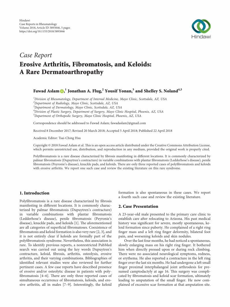
Case Report Erosive Arthritis, Fibromatosis, and Keloids: A Rare Dermatoarthropathy Fawad Aslam , 1 Jonathan A. Flug, 2 Yousif Yonan, 3 and Shelley S. Noland 4,5 1 Division of Rheumatology, Department of Internal Medicine, Mayo Clinic, Scottsdale, AZ, USA 2 Department of Radiology, Mayo Clinic, Scottsdale, AZ, USA 3 Department of Dermatology, Mayo Clinic, Scottsdale, AZ, USA 4 Division of Plastic Surgery, Department of Surgery, Mayo Clinic Hospital, Phoenix, AZ, USA 5 Department of Orthopedic Surgery, Mayo Clinic Hospital, Phoenix, AZ, USA Correspondence should be addressed to Fawad Aslam; [email protected] Received 8 December 2017; Revised 20 March 2018; Accepted 5 April 2018; Published 22 April 2018 Academic Editor: Tsai-Ching Hsu Copyright © 2018 Fawad Aslam et al. is is an open access article distributed under the Creative Commons Attribution License, which permits unrestricted use, distribution, and reproduction in any medium, provided the original work is properly cited. Polyfibromatosis is a rare disease characterized by fibrosis manifesting in different locations. It is commonly characterized by palmar fibromatosis (Dupuytren’s contracture) in variable combinations with plantar fibromatosis (Ledderhose’s disease), penile fibromatosis (Peyronie’s disease), knuckle pads, and keloids. ere are only three reported cases of polyfibromatosis and keloids with erosive arthritis. We report one such case and review the existing literature on this rare syndrome. 1.Introduction Polyfibromatosis is a rare disease characterized by fibrosis manifesting in different locations. It is commonly charac- terized by palmar fibromatosis (Dupuytren’s contracture) in variable combinations with plantar fibromatosis (Ledderhose’s disease), penile fibromatosis (Peyronie’s disease), knuckle pads, and keloids [1]. e aforementioned are all categories of superficial fibromatoses. Coexistence of fibromatosis and keloid formation is also very rare [2, 3], and it is not entirely clear if keloids are formally part of the polyfibromatosis syndrome. Nevertheless, this association is rare. To identify previous reports, a nonrestricted PubMed search was carried out using the key words Dupuytren’s contracture, keloid, fibrosis, arthritis, osteolysis, erosive arthritis, and their varying combinations. Bibliographies of identified relevant studies were also reviewed for further pertinent cases. A few case reports have described presence of erosive and/or osteolytic disease in patients with poly- fibromatosis [4–6]. ere are only three reported cases of simultaneous occurrence of fibromatosis, keloids, and ero- sive arthritis, all in males [7–9]. Interestingly, the keloid formation is also spontaneous in these cases. We report a fourth such case and review the existing literature. 2. Case Presentation A 23-year-old male presented to the primary care clinic to establish care after relocating to Arizona. His past medical history was significant for severe, mostly spontaneous, ke- loid formation since puberty. He complained of a right ring finger mass and a left ring finger deformity, bilateral foot pain, and worsening keloids and skin nodules. Over the last four months, he had noticed a spontaneous, slowly enlarging mass on his right ring finger. It bothered him when directly pressed upon or during rock climbing. ere were no associated neurological symptoms, redness, or erythema. He also reported a contracture in the left ring fingeroverthelastsixmonths.Hehadundergonealeftsmall finger proximal interphalangeal joint arthrodesis for pre- sumed camptodactyly at age 16. is surgery was compli- cated by fibromatosis and keloid scar formation, ultimately leading to amputation of the small finger. He now com- plained of excessive scar formation at that amputation site. Hindawi Case Reports in Rheumatology Volume 2018, Article ID 3893846, 5 pages https://doi.org/10.1155/2018/3893846
Transcript of ErosiveArthritis,Fibromatosis,andKeloids ...
Case ReportErosive Arthritis, Fibromatosis, and Keloids:A Rare Dermatoarthropathy
Fawad Aslam ,1 Jonathan A. Flug,2 Yousif Yonan,3 and Shelley S. Noland4,5
1Division of Rheumatology, Department of Internal Medicine, Mayo Clinic, Scottsdale, AZ, USA2Department of Radiology, Mayo Clinic, Scottsdale, AZ, USA3Department of Dermatology, Mayo Clinic, Scottsdale, AZ, USA4Division of Plastic Surgery, Department of Surgery, Mayo Clinic Hospital, Phoenix, AZ, USA5Department of Orthopedic Surgery, Mayo Clinic Hospital, Phoenix, AZ, USA
Correspondence should be addressed to Fawad Aslam; [email protected]
Received 8 December 2017; Revised 20 March 2018; Accepted 5 April 2018; Published 22 April 2018
Academic Editor: Tsai-Ching Hsu
Copyright © 2018 Fawad Aslam et al. +is is an open access article distributed under the Creative Commons Attribution License,which permits unrestricted use, distribution, and reproduction in any medium, provided the original work is properly cited.
Polyfibromatosis is a rare disease characterized by fibrosis manifesting in different locations. It is commonly characterized bypalmar fibromatosis (Dupuytren’s contracture) in variable combinations with plantar fibromatosis (Ledderhose’s disease), penilefibromatosis (Peyronie’s disease), knuckle pads, and keloids. +ere are only three reported cases of polyfibromatosis and keloidswith erosive arthritis. We report one such case and review the existing literature on this rare syndrome.
1. Introduction
Polyfibromatosis is a rare disease characterized by fibrosismanifesting in different locations. It is commonly charac-terized by palmar fibromatosis (Dupuytren’s contracture)in variable combinations with plantar fibromatosis(Ledderhose’s disease), penile fibromatosis (Peyronie’sdisease), knuckle pads, and keloids [1]. +e aforementionedare all categories of superficial fibromatoses. Coexistence offibromatosis and keloid formation is also very rare [2, 3], andit is not entirely clear if keloids are formally part of thepolyfibromatosis syndrome. Nevertheless, this association israre. To identify previous reports, a nonrestricted PubMedsearch was carried out using the key words Dupuytren’scontracture, keloid, fibrosis, arthritis, osteolysis, erosivearthritis, and their varying combinations. Bibliographies ofidentified relevant studies were also reviewed for furtherpertinent cases. A few case reports have described presenceof erosive and/or osteolytic disease in patients with poly-fibromatosis [4–6]. +ere are only three reported cases ofsimultaneous occurrence of fibromatosis, keloids, and ero-sive arthritis, all in males [7–9]. Interestingly, the keloid
formation is also spontaneous in these cases. We reporta fourth such case and review the existing literature.
2. Case Presentation
A 23-year-old male presented to the primary care clinic toestablish care after relocating to Arizona. His past medicalhistory was significant for severe, mostly spontaneous, ke-loid formation since puberty. He complained of a right ringfinger mass and a left ring finger deformity, bilateral footpain, and worsening keloids and skin nodules.
Over the last four months, he had noticed a spontaneous,slowly enlarging mass on his right ring finger. It botheredhim when directly pressed upon or during rock climbing.+ere were no associated neurological symptoms, redness,or erythema. He also reported a contracture in the left ringfinger over the last six months. He had undergone a left smallfinger proximal interphalangeal joint arthrodesis for pre-sumed camptodactyly at age 16. +is surgery was compli-cated by fibromatosis and keloid scar formation, ultimatelyleading to amputation of the small finger. He now com-plained of excessive scar formation at that amputation site.
HindawiCase Reports in RheumatologyVolume 2018, Article ID 3893846, 5 pageshttps://doi.org/10.1155/2018/3893846
He gave a two-year history of bilateral forefoot pain. Hisright fourth toe was the most painful. He reported that thistoe had become very stiff over this time. Other toes wereinvolved as well, and prolonged standing exacerbated thesymptoms. He did not report any redness, warmth, orswelling. He denied any acute joint pain episodes and handor back pain.
With respect to his keloids and skin nodules, he had hadthem since puberty. He noted that some were related to sitesof minor trauma, and others were spontaneous. He hadmultiple large keloids involving the chest, back, arms, andlegs. Recently, some had involved the toes as well. He deniedany genital involvement. Past treatments included intrale-sional steroid injections and radiation therapy with tem-porary improvements.
He was a lifelong nonsmoker and drank alcohol socially.He was an avid rock-climber. His family history was sig-nificant for keloid formation in his grandfather. A maternalcousin had rheumatoid arthritis. On review of systems, hedenied any fatigue, morning stiffness, night sweats, weightloss, inflammatory eye disease, cough, shortness of breath,back pain, nephrolithiasis, Raynaud’s phenomenon, orblood dyscrasias. He did not take any chronic medications.
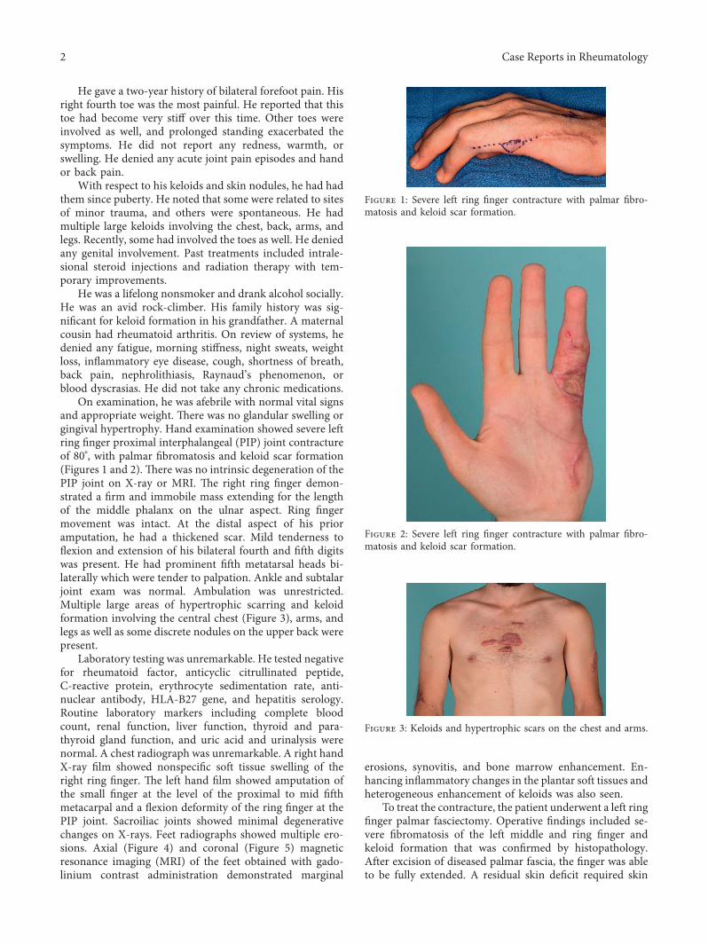
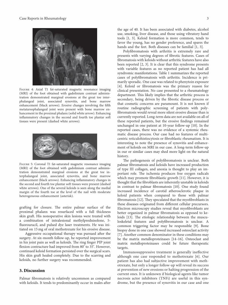
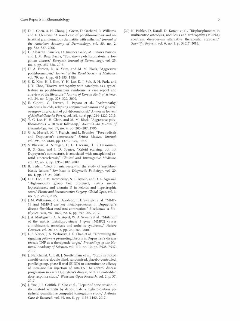
On examination, he was afebrile with normal vital signsand appropriate weight. +ere was no glandular swelling orgingival hypertrophy. Hand examination showed severe leftring finger proximal interphalangeal (PIP) joint contractureof 80°, with palmar fibromatosis and keloid scar formation(Figures 1 and 2). +ere was no intrinsic degeneration of thePIP joint on X-ray or MRI. +e right ring finger demon-strated a firm and immobile mass extending for the lengthof the middle phalanx on the ulnar aspect. Ring fingermovement was intact. At the distal aspect of his prioramputation, he had a thickened scar. Mild tenderness toflexion and extension of his bilateral fourth and fifth digitswas present. He had prominent fifth metatarsal heads bi-laterally which were tender to palpation. Ankle and subtalarjoint exam was normal. Ambulation was unrestricted.Multiple large areas of hypertrophic scarring and keloidformation involving the central chest (Figure 3), arms, andlegs as well as some discrete nodules on the upper back werepresent.
Laboratory testing was unremarkable. He tested negativefor rheumatoid factor, anticyclic citrullinated peptide,C-reactive protein, erythrocyte sedimentation rate, anti-nuclear antibody, HLA-B27 gene, and hepatitis serology.Routine laboratory markers including complete bloodcount, renal function, liver function, thyroid and para-thyroid gland function, and uric acid and urinalysis werenormal. A chest radiograph was unremarkable. A right handX-ray film showed nonspecific soft tissue swelling of theright ring finger. +e left hand film showed amputation ofthe small finger at the level of the proximal to mid fifthmetacarpal and a flexion deformity of the ring finger at thePIP joint. Sacroiliac joints showed minimal degenerativechanges on X-rays. Feet radiographs showed multiple ero-sions. Axial (Figure 4) and coronal (Figure 5) magneticresonance imaging (MRI) of the feet obtained with gado-linium contrast administration demonstrated marginal
erosions, synovitis, and bone marrow enhancement. En-hancing inflammatory changes in the plantar soft tissues andheterogeneous enhancement of keloids was also seen.
To treat the contracture, the patient underwent a left ringfinger palmar fasciectomy. Operative findings included se-vere fibromatosis of the left middle and ring finger andkeloid formation that was confirmed by histopathology.After excision of diseased palmar fascia, the finger was ableto be fully extended. A residual skin deficit required skin
Figure 1: Severe left ring finger contracture with palmar fibro-matosis and keloid scar formation.
Figure 2: Severe left ring finger contracture with palmar fibro-matosis and keloid scar formation.
Figure 3: Keloids and hypertrophic scars on the chest and arms.
2 Case Reports in Rheumatology
grafting for closure. +e entire palmar surface of theproximal phalanx was resurfaced with a full thicknessskin graft. His nonoperative skin lesions were treated witha combination of intralesional methylprednisolone, 5-fluorouracil, and pulsed dye laser treatments. He was ini-tiated on 15mg of oral methotrexate for his erosive disease.
Aggressive occupational therapy was pursued after thesurgery. At six-month follow-up, he reported improvementin his joint pain as well as keloids. +e ring finger PIP jointflexion contracture had improved from 80° to 35°. However,continued keloid formation persisted over the surgical scars.His skin graft healed completely. Due to the scarring andkeloids, no further surgery was recommended.
3. Discussion
Palmar fibromatosis is relatively uncommon as comparedwith keloids. It tends to predominantly occur in males after
the age of 40. It has been associated with diabetes, alcoholuse, smoking, liver disease, and those using vibratory handtools [1, 3]. Keloid formation is more common, tends tofavor the young, has no gender preference, and spares thehands and the feet. Both diseases can be familial [1, 3].
Polyfibromatosis with arthritis is extremely rare andpresents with varying degrees of fibrotic features. Cases offibromatosis with keloids without arthritic features have alsobeen reported [2, 3]. It is clear that this syndrome presentswith variable features as no reported patient has had allsyndromic manifestations. Table 1 summarizes the reportedcases of polyfibromatosis with arthritis. Incidence is pri-marily sporadic. One case was related to phenytoin exposure[4]. Keloid or fibromatosis was the primary reason forclinical presentation. No case presented to a rheumatologydepartment. +is likely implies that the arthritic process issecondary, being driven by the fibrotic disease process orthat cosmetic concerns are paramount. It is not known ifroutine radiographic screening of patients with poly-fibromatosis would reveal more silent erosive disease than iscurrently reported. Long-term data are not available on all ofthese reported patients, but the erosive findings remainedunchanged in one patient at 10-year follow-up [10]. In thereported cases, there was no evidence of a systemic rheu-matic disease process. Our case had no features of multi-centric reticulohistiocytosis or fibroblastic rheumatism. It isinteresting to note the presence of synovitis and enhance-ment of keloids on MRI in our case. A long-term follow-upin our or similar cases may shed more light on the naturalhistory.
+e pathogenesis of polyfibromatosis is unclear. Bothpalmar fibromatosis and keloids have increased productionof type III collagen, and anoxia is thought to play an im-portant role. +e ischemia produces free oxygen radicalswhich may promote fibroblastic growth [11]. However, it isthought that the fibroblasts are inherently unstable in keloidsin contrast to palmar fibromatosis [10]. One study foundincreased incidence of carotid atherosclerotic plaque inkeloid patients when compared to those with palmarfibromatosis [12]. +ey speculated that the myofibroblasts inthese diseases originated from different cellular precursors.Electron microscopy studies reveal that myofibroblasts arebetter organized in palmar fibromatosis as opposed to ke-loids [13]. +e etiologic relationship between the muscu-loskeletal features and polyfibromatosis is unclear. Acommon triggering factor may be responsible [9]. Bonebiopsy done in one case showed increased osteoclast activity[7]. Another common denominator in these conditions maybe the matrix metalloproteinases [14–16]. Osteoclast andmatrix metalloproteinases could be future therapeutictargets.
Immunosuppressive treatment is generally ineffectivealthough one case responded to methotrexate [6]. Ourpatient has also had subjective improvement with meth-otrexate, but only a longer follow-up will reveal its successat prevention of new erosions or halting progression of thecurrent ones. It is unknown if biological agents like tumornecrosis actor inhibitors (TNFi) are useful in this syn-drome, but the presence of synovitis in our case and one
Figure 5: Coronal T1 fat-saturated magnetic resonance imaging(MRI) of the foot obtained with gadolinium contrast adminis-tration demonstrated marginal erosions at the great toe in-terphalangeal joint, associated synovitis, and bone marrowenhancement (black arrows). Enhancing inflammatory changes inthe second and fourth toe plantar soft tissues were present (dashedwhite arrows). One of the several keloids is seen along the medialmargin of the fourth toe at the level of the nail with moderateheterogeneous enhancement (asterisk).
Figure 4: Axial T1 fat-saturated magnetic resonance imaging(MRI) of the foot obtained with gadolinium contrast adminis-tration demonstrated marginal erosions at the great toe inter-phalangeal joint, associated synovitis, and bone marrowenhancement (black arrows). Erosive changes involving the fifthmetatarsophalangeal joint were present with bone marrow en-hancement in the proximal phalanx (solid white arrow). Enhancinginflammatory changes in the second and fourth toe plantar softtissues were present (dashed white arrows).
Case Reports in Rheumatology 3

another [6] indicates its potential usefulness. However,none of the cases had prominent inflammatory jointsymptoms despite the aggressive radiological findings.Laboratory studies have shown the inhibitory effects ofTNFi in Dupuytren’s contracture by inhibiting myofibroblastactivity and the contractile apparatus [17]. A phase II clinicaltrial will assess the role of intralesional adalimumab in earlyDupuytren’s contracture [18]. +us, TNFi could possibly helpaddress both the musculoskeletal and dermatological mani-festations. Erosions could also possibly be repaired using anosteoclast development inhibitor like denosumab [19] but isuntested in this dermatoarthropathy. Bisphosphonates havebeen used to treat musculoskeletal manifestation in someosteolytic disorders [20].
In conclusion, polyfibromatosis with arthritis is ex-tremely rare with no established treatment. Our case will addto the limited repertoire of erosive polyfibromatosis caseswith keloids and serve to increase awareness and therebystimulate research and reporting of this rare syndrome.
Conflicts of Interest
+e authors declare that they have no conflicts of interest.
References
[1] N. P. Burrows and C. R. Lovell, “Disorders of connectivetissue,” in Rook’s Textbook of Dermatology, A. Rook andT. Burns, Eds., Wiley-Blackwell Hoboken, NJ, USA, 8thedition, 2010.
[2] R. Gonzalez-Martinez, S. Marin-Bertolin, and J. Amorrortu-Velayos, “Association between keloids and Dupuytren’sdisease: case report,” British Journal of Plastic Surgery, vol. 48,no. 1, pp. 47-48, 1995.
[3] L. Ly and I. Winship, “X-linked recessive polyfibromatosismanifesting with spontaneous keloid scars and Dupuytren’scontracture,” Australasian Journal of Dermatology, vol. 53,no. 2, pp. 148–150, 2012.
[4] G. E. Pierard and C. M. Lapiere, “Phenytoin dependent fi-brosis in polyfibromatosis syndrome,” British Journal ofDermatology, vol. 100, no. 3, pp. 335–341, 1979.
Table 1: Summary of polyfibromatosis cases with a musculoskeletal component.
Author/year/country Age(years)/gender Fibromatosis Keloids Musculoskeletal
features X-ray features MRI features
Pierard and Lapiere1979 [4], Belgium 20, male
Hand fibromatosis,knuckle pads, and“toughness” of skin
NA
Camptodactyly,tendon
calcification, facialhyoplasia,
osteolysis, andscoliosis
Osteolysis of distalulna, right ringfinger proximalphalanx, and first
metacarpal
NA
Fenton et al. 1986[7], United Kingdom 48, male
Palmarfibromatosis, andPeyronie’s disease
Keloids of arms,shoulders, andpresternal area
Hand and footstiffness
Erosions ofshoulders, hands,
and feetNA
Chen et al. 2006 [5],Australia 65, male
Progressive fibrosisand contractures of
wrist, elbows,knees, and ankles;linear fibrotic cords
NoneInflammatory painof wrist, elbows,knees, and ankles
Erosions of leftshoulder, elbows,wrists, hands, and
feet
NA
Kim et al. 2009 [8],South Korea 44, male
Palmar and plantarnodules andfibromatosis
Multiple keloidsof trunk andextremities
Asymptomatic Erosions andosteolysis of hands
Multiple handerosions
Cinotti et al. 2013[9], Italy 53, male
Flexioncontractures ofwrists, fingers,ankles, and toes;
gingivalhyperplasia andconjunctival
fibrosis
Multiple keloidsSevere hand andfoot deformitiesand facial changes
Osteolysis of wrists,hands, and toes;
ankylosisNA
Albarran et al. 2015[6], Spain 33, male
Palmar and plantarnodules, palmarfibromatosis, kneecontractures andfibromatosis, and
gingivalhyperplasia
None NA Erosion in rightfoot
Synovitis andeffusions inboth knees
Present case 2018,USA 23, male Finger flexion
contractures
Multiple keloidsof trunk andextremities
Mild foot painErosions of feet;hands without
erosions
Multipleerosions andsynovitis of
feet
4 Case Reports in Rheumatology
[5] D. L. Chen, A. H. Chong, J. Green, D. Orchard, R. Williams,and L. Clemens, “A novel case of polyfibromatosis and in-terstitial granulomatous dermatitis with arthritis,” Journal ofthe American Academy of Dermatology, vol. 55, no. 2,pp. S32–S37, 2006.
[6] C. Albarran Planelles, D. Jimenez Gallo, M. Linares Barrios,and J. M. Baez Baena, “Touraine’s polyfibromatosis: a for-gotten disease,” European Journal of Dermatology, vol. 25,no. 4, pp. 357-358, 2015.
[7] D. A. Fenton, D. A. Yates, and M. M. Black, “Aggressivepolyfibromatosis,” Journal of the Royal Society of Medicine,vol. 79, no. 8, pp. 482-483, 1986.
[8] S. K. Kim, H. J. Kim, Y. H. Lee, K. J. Suh, S. H. Park, andJ. Y. Choe, “Erosive arthropathy with osteolysis as a typicalfeature in polyfibromatosis syndrome: a case report anda review of the literature,” Journal of Korean Medical Science,vol. 24, no. 2, pp. 326–329, 2009.
[9] E. Cinotti, G. Ferrero, F. Paparo et al., “Arthropathy,osteolysis, keloids, relapsing conjunctival pannus and gingivalovergrowth: a variant of polyfibromatosis?,”American JournalofMedical Genetics Part A, vol. 161, no. 6, pp. 1214–1220, 2013.
[10] Y. C. Lee, H. H. Chan, and M. M. Black, “Aggressive poly-fibromatosis: a 10 year follow-up,” Australasian Journal ofDermatology, vol. 37, no. 4, pp. 205–207, 1996.
[11] G. A. Murrell, M. J. Francis, and L. Bromley, “Free radicalsand Dupuytren’s contracture,” British Medical Journal,vol. 295, no. 6610, pp. 1373–1375, 1987.
[12] S. Bhavsar, A. Nimigan, D. G. Hackam, D. B. O’Gorman,B. S. Gan, and J. D. Spence, “Keloid scarring, but notDupuytren’s contracture, is associated with unexplained ca-rotid atherosclerosis,” Clinical and Investigative Medicine,vol. 32, no. 2, pp. E95–E102, 2009.
[13] B. Eyden, “Electron microscopy in the study of myofibro-blastic lesions,” Seminars in Diagnostic Pathology, vol. 20,no. 1, pp. 13–24, 2003.
[14] D. E. Lee, R. M. Trowbridge, N. T. Ayoub, and D. K. Agrawal,“High-mobility group box protein-1, matrix metal-loproteinases, and vitamin D in keloids and hypertrophicscars,” Plastic and Reconstructive Surgery–Global Open, vol. 3,no. 6, p. e425, 2015.
[15] J. M. Wilkinson, R. K. Davidson, T. E. Swingler et al., “MMP-14 and MMP-2 are key metalloproteases in Dupuytren’sdisease fibroblast-mediated contraction,” Biochimica et Bio-physica Acta, vol. 1822, no. 6, pp. 897–905, 2012.
[16] J. A. Martignetti, A. A. Aqeel, W. A. Sewairi et al., “Mutationof the matrix metalloproteinase 2 gene (MMP2) causesa multicentric osteolysis and arthritis syndrome,” NatureGenetics, vol. 28, no. 3, pp. 261–265, 2001.
[17] L. S. Verjee, J. S. Verhoekx, J. K. Chan et al., “Unraveling thesignaling pathways promoting fibrosis in Dupuytren’s diseasereveals TNF as a therapeutic target,” Proceedings of the Na-tional Academy of Sciences, vol. 110, no. 10, pp. E928–E937,2013.
[18] J. Nanchahal, C. Ball, J. Swettenham et al., “Study protocol:a multi-centre, double blind, randomised, placebo-controlled,parallel group, phase II trial (RIDD) to determine the efficacyof intra-nodular injection of anti-TNF to control diseaseprogression in early Dupuytren’s disease, with an embeddeddose response study,” Wellcome Open Research, vol. 2, p. 37,2017.
[19] J. Yue, J. F. Griffith, F. Xiao et al., “Repair of bone erosion inrheumatoid arthritis by denosumab: a high-resolution pe-ripheral quantitative computed tomography study,” ArthritisCare & Research, vol. 69, no. 8, pp. 1156–1163, 2017.
[20] K. Pichler, D. Karall, D. Kotzot et al., “Bisphosphonates inmulticentric osteolysis, nodulosis and arthropathy (MONA)spectrum disorder–an alternative therapeutic approach,”Scientific Reports, vol. 6, no. 1, p. 34017, 2016.
Case Reports in Rheumatology 5

Stem Cells International
Hindawiwww.hindawi.com Volume 2018
Hindawiwww.hindawi.com Volume 2018
MEDIATORSINFLAMMATION
of
EndocrinologyInternational Journal of
Hindawiwww.hindawi.com Volume 2018
Hindawiwww.hindawi.com Volume 2018
Disease Markers
Hindawiwww.hindawi.com Volume 2018
BioMed Research International
OncologyJournal of
Hindawiwww.hindawi.com Volume 2013
Hindawiwww.hindawi.com Volume 2018
Oxidative Medicine and Cellular Longevity
Hindawiwww.hindawi.com Volume 2018
PPAR Research
Hindawi Publishing Corporation http://www.hindawi.com Volume 2013Hindawiwww.hindawi.com
The Scientific World Journal
Volume 2018
Immunology ResearchHindawiwww.hindawi.com Volume 2018
Journal of
ObesityJournal of
Hindawiwww.hindawi.com Volume 2018
Hindawiwww.hindawi.com Volume 2018
Computational and Mathematical Methods in Medicine
Hindawiwww.hindawi.com Volume 2018
Behavioural Neurology
OphthalmologyJournal of
Hindawiwww.hindawi.com Volume 2018
Diabetes ResearchJournal of
Hindawiwww.hindawi.com Volume 2018
Hindawiwww.hindawi.com Volume 2018
Research and TreatmentAIDS
Hindawiwww.hindawi.com Volume 2018
Gastroenterology Research and Practice
Hindawiwww.hindawi.com Volume 2018
Parkinson’s Disease
Evidence-Based Complementary andAlternative Medicine
Volume 2018Hindawiwww.hindawi.com
Submit your manuscripts atwww.hindawi.com