Dysrhythmia Monitoring Practices of Nurses on a Telemetry Unit
214
UNF Digital Commons UNF Graduate eses and Dissertations Student Scholarship 2010 Dysrhythmia Monitoring Practices of Nurses on a Telemetry Unit Susan Jane Schultz University of North Florida is Doctoral Project is brought to you for free and open access by the Student Scholarship at UNF Digital Commons. It has been accepted for inclusion in UNF Graduate eses and Dissertations by an authorized administrator of UNF Digital Commons. For more information, please contact Digital Projects. © 2010 All Rights Reserved Suggested Citation Schultz, Susan Jane, "Dysrhythmia Monitoring Practices of Nurses on a Telemetry Unit" (2010). UNF Graduate eses and Dissertations. 216. hps://digitalcommons.unf.edu/etd/216
Transcript of Dysrhythmia Monitoring Practices of Nurses on a Telemetry Unit

UNF Digital Commons
UNF Graduate Theses and Dissertations Student Scholarship
2010
Dysrhythmia Monitoring Practices of Nurses on aTelemetry UnitSusan Jane SchultzUniversity of North Florida
This Doctoral Project is brought to you for free and open access by theStudent Scholarship at UNF Digital Commons. It has been accepted forinclusion in UNF Graduate Theses and Dissertations by an authorizedadministrator of UNF Digital Commons. For more information, pleasecontact Digital Projects.© 2010 All Rights Reserved
Suggested CitationSchultz, Susan Jane, "Dysrhythmia Monitoring Practices of Nurses on a Telemetry Unit" (2010). UNF Graduate Theses andDissertations. 216.https://digitalcommons.unf.edu/etd/216
DYSRHYTHMIA MONITORING PRACTICES
DYSRHYTHMIA MONITORING PRACTICES
OF NURSES ON A TELEMETRY UNIT
by
Susan Jane Schultz
A project submitted to the School of Nursing in partial fulfillment of the requirements for the degree of
Doctor of Nursing Practice
UNIVERSITY OF NORTH FLORIDA
BROOKS COLLEGE OF HEALTH
Unpublished work © 2010 Susan Jane Schultz
DYSRHYTHMIA MONITORING PRACTICES
CERTIFICATE OF APPROVAL PAGE
The Doctor of Nursing Practice project of Susan Jane Schultz is approved:
Carol Ledbetter, PhD, APRN-BC, FAAN Committee Chairperson
Kathy Robinson, PhD, RN, CCNS (emeritus) Committee Person
Accepted for the Department:
Lillia Loriz, PhD, Director, School lrsing
s College of Health
Accepted for the University:
20 oet ').OID Date
til () () c---f '- C> I c>
Date
2Q 0 ct 2.<>10 Date
3 r!f-IU Date
Date
Date
Signature Deleted
Signature Deleted
Signature Deleted
Signature Deleted
Signature Deleted
Signature Deleted

DYSRHYTHMIA MONITORING PRACTICES
Acknowledgments
I wish to thank the following people for helping me to complete the Doctor of Nursing Practice project: Dr. Carol Ledbetter, for serving as my committee chairperson and helping to guide and support me through the entire process; Dr. Kathy Robinson and Dr. Gerard Hogan, for giving their input and advice; Dr. Robert Augspurger for his statistical expertise; and Katie LeGros for her editing assistance. This research study was funded in part by the Patricia H. Foster Graduate Nursing Fellowship and the University of Florida‘s Graduate Scholars program.
DYSRHYTHMIA MONITORING PRACTICES iv
Table of Contents Acknowledgements . . . . . . . . . . . . . . . . . . . . . . . . . . . . . . . . . . . . . . . . . . . . iii Abstract . . . . . . . . . . . . . . . . . . . . . . . . . . . . . . . . . . . . . . . . . . . . . . . . . . . . . vii Chapter 1 Introduction . . . . . . . . . . . . . . . . . . . . . . . . . . . . . . . . . . . . . . . . . . 1 Challenges . . . . . . . . . . . . . . . . . . . . . . . . . . . . . . . . . . . . . . . . . . . . . . 1 Abbreviated Literature Review . . . . . . . . . . . . . . . . . . . . . . . . . . . . . . 3 Project Description . . . . . . . . . . . . . . . . . . . . . . . . . . . . . . . . . . . . . . . 5 The Problem. . . . . . . . . . . . . . . . . . . . . . . . . . . . . . . . . . . . . . . . . . . . . 7 Research Questions . . . . . . . . . . . . . . . . . . . . . . . . . . . . . . . . . . . . . . . 8 Definition of Terms . . . . . . . . . . . . . . . . . . . . . . . . . . . . . . . . . . . . . . . 9 Chapter 2 Review of the Literature . . . . . . . . . . . . . . . . . . . . . . . . . . . . . . . . . 11 ECG Monitoring Practice Standards . . . . . . . . . . . . . . . . . . . . . . . . . 11 Research on ECG Monitoring Practice Standards . . . . . . . . . . . . . . . 14 ECG Teaching Methods for Nursing Staff . . . . . . . . . . . . . . . . . . . . . 15 ECG Teaching Methods for Nursing Students . . . . . . . . . . . . . . . . . . 18 Web-based or Online Methods to Nursing Staff . . . . . . . . . . . . . . . . 20 Best Practices in E-learning . . . . . . . . . . . . . . . . . . . . . . . . . . . . . . . . 22 Evaluation of the Evidence . . . . . . . . . . . . . . . . . . . . . . . . . . . . . . . . . 23 Chapter 3 Design and Methodology . . . . . . . . . . . . . . . . . . . . . . . . . . . . . . . 28 Sample . . . . . . . . . . . . . . . . . . . . . . . . . . . . . . . . . . . . . . . . . . . . . . . . 28 Methods . . . . . . . . . . . . . . . . . . . . . . . . . . . . . . . . . . . . . . . . . . . . . . . 31 Time Frame . . . . . . . . . . . . . . . . . . . . . . . . . . . . . . . . . . . . . . . . . . . . 32 Web-based Educational Program . . . . . . . . . . . . . . . . . . . . . . . . . . . . 32 Unit-based Collaborative Learning Activities . . . . . . . . . . . . . . . . . . 36 Project Evaluation Plan . . . . . . . . . . . . . . . . . . . . . . . . . . . . . . . . . . . 37 Feasibility . . . . . . . . . . . . . . . . . . . . . . . . . . . . . . . . . . . . . . . . . . . . . . 38 Income and Expenses . . . . . . . . . . . . . . . . . . . . . . . . . . . . . . . . . . . . . 40 Institutional Review Board . . . . . . . . . . . . . . . . . . . . . . . . . . . . . . . . . 40 Benefits and Risks . . . . . . . . . . . . . . . . . . . . . . . . . . . . . . . . . . . . . . . . 40 Confidentiality . . . . . . . . . . . . . . . . . . . . . . . . . . . . . . . . . . . . . . . . . . . 41 Data Analysis . . . . . . . . . . . . . . . . . . . . . . . . . . . . . . . . . . . . . . . . . . . 44

DYSRHYTHMIA MONITORING PRACTICES v
Chapter 4 Results . . . . . . . . . . . . . . . . . . . . . . . . . . . . . . . . . . . . . . . . . . . . . . 46 Participation in Study . . . . . . . . . . . . . . . . . . . . . . . . . . . . . . . . . . . . . 46 Description of the Sample . . . . . . . . . . . . . . . . . . . . . . . . . . . . . . . . . . 47 Test Content Validity and Reliability . . . . . . . . . . . . . . . . . . . . . . . . . 48 Pretest and Posttest Results. . . . . . . . . . . . . . . . . . . . . . . . . . . . . . . . . 49 Educational Program Evaluation Results . . . . . . . . . . . . . . . . . . . . . . 49 Unit-based Activities . . . . . . . . . . . . . . . . . . . . . . . . . . . . . . . . . . . . . 51 Patient Audit Results . . . . . . . . . . . . . . . . . . . . . . . . . . . . . . . . . . . . . 52 Chapter 5 Discussion . . . . . . . . . . . . . . . . . . . . . . . . . . . . . . . . . . . . . . . . . . . 55 Participation in Study . . . . . . . . . . . . . . . . . . . . . . . . . . . . . . . . . . . . . 55 Unit Champions . . . . . . . . . . . . . . . . . . . . . . . . . . . . . . . . . . . . . . . . . 57 Comfort with Online Learning . . . . . . . . . . . . . . . . . . . . . . . . . . . . . . 58 Incentives . . . . . . . . . . . . . . . . . . . . . . . . . . . . . . . . . . . . . . . . . . . . . . 59 Web-based Educational Program . . . . . . . . . . . . . . . . . . . . . . . . . . . . 59 Unit-based Activities . . . . . . . . . . . . . . . . . . . . . . . . . . . . . . . . . . . . . 60 Patient Audits . . . . . . . . . . . . . . . . . . . . . . . . . . . . . . . . . . . . . . . . . . 62 Unintended Consequences. . . . . . . . . . . . . . . . . . . . . . . . . . . . . . . . . . 64 Nursing Implications and Recommendations . . . . . . . . . . . . . . . . . . . 66 Limitations and Recommendations for Further Research . . . . . . . . . . 71 Conclusions . . . . . . . . . . . . . . . . . . . . . . . . . . . . . . . . . . . . . . . . . . . . 73

DYSRHYTHMIA MONITORING PRACTICES vi
Appendices . . . . . . . . . . . . . . . . . . . . . . . . . . . . . . . . . . . . . . . . . . . . . . . . . . . 74 Appendix A: AACN Practice Alert on Dysrhythmia Monitoring 74
Appendix B: AACN Dysrhythmia Monitoring Power Point Slides 76
Appendix C: Cardiac Monitoring Pocket Reference Card . . . . . . 81
Appendix D: AACN Audit Tool . . . . . . . . . . . . . . . . . . . . . . . . . 83
Appendix E: Permission from AACN . . . . . . . . . . . . . . . . . . . . . . 85
Appendix F: Appraisal Table . . . . . . . . . . . . . . . . . . . . . . . . . . . . 87
Appendix G: ACC/ ECC Rating System . . . . . . . . . . . . . . . . . . . 98
Appendix H: AACN Levels of Evidence . . . . . . . . . . . . . . . . . . . 98
Appendix I: ACC/AHA/ESC Levels of Evidence and Classifications 99
Appendix J: Principles of Good Practice, Evidence-based Strategies 100
Appendix K: Permission to Conduct Study (from nursing manager) 102
Appendix L: Nurses‘ Demographic Questionnaire . . . . . . . . . . . 103
Appendix M: Recruitment Announcements . . . . . . . . . . . . . . . . . 104
Appendix N: Informed Consent . . . . . . . . . . . . . . . . . . . . . . . . . . . 106
Appendix O: Format for Educational Program . . . . . . . . . . . . . . . 115
Appendix P: Audio Script for Educational Program . . . . . . . . . . 117
Appendix Q: Test Bank for Educational Program . . . . . . . . . . . . 124
Appendix R: Case Studies for Educational Program . . . . . . . . . . 141
Appendix S: Million Dollar Game for Educational Program . . . . 147
Appendix T: Unit-based Activities Poster . . . . . . . . . . . . . . . . . . 149
Appendix U: Permission to Use Published Materials . . . . . . . . . . 150
Appendix V: Content Validity Evaluation . . . . . . . . . . . . . . . . . . . 160
DYSRHYTHMIA MONITORING PRACTICES vii
Appendix W: Competency Skills Checklist . . . . . . . . . . . . . . . . . 162
Appendix X: Pretest and Posttest Questions . . . . . . . . . . . . . . . . . 163
Appendix Y: Pretest and Posttest Reliability (Educators) . . . . . . 171
Appendix Z: Education Program Evaluation Form . . . . . . . . . . . 173
Appendix AA: Income and Expenses for DNP Project . . . . . . . . . . 174
Appendix BB: IRB Approval Documents. . . . . . . . . . . . . . . . . . . . . 175
Appendix CC: Audit Check-off Sheet . . . . . . . . . . . . . . . . . . . . . . . 178
Appendix DD: Patients‘ Demographic Data . . . . . . . . . . . . . . . . . . 179
Appendix EE: SPSS Statistical Software Data . . . . . . . . . . . . . . . . 180
Appendix FF: Dysrhythmia Monitoring Nurses‘ Data . . . . . . . . . . 181
Appendix GG: Nurses‘ Demographic Survey Results . . . . . . . . . . . 183
Appendix HH: Pretest and Posttest Results (Nurses) . . . . . . . . . . . . 185
Appendix II: Pretest and Posttest Reliability (Nurses) . . . . . . . . . 186
Appendix JJ: Educational Program Evaluation Results . . . . . . . . . 188
Appendix KK: Patient Audits (Baseline, 6 Weeks, 18 Weeks) . . . . 189
Appendix LL: Patient Audit Results Audits . . . . . . . . . . . . . . . . . . 194
Appendix MM: Audits 4 Center Graph . . . . . . . . . . . . . . . . . . . . . . . 195
Appendix NN: Patient Demographics . . . . . . . . . . . . . . . . . . . . . . . 196
Appendix OO: Differences between Population Proportions . . . . . . 197
References . . . . . . . . . . . . . . . . . . . . . . . . . . . . . . . . . . . . . . . . . . . . . . . . . . . 199 Vita . . . . . . . . . . . . . . . . . . . . . . . . . . . . . . . . . . . . . . . . . . . . . . . . . . . . . . . 203

DYSRHYTHMIA MONITORING PRACTICES viii
Abstract
Standards of practice for hospital electrocardiogram monitoring were recommended in
2004 by the American Heart Association; however they are not widely followed. Many
nurses monitor in a single lead regardless of diagnosis and are unable to differentiate
wide QRS complex tachycardias. The purpose of this project was to evaluate the
effectiveness of an interactive web-based education program combined with unit-based
collaborative learning activities on both telemetry staff nurses‘ knowledge of
dysrhythmias and their monitoring practices for patients at risk for wide QRS complex
tachycardias. This interventional, one group before-and-after cohort study design
consisted of four components: interactive web-based educational program with a pretest
and posttest, unit-based collaborative activities, competency skills validation, and patient
audits of electrode placement and lead selection at baseline, six weeks, and 18 weeks.
There were 34 nurses who consented to participate, 16 started the program, and nine
finished all the components. The pretest scores ranged from 0 – 60% with median of
36.5%. The posttest scores ranged from 47 – 93% with median of 80%. The Wilcoxon
Signed Ranks test showed a significant difference between the pretest and posttest scores
(p = .008). The patient audit results did not indicate significant differences in proportions
of correct electrode placement and correct lead selection between baseline, 6 weeks, and
18 weeks. The program was effective in increasing nurses‘ knowledge about
dysrhythmias; however, it was not effective in changing monitoring behavior. More
research is needed to see if this type of program is more effective if it involves all the
staff on the unit who are responsible for monitoring, and if additional strategies are used,
such as unit champions and group rewards.

DYSRHYTHMIA MONITORING PRACTICES
Chapter 1: Introduction
Chapter 1 introduces the challenges and problems with electrocardiogram (ECG)
monitoring, provides an abbreviated literature review, and describes the project briefly.
It also includes the research questions and definition of terms.
Challenges
Electrocardiogram monitoring in hospitals has become more complex since it was
first introduced over 40 years ago (Drew & Funk, 2006). Practice standards for ECG
monitoring in hospital settings have been established for arrhythmias, ischemia, and
corrected QT (QTc) intervals (Drew et al., 2004). Due to these advances, nurses who
work in telemetry and critical care units in hospitals have an important responsibility to
monitor patients‘ cardiac rhythms appropriately and to intervene promptly.
Sudden cardiac death or cardiac arrest is defined as ―cessation of cardiac
mechanical activity and is confirmed by the absence of signs of circulation‖ (Lloyd-Jones
et al., 2009, p. e32). While ventricular fibrillation (VF) is listed as the cause of relatively
few deaths, the overwhelming majority of sudden cardiac deaths from coronary disease
(estimated at 310,000 per year) are nonetheless thought to be from VF (Lloyd-Jones et
al., 2009). In addition, an acute increase in QTc-interval length is associated with risk for
torsades de pointes, a type of polymorphic ventricular tachycardia (VT), which without
prompt defibrillation results in syncope and sudden death (Sommargren & Drew, 2007).
Consequently, it is important for nurses to recognize these dysrhythmias immediately.
DYSRHYTHMIA MONITORING PRACTICES 2
The discharge survival rates following in-hospital cardiac arrests are 27% among
children and 18% among adults (Lloyd-Jones et al., 2009). A total of 303 facilities
reported 21,748 in-hospital cardiac arrest events to the National Registry for
Cardiopulmonary Resuscitation in 2007. Of these, 93% were monitored or witnessed and
17.9% had VF or pulseless ventricular tachycardia as the first recorded rhythm.
Furthermore, 79% of the latter individuals received a defibrillation attempt within 3
minutes (Lloyd-Jones et al., 2009). Hospitals that adopt good monitoring practices may
impact these statistics favorably.
As wide complex tachycardias are typically caused by several mechanisms, one
may find it challenging to recognize the difference between VT and supraventricular
ventricular tachycardia (SVT) with aberrancy or bundle branch block (Urden, Stacy, &
Lough, 2010). It is important to correctly determine the cause of the tachycardia and
treat it appropriately because patients can become hemodynamically unstable very
quickly. Analysis of the ECG by a specialist is recommended (Urden et al., 2010; Zipes
et al., 2006).
Studies have shown that nurses often monitored in a single lead (regardless of
diagnosis), failed to properly prep the skin, misplaced electrodes, and were unable to
differentiate wide complex QRS tachycardias, all of which could lead to false alarms or
misdiagnoses (AACN Practice Alert, 2008; Drew & Funk, 2006; Funk et al., 2009; Keller
& Raines, 2005). To prevent error, nurses working on monitored units need to know how
to identify patients who are at risk for potentially lethal dysrhythmias, distinguish
between true and false alarms, correctly measure intervals, quickly recognize
dysrhythmias, and initiate the appropriate treatment promptly.

DYSRHYTHMIA MONITORING PRACTICES 3
The chief executive officers of many hospitals, including the one where this
project was conducted, are pursuing Magnet Hospital Recognition for Excellence in
Nursing Services, a program sponsored by the American Nurses Credentialing Center
(―Magnet Recognition Program Overview,‖ 2009). This project supports three of the 14
forces of magnetism: quality of care, quality improvement, and professional development
(Malloch & Porter-O‘Grady, 2010), which may help the hospital achieve its goal for
magnet recognition.
Abbreviated Literature Review
In 2004, the American Heart Association (AHA) published a scientific statement
recommending standards of practice for dysrhythmia monitoring (Drew et al., 2004).
Since large randomized clinical trials did not exist, recommendations were classified
according to level of evidence and were based on expert opinions. In addition, an
executive summary with recommendations on how to implement the AHA standards was
written by Drew and Funk (2006). Based on the data from these two documents, the
American Association of Critical-Care Nurses (AACN) wrote a practice alert with
specific recommendations for skin preparation, electrode placement, lead selection, and
QTc interval measurements (AACN Practice Alert, 2008).
The literature was reviewed to determine the best practices for educating staff
nurses on ECG or dysrhythmia monitoring. One multi-site randomized clinical trial,
called the PULSE Trial, is currently underway to evaluate implementation of the AHA
practice standards for dysrhythmia monitoring, but will not be completed until 2013
(Funk et al., 2009). Funk et al. are using an online ECG education program and unit-
based strategies led by unit champions to implement and sustain change.
DYSRHYTHMIA MONITORING PRACTICES 4
Two studies on effectiveness of written self-study packets for teaching
dysrhythmias to nursing staff showed conflicting results. Cadden (2007) found that a
self-study learning package, supplemented with unit-based materials and learning
activities with an educator, effectively developed staff competency regarding the
operation of ECG monitors and the interpretation of arrhythmias. Van Arsdale (1998),
however, concluded that a self-instruction reading packet was not as beneficial as
instructor-led classes, which he based on lower posttest scores and students' comments
that they would have preferred interaction with an instructor.
Jeffries (2005) and Morris et al. (2009) conducted two other studies with nursing
staff that evaluated the effectiveness of critical care courses, each including computer-
based ECG training. Both studies found that all participants successfully completed the
computer-based ECG course and demonstrated competency with ECG interpretation,
which was one component of the critical care course.
Due to the limited number of studies on nursing staff, studies that evaluated ECG
teaching methods with nursing students were also reviewed. Two studies of nursing
students compared interactive computer-based learning formats to traditional classroom
instruction on interpreting dysrhythmias and performing12-lead ECG (Jang, Hwang,
Park, Kim, & Kim, 2005; Jeffries, Woolf, & Linde, 2003). They found computer-based
formats to be just as effective as, if not more effective than traditional formats. Frith and
Kee (2003) compared the effectiveness of different instructional communication methods
between groups of nursing students who participated in a web-based cardiac rhythm
interpretation course. Their results showed that both groups passed the posttest exam and
DYSRHYTHMIA MONITORING PRACTICES 5
there were no significant differences between groups concerning exam scores or
completion rates.
Studies on the effectiveness of computer-based or web-based teaching methods
for ECG were also limited, so the search was consequently expanded to include studies
with nursing staff on subjects other than ECG. Belcher & Vonderharr (2005) found that a
web-based audio-streaming program on evidence-based practice provided a cost-effective
way to educate staff. Dumpe, Kanyak, and Hill (2007) studied the usefulness of an
online system to validate employees‘ competencies on the Health Information Privacy
and Portability Act (HIPPA) and other nursing competencies, which they discovered was
an effective way to reach large numbers of staff. Over 90% of the nursing staff reported
they were satisfied with the method and it was easy to use. Another study by Durkin
(2008) found interactive computer-based learning was more effective than text-only
computer-based learning on the topic of cranial nerve education.
Project Description
This project was designed to improve the quality of care of patients with ECG
monitoring and enhance the professional development of the nursing staff on one
telemetry unit. It supported the hospital‘s strategic plans for Magnet Recognition and the
implementation of evidenced-based practice. It also provided a standardized educational
program and competency assessment method which could potentially be implemented on
other telemetry or critical care units.
This interventional, one group before-and-after cohort study design consisted of
four components:

DYSRHYTHMIA MONITORING PRACTICES 6
1) An interactive web-based educational program about evidence-based practice
standards for dysrhythmia monitoring of wide QRS complexes with a pretest
and posttest, using the organization‘s existing online learning management
system.
2) Unit-based collaborative learning activities with other staff nurses, led by the
primary investigator, to reinforce knowledge of wide QRS complex
dysrhythmias and monitoring practices.
3) Validation of staff‘s competency using a skills checklist which included the
placement of electrodes, lead selection, QTc interval monitoring, QRS
morphology analysis, and nursing interventions for wide-QRS complexes.
4) Audits by the investigator of electrode placement and lead selection before the
interventions (education, unit-based activities, and staff competency
validation), at the conclusion of the interventions (6 weeks), and 12 weeks
after the interventions (week 18 of the study).
AACN posted a downloadable educational tool kit on their website that included
the practice alert, slides, audit tool, and pocket reference card (AACN Practice Alert,
2008; Audit Tool, 2008; Drew, 2002; Richards, 2008). These documents formed the
basis for this research and are attached in Appendix A, B, C, and D. Permission from
AACN to use these materials is included in Appendix E.
It is important to note both Drew et al. (2004) and Drew and Funk (2006) also
recommended continuous ST segment monitoring for patients with acute coronary
syndrome and percutaneous coronary interventions, with monitoring in leads other than
VI or II. ST segment monitoring was not included in this project because the nurses on
DYSRHYTHMIA MONITORING PRACTICES 7
the telemetry unit where it was conducted have already received education and
competency validation twice in the past two years on acute coronary syndrome and ST
segment monitoring. Patients who required ST segment monitoring in leads other than
VI or II were excluded.
The Problem
The discharge survival rate following in-hospital cardiac arrests is 18% among
adults (Lloyd-Jones et al., 2009). Studies have shown that nurses often monitored in a
single lead regardless of diagnosis, failed to properly prepare the skin, misplaced
electrodes, and were unable to differentiate wide complex QRS tachycardias, all of which
could lead to false alarms or misdiagnoses (AACN Practice Alert, 2008; Drew & Funk,
2006; Funk et al., 2009; Keller & Raines, 2005).
Evidence-based standards for dysrhythmia monitoring have been established
(Drew et al., 2004), but they have not yet been fully adopted in everyday practice on
telemetry units. The educator at the hospital where this project was conducted, identified
during clinical rounds that the nurses on the telemetry units required further education on
locating the correct placement for VI electrode, identifying best lead for monitoring
patients, measuring QTc intervals, recognizing bundle branch blocks, and differentiating
wide QRS complex tachycardia (T. Debile, personal communication, January 20, 2010).
Therefore, the purpose of this project was to evaluate the effectiveness of an
interactive web-based educational program combined with unit-based collaborative
learning activities on telemetry staff nurses‘ dysrhythmia knowledge and monitoring
practices for patients at risk for wide QRS complex tachycardias. There were two
outcomes of interest in this study. The first outcome was that nurses would demonstrate
DYSRHYTHMIA MONITORING PRACTICES 8
correct electrode placement and lead selection for arrhythmias, identify when and how to
measure QTc intervals, differentiate between wide QRS complexes tachycardias, and
describe the appropriate nursing interventions. This outcome was evaluated through the
use of pretest and posttest in the web-based educational program and a competency skills
checklist. The second outcome of interest was that patients would be monitored in the
optimal lead for their arrhythmias with electrodes placed properly on their chest. This
outcome was evaluated with an audit tool on electrode placement and lead selection at
baseline, 6 weeks, and 18 weeks.
The hypotheses for this study were:
1. There will be no significant differences in nurses‘ pretest and posttest scores.
2. There will be no significant differences in proportion of correct electrode
placement at baseline, 6 weeks, and 18 weeks.
3. There will be no significant differences in proportion of optimal lead selection at
baseline, 6 weeks, and 18 weeks.
Research Questions
The research questions that this study sought to answer were:
1. What is the effect of a web-based education program on telemetry nurses‘
dysrhythmia monitoring practices over a three month period?
2. What is the quality of evidence for the ―AACN Practice Alert: Dysrhythmia
Monitoring Practices‖ (2008)?
3. What is the quality of evidence for the Drew et al. (2004) ―Practice Standards for
ECG monitoring in Hospital Settings?‖

DYSRHYTHMIA MONITORING PRACTICES 9
4. What are the best practices for educating nursing staff on dysrhythmia
monitoring practices or electrocardiography?
Definition of Terms
Bundle branch block. Complete interruption of conduction through the right
bundle or the entire left bundle band, which results in QRS 0.12 seconds or longer in
duration (Urden, Stacy, & Lough, 2010, p. 376).
Cardiac death or cardiac arrest. Cessation of cardiac mechanical activity
confirmed by the absence of signs of circulation (Lloyd-Jones et al., 2009, p. e32).
QRS complex. Represents ventricular depolarization and is normally less than
0.10 second (Urden et al., 2010, p. 367).
QT interval. The total time interval from the onset of depolarization to the
completion of repolarization. It is measured from the beginning of the QRS complex to
the end of the T wave (Urden et al., 2010, p. 369)
QTc: The QT interval shortens with faster heart rates and lengthens with slower
heart rates. Therefore, it is often written as ‗corrected‘ value QTc, meaning the QT value
was mathematically corrected as if the heart rate were 60 beats/min. It is calculated by
dividing the measured QT interval by the square root of the RR cycle length. Normal
QTc is less than 0.46 second in women and less than 0.45 second in men. Prolongation
beyond 0.5 second increases the risk of polymorphic VT (Urden et al., 2010, p. 369)
Supraventricular Tachycardia (SVT). A varied group of dysrhythmias that
originate above the AV node, resulting in rates above 100 beats per minutes. Also
referred to as a narrow complex tachycardia with QRS that is less than 0.12 second
(Urden et al., 2010, p. 382).

DYSRHYTHMIA MONITORING PRACTICES 10
SVT with aberrant conduction. Supraventricular tachycardia with a QRS interval
of 0.12 second or wider, caused by a delay in conduction through the bundle branches or
an anomalous congenital accessory pathway. It is frequently misdiagnosed as VT (Urden
et al., 2010, p. 382).
Ventricular Tachycardia (VT). Dysrhythmia caused by a ventricular pacing site
firing at a rate of 100 times or more per minute, resulting in QRS complexes of 0.12
second or wider (Urden et al., 2010, p. 393).
Torsades de pointes. Polymorphic form of VT that is very rapid with QRS
complexes that appear to twist in a spiral pattern around the baseline. QT prolongation is
a contributing factor (Urden et al., 2010, p. 392).
Wide complex tachycardia. Heart rates above 100 with QRS complexes of 0.12
second or wider, which could be caused by VT or SVT with aberrant conduction (Urden
et al., 2010, p. 393).
DYSRHYTHMIA MONITORING PRACTICES 11
Chapter 2: Review of the Literature
The purpose of this chapter is to present a critical appraisal of the literature to
determine the best practices for instructing staff nurses on dysrhythmia recognition and
monitoring practices. A literature search was conducted using CINAHL, Medline, and
ERIC for nursing research articles utilizing key search terms of electrocardiography,
teaching methods, critical care nursing staff, and computer assisted instruction. Articles
in the past 10 years that contained at least two of these key search terms were critiqued if
appropriate. The following three websites were also accessed for evidence on guidelines:
Cochrane Collaboration (www.cochrane.org), National Guideline Clearinghouse
(www.guideline.gov), and American Association of Critical-Care Nurses
(www.aacn.org). (See Appraisal Table in Appendix F for detailed analysis on each
article that was reviewed.)
ECG Monitoring Practice Standards
In 2004, the AHA Science Advisory and Coordinating Committee approved a
scientific statement recommending standards of practice for hospital ECG monitoring to
provide clinicians with information they need in order to monitor children and adults
safely and effectively (Drew et al., 2004). Since randomized clinical trials on hospital
cardiac monitoring were nearly nonexistent, formal guidelines were not established. The
statement provided experts‘ opinions (from three AHA councils and one international
society) that were based on clinical experience and related research. Their

DYSRHYTHMIA MONITORING PRACTICES 12
recommendations for monitoring arrhythmias, ischemia, QTc intervals and staff training
were classified according to a rating system with three classes (see Appendix G). All
recommendations were rated either Class I (cardiac monitoring is indicated in most, if not
all, patients in this group) or Class II (cardiac monitoring may be of benefit in some
patients but is not considered essential for all patients).
Upon analysis with the Appraisal of Guidelines for Research and Evaluation
(AGREE) criteria (Hanson, Hoss, &Wesorick, 2008), it was determined that the article by
Drew et al. (2004) provided the best available evidence on dysrhythmia monitoring
practices available at the time. Their work became the foundation for the standard of
practice in subsequent years and other researchers frequently reference them. It was also
published on the National Guideline Clearinghouse website and was the most recent set
of standards identified and available on this subject.
Drew and Funk (2006) wrote an executive summary of the AHA Practice
Standards from Drew et al. (2004) with recommendations for nurses on how to
implement them into practice. The key nursing responsibilities were described for
arrhythmia monitoring, ST-segment ischemia monitoring, QTc interval monitoring, lead
selection, electrode placement, and staff training on ECG concepts and skills. Detailed
charts were provided on what content should be included in ECG education, although no
recommendations were made on what should be taught in basic or advanced courses.
The recommended topics in the guideline charts included electrophysiology concepts,
ECG dysrhythmias and abnormalities, and specific monitoring skills.
The AACN is the professional organization that provides certification, education,
and evidenced-based resources to nurses practicing in critical care areas, including

DYSRHYTHMIA MONITORING PRACTICES 13
progressive care and telemetry units. The AACN has issued over a dozen practice alerts
to help close the gap between research and practice and to standardize practice for acute
and critically ill patients. In 2008, they published ―AACN Practice Alert: Dysrhythmia
Monitoring.‖ The Practice Alert summarized the organization‘s recommendations for
dysrhythmia monitoring, including but not limited to:
selecting Lead VI to diagnose wide QRS complex and Lead II to diagnose atrial
activity and measure heart rate;
placing electrodes in proper placement for accurate diagnosis;
preparing patient‘s skin before attaching ECG electrodes; and
measuring the QTc interval and calculating the QTc using a consistent lead if at
high risk for torsades de pointes.
These recommendations were based on supporting evidence from research that
was rated level IV or V on a scale of V (Refer to Appendix H). Level IV was defined as
―limited clinical studies to support recommendations‖ (AACN Practice Alert, 2008, p. 2).
Level V was defined as ―clinical studies in more than one or two patient populations and
situations to support recommendations‖ (AACN Practice Alert, 2008, p. 2). The
references for the Practice Alert included the AHA Practice Standards from Drew et al.
(2004) and Drew and Funk (2006).
In a related evidenced-based medicine article on electrocardiography, Zipes et al.
(2006) reported on practice guidelines for management of ventricular arrhythmias,
including diagnosing, medications, implanted devices, ablation, and surgical
interventions developed by the American College of Cardiology, the American Heart
Association, and the European Society of Cardiology. They classified recommendations

DYSRHYTHMIA MONITORING PRACTICES 14
in three classes (I, II, III) and three levels of evidence (A, B, C) with 1,085 references
(See Appendix I). Important nursing implications that were included in this analysis
were how to manage specific arrhythmias, what symptoms to assess, and when to obtain
a resting 12-lead ECG (e.g. for all patients who were being evaluated for ventricular
arrhythmias). These guidelines may also be found on the National Guideline
Clearinghouse website.
Research on ECG Monitoring Practice Standards
Currently, one study called the PULSE Trial is being conducted to evaluate the
implementation of the AHA practice standards for ECG monitoring on nurses‘
knowledge, quality of care, and patient outcomes (Funk et al., 2009). It is a 5-year multi-
site randomized clinical trial on adult cardiac units at 17 hospital sites in the United
States with a projected completion data in 2013. The interventions involve an online
ECG education program as well as strategies to implement and sustain change, each led
by unit champions. The online education program consists of four interactive modules
alongside incentives to complete both the modules and posttests (paid leave time,
continuing education credit, and a $40 gift card). The unit champions are advanced
practice nurses who implement change strategies such as mentoring, case studies,
monitoring rounds, and random checks; these strategies, however, are still being
developed at this time (B. Drew, personal communication, January, 2010).
In the PULSE Trial (Funk et al., 2009), quality of care data to be collected is
electrode placement and lead selection by direct observation, arrhythmia detection and
interpretation by reviewing the monitors and nurses‘ documentation, and finally the
appropriateness of monitoring by reviewing current medical records. The outcomes to be

DYSRHYTHMIA MONITORING PRACTICES 15
evaluated are mortality, lengths of stay, and the rates of life-threatening arrhythmias or
new myocardial infarctions. One half of the hospitals were randomized to receive the
intervention during the first year, and the other half of the hospitals will receive
intervention during the second year. The quality of care and patient outcomes will be
measured at baseline, after one year, and at the end of two years.
Funk et al. (2009) presented their baseline data on 1,821 patients and it revealed
substandard ECG monitoring. This included incorrect electrode placement, inaccurate
rhythm interpretation, over-monitoring for arrhythmias, under-utilization of ischemia
monitoring, and failure to monitor for QTc prolongation when indicated.
ECG Teaching Methods for Nursing Staff
The preceding study by Funk et al. (2009) was the only study found that evaluated
the implementation of the AHA practice standards for dysrhythmia monitoring. The
effectiveness of the education program is not yet available, so a literature review was
conducted to determine the best practices for teaching nurses about ECG and monitoring
practices.
One qualitative study was conducted by Keller & Raines (2005) on nurses‘
perceptions of which arrhythmias should be categorized as basic, intermediate, or
advanced. A focus group methodology was conducted on a tiered schedule over one year
with 25 critical care nurses from three large metropolitan community hospitals. Critical
care nurses categorized heart blocks, aberrant conduction, and tachyarrhythmias as
advanced arrhythmia knowledge. The authors concluded there was a significant lack of
ability on the part of the nurses to recognize these arrhythmias. This study provided
DYSRHYTHMIA MONITORING PRACTICES 16
evidence for developing levels of arrhythmia competency and the need for ongoing
education after a basic dysrhythmia interpretation course.
Four different methods for teaching cardiac dysrhythmias were compared by Van
Arsdale (1998). The study was conducted at three moderate sized hospitals in a rural
setting over two years using 244 registered nurses from the emergency room, critical
care, and telemetry units. The two most effective methods for teaching arrhythmia
interpretation to staff nurses were those with instructor-led classes in two-hour sessions
conducted once a week over 10 weeks (Group 1) and those conducted twice a week over
five weeks (Group 2), as evidenced by significantly higher posttest scores than the
following two groups. All of the nurses from Group 3, who received a one week course
with two-hour sessions twice a day, indicated too much new information was presented
and they felt uncomfortable with their skills. Almost all of the nurses (91%) in Group 4,
who received the self-instruction reading packet, indicated some classroom sessions
would have been beneficial for asking questions or discussing rhythms. Limitations of
this study included that the nurses were not randomized to groups, analysis was not
provided on whether the groups were similar, and the course did not include bundle
branch blocks. In addition, the self-study packet did not include any form of computer-
assisted instruction.
Cadden (2007) described methods used to teach cardiac monitoring to 17 nursing
staff who worked on a stroke unit. Training was required because the unit was adding
cardiac monitoring and the nurses did not have prior training in ECG interpretation. This
study found that a self-study learning package, supplemented with bedside resource tools,
unit-based practice sessions, and collaborative learning activities, effectively developed

DYSRHYTHMIA MONITORING PRACTICES 17
staff competency for interpreting arrhythmias and operating ECG monitors. These
findings contrast with results found by Van Arsdale (1998), who did not include any unit-
based materials or activities to supplement the self-study packet. Some limitations of this
study included no control group and the psychometric evaluation of the exam instruments
was not reported.
The effectiveness of an online critical care course was studied by Jeffries (2005).
There were 15 participants who were taking the course primarily for professional
development and continuing education credit, except three individuals who were senior
baccalaureate students. The 10-week course included 10 interactive computer-based
modules, one of which was dysrhythmia recognition, 112 clinical hours with a preceptor
in the critical care unit, and access to a Virtual Center of Best Practices, or resource
center. This course was designed using an instructional model based on seven principles
of best practices of undergraduate education (Chickering & Gamson, 1987). The online
program consisted of mini-lectures, vignettes of patient scenarios, and interactive
activities such as games, a discussion board, a diary of reflections, and various web links.
Pilot testing of this course showed that all students were able to both successfully
complete the course with passing test scores and demonstrate the required competencies.
The investigator concluded that nurses could learn critical care concepts and skills
through e-learning. Limitations of this small pilot study were the passing criteria for the
tests were not defined and the skill competencies which were assessed by the preceptors
were not described in the article.
Morris et al. (2009) measured the outcomes of a critical care course, of which one
component was an online web-based ECG tutorial, designed to train the 173 critical care

DYSRHYTHMIA MONITORING PRACTICES 18
nursing staff who participated. Nurses were assigned to one of three groups based on
experience level and the orientation program for each group was tailored to their needs.
The program used multiple teaching strategies that included unit specific orientation with
preceptors, case studies, human patient simulations, three computerized-assisted online
learning modules, instructor-led modules, reference material on compact disc, and pocket
guides. One limitation of this study was that the results could not be attributed to any one
teaching strategy, since the model included all of them.
One strength of the above critical care orientation model by Morris et al. (2009)
was that they evaluated multiple outcomes in five cohorts over three years. The results
revealed improved retention rates, decreased turnover/vacancy rates, and increased
satisfaction of preceptors, educators, and managers. Three computerized assisted online
learning modules, including a web-based dysrhythmia tutorial, were essential parts of the
model and nurses achieved passing rates of 85% or higher. The simulations in the lab
and the pocket cards were rated the most useful by the nurses (each scoring 4.85 on a 5
point Likert scale, 5 indicating highly useful). The length of the orientation was
unchanged and the cost was increased, due to licensing fees for web-based programs and
the need for a fulltime education consultant. The authors concluded that the additional
cost was offset by increased retention of the nurses.
ECG Teaching Methods for Nursing Students
Since there were limited nursing research studies on evaluating effectiveness of
ECG training programs with nursing staff, and none of them compared traditional
formats to computer-based or web-based formats, the search was expanded to include
research on the effectiveness of ECG training programs with nursing students. For
DYSRHYTHMIA MONITORING PRACTICES 19
example, Jang et al. (2005) studied the effectiveness of an ECG course for nursing
students using a quasi-experimental design with pretest/posttest design. In a non-
randomized convenience sample, 54 students completed the web-based ECG course and
51 completed the traditional lecture method. Both programs lasted 16 weeks and were
conducted in different time periods. Differences were documented between web-based
and lecture-based teaching methods in two of four areas studied; the group who received
the web-based program had higher scores in ECG dysrhythmia interpretation but lower
scores in knowledge of ECG principles. No differences were seen between groups on
satisfaction and motivation.
Jeffries et al. (2003) compared a traditional instructor-led program to a student-led
program using a compact disk read only memory (CD ROM) to teach students how to
perform 12 lead ECGs. Both groups received the same self-study module, but the
experimental group received an interactive, multimedia CD ROM embedded with virtual
reality instead of traditional lecture and demonstration. The results showed that both
groups experienced significant improvement from pretest to posttest scores with no
considerable differences between groups. The authors concluded that instructor-led
classroom teaching and student-led self study with interactive multi-media CD ROM
were each equally effective in teaching skills for performing 12-lead ECGs. Both
methods were similar in students' self ratings of satisfaction and self-efficacy. The
researchers reported high validity and reliability of the questionnaires and inter-rater
reliability for the procedural checklist. Even though this course did not include
instruction about dysrhythmia recognition, it did include instruction on correct lead
placement, which is relevant to this project.

DYSRHYTHMIA MONITORING PRACTICES 20
An experimental study was conducted by Frith and Kee (2003) that compared the
effectiveness of different communication methods between the instructor and nursing
students in a Web-based course on cardiac rhythm interpretation. The outcomes
measured were students‘ cognitive learning on midterm and final exams, their satisfaction
with the computer-assisted format, and the motivation to complete the online course.
Both groups received the same online didactic content. Initially, 175 students were
randomly selected and assigned to a group, but only 75 students completed it (35 in
control and 40 in experimental). The control group received an internal-only
conversation method, which limited instructor communication with students regarding
directions or answering questions about content or technical problems. The experimental
group had access to frequent online communication between both the instructor and the
students. Students worked together on case studies, utilizing online chats to enhance
their understanding of the self-test. The instructor led chat sessions, contributed to
discussion forums, provided online office hours, and emailed answers to questions. The
results showed significant differences in satisfaction between groups, but not in exam
scores or completion rates. This may have been because the instructional design methods
were too similar.
Web-based or Online Methods to Nursing Staff on other Subjects
Only two studies were found on ECG teaching methods that compared
effectiveness between computer-assisted and traditional lecture format (Jang et al., 2005;
Jeffries et al., 2003). The other two studies with web-based ECG teaching methods
utilized multiple teaching strategies during critical care courses and the teaching methods
were not evaluated separately (Jeffries, 2005; Morris et al., 2009). As a result, the
DYSRHYTHMIA MONITORING PRACTICES 21
literature search was expanded to include other subjects besides ECG that were taught to
nursing staff using web-based formats.
Belcher and Vonderharr (2005) developed a web-based educational program to
teach nursing staff about evidence-based practice. The program included audio-streamed
content alongside video slides, graphics, text, and interaction with content and instructor
by email (although no staff nurses used the email). They found that a web-based program
provided a cost-effective way to educate staff on evidence-based practice principles.
This study lacked a control group for comparison, however, and evaluations were self-
reported by participants with no validation of competency or learning.
Another study on the usefulness of online programs via the organization‘s
learning management system was conducted by Dumpe et al. (2007). First it was used to
validate employees‘ competencies on the Health Information Privacy and Portability Act
(HIPPA). Later, a program was developed with 16 annual online competencies to be
completed by nursing staff (subjects not provided). Over 18,000 personnel completed the
HIPPA competency and 4,064 nurses logged onto the learning management system to
access the nursing competencies (percentage of staff was not provided). Surveys showed
that 90% of the nurses reported they were satisfied, or very satisfied, with the system;
92% reported it was easy, or very easy, to complete; and lastly, 87% reported they were
able to complete the competencies on their own unit.
Durkin (2008) compared differences between text-only computer-based learning
and interactive computer-based learning on cranial nerve education with 41 nurses on a
medical patient care unit. Nurses were randomly assigned to the groups and 31
completed all of the requirements (13 in text-only group and 18 in the interactive group).

DYSRHYTHMIA MONITORING PRACTICES 22
The interactive program included the same text document as the other group, but in
addition their program included humor, color, animation, review questions, interactive
games, and several cycles of repetition. Both groups showed significant improvement
between pretest and the first posttest, but only the interactive group had significantly
higher scores on the second posttest taken a few weeks later. The author concluded that
the interactive computer-based learning was more effective than text-only computer-
based learning when promoting learning and retention.
Best Practices in E-learning
Over the past decade, computer-based teaching methods (or e-learning) have
evolved from an experimental method to a ―mainstream staple for teaching everything
from life-saving medical procedures to spiritual vision‖ (Horton, 2006, p. 1). E-learning
is defined as ―the use of information and computer technologies to create learning
experiences‖ (Horton, 2006, p. 1). Horton described eight steps that should be followed
when designing e-learning courses:
1. Identify your underlying goal and align it with the organization‘s goals
2. Set learning objectives
3. Identify prerequisites
4. Decide the teaching sequence of our objectives
5. Create objects to accomplish objectives
6. Create tests
7. Select learning activities (―absorb, do, connect‖ as described below)
8. Evaluate and re-design but do not repeat

DYSRHYTHMIA MONITORING PRACTICES 23
Horton then recommended that three types of learning activities should be
incorporated into e-learning courses: absorb, do, and connect. Absorb activities are when
the learner is passive but mentally active, such as reading, listening, and watching. Do
activities involve active participation on the part of the learner with the content, such as
practicing a procedure, playing a game, or answering questions. Connect activities
require the individual to relate what they are learning to their work, lives, or prior
learning (Horton, 2006).
Two of the educational programs described above (Jeffries, 2005; Jeffries et al.,
2003) were based on the seven principles for good practice in undergraduate education
(Chickering & Gamson, 1987). These principles include:
1. Encourages student-faculty contact;
2. Develops reciprocity cooperation among students;
3. Uses active learning techniques;
4. Gives prompt feedback;
5. Emphasizes time on task;
6. Communicates high expectations;
7. Respects diverse talents and ways of learning (Chickering & Gamson, 1987, p. 3)
In an online critical care course, Jeffries (2005) made a direct comparison of
Chickering and Gamson‘s seven principles of best practices to the program components.
Students perceived that the principles of best practices in education were highly
incorporated into the online course. When teaching how to perform 12-lead ECGs,
Jeffries et al. (2003) based the learning method on Chickering and Gamson‘s seven
principles of best practices in education. The participants in both studies successfully

DYSRHYTHMIA MONITORING PRACTICES 24
completed the courses with passing test scores, met the required competencies, and
reported satisfaction with learning method.
These principles have been widely used and applied in education over the years
for classes and online education for other students besides nurses (Chickering &
Ehrmann, 1996; Koeckertiz, Malkiewicz, & Henderson, 2002). These two articles were
not research studies, but provided explanations of how all seven principles were
applicable to online programs.
Evaluation of the Evidence
The AHA Practice Standards (Drew et al., 2004) provided the best available
evidence to support the AACN Practice Alert recommendations for dysrhythmia
monitoring. Only one study was found that is currently being conducted to evaluate the
implementation of AHA Practice Standards for ECG monitoring (Funk et al., 2009).
Although the results are not yet available, their interactive online education modules and
unit-based change strategies, led by a unit champion, are similar to the design of this
study, which will utilize an interactive web-based education program and unit-based
collaborative learning activities that are led by the primary investigator.
There were no studies found on the best way to teach the dysrhythmia monitoring
practices contained in the AACN Practice Alert. Consequently, studies that investigated
methods for teaching ECG were critiqued. The studies on ECG instruction described in
the preceding section used different research designs and variable teaching methods
(from instructor-led to online, or a combination of both). Although most of these studies
did not provide specific information regarding which dysrhythmias were included in their
ECG courses, all of them were concerned with the initial instruction of nurses or nursing

DYSRHYTHMIA MONITORING PRACTICES 25
students. Accordingly, it is difficult to make direct comparisons between the best
practices for teaching dysrhythmia monitoring skills and advanced arrhythmia
interpretation to nursing staff.
After appraising the literature, the best practices for teaching cardiac arrhythmias
and monitoring skills to nursing staff in the hospital setting were determined to be
interactive computer-based programs when combined with unit-based activities and skills
validation (Cadden, 2007; Jeffries, 2005; Jeffries et al., 2003; Morris et al., 2009). These
programs should utilize the seven principles of good practice in undergraduate education
established by Chickering and Gamson (1987), which are also applicable to online
learning (Chickering & Ehrmann, 1996; Koechertiz et al. 2002).
Listed below are suggestions on how these principles were applied to this project
and the evidence in the literature that supported these strategies. Also refer to Appendix J
for a summary of principles of good practice and how they correlate with evidence-based
strategies for teaching nursing staff.
1. Student-educator contact was fostered in the online program by providing the
students with the instructor‘s email (Belcher &Vonderharr, 2005; Frith & Kee,
2003). The investigator also established regular times on the unit to reinforce
learning (Cadden, 2007; VanArsdale, 1998).
2. Cooperation among staff nurses was encouraged by planning opportunities for
nurses to collaborate on interpreting dysrhythmia strips or ECGs (Cadden, 2007).
Online discussion forums were not as useful for this hospital setting because
learners were only expected to access the program one time (Dumpe et al., 2007).
DYSRHYTHMIA MONITORING PRACTICES 26
3. Active learning activities were integrated throughout the online program by using
case studies, games, and self-check practice questions. Active learning was also
reinforced by requiring return demonstration of skills with competency
assessment checklists (Cadden, 2007; Durkin, 2008; Frith & Kee, 2003; Jeffries,
2005; Jeffries et al., 2003; Morris et al., 2009).
4. Prompt feedback to answers on practice questions was built into the online course
through the use of self-check questions interspersed between power point slides
(Jang et al., 2005; Frith & Kee, 2003). When the investigator met with the nurses
on the unit, immediate feedback was provided when each nurse completed an
objective from the skills checklist (Cadden, 2007; Jeffries, 2005; Morris et al.,
2009).
5. Time on task was provided through the online course because nurses could access
the program on the internet from home or work at the learner‘s convenience
(Belcher &Vonderharr, 2005; Dumpe et al., 2007; Durkin, 2008; Jang et al., 2005;
Jeffries, 2005). The program was designed with a ―home‖ button so learners
could return to the table of contents and navigate back and forth within the
program as they wished (Durkin, 2008; Jang et al., 2005; Jeffries, 2005; Jeffries et
al., 2003). Time on task was also reinforced by providing continuing education
credits for each individual who completed all components (Belcher &Vonderharr,
2005; Frith & Kee, 2003; Keller & Raines, 2005).
6. High expectations were communicated through learning objectives and evaluated
with pretest and/or posttest questions (Belcher &Vonderharr, 2005; Durkin, 2008;
Jang et al., 2005; Jeffries et al., 2003). A competency skills checklist was also
DYSRHYTHMIA MONITORING PRACTICES 27
used to reinforce what skills should be accomplished (Cadden, 2007; Dumpe et
al., 2007).
7. Respecting diverse ways of learning was promoted by incorporating a variety of
teaching methods, such as utilizing audiovisuals and graphics in the computer
program, supplying printed materials and pocket reference cards, and offering
interactive hands-on activities on the unit (Belcher & Vonderharr, 2005; Cadden,
2007; Durkin, 2008; Jang et al., 2005; Jeffries, 2005; Morris et al., 2009).
In conclusion, based on the findings in this literature review, there is evidence to
support using an interactive web-based learning format for teaching dysrhythmia
monitoring practices to nursing staff. The program should include Chickering and
Gamson‘s (1987) seven principles of good practice in undergraduate education. The
effectiveness can be enhanced by using unit-based collaborative activities led by an
instructor and validating nurses‘ competency with a skills checklist. Nurses‘
participation can be reinforced by offering continuing education credit.

DYSRHYTHMIA MONITORING PRACTICES 28
Chapter 3: Design and Methodology
The focus of this chapter is to explain the design and methodology of the study.
The design was an interventional one group before-and-after cohort study. The purpose
of this project was to evaluate the effectiveness of an interactive web-based education
program combined with unit-based collaborative learning activities upon telemetry staff
nurses‘ dysrhythmia knowledge and monitoring practices for patients at risk for wide
QRS complex tachycardias.
Sample
The sample consisted of staff nurses who have worked at least three months on a
telemetry unit (4 Center) at a private 500 bed religious-based hospital in northeast
Florida. Permission to conduct this study on the nursing unit was obtained from the
nursing manager (see Appendix K). The ages of the nurses ranged between 21 and 65.
All nurses who were hired on the unit completed a basic dysrhythmia course and passed a
dysrhythmia test during their first three months. The basic course did not include bundle
branch blocks or differentiating wide QRS complex tachycardias. There were some
nurses who floated to the unit from a pool or other telemetry units, and they were
included if they floated regularly to the unit (at least once every two weeks) and agreed to
participate. All nurses worked 12-hour shifts, either 7 AM to 7 PM or 7 PM to 7 AM.
Demographic data were obtained from the nurses who completed the web-based
education program. It included age, gender, ethnicity, highest educational degree, years

DYSRHYTHMIA MONITORING PRACTICES 29
licensed as a registered nurse, length of time worked on that unit, dysrhythmia education,
experience with online learning or web-based instruction, and whether English was a
second language or not (see Appendix L).
The patients on this adult 38-bed telemetry unit were all continuously monitored,
typically aged 40-90 years of old, although occasionally some were younger or older.
While most of the rooms were private, one room was semi-private. Most patients had
several chronic medical conditions and co-morbidities. The most common diseases
included coronary artery disease, congestive heart failure, diabetes, chronic obstructive
lung disease, renal failure, and anemia. A smaller percentage of the patients had a history
of recent surgeries, cardiac catheterizations, endoscopies, or other procedures.
Demographic information was obtained and reported as aggregate data on age, gender,
ethnicity, and admitting diagnoses for the patients who were audited.
Inclusion and exclusion criteria were established for both nurses and patients.
Inclusion criteria were as follows:
Nurses who worked at least three months on 4 Center and agreed to
participate in the study.
Nurses who floated regularly to the unit (at least once every two weeks) and
agreed to participate in the study.
All patients who were present on the day of the audits and did not need ST
segment monitoring in other leads besides VI or II, which included current
diagnoses of acute coronary syndrome, chest pain, angina, and myocardial
infarction.
DYSRHYTHMIA MONITORING PRACTICES 30
The exclusion criteria were as follows:
New orientees who started within the past three months and were still in the
new employee probationary period.
Patients who needed ST segment monitoring in leads other than VI or II,
which included current diagnoses of Acute Coronary Syndrome, Chest Pain,
Angina, and Myocardial Infarction.
This was not an experimental study and subjects were not randomized. Bias was
minimized by including all staff nurses and all patients on the unit who met the inclusion
criteria. The patient audits of electrode placement and lead selection were a part of
ordinary and standard care for patients being monitored on telemetry units.
All nurses who met the inclusion criteria were invited to participate. After
approval was obtained from the Institutional Review Boards (IRB) at the hospital and
University of North Florida (UNF), the investigator invited nurses to participate by
meeting with them on the unit, posting a flyer, and placing information in their mailboxes
(See Appendix M). The investigator then returned on both shifts to meet with them,
explain the study, and encourage them to participate. If they refused, the investigator did
not ask the nurses more than once to participate. Staff were given two weeks to complete
the learning program before unit based activities were scheduled.
The nurses‘ participation in the study and inclusion in the results were voluntary.
As an incentive to participate, laminated pocket reference cards and continuing education
credit for 2.5 hours were offered free of charge to those who completed all the
components.

DYSRHYTHMIA MONITORING PRACTICES 31
Methods
This interventional, one group before-and-after cohort study design consisted of
four components:
1) An interactive web-based educational program about evidence-based practice
standards for dysrhythmia monitoring of wide QRS complexes with a pretest
and posttest, using the organization‘s existing online learning management
system.
2) Unit-based collaborative learning activities with other nurses, led by the
primary investigator, to reinforce knowledge of wide QRS complex
dysrhythmias and monitoring practices.
3) Validation of staff‘s competency using a skills checklist that included the
placement of electrodes, lead selection, QTc interval monitoring, QRS
morphology analysis, and nursing interventions for wide-QRS complexes.
4) Audits by the investigator of electrode placement and lead selection prior to
interventions (education, unit-based activities, and staff competency
validation), when the interventions are concluded (6 weeks), and 12 weeks
later (week 18 of the study).
There were two outcomes of interest in this study. The first outcome was that
nurses would demonstrate correct electrode placement and lead selection for arrhythmias,
identify when and how to measure QTc intervals, differentiate between wide QRS
complexes tachycardias, and describe the appropriate nursing interventions. This
outcome was evaluated through the use of pretest and posttest in the web-based
educational program and a competency skills checklist. The second outcome of interest

DYSRHYTHMIA MONITORING PRACTICES 32
was patients would be monitored in the optimal lead for their arrhythmias with electrodes
placed properly on their chest. This outcome was evaluated with an audit tool on
electrode placement and lead selection at baseline, 6 weeks, and 18 weeks.
Time Frame
The time frame for this study was 18 weeks. The study was initiated in May,
2010, and completed in September, 2010. IRB approval was obtained in April from the
hospital and the UNF. The web-based educational program was created and loaded in the
hospital‘s learning management system during April and May.
When the web-based educational program was ready, the investigator conducted
the first audit of electrode placement and lead selection to obtain a baseline before the
interventions. Flyers were posted in the lounge and in their mailboxes (see Appendix M
for the Recruitment Announcements). The investigator informed nurses about the
program and gave them written instructions on how to access it. After the nurses
expressed an interest in participating in the study, the investigator obtained their written
consent (see Appendix N for Informed Consent). Nurses were allowed up to six weeks to
complete the educational program, unit-based activities, and competency skills checklist.
During that time the investigator went to the unit three or four times per week on both
shifts to remind nurses to complete it, offer assistance with registration, and scheduled
times to conduct the unit-based activities. At the end of 6 weeks, the second audit of
electrode placement and lead selection was done. The final audit was conducted at the
end of 18 weeks.

DYSRHYTHMIA MONITORING PRACTICES 33
Web-based Educational Program
The web-based educational program and unit-based collaborative learning
activities incorporated the seven principles of good practice in undergraduate education
(Chickering & Gamson, 1987; Chickering & Ehrmann, 1996; Koeckertiz et al., 2002).
Horton‘s eight steps for designing e-learning courses were also used, including absorb,
do, and connect activities (Horton, 2006).
The overall design of the program included introduction, demographic survey,
pretest, educational program, posttest, and course evaluation (see Appendix O for Format
of Learning Program). The demographic data from the nurses were obtained
electronically through the software before they began the educational program. The
pretest was imbedded in the software and required before they began the educational
program. A home page with a table of contents for the educational program was
accessible in order for them to navigate easily from one section to another and repeat a
section if they desired to do so. The program was set up in such way that the participant
must attempt all of the other sections before taking the posttest. A link was inserted for
the hospital‘s emergency standing orders from their procedure manual, for each nurse‘s
convenience. A reference list was also included at the end of the program.
The educational program was based on the PowerPoint ―Dysrhythmia Monitoring
Practices‖ downloaded from AACN‘s website (Richards, 2008) (as shown in Appendix
B). An audio script, based on articles in the reference list, was prepared for each slide of
the PowerPoint and the audio ―.wav‖ files were imbedded in the slides so that they played
automatically (See Appendix P for audio script). The learners could print the PowerPoint

DYSRHYTHMIA MONITORING PRACTICES 34
with script after they completed the pretest. The objectives of the educational program
were as follows.
1. Describe the skin preparation and correct placement for the five electrodes
monitoring system.
2. Identify the optimal lead to monitor patients for their diagnoses or arrhythmias.
3. Recognize the difference in QRS morphology between bundle branch block
aberrancy and ventricular ectopy.
4. Calculate the QTc interval from a single lead strip.
5. Describe which drugs or conditions prolong the QTc interval and the potential
complications that may result from a prolonged QTc interval.
6. Describe nursing interventions for SVT with bundle branch block aberrancy and
for ventricular tachycardia.
7. Analyze case studies with wide QRS complex tachycardias on 12 lead
electrocardiograms and differentiate between supraventricular tachycardia with
aberrancy and ventricular tachycardia.
The educational program also contained interactive activities developed by the
primary investigator, consistent with absorb, do, and connect activities recommended by
Horton (2006). The absorb activities consisted of reading and listening to the online
program. The do activities were done in the educational program through use of the self-
check questions interspersed throughout the slides, ending with a Million Dollar Game
(available in Lectora software). The do activities were also accomplished on the unit by
completing the skills checklist on the unit. The connect activities consisted of two cases
studies on torsades de pointes in the educational program, and unit based activities with

DYSRHYTHMIA MONITORING PRACTICES 35
two ECG case studies on a poster and ECGs on their patients on the unit. (Refer to
Appendices Q, R, S, and T for the questions in the Test Bank, Case Studies, Million
Dollar Game, and Unit-based Activities Poster).
Additional ECG figures were used with permission to enhance the self-check and
test questions (Drew, 2007; Jacobson, 2006; Jacobson, 2007; McClennen, Nathanson,
Safran, & Goldberger, 2003; Nathanson, McClennen, Safran, & Goldberger, 2007;
Pelter & Carey, 2006; Sommargren & Drew, 2007; Zipes et al., 2006). Copyright rules
were observed for fair usage of any ECGs used in the educational program or unit-based
activities and permission was obtained as necessary (Copyright Basics, 2008). Refer to
Appendix U for permission to use these published materials.
The content validity of the educational program was verified by three experts who
were familiar with the subject and served as the Clinical Resource Coordinators (nursing
educators) for the telemetry and critical care units (refer to Appendix V for content
validity evaluation). All nursing educators ―strongly agreed‖ that the materials used from
the AACN website were current and accurate. They ―strongly agreed‖ that the
interactivities and test questions were based on relevant literature and reinforced the
content in the power point slides. Two ―strongly agreed‖ that the audio script was
relevant and reinforced the content; one ―disagreed‖ primarily because she found it
difficult to read the slides and listen to the audio at the same time. When asked if the
instructional method was effective for this topic, two marked ―strongly agree‖ and the
other answered ―strongly disagree‖ because she thought it would be too hard for the staff
nurses who have not had prior 12 lead ECG training. Two of the nursing educators
DYSRHYTHMIA MONITORING PRACTICES 36
commented that they wished they had printed the program because they thought they
would have understood the content better if they could read it.
Unit-based Collaborative Learning Activities
The unit-based collaborative learning activities consisted of learning exercises
that the staff nurses completed together in small groups or with the investigator. The
activities included conducting audits on correct electrode placement and lead selection,
evaluating monitor strips and 12 lead ECGs in the current medical records, locating QTc
intervals on ECGs, analyzing dysrhythmias using pocket reference cards on cardiac
monitoring (Drew, 2002) and interpreting 12 lead ECGs displayed on a poster. On the
poster were two case studies with 12 lead ECGs (Nathanson et al., 2007), one showing
SVT with aberration, and the other VT. (See Appendix T for a photo of the poster
displayed on the unit).
The primary investigator was present on the unit between two and four times a
week on both shifts (25 visits over four weeks) and conducted unit based activities with
the nurses who reported they had completed the online program. The dates and times of
these activities were posted on the unit and offered during their working hours at non-
peak times. The primary investigator gave the laminated pocket reference cards to the
nurses who completed the learning program (see Appendix C). It took approximately 20-
30 minutes to complete the activities with the investigator.
The unit-based collaborative learning activities concluded with the completion of
a competency skills checklist with the investigator, which was conducted privately
between the nurse and investigator (see Appendix W). If nurses were unsure about
something, the investigator provided additional instruction and another opportunity to
DYSRHYTHMIA MONITORING PRACTICES 37
demonstrate competency if needed. When verifying placement of electrodes on a patient,
the instructor relayed expectations prior to entering the room, gave feedback nonverbally
while inside (such as pointing to the correct landmark on the patient‘s chest), and
discussed it later outside the patient room. A continuing education certificate for 2.5
contact hours was given to each participant who completed all of the components.
Project Evaluation Plan
The "Audit Tool for Electrode Placement and Lead Selection" and directions for
use are included as an attachment in Appendix D (Audit Tool, 2008). The investigator
audited all patients on the unit at baseline, 6 weeks, and 18 weeks. The questions on the
tool (listed below) were answered with yes or no responses, unless there was an improper
lead selection, in which case the incorrect lead was checked.
1. Are electrodes placed in the appropriate anatomical regions?
2. If electrodes are not properly placed, identify the incorrect lead(s) and document
any contraindications.
3. Has the appropriate lead been selected based on the patient's actual or potential
dysrhythmia? (Audit Tool, 2008, p. 1).
A pretest and posttest were used to evaluate the learner‘s knowledge of the
content (See Appendix X). The pretest consisted of 15 multiple-choice, true-false, or
matching questions and it could only be taken one time. The posttest consisted of 15
multiple-choice, multiple answers, true-false, or matching questions, similar to, but not
identical with, the pretest questions. Both tests had the same number of questions per
objective, which was between one and three questions per objective. The tests were

DYSRHYTHMIA MONITORING PRACTICES 38
evaluated for content validity and reliability by three nursing experts who were nursing
educators at that hospital and familiar with the topic (see Appendix Y).
The staff nurses were asked to complete an evaluation form after they finished the
educational program. They were informed that their evaluations would be reported in the
aggregate in table format. The form that was used was the same one used by the
hospital's education department to evaluate continuing education programs. It included
questions and answers with a five point Likert scale (see Appendix Z).
The nurses‘ competencies were validated with a competency skills checklist,
which was developed by the investigator with feedback from the nursing educators (see
Appendix W). It also included documentation of the unit-based collaborative activities.
The nurses' performance was rated according to the standard three-level criteria the
hospital has used in the past for other competency assessments (2 = Competent, 1 =
Needs Improvement, 0 = Not Competent). A satisfactory skills checklist was defined as
competent in all objectives.
Feasibility
This project was designed to improve the quality of care for patients with ECG
monitoring and enhance the professional development of the nursing staff on a telemetry
unit while simultaneously supporting the hospital‘s strategic plans for Magnet
Recognition and the implementation of evidenced-based practice. It also provided a
standardized educational program and competency assessment method that could be used
for nurses‘ annual competency assessment. Feedback on the program and skills
competency checklist was obtained from the nursing educators. The education program,

DYSRHYTHMIA MONITORING PRACTICES 39
unit-based learning activities, and competency skills checklist had the potential to be
implemented on other telemetry or critical care units.
For the past 3 years, the hospital has used an online learning management system
for staff education and annual competency evaluation. A computer with internet access is
required, which may be accessed from hospital computers or home. A user ID and
password (provided to all employees) are required to access the learning management
system. The investigator asked an educator authorized by the Director of Education to
retrieve the test scores, demographic data, and program evaluation summary. The
demographic data and evaluations of the education program were confidential and only
available as aggregate data.
The primary investigator provided the educational material, test questions,
informed consent, and demographic data in Microsoft Word or PowerPoint format to the
Instructional Designer in the Education Department at the hospital. The Instructional
Designer loaded it into the learning management system using software by Lectora. A
pilot test of the educational program was then conducted by the investigator and three
nursing educators to ensure correct functioning. Only one problem was found with a
self-check question and it was corrected before nurses were invited to participate. The
primary investigator‘s contact information was included in the educational program
software in case any nurses had questions about the content or problems occur with the
software. An application for continuing education credit was completed by the
investigator and the program was approved by the Director of Education for 2.5 contact
hours.

DYSRHYTHMIA MONITORING PRACTICES 40
Income and Expenses
The income for this project was $1,250 (from a graduate fellowship and
scholarship grant). The expenses for this project were estimated to be $2,723.69 for the
investigator and $425 for the hospital (see Appendix AA). The hospital did not charge
the investigator for preparing the educational program or certificate of continuing
education because it was being provided to the hospital‘s own employees.
The participants were not required to pay for any components of the study, such
as the web-based education program, unit-based activities, competency skills validation,
the pocket reference, or the contact hour certificate. There was no compensation given to
participants for routine costs or injuries that normally came up as a result of traveling to
their place of employment and completing continuing education requirements. They
were not reimbursed for their time or any expenses related to completing the web-based
education program. The unit-based activities and competency validation were completed
during the nurses‘ normal working hours.
Institutional Review Board
Applications for IRB approval were submitted to hospital and UNF (See
Appendix BB for IRB approval documents). It was approved by both facilities under
Expedited Review, Category #4 and #7 (Code of Federal Regulations, 2009).
Benefits and Risks
The possible benefits of this study were as follows:
1. Free continuing education certificate for 2.5 contact hours was given by the
investigator to all participants who consented to participate in the study and
completed all of the components.

DYSRHYTHMIA MONITORING PRACTICES 41
2. Free pocket reference card about cardiac monitoring was given to all nurses who
consented to participate.
3. Improved ability to differentiate wide QRS complex tachycardias and to initiate
the appropriate treatment.
4. Increased accuracy in placing electrodes and selecting optimal leads to monitor
patients.
One risk of this study was the potential for professional embarrassment, but it was
not anticipated. Testing and competency information was kept confidential from other
nurses and the nursing manager. The unit-based activities were conducted individually or
in small groups with the staff nurses who were working that shift. Nurses were given the
choice to participate at that time or reschedule for another time. When they were
demonstrating skills on the checklist with the investigator, it was done privately. If
nurses were unsure about something, the investigator provided additional instruction and
another opportunity to demonstrate competency if needed. When verifying placement of
electrodes on a patient, the instructor explained what was expected before going in the
room, gave feedback nonverbally while in the room (such as pointing to the correct
landmark on the patient‘s chest), and discussed it later outside the patient room.
Confidentiality
Steps were taken to protect confidentiality and to mitigate risks of the patients and
nurses. Records were kept confidential to the furthest extent as required by federal, state
and local law. Confidential documents and data collected during this study were scanned
and uploaded on a secure electronic server at the UNF, which was password protected

DYSRHYTHMIA MONITORING PRACTICES 42
and available only to the investigator. After the documents were scanned, they were
shredded or placed in a confidential shred bin at the hospital.
To maintain confidentiality of patient information, patient identifiers were not
recorded and data were not linked to individuals. During patient audits, no room
numbers were recorded on the audit tool. In order to assure that data were not duplicated
or omitted during audits, a separate Audit Check-off Sheet listing only the last two digits
of the four digit room numbers was used to check off which rooms were audited (see
Appendix CC). A unit census sheet was obtained in order to obtain demographic data
from only the patients that were audited. The gender, age, length of stay, and admitting
diagnoses were recorded on the Patient Demographic Data Collection Tool (see
Appendix DD). After the necessary information was collected and before leaving the
nursing unit, the census sheet and Audit Check-off Sheet were discarded in the
confidential shred bin on the nursing unit. During unit-based learning activities,
protection of patient confidentiality was maintained by reviewing only the monitor strips
or ECGs of patients assigned to those nurses and by not disclosing or documenting
patient identifiers.
The nurses‘ data were stored in the hospital‘s learning management system, which
had restricted access by employees who were authorized by the Director of Education at
the hospital. The investigator asked an authorized educator to retrieve the test scores,
demographic data, and program evaluation summary. The demographic data and
evaluations of the education program were confidential and only available as aggregate
data. The confidentiality of nurses‘ test scores was protected by using a numbering
system to code the individual nurses‘ names when the data was retrieved. The master list
DYSRHYTHMIA MONITORING PRACTICES 43
of nurses' code numbers was uploaded to UNF‘s secure server and the paper copy was
shredded. See Appendix EE for what data were recorded in the statistical software
program and saved on UNF's secure server.
The nurses‘ competency skills checklists, which included a list of the unit-based
activities, used the nurses‘ code number instead of a name. This was kept by the nurse
until after it was completed or the time period for interventions was over. Upon
completion, the investigator recorded the nurses' code number, the number of unit-based
activities done, and the number of attempts to complete the competency skills checklist.
The checklist was then scanned and uploaded to UNF's secure server, after which the
checklist was placed in a confidential shred bin on the unit. The nurses‘ scores on the
tests and competency skills checklist were kept confidential from other nurses and their
nursing manager. The investigator did not disclose confidential information of the
nurses' competency. The only situation in which the investigator might have disclosed
information was if patient safety was in imminent jeopardy (for example, if the nurse did
not identify new onset ventricular tachycardia and initiate immediate interventions).
Although this situation did not occur, if it had, the investigator would have discussed the
situation and the relevant competency with the nurse and nursing manager afterwards.
The nurses were able to print their own transcript of their test scores from the learning
management system and were allowed to make a copy of their signed competency skills
checklist for their own records.
The participants were informed that the following people may inspect and copy
the study records: the study sponsor (UNF), the hospital‘s Institutional Review Board

DYSRHYTHMIA MONITORING PRACTICES 44
(IRB), and the employees who are authorized by the Director of Education to access the
learning management system.
Data Analysis
The significance level that was used for all tests was 0.05. Statistical formulas
were used to estimate differences between population proportions (Daniel, 2005). The
null hypotheses and statistical analysis for each outcome in this study were as follows:
1. There will be no significant differences in nurses‘ pretest and posttest scores.
Null Ho: Pretest scores µ1 = µ2 posttest scores or µ1 - µ2 = 0
Alternate H1: Pretest scores µ1 ≠ µ2 posttest scores or µ1 - µ2 ≠ 0
Test type: Two-tailed, difference between two population means, matched pairs
before and after. Since this is not a random sample, normal distribution cannot be
assumed. The nonparametric Wilcoxson matched-pair signed-ranks test was
used.
2. There will be no significant differences in proportion (Ṕ) of correct electrode
placement at baseline (Ṕ1), 6 weeks (Ṕ2), and 18 weeks (Ṕ3)
Null Ho: Ṕ1 - Ṕ2 = 0 Ṕ1 - Ṕ3 = 0 Ṕ2 - Ṕ3 = 0
Alternate H1: Ṕ1 - Ṕ2 ≠ 0 Ṕ1 - Ṕ3 ≠ 0 Ṕ2 - Ṕ3 ≠ 0
Test type: Difference between two population proportions, Independent samples,
two-tailed, calculated using statistical formulas (Daniel, 2005, p. 260).
3. There will be no significant differences in proportion (Ṕ) of optimal lead selection
at baseline (Ṕ1), 6 weeks (Ṕ2), and 18 weeks (Ṕ3)
Null Ho: Ṕ1 - Ṕ2 = 0 Ṕ1 - Ṕ3 = 0 Ṕ2 - Ṕ3 = 0
Alternate H1: Ṕ1 - Ṕ2 ≠ 0 Ṕ1 - Ṕ3 ≠ 0 Ṕ2 - Ṕ3 ≠ 0

DYSRHYTHMIA MONITORING PRACTICES 45
Test type: Difference between two population proportions, Independent samples,
two-tailed, calculated using statistical formulas (Daniel, 2005, p. 260).
In conclusion, this interventional, one group before-and-after cohort study design
consisted of four components: interactive web-based educational program with a pretest
and posttest, unit-based collaborative activities, competency skills validation, and patient
audits of electrode placement and lead selection at baseline, six weeks, and 18 weeks.
The education program and unit-based activities, which were conducted over six weeks,
focused on demonstrating correct electrode placement and lead selection for arrhythmias,
identifying when and how to measure QTc intervals, differentiating wide QRS complexes
tachycardias, and describing the appropriate nursing interventions.

DYSRHYTHMIA MONITORING PRACTICES 46
Chapter 4: Results
The purpose of this chapter is to report the results of the study, which had two
outcomes of interest. The first outcome was the nurses would demonstrate correct
electrode placement and lead selection for arrhythmias, identify when and how to
measure QTc intervals, differentiate between wide QRS complexes tachycardias, and
describe the appropriate nursing interventions. This outcome was evaluated through the
use of pretest/posttest in the web-based educational program and a competency skills
checklist. The second outcome of interest was that patients would be monitored in the
optimal lead for their arrhythmias with electrodes placed properly on their chest. This
outcome was evaluated with an audit tool on electrode placement and lead selection at
baseline, 6 weeks, and 18 weeks.
The first outcome was met as evidenced by significant increases in pretest and
posttest scores and completion of competency skills checklists by nine nurses. The
second outcome was not met as evidenced by no significant increases between the first,
second, and third audits of electrode placement and lead selection. A detailed description
ensues to what extent each outcome was or was not met.
Participation in Study
There were 42 nurses who worked full-time or floated regularly to the unit.
Initially there was a good response from the staff nurses, as 34 out of 42 (81%) met the
inclusion criteria and agreed to participate. There were 8 (19%) nurses excluded because

DYSRHYTHMIA MONITORING PRACTICES 47
6 did not agree to participate and 2 had worked there less than three months. However,
only 9 of the 34 (26%) completed all the components. There were 6 (18%) who never
registered for the program, 12 (35%) registered but never started the pretest, and 7 (21%)
completed the pretest but did not finish the program. (See Appendix FF for data
collected on the nurses.)
Description of the Sample
There were eight nurses who completed all of the questions in the nurses‘
Demographic Survey. There was no information in the report from one nurse for
unknown reasons (refer to Appendix GG). The demographic survey was collected as
anonymous aggregate data from the learning management system, so it was not possible
to correlate results with demographics. The survey included questions on age, sex,
ethnicity, degree, years of experience in nursing and dysrhythmias interpretation, comfort
with online learning, and if English was a second language. A description of the sample
follows.
All of the participants in this study were female, which was not typical of the unit
because 3 (7%) were male. Four (50%) of the participants were Caucasian, 2 (25%) were
Asian or Pacific Islander, 1 (12.5%) was American Indian/Alaska Native and 1 (12.5%)
person selected other. There were 4 (50%) who had an Associate degree and 4 (50%)
who had a Bachelors degree. In regard to instruction in QT interval measurements, 5
(62.5%) reported that they had ―some instruction in QT intervals, but not comfortable
yet‖, 2 (25%) reported ―only a little instruction‖, and 1 (12.5%) reported ―no instruction‖.
There were 7 (87.5%) who reported they had studied online before and 1 (12.5%) who
had not. There were 7 (87.5%) who reported they were somewhat comfortable with

DYSRHYTHMIA MONITORING PRACTICES 48
online learning and 1 (12.5%) who reported very comfortable. English was a second
language for 3 (37.5%). The average age was 40.62, years licensed as an RN was 8,
years worked on the unit was 4, and years since first dysrhythmia class was 5.
Test Content Validity and Reliability
The tests were evaluated for content validity and reliability by three nursing
experts who were nursing educators at that hospital and familiar with the topic. First,
they individually completed the entire educational program. The educators pretest scores
ranged from 53 – 93% with a median of 80% and posttest scores ranged from 87 – 100%
with a median of 93%. The Wilcoxon Signed Ranks test on the nursing educators‘ test
scores did not show a significant difference from pretest to posttest (Z = -1.6333, p=
.102) (refer to Appendix Y). This lack of significance could be attributed to prior
knowledge of the material, a large range between pretest and posttest, and the very small
sample size.
The investigator then met with each nursing educator individually and asked them
to evaluate the program and tests. To establish content validity of the tests, they were
asked if they thought the pretest and posttest were based on the learner objectives and
relevant to the content being taught. All three educators checked ―strongly agree‖. To
establish reliability of the pretest and posttest, the three educators were given printed
copies of the tests, after they had completed the program. They completed both the
pretest and the posttest during the meeting without referring back to either the test or
reference material. The three educators‘ answers on the pretest and posttest were entered
in SPSS statistical software and coded so that the answer keys for the pretest and posttest
were the same. It was analyzed using the Cronbach's α test, which is a model of internal
DYSRHYTHMIA MONITORING PRACTICES 49
consistency, based on the average inter-item correlation. The closer the rating is to 1.0,
the higher the reliability (Daniel, 2005). The Cronbach‘s α test showed a reliability of
0.975 between the nursing educators‘ answers on the pretest and posttest questions (see
Appendix Y).
Pretest and Posttest Results
The nurses‘ knowledge of dysrhythmias and monitoring practices were evaluated
through the pretest and posttest in the interactive web-based education program. There
were 16 nurses who completed the pretest and nine who completed the posttest (refer to
Appendix HH). The pretest scores ranged from 0 - 60% with a median of 36.5%. The
posttest scores ranged from 47 - 93% with a median of 80%. The Wilcoxon Signed
Ranks test showed a significant difference between the pretest and posttest scores (Z= -
2.670, p = .008). Of the 16 who completed the pretest, there were 6 nurses who scored
zero. The test attempt details showed that the nurses who scored zero had completed the
pretest, but answered the questions wrong or left some unanswered. The questions that
were answered wrong or were left unanswered were ―select all that apply‖ questions or
QRS morphology questions that were not taught in their previous dysrhythmia classes.
Reliability testing was performed on the pretests and posttests taken by the nine
nurses who completed the program (see Appendix II). The Cronbach‘s α test showed a
reliability of 0.84 on the pretest and 0.963 on the posttest. The closer Cronbach‘s α is to
1, the higher the internal consistency. There were five questions in the pretest not
answered by some of the nurses; the software excluded the questions with missing data
and calculated the pretest reliability on 10 questions that were answered by all nurses.
DYSRHYTHMIA MONITORING PRACTICES 50
All of the questions in the posttest were answered by every nurse, so all 15 questions
were included in the calculation.
Educational Program Evaluation Results
There were 10 nurses who completed the program evaluation on the learning
management system, which was collected anonymously as aggregate data (refer to
Appendix JJ). One of the 10 nurses completed the evaluation after the 6 week time
period, but it was not possible to separate that nurse‘s responses from the others. Below
are the questions that were asked and the percentage of responses for Strongly Agree
(SA), Agree, (A), Neutral (N), Disagree (D), and Strongly Disagree (SD).
The posttest was based on the learner objectives (SA=20%, A=80%).
This type of learning is worthwhile (SA=20%, A=70%, D=10%).
The content met my professional educational needs (SA=30%, A=40%, D=30%).
The instructional method was effective for this topic (SA=10%, A-50%, N=10%,
D=30%).
I would recommend this module to others (SA=20%, A-50%, D=30%).
Self-study modules meet my needs for ongoing education (SA=20%, A-50%,
N=10%, D=20%).
The content was accurate and current (SA=40%, A=60%).
The verbal responses the investigator also received in person were as follows.
Several nurses said they thought the program was hard and two stated they would have
preferred attending a class. A couple of night nurses said they studied the program at
work and had trouble hearing the audio script or concentrating on it due to interruptions.
Two said they wished that they had printed the program because they learn better by
DYSRHYTHMIA MONITORING PRACTICES 51
reading than by studying online programs. Another said she didn‘t finish the program
because she thought the content was over her head and she admitted she had not had any
prior instruction on 12 Lead ECGs.
Unit-based Activities
The unit-based collaborative learning activities were led by the investigator and
included conducting audits on correct electrode placement and lead selection, evaluating
monitor strips and 12 lead ECGs in the current medical records, locating QTc intervals on
ECGs, analyzing dysrhythmias using pocket reference cards on cardiac monitoring
(Drew, 2002) and interpreting 12 lead ECGs displayed on a poster. The unit-based
collaborative learning activities concluded with the completion of a competency skills
checklist with the investigator (as shown previously in Appendix W).
All of the nurses who completed the online educational program also completed
each of the unit-based activities and the skills checklist. The activities are summarized
below in three categories: patient audits, ECG interpretation, and skills checklist.
During the patient audits the nurses did with the investigator, all of the nurses
were able to satisfactorily demonstrate correct skin prep, electrode placement, and lead
selection. The audits conducted by the nurses mirrored the results of the unit audits
conducted by the investigator. Correct electrode placement was observed in 6 out of 20
(30%). V1 was the electrode incorrectly placed in 14 out of 20 patient (70%). Lead
selection was correct in 12 out of 20 (60%). If incorrect electrode placement or lead
selection were noted, steps were taken to correct it.
The nurses practiced interpreting wide QRS complexes with the investigator.
They looked at single lead monitor strips, 12 lead ECGs from their patients‘ charts, and

DYSRHYTHMIA MONITORING PRACTICES 52
two 12 lead ECG case studies (Nathanson et al., 2007) on a poster created by the
investigator. The investigator showed them how to use the reference card to compare the
QRS morphology. Some of them needed a review of the material taught in the online
program, but they were able to answer questions correctly after the review and using the
reference card. The nurses told the investigator that the review of the unit helped them to
understand the complicated material better, especially how to analyze QRS morphology
when differentiating between aberration and ventricular dysrhythmias.
All of the nurses except for two received a ―Competent‖ rating on all eight of the
objectives on the skills checklist. Two nurses did not score at least 80% on the posttest,
so ―Needs Improvement‖ was recorded for that objective; they were, however, able to
verbalize or demonstrate competency on the rest of the objectives on the checklist. Those
two nurses also said they preferred classes over online modules and English was their
primary language. All nurses completed the skills competency checklist on the first
attempt.
Patient Audit Results
Patient audits of electrode placement and lead selection were conducted by the
investigator on the unit before the interventions (baseline), at the conclusion of the
interventions (6 weeks), and 12 weeks after the interventions (week 18 of the study).
(See Appendix KK). The correct locations of the electrodes were pictured on the audit
tool, as shown in Appendix D. The correct monitoring leads for dysrhythmia
identification were listed on the audit tool and were defined as ―Appropriate lead
selection includes the following: Lead V1 to distinguish VT from SVT with aberrant
conduction; left or right BBB; Lead II to monitor atrial activity‖ (Audit Tool, 2008 p. 1).

DYSRHYTHMIA MONITORING PRACTICES 53
The criteria did not specify the optimal lead for patients with pacemakers, so either lead
was considered appropriate as long as it had adequate QRS waveforms to calculate heart
rate.
All patients who met the inclusion criteria and were present on the unit at the time
of the audit were audited at baseline, 6 weeks, and 18 weeks (n=30, n=23, and n=24
respectively). Data on the patients‘ admitting diagnoses and cardiac rhythms were
collected in order to determine if the patient should be included and to determine if the
optimal lead was selected for their condition or dysrhythmia. The results of all three
audits and the patient demographic data are reported as aggregate data (refer to
Appendices LL, MM, NN).
The electrode placement was correct in 13 out of 30 (43%) of the patients at
baseline, 9 out of 23 (39%) at 6 weeks, and 9 out of 24 (38%) at 18 weeks. There were
no significant differences in the proportions of patients with correct electrode placement
between baseline/6 weeks (p = .76), baseline/18 weeks (p = .66), and 6 weeks/18 weeks
(p = .91). Of the misplaced electrodes, V1 was the most frequently misplaced (94%,
100%, and 100% at baseline, 6 weeks, and 18 weeks respectively), followed by Left Leg
(LL) (12%, 0%, and 7%) and Left Arm (LA) (6%, 7%, and 0%).
The lead selection was correct in 21 out of 30 (70%) of the patients at baseline, 15
out of 23 (65%) at 6 weeks, and 19 out of 24 (79%) patients at 18 weeks. There were no
significant differences in the proportions of correct leads selected between baseline/6
weeks (p = .72), baseline/18 weeks (p = .45), and 6 weeks/18 weeks (p = .29). When
percentages of patients monitored appropriately in VI and Lead II were looked at
separately, there were no statistically significant differences between audits. The

DYSRHYTHMIA MONITORING PRACTICES 54
percentage of patients who were monitored in V1 when it was indicated was 0%, 29%,
and 43% at baseline, 6 weeks, and 18 weeks respectively. The percentage of patients
who were monitored in Lead II when it was indicated was 78%, 81%, and 94% at
baseline, 6 weeks, and 18 weeks respectively.
In conclusion, the results of this study were as follows. Of the 42 nurses who
worked on the unit, 34 consented to participate, 16 started the program, and nine finished
all of the components. The pretest scores ranged from 0 – 60% with a median of 36.5%.
The posttest scores ranged from 47 – 93% with a median of 80%. The Wilcoxon Signed
Ranks test showed a significant difference between the pretest and posttest scores (p =
.008). The patient audit results did not indicate significant differences in proportions of
correct electrode placement and correct lead selection between baseline, 6 weeks, and 18
weeks. The unit-based collaborative learning activities and competency skills validation
reinforced content taught in the program.
DYSRHYTHMIA MONITORING PRACTICES 55
Chapter 5: Discussion
The focus of Chapter 5 is to analyze the results in comparison with other research
studies and to make recommendations for this site and other settings. Also included are
the limitations of this study and implications for further research.
Participation in Study
Initially, there was a good response from the staff nurses who said they were
interested (34 out of 42, 81%), however, only 9 of the 34 (26%) who agreed to participate
completed all of the components. To encourage participation, information about the
study was placed in their mailboxes and posted in the lounge. A colorful tri-fold poster
with ECGs and information about the study was accessible in their lounge throughout the
study (see Appendix T). In addition to this, the investigator assisted some nurses to
register for the program and showed others (after they completed the pretest) how to print
the educational program with the audio script. The investigator met the nurses face-to-
face on their unit numerous times (25 visits over first 6 weeks) during the middle portion
of their work shifts to explain the purpose of the study and to schedule or reschedule
additional times for unit-based activities. Incentives, in the form of a free laminated
reference card and 2.5 hours towards continuing education, were also offered.
One of the possible reasons for this low completion rate was that participation
was voluntary, so it was a low priority for them. Some of the reasons the nurses gave for
not starting or not completing it were due to vacations, weddings, illnesses, too busy at

DYSRHYTHMIA MONITORING PRACTICES 56
work or home, too busy studying for school, no computer at home, or not enough contact
hours. Two nurses on night shift resigned during the study period. The nursing educator
told the investigator that she has had trouble in the past getting the nurses to complete the
mandatory education, perhaps because there are so many required every year. The
investigator and the unit‘s nursing educator heard some nurses verbalizing to others that
the program was hard, so that might also have deterred some from starting or finishing
the program (T. Debile, personal communication, July, 2010).
A wide disparity was noted when comparing completion rates of this study to
other studies with web-based educational programs offered to nursing staff. Belcher and
Vonderharr (2005) reported a lower completion rate of 4% (64 nurses out of 1500 in the
hospital) in their voluntary web-delivered program to nursing staff on evidence based
practices. In the study by Dumpe et al. (2007), which studied the usefulness of a learning
management system to validate employee competencies, they reported that 4,064 nurses
accessed the online competency modules, but did not include what percentage of the total
nurses accessed it because mandated or if each completed all or the modules. Durkin
(2008) reported in her voluntary study on the differences between text-only and
interactive computer-based learning on cranial nerves that out of 85 nurses on the units,
31 started it and 41 finished it (76%). Jeffries (2005) reported a 100% a completion rate
(12 nurses and 3 nursing students) in an online critical care course, most of whom were
taking it for professional development. The completion rates for the voluntary web-based
programs to nursing staff in the literature review varied between 4%, 76%, and 100%, as
compared to 26% completion rate for the participants in this project.

DYSRHYTHMIA MONITORING PRACTICES 57
Unit Champions
Another possible reason for the initial high consent rate (81% of staff on unit) and
low completion rate (26% of staff who signed consents) may be because of the
investigator‘s relationship with the nurses and because she was the only person soliciting
nurse participation and leading the unit-based activities. The investigator was a peer to
the other nurses because she worked two shifts per month as a staff nurse in a PRN
position on that unit. The investigator may also have been perceived by the other nurses
as an expert in dysrhythmias because she was the former educator for the unit six years
ago and taught dysrhythmia interpretation to many of them while in that position as well
as afterwards (because the videotape of the dysrhythmia class was still being used to train
new staff). The investigator‘s relationship with the staff may have influenced many to
agree to participate but it was not enough to overcome the multiple factors that led to
only 26% of them completing the project.
While the nursing manager and nursing educator were supportive of the program
by encouraging the nurses to participate and not scheduling other mandatory education
programs during the initial six weeks of this study, they did not actively participate on the
unit. There were two assistant nurse managers who signed consent forms, but neither one
completed the study and they were not involved in encouraging the staff to do it. This
design with one instructor to conduct the unit-based activities was similar to Cadden‘s
study (2007); however, Cadden‘s program was mandatory for the nursing staff. Jeffries
(2005) and Morris et al. (2009) utilized unit preceptors to supervise practicum or
orientation hours and validate learner‘s skill competency.

DYSRHYTHMIA MONITORING PRACTICES 58
In the PULSE trial currently being conducted by Funk et al. (2009), their study
design calls for ―unit champions‖ or ―super users,‖ who are mentors that conduct random
checks, provide incentives, and reinforce learning with case studies or other activities.
Their results are still pending. Perhaps this study would have had a different outcome if
there were some unit champions on each shift (besides the investigator) to encourage
staff to participate and help lead unit-based activities.
Comfort with Online Learning
Another possible reason for the low participation rate was that the nurses may not
have felt comfortable with online learning formats. Out of the 8 nurses who completed
the demographic survey, 7 (87.5%) reported they were ―somewhat comfortable‖ and 1
(12.5%) reported ―very comfortable.‖ The comfort level of the other nurses was
unknown. The learning management system had been used for mandatory education over
the past three years, although some of them had not used it often enough to remember
their user information. Some reported difficulty registering for the program, so the
investigator assisted them with signing up on a computer in the nursing station. In the
study by Dumpe et al. (2007), 90% of the nurses reported they were satisfied, or very
satisfied, with the online method of instruction.
Perhaps, if there was a computerized learning resource center at the hospital
where staff could complete learning management system modules without unit
interruptions, more staff would have accessed and completed it. The hospital where this
study was conducted has started developing a center, but it was not completed at the time
of this study. It would also be important to have a support team or technicians to assist
staff in the learning resource center.

DYSRHYTHMIA MONITORING PRACTICES 59
Incentives
Another factor that may have affected the nurses‘ participation was that the
incentives may not have been valued by the nurses. For the first six weeks, two
incentives were used: free continuing education for 2.5 hours and a laminated pocket
reference card. Four other studies provided free continuing education credit to
participants (Belcher & Vonderharr, 2005; Frith & Kee, 2003; Funk et al., 2009; Keller &
Raines, 2005). Funk et al. (2009) also provided $40 gift cards and paid leave time to
complete the modules as incentive.
Web-based Educational Program
The web-based educational program used in this study was found to be effective
in increasing nurses‘ knowledge of dysrhythmias and monitoring practices for patients at
risk for wide QRS complex tachycardias. This online program incorporated the seven
principles of good practice in undergraduate education (Chickering & Gamson, 1987) and
the eight steps for designing e-learning courses, including absorb, do, and connect
activities (Horton, 2006). The results of this study substantiated the findings of other
research studies that Chickering and Gamson‘s seven principles provided an effective
framework for online programs to nursing staff or students (Jeffries, 2005; Jeffries et al.,
2003).
There were two nurses who did not score at least 80% on the posttest. Some of
the reasons they gave for why they did not do well was because they did it while working
night shift and had frequent interruptions or trouble hearing the script. One said she
learned better by reading or attending classes than by taking online courses. In addition,
30% of the participants selected ―disagree‖ on the program evaluation when asked if the

DYSRHYTHMIA MONITORING PRACTICES 60
instructional method was effective and that they would recommend it to others. This
web-based educational program implemented Chickering and Gamson‘s (1987) seventh
principle of good practice ―Respects diverse talents and ways of learning‖ by
incorporating a variety of teaching strategies, such as printed material, audio, visual, and
interactive activities. Other studies used a similar variety of strategies in their online
programs (Belcher &Vonderharr, 2005; Durkin, 2008; Jang et al., 2005; Jeffries, 2005;
Jeffries et al., 2003; Morris et al., 2009). However, some learners may prefer traditional
face-to-face instructional methods, so perhaps it would be beneficial to offer classes on
this subject or compare effectiveness of classes versus online instruction, as was done by
other researchers (Jang et al., 2005; Jeffries et al., 2003).
Unit-based Activities
The nurses were required to complete the educational program before doing the
unit-based activities with the investigator, in order to prevent affecting pretest or posttest
results. The unit-based activities were beneficial for reinforcing what they learned in the
educational program and providing prompt feedback. Other studies found that it was
effective to require return demonstration of skills with competency assessment checklists
and/or human patient simulation (Cadden, 2007; Jeffries, 2005; Jeffries et al., 2003;
Morris et al., 2009).
While validating the nurses‘ competencies with the skills on the checklist, the
investigator found that they remembered some things very well, such as V1 placement,
best lead to select for dysrhythmias, and where to measure QT interval. Some nurses
needed a review on subjects like normal QTc interval and treatments for wide complex
tachycardias, while almost all of them needed practice in analyzing QRS morphology.
DYSRHYTHMIA MONITORING PRACTICES 61
They told the investigator that the laminated reference card helped them differentiate
between SVT with aberration and VT and that they understood it better after reviewing it
with the investigator. The nurses informed the investigator that they had seen the poster
but had not studied the ECGs on the poster until they completed the unit-based activities
with the investigator. The main reasons they gave for not studying the poster was
because they were so busy on the unit when they worked and they were too tired after
their 12-hour shift to look at it later. Other studies used references on the unit such as
posters, flyers, and reference cards and found them to be beneficial supplements to the
education program (Belcher & Vonderharr, 2005; Cadden, 2007; Jeffries, 2005; Morris et
al., 2009).
One incongruent observation was made regarding electrode placement and lead
selection. Even though the nurses had finished the online program and assessed their
patients before the investigator did the audits with them during their shift, only 6 out of
20 patients (30%) had the V1 electrode in the correct location and 12 out of 20 (60%)
were monitored in the correct lead. The nurses were able to demonstrate correct location
for V1 and state the best lead to select to monitor their patients, but they did not
demonstrate a change in their practice after completing the online educational program.
This illustrates that knowledge alone does not beget compliance and that educational
programs are more effective when combined with unit based activities and skills
competency validation (Cadden, 2007; Jeffries, 2005; Jeffries et al., 2003; Morris et al.,
2009). The PULSE trial currently underway by Funk et al. (2009) also incorporates a
model that includes online education plus unit-based strategies led by unit champions.

DYSRHYTHMIA MONITORING PRACTICES 62
Patient Audits
There is only one comparable study underway that is researching the effectiveness
of a training program on ECG monitoring practices, which is the PULSE trial by Funk et
al. (2009). In their preliminary results of the baseline data of 1821 patients from 17
hospital sites, they found that 76% of the patients were monitored with the 5 lead wire
system, as was used in all patients at the hospital in this research project. At this time,
their results provide the best available benchmark data to use for comparison. Of those
using the 5 lead wire system, inaccurate position of electrodes was found in V1 for 75.6%
of the patients, LL 27.1%, LA 19.5%, RA 15.3% and RL 0.7% (Funk et al., 2009). In
this study, inaccurate placement of V1 was also the most commonly misplaced electrode
in 53-70% of the patients, which is lower than the findings by Funk et al. (2009). The
other electrodes in this study were inaccurately placed in less than 12% of the patients.
One reason for the lower results in this study could be related to the small sample size in
this study compared to the Funk et al.‘s multi-site study.
It is not possible to compare proportions of correct lead selection with Funk et
al.‘s (2009) study because they did not report percentages that were correct or incorrect in
their baseline data. Funk et al. did report that 71% of the patients were monitored in
Lead II and 22% in V1. When compared to this study, the results were similar for Lead
II (78-83%) but dissimilar for V1 (which ranged from 0% at baseline to 17- 22% at the
other audits). One explanation for the increase in patients being monitored in V1 during
the audits at 6 and 18 weeks could be due to the training that was provided to all monitor
technicians by the hospital educator during the first weeks of the study, which included
monitoring recommendations consistent with AACN‘s Practice Alert (2008).
DYSRHYTHMIA MONITORING PRACTICES 63
The most likely explanation for why no significant differences were seen in
electrode placement and lead selection after six weeks was because not enough
employees were educated on the correct procedures. Only 9 out of 42 nurses (24%) who
worked on the unit completed the educational program and unit-based activities. The
PULSE trial underway by Funk et al. (2009) includes education and training to all levels
of staff on the nursing unit.
There were other personnel on the unit who were responsible for electrode
placements and lead selection, but they were excluded from this study. The Associate
Care Providers (ACPs) frequently apply or replace the electrodes. Monitor technicians
(techs) select or adjust the monitoring lead based on nurses‘ input and the presence of
artifact. Educating and validating competency on the entire staff at the same time may
result in a significant difference in patient outcomes of electrode placement and lead
selection.
Unintended Consequences
The nursing educator, who plans education for the telemetry staff, informed the
investigator that all monitor techs were required to attend in-services during the first six
weeks of the study (T. Debile, personal communication, July, 2010). This training was
the result of an incident that occurred on another telemetry unit when artifact on a paced
rhythm was misinterpreted and the patient‘s electrodes and condition were not checked in
a timely manner. The in-service for the monitor techs included dysrhythmia and
pacemaker interpretation, hospital monitoring policies, communication policies for
artifact, and optimal lead selection. The instructions on lead selection were consistent
with recommendations by AACN and the education provided to the nurses in this study.
DYSRHYTHMIA MONITORING PRACTICES 64
The investigator observed a difference in unanalyzable rhythms between the first
and second audit, but since that was not one of the outcomes included in the approved
protocol for this study, it was not included in the study results. On the first audit, 4 out of
30 (13%) tracings were unanalyzable, 2 due to artifact (6.5%) and 2 due to alarms
silenced inappropriately (6.5%). The alarms were off because processing was suspended
when patients left the unit and they were not resumed immediately when the patients
returned. This was considerably higher than the percentage noted in the baseline data of
the PULSE trial, which was 2.7% for unanalyzable tracings and 2% for alarms silenced
inappropriately. On the second and third audits, which occurred after the monitor tech
training, there were zero unanalyzable tracings. This may have been due to the training
program, or it may have been due to the differences in employees who were on duty at
the time of the audit. These findings reinforce the need to educate all employees on a
telemetry unit who have responsibility for cardiac monitoring. They also highlight the
need to monitor data on the percentage of unanalyzable tracings and inappropriately
silenced alarms.
The unit nursing educator also informed the investigator that education and
competency validation on skin preparation, electrode placement, and communication
with monitor techs was initiated for all nurses and ACPs in August (during weeks 14-18
of this study). Some of the material in the web-based education program was used by the
educator to create a reading self-study module that all ACPs and nurses received, but the
competency validation for ACPs and nurses did not take place as planned prior to the
final audit at 18 weeks. Since there were no significant differences in correct electrode

DYSRHYTHMIA MONITORING PRACTICES 65
placement between the three audits, it may be concluded that the training did not affect
the results of the final patient audits.
Another unintended consequence was that nurses were not expected to calculate
QTc intervals on their high risk patients as recommended in the guidelines by Drew et al.
(2004) and the AACN Practice Alert (2008). The cardiologists at the hospital where this
study was conducted informed the nurse educator and investigator that they did not
support teaching the nurses how to calculate QTc because they did not believe monitor
tracings were as accurate as 12 lead ECGs. The cardiologists said they would be ordering
and examining 12 lead ECGs when necessary. The investigator explained the importance
of monitoring trends on QTc intervals in a consistent lead every shift for high risk
patients, but the nurse educator and nurse manager did not recommend including it on the
skills checklist. A compromise was made by including instruction on how to calculate
QTc in the educational program, but this was not required on the competency skills
checklist. Nurses were expected to locate QTc interval on the ECG, but not to
demonstrate how to measure QT intervals or calculate QTc manually.
There were no known incidences of torsades de points during this study, although
at least one high risk patient was identified during the second unit audit. This patient had
three risk factors for torsades de points because she was receiving an antiarrhythmic that
prolonged QT interval, had a prolonged QTc on 12 lead ECG, and had hypokalemia
(which was being treated). However, there was not a cardiologist on her consult list
because she was admitted for a non-cardiac problem. The patient‘s admitting physician
was a hospitalist and there was no mention of this problem in his admission assessment
or progress notes. The investigator brought this to the attention of the primary nurse,

DYSRHYTHMIA MONITORING PRACTICES 66
who was not aware of the prolonged QTc, and he said he would notify the admitting
physician. The investigator did not hear of any bad outcomes with that patient, but it
illustrated the need to have staff nurses educated on QTc monitoring for high risk
individuals, especially when there isn‘t a Cardiologist on the case.
Nursing Implications and Recommendations
The strategic plan for the hospital includes obtaining Magnet Recognition and
promoting implementation of evidenced-based practice. Nursing administrators
encourage nursing staff to become involved in shared governance by participating in Unit
Practice Councils and Magnet Councils (which are focused on quality, research, nursing
practice, education, and environment). An expansion of this project into all monitored
units of the hospital could be compatible with both initiatives.
In addition, the hospital administrators were encouraging nurses to participate in
the Clinical Practice Developmental Program, which is a pay-for-performance plan that
rewards nurses with increases in salary if they obtain certification and participate in
educational and leadership activities. Staff nurses who become unit-based champions
could earn points towards their Clinical Practice Developmental Program. Expansion
throughout all monitored units was beyond the scope of this project and it would require
participation and follow-up by several nurse managers and educators.
The first recommendation for this site and other hospitals is to revise their
monitoring policy so that it includes the recommendations for QT interval monitoring as
recommended by Drew and Funk (2006). The nursing manager or unit educator could be
responsible for updating the policy. The policy at this site already included what
information should be communicated between nurses and monitor techs, which
DYSRHYTHMIA MONITORING PRACTICES 67
conditions require continuous monitoring by a nurse when transported to other
departments, alarm protocols, and when to use ST segment ischemia monitoring. The
policy should have included the top four priorities for QT monitoring that Drew and Funk
(2006) identified, which are listed below.
1. Patients started on antiarrhythmic drugs known to cause torasades de points
(especially disopyramide, dofetilide, ibutilide, procainamide, quinidine, and
sotalol).
2. Patients who overdose from potentially proarrhythmic agent.
3. Patients who have new-onset bradyarrhythmias (e.g., complete heart block, long
sinus pauses).
4. Patients who have severe hypokalemia or hypomagnesemia (p. 161).
In addition to the above, another nursing implication was delineated in a new
scientific statement that was recently released by Drew et al. (2010) for prevention of
torsades de pointes in hospital settings. Drew et al. (2010) stated,
Of utmost importance, however, is that a hospital protocol be established so that a
single consistent method is used by all healthcare professionals charged with the
responsibility for cardiac monitoring. The protocol should stipulate the equipment
to use for QT measurement, the method to determine the end of the T wave, the
formula for heart rate correction, lead-selection criteria, (e.g., the lead that has a
visible T wave with a clear-cut ending), and the importance of measuring the
same lead in the same patient over time (p. 1055).
The second recommendation is to expand the training on dysrhythmia monitoring
practices at the appropriate level for all monitor techs, associate care providers (ACPs),
DYSRHYTHMIA MONITORING PRACTICES 68
and nurses who work in monitored units. Units that should be included are telemetry
units, intensive care units, progressive care units, emergency departments, and anywhere
else that cardiac monitoring is performed. All of the educators for those areas could
collaborate to develop and teach the courses. The educators could decide to do it in
phases by concentrating on educating selected staff or units in a stepwise fashion (i.e.,
nurses first, followed by ACPs and monitor techs). It should be mandatory for all staff
and enforced by nursing managers, with consequences for failure to complete the
competency validation by a particular date.
The ACPs should be taught (or reeducated) about skin preparation, electrode
placement, and lead selection. The training should include return demonstration of the
skills, so it should be offered as a face-to-face class, or a combination of online modules
on the learning management system with skills competency validation on the unit. The
monitor techs should receive a basic dysrhythmia course similar to what is provided to
the nurses, as well as additional hands-on training on how to operate the monitors and
what to do about artifact.
For nurses, their dysrhythmia and monitoring education should include the
following four components:
1) basic dysrhythmia course to include skin preparation, electrode placement, lead
selection, and hospital monitoring policies;
2) emergency standing orders and drip calculations;
3) ST segment monitoring; and
4) QTc monitoring and differentiating between wide QRS complex tachycardias.

DYSRHYTHMIA MONITORING PRACTICES 69
The nurses should be given a time frame to complete the modules, along with a list of
skills to demonstrate on the unit for each module. The third and fourth components
should be done after they have demonstrated competency with basic dysrhythmia
interpretation. Nurses‘ preferred learning styles may need to be considered and some
classes could be offered face-to-face if needed.
For the nurses‘ education on the first component, a new interactive web-based
learning module could be developed or purchased to replace the six-year old videotaped
dysrhythmia class. There are already some dysrhythmia modules provided on the
learning management system which are not used, so a group of staff nurses should be
asked to evaluate their usefulness. For the second and third components, new nursing
staff could view the two existing modules on ST segment monitoring and emergency
standing orders/drip calculations that were used for annual competency of other nurses‘
validation in the past.
To educate nurses on the fourth component of QTc monitoring and wide QRS
complex tachycardias, the web-based educational program used in this research project
could be modified by removing the pretest, because it is not necessary for ongoing
education, and it may have deterred some nurses from finishing the program. Continuing
education credit was an important incentive to some nurses, so it should continue to be
offered. In order to make it easier to distribute continuing education certificates to large
numbers of nurses, the application for continuing education could be resubmitted
eliminating the 30 minutes devoted to unit-based activities. The nurses may then print
their own certificate for two contact hours after completion of the posttest. The unit-
DYSRHYTHMIA MONITORING PRACTICES 70
based activities and skills validation should still be conducted because they reinforced the
content taught in the program and helped nurses apply what they learned to their patients.
One method to promote completion of both the online educational program and
unit-based activities would be to make it a part of their orientation or yearly mandatory
competency assessment. A checklist similar to the one used in this research project could
be used for nurses and modified for ACPs and monitor techs. The educators and assistant
nurse managers are usually the ones responsible for competency validation and
enforcement of compliance with mandatory education.
It would be beneficial for the hospital to provide a learning center with computers
and technical support. It would help to reduce some of the barriers encountered in this
study, such as distractions, noise, computer access when off the unit, and technical
hardware or software problems. The telemetry educator informed the investigator that
construction has already started towards establishing a computer room for staff to use for
continuing education. The staff in the centralized Education Department would be
responsible for the roll-out of the computerized learning resource center.
Another recommendation to improve participation of staff is to ask for volunteers
from each job class and shift to be ―unit champions,‖ who would motivate the staff and
lead unit-based collaborative learning activities similar to ones that were done in this
research project. The unit educator or assistant nurse managers should validate the unit
champion‘s skill competency and then determine if the champion may document other
staffs‘ competency with the skills.
For ongoing reinforcement, the unit champions, assistant nurse managers, or
educators should conduct periodic patient audits of electrode placement, lead selection,

DYSRHYTHMIA MONITORING PRACTICES 71
unanalyzable tracings, and inappropriately silenced alarms once a month initially, then
every 6-12 months. Graphs could be posted on the unit comparing their unit with other
units and the results could be discussed in staff meetings by the nursing manager. If an
employee is consistently not following the policy, the nursing manager should provide
individual feedback and assist the nurse to develop an action plan for improvement. If
the unit shows an improvement towards meeting or exceeding the hospital average,
incentives or rewards could be provided to the whole unit, as has been done in the past at
this particular hospital (e.g., awarding pizza parties, ice cream socials, or public
recognition by administration).
Limitations and Recommendations for Further Research
There were several limitations to this study. First, it was conducted with a very
small sample size of nurses on one telemetry unit and results cannot be generalized to
other groups. It should be replicated with larger sample sizes in other telemetry units, as
well as emergency departments, intensive care units, and other locations where patients
are monitored.
Second, nurses were the only staff members who received the education and were
included in the study. The patient outcomes of electrode placement and lead selection
were impacted by other staff workers, such as ACPs and monitor techs, who were also
responsible for electrode placement and lead selection at the monitors. In addition,
electrodes were often placed on the patients in other departments, such as admissions
from the emergency or transfers from intensive care units.
A third limitation to this study was that it only evaluated the monitoring practices
related to wide QRS complex tachycardias. The other components in the practice

DYSRHYTHMIA MONITORING PRACTICES 72
standards for ECG monitoring in hospitals by Drew and Funk (2006) included
appropriateness of arrhythmia monitoring, analyzability of ECG tracings, accuracy of
nurse‘s arrhythmia interpretation, use of ST segment monitoring for ischemia, and
training of staff. The PULSE study currently underway by Funk et al. (2009) is more
comprehensive and includes each of these components.
A fourth limitation is that the study did not look at other quality patient outcomes
or financial impacts. Other outcomes being evaluated by Funk et al. (2009) included life
threatening dysrhythmias, patient mortality, transfers to intensive care units, increased
length of stay, and costs over a three year time period (concluding in 2012).
The recommendations for further research are summarized below.
Replicate with all staff on other telemetry units, intensive care units, and
emergency rooms.
Evaluate effectiveness when other strategies are used, such as unit champions or
group rewards.
Compare classroom & online methods for instructing associate personnel on
electrode placement and instructing nurses on advanced dysrhythmias, such as
wide QRS complex tachycardias.
Evaluate outcomes after all staff on the unit and/or in the hospital are trained.
Expand design to include all components of the practice standards (analyzability,
accuracy of interpretation, ST segment ischemic monitoring).
Evaluate quality patient outcomes over the long term (incidences of life-
threatening dysrhythmias, transfers to intensive care, length of stay, mortality).

DYSRHYTHMIA MONITORING PRACTICES 73
Conclusions
The interactive web-based educational program used in this DNP project was
effective in increasing nurses‘ knowledge about dysrhythmias and monitoring practices
for patients at risk for wide QRS complex tachycardias. However, the project was not
effective in changing monitoring behavior related to electrode placement and lead
selection on the unit. This may be related to the small percentage of staff on the unit who
completed the project. The unit-based collaborative learning activities and competency
skills validation helped reinforce the content of the educational program. In order to
improve patient outcomes, this type of program may be more effective if it were to
involve all of the staff members on the unit who are responsible for applying electrodes
and selecting the monitoring leads. More research is needed to establish if this type of
program is more effective in improving patient outcomes when all staff are included and
if additional strategies are used, such as unit champions and group rewards.

DYSRHYTHMIA MONITORING PRACTICES 74
Appendix A: AACN Practice Alert on Dysrhythmia Monitoring
I ~BAACN PRACTICE ALERT DYSRHYTHMIA MONITORING
Expected Practice: Ii!! Select the best monitoring leads for dysrhythmia identification (display two leads when possible).
0 Lead V I to diagnose wide QRS complex. 0 Lead II to diagnose atrial activity and measure heart rate.
Ii!! Proper eledrode placement is required for accurate diagnosis (Figure 1). Ii!! Prepare the patient's skin before attaching ECG electrodes. Ii!! Measure aT interval and calculate aTe using a consistent lead if high risk for T orsades de Pointes.
Scope and Impact of the Problem: • Studies show that nurses often monitor in a single lead regardless of diagnosis. 1·2 · Failure to properly prep skin prior to electrode placement may cause inappropriate monitoring alarms.3-4 · \NtIen an electrode is misplaced by as little as 1 intercostal space, QRS morphology can change and
misdiagnosis may occur (I.e., ventricular tachycardia £VT] may be misidentified as supraventricular tachycardia [SVT] or vise versa).s
Su~~orting Evidence: Iv.;"~ · V I is the lead of choice to diagnose wide QRS complexes (VT vs. SVT with c..., -.!, , . '\ aberrant conduction; left vs. right BBB). A 5 lead monitoring system is required to
"G monitor V leads. MCl, may differ in QRS morphology as compared to Vi and should be used only when a 5 lead monitoring system is unavailable. 11-10 (level V) · Vv'hen V, electrode placement is not possible, Va may be used. 7.11 (Level IV) \"> . -f · Electrode sitvreparation includes clipping excessive hair and cleansing oily skin '1 ." with alcohol. (level lV) ) · OTc >0.50 sec (500 ms) is dangerously prolonged and associated with risk for :;~ . :.;,: Torsades de Pointes. The aT interval should be corrected for heart rate (aTc) ,
, -4 ( and monitored with any of the following:a.-IO.12.15 (Level IV) \1 ;, 0 Antidysrhythmic, antibiotic, antipsychotic, and other drugs that prolong
QTe 0 Severe bradycardia Figure I 0 Hypokalemia or hypomagnesemia 0 Any drug overdose · Perform an atrial electr~ram (AEG) in cardiac surgical patients with atrial epicardial wires to assist in
identifying atrial activity. 17 (level VJ
Pediatric Specific Abnormal prolongation: aTc >0.40 sec ± 10%. Pediatric limits are age specifIC and shorter than adult ranges.18
Actions for Nursing Practice: · Ensure that your organization has written policies and procedures related to cardiac monitoring. • Provide appropriate ECG education for staff. · Develop proficiency standards for all staff involved with ECG monitoring to ensure accurate and effective
monitoring.
1\ ,' lr l~ll · ·\!\: A'-;SOCI \r1o."\" ! \:I~ITlCJ\!.·C\nl : l"'UI{Sr:..:; 1012
''''''''' 0<12008 A. BoorgalJt

DYSRHYTHMIA MONITORING PRACTICES 75

DYSRHYTHMIA MONITORING PRACTICES 76
Appendix B: AACN Dysrhythmia Monitoring Power Point Slides
Practice Alert Dysrhythmia Monitoring
Authors & Reviewers: Nlncy M Richards. RN. e NS, MSN, ceRN. CCNS
Issued April 2008
" p, ....... _', Q,.....,.,.... ~. :" ," I:." .......
Skin Preparation
e + Skin oil and debris can cause noisy
.... ~ signals
-:·Clip excessive hair before placing ~ electrodes = ·:-Clean skin with alcohol or washcloth ~ m remove skon 0115 andlor debns
r : ~ ~~ , p-.-.., ~-.... ./::~::t~" 3
== Electrode Placement .... a .... ~
G • Precordial Leads
iE .:. Dependent on patient's needs and goals of monitoring
-= ':'Consider marking electrode location with
!9l indelible ink - EnSIXes electrodes WIll be placed in same m POSttioll.
~ + Precordialleads misplaced by 1 ICS can
change the QRS morphology
............ . ~-... ~e~ .. ,
= ... :a! ... ~
i .... :z::
!9l m
~
Lecture Content
Electrode Placement + Umb leads (1,11 ,111 )
.:. Place to decrease muscle artifact during limb movement
+ Placement .:. Right Arm (RA) infra-davicular fossa clOse to right
shOulder ':' l en Ann (LA) infra-davicular fossa close to left
shoulder .:. Left leg (ll) below rib cage on left s ide of
abdomen +!' Ground (RL) anywhere on torso
Dysrhythmia Monitoring
+Lead V, to distinguish Ventricular Tachycardia (VT) from Supraventricular Tachycardia (SVT) with aberrant conduction
+ V, lead of choice for dysrhythmia monitoring -:-Lead II or II I if patient condition indicates
need to monitor for atria l dysrhythmias
--.~~ ."':tl:~-. ... .. "",ur .....

DYSRHYTHMIA MONITORING PRACTICES 77
m I I
~
I I I
m
Dysrhythmia Monitoring lead Placement
• V, (5 lead system)
-c. 4" intercostal space (ICS) to the right of the stemum
• Mel, (3 lead system)
-:_4'" intercostal space (ICS) to the right of the stemum
~ - '
5 Lead Electrode Placement
" l \....... 5 IUd .y. ..... altow for
~I ~ -----. . ~ ' -';'--;':<~-' the .... 11mb Iuds pi .. . ~ -. '~ _...,; iC " '\:o,.l(.:<A>- . one ptoCOnIIol M lead,
. :~~:! .~'i:j. S,,==~ntfor , . iJ,~~ 5LNd_ng
I: -', .:_~~ ...... aN r' "~0 ..., ___ .-3 . ~1 .. Id.,.. .... tor " \ monttoftng QRS
It -Got !.!. -t morphology
~--~ ......... ---._eo.._ " -~- ~ •
ORS Morphology
....... .-.' ~-~'-
--.~....-. --- ~"" ____ Ooot ..
m a Ii I
m
!i ~
i I
m
3 Lead Electrode Placement
.Slmple 3· electrode lead system
.Electrode placement for MCl,
.Only 1 lead can be monitored with a 3 lead system
ORS Morphology Ventricular Tachycardia
+V,orMCl, .V,or MCt. -o.~Rwave • Biphaic rS wIttl R:S + NoId'MId R ...... With 1'IItio<1.0 _Ioft_
~_a
• BiptIuic RS .~as ._qR ~-qR
+ Any of the fobW'Ig in V 1 • ~ deftection! "'v, ""'" • R '"' :JOrN ·SUq,dot~S -• QRS onMt b S MCIr ......
--.~ ......... ~
ORS Morphology SVT with Aberration
.V, or MCl, .V.orMCLo <-Bimodal rR' or <-Triphasic qRs with
triphaslc .. R' R:s ratio ~ 1.0 -:-AU of the follOYt'ing in <O>lntrintiooid
V'O(V1 deftection ~ 50 ms ·R<30msornoR • s~ S deIc::erW • QRS onMI. to 5 necIr
< eomuodnoQin v,
"
........... ~ ......... ~~,2
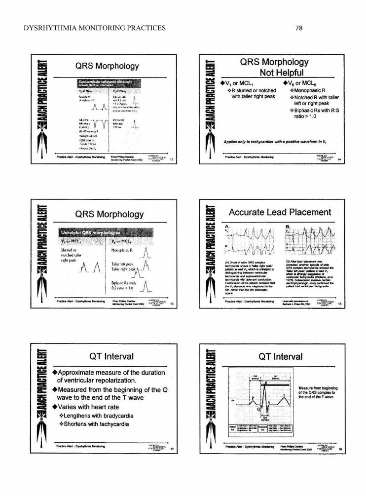
DYSRHYTHMIA MONITORING PRACTICES 78
i ~
& -= 9!
m
i ~ G I! -= !I
m
QRS Morphology
".""'+-.'.' 1·,""",-, ~',..~ I ~,! ~ ', .(...... .... 1 . .-.J\.. ~ """,. """,rio<";'. -- I fl"O'_"~""''''' ''"
AI ." ... -1. r.-,/-l ..... ,-~':;:,;. V \ J ~,:' J" ·k<JO .... ... 1 ·\or1i#<I .. "", ''.;«1_", s_·~ ...
' '''''' .. 0 .. ''.
ORS Morphology
Slurr~d 01'" NonopI\J ~l( R II()(d~uR~ f,
-'-
""" "'" A A T)/Jfr ~l PNk "'-Mer r~l(~rt
Jl , BiplI';l$k Rs wKI. IUni,IQ;> 1.(1 - ........ ~~ _ ........ 0:-
--'_CMXICI;I ~~
OT Interval
+Approximate measure of the duration of ventricular repolarization .
+Measured from the beginning of the Q wave to the end of the T wave
+Varies with heart rate ~·Lengthens with bradycardia ·:·Shortens with tachycardia
"
1'Ncb-,-, .~ -..... , .. :w~,~ H
Iii: .... --.... I .... = m
~
Ii .... -i -= :I
m
ORS Morphology Not Helpful
+V, or MCl, +V. or MCL. .:. R slurred or notched -:'MonophasK; R
with taller light peak .:. Notched R with taller lett or right peak
.:. Biphaslc Rs With RS ratio> 1.0
Accurate Lead Placement A.
~i-tlliVf~ Y, : I~ , ,I1 j li ' It t·V" --' l!J!v : \!j-\ 1'1 11"1 ·+ . . ~ ' ,I' .!' I " " , , -" -I ,II l i i.: JII /i i \ U1 , I ~V\tt If' i' -~lJ : VI J- t I I, _ .I I 1_ n. i
1Al0000oI_QRS-..... (B}AIIof_~_ --.--.- ... """,' - ............ "'~ ~ .. _v~ ......,.. • ..,.,., 1t'I aM ...... ~_ .. _ ... --.·..-n .. _ v, ............. -~ .." .. *""fh ....... '" --- ......... ___ ~OII ..
~ .... ~~ j978).~ ........ ....-.: ~d"poIin~'" p)oIOIUiJIt""'t~'" .. v, __ ............ ....... '-1--....---. . 5Ih..- ....... .........., ---.~---.
.-__ .. ...., _Lc.-IIIN,I'ICI ,"'~
OT Interval
-, , • I . .j
• _ . .I . ..l -I
5 I -- -0: : : --.~ ,"'-' '..21.!
_., ..... .. ...... .... "-",
MMsIn from begirvWIg of 11M: QRS COI'Il)Iel to .,. end of It. T wave
"
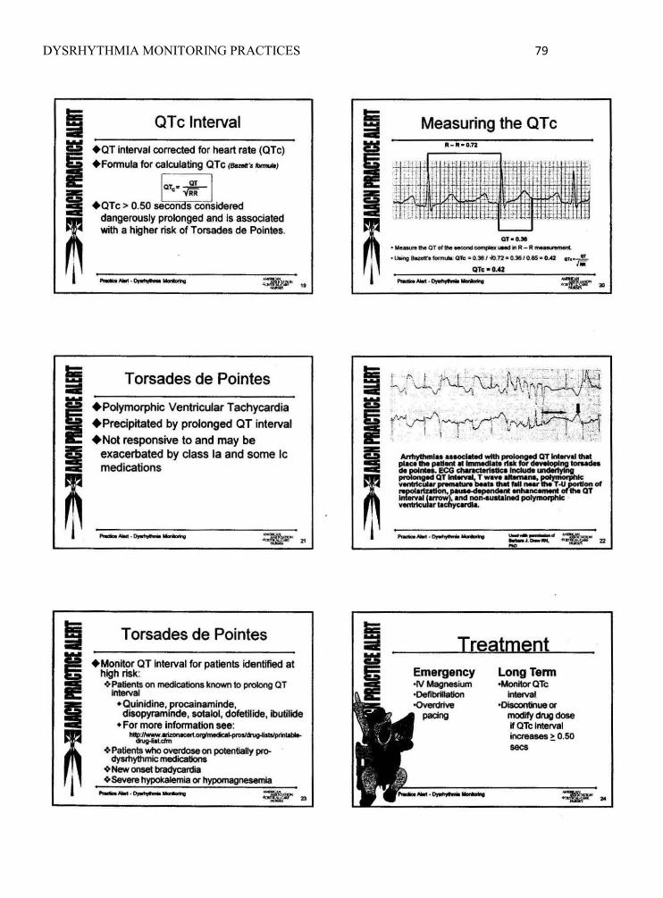
DYSRHYTHMIA MONITORING PRACTICES 79
Ii I!:S
i I
m
OTc Interval
+OT interval corrected for heart rate (OTc) +Fonnula for calculating OTc (-"-1
a, 0f.;!--• RR
+OTc > 0,50 sacon 5 considered dangerously prolonged and Is associated with a higher risk of T orsades de Pointe.,
.......,.J,IooI .~ ......... ~Jl"' it
T orsades de Pointes
+PoIymorphic Ventricular Tachycandia +Precipitated by prolonged OT interval +Not responsive to and may be
exacerbated by class la and some Ic medications
Torsades de Pointes
+MonHor aT interval for patients identified at high risk: <OoPatients on medications known to prok)ng QT
Inta<val .Ouintdine,procainaminde,
dlaopyram,nde, sotalol, dofetilide, ibutilide • For more infonnation see:
1.IIlIIp"J1wWw.lrizoMoertotgl~ _ ... ~ Patients who overdooe on poIe<1tiany pro-
dysthythmic medicetiono ~New"- brady<:aroia ~Sevent hypokalemia Of hypomagnesemia
i I Ii
m
Measuring the OTc 111: - 111:·0.12
I ,.
I ' 'I ,
I I
QT ....
• M_tI'M QT of ..... --.:I compIa UMd III R- II....-..-c. olJlir1r;J kath JonnuIiI: QTc a 0,311 1-io.n_a.3IS / O.as_ oAZ .T"~ <-QTe-OAl ~AM.~"""" ~z
Treatment Emergency 'II/~ium ·Defibrillation oQven:trlve
pacing
LongTenn oMonitorQTc
Interval ·DIooontInue Of
modify drug dose W QTe Interval increases ~ 0.50 sees

DYSRHYTHMIA MONITORING PRACTICES 80

DY
SRH
YTH
MIA
MO
NITO
RIN
G PR
AC
TICES
81
Appendix C
: Cardiac M
onitoring Pocket Reference C
ard
A •
" .. -~ .... . .-. '. . .. • . . 10 : . .. I i ' .. :i .. 0." .. - .ec_
'.20 $ftOncl ---,
rJ~: : .-:...~L : .. : . : ~ _~: ---~ .. ,-- .
Cardiac Monitoring (rev)
1....,';;-;;.;' , V
Barbara Drew, RN, PhD University of California
San Francisco
PHILIPS \ .... Inm~ ,\:"\
.• ~,.,( J( ,,'I It I '\. , ' l l! 11N_'AI ·4,J\J.!f·
:-"Uk~r~ A S.ppkmrlll 10 Crili.c,", C_" N"nee
Philips PN #5990-0487 Printed in USA March 15. 2002
Rate
Rhythm
Pwave
Shape
Size
Axis
Deflection
PR interval
ORS
a wave
AmpWtude
Axis
aT interval
ST segment
Twave
Deflection
Normal ECG criteria Atrial and ventricular rates are the same. In adults, 60 10 100 cycles/min; in infants and chil-dren, within normal limits lor age
Regular, with variance between p.p and R-A IntelVals less lhan 0. 16 second
Present and 1:1 with the QRS
Uniformly rounded without peaking or notches
Amplitude <3.0 mm, width 1.5 • 2.5 mm or duralioo of 0.06 - 0.11 second
o to +90 degrees
Upright in leads I, II, aVF, V4 through V6 : inverted in aVR ; may be flat, inverted, or blphasic in leads III, VI , and V2
Consistent; in adults, 0.12 - 0.20 second; in infants and children, 0.11 - 0.18 second
Follows the P wave; ORS interval is 0.04 - 0.10 second
Duration is -dl.OJ second; depth is 1-2 mm in leads I, aVL, V5' and VB; deep OR or as in aVA and possibly in lead III
5·25 mm in limb leads, 5-30 mm in V, and Ve, 7-30 mm in V2 and V5' 9-30 mm in V3 and V4
-3~ 10 ... 100 degrees
Interval <5Q01o preceding A-R ; a Te -dl.42 second (men) and -dl.43 second (women)
Fallows isoeleclric l ine, Slight curve at proximal portion 01 the T wave
Not depressed more l han 1 mm
May be normally elevated 1-2 mm in V, through Vo Asymmetric and slightly rounded, without sharp points or large notches
ShOuld be in the same direction as QAS: ~ngtt ., leads I, II, aVF, V4 \hroLgh~; inverted in aVR; varied in leads III, aVL, and 1 ttvough V3
Standard electrode placement with a 5-lead set
Angle ol lDuis
RA (while)
For VI (bro~~ ~c For V6 (brown)
. 0" L ... . (green) (red)
Leads recommended for arrhythmia monitoring
For monitors with 5..feadWire patient cabies 1st choice Single-lead monitoring: V, Dual-lead monitorilg : v, ... II
2nd choice Substitute V6'or V, when the patient cannot have an electrode at the sternal border or when QAS amplitude is not adequate for optimized computerized arrhythmia monitoring.
Leads recommended for ischemia monitoring
Inferior myocardial infarction or RCA angioplasty/stent
Anterior myocardial infarction or LAD angioplasty/stent
Posterior or LCX anglopiasty/stent
1t,II1, oraVF
V2 or V3
V2 or V3
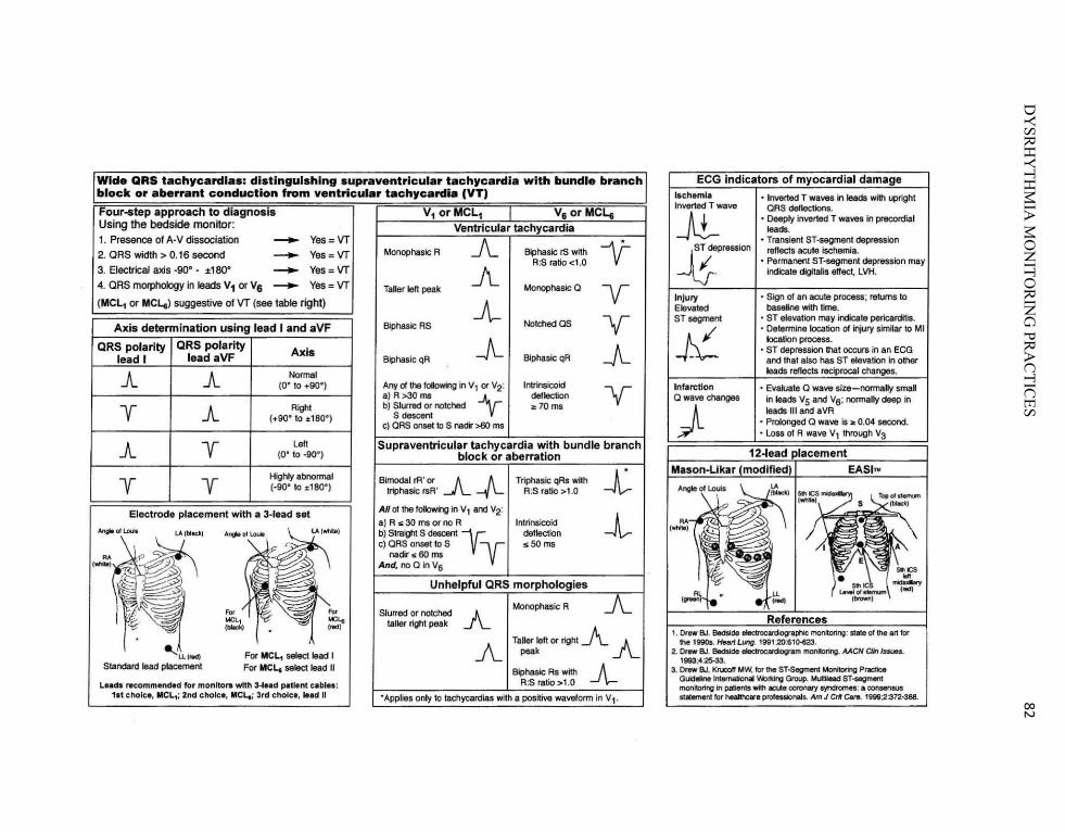
DY
SRH
YTH
MIA
MO
NITO
RIN
G PR
AC
TICES
82
Wide QRS tachycarcUa.: distinguishing supraventricular tachycardia with bundle branch block or aberrant conduction from ventricular tachycardia (VT) Four.tep approach to diagnosIs V1 0rMCL1 I V. or MCLo Using the bedside monitor: Ventricular tachycardia 1. Presence o f A·V dissocialion - Yes=VT A -y-2. ORS widlh > 0.16 second ---- Yes = VT Monophasic R Biphaslc IS with
A:S ratio < 1.0 3. Electrical axis _900 - ±180" - Yes = VT A 4. OAS mQlPhology in leads V1 or Va - Yes = VT Taller left peak Monophasic Q Y (MCL, or MCLt) suggestive of VT (see lable right) -A-
Axis determination using lead I and aVF Biphasic AS No"""" as V-QRS polarity QRS polarity Axis -A- -A-lead I lead aVF Biphasic qR Biphasic QR
JL JL Nonnal (0" 10 +90" ) Any of the fOllowing in v t or V2: Intrinslcoid V-a) R >30 ms -y deflection
y JL Right b) Slurred or notched ~70ms
(+90" 10 :1:180") S descent c) CAS onset to S nadir >80 ms
JL y La" Supraventricular tachycardia with bundle branch (0· to _go·) block or aberration
y y Highly abnormal Bimodal "" '" .A--/L Triphasic qAs with -1-(-gO" 10 :1:180") t~hasic rsR ' A:S ratio > 1.0
Electrode placement with a 3·lead set AJf Ollhefoiowing In Vt and V2:
-!-~~~ ~ .;;~) a)R,,30msornoR Intrinsicoid
AngM 01 l..ooit LA (bl.cll:) b) Stralgtrt S descent -r- deflection c) CRS onset to S V .5O ms
nadir ,,60 ms
'~ And. no Q in V6
Unhelpful QRS morphologies
... ' .. Slurred or notched A Monophasic R A
i.4Cl1 t.lCLe taller rlghl peak
Taller laft or right A A ~Ll (otdl For MeLt seIed lead I A peak
Standard lead placement For MeL. select lead II Biphaslc As with -A-LI.ds recommend.d for monitors with 3 .... d pitllnt c.bl .. : R:S ratio>1.0 1,t cnolc •. MCL,; 2nd cholcl, MCL.; 3td chole., ... d II 'Applies only to tachycardias with a positive waveform In V l '
Inverted T wave
--At-Injury Elevated ST segment
J{. Infarction a wave changes
)
• Inverted T waves in leads with upright QRS dellections.
• Deeply inverted T waves in precordial lea .. ,
• Transient ST-segment depression ref1ects acute ischemia.
• Permanent Sf-segment depression may indicate digitalis effect. LVH.
• Sign of an acute process; returns to baseline with time.
• ST elevation may indicate pericarditis. • Determine location of injury slmHar to
location process. • ST depression thaI occurs in an ECG
and that also has ST etevation in other
• Evaluate Q wave size- norma..,. smal in )&ads Vs and V6; normally deep in leads III and a VA
• Prolonged a wave Is ~ 0.04 second. • loss of A
I . o..ew BJ. 6ectsicle eIectnx:~ moniIorin;: Slate of !he art tor !he li9Ot. HeatfLung. 1991:20:6t0.623.
2. 0.. .. BJ. Bedside ~ 1I'IOIlitoring. MCN a;, ruuel. 1993;4:25-33.
3. Drew 8./. Kru:oII JAW. lot the ST-5egrrIert UoMomg Practice Guideline lntemaliOnal W;u1dng Group. ~ ST-segment monitoring in pilliet'lts with .::ure coronafY S'1f)dromn:. COI'IMMUS stldemen1 tor heallhclre prolestio~,. Am J Cit Cant. 19W;2:372-3e8.

DYSRHYTHMIA MONITORING PRACTICES 83
Appendix D: AACN Audit Tool
• AlCN PRACTICE ALERT ELECTRODE PLACEMENT AND LEAD SELECTION AUDIT TOOL
Purpose: To determine compliance with correct electrode placement and lead selection Instructions: Modify per unit preference.
Date: _____ Time: _____ Room number: ____ _
Nume: ____________ ___
1. Are electrodes placed in the appropriate anatomical region (See illustration) 0 Yes 0 No
\.~!"
,~~h~?t\ \St~· : ,::.;=);
c r (:::\ ~-~ '.?----;:;;;~ .. -: ' ~:;
Rl It I
If answer is Yes, move to question 3
2. If electrodes are not properly placed, identify the incorrect lead(s) and document any contraindications.
Incorrect lead(s): 0 V_ 0 LA 0 RA 0 LL 0 RL
Contra indications: 0 Yes 0 No
Contraindications include: presence of incisionslwoundsllines, implantable devices, or an anatomical abnonnality.
3. Has the appropriate lead been selected based on the patient's actual or potential dysrhy1hmia?
Yes No
Appropriate Lead Selection includes the following: Lead V, to distinguish VT from SVT with aberrant conduction; lett or right BBB; Lead /I to monitor atrial activity.
,\MERI C':AN ASSOCIATION ,j CRITICAL·C"IRE NURSES 1of2 Issued 0</2008
D. KrarnIich

DYSRHYTHMIA MONITORING PRACTICES 84
ELECTRODE PLACEMENT AND LEAD SELECTION AUDIT INSTRUCTIONS
PURPOSE To determine compliance with correct electrode placement and lead selection.
PROCEDURE 1. Assess each patient on the unit for correct electrode placement and lead selection according
to the following criteria: a. Are electrodes placed in the appropriate anatomical regions (use the illustration
below)? b. If electrodes are not properly placed, which leads are incorrect and are there
contraindications (presence of incisions/woundsllines, implantable devices, or an anatomical abnormality)?
c. Has the appropriate monitoring lead been selected based on the patient's actual or potential dysrhythmia (Lead V, to distinguish VT from SVT with aberrant conduction ; left or right BBB; Lead II to monitor atrial activity)?
2. To determine the % compliance with correct electrode placement, divide the number of audits where the electrodes were placed correctly by the total number of patients audited. For example, if 10 patients were audited, and 8 of those patients had correct electrode placement, the % compliance would be 80%.
3. Follow the same procedure for determining % compliance for appropriate lead selection. 4. Audits of electrode placement may be performed collectively (either all electrodes are
correctly placed or not) or individually (do a separate audit for each electrode) according to unit needs and preferences.
AMERICM, ASSOCIAT IO N.{C RITICAI.·CARE " URSES 2c/2 '_0412008
o. Kramlich

DYSRHYTHMIA MONITORING PRACTICES 85
Appendix E: Permission from AACN
From: [email protected] [mailto:[email protected]] Sent: Wed 1/13/2010 3:49 PM To: Schultz, Susan Subject: Re: Practice Alert permission
Susan, You may download the Practice Alert and tools for PA and the toolkit connected with itfrom the Website and utilize the information for your course. They must maintain the American Association of Critical-Care Nurses Practice Alert title at the top. You may NOT edit the content for purposes of your course. There are no additional forms or fees for this. Acknowledgement of permission for multiple copies can be made either on the document or in your program. You may post a link to the AACN website that links to the practice alerts page from your e-learning site for students to access the practice alerts. Pamela Shellner, RN MA Clinical Practice Specialist 101 Columbia Aliso Viejo, CA 92656 phone 800-394-5995 x321 fax 949-448-5551 [email protected] "Act with Intention" Beth Hammer
From: Schultz, Susan Sent: Wed 1/13/2010 10:25 AM To: [email protected]; [email protected] Subject: Practice Alert permission I'm planning on conducting research for a Doctor of Nursing Practice degree on dysrhythmia monitoring practices based on AACN's Practice Alert. I would like permission from American Association of Critical-Care Nurses to use the following resources posted on www.aacn.org : AACN Practice Alert: Dysrhythmia Monitoring AACN Practice Alert: Electrode Placement and Lead Selection Audit Tool PowerPoint entitled "Practice Alert Dysrhythmia Monitoring" by Nancy M. Richards Pocket reference: Cardiac Monitoring, AACN 2002, by Barbara Drew. I plan to use the materials to educate telemetry staff nurses via an online learning module that is accessible only by the nurses in that hospital. I will not alter the content or parts of the documents that give credit to American Association of Critical-Care Nurses. Please advise me if I should contact someone else for permission or if further information is required. Thank you Susan Schultz, RN, MSN, CNE, CCRN DNP Student at University of North Florida, Jacksonville, FL

DYSRHYTHMIA MONITORING PRACTICES 86
Signature Deleted

DYSRHYTHMIA MONITORING PRACTICES 87
Appendix F: Appraisal Tables
Authors Study Purpose
Sampling Method, Size
Intervention Group
Control Group
Outcomes Measured
Results Major Strengths Major Weaknesses
Belcher &Vonderharr (2005) Descriptive Interventional Study Evaluated web-delivered program to nursing staff on evidence based practice (guided by Gagne’s instructional design principles)
64 staff nurses (out of 1500 in the hospital) voluntarily completed the audio and visual portion of the program and written evaluation. One hour CEU offered free. University students also used the program in an online course, but their evaluations were excluded from this article.
One hour online program (in WebCT) was developed by university faculty with hospital educator input. It included audio-streaming coordinated with video slides. Also had graphics, text, and interaction with content and instructor by email (although no one used email). Program objectives were about defining research-based practice, roles, processes, and how to implement.
none 1) Written course evaluations on participants’ assessment of their achievement of learning outcomes, satisfaction and overall effectiveness. 2) Estimates of cost-effectiveness of web-based compared to traditional lecture
“All the staff nurses agreed that the learning objectives were achieved, they were satisfied with the program, and their personal learning needs were met.” “Web-based program was marginally more cost effective ($30 less), given our small number of users”
Even though it was not an ECG educational program, it described detailed components of web-based program and how it was developed. Self-paced program, which allowed them to rewind and repeat. Program was standardized and accessible by multiple learners. Developed collaboratively between university and hospital.
Evaluations were self-reported perceptions with no validation of competency or learning. Vague summary of results with no statistical analysis. Not randomized. No control group for comparison. Cost effectiveness based on estimates and showed marginal difference, perhaps due to small percentage of staff who participated.
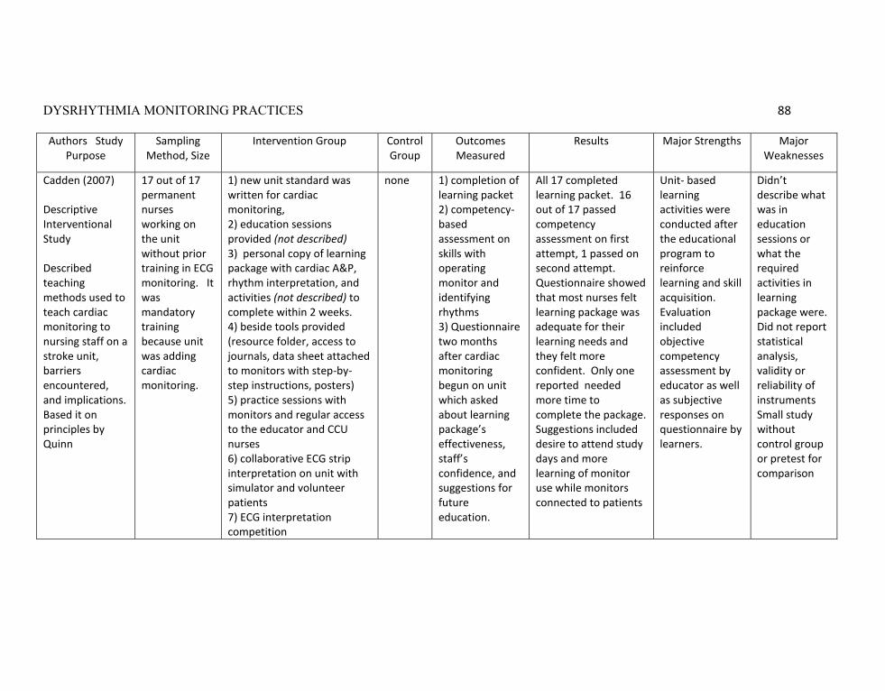
DYSRHYTHMIA MONITORING PRACTICES 88
Authors Study Purpose
Sampling Method, Size
Intervention Group Control Group
Outcomes Measured
Results Major Strengths Major Weaknesses
Cadden (2007) Descriptive Interventional Study Described teaching methods used to teach cardiac monitoring to nursing staff on a stroke unit, barriers encountered, and implications. Based it on principles by Quinn
17 out of 17 permanent nurses working on the unit without prior training in ECG monitoring. It was mandatory training because unit was adding cardiac monitoring.
1) new unit standard was written for cardiac monitoring, 2) education sessions provided (not described) 3) personal copy of learning package with cardiac A&P, rhythm interpretation, and activities (not described) to complete within 2 weeks. 4) beside tools provided (resource folder, access to journals, data sheet attached to monitors with step-by-step instructions, posters) 5) practice sessions with monitors and regular access to the educator and CCU nurses 6) collaborative ECG strip interpretation on unit with simulator and volunteer patients 7) ECG interpretation competition
none 1) completion of learning packet 2) competency-based assessment on skills with operating monitor and identifying rhythms 3) Questionnaire two months after cardiac monitoring begun on unit which asked about learning package’s effectiveness, staff’s confidence, and suggestions for future education.
All 17 completed learning packet. 16 out of 17 passed competency assessment on first attempt, 1 passed on second attempt. Questionnaire showed that most nurses felt learning package was adequate for their learning needs and they felt more confident. Only one reported needed more time to complete the package. Suggestions included desire to attend study days and more learning of monitor use while monitors connected to patients
Unit- based learning activities were conducted after the educational program to reinforce learning and skill acquisition. Evaluation included objective competency assessment by educator as well as subjective responses on questionnaire by learners.
Didn’t describe what was in education sessions or what the required activities in learning package were. Did not report statistical analysis, validity or reliability of instruments Small study without control group or pretest for comparison

DYSRHYTHMIA MONITORING PRACTICES 89
Authors Study Purpose
Sampling Method, Size
Intervention Group
Control Group
Outcomes Measured
Results Major Strengths Major Weaknesses
Dumpe, Kanyak, & Hill (2007) Studied usefulness of a Learning Management System (LMS) to validate multidiscipli-nary employee competencies on HIPAA. Later, developed program for 16 annual online competencies to be completed by nursing staff.
HIPAA mandatory for over 18,000 total hospital employees Nursing competencies were accessed by 4064 nurses (total population unknown)
All employees for HIPPA and all nursing staff for 16 online competencies.
N/A Completion and satisfaction rates.
Over 2 months, 18,000 employees successfully completed the HIPAA online course (but don’t know percentage). Participant survey regarding their satisfaction showed 75% were satisfied or very satisfied with the online course. 65% preferred online training to traditional instructor-led training. 4064 nursing personnel accessed the online nursing competencies. 90% reported satisfied or very satisfied with the system 92% reported it was easy or very easy to complete 87% reported they were able to complete the competencies on their own unit.
LMS was an effective way to standardize competencies and reach large numbers of staff for HIPPA training and nursing competency assessment.
Did not report statistics or percentages of staff that completed the programs. Didn’t describe what the subjects were for the nurses’ annual competency assessments.
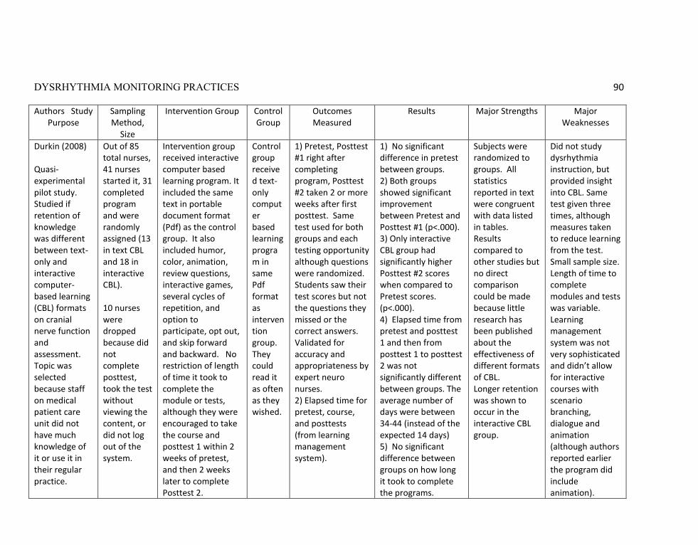
DYSRHYTHMIA MONITORING PRACTICES 90
Authors Study Purpose
Sampling Method,
Size
Intervention Group Control Group
Outcomes Measured
Results Major Strengths Major Weaknesses
Durkin (2008) Quasi-experimental pilot study. Studied if retention of knowledge was different between text-only and interactive computer-based learning (CBL) formats on cranial nerve function and assessment. Topic was selected because staff on medical patient care unit did not have much knowledge of it or use it in their regular practice.
Out of 85 total nurses, 41 nurses started it, 31 completed program and were randomly assigned (13 in text CBL and 18 in interactive CBL). 10 nurses were dropped because did not complete posttest, took the test without viewing the content, or did not log out of the system.
Intervention group received interactive computer based learning program. It included the same text in portable document format (Pdf) as the control group. It also included humor, color, animation, review questions, interactive games, several cycles of repetition, and option to participate, opt out, and skip forward and backward. No restriction of length of time it took to complete the module or tests, although they were encouraged to take the course and posttest 1 within 2 weeks of pretest, and then 2 weeks later to complete Posttest 2.
Control group received text-only computer based learning program in same Pdf format as intervention group. They could read it as often as they wished.
1) Pretest, Posttest #1 right after completing program, Posttest #2 taken 2 or more weeks after first posttest. Same test used for both groups and each testing opportunity although questions were randomized. Students saw their test scores but not the questions they missed or the correct answers. Validated for accuracy and appropriateness by expert neuro nurses. 2) Elapsed time for pretest, course, and posttests (from learning management system).
1) No significant difference in pretest between groups. 2) Both groups showed significant improvement between Pretest and Posttest #1 (p<.000). 3) Only interactive CBL group had significantly higher Posttest #2 scores when compared to Pretest scores. (p<.000). 4) Elapsed time from pretest and posttest 1 and then from posttest 1 to posttest 2 was not significantly different between groups. The average number of days were between 34-44 (instead of the expected 14 days) 5) No significant difference between groups on how long it took to complete the programs.
Subjects were randomized to groups. All statistics reported in text were congruent with data listed in tables. Results compared to other studies but no direct comparison could be made because little research has been published about the effectiveness of different formats of CBL. Longer retention was shown to occur in the interactive CBL group.
Did not study dysrhythmia instruction, but provided insight into CBL. Same test given three times, although measures taken to reduce learning from the test. Small sample size. Length of time to complete modules and tests was variable. Learning management system was not very sophisticated and didn’t allow for interactive courses with scenario branching, dialogue and animation (although authors reported earlier the program did include animation).
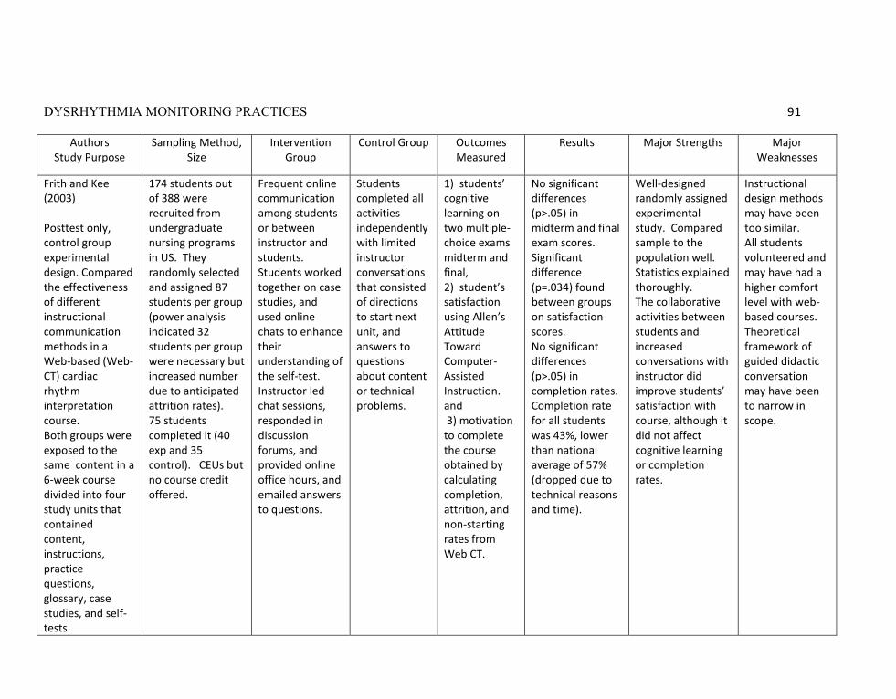
DYSRHYTHMIA MONITORING PRACTICES 91
Authors Study Purpose
Sampling Method, Size
Intervention Group
Control Group Outcomes Measured
Results Major Strengths Major Weaknesses
Frith and Kee (2003) Posttest only, control group experimental design. Compared the effectiveness of different instructional communication methods in a Web-based (Web-CT) cardiac rhythm interpretation course. Both groups were exposed to the same content in a 6-week course divided into four study units that contained content, instructions, practice questions, glossary, case studies, and self-tests.
174 students out of 388 were recruited from undergraduate nursing programs in US. They randomly selected and assigned 87 students per group (power analysis indicated 32 students per group were necessary but increased number due to anticipated attrition rates). 75 students completed it (40 exp and 35 control). CEUs but no course credit offered.
Frequent online communication among students or between instructor and students. Students worked together on case studies, and used online chats to enhance their understanding of the self-test. Instructor led chat sessions, responded in discussion forums, and provided online office hours, and emailed answers to questions.
Students completed all activities independently with limited instructor conversations that consisted of directions to start next unit, and answers to questions about content or technical problems.
1) students’ cognitive learning on two multiple-choice exams midterm and final, 2) student’s satisfaction using Allen’s Attitude Toward Computer-Assisted Instruction. and 3) motivation to complete the course obtained by calculating completion, attrition, and non-starting rates from Web CT.
No significant differences (p>.05) in midterm and final exam scores. Significant difference (p=.034) found between groups on satisfaction scores. No significant differences (p>.05) in completion rates. Completion rate for all students was 43%, lower than national average of 57% (dropped due to technical reasons and time).
Well-designed randomly assigned experimental study. Compared sample to the population well. Statistics explained thoroughly. The collaborative activities between students and increased conversations with instructor did improve students’ satisfaction with course, although it did not affect cognitive learning or completion rates.
Instructional design methods may have been too similar. All students volunteered and may have had a higher comfort level with web-based courses. Theoretical framework of guided didactic conversation may have been to narrow in scope.
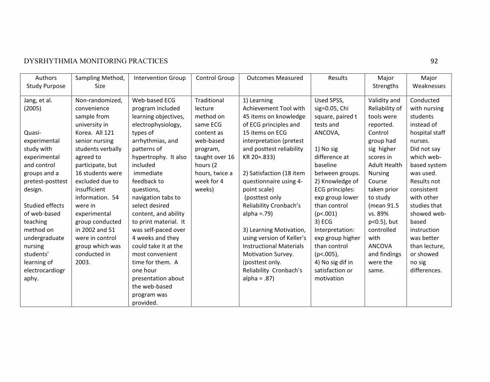
DYSRHYTHMIA MONITORING PRACTICES 92
Authors Study Purpose
Sampling Method, Size
Intervention Group Control Group Outcomes Measured Results Major Strengths
Major Weaknesses
Jang, et al. (2005) Quasi-experimental study with experimental and control groups and a pretest-posttest design. Studied effects of web-based teaching method on undergraduate nursing students' learning of electrocardiography.
Non-randomized, convenience sample from university in Korea. All 121 senior nursing students verbally agreed to participate, but 16 students were excluded due to insufficient information. 54 were in experimental group conducted in 2002 and 51 were in control group which was conducted in 2003.
Web-based ECG program included learning objectives, electrophysiology, types of arrhythmias, and patterns of hypertrophy. It also included immediate feedback to questions, navigation tabs to select desired content, and ability to print material. It was self-paced over 4 weeks and they could take it at the most convenient time for them. A one hour presentation about the web-based program was provided.
Traditional lecture method on same ECG content as web-based program, taught over 16 hours (2 hours, twice a week for 4 weeks)
1) Learning Achievement Tool with 45 items on knowledge of ECG principles and 15 items on ECG interpretation (pretest and posttest reliability KR 20=.833) 2) Satisfaction (18 item questionnaire using 4-point scale) (posttest only Reliability Cronbach’s alpha =.79) 3) Learning Motivation, using version of Keller's Instructional Materials Motivation Survey. (posttest only. Reliability Cronbach’s alpha = .87)
Used SPSS, sig=0.05, Chi square, paired t tests and ANCOVA, 1) No sig difference at baseline between groups. 2) Knowledge of ECG principles: exp group lower than control (p<.001) 3) ECG Interpretation: exp group higher than control (p<.005), 4) No sig dif in satisfaction or motivation
Validity and Reliability of tools were reported. Control group had sig higher scores in Adult Health Nursing Course taken prior to study (mean 91.5 vs. 89% p<0.5), but controlled with ANCOVA and findings were the same.
Conducted with nursing students instead of hospital staff nurses. Did not say which web-based system was used. Results not consistent with other studies that showed web-based instruction was better than lecture, or showed no sig differences.

DYSRHYTHMIA MONITORING PRACTICES 93
Authors Study Purpose
Sampling Method, Size
Intervention Group Outcomes Measured
Results Major Strengths
Major Weaknesses
Jeffries (2005) Pilot Study, Descriptive Intervention Designed an online critical care course, using an instruction model based on Chickering and Gamson’s seven principles of best practices in education. Usability testing was done before pilot study.
15 participate, 12 RNs and 3 senior BSN students with diverse backgrounds and experience. Did not say how recruited. Ages ranged 20-49. Most were RNs taking course for professional development. Two thirds lived more than 100 miles from campus. Course lasted 10 weeks.
Model included three components: 1) core didactic online program with mini-lectures, vignettes of patient scenarios, interactive activities such as games, discussion board, diary of reflections, and web links that learners could select as needed. Program incorporated three dimensions of general principles, process, and critical thinking. Student-centered, allowing selection of content, activities, and materials. 2) Practicum of 112 clinical hours with preceptor in critical care unit 3) Access to Virtual Center of Best Practices (VCBP) which served as a learning resource center and provided standards or protocols, current research, and asynchronous consultation with clinical experts.
1) competency in reaching learning outcomes on 10 modules as measured by multiple choice pretests and post tests for each modules and final exam. Correct answers with rationales displayed on pretest 2) skills acquisition assessment by preceptor 3) Students’ perception of educational practices 4) Students’ satisfaction 5) convenience of course (For 3, 4, 5, used EEUWIN instrument)
1) All students successfully completed the course with passing scores (didn’t clarify what was passing). 2) All students met the required competencies 3) Students perceived that the principles of best practices in education were highly incorporated into the online course (means from pilot study were higher than means national benchmark). 4) Mean course satisfaction was 4.17 out of 5 (SD=0.80). 5). Mean for perceived convenience was 4.01 out of 5 (SD = 0.82)
Included direct comparison of Chickering and Gamson’s seven principles of best practices to the program components and students’ ratings of how well program met the principles. Pilot study showed that critical care concepts and skills could be learned through e-learning.
Small pilot study with mix of RNs and student nurses. Did not say how students recruited. Didn’t specify what cardiac dysrhythmias were included in the critical care course.
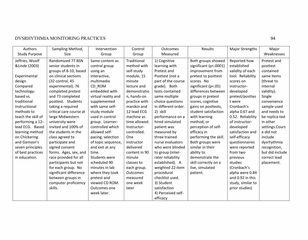
DYSRHYTHMIA MONITORING PRACTICES 94
Authors Study Purpose
Sampling Method, Size
Intervention Group
Control Group
Outcomes Measured
Results Major Strengths Major Weaknesses
Jeffries, Woolf &Linde (2003) Experimental design. Compared technology-based vs. traditional instructional methods to teach the skill of performing a 12-lead ECG. Based learning method on Chickering and Gamson’s seven principles of best practices in education.
Randomized 77 BSN senior students in groups of 8-10, based on clinical sections (32 control, 45 experimental). 76 completed pretest and 73 completed posttest. Students taking a required critical care course at large Midwestern university were invited and 100% of the students in the class agreed to participate and signed consent forms. Ages, sex, and race provided for all participants but not for each group. No significant difference between groups in computer proficiency skills.
Same content as control group using an interactive, multimedia CD_ROM embedded with virtual reality and supplemented with same self-study module used in control group. Learner- controlled which allowed self-pacing, selection of topic sequence, and exit at any time. Students were scheduled 90 minutes in lab where they took pretest and viewed CD ROM. Outcomes one week later.
Traditional method with self-study module, 15 minute lecture and demonstration, hands-on practice with manikin and 12-lead ECG machine as time allowed. Instructor- controlled. One instructor delivered content in 90 minute classes to each group. Outcomes measured one week later
1) Cognitive learning with Pretest and Posttest (not a part of the course grade). Both tests contained same multiple choice questions in different order. 2) skill performance on a hired simulated patient was measured by three trained nurse evaluators who were blinded to group (inter-rater reliability established). A weighted 22-item procedural checklist used. 3) Student satisfaction 4) Perceived self-efficacy
Both groups showed significant (p<.0001) improvement from pretest to posttest scores. No significant (p<.05) differences between groups in pretest scores, cognitive gains on posttests, student satisfaction with learning method, or perception of self-efficacy in performing the skill. Both groups were similar in their ability to demonstrate the skill correctly on a live, simulated patient.
Reported how established validity of each tool. Reliability scores on instructor-developed pretest/posttest were Cronbach’s alpha 0.67 and 0.52. Reliability of instructor- developed satisfaction and self-efficacy questionnaires were reported from two previous studies (Cronbach’s alpha were 0.84 and 0.92 in this study, similar to prior studies)
Pretest and posttest contained same items (threat to internal validity). Single convenience sample used and needs to be replica-ted in other settings.Course did not include dysrhythmia recognition, but did include correct lead placement.

DYSRHYTHMIA MONITORING PRACTICES 95
Authors Study Purpose
Sampling Method, Size
Intervention Group Outcomes Measured
Results Major Strengths Major Weaknesses
Keller & Raines (2005) Qualitative study. Focus group methodology, conducted on a tiered schedule over 1 year. Purpose was to elicit perceptions of nurses about the level of knowledge needed to recognize a cardiac arrhythmia. Provided evidence for developing levels of arrhythmia competency
25 critical care nurses from three large metropolitan community hospitals were asked to volunteer and incentive was arrhythmia update program with continuing education (CE) credits. Purposeful sampling with group-specific criteria used to create groups between 4-8 people per group.
Group 1: 5 ACLS instructors Group 2: 4 nurses with less than one year critical care experience. Group 3: 4 nurses with more than 1 year critical care experience Group 4: 8 nurses with more than 1 year critical care experience Group 5: 4 nurses with more than 1 year critical care experience
Nurse’s perceptions whether ECG rhythm strips represented basic or advanced knowledge. A third category of intermediate knowledge was added.
Consensus was reached on 17 of the 30 strips, with 12 categorized as basic and 5 as advanced. Of the remaining 13, there continued to be lack of consensus. These arrhythmias included torsades de pointe, heart blocks, atrial fibrillation with right BBB, ventricular tachycardia, and WPW with atrial fibrillation. A chart listing which arrhythmias could be categorized as basic, intermediate, and advanced was displayed. They concluded there was significant lack of ability of nurses to recognize and differentiate heart blocks, aberrant conduction, and tachyarrhythmias.
Group methodology explained well. Verbatim extracts from transcripts of group discussions were included to back up author’s conclusions.
Sample not randomized. Incentive of CE credits for arrhythmia update may have adversely affected sample by attracting more nurses who didn’t know arrhythmias very well.
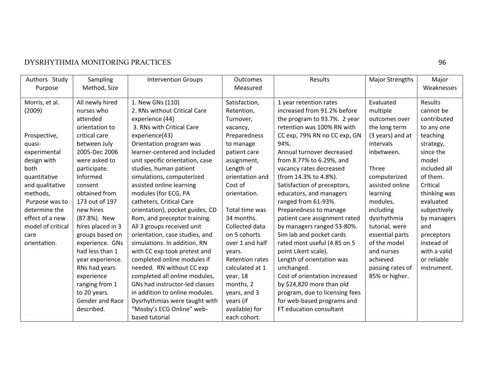
DYSRHYTHMIA MONITORING PRACTICES 96
Authors Study Purpose
Sampling Method, Size
Intervention Groups Outcomes Measured
Results Major Strengths Major Weaknesses
Morris, et al. (2009) Prospective, quasi-experimental design with both quantitative and qualitative methods, Purpose was to determine the effect of a new model of critical care orientation.
All newly hired nurses who attended orientation to critical care between July 2005-Dec 2006 were asked to participate. Informed consent obtained from 173 out of 197 new hires (87.8%). New hires placed in 3 groups based on experience. GNs had less than 1 year experience. RNs had years experience ranging from 1 to 20 years. Gender and Race described.
1. New GNs (110) 2. RNs without Critical Care experience (44) 3. RNs with Critical Care experience(43) Orientation program was learner-centered and included unit specific orientation, case studies, human patient simulations, computerized assisted online learning modules (for ECG, PA catheters, Critical Care orientation), pocket guides, CD Rom, and preceptor training. All 3 groups received unit orientation, case studies, and simulations. In addition, RN with CC exp took pretest and completed online modules if needed. RN without CC exp completed all online modules, GNs had instructor-led classes in addition to online modules. Dysrhythmias were taught with “Mosby’s ECG Online” web-based tutorial
Satisfaction, Retention, Turnover, vacancy, Preparedness to manage patient care assignment, Length of orientation and Cost of orientation. Total time was 34 months. Collected data on 5 cohorts over 1 and half years. Retention rates calculated at 1 year, 18 months, 2 years, and 3 years (if available) for each cohort.
1 year retention rates increased from 91.2% before the program to 93.7%. 2 year retention was 100% RN with CC exp, 79% RN no CC exp, GN 94%. Annual turnover decreased from 8.77% to 6.29%, and vacancy rates decreased (from 14.3% to 4.8%). Satisfaction of preceptors, educators, and managers ranged from 61-93%. Preparedness to manage patient care assignment rated by managers ranged 53-80%. Sim lab and pocket cards rated most useful (4.85 on 5 point Likert scale). Length of orientation was unchanged. Cost of orientation increased by $24,820 more than old program, due to licensing fees for web-based programs and FT education consultant
Evaluated multiple outcomes over the long term (3 years) and at intervals inbetween. Three computerized assisted online learning modules, including dysrhythmia tutorial, were essential parts of the model and nurses achieved passing rates of 85% or higher.
Results cannot be contributed to any one teaching strategy, since the model included all of them. Critical thinking was evaluated subjectively by managers and preceptors instead of with a valid or reliable instrument.

DYSRHYTHMIA MONITORING PRACTICES 97
Authors Study Purpose
Sampling Method, Size
Intervention Groups
Outcomes Measured
Results Major Strengths Major Weaknesses
Van-Arsdale, (1998) Comparative study, posttest only, multiple treatment group design. Evaluated four different teaching methods in the development of skills necessary to identify cardiac dysrhythmias
Not randomized. Included 244 R.N.s from ER, CCU, and telemetry units from three hospitals (200-500 beds) in a rural setting over a 2 year period of time. Nurses were assigned to one of four groups depending on when they were employed and which method was being used at that time. Only nurse who had never attended a program on dysrhythmia interpretation were included.
All groups received a 20-hour basic dysrhythmia course with same objectives, content, and reference book. All groups were taught by the same critical care clinical specialist. Group 1: 72 RNs 2-hour sessions once a week over 10 weeks Group 2: 48 RNs 2-hour sessions twice a week over 5 weeks Group 3: 60 RNs 2-hour sessions twice a day over 5 days Group 4: 64 RNs Self-instructional module (reading packet)to be completed within 10-weeks (reading packet)
1) Posttest. 90% of the questions dealt with interpretation of rhythm strips. Explained how validity was established. Reliability was .90. 2) Course evaluation surveys by students
1) ANOVA showed there were significant differences between groups on posttest scores (p= .0001) 2) Follow-up (Tukey) revealed that Group 1 and 2 scored significantly higher on the posttest than Group 3 and 4. 3) 90% from Group 1 and 2 indicated objectives were attained, length appropriate, and felt prepared. 4) 100% from Group 3 indicated too much new information was presented and they felt uncomfortable with their skills. 5) 91% from Group 4 indicated some classroom sessions would be beneficial to ask questions or discuss rhythms.
All statistics reported in text were congruent with data listed in tables. Group interactions not a factor because courses not taught simultaneously. Consistent educational materials and posttests were used for each group. Posttest had high reliability (Kuder-Richardson .9).
Study was conducted over two years prior to 1998. Self-instruction module was only a reading packet, with no computer- based learning strategies (perhaps because they were not readily available then) Analysis of group characteristics was not provided so don’t know if all groups were similar. Course included basic dysrhythmia categories but not bundle branch blocks.

DYSRHYTHMIA MONITORING PRACTICES 98
Appendix G: ACC/ ECC Rating System
American College of Cardiology Emergency Cardiac Care Committee Rating System
Class I: Cardiac monitoring is indicated in most, if not all, patients in this group. Class II: Cardiac monitoring may be of benefit in some patients but is not considered essential for all patients. Class III: Cardiac monitoring is not indicated because a patient's risk of a serious event is so low that monitoring has no therapeutic benefit (Taken from Drew et al., 2004)
AppendixH: AACN Levels of Evidence
American Association of Critical-Care Nurses’ Grading Level of Evidence Level I: Manufacturer‘s recommendations only Level II: Theory based, no research data to support recommendations;
Recommendations from expert consensus group may exist Level III: Laboratory data, no clinical data to support recommendations Level IV: Limited clinical studies to support recommendations Level V: Clinical studies in more than one or two patient populations and situations
to support recommendations Level VI: Clinical studies in a variety of patient populations and situations to support
recommendations. (Taken from AACN Practice Alert, 2008)
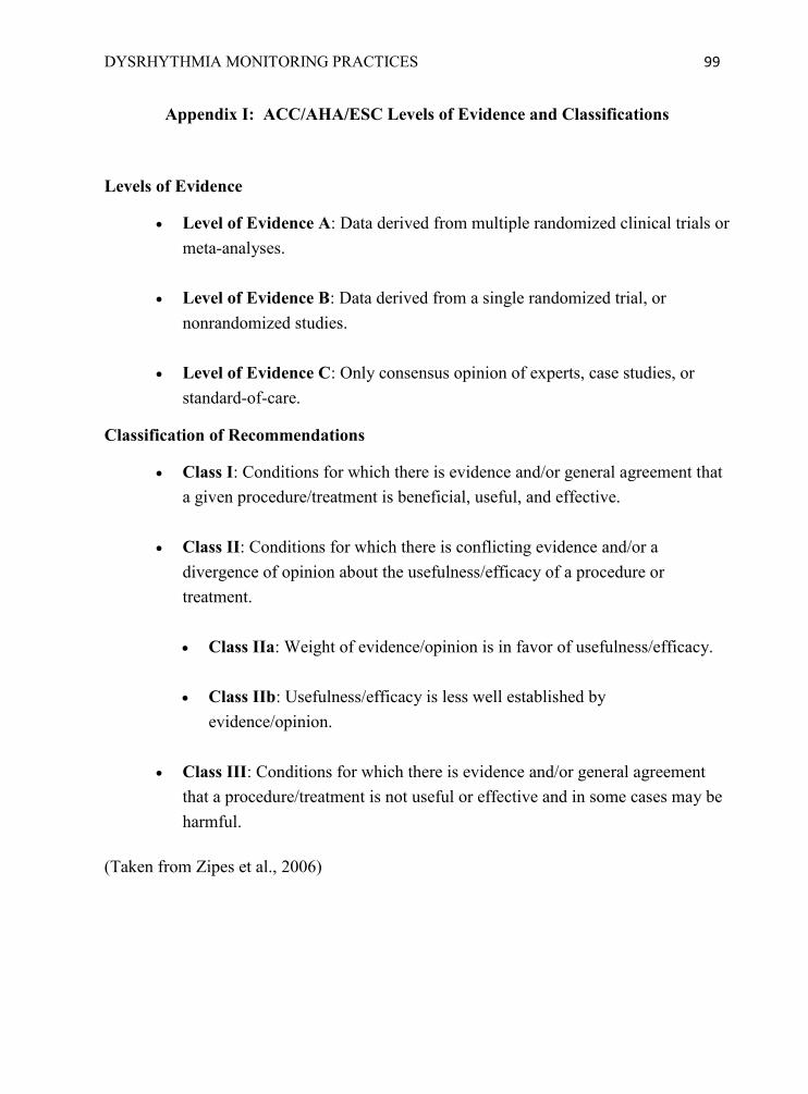
DYSRHYTHMIA MONITORING PRACTICES 99
Appendix I: ACC/AHA/ESC Levels of Evidence and Classifications
Levels of Evidence
Level of Evidence A: Data derived from multiple randomized clinical trials or meta-analyses.
Level of Evidence B: Data derived from a single randomized trial, or nonrandomized studies.
Level of Evidence C: Only consensus opinion of experts, case studies, or standard-of-care.
Classification of Recommendations
Class I: Conditions for which there is evidence and/or general agreement that a given procedure/treatment is beneficial, useful, and effective.
Class II: Conditions for which there is conflicting evidence and/or a divergence of opinion about the usefulness/efficacy of a procedure or treatment. Class IIa: Weight of evidence/opinion is in favor of usefulness/efficacy.
Class IIb: Usefulness/efficacy is less well established by
evidence/opinion.
Class III: Conditions for which there is evidence and/or general agreement that a procedure/treatment is not useful or effective and in some cases may be harmful.
(Taken from Zipes et al., 2006)

DYSRHYTHMIA MONITORING PRACTICES 100
Appendix J: Principles of Good Practice andEvidence-based Strategies
Seven Principles of Good Practice 1
Evidence-based Strategies Applicable to Staff Nurses References
1. Encourages student-faculty contact
Provide the instructor‘s contact information for the students in the program.
Establish regular times for educator to be online or present to reinforce learning.
Belcher &Vonderharr, 2005; Frith&Kee, 2003.
Cadden, 2007; Frith&Kee, 2003.
2. Develops reciprocity and cooperation among students.
Plan opportunities for nurses to collaborate on interpreting case studies and ECGs
Use online chats or discussion forums when learners are expected to access the program more than once.
Cadden, 2007; Frith&Kee, 2003; Morris et al., 2009.
Dumpe, Kanyak, & Hill, 2007; Frith&Kee, 2003; Jeffries, 2005.
3. Uses active learning techniques.
Incorporate case studies, games, and self-check practice questions in the online educational program.
Require return demonstration of skills with competency assessment checklists and/or human patient simulation.
Durkin, 2008; Frith&Kee, 2003; Jeffries, 2005; Jeffries, Woolf, &Linde, 2003.
Cadden, 2007; Jeffries, 2005; Jeffries, Woolf, &Linde, 2003; Morris et al., 2009.
4. Gives prompt
feedback. Provide answers on practice questions throughout
the online course and after the posttest.
Offer unit training with a preceptor or educator who gave them daily feedback.
Durkin, 2008; Frith&Kee, 2003; Jang et al., 2005.
Cadden, 2007; Jeffries, 2005; Morris et al., 2009.

DYSRHYTHMIA MONITORING PRACTICES 101
Seven Principles of Good Practice 1
Evidence-based Strategies Applicable to Staff Nurses References
5. Emphasizes time on task.
Develop web-based courses that may be accessed at the learner‘s convenience from home or work.
Design software that displays table of contents and allows learners to navigate back and forth through the program.
Provide continuing education credits
Belcher &Vonderharr, 2005; Dumpe, Kanyak, & Hill, 2007; Durkin, 2008; Jang et al., 2005; Jeffries, 2005.
Durkin, 2008; Jang et al., 2005; Jeffries, 2005; Jeffries, Woolf, &Linde, 2003.
Belcher &Vonderharr, 2005; Frith&Kee, 2003; Keller & Raines, 2005.
6. Communicates
high expectations.
State the learning objectives of the program clearly and evaluate how well they were met with pretest and/or posttest questions.
Require competency-based skills validation.
Durkin, 2008; Frith&Kee, 2003; Jang et al., 2005; Jeffries, 2005; Jeffries Woolf, &Linde, 2003.
Cadden, 2007; Dumpe, Kanyak, & Hill, 2007; Jeffries, 2005; Jeffries, Woolf, &Linde, 2003; Morris et al., 2009.
7. Respects diverse talents and ways of learning
Incorporate variety of teaching methods such as printed materials for self-study, pocket cards, references on the unit, audio, visual, graphics, animation, virtual reality, hands-on, and interactive activities.
Belcher &Vonderharr, 2005; Cadden, 2007; Durkin, 2008; Jang et al., 2005; Jeffries, 2005; Jeffries, Woolf, &Linde, 2003; Morris et al., 2009.
1Chickering, A. W. &Gamson, Z. F. (1987, March), Seven principles for good practice in undergraduate education. American Association for Higher Education (AAHE) Bulletin, 39 (7), 3-7.

DYSRHYTHMIA MONITORING PRACTICES 102
Appendix K: Permission to Conduct Study
Signature Deleted

DYSRHYTHMIA MONITORING PRACTICES 103
Appendix L: Nurses’ Demographic Questionnaire 1. Gender a. Male b. Female 2. Ethnicity a. American Indian or Alaska Native b. Asian or Pacific Islander c. Black or African American d. Hispanic or Latino e. White or Caucasian 3. Highest educational degree: a. Associates b. Bachelors c. Masters d. Doctoral 4. How much instruction have you received before today on QRS morphology and QT interval monitoring? a. no instruction b. only a little instruction c. some instruction but not comfortable yet d. enough instruction to feel comfortable e. sufficient instruction to be able to teach others 5. Have you ever studied online or web-based educational programs? a. Yes b. No 6. How comfortable are you with online learning or web-based instruction? a. Not comfortable at all b. Somewhat comfortable c. Very comfortable 7. Is English a second language for you? a. Yes b. No 8. How old are you? ____________ 9. Years licensed as RN: ______________ 10. Length of time worked on this unit: ______________ 11. How many years ago was your first class on dysrhythmia interpretation? _________
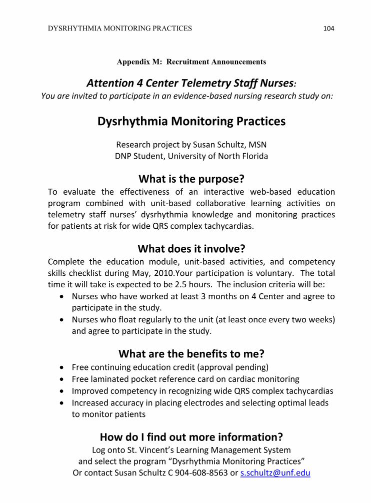
DYSRHYTHMIA MONITORING PRACTICES 104
Appendix M: Recruitment Announcements
Attention 4 Center Telemetry Staff Nurses:
You are invited to participate in an evidence-based nursing research study on:
Dysrhythmia Monitoring Practices
Research project by Susan Schultz, MSN DNP Student, University of North Florida
What is the purpose? To evaluate the effectiveness of an interactive web-based education program combined with unit-based collaborative learning activities on telemetry staff nurses’ dysrhythmia knowledge and monitoring practices for patients at risk for wide QRS complex tachycardias.
What does it involve? Complete the education module, unit-based activities, and competency skills checklist during May, 2010.Your participation is voluntary. The total time it will take is expected to be 2.5 hours. The inclusion criteria will be:
Nurses who have worked at least 3 months on 4 Center and agree to participate in the study.
Nurses who float regularly to the unit (at least once every two weeks) and agree to participate in the study.
What are the benefits to me? Free continuing education credit (approval pending)
Free laminated pocket reference card on cardiac monitoring
Improved competency in recognizing wide QRS complex tachycardias
Increased accuracy in placing electrodes and selecting optimal leads to monitor patients
How do I find out more information? Log onto St. Vincent’s Learning Management System
and select the program “Dysrhythmia Monitoring Practices” Or contact Susan Schultz C 904-608-8563 or [email protected]

DYSRHYTHMIA MONITORING PRACTICES 105
Dysrhythmia Monitoring Practices
Research Project with Susan Schultz, MSN, DNP Student, University of North Florida
PROJECT TIMELINE
May 17- 20, 2010 Conduct first audit of electrode placement and lead selection May 20 – June 13 Sign consent forms and study educational module on LMS June 2 – June 26 Work on unit-based activities and skills checklist Susan Schultz is planning to be on 4 Center during these times: Wed. June 2, 1:00 – 3:00 pm 11:00 pm – 12:30 am Thu. June 3 ------------------ 11:00 pm – 12:30 am Fri. June 4 1:00 – 3:00 pm 11:00 pm – 12:30 am Sat. June 5 1:00 – 3:00 pm ----------------------- Wed. June 9 1:00 – 3:00 pm, 11:00 pm – 12:30 am Thur. June 10 1:00 – 3:00 pm, 11:00 pm – 12:30 am Fri. June 11 1:00 – 3:00 pm, 10:30 pm – 12:30 am Sat. June 12 1:00 – 3:00 pm, Mon. June 14 4:00 – 5:00 pm, 10:30 pm – 12:30 am Wed. June 16 1:00 – 3:00 pm, 10:30 pm – 12:30 am Thur. June 17 1:00 – 3:00 pm, 10:30 pm – 12:30 am Fri. June 18 1:00 – 3:00 pm, 10:30 pm – 12:30 am Tue. June 22 ------------------ 10:30 pm – 12:30 am Thu. June 24 1:00 – 3:00 pm ------------------------ Fri. June 25 ------------------ 10:30 pm – 12:30 am Sat. June 26 12:00 – 1:00 pm -------------------------- June 24-26 Conduct second audit of electrode placement and lead selection Sept. 1-3 Conduct third audit of electrode placement and lead selection
Contact Information: Susan Schultz, Cell (904) 608-8563,[email protected]
DYSRHYTHMIA MONITORING PRACTICES 106
Appendix N: Informed Consent
(IRB - 4)
INFORMED CONSENT
Informed Consent to Participate in a Research Study
Title: Dysrhythmia Monitoring Practices of Nurses on a Telemetry Unit Protocol #: St. Vincent‘s #10-05-01; UNF #10-031 Sponsor: University of North Florida (UNF), Jacksonville, Florida Principal Investigator: Susan J. Schultz, MSN, CNE, CCRN Contact Information: 904-608-8563 or [email protected] Supervising Faculty: Carol Ledbetter, PhD, FNP, BC, FAAN, Professor UNF Contact Information: 904-620-1212 or [email protected] Introduction You are asked to take part in a research study that looks at dysrhythmia monitoring practices of nurses. Susan Schultz, doctoral student at University of North Florida, will be in charge of this study. You have been asked to be in this study because you are a nurse who works at St. Vincent‘s on a telemetry unit. Your being in this study is completely voluntary. That means you do not have to be in this study if you do not want to. You should read the information below. You may ask questions about things that are not clear before deciding to take part in this study. We encourage you to talk to your coworkers or manager before you decide. Why is this study being done? This study is being done because nurses who work in telemetry units in hospitals have an important responsibility to monitor patients‘ cardiac rhythms appropriately and to

DYSRHYTHMIA MONITORING PRACTICES 107
intervene promptly. However, many nurses often monitor in a single lead regardless of diagnosis and are unable to differentiate wide QRS complex tachycardias. The American Association of Critical-Care Nurses has established recommendations for monitoring patients at risk of wide QRS complex tachycardias and this study will be based on their recommendations. The purpose of this study is to evaluate the effectiveness of an interactive web-based education program combined with unit-based collaborative learning activities on telemetry staff nurses‘ dysrhythmia knowledge and monitoring practices for patients at risk for wide QRS complex tachycardias. How many people will take part in this study? About 35 staff nurses may be in this study. You may be able to be in the study if you meet the inclusion criteria. You will be given 4 weeks to complete the education and activities in this study. The entire study could last up to 18 weeks. It requires between one and three visits to the hospital, some of which may be completed during your scheduled working hours. What is involved in this study? This interventional, one group before-and-after cohort study design will consist of four components: 1) An interactive web-based educational program about evidence-based practice
standards for dysrhythmia monitoring of wide QRS complexes with a pretest and posttest, using St. Vincent‘s online Learning Management System.
2) Unit-based collaborative learning activities with other staff nurses, led by the primary investigator, to reinforce knowledge of wide QRS complex dysrhythmias and monitoring practices. Examples of activities are conducting audits, calculating QTc intervals, and interpreting dysrhythmias and 12 lead electrocardiograms.
3) Validation of staff‘s competency using a skills checklist which will include placement of electrodes, lead selection, QTc interval monitoring, QRS morphology analysis, and nursing interventions for wide QRS complexes
4) Audits done by the investigator of electrode placement and lead selection before the interventions (education, unit-based activities, and staff competency validation), at the conclusion of the interventions, and 12 weeks after the interventions.
The procedures are based on evidence-based practice and standards of care established by the American Association of Critical-Care Nurses. None of the procedures are experimental.

DYSRHYTHMIA MONITORING PRACTICES 108
What are the risks or discomforts of being in this study? Your participation in this study is voluntary and it will not affect your employment. There is the potential for professional embarrassment (due to insufficient knowledge about the subject), but it is not anticipated. Your testing and competency information will be kept confidential. The unit-based activities will be conducted in small groups with the staff nurses who are working that shift and you will be given a choice whether to participate with them or to do it another time. When you are demonstrating skills on the checklist with the investigator, it will be done privately. If you are unsure about something, the investigator will provide additional instruction and another opportunity to demonstrate competency if needed. When verifying placement of electrodes on a patient, the instructor will explain what is expected before going in the room, give you feedback nonverbally while in the room (example: by pointing to the correct landmark on the patient‘s chest), and discuss it later with you outside the patient room. What if you are pregnant or may become pregnant? This study will not involve any risks to pregnant women or unborn children. What are the possible benefits?
The possible benefits of the study are: Free continuing education certificate for contact hours will be given by the
investigator to all participants who consent to participate in the study and complete all of the components (education program pretest and posttest, program evaluation, unit-based collaborative activities, and competency skills checklist).
Free laminated pocket reference card about cardiac monitoring will be given to all nurses who consent to participate.
Improved ability to differentiate wide QRS complex tachycardias and to initiate the appropriate treatment.
Increased accuracy in placing electrodes and selecting optimal leads to monitor patients
Since this is a research study, there is no guarantee that the education program will be of any help to you. Are there other treatment options? Being in this study – or saying no – is up to you. If you do not agree to participate in the study, you may access some of the educational materials from the American Association of Critical-Care Nurses at their website www.aacn.org

DYSRHYTHMIA MONITORING PRACTICES 109
What about new findings? The principle investigator, Susan Schultz, will tell you of any new findings developed during the study. These new findings may make you change your mind about wanting to stay in the study. Is there any compensation if you are injured? The sponsor or principle investigator will not pay for health care costs your health insurance does not cover if you are injured because of this study. This will include emergency treatment and follow-up care. The sponsor will not pay for treatment of problems the study did not cause. You will not be paid for lost pay, lost time, or pain and suffering. You do not waive any legal rights by signing this consent form. How will your records be kept confidential? If you agree to be in this study, your performance records will be kept confidential to the extent provided by federal, state and local law. Nothing about you or your performance on the tests, unit-based activities, or competency skills checklist will be made public or shared with other staff, except as stated in this consent. Confidential documents and data collected during this study will be scanned at the hospital and uploaded on a secure electronic server at UNF, which is password protected and available only to the investigator. After the documents are scanned, they will be shredded or placed in a confidential shred bin at the hospital. The investigator will store for up to three years the confidential scanned documents and electronic statistical data in UNF's secure electronic server. If the scanner at the hospital is not immediately available, then the documents will be completely deidentified and stored temporarily in a locked briefcase in a padlocked metal locker in the 4 Center nurses' conference room. The briefcase and locker will only be accessible by the investigator. Your testing data will be stored in the hospital‘s Learning Management System (LMS), which has restricted access by employees who are authorized by the Director of Education at the hospital. The investigator will ask an authorized educator to retrieve the test scores, demographic data, and program evaluation summary. The demographic data and evaluations of the education program will be confidential and only available as aggregate data. To protect confidentiality of test scores, the investigator will use a numbering system to code the individual nurses‘ names Only the nurses‘ code number will be recorded in the software. The master list of nurses' code numbers will be uploaded to UNF‘s secure server and then shredded. The competency skills checklists will also use your code number instead of a name. It will be kept by you until after it is completed or the time period for interventions is over. You may make a copy of your competency skills checklist if you want it for your records. After it is completed, the investigator will record your code number, number of unit-
DYSRHYTHMIA MONITORING PRACTICES 110
based activities done, and number of attempts to complete competency skills checklist. Then the checklist will be scanned and uploaded to UNF's secure server, after which the checklist will be placed in confidential shred bin on the unit. Your performance on the tests and competency skills checklist will be kept confidential from other nurses and the nursing manager. The investigator will not disclose confidential information of your competency unless patient safety is in imminent jeopardy (for example a nurse does not identify new onset ventricular tachycardia and initiate immediate interventions). In that case, the investigator may discuss the situation and the relevant competency with you and your nursing manager afterwards. The following people may inspect and copy your records: the study sponsor (University of North Florida) St. Vincent‘s Medical Center Institutional Review Board (IRB) {a group of doctors,
health professionals, and community representatives who review the research study to protect your rights}
St. Vincent‘s Medical Center employees who are authorized by Director of Education to access the Learning Management System records (in order to retrieve test scores, demographic data, and evaluation summary)
As part of this study, the principle investigator will keep records of your being in the study. These records may include: results of tests you have taken during the study. information about which unit-based activities were completed information on whether your performance of the skills on the competency checklist
were satisfactory or unsatisfactory There are rules that say your study records cannot be used or given to others without your approval. You may not be in the study unless you give your approval. If you sign this consent you will be agreeing to the terms below: Some or all of the test results will be given to the sponsor, University of North Florida.
These results may be given to people that are helping to make sure the study is done right. Your study records will be given a code number by the investigator. Your name will not be in the study records.
There may be people from University of North Florida visiting the hospital to look at
how the study is doing. They may look at your study records. SVMC‘s IRB may also look at your study records. This committee looks at how each
research study is done. The IRB will look at your study records only for reasons associated with this study. Your name will be kept confidential. The IRB will not give your study records to anyone else unless law permits.
DYSRHYTHMIA MONITORING PRACTICES 111
Susan Schultz may use and disclose your testing information for the following reasons:
1. for this study 2. for future studies 3. for printing in professional journals 4. for talks about this study 5. for control reasons 6. for payment support reasons
You will not be named in anything written about this study. Unless asked for by law, Susan Schultz will not let your study records go to anyone other than the people mentioned above. You will have the right to look at your records while the study is in progress and after the study has ended. This consent does not expire. This means that your information can be used at any time in the future. However, you have the right to revoke this consent at any time. To revoke this consent, you must send an email to Susan Schultz at [email protected] Even though you can revoke your consent, you must understand that we cannot take back any uses or disclosures of your information we have already made in reliance on your consent. You will not be able to stay in the study if you revoke this consent. You do not have to revoke your consent if you drop out of the study. The principle investigator may still use the information in your study records even if you drop out or revoke your consent. In that case, your information will only be used to protect the study. If you decide to drop out of the study, information already in your records may still be used. There may be other items in your records used as part of this study. You may be able to look at your information as allowed by law. The care you were given may need to be kept confidential until it is studied. It is possible that the information disclosed under this consent may be re-disclosed by someone who receives it. In that case, privacy laws may no longer protect the information. Who do you call if you have questions or problems? If you have any questions about the study or develop a study-related problem or question, you should contact the following principle investigator: Susan Schultz, Cell: 904-608-8563 Email: [email protected]
DYSRHYTHMIA MONITORING PRACTICES 112
If you have any questions about your rights as a research subject, you should contact the hospital Institutional Review Board at (904) 308-8124 or Katherine Kasten chair of UNF‘s Institutional Review Board at (904) 620-2498. What are your rights? Your participation in this study is voluntary. This means you do not have to be in this research study if you do not want to, and you can drop out of the study at anytime. You will not be penalized or lose any benefits if you do not participate in the study or if you drop out of the study. Your enrollment, eligibility, or employment will not be conditioned on whether you sign this consent. You can agree to be in the study now and change your mind later. You will not be penalized or lose any benefits if you change your mind. If you do drop out of the study, you may speak with your nursing manager about other choices. The following people may take you out of the study at any time without your consent: the principle investigator the sponsor University of North Florida the hospital IRB or UNF‘s IRB Costs You Will Be Responsible For: You will be required to pay for routine costs that normally come up as a result of your employment and completion of continuing education requirements. These are costs you would incur whether or not you take part in this study. There will be no compensation for costs that come up as a result of your participation in this study, including travel expenses and time to complete the web-based education program. The unit-based activities and competency validation may be done during normal working hours, but if you do them outside of your scheduled work hours, you will not be reimbursed. Costs You Will Not Be Responsible For: You will not be required to pay for testing and services done only because of the study. For example, you will not be charged for the following: web-based education program, unit-based activities, competency skills validation, pocket reference card, or contact hour certificate.

DYSRHYTHMIA MONITORING PRACTICES 113
Questions About Costs: If you have any questions about possible expenses, please talk with the principle investigator.
Dysrhythmia Monitoring Practices of Nurses on a Telemetry Unit Agreement to be in the study: Being in this study is up to you. You have the right to say no or stop your consent without putting your employment in danger. This form has information to help you decide to be in this study. Please ask Susan Schultz if you have any questions that have not been answered. Please answer Yes or No to the following questions: 1. Have you understood the consent form? 1. __________ 2. Have you had a chance to ask questions and discuss this study? 2. __________ 3. Have you been satisfied with the answers to your questions? 3. __________ 4. Have you been given enough information about the study? 4. __________ 5. Do you know that you are free to leave the study at any time? without having to give a reason and without affecting your employment? 5. __________ 6. Do you understand that your records may be looked at by the company sponsoring the study and by the IRB authorities? 6. __________ 7. By your signatures below, you are asserting that you are
over the age of 18 and would like to participate in this research study. 7. __________
If you answered No to any of the seven questions listed above, you should not sign this informed consent.

DYSRHYTHMIA MONITORING PRACTICES 114
STATEMENT OF CONSENT I have read this form. I know why the study is being done and what I have to do. I have been told about the risks I may face. I have been given the answers to all my questions. I want to be in this study, and I agree freely to take part. I authorize my testing information to be used and disclosed in accordance with the terms of this document. I agree that the sponsor may keep, publish, use, or disclose the results of this study. I will receive a signed copy of this consent form with all blanks filled in. I voluntarily consent to take part in this research study. _______________________________________ ________________________ Employee Name Printed Date and Time _______________________________________ _________________________ Signature of Employee Date and Time _______________________________________ _________________________ Signature of Witness Date and Time _______________________________________ _________________________ Signature of Investigator Date and Time
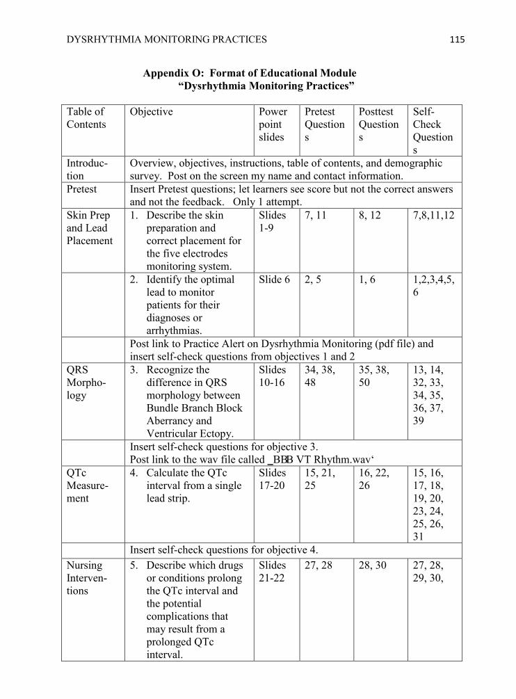
DYSRHYTHMIA MONITORING PRACTICES 115
Appendix O: Format of Educational Module “Dysrhythmia Monitoring Practices”
Table of Contents
Objective Power point slides
Pretest Questions
Posttest Questions
Self-Check Questions
Introduc-tion
Overview, objectives, instructions, table of contents, and demographic survey. Post on the screen my name and contact information.
Pretest Insert Pretest questions; let learners see score but not the correct answers and not the feedback. Only 1 attempt.
Skin Prep and Lead Placement
1. Describe the skin preparation and correct placement for the five electrodes monitoring system.
Slides 1-9
7, 11 8, 12 7,8,11,12
2. Identify the optimal lead to monitor patients for their diagnoses or arrhythmias.
Slide 6
2, 5 1, 6 1,2,3,4,5,6
Post link to Practice Alert on Dysrhythmia Monitoring (pdf file) and insert self-check questions from objectives 1 and 2
QRS Morpho-logy
3. Recognize the difference in QRS morphology between Bundle Branch Block Aberrancy and Ventricular Ectopy.
Slides 10-16
34, 38, 48
35, 38, 50
13, 14, 32, 33, 34, 35, 36, 37, 39
Insert self-check questions for objective 3. Post link to the wav file called ‗BBB VT Rhythm.wav‘
QTc Measure-ment
4. Calculate the QTc interval from a single lead strip.
Slides 17-20
15, 21, 25
16, 22, 26
15, 16, 17, 18, 19, 20, 23, 24, 25, 26, 31
Insert self-check questions for objective 4. Nursing Interven-tions
5. Describe which drugs or conditions prolong the QTc interval and the potential complications that may result from a prolonged QTc interval.
Slides 21-22
27, 28 28, 30 27, 28, 29, 30,

DYSRHYTHMIA MONITORING PRACTICES 116
Table of Contents
Objective Power point slides
Pretest Questions
Posttest Questions
Self-Check Questions
6. Describe nursing interventions for SVT with Bundle Branch Block Aberrancy and for Ventricular Tachycardia.
Slides 23-25
40, 41, 44
40, 42, 45
9, 10, 40, 41, 42, 43, 44, 45
Post link to file of St. Vincent‘s Emergency Standing Orders Directive 3.13 in Clinical Standards Module (file is preferred, not an intranet link, so that they can view it from home) Insert self-check questions for objective 5 and 6.
Case Studies
7. Analyze case studies with wide QRS complex tachycardias on 12 lead electrocardiograms and differentiate between Supraventricular Tachycardia with aberrancy and Ventricular Tachycardia.
N/A N/A N/A 46-53 (case studies) Game: all 10 questions about ECGs
Insert case study questions 46-53. Note that 49 and 53 have instructions to click on each answer choice to see the results of their interventions (if possible!)
Million Dollar Game
Test your knowledge with this game on the main points from the learning module. If you answer all 10 questions correctly, then you ―earn‖ a million dollars! You may take it as many times as you want.
Posttest Insert Posttest. Allow only one attempt. When done, allow them to see score, correct answers, and feedback. Set up program so that they have to view all the slides, self-check questions, case studies, and game before the posttest will open.
References
See references below.
Program Evaluation
Use standard program evaluation for online courses. Make this required before they can print certificate of completion.
Completion Certificate
Printable only after all the components have been completed. They will also need to complete unit-based activities before they get the continuing education certificate from the instructor.

DYSRHYTHMIA MONITORING PRACTICES 117
Appendix P: Audio Script for Educational Module Slide 1 Credits: The power point slides are used with permission from the American Association of Critical Care Nurses. A copy can be downloaded from their website at www.aacn.org The audio and script were added by Susan Schultz using the sources in the reference list. Slide 2 The purpose of this presentation is to discuss the evidenced-based practices for optimal skin preparation, lead placement, ventricular dysrhythmia monitoring, and QT interval measurement. Slide 3 ―Carefully preparing the skin by cleaning the electrode sites with soap and water and drying with a rough washcloth or gauze is worth the extra minutes, because of the time that is saved in responding to false alarms. If the patient is diaphoretic or the skin excessively oily, an alcohol pad may be used, but this may dehydrate and cause increased skin resistance. Excessive hair at electrode sites should be clipped rather than shaved because shaving may cause nicking and irritation‖ (Drew & Funk, 2006, p. 165). Slide 4 Do not place electrodes over large muscle groups, such as deltoids or quadriceps, as this may create a noisy signal. ―The RA and LA electrodes should be placed just below the clavicle and as close as possible to the shoulder. … If arm electrodes are moved medially toward the sternum, more striking changes in waveforms occur, which make the resultant 12-lead ECG invalid. RA and LA electrodes can be placed high on the patient‘s upper back as long as they are close to the respective shoulders‖ (Drew & Funk, 2006, p. 163). ―RL and LL electrodes should be placed on the right and left abdomen below the rib cages. Using this placement, the 12 lead ECG is similar, but not identical, to the standard 12-lead ECG. … It should also be pointed out that the RL electrode [only provides a ground] and does not contribute to the waveform morphology; therefore, it can be placed anywhere on the body‖ (Drew & Funk, 2006, p. 163). Slide 5 When a 12-lead ECG is performed, the precordial leads, also called ―V‖ leads, should be carefully placed in the correct intercostal spaces. If serial ECGs are to be performed, marking the location with indelible ink is advisable to make sure they are placed in the same position every time. If the precordial leads are misplaced by 1 intercostal space, it can change the QRS morphology and ST segment elevation, and possibly lead to misdiagnosis (Drew et al., 2004). Slide 6 ―Lead V1 is considered the best lead for diagnosing right and left bundle-branch block, … and to distinguish ventricular tachycardia from supraventricular tachycardia with aberrant ventricular conduction‖ (Drew et al., 2006, p. 2734)
DYSRHYTHMIA MONITORING PRACTICES 118
―Lead II [is best used] to diagnose atrial activity and measure heart rate‖ because of the upright rounded P waves and tall R waves (AACN Practice Alert, 2008, p. 1). Slide 7 To monitor a patient in V1 with the 5 electrode lead system, the ―C‖ or Chest electrode should be placed in the 4th intercostal space on the Right Sternal Border. The same location is used for MCL1. See next 2 slides for a picture and description of how to accurately locate the 4th intercostal space. Slide 8 The 3-electrode lead system can be used to obtain a Modified Chest Lead 1, which is a bipolar substitute for the V1 lead. As shown in the picture, the RA electrode is moved to the left shoulder and the LA electrode is placed in the 4th ICS, RSB. When Lead I is selected on the monitor, it generates an image from the RA electrode (which is negative) to the LA electrode on the 4th ISC (which is positive). ―MCL1 has been shown to differ in QRS morphology in 40% of patients with ventricular tachycardia and as such is not recommended for diagnosing wide QRS complex tachycardia. Bipolar lead monitoring also is inadequate for ST-segment monitoring because it does not provide multi-lead monitoring or precordial leads, which often are the most sensitive for detecting ischemia‖ (Drew et al., 2004, p. 2734). Slide 9 “The single most important landmark for accurate precordial lead placement is the sternal angle (also referred to as the Angle of Louis). This bony prominence in the upper sternum is caused by fusion of the upper and lower sternum in utero. The sternal angle marked the point where the second rib joins the sternum‖ (Drew & Funk, 2006, p. 164). The best way to find this landmark is a follows: With the patient is in a recumbent position, the suprasternal notch at the base of the throat is located. Just below this notch is the flat part of the upper sternum, which is called the manubrium. When placing the index and middle fingers together with fingertips points laterally, the sternal angel can be felt just below these two fingers. The fingers are next moved up over the body prominence of the sternal angel to just below it. Next the fingers are moved to the right side of the sternum, which corresponds to the second intercostal space. From the second intercostal space, it is easy to palpate down to the third and fourth intercostal spaces. Lead V1 is placed in the fourth intercostal space at the right sternal border‖ (Drew & Funk, 2006, p. 164). Slide 10 Wide QRS complex tachycardias can be caused by ventricular tachycardia or supra-ventricular tachycardia with abnormal conduction through the bundle branches. It is important to differentiate the cause of the wide complex tachycardia, because the treatments are different. In fact, the incorrect medication could cause severe hypotension or loss of consciousness (Urden, Stacy, & Lough, 2010).

DYSRHYTHMIA MONITORING PRACTICES 119
―Contrary to popular belief, hemodynamic stability or instability does not help to differentiate between VT and SVT with a wide QRS complex‖ (Urden et al., 2010, p. 395). Generally, nurses expect patients to remain conscious with an adequate BP during SVT and to become unconscious and pulseless during VT. However, in some cases VT may be well tolerated, especially if the rate is less than 150. And some patients with a rapid SVT may have severe reduction in cardiac output and become hemodynamically unstable. When a patient‘s rhythm strip exhibits a wide QRS complex tachycardia, a 12-lead ECG is better than any single lead and should be obtained (Drew & Funk, 2006; Zipes et al., 2006). But continuous 12-lead ECG monitoring is not always readily available. Fortunately, there are many useful clues about the shape of the QRS (or morphology) that can be seen in V1 or V6. V1 is the preferred lead for continuous monitoring of patients with wide QRSes, but V6 may be used if V1 is unavailable (Jacobson, 2007). The next slide shows examples of each. Slide 11 First, here is a brief review of QRS morphology: ―The letter Q is used to describe an initial negative deflection; in other words, only if the first deflection from the baseline is negative will it be labeled a Q wave. The letter R applies to any positive deflection. If there are two positive deflections in one QRS complex, the second is labeled R prime… The letter S refers to any subsequent negative deflections‖ (Urden et al., 2010, p. 335). The normal QRS in V1 should be less than 0.10 seconds with a small positive r wave and a large negative S wave. All the QRSes pictured on this slide are wider than 0.10 and are considered to have originated in the ventricle (which means they could be PVCs or V Tach). The first QRS in the upper left corner of this diagram is entirely positive with one peak, and that is called a monophasic R wave. The QRS below it is an R wave with two points, and when the first one is taller, it‘s called a ―taller left peak‖, which indicates a ventricular beat. The third and fourth QRSes on the left hand side are biphasic, which means 2 directional. The last QRS on the left side is primarily negative, but it is important to notice that it initially starts with a positive deflective (r wave). An R wave wider than 30 msec (or wider than one little box of 0.04 seconds) is indicative of a ventricular beat. Other indications that the impulse is ventricular in origin is a slurred or notched S on the way down. Both the wide R wave and slurred or notched S descent cause a prolongation from the onset of the QRS to the deepest point of the S (called the nadir). When the nadir is greater than 60 msec (or 0.06 seconds), then it indicates a ventricular beat (Jacobson, 2007). As you can see on the right hand side of the diagram, most of the QRSes seen in V6 are negative. If the wave form is entirely negative, then it is called a either a monophasic Q or a notched QS. Small and large cap letters are used to indicate relative sizes of the wave forms.
DYSRHYTHMIA MONITORING PRACTICES 120
Slide 12 Supra Ventricular Tachycardias with Aberration have different QRS morphologies in leads V1 and V6 than ventricular beats. Aberration means abnormal conduction caused by a bundle branch block or accessory pathway. The QRS measurements are prolonged in both SVT with aberration and VT. However, QRS measurements of less than 0.14 favor aberrant conduction. QRS measurements of 0.16 or greater strongly suggest VT (Urden, et al., 2006). AV dissociation is another clue to help differentiate supraventricular dysrhythmias from ventricular dysrhythmias. SVTs may have visible P waves or atrial flutter waves that are associated with the QRS. When a Premature Atrial Contraction is followed by a wide QRS, that may be a sign of an intermittent BBB which occurred because one of the bundle branches was still refractory when the premature impulse started. One other condition that favors SVT is a ventricular rate over 200 (Jacobson, 2006; Urden et al., 2010). Slide 13 The classic clues for RBBB are a bimodal rR‘ortriphasicrsR‘ (as displayed in left upper side of diagram). Bimodal means the QRS goes in 2 directions. Triphasic means it has 3 deflections from baseline, such as it goes up, then it goes down below baseline, then it goes back up again. When a QRS has more than one positive deflection, then the smaller one is written in lower case r and the taller ones is written upper case, or a capital letter R. An apostrophe or PRIME symbol is usually written after the second R wave. In the lower left side of the diagram the classic clues for LBBB are displayed. This primarily negative wave form is different from ventricular beats in 3 ways: 1) R wave is < 30 msec wide or it has no R wave at all, 2) it has a straight S descent (sometimes called a slick downstroke), and 3) the measurement of the QRS onset to the nadir of the S (deepest point) is less than 60 msec (or 0.06 sec). To be considered LBBB, it should have all three components as well as no Q in Lead V6 (Jacobson, 2007). In V6, the most memorable part of the QRS morphology that represents RBBB is the qRS complex with a ―wide S‖ wave, caused by late right ventricular depolarization. A wide S wave makes the R:S ratio more than 1:1. The intrinsicoid deflection is a measurement from the onset of the QRS to the tallest point of the R wave. When it is < 50 ms, then it favors supraventricular origin (Jacobson, 2007). All the QRS morphologies pictured on this diagram point to dysrhythmias that started above the ventricles and encountered delays as it traveled down the bundle branches, also may also be called SVT with aberration. Slide 14 If you see any of the QRS morphologies described here, then they are not helpful in differentiating between SVT with aberration or ventricular tachycardia. This applies only
DYSRHYTHMIA MONITORING PRACTICES 121
to tachycardias with a positive waveform in V1. Lead II does not provide any helpful QRS morphology and sometimes the QRSes can look identical even when etiology is different. It would be a good idea to recheck the placement of the V electrodes on the patient‘s chest, in particular the Chest lead. When the V lead is placed one intercostal space too high or too low, it can show unhelpful or misleading QRS morphology. Slide 15 The QRSes pictured here are not helpful in determining the origin. Remember to look for the clues in V1 and V6 that were shown on previous slides to help differentiate wide QRS complex tachycardias. When uncertain, a 12-lead ECG is recommended to confirm diagnoses. However, ―There are some tachycardias that do not follow any of the rules and cannot be diagnosed by 12 lead ECG…. An electrophysiology study is needed in such cases to determine for sure the mechanism of the tachycardia‖ (Jacobson, 2007, p. 95). So here is a rhyme to help you remember for all time. When looking at V1…
A slick downstroke or rsR prime, are Bundle Branch Block, so take your time. A notched downstroke or taller left peak, warn of V. tach. Help go seek!
Slide 16 In this example, the lead placement affected the QRS morphology and may have caused delayed recognition of life-threatening ventricular tachycardia. Remember to check the placement of V1 every shift. A good time to do this is when you are listening to your patients‘ heart sounds. If you carry some spare electrodes in your pocket, it will only take a few seconds to correct it (and possibly save the patient‘s life!) Slide 17 ―The QT interval is an approximate measure of the duration of ventricular repolarization. A prolonged QT interval indicates an alteration in cardiac membrane channel function, and can occur as a congenital or acquired disorder‖ (Sommargren & Drew, 2007, p. 285). ―Ideally, the same lead should be used for QT monitoring over time because of variation in QT-interval length across the 12 leads. …. Choose an ECG lead in which the T-wave end is well defined…. Lead II usually has a large positive T wave, and if U waves are present they are more likely to be separated from the T wave, resulting in a clearly defined T-wave end‖ (Sommargren & Drew, 2007, p. 286). The QT interval normally shortens with faster heart rates and lengthens with slower heart rates. Slide 18 The QT Interval is measured from beginning of the QRS complex to the end of the T wave. When the T wave is notched, the end of the T wave should be used as the end of the QT interval. When a U wave occurs after the T wave has reached the baseline, the end of the T wave should be used as the end of the QT interval (exclude the U wave)
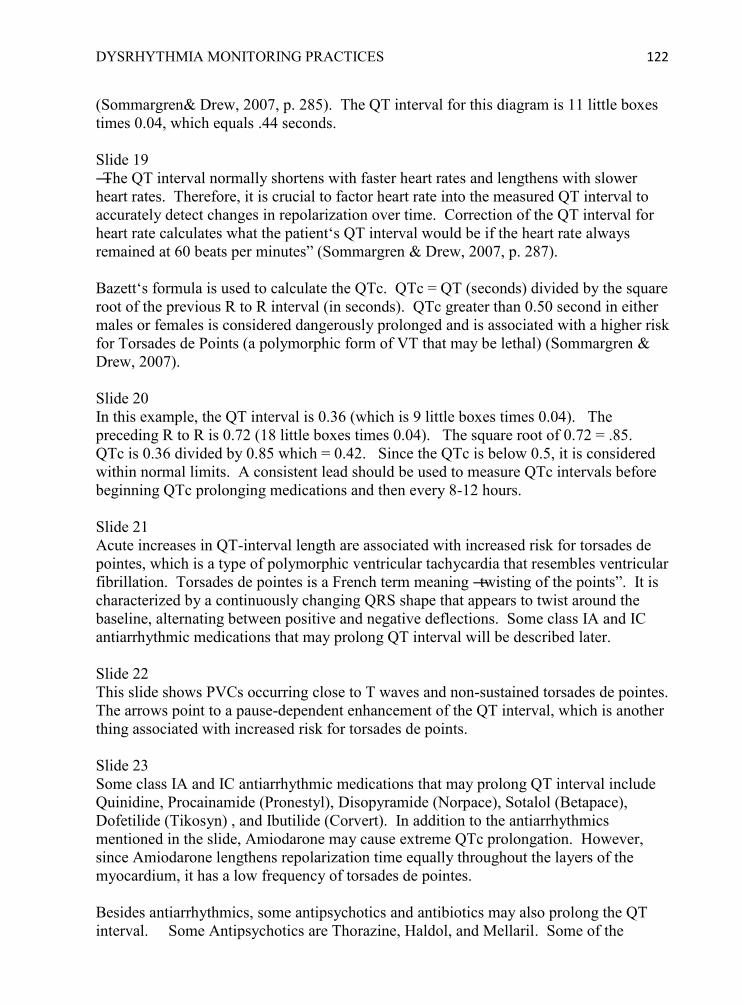
DYSRHYTHMIA MONITORING PRACTICES 122
(Sommargren& Drew, 2007, p. 285). The QT interval for this diagram is 11 little boxes times 0.04, which equals .44 seconds. Slide 19 ―The QT interval normally shortens with faster heart rates and lengthens with slower heart rates. Therefore, it is crucial to factor heart rate into the measured QT interval to accurately detect changes in repolarization over time. Correction of the QT interval for heart rate calculates what the patient‘s QT interval would be if the heart rate always remained at 60 beats per minutes‖ (Sommargren & Drew, 2007, p. 287). Bazett‘s formula is used to calculate the QTc. QTc = QT (seconds) divided by the square root of the previous R to R interval (in seconds). QTc greater than 0.50 second in either males or females is considered dangerously prolonged and is associated with a higher risk for Torsades de Points (a polymorphic form of VT that may be lethal) (Sommargren & Drew, 2007). Slide 20 In this example, the QT interval is 0.36 (which is 9 little boxes times 0.04). The preceding R to R is 0.72 (18 little boxes times 0.04). The square root of 0.72 = .85. QTc is 0.36 divided by 0.85 which = 0.42. Since the QTc is below 0.5, it is considered within normal limits. A consistent lead should be used to measure QTc intervals before beginning QTc prolonging medications and then every 8-12 hours. Slide 21 Acute increases in QT-interval length are associated with increased risk for torsades de pointes, which is a type of polymorphic ventricular tachycardia that resembles ventricular fibrillation. Torsades de pointes is a French term meaning ―twisting of the points‖. It is characterized by a continuously changing QRS shape that appears to twist around the baseline, alternating between positive and negative deflections. Some class IA and IC antiarrhythmic medications that may prolong QT interval will be described later. Slide 22 This slide shows PVCs occurring close to T waves and non-sustained torsades de pointes. The arrows point to a pause-dependent enhancement of the QT interval, which is another thing associated with increased risk for torsades de points. Slide 23 Some class IA and IC antiarrhythmic medications that may prolong QT interval include Quinidine, Procainamide (Pronestyl), Disopyramide (Norpace), Sotalol (Betapace), Dofetilide (Tikosyn) , and Ibutilide (Corvert). In addition to the antiarrhythmics mentioned in the slide, Amiodarone may cause extreme QTc prolongation. However, since Amiodarone lengthens repolarization time equally throughout the layers of the myocardium, it has a low frequency of torsades de pointes. Besides antiarrhythmics, some antipsychotics and antibiotics may also prolong the QT interval. Some Antipsychotics are Thorazine, Haldol, and Mellaril. Some of the

DYSRHYTHMIA MONITORING PRACTICES 123
antibiotics are Erythromycin and Levaquin. Refer to the website for additional medications. The risk is increased if patients are receiving more than 1 drug known to prolong QTc, if they overdose on one of those medications, if they have renal or hepatic failure, or if they take another drug that impairs its metabolism (such as dofetilide and cimetidine). Patients with slow heart rhythms or long pauses, such as complete heart block or sinus pauses, are considered to be at high risk because the initiation of torsades de pointes‘ pause-dependent characteristic. Lastly, it is important to monitor and correct hypokalemia or hypomagnesemia, especially in patients receiving any of the medications known to prolong QT interval. Diuretics and malnutrition also exacerbate electrolyte imbalances. (Sommargren & Drew, 2007) Slide 24 Emergency treatment of torsades de pointes may include IV magnesium, defibrillation, or overdrive pacing (Zipes et al., 2006). If the patient is unstable or nonresponsive, call Code Blue and initiate Emergency Standing Orders. Immediate defibrillation is indicated for pulseless V Tach or V Fib. Emergency Synchronized Cardioversion is indicated for unstable VT with a pulse. For wide complex tachycardias with a pulse, the emergency standing orders allow for Lidocaine to be given. However, in the presence of suspected torsades de points, a Cardiologist should be consulted before administering antiarrhythmics. A Magnesium drip or overdrive pacing should be considered. Other things that the nurse may order per emergency standing orders are STAT 12 Lead ECG (for chest pain or rhythm change) and Chem 7 (for electrolyte levels). It is also recommended that you consult the Rapid Response Nurse if prolongation of QTc occurs or other warning signs of torsades de pointes are suspected. Refer to the Emergency Standing Orders, Directive 3.13 in St. Vincent‘s Clinical Standards Manual (on the intranet). Slide 25 Please read the Emergency Standing Orders, complete all the self-check questions, case studies, and the Million Dollar Game, then proceed to the posttest.

DYSRHYTHMIA MONITORING PRACTICES 124
Appendix Q: Test Bank for Educational Program
Below are questions that will be in the online test pool for pretest, posttest, and self-check questions. Questions may be multiple choice, true or false, and matching.
1. Select the best lead to diagnose atrial activity and measure heart rate.
A. I B. II C. III D. V1
Correct answer: B Feedback: Lead II is best to diagnose atrial activity and measure heart rate because of the upright rounded P waves and tall R waves. Lead V1 is best to diagnose wide QRS complexes, which occur in bundle branch block aberrancy or ventricular rhythms (AACN Practice Alert, 2008, p. 1). 2. Select the best lead to diagnose wide QRS complexes.
A. I B. II C. III D. V1
Correct answer D Feedback: Lead V1 is best to diagnose wide QRS complexes, which occur in bundle branch block aberrancy or ventricular rhythms. Lead II is best to diagnose atrial activity and measure heart rate because of the upright rounded P waves and tall R waves(AACN Practice Alert, 2008, p. 1). 3. An advantage of the 5 lead monitoring system is that it allows for the recording of any of the limb leads plus one precordial (V) lead.
A. True B. False
Correct answer: A Feedback: The 5 lead system is recommended over the 3 lead system for monitoring QRS morphology because it includes on precordial (V) lead (Drew, et al., 2004). 4. An advantage of the 3 lead monitoring system is that it allows for the recording of any of the limb leads plus one precordial (V) lead.
A. True B. False

DYSRHYTHMIA MONITORING PRACTICES 125
Correct answer: B Feedback: The 5 lead system is recommended over the 3 lead system for monitoring QRS morphology because it includes on precordial (V) lead (Drew, et al., 2004). 5. Which of the following patient conditions would be best lead to monitor in V1? (Select all that apply) _____a Atrial fibrillation with intermittent wide QRS complexes _____b First, Second or Third Degree AV heart block _____c Supra-Ventricular Tachycardia with aberrant conduction _____d Sinus Bradycardia or Sinus Rhythm with pauses Correct answers: A, C Feedback: Lead V1 is best to diagnose wide QRS complexes, which occur in bundle branch block aberrant conduction or ventricular rhythms such as PVC, VT, and Torsades de Pointes. Lead II is best to diagnose atrial activity, heart blocks, and measure heart rate because of the upright rounded P waves and tall R waves (AACN Practice Alert, 2008). 6. Which of the following patient conditions would be best lead to monitor in V1? (Select all that apply) _____ Normal Sinus Rhythm with 5 beat run of Ventricular Tachycardia _____ Possible Torsades de Pointes _____ Diagnosing Atrial Flutter versus Atrial Fibrillation _____ Sinus Tachycardia with intermittent right Bundle Branch Block Correct answers: A, B, D Feedback: Lead V1 is best to diagnose wide QRS complexes, which occur in bundle branch block aberrant conduction or ventricular rhythms such as PVC, VT, and Torsades de Pointes. Lead II is best to diagnose atrial activity, heart blocks, and measure heart rate because of the upright rounded P waves and tall R waves (AACN Practice Alert, 2008). 7. Match the description of the electrode placement to the correct electrode. _____ below rib cage on left side of abdomen _____ infra-clavicular fossa close to left shoulder _____ anywhere on torso, usually lower right side of chest or abdomen _____ 4th Inter Costal Space, Right Sternal Border _____ infra-clavicular fossa close to right shoulder A. RA B. LA C. C D. LL E. RL
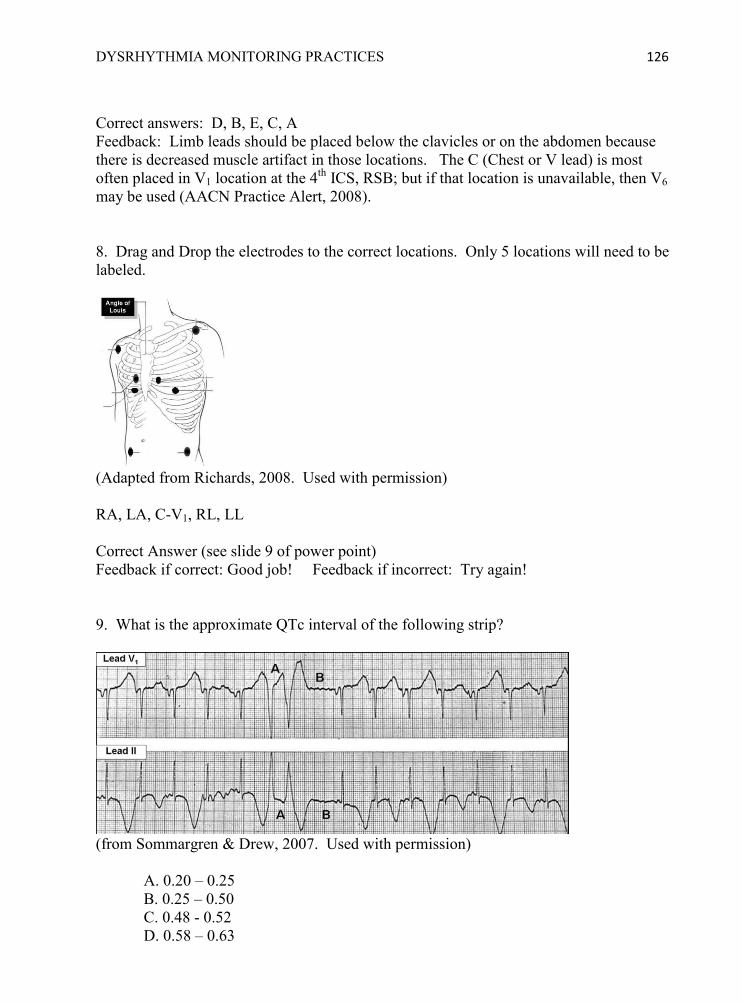
DYSRHYTHMIA MONITORING PRACTICES 126
Correct answers: D, B, E, C, A Feedback: Limb leads should be placed below the clavicles or on the abdomen because there is decreased muscle artifact in those locations. The C (Chest or V lead) is most often placed in V1 location at the 4th ICS, RSB; but if that location is unavailable, then V6 may be used (AACN Practice Alert, 2008). 8. Drag and Drop the electrodes to the correct locations. Only 5 locations will need to be labeled.
(Adapted from Richards, 2008. Used with permission) RA, LA, C-V1, RL, LL Correct Answer (see slide 9 of power point) Feedback if correct: Good job! Feedback if incorrect: Try again! 9. What is the approximate QTc interval of the following strip?
(from Sommargren & Drew, 2007. Used with permission)
A. 0.20 – 0.25 B. 0.25 – 0.50 C. 0.48 - 0.52 D. 0.58 – 0.63

DYSRHYTHMIA MONITORING PRACTICES 127
Correct answer = D Feedback: QTc is prolonged >.50 second. ―ECG signs of impending torsades de pointes in a patient with QTc over .50 are (A) a ventricular couplet, followed by (B) a compensatory pause. Also note T-wave alternans, which is most apparent in the Lead II rhythm strip‖ (Sommargren & Drew, 2007, p. 290). 10. The same patient from the previous question exhibited this change in rhythm. Interpret the following dysrhythmia.
(from Sommargren & Drew, 2007. Used with permission)
A. SVT with aberration B. Monomorphic ventricular tachycardia C. Torsades de Pointes D. Ventricular Fibrillation
Correct answer C Feedback: ―In a subsequent rhythm strip from the patient shown in previous questions. (A) a ventricular couplet, followed by (B) a compensatory pause; (C) torsades de pointes is triggered on the T wave following the pause‖ (Sommargren & Drew, 2007, p. 290). Note the alternating direction of the QRS complexes, initially negative deflection, then ―twisting‖ to positive deflection, and ―twisting‖ again back to negative. 11. What is the best way to prepare the skin before placing electrodes?
A. Clip excessive hair and clean skin with alcohol or washcloth before placing electrodes. B. Clean skin with soap and water only if there is visible sweat, oil, or powder. C. Apply electrodes; if artifact noted, then clean the skin and replace electrodes. D. If chest is hairy, shave with shaving cream and razor, dry thoroughly, then apply electrodes.
Correct answer: A

DYSRHYTHMIA MONITORING PRACTICES 128
Feedback: Sweat, oil, lotion, powder, and dead skin cells may not be visible, so the skin should be routinely cleaned before applying electrodes. Shaving skin with a razor can cause irritation, so it is better to clip the excess hair (Drew & Funk, 2006).
12. Place the steps for applying electrodes in the proper order.
A. Apply new electrodes and press firmly to skin B. Clip excessive hair from selected locations C. Visually select locations without implanted devices, dressings, or muscle artifact that could interfere with monitoring D. Clean skin with alcohol or washcloth and dry thoroughly
Correct order: C, B, D, A Feedback: Sweat, oil, lotion, powder, and dead skin cells may not be visible, so the skin should be routinely cleaned before applying electrodes. Shaving skin with a razor can cause irritation, so it is better to clip the excess hair (Drew & Funk, 2006). 13. When the V1 electrode is misplaced by as little as one intercostal space, QRS morphology can change and misdiagnosis may occur.
A. True B. False
Correct answer: A Feedback: Inaccurate lead placement may produce QRS morphology that is unhelpful or misleading in differentiating SVT with aberrant conduction or Ventricular Tachycardia (Drew, et al., 2004). 14. When the V1 electrode is placed one intercostal space above or below the preferred location, QRS morphology will be unchanged.
A. True B. False
Correct answer: B Feedback: Inaccurate lead placement may produce QRS morphology that is unhelpful or misleading in differentiating SVT with aberrant conduction or Ventricular Tachycardia (Drew et al., 2004). 15. Calculate the QT, R-R, and QTc for this strip (√ = square roots. Accurate divisions have been provided in the answers; you just need to select the correct intervals)

DYSRHYTHMIA MONITORING PRACTICES 129
(From Sommargren & Drew, 2007. Used with permission.)
A. QT = .24, R-R = .48; √.48 = .693; QTc = .24 / .693 = .35 seconds B. QT = .36, R-R = .60; √.6 = .775; QTc = .36 / .775 = .46 seconds C. QT = .44, R-R = .80; √.8 = .894; QTc = .44 / .894 = .49 seconds D. QT = .56, R-R = .80; √8 = .894; QTc = .56 / .894 = .63 seconds
Correct answer: C Feedback: QT is 11 boxes x .04. Preceding R to R is 20 boxes x .04 16. Calculate the QT and QTc interval for this strip. (√ = square roots. Accurate divisions have been provided in the answers; you just need to select the correct intervals)
(From Drew, 2007. Used with permission.)
A. QT = .24, R-R = .48; √.48 = .693; QTc = .24 / .693 = .35 seconds B. QT = .40, R-R = .80; √.8 = .894. QTc = .40 / .894 = .48 seconds C. QT = .44, R-R = .60; √.6 = .775; QTc = .44 / .775 = .57 seconds D. QT = .56, R-R = .80; √8 = .894; QTc = .56 / .894 = .63 seconds
Correct answer B Feedback QT is 10 boxes x 0.04; preceeding R to R is 20 boxes x 0.04 17. The QT interval is measured from beginning of QRS to end of T wave
A. True B. False

DYSRHYTHMIA MONITORING PRACTICES 130
Correct answer: A Feedback: The QT interval is an approximate measure of the duration of ventricular repolarization and is measured from beginning of QRS to end of T wave. (Sommargren & Drew, 2007) 18. The QT interval is measured from beginning of QRS to beginning of T wave.
A. True F. False
Correct answer: B Feedback: The QT interval is an approximate measure of the duration of ventricular repolarization and is measured from beginning of QRS to end of T wave (Sommargren & Drew, 2007). 19. The QTinterval lengthens with bradycardia and shortens with tachycardia.
A. True B. False
Correct answer: A Feedback: QT shortens with tachycardia because repolarization is quicker (Sommargren & Drew, 2007).
20. The QT interval shortens with bradycardia and lengthens with tachycardia.
A. True B. False
Correct answer: B Feedback: QT shortens with tachycardia because repolarization is quicker (Sommargren & Drew, 2007). 21. Which of the following are true statements (select all that apply).
A. QT interval is measured from beginning of QRS to end of T wave B. QTc interval is the QRS interval plus the T wave C. QT interval lengthens with bradycardia and shortens with tachycardia. D. QT interval shortens with bradycardia and lengthens with tachycardia. E. QTcinterval is the QT interval corrected for heart rate
Correct answer: A, C, E Feedback: The QT interval is an approximate measure of the duration of ventricular repolarization and is measured from beginning of QRS to end of T wave. QT shortens with tachycardia because repolarization is quicker, which makes it necessary to correct the QT interval for the heart rate (Sommargren & Drew, 2007).

DYSRHYTHMIA MONITORING PRACTICES 131
22. Which of the following are true statements (select all that apply). A. QTc interval is the QRS interval plus the T wave B. QT interval shortens with bradycardia and lengthens with tachycardia. C. QTcinterval is the QT interval corrected for heart rate D. QT interval is measured from beginning of QRS to end of T wave E. QT interval lengthens with bradycardia and shortens with tachycardia.
Correct answer: C, D, E Feedback: The QT interval is an approximate measure of the duration of ventricular repolarization and is measured from beginning of QRS to end of T wave. QT shortens with tachycardia because repolarization is quicker, which makes it necessary to correct the QT interval for the heart rate (Sommargren & Drew, 2007). 23. The QTcinterval is the QT interval corrected for heart rate.
A. True B. False
Correct answer: A Feedback: The QTc interval is the QT interval corrected for heart rate because QT intervals shorten when heart rate is faster (Sommargren & Drew, 2007). 24. The QTc interval is the QRS interval plus the T wave.
A. True B. False
Correct answer: B Feedback: The QTc interval is the QT interval corrected for heart rate because QT intervals shorten when heart rate is faster (Sommargren & Drew, 2007). 25. What QTc interval is associated with Torsades de Pointes?
A. > 0.20 sec (200 msec) B. > 0.24 sec (240 msec) C. > 0.50 sec (500 msec) D. < 0.12 sec (120 msec)
Correct answer: C Feedback: QTc interval greater than 0.50 sec (500 msec) is dangerously prolonged and associated with risk for Torsades de Pointes (Sommargren & Drew, 2007). 26. What QTc interval is associated with Torsades de Pointes?
A. < 0.12 sec (120 msec) B. > 0.20 sec (200 msec) C. > 0.24 sec (240 msec) D. > 0.50 sec (500 msec)

DYSRHYTHMIA MONITORING PRACTICES 132
Correct answer: D Feedback: QTc interval greater than 0.50 sec (500 msec) is dangerously prolonged and associated with risk for Torsades de Pointes (Sommargren & Drew, 2007). 27. Which drugs may prolong the QTc?
A. Antiarrhythmics (Procainamide/Pronestyl, Sotalol/Betapace) B. Antipsychotics (Chlorpromazine/Thorazine, Haloperidol/Haldol) C. Antibiotics (Erythromycin/EES, Levofloxacin/Levaquin) D. All of the above
Correct answer: D Feedback: Selected antiarrhythmics, antipsychotics, and antibiotics are some of the drugs that can increase risk of prolonged QTc (Sommargren & Drew, 2007). 28. Which of the following conditions should have QTc interval monitoring?
A. New onset or severe bradycardia B. Hypokalemia or hypomagnesemia C. Overdose on potentially prodysrhythmic medications D. All of the above
Correct answer: D Feedback: Severe bradycardia, hypokalemia or hypomagnesemia, and overdose on potentially prodysrhythmic medications can all potentiate prolongation of QT interval (Sommargren & Drew, 2007). 29. Which of the following patients should have QTc interval monitoring? (select all that apply)
A. 58 year-old male with acute MI and on Lidocaine drip 2 mg/min, Potassium 4.0 B. 36 year-old female admitted with overdose on Chlorpromazine/Thorazine
C. 54 year-old female with new onset atrial fibrillation and started on oral Sotalol/Betapace
D. 49 year-old alcoholic male with cardiomyopathy taking Quinidine and has Magnesium of 1.8
E. 75 year-old female admitted with pneumonia and started on Levofloxacin/Levaquin
Correct answer: B, C, D, E Feedback: Medication (certain antiarrhythmics, antipsychotics, and antibiotics), hypokalemia or hypomagnesemia, and overdose on potentially prodysrhythmic medications can all potentiate prolongation of QT interval. Lidocaine and a normal potassium level do not elevate risk of Torsades de Pointes (Sommargren & Drew, 2007). 30. Which of the following patients should have QTc interval monitoring? (select all that apply)
A. 75 year-old female admitted with pneumonia and started on Erythromycin (EES)
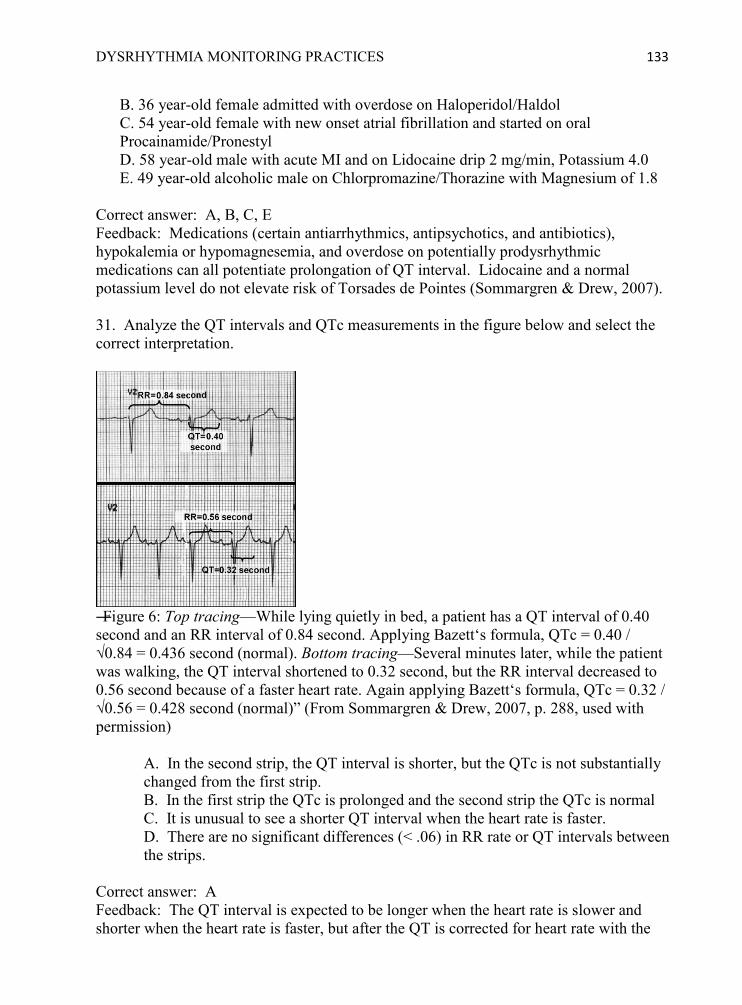
DYSRHYTHMIA MONITORING PRACTICES 133
B. 36 year-old female admitted with overdose on Haloperidol/Haldol C. 54 year-old female with new onset atrial fibrillation and started on oral Procainamide/Pronestyl D. 58 year-old male with acute MI and on Lidocaine drip 2 mg/min, Potassium 4.0 E. 49 year-old alcoholic male on Chlorpromazine/Thorazine with Magnesium of 1.8
Correct answer: A, B, C, E Feedback: Medications (certain antiarrhythmics, antipsychotics, and antibiotics), hypokalemia or hypomagnesemia, and overdose on potentially prodysrhythmic medications can all potentiate prolongation of QT interval. Lidocaine and a normal potassium level do not elevate risk of Torsades de Pointes (Sommargren & Drew, 2007). 31. Analyze the QT intervals and QTc measurements in the figure below and select the correct interpretation.
―Figure 6: Top tracing—While lying quietly in bed, a patient has a QT interval of 0.40 second and an RR interval of 0.84 second. Applying Bazett‘s formula, QTc = 0.40 / √0.84 = 0.436 second (normal). Bottom tracing—Several minutes later, while the patient was walking, the QT interval shortened to 0.32 second, but the RR interval decreased to 0.56 second because of a faster heart rate. Again applying Bazett‘s formula, QTc = 0.32 / √0.56 = 0.428 second (normal)‖ (From Sommargren & Drew, 2007, p. 288, used with permission)
A. In the second strip, the QT interval is shorter, but the QTc is not substantially changed from the first strip. B. In the first strip the QTc is prolonged and the second strip the QTc is normal C. It is unusual to see a shorter QT interval when the heart rate is faster. D. There are no significant differences (< .06) in RR rate or QT intervals between the strips.
Correct answer: A Feedback: The QT interval is expected to be longer when the heart rate is slower and shorter when the heart rate is faster, but after the QT is corrected for heart rate with the
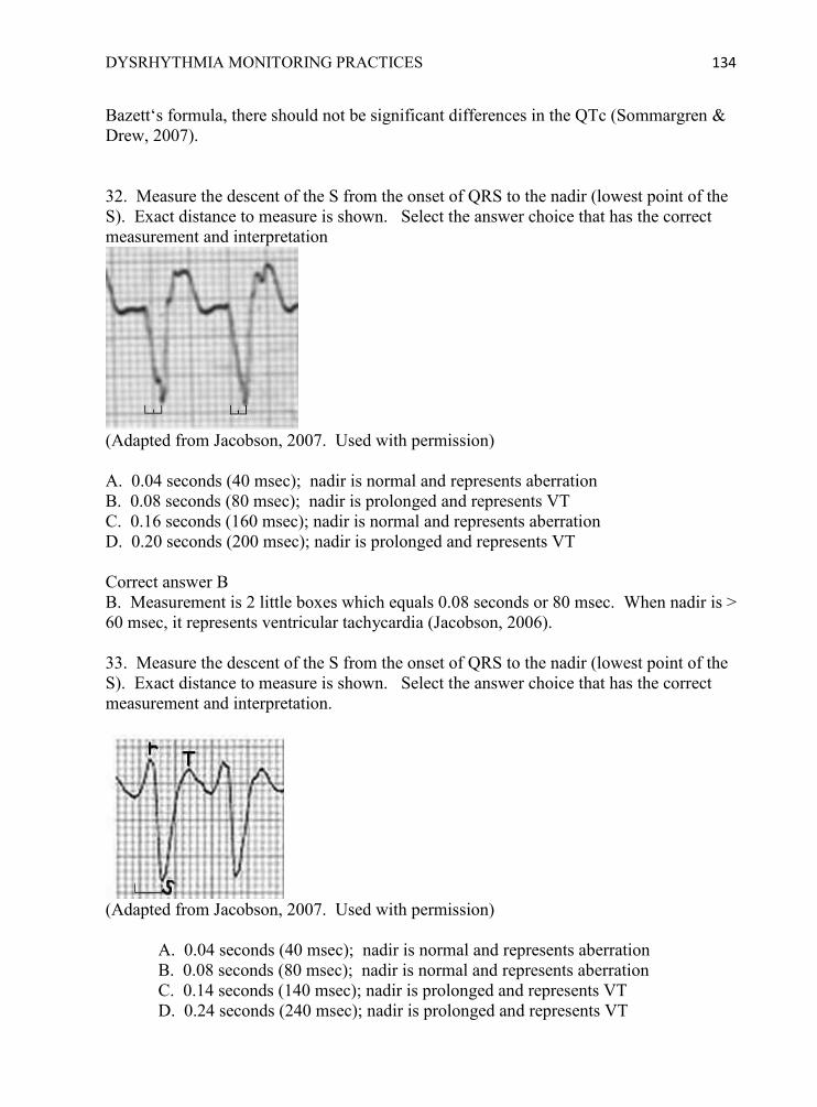
DYSRHYTHMIA MONITORING PRACTICES 134
Bazett‘s formula, there should not be significant differences in the QTc (Sommargren & Drew, 2007). 32. Measure the descent of the S from the onset of QRS to the nadir (lowest point of the S). Exact distance to measure is shown. Select the answer choice that has the correct measurement and interpretation
(Adapted from Jacobson, 2007. Used with permission) A. 0.04 seconds (40 msec); nadir is normal and represents aberration B. 0.08 seconds (80 msec); nadir is prolonged and represents VT C. 0.16 seconds (160 msec); nadir is normal and represents aberration D. 0.20 seconds (200 msec); nadir is prolonged and represents VT Correct answer B B. Measurement is 2 little boxes which equals 0.08 seconds or 80 msec. When nadir is > 60 msec, it represents ventricular tachycardia (Jacobson, 2006). 33. Measure the descent of the S from the onset of QRS to the nadir (lowest point of the S). Exact distance to measure is shown. Select the answer choice that has the correct measurement and interpretation.
(Adapted from Jacobson, 2007. Used with permission)
A. 0.04 seconds (40 msec); nadir is normal and represents aberration B. 0.08 seconds (80 msec); nadir is normal and represents aberration C. 0.14 seconds (140 msec); nadir is prolonged and represents VT D. 0.24 seconds (240 msec); nadir is prolonged and represents VT

DYSRHYTHMIA MONITORING PRACTICES 135
Correct answer C Feedback: Measurement is 3 ½ little boxes which equals 0.14 seconds (140 msec). When nadir is > 60 msec, it represents ventricular tachycardia (Jacobson, 2007). 34. What does a triphasicrsR‘ morphology (shown below) in V1 indicate?
(Adapted from Jacobson, 2007. Used with permission)
A. RBBB aberration B. LBBB aberration C. Ventricular beat D. None of the above
Correct answer: A Feedback: RBBB in V1 shows a classic bimodal rR‘ or triphasicrsR‘ morphology (Jacobson, 2007). 35. Measure the descent of the S from the onset of QRS to the nadir (lowest point of the S). Exact distance to measure is shown. Select the answer choice that has the correct measurement and interpretation.
(Adapted from Jacobson, 2007. Used with permission)
A. 0.04-0.06 seconds (40-60 msec); nadir is normal and represents LBBB aberration B. 0.10 seconds (100 msec); nadir is normal and represents RBBB aberration C. 0.14 seconds (140 msec); nadir is prolonged and represents VT D. 0.24 seconds (240 msec); nadir is prolonged and represents VT
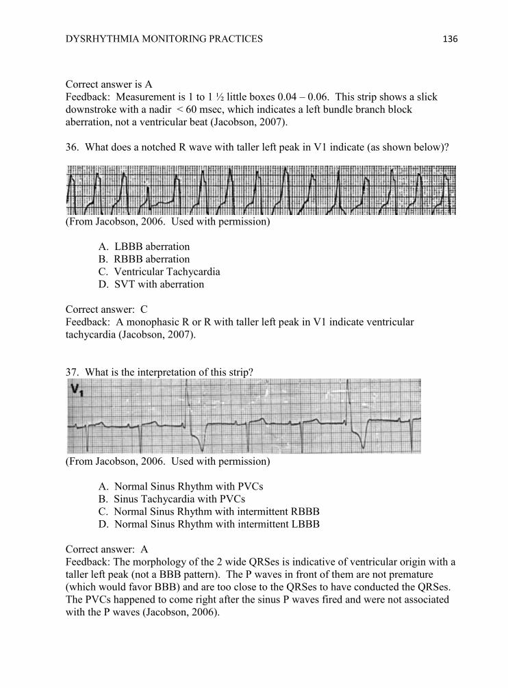
DYSRHYTHMIA MONITORING PRACTICES 136
Correct answer is A Feedback: Measurement is 1 to 1 ½ little boxes 0.04 – 0.06. This strip shows a slick downstroke with a nadir < 60 msec, which indicates a left bundle branch block aberration, not a ventricular beat (Jacobson, 2007). 36. What does a notched R wave with taller left peak in V1 indicate (as shown below)?
(From Jacobson, 2006. Used with permission)
A. LBBB aberration B. RBBB aberration C. Ventricular Tachycardia D. SVT with aberration
Correct answer: C Feedback: A monophasic R or R with taller left peak in V1 indicate ventricular tachycardia (Jacobson, 2007). 37. What is the interpretation of this strip?
(From Jacobson, 2006. Used with permission)
A. Normal Sinus Rhythm with PVCs B. Sinus Tachycardia with PVCs C. Normal Sinus Rhythm with intermittent RBBB D. Normal Sinus Rhythm with intermittent LBBB
Correct answer: A Feedback: The morphology of the 2 wide QRSes is indicative of ventricular origin with a taller left peak (not a BBB pattern). The P waves in front of them are not premature (which would favor BBB) and are too close to the QRSes to have conducted the QRSes. The PVCs happened to come right after the sinus P waves fired and were not associated with the P waves (Jacobson, 2006).

DYSRHYTHMIA MONITORING PRACTICES 137
38. Match the strip with the correct interpretation using QRS morphology and AV dissociation cues.
(From Jacobson, 2006. Used with permission)
A. Sinus rhythm with 2 pairs of PVCs. B. Sinus tachycardia with LBBB aberration. C. Ventricular Tachycardia (positive deflection). D. Ventricular Tachycardia (negative deflection)
Correct answers: 1=D, 2=B, 3=C, 4=A Feedback: 1: QRS morphology shows slurred or notched S decent, typical of VT. ―AV dissociation is clearly present throughout the strip‖ 2: QRS is typical of LBBB morphology with straight S descent. ―P waves can be seen near the end of the T wave and preceding every QRS complex‖ 3. Although it is in Lead II and QRS morphology is not helpful, ―independent P waves can be seen indicating AV dissociation, and the 8th beat is a fusion beat‖ 4: Monophasic shape of wide QRSs is typical of VT. ―The first beat of the second pair is a fusion beat. The P wave preceding the fusion beat is not premature; it is the normal sinus P that happens to precede an end-diastolic PVC, resulting in fusion‖ (Jacobson, 2006, p. 465).

DYSRHYTHMIA MONITORING PRACTICES 138
39. Study the QRS morphologies below and choose the WRONG statement.
(From Jacobson, 2007. Used with permission)
A. LBBB in V1 shows all of the following: R < 30 ms or no R, straight S descent, and QRS onset to S nadir < 60 ms B. RBBB in V1 shows a classic bimodal rR‘ or triphasicrsR‘ morphology C. LV Tachycardia in V1 shows a monophasic R or an R with a taller left peak D. Lead II provides useful QRS morphology clues to differentiate BBB and VT. E. RV Tachycardia in V1 shows R > 30ms, or Slurred or notched S descent, or QRS onset to S nadir >60 ms
Correct answer: D Feedback: D is wrong because Lead II does NOT provide useful clues. In this example all the QRSes in Lead II look similar and have a negative deflection (Jacobson, 2007). 40. What treatments have been shown to be effective for Torsades de Points?
A. IV Magnesium B. Defibrillation C. Overdrive pacing D. All of the above
Correct answer: D Feedback: IV magnesium, defibrillation, and overdrive pacing have all been shown to be effective in Torsades de Points (Zipes, et al., 2006). 41. What are appropriate treatments for wide-complex ventricular tachycardia without a pulse (per hospital Emergency Standing Orders)? (select all that apply)
DYSRHYTHMIA MONITORING PRACTICES 139
A. Call Code Blue and begin CPR B. Defibrillate immediately with 360 joules or equivalent with a biphasic defibrillator C. Give drug trial of Lidocaine before defibrillating. D. Initial external pacemaker at rate of 60-80 with milliamps 10% above the threshold
Correct answer: A, B Feedback: Pulselses VT and VF should be treated with Code Blue and immediate defibrillation (See Directive 3.13, St. Vincent‘s Clinical Standard Manual). 42. What treatment is indicated for unstable Ventricular Tachycardia with a pulse (per hospital Emergency Standing Orders)?
A. IV Amiodarone bolus and drip B. Defibrillation with 360 joules or equivalent with a biphasic defibrillator C. Synchronized Cardioversion D. All of the above
Correct answer: C Feedback: For unstable wide-complex tachycardias with a pulse, Emergency Standing Orders list vagal maneuvers, Lidocaine and emergency synchronized cardioversion. (See Directive 3.13, St. Vincent‘s Clinical Standard Manual). 43. What treatments may be indicated for Supra-Ventricular Tachycardia with a narrow QRS complex, rate 168, BP 90/50 (per hospital Emergency Standing Orders)? (Select all that apply)
A. Ask patient to cough and bear down B. Give Lidocaine 1-1.5 mg/kg IVP C. Prepare for emergency synchronized cardioversion D. Defibrillation with 360 joules or equivalent with a biphasic defibrillator
Correct answers: A, C Feedback: For unstable SVT ( narrow complex with a pulse) vagal maneuvers, Adenosine IVP, and synchronized cardioversion may be indicated. (See Directive 3.13, St. Vincent‘s Clinical Standard Manual).

DYSRHYTHMIA MONITORING PRACTICES 140
44. Which intervention is the most important for the nurse to initiate first when a new wide QRS complex tachycardia is identified by the monitor tech?
A. Look at the strip with the monitor tech B. Order STAT 12 Lead ECG C. Call the Rapid Response Nurse D. Assess the patient
Correct answer: D Feedback: The most important intervention is to assess how the patient is tolerating the rhythm (check LOC, pulse, and blood pressure) because the findings will guide the next actions. 45. Which intervention is the most important for the nurse to initiate first when a new wide QRS complex tachycardia is identified by the monitor tech?
A. Assess the patient B. Look at the strip with the monitor tech C. Ask Unit Clerk to call for STAT 12 Lead ECG D. Call Code Blue
Correct answer: A Feedback: The most important intervention is to assess how the patient is tolerating the rhythm (check LOC, pulse, and blood pressure) because the findings will guide the next actions.
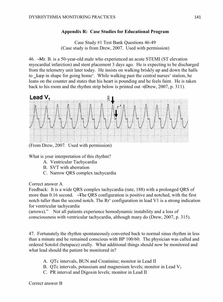
DYSRHYTHMIA MONITORING PRACTICES 141
Appendix R: Case Studies for Educational Program
Case Study #1 Test Bank Questions 46-49 (Case study is from Drew, 2007. Used with permission)
46. ―Mr. B. is a 50-year-old male who experienced an acute STEMI (ST elevation myocardial infarction) and stent placement 3 days ago. He is expecting to be discharged from the telemetry unit later today. He insists on walking briskly up and down the halls to ‗keep in shape for going home‘. While walking past the central nurses‘ station, he leans on the counter and states that his heart is pounding and he feels faint. He is taken back to his room and the rhythm strip below is printed out ―(Drew, 2007, p. 311).
(From Drew, 2007. Used with permission) What is your interpretation of this rhythm?
A. Ventricular Tachycardia B. SVT with aberration C. Narrow QRS complex tachycardia
Correct answer A Feedback: It is a wide QRS complex tachycardia (rate, 188) with a prolonged QRS of more than 0.16 second. ―The QRS configuration is positive and notched, with the first notch taller than the second notch. The Rr‘ configuration in lead V1 is a strong indication for ventricular tachycardia (arrows).‖ Not all patients experience hemodynamic instability and a loss of consciousness with ventricular tachycardia, although many do (Drew, 2007, p. 315). 47. Fortunately the rhythm spontaneously converted back to normal sinus rhythm in less than a minute and he remained conscious with BP 100/60. The physician was called and ordered Sotolol (betapace) orally. What additional things should now be monitored and what lead should the patient be monitored in?
A. QTc intervals, BUN and Creatinine; monitor in Lead II B. QTc intervals, potassium and magnesium levels; monitor in Lead V1 C. PR interval and Digoxin levels; monitor in Lead II
Correct answer B

DYSRHYTHMIA MONITORING PRACTICES 142
Feedback: Now that Mr B. has been started on a drug that can prolong ventricular repolarization and lead to torsades de pointes, the nurse should document the baseline QTc interval before sotolol is started and at least every 8 to 12 hours thereafter (Drew, 2007, p. 315). (put on new slide) QTc Review: ―If Mr. B. has an increase of 0.06 second or more in his QTc interval from the predrug to the postdrug period, the nurse should be concerned and watch for QT-related arrhythmias such as polymorphic ventricular premature beats and couplets, T wave alternans, and nonsustainedtorsades de pointes. If the QTc interval lengthens to 0.50 second or more, the nurse should notify the physician to consider stopping the offending drug (sotolol). The nurse should also make sure Mr B. does not have Hypokalemia and that he is not having severe bradycardia or long pauses, which could trigger torsades de pointes and cardiac arrest‖ (Drew, 2007, p. 315). 48. Mr. B.‘s cardiac monitor arrhythmia alarm sounds and the following tracing is automatically printed out. What is your interpretation of the rhythm?
(From Drew, 2007. Used with permission)
A. SVT with aberration B. Ventricular tachycardia, monophasic C. Torsades de Pointes
Correct answer C Feedback: ―Episodes of drug-induced torsades de pointes. The first beat on the top tracing is a sinus beat followed by 3 polymorphic ventricular premature beats. The beat after the pause is a junctional escape beat that is dissociated from the sinus P wave that just precedes it. Then, nonsustainedTorsades de Pointes occurs. It has the characteristic ‗twisting of the points‘ pattern (i.e., QRS complexes point in one direction initially and
DYSRHYTHMIA MONITORING PRACTICES 143
then switch to the opposite direction [arrows]). Another episode occurs on the bottom rhythm strip. The QT interval measures 0.64 second; the RR interval measures 0.96 second. The QTC is 0.65 second, which is more than the critical threshold of 0.50 second. This rhythm strip indicates that Mr. B. is at a high risk for developing sustained torsades de pointes and cardiac arrest unless the nurse acts immediately‖ (Drew 2007, p. 315). 49. What should the nurse do? (click on each answer choice to see the results)
A. Assess the patient, order STAT 12 lead ECG, and notify Rapid Response Nurse or M.D. B. Assess the patient, ask him to cough or bear down, administer Adenosine 6 mg IVP for narrow complex tachycardia per Emergency Standing Orders. C. No intervention is necessary because he is already on antiarrhythmic medication and the arrhythmia was not sustained. D. When in danger, when in doubt, run in circles, scream and shout.
Correct answer A Feedback for Answer A: Mr. B‘s BP remained alert and responsive with a BP above 100/60. A 12-lead ECG confirmed prolongation of QTc above 0.50. A Magnesium drip was ordered and begun. Sotolol was discontinued, and within 24 hours he had no further ventricular arrhythmias and was discharged. The nurse was commended for her prompt recognition of Torsades de Pointe and taking corrective actions. Feedback for Answer B: Mr. B‘s BP remained alert and responsive with a BP above 100/60. Immediately after IV Adenosine given, the patient went into third degree AV block and ventricular standstill, so a Code Blue was called. He was successfully revived and transferred to ICU. The nurse was required to complete a remediation program on arrhythmia interpretation and treatment. Feedback for Answer C: Thirty minutes later the monitor alarms sounded again and this time the Torsades de Pointes deteriorated into ventricular fibrillation and cardiac arrest. A Code Blue was called but the patient died. The nurse was required to complete a remediation module on arrhythmia interpretation and treatment. Feedback for Answer D: The other nurses came to see what all the screaming was about, assessed the patient, and called the Rapid Response Nurse, who initiated appropriate treatment. The nurse who was running in circles and screaming was later fired.

DYSRHYTHMIA MONITORING PRACTICES 144
Case Study #2 Test Bank Questions 50-53 (From Pelter & Carey, 2006. Used with permission)
“Scenario: This ECG was obtained in a 79-year-old woman admitted to the intensive care unit for acute exacerbation of chronic obstructive pulmonary disease and pneumonia treated with erythromycin. The bedside monitor alarmed for ―V Tach.‖ The nurse could not assess the patient‘s mental status because the patient was intubated and sedated with propofol. The patient‘s blood pressure was 109/48 mm Hg and her pulse oximeter reading was 93%. The rhythm spontaneously returned to normal sinus rhythm after 30 seconds‖ (Pelter & Carey, 2006, p. 437).
(From Pelter & Carey, 2006. Used with permission) 50. What is your interpretation of this rhythm?
A. SVT with aberration B. Ventricular tachycardia, monophasic C. Torsades de pointes (TdP)
Correct Answer C. Feedback: ―The strip begins with a ventricular triplet. These complexes are further labeled as multiform because of the different QRS morphologies. The fourth beat is a normal sinus beat with a long QT interval (0.64 seconds). This beat is followed by an R-on-T premature ventricular contraction (PVC) inducing torsades de pointes (TdP)‖ (Pelter & Carey, 2006, p. 438). 51. Select the best lead to monitor this patient‘s dysrhythmia.
A. I B. II C. III D. V1

DYSRHYTHMIA MONITORING PRACTICES 145
Correct Answer D Feedback: Lead V1 is best to diagnose wide QRS complexes, which occur in bundle branch block aberrancy or ventricular rhythms. Lead II is best to diagnose atrial activity and measure heart rate because of the upright rounded P waves and tall R waves (AACN Practice Alert, 2008, p. 1) 52. What predisposing factors did this patient have that increased her risk of developing torsades de pointes (TdP)? (select all that apply)
A. Chronic obstructive pulmonary disease and pneumonia B. Erythromycin C. Sedated with propofol D. Probably prolonged QTc (as seen with the QT in the one recorded sinus beat).
Correct answers: B and D Feedback: ―One drug that can cause TdP is erythromycin, a likely cause in this patient. Importantly, because the QTc interval is typically longer in women than in men, women may be more vulnerable to the effects of drugs that prolong the QT interval. This example shows the characteristic onset of TdP immediately preceding the arrhythmia; ventricular ectopy (3 beats in this example), followed by a pause, then a sinus beat, and finally an R-on-T PVC‖ (Pelter & Carey, 2006, p. 438). 53. What are the appropriate nursing actions that should be done for this patient? (click on each answer choice to see the results)
A. Give next scheduled dose of erythromycin early to counteract the pneumonia. B. Hold sliding scale Potassium if K is 3.5 mEq until Creatinine levels can be checked C. Call a Code Blue and defibrillate immediately with 360 joules D. Assess the patient, hold erythromycin, check electrolyte levels, notify Rapid Response Nurse and M.D, and put crash cart outside the room.
Correct answer D. Feedback for A: About 15 minutes after giving erythromycin, the patient goes back into Torsades de Pointes and this time it progresses to Ventricular Fibrillation. A Code Blue is called but the patient dies. The nurse was later fired. Feedback for B: About an hour later the patient goes back into torsades de pointes and this time she is symptomatic with BP of 80/50. A Code Blue is called, emergency synchronized cardioversion is done, and a Magnesium drip is begun. The patient remained in ICU for several more days and was finally discharged one week later. The nurse was required to complete a remediation module on arrhythmia interpretation and treatment.

DYSRHYTHMIA MONITORING PRACTICES 146
Feedback for C: After you call for help and attach the debrillation patches to her chest, another nurse yells at you, ―What are you doing? Don‘t shock her! She‘s back in Normal Sinus Rhythm!‖ The Code Blue is cancelled and a Cardiology consult is ordered. Erythromycin is discontinued and a new antibiotic started. Magnesium and potassium boluses are given. The nurse was required to complete a remediation module on arrhythmia interpretation and treatment. Feedback for D: Correct! ―Because TdP can quickly degenerate into ventricular fibrillation, the crash cart should be readily available for immediate defibrillation, offending agent such as the erythromycin should be D/C, and magnesium and antiarhythmic drugs should be given. The nurse should also carefully measure the QT interval as per the unit protocol‖ (Pelter & Carey, 2006). The nurse was commended for her prompt recognition of torsades de pointes and was later promoted to Assistant Nurse Manager.
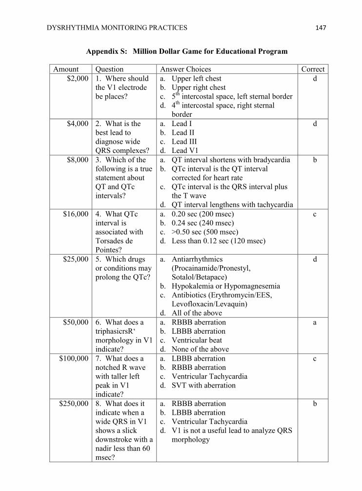
DYSRHYTHMIA MONITORING PRACTICES 147
Appendix S: Million Dollar Game for Educational Program
Amount Question Answer Choices Correct $2,000 1. Where should
the V1 electrode be places?
a. Upper left chest b. Upper right chest c. 5th intercostal space, left sternal border d. 4th intercostal space, right sternal
border
d
$4,000 2. What is the best lead to diagnose wide QRS complexes?
a. Lead I b. Lead II c. Lead III d. Lead V1
d
$8,000 3. Which of the following is a true statement about QT and QTc intervals?
a. QT interval shortens with bradycardia b. QTc interval is the QT interval
corrected for heart rate c. QTc interval is the QRS interval plus
the T wave d. QT interval lengthens with tachycardia
b
$16,000 4. What QTc interval is associated with Torsades de Pointes?
a. 0.20 sec (200 msec) b. 0.24 sec (240 msec) c. >0.50 sec (500 msec) d. Less than 0.12 sec (120 msec)
c
$25,000 5. Which drugs or conditions may prolong the QTc?
a. Antiarrhythmics (Procainamide/Pronestyl, Sotalol/Betapace)
b. Hypokalemia or Hypomagnesemia c. Antibiotics (Erythromycin/EES,
Levofloxacin/Levaquin) d. All of the above
d
$50,000 6. What does a triphasicrsR‘ morphology in V1 indicate?
a. RBBB aberration b. LBBB aberration c. Ventricular beat d. None of the above
a
$100,000 7. What does a notched R wave with taller left peak in V1 indicate?
a. LBBB aberration b. RBBB aberration c. Ventricular Tachycardia d. SVT with aberration
c
$250,000 8. What does it indicate when a wide QRS in V1 shows a slick downstroke with a nadir less than 60 msec?
a. RBBB aberration b. LBBB aberration c. Ventricular Tachycardia d. V1 is not a useful lead to analyze QRS
morphology
b

DYSRHYTHMIA MONITORING PRACTICES 148
$500,000 9. What does it indicate if V1 shows R>30 msec, or slurred or notched S descent, or QRS onset to S nadir > 60 msec?
a. RBBB aberration b. LBBB aberration c. Ventricular Tachycardia d. SVT with aberration
c
$1,000,000 10. Which intervention is the most important for the nurse to initiate first when a new wide QRS complex tachycardia is identifies by the monitor tech?
a. Assess the patient‘s LOC and VS b. Order STAT 12 lead ECG c. Call the Rapid Response Nurse d. Defibrillate immediately with 360
joules or equivalent biphasic
a

DYSRHYTHMIA MONITORING PRACTICES 149
Appendix T: Unit-based Activities Poster

DYSRHYTHMIA MONITORING PRACTICES 150
Appendix U: Permission to Use Published Materials
Thank you for your order! A confirmation for your order will be sent to your account email
address. If you have questions about your order, you can call us at 978-646-2600, M-F between
8:00 AM and 6:00 PM (Eastern), or write to us at [email protected].
Confirmation Number: 2604279
Order Date: 03/11/2010
If you pay by credit card, your order will be finalized and your card will be charged within 24
hours. If you pay by invoice, you can change or cancel your order until the invoice is generated.
Payment Information
Susan Schultz [email protected] +1 (904)7712397 Payment Method:
Order Details
AMERICAN JOURNAL OF CRITICAL CARE Order detail ID:40724391
Permission Status: Granted
ISSN: 1062-3264 Publication year: 2006 Publication Type: Journal Publisher: THE ASSOCIATION, Rightsholder: AMERICAN ASSOCIATION OF CRITICAL-CARE NURSES Author/Editor: Pelter, Michele
Permission type: Use in e-mail, intranet/extranet/internet postings… Requested use: Intranet Article/Chapter: Long QT Syndrome, Vol. 15, No. 4, p. 437-438, 2006 Date to be posted: 05/01/2010 Duration of posting: Up to 180 days
Rightsholder terms apply (see terms and conditions)
Order Total: $ 103.00
Copyright 2010 Copyright Clearance Centerwww.copyright.com
DYSRHYTHMIA MONITORING PRACTICES 151
Title: Pulling It All Together: Case Studies on ECG Monitoring
Author: Barbara Drew Publication: AACN Advanced Critical
Care Publisher: Wolters Kluwer Health Date: Jan 1, 2007 Copyright © 2007, (C) 2007 Lippincott Williams
Logged in as: Susan Schultz Account #:
Order Completed
Thank you very much for your order. This is a License Agreement between Susan J Schultz ("You") and Wolters Kluwer Health ("Wolters Kluwer Health"). The license consists of your order details, the terms and conditions provided by Wolters Kluwer Health, and the payment terms and conditions. Get the printable license.
License Number 2396231415658 License date Mar 25, 2010 Licensed content publisher
Wolters Kluwer Health
Licensed content publication
AACN Advanced Critical Care
Licensed content title Pulling It All Together: Case Studies on ECG Monitoring Licensed content author Barbara Drew Licensed content date Jan 1, 2007 Volume number 18 Issue Number 3 Type of Use Training Materials Requestor type Not-for-profit Format Electronic Portion Figures/table/illustration Number of figures/tables/illustrations
5
Will you be translating? No Number of copies 100
DYSRHYTHMIA MONITORING PRACTICES 152
Billing type Credit card info Credit card expiration Total 156.23 USD
Copyright © 2010 Copyright Clearance Center, Inc. All Rights Reserved. Privacy statement. Comments? We would like to hear from you. E-mail us at [email protected] WOLTERS KLUWER HEALTH LICENSE TERMS AND CONDITIONS Mar 11, 2010
This is a License Agreement between Susan J Schultz ("You") and Wolters Kluwer Health ("Wolters Kluwer Health") provided by Copyright Clearance Center ("CCC"). The license consists of your order details, the terms and conditions provided by Wolters Kluwer Health, and the payment terms and conditions.
License Number 2385990134323
License date Mar 11, 2010
Licensed content publisher Wolters Kluwer Health
Licensed content publication
AACN Advanced Critical Care
Licensed content title Tools for Teaching Arrhythmias: Wide QRS Beats and Rhythms-Part I; P Waves, Fusion, and Capture Beats
Licensed content author Carol Jacobson
Licensed content date Jan 1, 2006
Volume Number 17
Issue Number 4
Type of Use Training Materials
Requestor type Not-for-profit
Format Electronic
Portion Figures/table/illustration

DYSRHYTHMIA MONITORING PRACTICES 153
Number of figures/tables/illustrations
5
Will you be translating? No
Number of copies 100
Billing Type
Credit Card Info
Credit Card Expiration
Total 156.23 USD
Terms and Conditions
Terms and Conditions 1. A credit line will be prominently placed and include: for books - the author(s), title
of book, editor, copyright holder, year of publication; For journals - the author(s), title of article, title of journal, volume number, issue number and inclusive pages.
2. The requestor warrants that the material shall not be used in any manner which may be considered derogatory to the title, content, or authors of the material, or to Wolters Kluwer/Lippincott, Williams & Wilkins.
3. Permission is granted for one time use only as specified in your correspondence. Rights herein do not apply to future reproductions, editions, revisions, or other derivative works. Once term has expired, permission to renew must be made in writing.
4. Permission granted is non-exclusive, and is valid throughout the world in the English language and the languages specified in your original request.
5. Wolters Kluwer Health/ Lippincott, Williams & Wilkins, cannot supply the requestor with the original artwork or a "clean copy."
6. The requestor agrees to secure written permission from the author (for book material only).
7. Permission is valid if the borrowed material is original to a LWW imprint (Lippincott-Raven Publishers, Williams & Wilkins, Lea & Febiger, Harwal, Igaku-Shoin, Rapid Science, Little Brown & Company, Harper & Row Medical, American Journal of Nursing Co, and Urban & Schwarzenberg - English Language).
8. If you opt not to use the material requested above, please notify Rightslink within 90 days of the original invoice date.
v1.0
DYSRHYTHMIA MONITORING PRACTICES 154
WOLTERS KLUWER HEALTH LICENSE TERMS AND CONDITIONS Mar 11, 2010
This is a License Agreement between Susan J Schultz ("You") and Wolters Kluwer Health ("Wolters Kluwer Health") provided by Copyright Clearance Center ("CCC"). The license consists of your order details, the terms and conditions provided by Wolters Kluwer Health, and the payment terms and conditions.
License Number 2385970744823
License date Mar 11, 2010
Licensed content publisher Wolters Kluwer Health
Licensed content publication
AACN Advanced Critical Care
Licensed content title Tools for Teaching Arrhythmias: Wide QRS Beats and Rhythms-Part II, QRS Morphology Clues
Licensed content author Carol Jacobson
Licensed content date Jan 1, 2007
Volume Number 18
Issue Number 1
Type of Use Training Materials
Requestor type Not-for-profit
Format Electronic
Portion Figures/table/illustration
Number of figures/tables/illustrations
10
Will you be translating? No
Number of copies 100
Billing Type Credit Card Info Credit Card Expiration Total 312.46 USD Terms and Conditions

DYSRHYTHMIA MONITORING PRACTICES 155
Terms and Conditions 1. A credit line will be prominently placed and include: for books - the author(s), title
of book, editor, copyright holder, year of publication; For journals - the author(s), title of article, title of journal, volume number, issue number and inclusive pages.
2. The requestor warrants that the material shall not be used in any manner which may be considered derogatory to the title, content, or authors of the material, or to Wolters Kluwer/Lippincott, Williams & Wilkins.
3. Permission is granted for one time use only as specified in your correspondence. Rights herein do not apply to future reproductions, editions, revisions, or other derivative works. Once term has expired, permission to renew must be made in writing.
4. Permission granted is non-exclusive, and is valid throughout the world in the English language and the languages specified in your original request.
5. Wolters Kluwer Health/ Lippincott, Williams & Wilkins, cannot supply the requestor with the original artwork or a "clean copy."
6. The requestor agrees to secure written permission from the author (for book material only).
7. Permission is valid if the borrowed material is original to a LWW imprint (Lippincott-Raven Publishers, Williams & Wilkins, Lea &Febiger, Harwal, Igaku-Shoin, Rapid Science, Little Brown & Company, Harper & Row Medical, American Journal of Nursing Co, and Urban & Schwarzenberg - English Language).
8. If you opt not to use the material requested above, please notify Rightslink within 90 days of the original invoice date.
v1.0

DYSRHYTHMIA MONITORING PRACTICES 156
Title: Preventing Torsades de Pointes by Careful Cardiac Monitoring in Hospital Settings
Author: Claire Sommargren and Barbara Drew
Publication: AACN Advanced Critical Care
Publisher: Wolters Kluwer Health Date: Jan 1, 2007 Copyright © 2007, (C) 2007 Lippincott Williams
Logged in as: Susan Schultz Account #:
Order Completed
Thank you very much for your order. This is a License Agreement between Susan J Schultz ("You") and Wolters Kluwer Health ("Wolters Kluwer Health"). The license consists of your order details, the terms and conditions provided by Wolters Kluwer Health, and the payment terms and conditions. Get the printable license.
License Number 2396230798408 License date Mar 25, 2010 Licensed content publisher
Wolters Kluwer Health
Licensed content publication
AACN Advanced Critical Care
Licensed content title Preventing Torsades de Pointes by Careful Cardiac Monitoring in Hospital Settings
Licensed content author Claire Sommargren and Barbara Drew Licensed content date Jan 1, 2007 Volume number 18 Issue Number 3 Type of Use Training Materials Requestor type Not-for-profit Format Electronic Portion Figures/table/illustration Number of figures/tables/illustrations
5

DYSRHYTHMIA MONITORING PRACTICES 157
Will you be translating? No Number of copies 100 Billing type Credit card info Credit card expiration Total 156.23 USD
Copyright © 2010 Copyright Clearance Center, Inc. All Rights Reserved. Privacy statement. Comments? We would like to hear from you. E-mail us at [email protected]

DYSRHYTHMIA MONITORING PRACTICES 158
WOLTERS KLUWER HEALTH LICENSE TERMS AND CONDITIONS Mar 18, 2010 Order Number 500560123
Order date Mar 18, 2010
Licensed content publisher Wolters Kluwer Health
Licensed content publication
AACN Advanced Critical Care
Licensed content title Preventing Torsades de Pointes by Careful Cardiac Monitoring in Hospital Settings
Licensed content author Claire Sommargren and Barbara Drew
Licensed content date Jan 1, 2007
Volume Number 18
Issue Number 3
Type of Use Classroom Materials
Requestor type Academic/Educational
Format Electronic
Portion Full article
Number of pages requested
1
Will you be translating? No
Number of copies 1
Course name Dysrhythmia Monitoring Practices
Institution name St. Vincent's Medical Center
Instructor name Susan Schultz
Expected course start date May 2010
Total Not Available
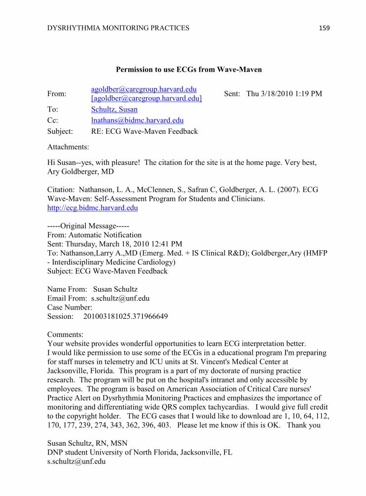
DYSRHYTHMIA MONITORING PRACTICES 159
Permission to use ECGs from Wave-Maven
From: [email protected] [[email protected]]
Sent: Thu 3/18/2010 1:19 PM
To: Schultz, Susan
Subject: RE: ECG Wave-Maven Feedback
Attachments: Hi Susan--yes, with pleasure! The citation for the site is at the home page. Very best, Ary Goldberger, MD Citation: Nathanson, L. A., McClennen, S., Safran C, Goldberger, A. L. (2007). ECG Wave-Maven: Self-Assessment Program for Students and Clinicians. http://ecg.bidmc.harvard.edu -----Original Message----- From: Automatic Notification Sent: Thursday, March 18, 2010 12:41 PM To: Nathanson,Larry A.,MD (Emerg. Med. + IS Clinical R&D); Goldberger,Ary (HMFP - Interdisciplinary Medicine Cardiology) Subject: ECG Wave-Maven Feedback Name From: Susan Schultz Email From: [email protected] Case Number: Session: 201003181025.371966649 Comments: Your website provides wonderful opportunities to learn ECG interpretation better. I would like permission to use some of the ECGs in a educational program I'm preparing for staff nurses in telemetry and ICU units at St. Vincent's Medical Center at Jacksonville, Florida. This program is a part of my doctorate of nursing practice research. The program will be put on the hospital's intranet and only accessible by employees. The program is based on American Association of Critical Care nurses' Practice Alert on Dysrhythmia Monitoring Practices and emphasizes the importance of monitoring and differentiating wide QRS complex tachycardias. I would give full credit to the copyright holder. The ECG cases that I would like to download are 1, 10, 64, 112, 170, 177, 239, 274, 343, 362, 396, 403. Please let me know if this is OK. Thank you Susan Schultz, RN, MSN DNP student University of North Florida, Jacksonville, FL [email protected]

DYSRHYTHMIA MONITORING PRACTICES 160
Appendix V: Content Validity Evaluation “Dysrhythmia Monitoring Practices” by Susan Schultz
Evaluator name: Educator 1_________ Date: _7/15/10______ Job title: Clinical Resource Coordinator Area of specialty: _Critical Care_______ Years in current position: ____10______ Years licensed as a nurse: ___32_____ Educational degrees/years: ___ADN, BSN, MSN_____________________ Certifications: _____CCRN____________________________________ Number of years experience with dysrhythmia interpretation: __31_____________ Evaluator name: Educator 2______________ Date: 7/7/10________________ Job title: Clinical Resource Coordinator Area of specialty: med/surg/tele____ Years in current position: _____2________ Years licensed as a nurse: __29_______ Educational degrees/years: BSN, Diploma__________________________ Certifications: RN,C-Medical surgical nurse____________________________ Number of years experience with dysrhythmia interpretation: ___7______________ Evaluator name: Educator 3______________ Date: _7/29/10______ Job title: __Clinical Resource Coordinator _ Area of specialty: _Telemetry_______ Years in current position: ____5______ Years licensed as a nurse: ___27_____ Educational degrees/years: ___BSN_____________________ Certifications: _____PCCN____________________________________ Number of years experience with dysrhythmia interpretation: __27_____________
Strongly Agree
Agree Neutral Disagree Strongly Disagree
1. The content in the power point from the American Association of Critical Care-Nurses‘ was current and accurate.
3
2. The audio script was from relevant literature and reinforced the content in the power point slides.
2 1
3. The interactivities interspersed throughout the slides (self-check questions, case studies, and game) were from relevant literature and reinforced the content in the power point slides.
3
4. The pretest and posttest were based on the learner objectives and relevant to the content taught in the module.
3
5. The instructional method was effective for this topic.
2 1

DYSRHYTHMIA MONITORING PRACTICES 161
Would you recommend this learning module to the nurses in your units? Yes, but it would require time commitment It would be too hard for the nurses on the unit Yes, definitely What did you like the most about it? Liked interactive questions The games and test that were spread out Challenge—raised my level of expertise in tele filed. Organization of module; the audio was great What modifications do you suggest?
I should have printed the material. I would have gotten more from it – could highlight key points and probably retained.
Would have power point slides that more closely matched the voice over. I found it hard to follow the voice over. I needed something in writing to be able to study in order to really understand the material.
None

DYSRHYTHMIA MONITORING PRACTICES 162
Appendix W: Competency Skills Checklist for Dysrhythmia Monitoring Practices
Nurses‘ Code Number: __________________________
Competency Objectives Rating
Key Method Date &
Validator Initials
1. Complete web-based education program with at least 80% on the posttest.
2. Complete unit-based collaborative learning activities: _____ Conduct audit of electrode placement and lead section on
two patients (attach audit tools) Date:____________ _____ Interpret wide QRS complexes on monitor strip or 12
Lead ECG. Interpretation: ___________________ _____ ECG Poster Challenge (#1) Interpretation: _____ ECG Poster Challenge (#2) Interpretation:
3. Demonstrate correct placement of 5 electrodes on a patient, in particular V1 in the 4ICS RSB
4. State optimal lead to monitor patient for their diagnoses or arrhythmia.
5. Describe 3 drugs or conditions that warrant QTc monitoring. 6. Locate the QTc interval on 12 lead ECG and evaluate what it means.
7. Analyze QRS morphology and differentiate between Bundle Branch Block Aberrancy and Ventricular Ectopy (with the use of pocket reference card or other references).
8. Describe nursing interventions for SVT with Bundle Branch Block Aberrancy and for Ventricular Tachycardia.
Rating Key: 2=Competent, 1=Needs Improvement, 0-Not Competent Method of evaluation: Verbalized, Tested, Skill observation, Simulation Comments and/or Action Plan: ________________________________________________________________________________________________________________________________________________________________________________________________________________________ Validator‘s Initials and Signature: _____ ________________________________

DYSRHYTHMIA MONITORING PRACTICES 163
Appendix X: Pretest and Posttest Questions
Dysrhythmia Monitoring Practices Pretest Questions
1. Match the description of the electrode placement to the correct electrode. A. RA _____ below rib cage on left side of abdomen B. LA _____ infra-clavicular fossa close to left shoulder C. C _____ anywhere on torso, usually lower right side of chest or abdomen D. LL _____ 4th Inter Costal Space, Right Sternal Border E. RL _____ infra-clavicular fossa close to right shoulder 2. What is the best way to prepare the skin before placing electrodes?
A. Clip excessive hair and clean skin with alcohol or washcloth before placing electrodes. B. Clean skin with soap and water only if there is visible sweat, oil, or powder. C. Apply electrodes; if artifact noted, then clean the skin and replace electrodes. D. If chest is hairy, shave with shaving cream and razor, dry thoroughly, then apply electrodes.
3. Select the best lead to diagnose wide QRS complexes.
A. I B. II C. III D. V1
4. Which of the following patient conditions would be best lead to monitor in V1? (Select all that apply)
A. Atrial fibrillation with intermittent wide QRS complexes B. First, Second or Third Degree AV heart block C. Supra-Ventricular Tachycardia with aberrant conduction D. Sinus Bradycardia or Sinus Rhythm with pauses
5. What does a triphasicrsR‘ morphology (shown below) in V1 indicate?
(Adapted from Jacobson, 2007. Used with permission)

DYSRHYTHMIA MONITORING PRACTICES 164
A. RBBB aberration B. LBBB aberration C. Ventricular beat D. None of the above
6. Match the strip with the correct interpretation using QRS morphology and AV dissociation cues.
(From Jacobson, 2006. Used with permission)
A. Sinus rhythm with 2 pairs of PVCs. B. Sinus tachycardia with LBBB aberration. C. Ventricular Tachycardia (positive deflection). D. Ventricular Tachycardia (negative deflection)
7. Mr. B.‘s cardiac monitor arrhythmia alarm sounds and the following tracing is automatically printed out. What is your interpretation of the rhythm?
A. SVT with aberration B. Ventricular tachycardia, monophasic C. Torsades de Pointes

DYSRHYTHMIA MONITORING PRACTICES 165
(From Drew, 2007. Used with permission) 8. Calculate the QT, R-R, and QTc for this strip (√ = square roots. Accurate divisions have been provided in the answers; you just need to select the correct intervals)
From Sommargren & Drew, 2007. Used with permission.)
A. QT = .24, R-R = .48; √.48 = .693; QTc = .24 / .693 = .35 seconds B. QT = .36, R-R = .60; √.6 = .775; QTc = .36 / .775 = .46 seconds C. QT = .44, R-R = .80; √.8 = .894; QTc = .44 / .894 = .49 seconds D. QT = .56, R-R = .80; √8 = .894; QTc = .56 / .894 = .63 seconds
9. Which of the following are true statements (select all that apply).
A. QT interval is measured from beginning of QRS to end of T wave B. QTc interval is the QRS interval plus the T wave C. QT interval lengthens with bradycardia and shortens with tachycardia. D. QT interval shortens with bradycardia and lengthens with tachycardia. E. QTcinterval is the QT interval corrected for heart rate
10. What QTc interval is associated with Torsades de pointes?
A. > 0.20 sec (200 msec) B. > 0.24 sec (240 msec) C. > 0.50 sec (500 msec) D. < 0.12 sec (120 msec)

DYSRHYTHMIA MONITORING PRACTICES 166
11. Which drugs may prolong the QTc? A. Antiarrhythmics (Procainamide/Pronestyl, Sotalol/Betapace) B. Antipsychotics (Chlorpromazine/Thorazine, Haloperidol/Haldol) C. Antibiotics (Erythromycin/EES, Levofloxacin/Levaquin) D. All of the above
12. Which of the following patients should have QTc interval monitoring? (select all that apply)
A. 58 year-old male with acute MI and on Lidocaine drip 2 mg/min, Potassium 4.0 B. 36 year-old female admitted with overdose on Chlorpromazine/Thorazine C. 54 year-old female with new onset atrial fibrillation and started on oral Sotalol/Betapace D. 49 year-old alcoholic male with cardiomyopathy taking Quinidine and Magnesium 1.8 E. 75 year-old female admitted with pneumonia and started on Levofloxacin/Levaquin
13. What treatments have been shown to be effective for Torsades de points?
A. IV Magnesium B. Defibrillation C. Overdrive pacing D. All of the above
14. What are appropriate treatments for wide-complex ventricular tachycardia without a pulse (per hospital Emergency Standing Orders)? (select all that apply)
A. Call Code Blue and begin CPR B. Defibrillate immediately with 360 joules or equivalent with a biphasic defibrillator C. Give drug trial of Lidocaine before defibrillating. D. Initial external pacemaker at rate of 60-80 with milliamps 10% above the threshold
15. Which intervention is the most important for the nurse to initiate first when a new wide QRS complex tachycardia is identified by the monitor tech?
A. Look at the strip with the monitor tech B. Order STAT 12 Lead ECG C. Call the Rapid Response Nurse D. Assess the patient
Answers to Pretest: 1.D, B, E, C, A; 2.A; 3.D; 4.A, C; 5.A; 6.D, B, C, A; 7.C; 8.C; 9.A, C, E; 10.C; 11.D; 12.B, C, D, E; 13.D; 14.A, B; 15.D.

DYSRHYTHMIA MONITORING PRACTICES 167
Dysrhythmia Monitoring Practices Posttest Questions
1. Drag and Drop the electrodes to the correct locations (or write them next to the correct location). Only 5 locations will need to be labeled. RA, LA, C-V1, RL, LL
(Adapted from Richards, 2008. Used with permission) 2. Place the steps for applying electrodes in the proper order (select the answer choice with the steps in the correct order).
W. Apply new electrodes and press firmly to skin X. Clip excessive hair from selected locations Y. Visually select locations without implanted devices, dressings, or
muscle artifact that could interfere with monitoring Z. Clean skin with alcohol or washcloth and dry thoroughly
A. Y, X, Z, W B. Y, Z, X, W C. W, X, Y, Z D. Z, X, Y, W
3. Select the best lead to diagnose atrial activity and measure heart rate.
A. I B. II C. III D. V1
4. Which of the following patient conditions would be best lead to monitor in V1? (Select all that apply)
A. Normal Sinus Rhythm with 5 beat run of Ventricular Tachycardia B. Possible Torsades de Pointes C. Diagnosing Atrial Flutter versus Atrial Fibrillation D. Sinus Tachycardia with intermittent right Bundle Branch Block

DYSRHYTHMIA MONITORING PRACTICES 168
5. Measure the descent of the S from the onset of QRS to the nadir (lowest point of the S). Exact distance to measure is shown. Select the answer choice that has the correct measurement and interpretation.
(Adapted from Jacobson, 2007. Used with permission)
A. 0.04-0.06 seconds (40-60 msec); nadir is normal and represents LBBB aberration B. 0.10 seconds (100 msec); nadir is normal and represents RBBB aberration C. 0.14 seconds (140 msec); nadir is prolonged and represents VT D. 0.24 seconds (240 msec); nadir is prolonged and represents VT
6. Match the strips with the correct interpretations using QRS morphology and AV dissociation cues. (draw lines to connect the letters and numbers)
A. Sinus rhythm with 2 pairs of PVCs. 1 B. Sinus tachycardia with LBBB aberration. 2 C. Ventricular Tachycardia (positive deflection). 3 D. Ventricular Tachycardia (negative deflection) 4
(From Jacobson, 2006. Used with permission) 7. What is your interpretation of the rhythm below?
A. SVT with aberration B. Ventricular tachycardia, monophasic C. Torsades de Pointes (TdP)

DYSRHYTHMIA MONITORING PRACTICES 169
(From Pelter & Carey, 2006. Used with permission) 8. Calculate the QT and QTc interval for this strip. (√ = square roots. Accurate divisions have been provided in the answers; you just need to select the correct intervals)
(From Drew, 2007. Used with permission.)
A. QT = .24, R-R = .48; √.48 = .693; QTc = .24 / .693 = .35 seconds B. QT = .40, R-R = .80; √.8 = .894. QTc = .40 / .894 = .48 seconds C. QT = .44, R-R = .60; √.6 = .775; QTc = .44 / .775 = .57 seconds D. QT = .56, R-R = .80; √8 = .894; QTc = .56 / .894 = .63 seconds
9. Which of the following are true statements (select all that apply).
A. QTc interval is the QRS interval plus the T wave B. QT interval shortens with bradycardia and lengthens with tachycardia. C. QTcinterval is the QT interval corrected for heart rate D. QT interval is measured from beginning of QRS to end of T wave E. QT interval lengthens with bradycardia and shortens with tachycardia.
10. What QTc interval is associated with Torsades de Pointes? A. < 0.12 sec (120 msec) B. > 0.20 sec (200 msec) C. > 0.24 sec (240 msec) D. > 0.50 sec (500 msec)
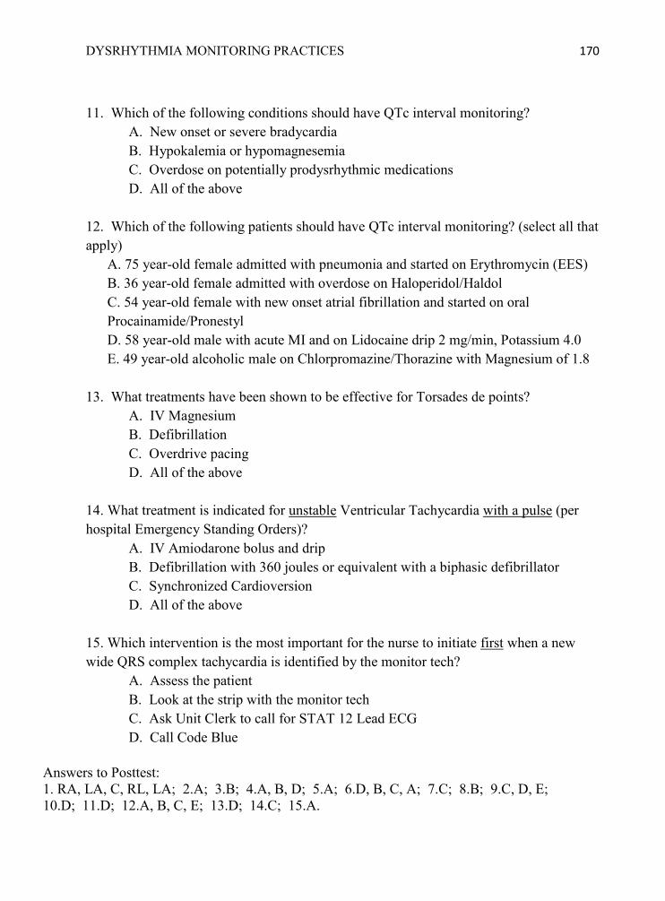
DYSRHYTHMIA MONITORING PRACTICES 170
11. Which of the following conditions should have QTc interval monitoring?
A. New onset or severe bradycardia B. Hypokalemia or hypomagnesemia C. Overdose on potentially prodysrhythmic medications D. All of the above
12. Which of the following patients should have QTc interval monitoring? (select all that apply)
A. 75 year-old female admitted with pneumonia and started on Erythromycin (EES) B. 36 year-old female admitted with overdose on Haloperidol/Haldol C. 54 year-old female with new onset atrial fibrillation and started on oral Procainamide/Pronestyl D. 58 year-old male with acute MI and on Lidocaine drip 2 mg/min, Potassium 4.0 E. 49 year-old alcoholic male on Chlorpromazine/Thorazine with Magnesium of 1.8
13. What treatments have been shown to be effective for Torsades de points?
A. IV Magnesium B. Defibrillation C. Overdrive pacing D. All of the above
14. What treatment is indicated for unstable Ventricular Tachycardia with a pulse (per hospital Emergency Standing Orders)?
A. IV Amiodarone bolus and drip B. Defibrillation with 360 joules or equivalent with a biphasic defibrillator C. Synchronized Cardioversion D. All of the above
15. Which intervention is the most important for the nurse to initiate first when a new wide QRS complex tachycardia is identified by the monitor tech?
A. Assess the patient B. Look at the strip with the monitor tech C. Ask Unit Clerk to call for STAT 12 Lead ECG D. Call Code Blue
Answers to Posttest: 1. RA, LA, C, RL, LA; 2.A; 3.B; 4.A, B, D; 5.A; 6.D, B, C, A; 7.C; 8.B; 9.C, D, E; 10.D; 11.D; 12.A, B, C, E; 13.D; 14.C; 15.A.

DYSRHYTHMIA MONITORING PRACTICES 171
Appendix Y: Pretest and Posttest Scores and Reliability (Educators) Educators’ Pretest and Posttest Scores Educator Pretest Posttest 1 93 100 2 53 93 3 80 87
Descriptive Statistics N Mean Std. Deviation Minimum Maximum Median Pretest 3 75.33 20.404 53 93 80 Posttest 3 93.33 6.506 87 100 93 Wilcoxon Signed Ranks Test Ranks N Mean Rank Sum of Ranks Posttest - Pretest Negative Ranks 0a .00 .00
Positive Ranks 3b 2.00 6.00 Ties 0c Total 3
a. Posttest < Pretest b. Posttest > Pretest c. Posttest = Pretest
Test Statisticsb Posttest - Pretest Z -1.633a Asymp. Sig. (2-tailed) .102
a. Based on negative ranks. b. Wilcoxon Signed Ranks Test
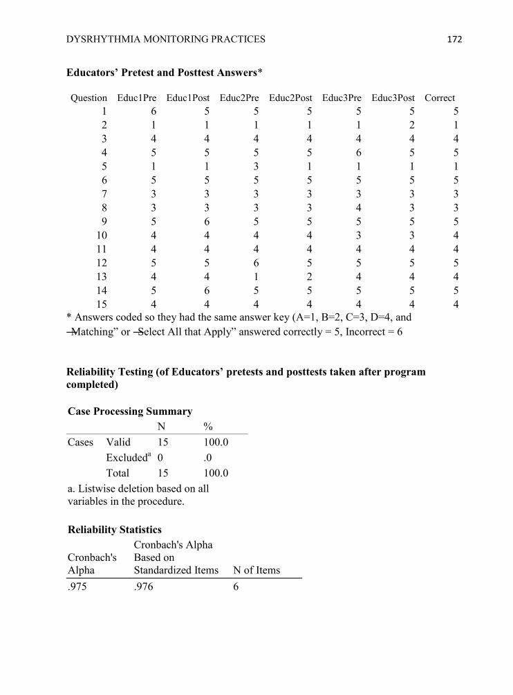
DYSRHYTHMIA MONITORING PRACTICES 172
Educators’ Pretest and Posttest Answers*
Question Educ1Pre Educ1Post Educ2Pre Educ2Post Educ3Pre Educ3Post Correct 1 6 5 5 5 5 5 5 2 1 1 1 1 1 2 1 3 4 4 4 4 4 4 4 4 5 5 5 5 6 5 5 5 1 1 3 1 1 1 1 6 5 5 5 5 5 5 5 7 3 3 3 3 3 3 3 8 3 3 3 3 4 3 3 9 5 6 5 5 5 5 5
10 4 4 4 4 3 3 4 11 4 4 4 4 4 4 4 12 5 5 6 5 5 5 5 13 4 4 1 2 4 4 4 14 5 6 5 5 5 5 5 15 4 4 4 4 4 4 4
* Answers coded so they had the same answer key (A=1, B=2, C=3, D=4, and ―Matching‖ or ―Select All that Apply‖ answered correctly = 5, Incorrect = 6 Reliability Testing (of Educators’ pretests and posttests taken after program completed) Case Processing Summary N % Cases Valid 15 100.0
Excludeda 0 .0 Total 15 100.0
a. Listwise deletion based on all variables in the procedure. Reliability Statistics
Cronbach's Alpha
Cronbach's Alpha Based on Standardized Items N of Items
.975 .976 6

DYSRHYTHMIA MONITORING PRACTICES 173
Appendix Z: Education Program Evaluation Form
Strongly Agree
Agree Neutral Disagree Strongly Disagree
The posttest was based on the learner objectives.
1 8
This type of learning is worthwhile.
1 7 1
The content met my professional educational needs.
2 4 3
The instructional method was effective for this topic.
1 4 3
I would recommend this module to others.
1 5 3
Self-study modules meet my needs for ongoing education.
1 5 1 2
The content was accurate and current.
3 6
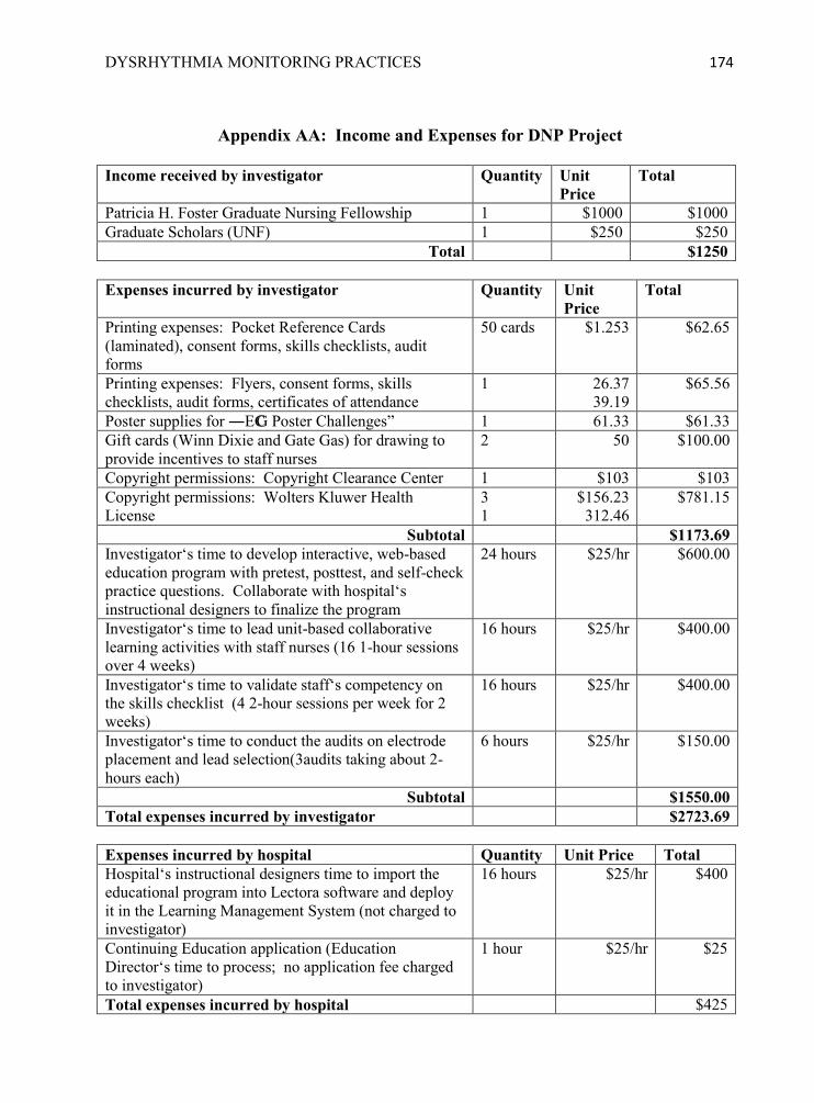
DYSRHYTHMIA MONITORING PRACTICES 174
Appendix AA: Income and Expenses for DNP Project
Income received by investigator Quantity Unit
Price Total
Patricia H. Foster Graduate Nursing Fellowship 1 $1000 $1000 Graduate Scholars (UNF) 1 $250 $250
Total $1250
Expenses incurred by investigator Quantity Unit Price
Total
Printing expenses: Pocket Reference Cards (laminated), consent forms, skills checklists, audit forms
50 cards $1.253 $62.65
Printing expenses: Flyers, consent forms, skills checklists, audit forms, certificates of attendance
1 26.37 39.19
$65.56
Poster supplies for ―ECG Poster Challenges‖ 1 61.33 $61.33 Gift cards (Winn Dixie and Gate Gas) for drawing to provide incentives to staff nurses
2 50 $100.00
Copyright permissions: Copyright Clearance Center 1 $103 $103 Copyright permissions: Wolters Kluwer Health License
3 1
$156.23 312.46
$781.15
Subtotal $1173.69 Investigator‘s time to develop interactive, web-based education program with pretest, posttest, and self-check practice questions. Collaborate with hospital‘s instructional designers to finalize the program
24 hours $25/hr $600.00
Investigator‘s time to lead unit-based collaborative learning activities with staff nurses (16 1-hour sessions over 4 weeks)
16 hours $25/hr $400.00
Investigator‘s time to validate staff‘s competency on the skills checklist (4 2-hour sessions per week for 2 weeks)
16 hours $25/hr $400.00
Investigator‘s time to conduct the audits on electrode placement and lead selection(3audits taking about 2-hours each)
6 hours $25/hr $150.00
Subtotal $1550.00 Total expenses incurred by investigator $2723.69
Expenses incurred by hospital Quantity Unit Price Total Hospital‘s instructional designers time to import the educational program into Lectora software and deploy it in the Learning Management System (not charged to investigator)
16 hours $25/hr $400
Continuing Education application (Education Director‘s time to process; no application fee charged to investigator)
1 hour $25/hr $25
Total expenses incurred by hospital $425

DYSRHYTHMIA MONITORING PRACTICES 175
Appendix BB: IRB Approval Documents
UNIVERSITY of NORTH FLO RIDA_
Office ofRescarch and Sponsored Programs 1 UNF Drive Jacksonville, FL 32224-2665 904-620-24SS FAX 904-62()..24S7 Equal OpportunitylEquai AcceulAffirmative Action Ins1iMion
MEMORANDUM
Apri l 30, 2010
Ms. Susan J. Schu ltz
Dr. Carol Ledbetter Nu~ing
Dr. Katherine Kasten, Chairperson UNF Institutional Review Board
Review by the UNF Institutional Review Board IRB# 1 0-031: "Dysrhythmia Monitoring Practices of Nurses on a Telemetry Unit"
This is to advise you that your project, "Dysrhythmia Monitoring Practices of Nurses on a Telemetry Un it," has undergone "expedited, category #7" review on behalf of the UNF Institutional Review Board and was approved.
This approval applies to your project in the foml and content as submitted to the IRB for review. Any variations or modifications to the approved protocol andlor infonned consent fonns as they relate to dealing with human subjects must be cleared with the lRB prior to implementing such changes. Any unanticipated problems involving risk and any occurrence of serious harm to subjects and others shall be reported promptly to the IRB.
Your study bas been approved for a period of 12 months. !fyour project continues for more than one year, you are required to provide 8 Continu ing Status Report to the UNF IRE prior to 0313012011 if your study will be continuing past 0412912011. We suggest you submit your slarus report J I monrhsfrom the date of your approval dale as noted above 10 allow time for review and processing.
As you may know, CITI Course Completion R eports are valid for 3 years. Your completion report is valid through 11/121201 2. If your completioll report expires soon please take CITI 's refresher course. Once you complete all of the CITI modules a completion report will be emailed to our office. For faster file updating purposes, however, please notify th is office when you complete your CITl refresher course.
Should you have questions regarding your project or any other IRS issues, please contact the Office of Research and Sponsored Programs at 904.620.2455.
Thank you, UN!' ;R{3 lI!umbar:
Research Integrity Staff App!"Ovc~ D~ta: t..1-30 - IO
Revision Dele:
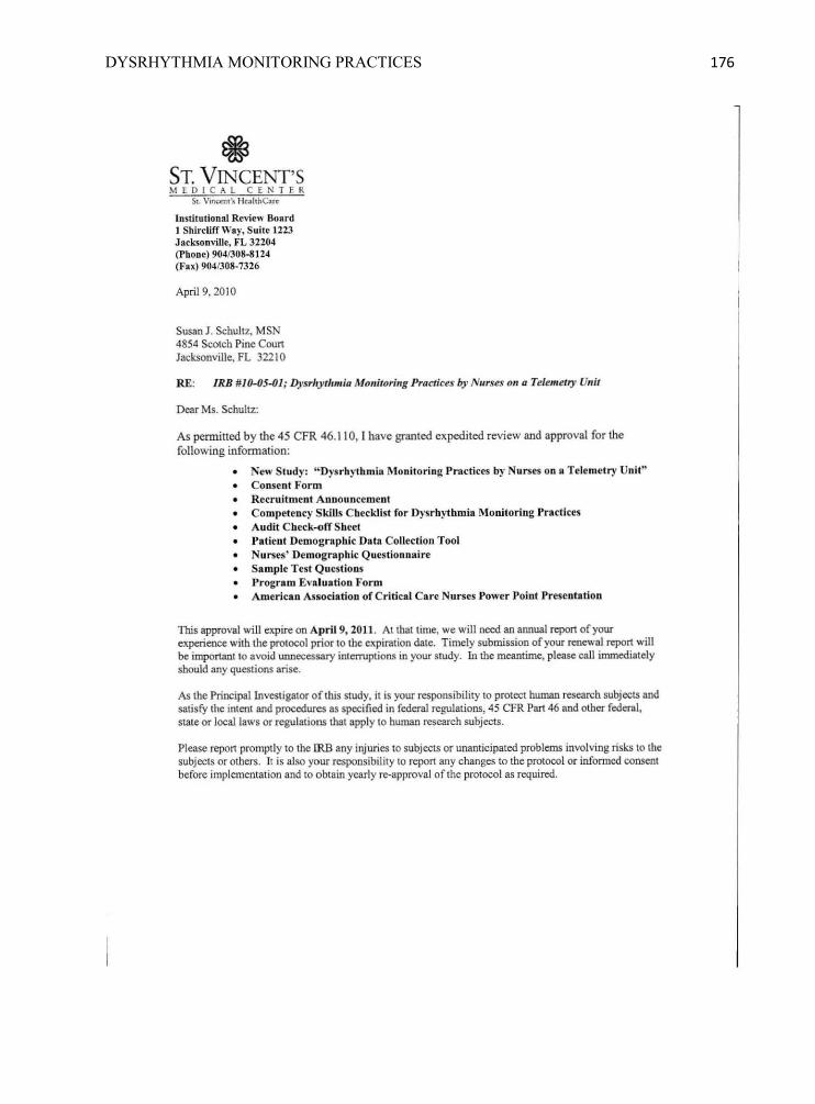
DYSRHYTHMIA MONITORING PRACTICES 176
@ ST. V INCENT'S MEDI C AL CENTER
50:. V;nalll'~ He .. llha.r~
Institutional Review 8011.rd J ShircUff Way, Suite 1223 Jacksonville, Fl 32204 (Phone) 9041308-8 124 (Fax) 904/308·7326
April 9. 2010
Susan J. Schultz. MSN 4854 Scotch Pine Coun Jacksonville, FL 32210
RE: IRB #10-05-01; Dysrhy thmia Monitoring Practices by N urses on Q Telemetry UnU
Dear Ms. Schultz:
As pennitted by the 45 CFR 46.1 ) 0, I have granted expedited review and approval for the following infonnation:
• New Study: " Dysrh)1hmia Monitoring Practices by Nurses on a Telemetry Unit" Consent Form
• Recruitment Announcement • Competency Skills Checklist fo r Dysrb)1hmia Monitoring Practices • Audit Cbeck-off Sheet
Patient Demographic Data Collection Tool Nu rses' Demograph ic Q uestionnaire
• Sample Test Q uestions Program Evaluation Form American AssociatioD of Critical Care Nurses Power Point Presentation
ntis approval will expire on April 9, 2011. At that time, we will need an annual report of your experience with the protocol prior to the expiration date. Timely submission of your renewal report will be important 10 avoid unnecessary interruptions in your study. In the meantime, please call immediately should any questions arise.
As the Principal Investigator of this study, it is your responsibility to protect human research subjects and satisfy the intent and procedures as specified in federal regulat ions, 45 CFR Part 46 and other federal, state or loca1 1aws or regulations thai apply to human research subjects.
Please repon promptly to the lRB any injuries to subjects or unanticipated problems involving risks to the subjects or others. It is also your responsibil ity to report any changes to the protocol or infonncd consent before implementation and to obtain yearly re·approval of the protocol as required.
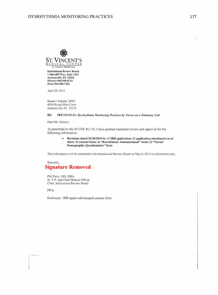
DYSRHYTHMIA MONITORING PRACTICES 177
• ST. VINCENT'S M E DI C A l. CE N T ER
St. Vincent 's Hc"hhCa rc
Institution al Review Board 1 Shirclirr Way. Suite 1223 J arksonvillr, FL 32204 (Phone) 904/308·8124 (Fax) 904/308-7326
April 29. 2010
Susan J. Schultz. MSN 4854 SCOIch Pine Coun Jacksonville. FL 32210
RE: IRR #10-05-01: Dysr/,y fJIIII ;U Monitoring Practices by Nurses Oil u Telemetry VIII',
Dear Ms. Schultz:
As permitted by the 45 CFR 46. 110. [ have granted expedited review and approval for the foll owing information:
Revisions dated 04/28/20 10 to: 1) tRll a pplication; 2) applicll tioll atta chment cover sheet; 3) consent form ; 4) " Recruitment Announcement" fo r m; 5) " Nurses' Demograph ic Questionnai re" form
TlUs information will be submitted to the lnsti tutional Review Board on May 6. 2010 as infonnation only.
Phil Perry. MD. MBA Sr. V.P. and Chief Mcdical Officer Chair, Institutional Review Board
PP/ts
Enclosure: lRB approved/stamped consent fonn
Signature Removed

DYSRHYTHMIA MONITORING PRACTICES 178
Appendix CC: Audit Check-off Sheet
Number Audit Done (√ or NA)
Data recorded (√ or NA)
Number Audit Done (√ or NA)
Data recorded (√ or NA)
1 21 2 22 3 23 4 24 5 25 6 26 7 27 8 28 9 29 10 30 11 31 12 32 13 33 14 34 15 35 16 36 17 37 18 38 19 39 20 40 When audit is finished, this check-off sheet will be discarded in confidential bin before leaving the unit.

DYSRHYTHMIA MONITORING PRACTICES 179
Appendix DD: Patient Demographic Data Collection Tool
Number of Males: ________________ Number of Females: ________________ Ages: ________________________________________________________________ Length of stay (in days): ________________________________________________ Admitting diagnoses: ____________________________________________________ ______________________________________________________________________ ______________________________________________________________________ ______________________________________________________________________

DYSRHYTHMIA MONITORING PRACTICES 180
Appendix EE: SPSS Statistical Software Data Nurse‘s codenumber
Consent Signed (0 = No, 1= Yes)
Pretest (percentage)
Posttest (percentage)
Number of unit-based activities done (0, 1, 2, 3, 4)
Competency skills checklist completed (0 = No, 1= Yes)
Number of attempts to complete competency skills checklist (0, 1, 2)
Status of nurses‘ participation in educational program:
0 = Never registered
1 = Registered but never started pretest
2 = Registered, pretest in progress, no posttest
3 = Pretest completed, posttest in progress
4 = Pretest and posttest completed
5 = Excluded
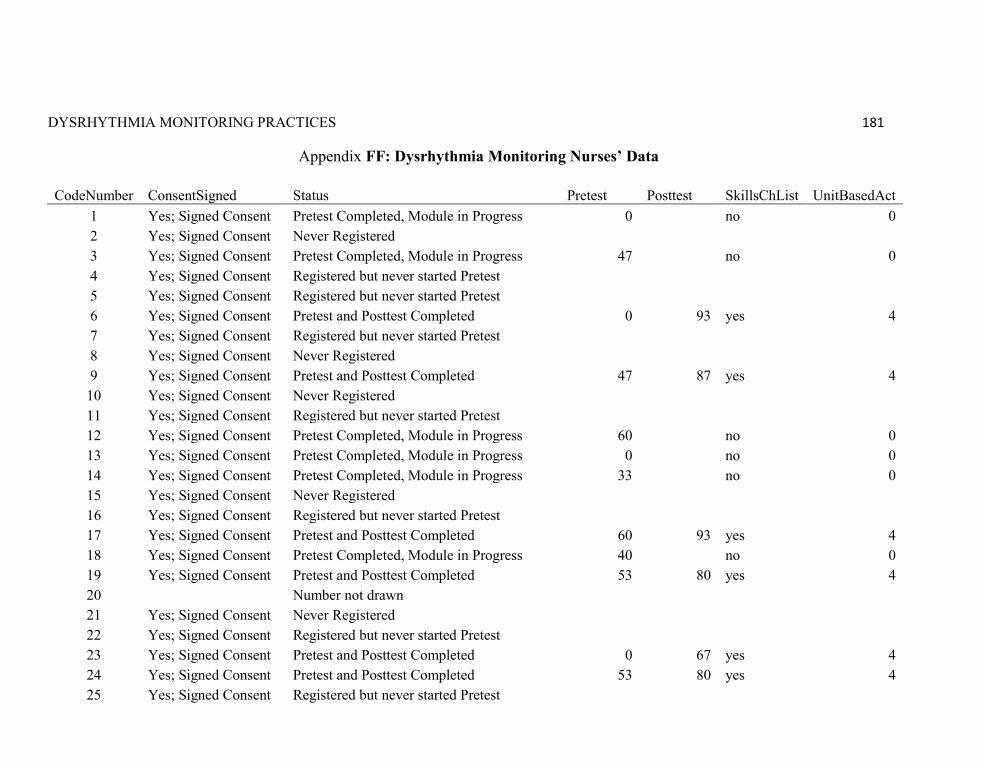
DYSRHYTHMIA MONITORING PRACTICES 181
Appendix FF: Dysrhythmia Monitoring Nurses’ Data
CodeNumber ConsentSigned Status Pretest Posttest SkillsChList UnitBasedAct 1 Yes; Signed Consent Pretest Completed, Module in Progress 0
no 0
2 Yes; Signed Consent Never Registered 3 Yes; Signed Consent Pretest Completed, Module in Progress 47
no 0
4 Yes; Signed Consent Registered but never started Pretest 5 Yes; Signed Consent Registered but never started Pretest 6 Yes; Signed Consent Pretest and Posttest Completed 0 93 yes 4
7 Yes; Signed Consent Registered but never started Pretest 8 Yes; Signed Consent Never Registered 9 Yes; Signed Consent Pretest and Posttest Completed 47 87 yes 4
10 Yes; Signed Consent Never Registered 11 Yes; Signed Consent Registered but never started Pretest 12 Yes; Signed Consent Pretest Completed, Module in Progress 60
no 0
13 Yes; Signed Consent Pretest Completed, Module in Progress 0
no 0 14 Yes; Signed Consent Pretest Completed, Module in Progress 33
no 0
15 Yes; Signed Consent Never Registered 16 Yes; Signed Consent Registered but never started Pretest 17 Yes; Signed Consent Pretest and Posttest Completed 60 93 yes 4
18 Yes; Signed Consent Pretest Completed, Module in Progress 40
no 0 19 Yes; Signed Consent Pretest and Posttest Completed 53 80 yes 4 20
Number not drawn
21 Yes; Signed Consent Never Registered 22 Yes; Signed Consent Registered but never started Pretest 23 Yes; Signed Consent Pretest and Posttest Completed 0 67 yes 4
24 Yes; Signed Consent Pretest and Posttest Completed 53 80 yes 4 25 Yes; Signed Consent Registered but never started Pretest
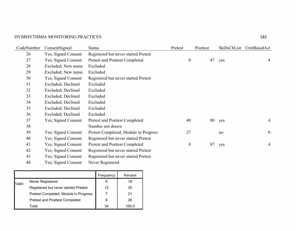
DYSRHYTHMIA MONITORING PRACTICES 182
CodeNumber ConsentSigned Status Pretest Posttest SkillsChList UnitBasedAct 26 Yes; Signed Consent Registered but never started Pretest
27 Yes; Signed Consent Pretest and Posttest Completed 0 47 yes 4 28 Excluded; New nurse Excluded
29 Excluded; New nurse Excluded 30 Yes; Signed Consent Registered but never started Pretest 31 Excluded; Declined Excluded 32 Excluded; Declined Excluded 33 Excluded; Declined Excluded 34 Excluded; Declined Excluded 35 Excluded; Declined Excluded 36 Excluded; Declined Excluded 37 Yes; Signed Consent Pretest and Posttest Completed 40 80 yes 4
38
Number not drawn 39 Yes; Signed Consent Pretest Completed, Module in Progress 27
no 0
40 Yes; Signed Consent Registered but never started Pretest 41 Yes; Signed Consent Pretest and Posttest Completed 0 87 yes 4
42 Yes; Signed Consent Registered but never started Pretest 43 Yes; Signed Consent Registered but never started Pretest 44 Yes; Signed Consent Never Registered
Frequency Percent
Valid Never Registered 6 18
Registered but never started Pretest 12 35
Pretest Completed, Module in Progress 7 21
Pretest and Posttest Completed 9 26
Total 34 100.0
DYSRHYTHMIA MONITORING PRACTICES 183
Appendix GG: Nurses’ Demographic Data Frequency Tables
Gender
Frequency Percent Valid Percent
Cumulative Percent
Valid Female 8 100.0 100.0 100.0
Ethnicity
Frequency Percent Valid Percent
Cumulative Percent
Valid American Indian/Alaska Native 1 12.5 12.5 12.5
Asian or Pacific Islander 2 25.0 25.0 37.5
White or Caucasian 4 50.0 50.0 87.5 Other 1 12.5 12.5 100.0 Total 8 100.0 100.0
Degree
Frequency Percent Valid Percent
Cumulative Percent
Valid Associates 4 50.0 50.0 50.0 Bachelors 4 50.0 50.0 100.0 Total 8 100.0 100.0
QT Instruction
Frequency Percent Valid Percent
Cumulative Percent
Valid No instruction 1 12.5 12.5 12.5 Only a little instruction 2 25.0 25.0 37.5 Some instruction but not comfortable yet 5 62.5 62.5 100.0
Total 8 100.0 100.0

DYSRHYTHMIA MONITORING PRACTICES 184
Studied Online
Frequency Percent Valid Percent
Cumulative Percent
Valid Yes 7 87.5 87.5 87.5 No 1 12.5 12.5 100.0 Total 8 100.0 100.0
Comfort online learning
Frequency Percent Valid Percent
Cumulative Percent
Valid Somewhat comfortable 7 87.5 87.5 87.5
Very comfortable 1 12.5 12.5 100.0 Total 8 100.0 100.0
Age and Years of Experience
N Range Minimum Maximum Mean Std. Deviation
Age 8 28 23 51 40.62 9.039 Years RN License 8 28 1 29 8.25 9.925 Years worked on Unit 8 12 1 13 4.38 4.274
Years since first class 8 14 1 15 5.38 5.449
Valid N (listwise) 8
Enrollment Status
Frequency Percent Valid
Percent Cumulative
Percent Valid Never Registered 6 14.3 14.3 14.3
Registered but never started Pretest 12 28.6 28.6 42.9 Pretest Completed, Module in Progress 6 14.3 14.3 57.1 Pretest and Posttest Completed 10 23.8 23.8 81.0 Excluded 8 19.0 19.0 100.0 Total 42 100.0 100.0
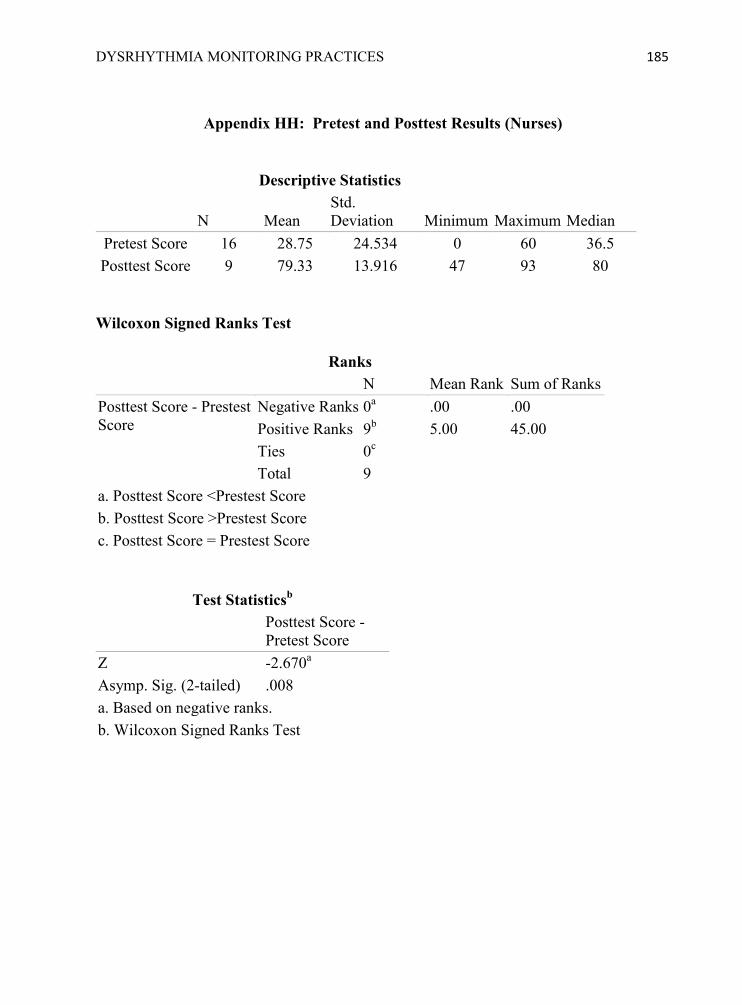
DYSRHYTHMIA MONITORING PRACTICES 185
Appendix HH: Pretest and Posttest Results (Nurses)
Descriptive Statistics
N Mean Std. Deviation Minimum Maximum
Median
Pretest Score 16 28.75 24.534 0 60 36.5 Posttest Score 9 79.33 13.916 47 93 80
Wilcoxon Signed Ranks Test
Ranks N Mean Rank Sum of Ranks Posttest Score - Prestest Score
Negative Ranks 0a .00 .00 Positive Ranks 9b 5.00 45.00 Ties 0c Total 9
a. Posttest Score <Prestest Score b. Posttest Score >Prestest Score c. Posttest Score = Prestest Score
Test Statisticsb Posttest Score -
Pretest Score Z -2.670a Asymp. Sig. (2-tailed) .008 a. Based on negative ranks. b. Wilcoxon Signed Ranks Test

DYSRHYTHMIA MONITORING PRACTICES 186
Appendix II: Pretest and Posttest Reliability (Nurses) Nurses’ Pretest Answers Questions Nurse6 Nurse9 Nurse17 Nurse19 Nurse23 Nurse24 Nurse27 Nurse37 Nurse41 Correct
1 B C C C B C B C A C 2 B A A A B A B A C A 3 A A B B A A A A A B 4 B A A B B A B A B C 5
A C C
A
B
A
6 B C C C B C B C A C 7 A A B B A A A A B C 8
A C C
A
A D C
9 D D C A D D D D A C 10 A C B C B C B C B C 11
D D A
D
D C D
12 B B A D
B
B
C 13 B A A A B A B A C D 14 B B C C B C B B A C 15
D D D
D
D B D
Case Processing Summary Reliability Statistics
N % Cronbach's
Alpha Cronbach's Alpha Based on
Standardized Items N of Items
Cases Valid 10 66.7 .840 .813 9
Excludeda 5 33.3
Total 15 100.0
a. Listwise deletion based on all variables in the procedure.

DYSRHYTHMIA MONITORING PRACTICES 187
Nurses’ Posttest Answers Questions Nurse6 Nurse9 Nurse17 Nurse19 Nurse23 Nurse24 Nurse27 Nurse37 Nurse41 Correct
1 C C C C C C C C C C 2 A A A A A A A C B A 3 C C C C C C C C C C 4 C B C C A C A C C C 5 A A A B A B A A A A 6 C C C C B C B C C C 7 C C C C C C B C C C 8 B B B B C B C B B B 9 C C C A B A B C C C 10 D D D D D D D D D D 11 D D D D D D C D D D 12 C D D C D C D D D C 13 C C C C C C C A C C 14 B C C B C B C C C C 15 A A A A A A B A A A
Case Processing Summary Reliability Statistics
N % Cronbach's
Alpha Cronbach's Alpha Based on Standardized Items N of Items
Cases Valid 15 100 .963 .964 9 Excludeda 0 0 Total 15 100.0
a. Listwise deletion based on all variables in the procedure.

DYSRHYTHMIA MONITORING PRACTICES 188
Appendix JJ: Education Program Evaluation Results
Strongly
Agree Agree Neutral Disagree Strongly
Disagree The posttest was based on the learner objectives.
2 8
This type of learning is worthwhile.
2 7 1
The content met my professional educational needs.
3 4 3
The instructional method was effective for this topic.
1 5 1 3
I would recommend this module to others.
2 5 3
Self-study modules meet my needs for ongoing education.
2 5 1 2
The content was accurate and current.
4 6
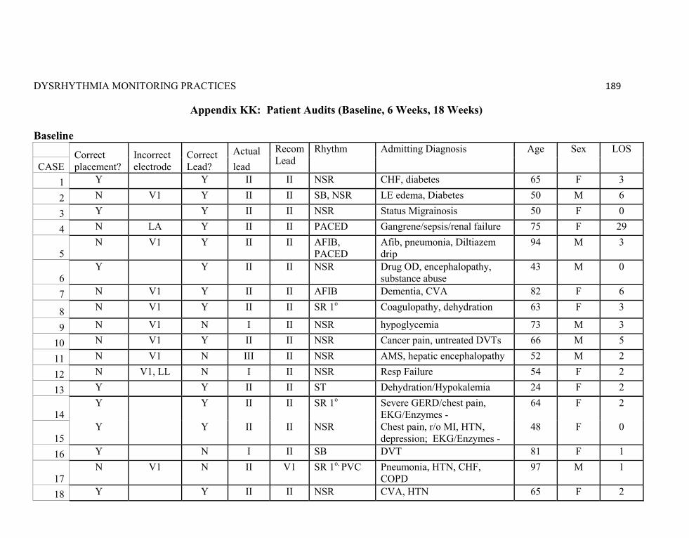
DYSRHYTHMIA MONITORING PRACTICES 189
Appendix KK: Patient Audits (Baseline, 6 Weeks, 18 Weeks) Baseline
Correct placement?
Incorrect electrode
Correct Lead?
Actual Recom Lead
Rhythm Admitting Diagnosis Age Sex LOS
CASE lead 1 Y Y II II NSR CHF, diabetes 65 F 3
2 N V1 Y II II SB, NSR LE edema, Diabetes 50 M 6
3 Y Y II II NSR Status Migrainosis 50 F 0
4 N LA Y II II PACED Gangrene/sepsis/renal failure 75 F 29
5 N V1 Y II II AFIB,
PACED Afib, pneumonia, Diltiazem drip
94 M 3
6 Y Y II II NSR Drug OD, encephalopathy,
substance abuse 43 M 0
7 N V1 Y II II AFIB Dementia, CVA 82 F 6
8 N V1 Y II II SR 1o Coagulopathy, dehydration 63 F 3
9 N V1 N I II NSR hypoglycemia 73 M 3
10 N V1 Y II II NSR Cancer pain, untreated DVTs 66 M 5
11 N V1 N III II NSR AMS, hepatic encephalopathy 52 M 2
12 N V1, LL N I II NSR Resp Failure 54 F 2
13 Y Y II II ST Dehydration/Hypokalemia 24 F 2
14 Y Y II II SR 1o Severe GERD/chest pain,
EKG/Enzymes - 64 F 2
15 Y Y II II NSR Chest pain, r/o MI, HTN,
depression; EKG/Enzymes - 48 F 0
16 Y N I II SB DVT 81 F 1
17 N V1 N II V1 SR 1o, PVC Pneumonia, HTN, CHF,
COPD 97 M 1
18 Y Y II II NSR CVA, HTN 65 F 2
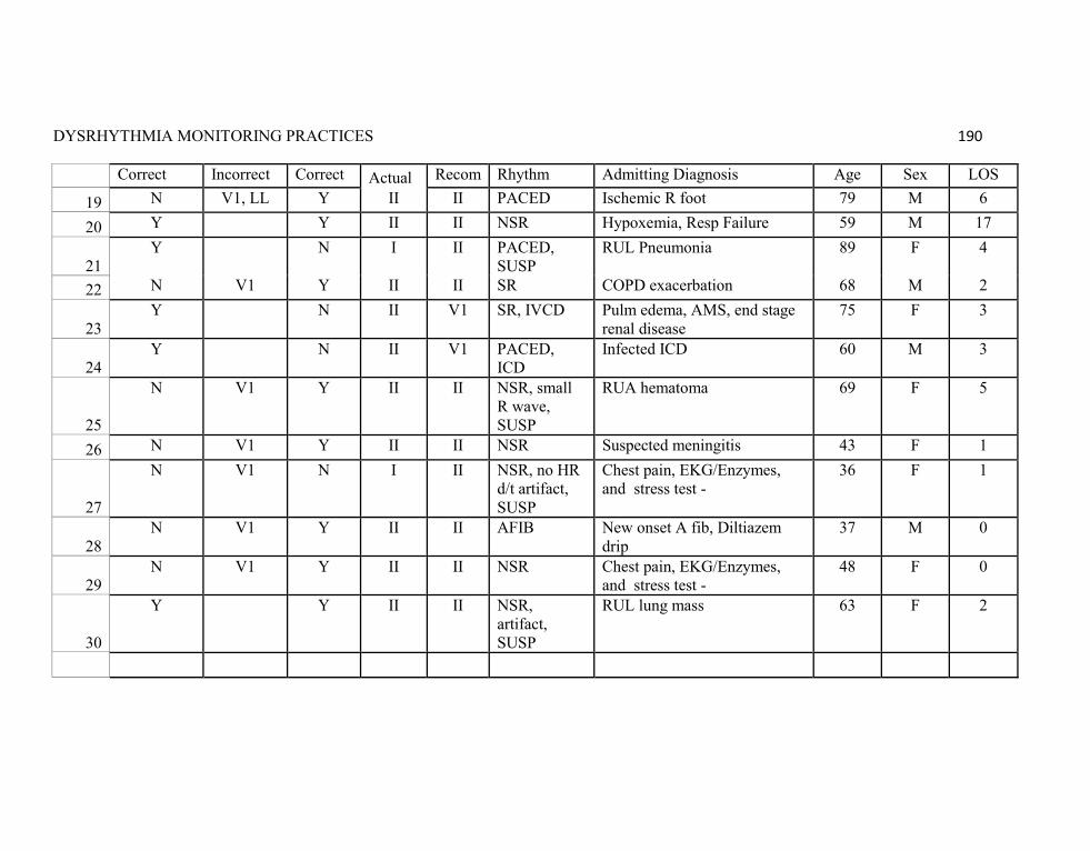
DYSRHYTHMIA MONITORING PRACTICES 190
Correct placement?
Incorrect electrode
Correct Lead?
Actual Recom Lead
Rhythm Admitting Diagnosis Age Sex LOS
19 N V1, LL Y II II PACED Ischemic R foot 79 M 6
20 Y Y II II NSR Hypoxemia, Resp Failure 59 M 17
21 Y N I II PACED,
SUSP RUL Pneumonia 89 F 4
22 N V1 Y II II SR COPD exacerbation 68 M 2
23 Y N II V1 SR, IVCD Pulm edema, AMS, end stage
renal disease 75 F 3
24 Y N II V1 PACED,
ICD Infected ICD 60 M 3
25
N V1 Y II II NSR, small R wave, SUSP
RUA hematoma 69 F 5
26 N V1 Y II II NSR Suspected meningitis 43 F 1
27
N V1 N I II NSR, no HR d/t artifact, SUSP
Chest pain, EKG/Enzymes, and stress test -
36 F 1
28 N V1 Y II II AFIB New onset A fib, Diltiazem
drip 37 M 0
29 N V1 Y II II NSR Chest pain, EKG/Enzymes,
and stress test - 48 F 0
30
Y Y II II NSR, artifact, SUSP
RUL lung mass 63 F 2
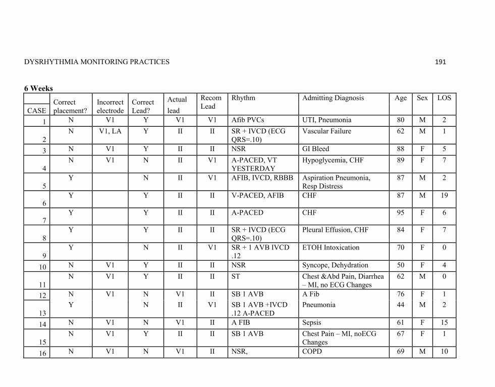
DYSRHYTHMIA MONITORING PRACTICES 191
6 Weeks
Correct placement?
Incorrect electrode
Correct Lead?
Actual Recom Lead
Rhythm Admitting Diagnosis Age Sex LOS
CASE lead 1 N V1 Y V1 V1 Afib PVCs UTI, Pneumonia 80 M 2
2 N V1, LA Y II II SR + IVCD (ECG
QRS=.10) Vascular Failure 62 M 1
3 N V1 Y II II NSR GI Bleed 88 F 5
4 N V1 N II V1 A-PACED, VT
YESTERDAY Hypoglycemia, CHF 89 F 7
5 Y N II V1 AFIB, IVCD, RBBB Aspiration Pneumonia,
Resp Distress 87 M 2
6 Y Y II II V-PACED, AFIB CHF 87 M 19
7 Y Y II II A-PACED CHF 95 F 6
8 Y Y II II SR + IVCD (ECG
QRS=.10) Pleural Effusion, CHF 84 F 7
9 Y N II V1 SR + 1 AVB IVCD
.12 ETOH Intoxication 70 F 0
10 N V1 Y II II NSR Syncope, Dehydration 50 F 4
11 N V1 Y II II ST Chest &Abd Pain, Diarrhea
– MI, no ECG Changes 62 M 0
12 N V1 N V1 II SB 1 AVB A Fib 76 F 1
13 Y N II V1 SB 1 AVB +IVCD
.12 A-PACED Pneumonia 44 M 2
14 N V1 N V1 II A FIB Sepsis 61 F 15
15 N V1 Y II II SB 1 AVB Chest Pain – MI, noECG
Changes 67 F 1
16 N V1 N V1 II NSR, COPD 69 M 10
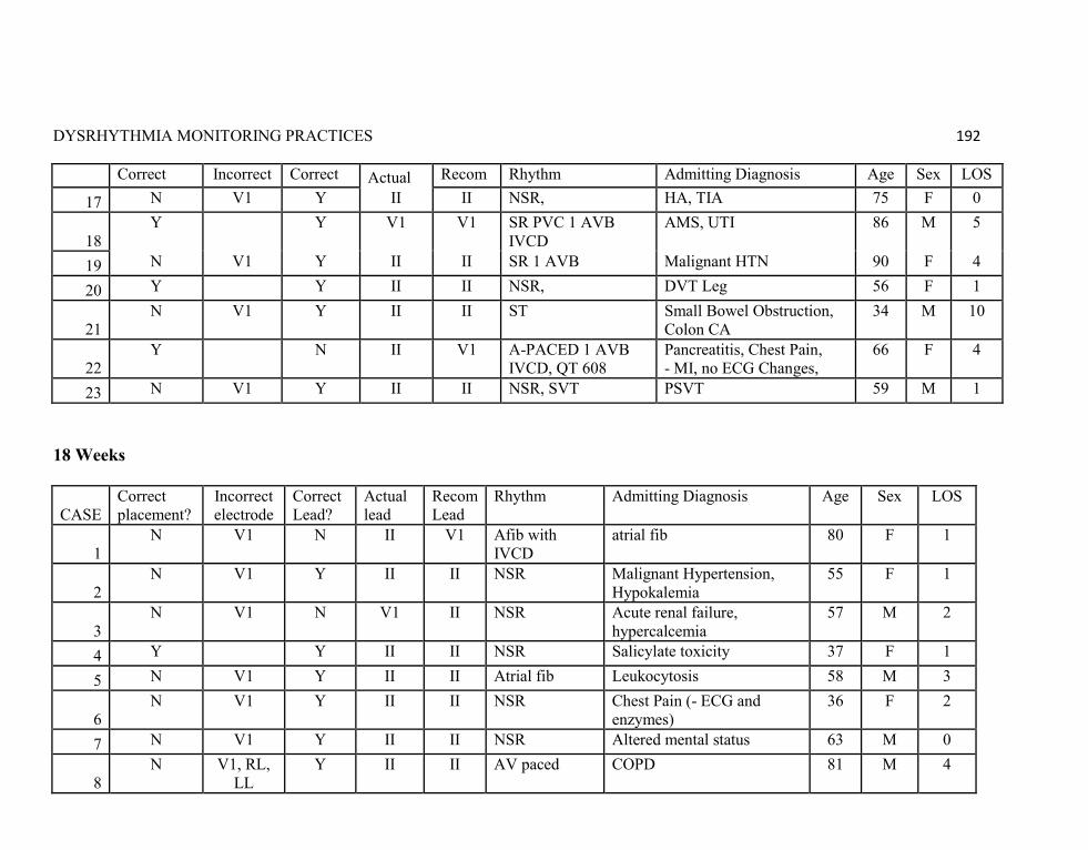
DYSRHYTHMIA MONITORING PRACTICES 192
Correct placement?
Incorrect electrode
Correct Lead?
Actual Recom Lead
Rhythm Admitting Diagnosis Age Sex LOS
17 N V1 Y II II NSR, HA, TIA 75 F 0
18 Y Y V1 V1 SR PVC 1 AVB
IVCD AMS, UTI 86 M 5
19 N V1 Y II II SR 1 AVB Malignant HTN 90 F 4
20 Y Y II II NSR, DVT Leg 56 F 1
21 N V1 Y II II ST Small Bowel Obstruction,
Colon CA 34 M 10
22 Y N II V1 A-PACED 1 AVB
IVCD, QT 608 Pancreatitis, Chest Pain, - MI, no ECG Changes,
66 F 4
23 N V1 Y II II NSR, SVT PSVT 59 M 1 18 Weeks
CASE Correct placement?
Incorrect electrode
Correct Lead?
Actual lead
Recom Lead
Rhythm Admitting Diagnosis Age Sex LOS
1 N V1 N II V1 Afib with
IVCD atrial fib 80 F 1
2 N V1 Y II II NSR Malignant Hypertension,
Hypokalemia 55 F 1
3 N V1 N V1 II NSR Acute renal failure,
hypercalcemia 57 M 2
4 Y Y II II NSR Salicylate toxicity 37 F 1
5 N V1 Y II II Atrial fib Leukocytosis 58 M 3
6 N V1 Y II II NSR Chest Pain (- ECG and
enzymes) 36 F 2
7 N V1 Y II II NSR Altered mental status 63 M 0
8 N V1, RL,
LL Y II II AV paced COPD 81 M 4

DYSRHYTHMIA MONITORING PRACTICES 193
CASE Correct placement?
Incorrect electrode
Correct Lead?
Actual lead
Recom Lead
Rhythm Admitting Diagnosis Age Sex LOS
9 N V1 N II V1 NSR with
IVCD Chest Pain (- ECG and enzymes), Pneumonia
52 M 2
10 Y Y V1 V1 ST with IVCD Diarrhea, dehydration 71 F 20
11 N V1 Y II II ST SOB 74 M 1
12 N V1, LL Y II II A-Flutter
(Classes Full) Chest Pain (-ECG, enzymes), CHF
83 F 9
13 N V1 Y V1 V1 V paced Pneumonia 89 M 2
14 Y Y II II NSR, (Classes
Full) CAD, SOB 64 F 1
15 Y Y II II NSR GI Bleed, Abd pain 53 F 1
16 Y N II V1 SB with IVCD Hypoglycemia 86 M 5
17 Y Y II II NSR Abd pain, Asthma 47 F 14
18 Y Y II II NSR Dehydration, elevated LFT 36 F 5
19 Y Y II II NSR Odontogenic infection 31 F 2
20 N V1 Y II II NSR N/V Abd pain 59 F 1
21 N V1 Y II II NSR CHF, Cirrhosis 47 F 1
22 N V1 N II V1 NSR with PVC Syncope, Periorbital
Hematoma 75 F 8
23 N V1 Y II II Atrial Fib Sepsis 66 F 9
24 Y Y V1 V1 A-paced Left Shoulder pain 73 F 9

DYSRHYTHMIA MONITORING PRACTICES 194
Appendix LL: Patient Audit Results
Item Baseline
2-6 Weeks (by nurses)
6 Weeks
18 Weeks
Number included
30 20 23 24
Placement correct
13(43%) 6 (30%) 9 (39%) 9 (38%)
Placement Incorrect
17 (57%) 14 (70%) 14 (61%) 15 (63%)
V1 Incorrect Electrode
16/30 (53%) 16/17 (94%)
14/20 (70%) 14/14 (100%)
14/23 (61%) 14/14 (100%)
15/24 (63%) 15/15 (100%)
LL Incorrect Electrode
2/30 (6%) 2/17 (12%)
0 0 1/24 (4%) 1/15 (7%)
LA Incorrect Electrode
1/30 (3%) 1/17 (6%)
0 1/23 (4%) 1/14 (7%)
0
Monitored in Lead II
24 (80%) N/A 18 (78%) 20(83%)
Monitored in V1
0 (0%) N/A 5 (22%) 4 (17%)
Monitored in other leads
6 (20%) N/A 0 0
Correct lead
21 (70%) 12 (60%) 15 (65%) 19 (79%)
Incorrect lead
9 (30%) 8 (40%) 8 (35%) 5 (21%)
Lead V1 used when indicated
0/3 (0%) N/A 2/7 (29%) 3/7 (43%)
Lead II used when indicated
21/27 (78%) N/A 13/16 (81%) 16/17 (94%)

DYSRHYTHMIA MONITORING PRACTICES 195
Appendix MM: Audits of 4 CenterGraph
* no statistically significant differences between baseline/6 weeks, baseline/18 weeks, and 6 weeks/18 weeks (p> 0.05)
43% 39% 38%
70%65%
79%
0%
10%
20%
30%
40%
50%
60%
70%
80%
90%
100%
Baseline* 6 weeks* 18 weeks*
Audits of 4 CenterElectrode Placement and Lead Selection
Correct Placement
Correct Lead
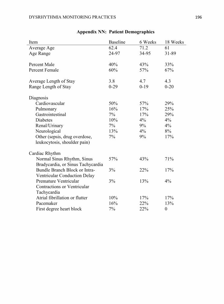
DYSRHYTHMIA MONITORING PRACTICES 196
Appendix NN: Patient Demographics
Item Baseline 6 Weeks 18 Weeks Average Age 62.4 71.2 61 Age Range 24-97 34-95 31-89 Percent Male 40% 43% 33% Percent Female 60% 57% 67% Average Length of Stay 3.8 4.7 4.3 Range Length of Stay 0-29 0-19 0-20 Diagnosis
Cardiovascular 50% 57% 29% Pulmonary 16% 17% 25% Gastrointestinal 7% 17% 29% Diabetes 10% 4% 4% Renal/Urinary 7% 9% 4% Neurological 13% 4% 8% Other (sepsis, drug overdose, leukocytosis, shoulder pain)
7% 9% 17%
Cardiac Rhythm
Normal Sinus Rhythm, Sinus Bradycardia, or Sinus Tachycardia
57% 43% 71%
Bundle Branch Block or Intra-Ventricular Conduction Delay
3% 22% 17%
Premature Ventricular Contractions or Ventricular Tachycardia
3% 13% 4%
Atrial fibrillation or flutter 10% 17% 17% Pacemaker 16% 22% 13% First degree heart block 7% 22% 0
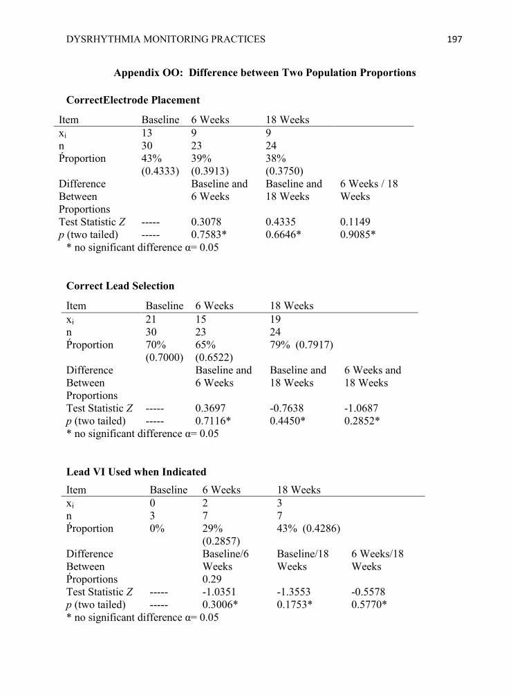
DYSRHYTHMIA MONITORING PRACTICES 197
Appendix OO: Difference between Two Population Proportions
CorrectElectrode Placement
Item Baseline 6 Weeks 18 Weeks xi 13 9 9 n 30 23 24 Ṕroportion 43%
(0.4333) 39% (0.3913)
38% (0.3750)
Difference Between Proportions
Baseline and 6 Weeks
Baseline and 18 Weeks
6 Weeks / 18 Weeks
Test Statistic Z ----- 0.3078 0.4335 0.1149 p (two tailed) ----- 0.7583* 0.6646* 0.9085*
* no significant difference α= 0.05
Correct Lead Selection
Item Baseline 6 Weeks 18 Weeks xi 21 15 19 n 30 23 24 Ṕroportion 70%
(0.7000) 65% (0.6522)
79% (0.7917)
Difference Between Proportions
Baseline and 6 Weeks
Baseline and 18 Weeks
6 Weeks and 18 Weeks
Test Statistic Z ----- 0.3697 -0.7638 -1.0687 p (two tailed) ----- 0.7116* 0.4450* 0.2852* * no significant difference α= 0.05 Lead VI Used when Indicated Item Baseline 6 Weeks 18 Weeks xi 0 2 3 n 3 7 7 Ṕroportion 0% 29%
(0.2857) 43% (0.4286)
Difference Between Ṕroportions
Baseline/6 Weeks 0.29
Baseline/18 Weeks
6 Weeks/18 Weeks
Test Statistic Z ----- -1.0351 -1.3553 -0.5578 p (two tailed) ----- 0.3006* 0.1753* 0.5770* * no significant difference α= 0.05

DYSRHYTHMIA MONITORING PRACTICES 198
Lead II Used when Indicated
Item Baseline 6 Weeks 18 Weeks xi 21 13 16 n 27 16 17 Ṕroportion 78%
(0.7778) 81% (0.8125)
94% (0.9412)
Difference Between Proportions
Baseline/6 Weeks
Baseline/18 Weeks
6 Weeks/18 Weeks
Test Statistic Z ----- -0.2705 -1.4429 -1.1319 p (two tailed) ----- 0.7867* 0.1491* 0.2577* * no significant difference α= 0.05

DYSRHYTHMIA MONITORING PRACTICES 199
References
AACN practice alert: Dysrhythmia monitoring. (2008). Critical Care Nurse 28(5), 90-
91. Also available online from American Association of Critical-Care Nurses website at http://www.aacn.org/WD/Practice/Docs/Dysrhythmia_Monitoring_04-2008.pdf
Audit Tool: Electrode placement and Lead Selection (2008). American Association of
Critical-Care Nurses Retrieved from http://www.aacn.org/WD/Practice/Docs/PracticeAlerts/Electrode_Placement-Lead_Selection_Audit_Tool_04-2008.pdf
Belcher, J., & Vonderhaar, K. J. (2005). Staff education: Web-delivered research-based
nursing staff education for seeking magnet status. Journal of Nursing Administration, 35(9), 382-386.
Cadden, S. M. (2007). Educating nurses about cardiac monitoring in a stroke unit.
Journal for Nurses in Staff Development, 23(1), 20-25. Chickering, A. W., & Gamson, Z. F. (1987). Seven principles for good practice in
undergraduate education. American Association for Higher Education (AAHE) Bulletin, 39 (7), 3-7.Retrieved from http://www.aahea.org/bulletins/articles/sevenprinciples1987.htm
Chickering, A. W., & Ehrmann, S. C. (1996). Implementing the seven principles:
Technology as lever. American Association for Higher Education (AAHE) Bulletin, 49(2), 3-6. Retrieved from http://www.aahea.org/bulletins/articles/sevenprinciples.htm
Code of Federal Regulations. (2009). Title 45 Public Welfare, Part 46 Protection of
human subjects. Department of Health and Human Services. Retrieved from, http://www.hhs.gov/ohrp/documents/OHRPRegulations.pdf and http://www.hhs.gov/ohrp/humansubjects/guidance/expedited98.htm
Copyright basics: Fair use. (2008). The campus guide to copyright compliance.
Copyright Clearance Center, Inc. Retrieved from http://www.copyright.com/Services/copyrightoncampus/basics/fairuse_rules.html
Daniel, W. W. (2005). Biostatistics: A foundation for analysis in the health sciences.
(8th Ed.). Hoboken, NJ: Wiley. Drew, B. J. (2002). Cardiac monitoring (rev), pocket card. Phillips PN 5990-0487,
American Association of Critical-Care Nurses, Downloaded from http://www.aacn.org/WD/Practice/Docs/REV4_02_pocket_card.pdf
DYSRHYTHMIA MONITORING PRACTICES 200
Drew, B. J. (2007). Pulling it all together: Case studies on ECG monitoring. AACN Advanced Critical Care, 18(3), 306-317.
Drew, B. J., Ackerman, M. J., Funk, M., Gibler, W. B., Kligfield, P., Menon, V. …
Zareba, W. (2010). Prevention of torsade de pointes in hospital settings: A scientific statement from the American Heart Association and the American College of Cardiology. Circulation, 121, 1047-1060. doi: 10.1161/CIRCULATIONAHA.109.19270
Drew, B. J., Califf, R. M., Funk, M., Kaufman, E. S., Krucoff, M. W., Laks, M. M.
…Van Hare, G. F. (2004). Practice standards for electrocardiographic monitoring in hospital settings. Circulation. 110: 2721-2746. doi: 10.1161/01.CIR0000145144.56673.59 Also available at National Guideline Clearinghouse website http://guideline.gov/summary/summary.aspx?doc_id=6181&nbr=003980&string=Practice+AND+standards+AND+electrocardiographic
Drew, B. J., & Funk, M. (2006). Practice standards for ECG monitoring in hospital
settings: Executive summary and guide for implementation. Critical Care Nursing Clinics of North America. 18, 157-168.
Dumpe, M. L., Kanyak, N., & Hill, K. (2007). Use of an automated learning management
system to validate nursing competencies. Journal for Nurses in Staff Development. 23(4), 183-185.
Durkin, G. J. (2008). A comparison of the effectiveness of computer-based learning
courses among nursing staff. Journal for Nurses in Staff Development, 24(2), 62-66. Frith, K. H., & Kee, C. C. (2003). The effect of communication on nursing student
outcomes in a web-based course. Journal of Nursing Education, 42(8), 350-358. Funk, M., Hurley, E., May, J., Winkler, C., Stephens, K., Fennie, K., … Drew. (2009)
Substandard quality of ECG monitoring in current clinical practice: Preliminary results of the PULSE Trial. Paper presented at the American Heart Association‘s Scientific Sessions in Orlando, FL
Hanson, D., Hoss, B. L., & Wesorick, B. (2008). Evaluating the evidence: Guidelines.
AORN Journal, 88(2), 184-196. Horton, W. (2006). E-learning by design. San Francisco, CA: Pfeiffer. Jacobson, C. (2006). Tools for teaching arrhythmias: Wide QRS beats and rhythms—Part
I; P waves, fusion, and capture beats. AACN Advanced Critical Care, 17(4), 462-465.

DYSRHYTHMIA MONITORING PRACTICES 201
Jacobson, C. (2007). Tools for teaching arrhythmias: Wide QRS beats and rhythms—Part II; QRS morphology clues. AACN Advanced Critical Care, 18(1), 91-96.
Jang, K. S., Hwang, S. Y., Park, S. J., Kim, Y. M., & Kim, M. J. (2005). Research briefs:
Effects of web-based teaching method on undergraduate nursing students' learning of electrocardiography. Journal of Nursing Education, 44(1), 35-39.
Jeffries, P. R. (2005). Development and testing of a hyperlearning model for design of an
online critical care course. Journal of Nursing Education, 44(8), 366-372. Jeffries, P. R., Woolf, S., & Linde, B. (2003). Technology-based vs. traditional: A
comparison of two instructional methods to teach the skill of performing a 12-lead ECG. Nursing Education Perspectives, 24(2), 70-74.
Keller, K. B. & Raines, D. A. (2005). Arrhythmia knowledge: A qualitative study.
Heart & Lung: The Journal of Acute and Critical Care, 34 (5), 309-316. Koeckertiz, J., Malkiewicz, J., & Henderson, A. (2002). The seven principles of good
practice: Applications for online education in nursing. Nurse Educator, 27(6), 283-287.
Lloyd-Jones, D., Adams, R., Carnethon, M., De Simone, G., Ferguson, T. B., Flegal, K.,
… Hong, Y. (2009). Heart disease and stroke statistics 2009 update: A report from the American Heart Association Statistics Committee and Stroke Statistics Subcommittee. Circulation, 119: e21-e181. doi: 10.1161/CIRCULATIONAHA.108.191261.
Magnet Recognition Program Overview. (2009). American Nurses Credentialing Center.
Retrieved from http://www.nursecredentialing.org/Magnet/ProgramOverview.aspx
Malloch, K., & Porter-O‘Grady, T. (2010). Introduction to evidence-based practice in
nursing and health care (2nd ed.). Boston, MA: Jones and Bartlett. McClennen, S., Nathanson, L. A., Safran, D., & Goldberger, A. L. (2003). ECG wave-
maven: An internet-based electrocardiography self-assessment program for students and clinicians. Medical Education Online, 8(2), 1-5, available from http://www.med-ed-online.org
Morris, L. L., Pfeifer, P., Catalano, R., Fortney, R., Nelson, G., Rabito, R., & Harap, R.
(2009). Outcome evaluation of a new model of critical care orientation. American Journal of Critical Care, 18(3), 252-259.
Nathanson, L. A., McClennen, S., Safran, C., & Goldberger, A. L. (2007). ECG Wave-
Maven: Self-Assessment Program for Students and Clinicians. http://ecg.bidmc.harvard.edu

DYSRHYTHMIA MONITORING PRACTICES 202
Richards, N. M. (2008). Practice alert dysrhythmia monitoring. Power point slide
educational presentation, American Association of Critical-Care Nurses, retrieved from http://www.aacn.org/WD/Practice/Content/practicealerts.pcms?menu=Practice
Pelter, M. M., & Carey, M. G. (2006). Long QT syndrome. American Journal of Critical
Care, 15(4), 437-438. Sommargren, C. E., & Drew, B. J. (2007). Preventing torades de pointes by careful
cardiac monitoring in hospital settings. AACN Advanced Critical Care, 18(3), 285-293.
Urden, L., Stacy, K. M., & Lough, M. E. (2010). Critical Care Nursing (6th ed.). St. Louis, MO: Mosby Elsevier. VanArsdale, S. K. (1998). Teaching basic dysrhythmias: What is the best approach?
Journal of Continuing Education in Nursing, 29(5), 217-220. Zipes, D. P., Camm, A. J., Borggrefe, M., Buxton, A. E, Chaitman, B., Fromer, M. ...
Zamorano, J. L. (2006). ACC/AHA/ESC 2006 guidelines for management of patients with ventricular arrhythmias and the prevention of sudden cardiac death: A report of the American College of Cardiology/American Heart Association Task Force and the European Society of Cardiology Committee for Practice Guidelines. Journal of the American College of Cardiology, 48(5), 247-346. doi: 10.1161/CIRCULATIONAHA.106.178233Also available at National Guideline Clearinghouse website http://guideline.gov/summary/summary.aspx?doc_id=9725&nbr=005208&string=ACC%2fAHA%2fESC+AND+2006+AND+ventricular+AND+arrhythmias

DYSRHYTHMIA MONITORING PRACTICES 203
P H O N E W ( 9 0 4 ) 7 6 6 - 6 5 0 2 , C ( 9 0 4 ) 6 0 8 - 6 5 8 3
E - M A I L s sc hu l t z@ f sc j . e du
S U S A N J A N E S C H U L T Z D N P , C N E , C C R N
December, 2010
SUMMARY OF QUALIFICATIONS
Currently work as Professor of Nursing in A.S.N. and B.S.N
programs at Florida State College at Jacksonville, teaching nursing students medical/surgical or critical care concepts and supervising their clinical performance.
Completed Doctor of Nursing Practice program in December, 2010.
Certified as Certified Nurse Educator, Critical Care Registered Nurse, and Online Professor
Maintain clinical competency by working PRN as staff nurse on telemetry units
Use creative teaching techniques and human patient simulators to stimulate learning and retention.
EDUCATION
2008 – 2010 University of North Florida Jacksonville, Florida
Doctor of Nursing Practice
Administrator tract
1988-1992 University of Florida Gainesville, Florida
Master of Science in Nursing
Emphasis on role of Educator in Adult Health Nursing with focus on cardiovascular diseases.
Completed and published thesis on research about effectiveness of educational and behavioral strategies in cardiac risk factor modification
1987 or 1988 Florida Community College Jacksonville, Florida
Statistics course required for MSN program
1978-1982 Marycrest College Davenport, Iowa
Bachelor of Science in Nursing and Music Minor (cum laude)

DYSRHYTHMIA MONITORING PRACTICES 204
PROFESSIONAL EXPERIENCE
Florida State College at Jacksonville, Jacksonville, Florida
2004 – Present Professor of Nursing
Teach B.S.N. students critical care concepts and procedures, and supervise their practicums since 2009
Teach A.S.N. students medical/surgical concepts, nursing skills, and supervise their clinical performance.
Utilize Human Patient Simulators and instruct other faculty how to integrate it in their curriculum.
Chairperson of Nursing Curriculum Committee (A.S.N.)
Chairperson for National League of Nursing Accrediting Commission self-study report for A.S.N. program
Course Coordinator for 4th term (A.S.N.)
St. Vincent’s Medical Center Jacksonville, Florida
2004 – present Staff Nurse Telemetry Units PRN 4C/4E Telemetry
Deliver and manage quality care for team of 4-6 telemetry patients with variety of medical or surgical diagnoses PRN (two shifts per month)
1998 – 2004 Nursing Clinical Educator, Telemetry/ Medical/Surgical Units
Developed, taught, and evaluated effectiveness of orientation, dysrhythmia, and internship courses
Collaborated with multi-disciplinary teams to analyze fall and skin breakdown prevention programs and assisted with implementation and evaluation of new policies to improve patient safety and quality of care
Developed new protocols with multi-disciplinary teams for monitoring patients on cardiac IV drips and educating patients before joint replacement surgery
1997 – 1998 Critical Care Staff Nurse, CCU and 4 East ICU
1992 – 1997 Nursing Educator, Telemetry Units
DYSRHYTHMIA MONITORING PRACTICES 205
ACCREDITATIONS AND AWARDS
Certified Nurse Educator, by National League of Nursing, 2008 – present.
Critical Care Registered Nurse, by American Association of Critical-Care Nurses, 1998 – present
Online Professor Certification, Florida Community College at Jacksonville, Jacksonville, FL, anticipated completion December, 2008.
Innovative Excellence in Teaching, Learning, and Technology, Award Recipient April, 2008, at 19th International Conference on College Teaching and Learning, Florida Community College at Jacksonville, FL for development of songs and raps to stimulate learning, posted on faculty web page: www1.fccj.org/sschultz
Advanced Cardiac Life Support, by American Heart Association, Instructor: 2001-2003, Provider: 1992 – present
Basic Cardiac Life Support, by American Heart Association, Instructor: 1988-2003, Provider: 1982 – present
PRESENTATIONS AND PUBLICATIONS
“Dysrhythmia Monitoring Practices -- Research Proposal”, poster presentation at JANIE conference, Jacksonville, FL, on March 12, 2010. Poster will also be presented at Sigma Theta Tau International, Lambda Rho Chapter-at-Large, Jacksonville, FL, on April 26, 2010.
“Stimulate when you Simulate”, presented at Florida Community College, Jacksonville, FL on March 10 and 17, 2007 for 2 Contact Hours.
“Make a Sensation with Medication Education”, presented three times: National Institute for Staff & Organizational Development (NISOD), Austin, TX in May 26, 2009; International Conference on College Teaching and Learning, Ponte Vedra, FL on April 5, 2007; and Florida Summit on Nursing Education, Valencia Community College, Orlando, FL on May 16, 2007 for 1.5 Contact Hours
“Rhythms, Rhymes, and Raps”, Creative Solutions Poster Presentation at National Teaching Institute of Critical Care Nurses Association, 2002
“Who Knows the E.S.O.s?” (Emergency Standing Orders), Creative Solutions Poster Presentation at National Teaching Institute of Critical Care Nurses Association, 2001