Differentiation HC2S2 of the v-myc oncogenev-myc oncogene is transcribed in a tetracycline-regulated...
6
Proc. Natl. Acad. Sci. USA Vol. 93, pp. 1518-1523, February 1996 Neurobiology Differentiation of the immortalized adult neuronal progenitor cell line HC2S2 into neurons by regulatable suppression of the v-myc oncogene (retroviral vector/tetracycline/electrophysiology) MINORU HOSHIMARUtt, JASODHARA RAYt, DINAH W. Y. SAH§, AND FRED H. GAGEt¶ tLaboratory of Genetics, The Salk Institute for Biological Studies, P.O. Box 85800, San Diego, CA 92186-5800; and §Signal Pharmaceuticals, San Diego, CA Communicated by Roger Guillemin, Scripps Memorial Hospital, La Jolla, CA, October 19, 1995 ABSTRACT A regulatable retroviral vector in which the v-myc oncogene is driven by a tetracycline-controlled trans- activator and a human cytomegalovirus minimal promoter fused to a tet operator sequence was used for conditional immortalization of adult rat neuronal progenitor cells. A single clone, HC2S2, was isolated and characterized. Two days after the addition of tetracycline, the HC2S2 cells stopped proliferating, began to extend neurites, and expressed the neuronal markers tau, NeuN, neurofilament 200 kDa, and glutamic acid decarboxylase in accordance with the reduced production of the v-myc oncoprotein. Differentiated HC2S2 cells expressed large sodium and calcium currents and could fire regenerative action potentials. These results suggest that the suppression of the v-myc oncogene may be sufficient to make proliferating cells exit from cell cycles and induce terminal differentiation. The HC2S2 cells will be valuable for studying the differentiation process of neurons. Much of our understanding of the molecular mechanisms that control the development and the function of nervous system cells has been derived from studies of cells in culture. Clonal cultures of progenitor cells are useful for studying aspects of the differentiation pathways leading to the formation of ma- ture neurons or glia. Primary cultures of progenitor cells, being heterogeneous in nature, do not offer such an opportunity. As an alternative approach, neural cell lines have been generated by various techniques (1-5). One common approach has been to immortalize cells by transducing neuroepithelial or neural progenitor cells from developing brain with retroviral vectors encoding the simian virus 40 (SV40) large tumor (T) antigen or myc oncogenes (1-12). Studies have shown that the immor- talization process arrests cells at specific stages of development and prevents their terminal differentiation (9). As a result, cells at specific stages in the development can be immortalized and used to study the nature and potentiality of the cells at this particular stage in the lineage and how they can be further differentiated down that particular pathway. In neural cell lines developed by constitutively expressing oncogenes such as myc, the mitotic activity of the oncogene is always present, and cells proliferate continuously in culture (6, 9, 13). Differentiation of the immortalized progenitor cells into neurons may require sufficient down-regulation of the onco- gene, but attempts to induce such terminal differentiation often induce apoptosis (14). However, partial differentiation of c-myc-immortalized cells has been achieved by treatment with growth factors (6). To obtain a regulatable expression of the oncogene, a temperature-sensitive mutant of SV40 large T antigen (tsA58) has been used for conditional immortalization (3, 7, 8, 10-12, 15). At a permissive temperature when the large T antigen is expressed, the immortalized cells are undifferen- The publication costs of this article were defrayed in part by page charge payment. This article must therefore be hereby marked "advertisement" in accordance with 18 U.S.C. §1734 solely to indicate this fact. tiated and are multipotent in nature (3, 7, 8), as has been seen with myc-immortalized cells (6). Although at nonpermissive temperature the expression of T antigen is considerably down- regulated and the cells are not under the mitotic drive of the oncogene, the temperature shift results in only incomplete I differentiation of cells into neurons (7, 8, 11, 12, 15). A combination of factors and substrates is needed to further differentiate the cells in vitro (12). A simpler system in which the regulatable suppression of oncogene expression in immor- talized neuronal progenitor cells could allow their differenti- ation into neurons without the help of complex factors and substrates would be useful. To build a system in which the oncogene expression can be regulated by exogenous agents, we have taken advantage of a tetracycline-controlled gene expression system (16-19). In this system, a tetracycline-controlled transactivator (tTA), which is a fusion protein of the repressor (tetR) of the Tn1O-derived tetracycline-resistance operon of Escherichia coli and the acidic domain of VP16 of herpes simplex virus, strongly activates transcription from PhCMV*-I, a minimal promoter from human cytomegalovirus (hCMV) fused to the tetracy- cline (tet) operator sequences in the absence of tetracycline. Low concentrations of tetracycline (0.01-1.0 ,.tg/ml), at which no toxic effect is evident, almost completely abolishes tran- scription activation by tTA (16, 17). Using this vector system, we constructed a retroviral vector (LINXv-myc) in which the v-myc oncogene is transcribed in a tetracycline-regulated fashion. Neural progenitor cells cultured from adult rat hip- pocampus were transduced with the retroviral vector LINXv- myc, and a stably transfected colony was isolated and grown. Here we report that tetracycline suppresses the production of v-myc and allows the progenitor cells to terminally differen- tiate into neurons. Since the progenitor cell line differentiates into neurons only, we may have immortalized a cell that has already been committed to a neuronal lineage. This cell line will be useful for studying the mechanisms of cell differenti- ation in vitro. EXPERIMENTAL PROCEDURES Vector Construction. The retroviral vector LINXv-myc was constructed as follows: the 1.02-kb EcoRI-BamHI fragment of the tTA was excised from pUHD15-1 (16) and inserted into pHENA (20) just upstream of the internal ribosome entry site (IRES) of the encephalomyocarditis virus. The resultant 2.78-kb EcoRV-BamHI fragment containing tTA, IRES, and Abbreviations: FGF-2, basic fibroblast growth factor; tTA, tetracy- cline-controlled transactivator; IRES, internal ribosomal entry site; GAD, glutamic acid decarboxylase; GFAP, glia fibrillary acidic pro- tein; SV40, simian virus 40; T, tumor; LTR, long terminal repeat; NFH, neurofilament 200 kDa. tPresent address: Department of Neurosurgery, Kyoto University Hospital, 54 Shogoin Kawahara-cho, Sakyo-ku, Kyoto, 606 Japan. ITo whom reprint requests should be addressed. 1518 Downloaded by guest on July 30, 2020
Transcript of Differentiation HC2S2 of the v-myc oncogenev-myc oncogene is transcribed in a tetracycline-regulated...

Proc. Natl. Acad. Sci. USAVol. 93, pp. 1518-1523, February 1996Neurobiology
Differentiation of the immortalized adult neuronal progenitor cellline HC2S2 into neurons by regulatable suppression of thev-myc oncogene
(retroviral vector/tetracycline/electrophysiology)
MINORU HOSHIMARUtt, JASODHARA RAYt, DINAH W. Y. SAH§, AND FRED H. GAGEt¶tLaboratory of Genetics, The Salk Institute for Biological Studies, P.O. Box 85800, San Diego, CA 92186-5800; and §Signal Pharmaceuticals, San Diego, CA
Communicated by Roger Guillemin, Scripps Memorial Hospital, La Jolla, CA, October 19, 1995
ABSTRACT A regulatable retroviral vector in which thev-myc oncogene is driven by a tetracycline-controlled trans-activator and a human cytomegalovirus minimal promoterfused to a tet operator sequence was used for conditionalimmortalization of adult rat neuronal progenitor cells. Asingle clone, HC2S2, was isolated and characterized. Two daysafter the addition of tetracycline, the HC2S2 cells stoppedproliferating, began to extend neurites, and expressed theneuronal markers tau, NeuN, neurofilament 200 kDa, andglutamic acid decarboxylase in accordance with the reducedproduction of the v-myc oncoprotein. Differentiated HC2S2cells expressed large sodium and calcium currents and couldfire regenerative action potentials. These results suggest thatthe suppression of the v-myc oncogene may be sufficient tomake proliferating cells exit from cell cycles and induceterminal differentiation. The HC2S2 cells will be valuable forstudying the differentiation process of neurons.
Much of our understanding of the molecular mechanisms thatcontrol the development and the function of nervous systemcells has been derived from studies of cells in culture. Clonalcultures of progenitor cells are useful for studying aspects ofthe differentiation pathways leading to the formation of ma-ture neurons or glia. Primary cultures of progenitor cells, beingheterogeneous in nature, do not offer such an opportunity. Asan alternative approach, neural cell lines have been generatedby various techniques (1-5). One common approach has beento immortalize cells by transducing neuroepithelial or neuralprogenitor cells from developing brain with retroviral vectorsencoding the simian virus 40 (SV40) large tumor (T) antigenor myc oncogenes (1-12). Studies have shown that the immor-talization process arrests cells at specific stages of developmentand prevents their terminal differentiation (9). As a result, cellsat specific stages in the development can be immortalized andused to study the nature and potentiality of the cells at thisparticular stage in the lineage and how they can be furtherdifferentiated down that particular pathway.
In neural cell lines developed by constitutively expressingoncogenes such as myc, the mitotic activity of the oncogene isalways present, and cells proliferate continuously in culture (6,9, 13). Differentiation of the immortalized progenitor cells intoneurons may require sufficient down-regulation of the onco-gene, but attempts to induce such terminal differentiationoften induce apoptosis (14). However, partial differentiationof c-myc-immortalized cells has been achieved by treatmentwith growth factors (6). To obtain a regulatable expression ofthe oncogene, a temperature-sensitive mutant of SV40 large Tantigen (tsA58) has been used for conditional immortalization(3, 7, 8, 10-12, 15). At a permissive temperature when the largeT antigen is expressed, the immortalized cells are undifferen-
The publication costs of this article were defrayed in part by page chargepayment. This article must therefore be hereby marked "advertisement" inaccordance with 18 U.S.C. §1734 solely to indicate this fact.
tiated and are multipotent in nature (3, 7, 8), as has been seenwith myc-immortalized cells (6). Although at nonpermissivetemperature the expression of T antigen is considerably down-regulated and the cells are not under the mitotic drive of theoncogene, the temperature shift results in only incomplete
I differentiation of cells into neurons (7, 8, 11, 12, 15). Acombination of factors and substrates is needed to furtherdifferentiate the cells in vitro (12). A simpler system in whichthe regulatable suppression of oncogene expression in immor-talized neuronal progenitor cells could allow their differenti-ation into neurons without the help of complex factors andsubstrates would be useful.To build a system in which the oncogene expression can be
regulated by exogenous agents, we have taken advantage of atetracycline-controlled gene expression system (16-19). In thissystem, a tetracycline-controlled transactivator (tTA), which isa fusion protein of the repressor (tetR) of the Tn1O-derivedtetracycline-resistance operon of Escherichia coli and theacidic domain of VP16 of herpes simplex virus, stronglyactivates transcription from PhCMV*-I, a minimal promoterfrom human cytomegalovirus (hCMV) fused to the tetracy-cline (tet) operator sequences in the absence of tetracycline.Low concentrations of tetracycline (0.01-1.0 ,.tg/ml), at whichno toxic effect is evident, almost completely abolishes tran-scription activation by tTA (16, 17). Using this vector system,we constructed a retroviral vector (LINXv-myc) in which thev-myc oncogene is transcribed in a tetracycline-regulatedfashion. Neural progenitor cells cultured from adult rat hip-pocampus were transduced with the retroviral vector LINXv-myc, and a stably transfected colony was isolated and grown.Here we report that tetracycline suppresses the production ofv-myc and allows the progenitor cells to terminally differen-tiate into neurons. Since the progenitor cell line differentiatesinto neurons only, we may have immortalized a cell that hasalready been committed to a neuronal lineage. This cell linewill be useful for studying the mechanisms of cell differenti-ation in vitro.
EXPERIMENTAL PROCEDURESVector Construction. The retroviral vector LINXv-myc was
constructed as follows: the 1.02-kb EcoRI-BamHI fragment ofthe tTA was excised from pUHD15-1 (16) and inserted intopHENA (20) just upstream of the internal ribosome entry site(IRES) of the encephalomyocarditis virus. The resultant2.78-kb EcoRV-BamHI fragment containing tTA, IRES, and
Abbreviations: FGF-2, basic fibroblast growth factor; tTA, tetracy-cline-controlled transactivator; IRES, internal ribosomal entry site;GAD, glutamic acid decarboxylase; GFAP, glia fibrillary acidic pro-tein; SV40, simian virus 40; T, tumor; LTR, long terminal repeat;NFH, neurofilament 200 kDa.tPresent address: Department of Neurosurgery, Kyoto UniversityHospital, 54 Shogoin Kawahara-cho, Sakyo-ku, Kyoto, 606 Japan.ITo whom reprint requests should be addressed.
1518
Dow
nloa
ded
by g
uest
on
July
30,
202
0

Proc. Natl. Acad. Sci. USA 93 (1996) 1519
neomycin phosphotransferase was inserted into the polylinkersite (Hap I and BamHI) of LXSHD (21). Then, the portion ofLXSHD spanning the SV40 early promoter and histidinoldehydrogenase cDNA was replaced by a PhCMV*-1-v-myc (3.32kb) fragment. PhCMv-l was derived from pUHD10-3, and a2.87-kb Sac I-Sph I fragment of v-myc was derived frompMC38 (American Type Culture Collection).
Cell Culture. All packaging and producer cell lines werecultured in Dulbecco's modified Eagle's medium (DMEM)with 10% fetal bovine serum. qp-2 cells were transfected with10 ,ug of LINXv-myc plasmid DNA by the calcium phosphateprocedure. Virus-containing medium collected from qi-2 cells2 days after transfection was used to infect the amphotropicPA317 cells. One day after the infection, the cells were split ata 1:10 ratio, plated, and selected for G418 (400 jig/ml)resistance. Colonies were picked after selection for 8 days andtested for the proper integration of the vector, production ofv-myc, and the titer of the retrovirus.
Cells from adult (3-month-old) rat hippocampus were iso-lated and cultured as described (22) with the following mod-ifications. Cells isolated from tissue after enzymatic dissocia-tion were resuspended in DMEM/Ham's F-12 (1:1, vol/vol)high glucose medium (Irvine Scientific) containing 10% fetalbovine serum and then plated onto uncoated plastic tissueculture flasks (1 X 10⁶ cells per 75 cm2 flask). The cells weregrown at 37°C in a 5% CO2 incubator. The next day, theserum-containing medium was replaced with serum-freeDMEM/Ham's F-12 medium containing N-2 supplement (in-sulin at 5 ,ug/ml, human transferrin at 50 ,tg/ml, 20 nMprogesterone, 100 ,uM putrescine, 30 nM sodium selenite, 2.5mM glutamine; GIBCO) and basic fibroblast growth factor(FGF-2; human recombinant) at 20 ng/ml. Confluent culturesof cells were passaged to polyornithine/laminin-coated platesand cultured. Proliferating cultures, maintained for about ayear through 19 passages were split at a 1:3 ratio and 1 day laterinfected for 20 hr with a mixture of one volume of theconditioned medium of the producer cell line and two volumesof DMEM/Ham's F-12 containing N-2 supplements, FGF-2 at20 ng/ml, and Polybrene at 4 ,tg/ml. The infected cells weresplit at a 1:4 ratio and selected in the presence of G418 (100Kg/ml).Northern Blot Analyses. Total RNA was isolated by the
CsCl/guanidinium thiocyanate method (23). Fifteen micro-grams of total RNA was separated on formaldehyde/agarose(1.5%) gels and transferred onto a Magnagraph nylon mem-brane and probed with randomly primed v-myc or cyclophilin(pBlB15).Immunofluorescence Staining. Cells were plated in the
absence or presence of tetracycline (1 ,ug/ml) onto Lab-Tekglass chamber slides (Nunc) coated with polyornithin/lamininand cultured for 1 (without tetracycline) or 5 (with tetracy-cline) days and then fixed for 10 min with 4% paraformalde-hyde. The cells were incubated sequentially with the primaryantibody in PBS containing 4% donkey serum and 0.3% TritonX-100 overnight at 4°C followed by fluorescein isothiocyanate-conjugated secondary antibody (Jackson ImmunoresearchLaboratories; used at 1:500) for 4 hr at room temperature.Slides were mounted in 24% glycerol and 9.6% poly(vinylalcohol) containing 2.5% 1.4-diazobycyclo[2.2.2]octane. Themonoclonal antibodies used were to tau (BoehringerMannheim; used at 1:250), neurofilament 200 kDa (NFH)(clone RT97; Boehringer Mannheim; used at 2 mg/ml), andNeuN (used at 1:5; ref. 24); polyclonal antibodies used were tovimentin (Amersham; used at 1:10), glial fibrillary acidicprotein (GFAP) (Chemicon; used at 1:2000), nestin (used at1:10,000), and v-myc (Upstate Biotechnology; used at 1:8000).For immunofluorescent staining for bromodeoxyuridine (Brd-Urd), cells were incubated with BrdUrd (Amersham; used at1:1000) for 18 hr, fixed with 95% ethanol/5% acetic acid, andthen incubated with monoclonal anti-BrdUrd antibody (Am-
ersham; undiluted). Immunoreactivity was detected with anti-mouse biotinylated IgG followed by streptavidin-Texas Redconjugate.
Confocal microscopic images of cells were obtained using aBio-Rad MRC600 confocal microscope equipped with a kryp-ton/argon laser and coupled to a Zeiss Axiovert microscope.Images were collected sequentially using the K1/K2 filterblocks matched to the appropriate excitation filter for eachchannel. Transmitted light images of differential interferencecontrast optics were captured in registration with the fluores-cent signals using the transmitted field detector. Instrumentsettings to preclude background fluorescent signals were setagainst a negative control well where the primary antibodieshad been omitted; gain and black levels were adjusted topreclude any signal from this well before imaging other wellsfor positive signal. Collected digital images were composited inADOBE PHOTOSHOP 3.0 and printed on a Fujix Pictrography3000. Phase-contrast images were photographed on a NikonDiaphot using T-Max film and the negatives digitized using aLeaf Lumina. These images were printed as described above.
Reverse PCR Analysis. One hundred nanograms of totalRNA was reverse transcribed using avian myeloblastosis virusreverse transcriptase (Promega) with random hexamers (10,tM) as primers in a 20-,l reaction mixture containing 10 mMTris Cl (pH 8.4), 50 mM KCl, 3.8 mM MgCl2, 1 mM eachdATP, dTTP, dCTP, and dGTP, and 20 units of RNasin(Promega). After 75 min at 42°C, the reaction was terminatedby heat inactivation at 95°C for 5 min. For PCR amplification,several sets of specific oligonucleotide pairs (10 ng/,l) wereincubated with the above reaction mixture and 5 units of Taqpolymerase (Perkin-Elmer) in a 100-,l reaction mixture con-taining 10 mM Tris Cl (pH 8.4), 50 mM KCl, 1 mM MgCl2, and2 ,uCi of [a-32P]dCTP (1 Ci = 37 GBq). Cycle parameters were2 min at 94°C, 2 min at 60°C, and 2 min at 72°C for 23 cyclesfollowed by a final 10-min incubation at 72°C. Forty microlitersof each reaction was analyzed by electrophoresis on 8%polyacrylamide gels followed by autoradiography. Controlexperiments using primers for the rat ribosomal protein L27a(internal control) showed that the amount of amplified PCRproduct was directly proportional to the amount of input RNAafter 23 cycles of amplification (data not shown). The se-quences of genomic DNA between two primers were selectedso that they contained one or two introns for discriminationbetween products from RNA and contaminating genomicDNA. The following oligonucleotides were used as primers(nucleotide positions are shown in parentheses): rat ribosomalprotein L27a (25) 5' primer: 5'-ATCGGTAAGCACCG-CAAGCA-3' (69-88), 3' primer: 5'-GGGAGCAACTCCAT-TCTTGT-3' (302-283); rat GFAP (26) 5' primer: 5'-ACCT-CGGCACCCTGAGGCAG-3' (459-478), 3' primer: 5'-CCA-GCGACTCAACCTTCCTC-3' (599-580); rat glutamic aciddecarboxylase (GAD) (27) 5' primer: 5'-AAGGTTTTGGAC-TTCCACCAC-3' (589-609), 3' primer: 5'-CATAAGAA-CAAACACGGGTGC-3' (855-835); rat NFH (28) 5' primer:5'-GAGGAGATAACTGAGTACCG-3' (247-266), 3' primer:5'-CCAAAGCCAATCCGACACTC-3' (548-529).
Electrophysiology. The whole-cell configuration of thepatch clamp technique was used to study voltage-gated cur-rents. Pipettes (3- to 5-Mfl resistance) were pulled fromBoralex glass (Rochester Scientific), coated with Sylgard (DowCorning Corp.) and fire-polished. They were filled with inter-nal solution containing 108 mM cesium methanesulfonate, 4mM MgCl2, 9 mM EGTA 9 mM Hepes, 4 mM ATP, 14 mMcreatine phosphate (Tris salt), 0.3 mM GTP (Tris salt), andcreatine phosphokinase (50 units/ml) at pH 7.4 with CsOH.Whole-cell recordings were initially established in the bathsolution [Tyrode's: 150 mM NaCl, 4 mM KCl, 2mM MgCl2, 10mM glucose, 10 mM Hepes (pH 7.4 with NaOH), 2 mM CaCl2and, in some cases, 4 mM BaCl2 added]. Sodium currents werecharacterized in this bath solution, whereas calcium currents
Neurobiology: Hoshimaru et al.
Dow
nloa
ded
by g
uest
on
July
30,
202
0

1520 Neurobiology: Hoshimaru et al.
were assessed with external solution containing 160 mMtetraethylammonium chloride, 10 mM BaCl2, and 10 mMHepes (pH 7.4) with tetraethylammonium hydroxide and 1 ,Mtetrodotoxin (Sigma) added. External solutions flowed froman array of microcapillary tubes (internal diameter, 140 ,um),driven by gravity; solution exchange was complete in less than500 msec.
Whole-cell currents were recorded using an Axopatch 200Apatch clamp amplifier and the BASIC-FASTLAB interface system(Indec Systems, Santa Cruz, CA). Voltage-dependent currentswere filtered at 10 kHz (4-pole Bessel low-pass) and digitizedevery 25 ,us (sodium current) or 50 ,us (calcium current). Seriesresistance compensation was employed, typically for 80-95%of the series resistance measured from the uncompensatedcapacity transient (dividing the decay time constant by cellcapacitance) or from the potentiometer used for nulling thecapacity transient. Data were accepted for sodium and calciumcurrent only if the remaining voltage error (calculated as thecurrent times the uncompensated series resistance) was <1mV and if voltage control was adequate as judged by a gradedincrease in peak current as test depolarizations were increased.Reported potentials have been corrected for a liquid junctionpotential of -10 mV between the internal solution and theTyrode's solution in which the pipette current was zeroed
A LTR IRES PhCMV-l LTR
tTA neo v-myc
JrtTA 0 0 q TE (@))
.0~~~~~A*\*
\**-TET
\PlCMV*-1
B LTR IRES 'I-, LTR
tTA neo I 3
v-myc7.
before sealing onto the cell. Sodium and calcium channelcurrents were corrected for leak and capacitative currents bysubtraction of an appropriately scaled current elicited by ahyperpolarization from -80 mV to -90 mV. All experimentswere done at 21-25°C. Statistics are given as the mean ± SEM.
RESULTSDesign of the Vector and Generation of the Producer Cell
Line. The control elements of tetracycline-resistance operonencoded in E. coli TnlO have been used to generate atetracycline-regulatable vector system (16, 17). In this vectorthe prokaryotic tet repressor was converted to a eukaryotictransactivator by fusion of the repressor with the activatingdomain (C-terminal) of herpes simplex virus VP16 protein.This transactivator strongly activates transcription from theminimal promoter PhCMv*-l fused to tet operator sequences(16). Although this promoter has very low basal activity inHeLa cells or in most tissues of transgenic mice, the synthesisof tTA activates the PhCMv*-l promoter (16-19). Using thisvector system, we constructed a retroviral vector to express thev-myc oncogene from the PhCMv*-l promoter in a tetracycline-regulated fashion (Fig. 1A). The long terminal repeat (LTR)of Moloney murine sarcoma virus transcribed a 7.8-kb mRNA
C
.5 Kb
.8 Kb
l --&X 3.5 Kb
o 7.8 Kb
D Tc (-) Tc (+)
v-mycc
BrdU ; -. __ m l
FIG. 1. Structure of LINXv-myc. (A) The LTR transcribes the 7.8-kb mRNA, which produces two proteins: tTA and neomycin phosphotrans-ferase (neo), with the assistance of the encephalomyocarditis virus IRES. (B) In turn, tTA activates the tTA-dependent promoter (PhCMv'--) andtranscribes the 3.5-kb mRNA, which produces the v-myc oncoprotein. (C) Tetracycline inhibits the ability of tTA to transcribe the 3.5-kb mRNA.(D) Immunofluorescence staining of HC2S2 cells for v-myc and BrdUrd. The HC2S2 cells were grown in the absence of tetracycline [Tc(-)] for1 day (a and c) and in the presence of tetracycline [Tc(+)] (1 ,ug/ml) for 2 days (d) or 3 days (b). The cells were stained with anti-v-myc antibody(a and b) or incubated with BrdUrd for 18 hr and stained with anti-BrdUrd antibody (c and d).
I
Proc. Natl. Acad. Sci. USA 93 (1996)
*0g4.
I
Dow
nloa
ded
by g
uest
on
July
30,
202
0

Proc. Natl. Acad. Sci. USA 93 (1996) 1521
containing both tTA and neomycin phosphotransferase genesby means of an IRES (Fig. 1B). The tTA binds to tet operatorsequences present in the hybrid promoter in the absence oftetracycline and stimulated transcription from PhcMv-l toyield a 3.5-kb mRNA, which produced the v-myc oncoprotein(Fig. 1B). Tetracycline (1 ,ug/ml) suppressed the function ofthe tTA almost completely and inhibited the production ofv-myc (Fig. 1C). An amphotropic producer cell line producingLINXv-myc retrovirus was selected and expanded. The virushad a titer of 105 colony-forming units per ml for NIH 3T3 cellsand did not contain helper viruses.
Isolation of Immortalized Neuronal Progenitor Cells. Cul-tured adult rat hippocampal cells were infected with theLINXv-myc retrovirus and selected for stable transfectants.One culture consisting of several colonies, HC2, showed somecells with neuronal morphology 3 days after the addition oftetracycline at 1 ,ug/ml. HC2 cells were plated at low density(1:1000), and a single colony was isolated (HC2S2).The HC2S2 cells were polygonal and had very small pro-
cesses (Fig. 2A). Southern blot analysis showed that these cellshad a single retroviral genome integration site and thereforecan be considered a single clone (data not shown). The HC2S2cells grew very rapidly (doubling time of 12 hr) and were notcontact inhibited for growth. However, 2 days after theaddition of tetracycline at 1.0 ,ug/ml, the cells stopped divid-ing. Most of the cells became phase-bright and started toextend processes, which began to be interconnected by 3 daysafter the addition of tetracycline (Fig. 2B). BrdUrd incorpo-ration studies showed that, when grown in the absence oftetracycline, essentially all HC2S2 cells incorporated BrdUrd(Fig. LDa). However, tetracycline treatment for 2 days fol-lowed by incubation with BrdUrd resulted in only 1-2% of thecells incorporating the label (Fig. lDd).
Northern blot analysis showed the production of largeamounts of 3.5-kb v-myc transcript in HC2S2 cells grown in theabsence of tetracycline. However, amounts of the transcriptwere reduced after the addition of tetracycline. These resultssuggest that tetracycline can regulate the transcription ofv-mycfrom the hybrid promoter in the HC2S2 cells (Fig. 2C). Thetranscription from the LTR promoter was also reduced (Fig.2C). At the protein level, the expression of v-myc oncoproteinin the nucleus of the HC2S2 cells was also down-regulated afterthe addition of tetracycline (Fig. 1 Da and Db). Although the
HCN/C LINXv-myc
Tc(-) Tc(+)
suppression of v-myc production by tetracycline was enough tostop proliferation of the HC2S2 cells and induce differentia-tion, the suppression of the oncogene was not complete.
Suppression of the v-myc Production Is Sufficient to Dif-ferentiate Immortalized Neuronal Progenitor Cells into Neu-rons. The expression of the neural precursor cell markersnestin and vimentin and the neuronal markers tau, NeuN, andNFH in HC2S2 cells under proliferative and differentiatingconditions was examined. When grown in the absence oftetracycline, cells expressed nestin and vimentin (data notshown). Although a few cells were cytoplasmically stained bythe anti-NFH antibody (Fig. 3A), no staining of cells by theanti-tau or anti-NeuN antibodies was observed (Fig. 3 C andE). Five days after the addition of tetracycline, almost all cellsin culture became positive for NFH, and over half of the cellsbecame positive for tau and NeuN (Fig. 3 B, D, and F). Cellbodies as well as processes were stained by the anti-NFH andanti-tau antibodies (Fig. 3 B and D), and nuclei were stainedby the anti-NeuN antibody (Fig. 3F). However, nestin andvimentin staining was no longer present in the cells (data notshown).
In parallel to the immunological studies, PCR analysesshowed the expression of differentiated neural cell markers byHC2S2 cells. Designs for primers predicted PCR products of234 bp for the ribosomal protein L27a, 267 bp for the adult typeof GAD, 353 bp for the embryonic type of GAD, 141 bp forGFAP, and 302 bp for NFH. The primer design for GAD candiscriminate between the embryonic type of GAD, which is atruncated inactive enzyme, and the adult type of GAD, whichis an active enzyme (29). The PCR study demonstrated amarked increase of both types of GAD mRNA in the differ-entiated HC2S2 cells 3 days after the addition of tetracycline(Fig. 4). A very weak NFH PCR product was detected in theHC2S2 cells grown without tetracycline and paralleled thefinding that only a few cells were immunoreactive for anti-NFH. However, there was an increase of the NFH PCRproduct after the addition of tetracycline (Fig. 4). GFAPmRNA could not be detected in cells grown with or withouttetracycline (Fig. 4).Immortalized Cells Express Sodium and Calcium Currents
After Suppression of v-myc Production. Electrophysiological
Tc (-) Tc (+)
NFH
Y>- 7.8kbmRNA
-3.5kbmRNA
Tau
- 28S
FIG. 2. The cultures derived from a single clone (HC2S2) weregrown in the absence of tetracycline for 2 days (A) or in the presenceof tetracycline (1 jig/ml) for 3 days (B). (C) The Northern blot analysisshows the transcripts from the LTR (7.8-kb mRNA) and the transcriptsfrom PhCMV.1 (3.5-kb mRNA) in the cells grown in the absence[Tc(-)] or presence [Tc(+)] of tetracycline. The photograph of the gelstained with ethidium bromide is presented as an internal control.
FIG. 3. Immunofluorescence staining for neuronal markers. Thecells were grown in the absence of tetracycline [Tc(-)] for 1 day (A,C, and E) or in the presence of tetracycline [Tc(+)] (1 jig/ml) for 5days (B, D, and F). The cells were stained with anti-NFH antibody (Aand B), anti-tau antibody (C and D), and anti-NeuN antibody (E and F).
- 18S NeuN
Neurobiology: Hoshimaru et al.
Dow
nloa
ded
by g
uest
on
July
30,
202
0
1522 Neurobiology: Hoshimaru et al.
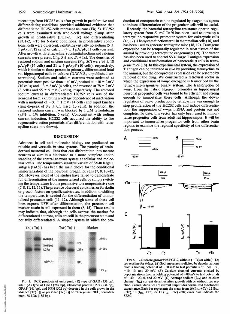
recordings from HC2S2 cells after growth in proliferative anddifferentiating conditions provided additional evidence thatdifferentiated HC2S2 cells represent neurons (Fig. 5). HC2S2cells were examined with whole-cell voltage clamp aftergrowth in proliferative (FGF-2, -Tc) and differentiating(FGF-2, +Tc for 6 days) conditions. In proliferative condi-tions, cells were quiescent, exhibiting virtually no sodium (5 +
1 pA/pF; 12 cells) or calcium (4 ± 1 pA/pF; 11 cells) current.After growth with tetracycline, substantial sodium and calciumcurrents were present (Fig. 5 A and B; +Tc). The densities ofrestored sodium and calcium currents (Fig. SC) were 96 + 18pA/pF (16 cells) and 21 + 3 pA/pF (10 cells), respectively,which is similar to those present in primary, differentiated fetalrat hippocampal cells in culture (D.W.Y.S., unpublished ob-servations). Sodium and calcium currents were activated atpotentials more positive than -40 mV, peaked at -10 ± 2 mV(10 cells) and -3 + 2 mV (4 cells), and reversed at 78 + 2 mV(8 cells) and 55 ± 9 mV (3 cells), respectively. The restoredsodium current in differentiated HC2S2 cells was of theneuronal form, exhibiting a voltage dependence of inactivationwith a midpoint of -60 ± 1 mV (14 cells) and rapid kinetics(time-to-peak of 0.8 ± 0.1 msec; 13 cells). In addition, therestored sodium current was blocked by 1 ,tM tetrodotoxin(95% ± 1% inhibition, 6 cells). Concomitant with sodiumcurrent induction, HC2S2 cells acquired the ability to fireregenerative action potentials after differentiation with tetra-cycline (data not shown).
DISCUSSIONAdvances in cell and molecular biology are predicated onreliable and versatile in vitro systems. The paucity of brain-derived neuronal cell lines that can differentiate into matureneurons in vitro is a hindrance to a more complete under-standing of the central nervous system at cellular and molec-ular levels. The temperature-sensitive variant of SV40 large Tantigen (tsA58) has been the main choice for the conditionalimmortalization of the neuronal progenitor cells (7, 8, 10-12,15). However, most of the studies have failed to demonstratefull differentiation of the immortalized cells by simply switch-ing the temperature from a permissive to a nonpermissive one(7, 8, 11, 12, 15). The presence of several cytokines, or forskolinor growth factors on specific substrates, in addition to shiftingthe temperature, is needed for the differentiation of immor-talized precursor cells (11, 12). Although some of these celllines express NFH after differentiation, the precursor cellmarker nestin is still expressed in them (8, 15). These resultsmay indicate that, although the cells express the marker fordifferentiated neurons, cells are still in the precursor state andnot fully differentiated. A simpler system in which the pro-
Tc(-) Tc(+)
* GAD(E)
GAD(A), L27a
Tc(-) Tc(+)
, NFH
_II L27a
duction of oncoprotein can be regulated by exogenous agentsto induce differentiation of the progenitor cells will be useful.
Recently, the bacterial tetracycline-resistance operon regu-latory system from E. coli TnlO has been used to develop atetracycline-responsive promoter system for eukaryotic cells(16, 17). The system functions well in mammalian cells (16) andhas been used to generate transgenic mice (18, 19). Transgeneexpression can be temporally regulated in most tissues of theanimal by providing tetracycline exogenously (19). The vectorhas also been used to control SV40 large T antigen expressionand conditional transformation of pancreatic ,B cells in trans-genic mice (18). In this experimental system, the expression ofT antigen can be inhibited in vivo by providing tetracycline tothe animals, but the oncoprotein expression can be restored byremoval of the drug. We constructed a retroviral vector inwhich the expression of v-myc oncogene is transcribed by thetetracycline-responsive binary system. The transcription ofv-myc from the hybrid PhCMv*-1 promoter in hippocampalneuronal progenitor cells was found to be efficient and strongenough to immortalize these cells. Although the down-regulation of v-myc production by tetracycline was enough tostop proliferation of the HC2S2 cells and induce differentia-tion, the suppression of v-myc mRNA and protein was notcomplete. To date, this vector has only been used to immor-talize progenitor cells from adult rat hippocampus. It will beimportant to immortalize progenitor cells from other brainregions to examine the regional specificity of the differentia-tion process.
A 30 mV
-50 mV
B 20mV
40 mV
-80 mV
-Tc -
1 ms 10 Ms
+Tc
C
Marker
i1, 369bp
150-
C 100-
Z5a0Z3 501
.f 246bp
-Tc
T ElQ.C:a..
co
+Tc
30-
20-
10-
T
-Tc +TcO0
-(GFAP) NFL
*p 123bp
FIG. 4. PCR products of embryonic (E) type of GAD (353 bp),adult (A) type of GAD (267 bp), ribosomal protein L27a (234 bp),GFAP (141 bp), and NFH (302 bp) detected in the cells grown in theabsence [Tc(-)] or presence [Tc(+)] of tetracycline. NFL, neurofila-ment 68 kDa (155 bp).
FIG. 5. Cells were grown with FGF-2, without (-Tc) or with (+Tc)tetracycline for 6 days. (A) Sodium currents elicited by depolarizationsfrom a holding potential of -80 mV to test potentials of -50, -30,-10, 10, and 30 mV. (B) Calcium channel currents elicited bydepolarizations from a holding potential of -80 mV to test potentialsof -40, -20, 0, and 20 mV. (C) Average sodium (INa) and calciumchannel (IBa) current densities after growth with or without tetracy-cline. Current densities are current amplitudes normalized to total cellcapacitance. Each bar represents the mean from 16 (INa, +Tc), 12 (INa,-Tc), 10 (IBa, +Tc), or 11 (IBa, -Tc) cells; error bars indicate theSEM.
Proc. Natl. Acad. Sci. USA 93 (1996)
Dow
nloa
ded
by g
uest
on
July
30,
202
0

Proc. Natl. Acad. Sci. USA 93 (1996) 1523
The differentiation of HC2S2 cells to neurons was demon-strated by morphological and immunocytochemical character-istics of cells. Neurons with phase-bright cell bodies andinterconnected long and thin processes expressing specificneuronal markers, tau, NFH, and NeuN were observed. Inaddition, unlike temperature-sensitive tsA58 immortalizedcells, the HC2S2 cells stop expressing nestin upon differenti-ation (data not shown). Electrophysiological studies haveprovided further evidence for the differentiation of HC2S2cells into neurons. Cells treated with tetracycline acquiredlarge sodium currents and the ability to fire action potentials.Moreover, the induced sodium current was of the neuronalform, exhibiting rapid kinetics and a midpoint of inactivationof -60 mV (compared to a midpoint at -80 to - 85 mV in glialcells; ref. 30). The restored calcium current was of the high-threshold class, based on the voltage dependence of activationand lack of inactivation (31). Thus differentiated HC2S2 cellsexhibit functional properties essential to central nervous sys-tem neurons: sodium current, which is necessary for initiationand propagation of the action potential, and calcium current,which is required for neurotransmitter release. Taken to-gether, these results suggest that HC2S2 cells were derivedfrom an immortalized neuronal progenitor cell and that theycan be differentiated into neurons after suppression of thev-myc oncogene. Moreover, since these cells are derived fromthe adult hippocampus, progenitor cells with the potential forneural differentiation must exist in the adult nervous systemand they are amenable to isolation and expansion in vitro. It isnow possible to study the mechanism of the cell cycle arrest andsubsequent differentiation into neurons that is induced by thedown-regulation of the overexpressed v-myc oncogene.
We thank Drs. I. Verma, T. Hunter, S. Thode, and T. Palmer fortheir valuable comments on the manuscript; Dr. D. Peterson for hishelp in confocal microscopy and photography; and M. L. Gage for herhelp in preparation of the manuscript. We also thank Dr. A. Baird forrecombinant FGF-2, Dr. H. Bujard for pUHD15-1 and pUHD 10-3plasmids, Drs. T. Palmer and D. Miller for pHENA and LXSHDplasmids, Dr. J. G. Sutcliff for pBlB15 plasmid, Dr. R. J. Mullen forNeuN, and Dr. R. D. G. McKay for nestin antibody. The work has beensupported by grants from NIA (PO1 AG10435 to F.H.G. and J.R.) andthe Hollfelder Foundation (F.H.G.). M.H. was supported by theMinistry of Education, Science and Culture of Japan, Japan BrainFoundation, and the Shimazu Foundation.
1. Cepko, C. (1988) Trends Neurosci. 11, 6-8.2. Cepko, C. L. (1989) Annu. Rev. Neurosci. 12, 47-65.3. Lendhal, U. & McKay, R. D. G. (1990) Trends Neurosci. 13,
132-137.4. Gage, F. H., Ray, J. & Fisher, L. J. (1995) Annu. Rev. Neurosci.
18, 159-192.
5. Snyder, E. Y. (1994) Curr. Opin. Neurobiol. 4, 742-751.6. Bartlett, P. F., Reid, H. H., Bailey, K. A. & Bernard, 0. (1988)
Proc. Natl. Acad. Sci. USA 85, 3255-3259.7. Frederiksen, K., Jat, P. S., Valtz, N., Levy, D. & McKay, R. (1988)
Neuron 1, 439-448.8. Renfranz, P. J., Cunningham, M. G. & McKay, R. D. G. (1988)
Cell 66, 713-729.9. Ryder, E. F., Snyder, E. Y. & Cepko, C. L. (1990) J. Neurobiol.
21, 356-375.10. Eves, E. M., Tucker, M. S., Roback, J. D., Downen, M., Rosner,
M. R. & Wainer, B. H. (1992) Proc. Natl. Acad. Sci. USA 89,4373-4377.
11. Mehler, M. F., Rozental, R., Dougherty, M., Spray, D. C. &Kessler, J. A. (1993) Nature (London) 362, 62-65.
12. White, L. A., Eaton, M. J., Castro, M. C., Klose, K. J., Globus,M. Y., Shaw, G. & Whittemore, S. R. (1994) J. Neurosci. 14,6744-6753.
13. Marcu, K. B., Bossone, S. A. & Patel, A. J. (1992) Annu. Rev.Biochem. 61, 809-860.
14. Rubin, L. L., Philpott, K. L. & Brooks, S. F. (1993) Curr. Biol. 3,391-394.
15. Whittemore, S. R. & White, L. A. (1993) Brain Res. 615, 27-40.16. Gossen, M. & Bujard, H. (1992) Proc. Natl. Acad. Sci. USA 89,
5547-5551.17. Gossen, M., Bonin, A. L. & Bujard, H. (1993) Trends Biochem.
Sci. 18, 471-475.18. Efrat, S., Fusco-DeMane, D., Lemberg, H., Al Emran, 0. &
Wang, X. (1995) Proc. Natl. Acad. Sci. USA 92, 3576-3580.19. Furth, P. A., St Onge, L., Boger, H., Gruss, P., Gossen, M.,
Kistner, A., Bujard, H. & Hennighausen, L. (1994) Proc. Natl.Acad. Sci. USA 91, 9302-9306.
20. Palmer, T. D., Miller, A. D., Reeder, R. H. & McStay, B. (1993)Nucleic Acids Res. 21, 3451-3457.
21. Stockschlaeder, M. A., Storb, R., Osborne, W. R. & Miller, A. D.(1991) Hum. Gene Ther. 2, 33-39.
22. Ray, J. & Gage, F. H. (1994) J. Neurosci. 14, 3548-3564.23. Ausubel, F. M., Brent, R., Kingston, R. E., Moore, D. D., Seid-
man, J., Smith, J. A. & Struhl, K., eds. (1989) Short Protocols inMolecular Biology (Wiley, New York), pp. 142-143.
24. Mullen, R. J., Buck, C. R. & Smith, A. M. (1992) Development(Cambridge, UK) 116, 201-211.
25. Wool, I. G., Chan, Y.-L., Paz, V. & Olvera, J. (1990) Biochim.Biophys. Acta 1050, 69-73.
26. Feinstein, D. L., Weinmaster, G. A. & Milner, R. J. (1992) J.Neurosci. Res. 32, 1-14.
27. Wyborski, R. J., Bond, R. W. & Gottlieb, D. I. (1990) Mol. BrainRes. 8, 193-198.
28. Breen, K. C., Robinson, P. A., Wion, D. & Anderton, B. H.(1988) FEBS Lett. 241, 213-218.
29. Bond, R. W., Wyborski, R. J. & Gottlieb, D. I. (1990) Proc. Natl.Acad. Sci. USA 87, 8771-8775.
30. Barres, B. A., Chun, L. L. Y. & Corey, D. P. (1989) Neuron 2,1375-1388.
31. Bean, B. P., Mintz, I. M., Regan, L. J., Sah, D. W. Y. & Adams,M. E. (1993) Drugs Dev. 2, 61-70.
Neurobiology: Hoshimaru et al.
Dow
nloa
ded
by g
uest
on
July
30,
202
0

![Analyzing the effect of c-Myc oncogene and matrix ......expression of the c-Myc oncogene and matrix metolloproteninase-2 [MMP2] on the metastasis and prognosis of the malign melanoma](https://static.fdocuments.net/doc/165x107/60a7fab3d79f715ad65b87dd/analyzing-the-effect-of-c-myc-oncogene-and-matrix-expression-of-the-c-myc.jpg)