Pancreatic Cystic Lesions: What are They and When do I Need to Worry?

542 Copyright © 2019 The Korean Society of Radiology
INTRODUCTION
The occurrence of incidentally detected pancreatic cystic lesions (PCLs) is continuously increasing due to the widespread use of diagnostic imaging, including computed tomography (CT) and magnetic resonance imaging (MRI).
Diagnosis and Surveillance of Incidental Pancreatic Cystic Lesions: 2017 Consensus Recommendations of the Korean Society of Abdominal RadiologyEun Sun Lee, MD1, Jung Hoon Kim, MD2, Mi Hye Yu, MD3, Seo-Youn Choi, MD4, Hyo-Jin Kang, MD2, Hyun Jeong Park, MD1, Yang Shin Park, MD5, Jae Ho Byun, MD6, Sang Soo Shin, MD7, Chang Hee Lee, MD5; Korean Society of Abdominal Radiology1Department of Radiology, Chung-Ang University Hospital, Seoul, Korea; 2Department of Radiology, Seoul National University Hospital, Seoul, Korea; 3Department of Radiology, Konkuk University Medical Center, Seoul, Korea; 4Department of Radiology, Soonchunhyang University Bucheon Hospital, Bucheon, Korea; 5Department of Radiology, Korea University Guro Hospital, Seoul, Korea; 6Department of Radiology, Asan Medical Center, Seoul, Korea; 7Department of Radiology, Chonnam National University Hospital, Gwangju, Korea
The occurrence of incidentally detected pancreatic cystic lesions (PCLs) is continuously increasing. Radiologic examinations including computed tomography and magnetic resonance imaging with magnetic resonance cholangiopancreatography have been widely used as the main diagnostic and surveillance methods for patients with incidental PCLs. Although most incidentally detected PCLs are considered benign, they have the potential to become malignant. Currently, we have several guidelines for the management of incidental PCLs. However, there is still debate over proper management, in terms of accurate diagnosis, optimal follow-up interval, and imaging tools. Because imaging studies play a crucial role in the management of incidental PCLs, the 2017 consensus recommendations of the Korean Society of Abdominal Radiology for the diagnosis and surveillance of incidental PCLs approved 11 out of 16 recommendations. Although several challenges remain in terms of optimization and standardization, these consensus recommendations might serve as useful tools to provide a more standardized approach and to optimize care of patients with incidental PCLs.Keywords: Pancreas; Cysts; Consensus; Magnetic resonance imaging; Computed tomography
Received September 14, 2018; accepted after revision November 30, 2018.Corresponding author: Jung Hoon Kim, MD, Department of Radiology, Seoul National University Hospital, Seoul National University College of Medicine, 101 Daehak-ro, Jongno-gu, Seoul 03080, Korea. • Tel: (822) 2072-1969 • Fax: (822) 743-6385• E-mail: [email protected] is an Open Access article distributed under the terms of the Creative Commons Attribution Non-Commercial License (https://creativecommons.org/licenses/by-nc/4.0) which permits unrestricted non-commercial use, distribution, and reproduction in any medium, provided the original work is properly cited.
The reported incidence rate of PCLs ranges from 13.5% to 19.6% and is 24.3% at autopsy (1-3). Furthermore, there is a strong positive correlation between patient age and the frequency of detected PCLs (4-8). PCLs form a heterogeneous group of tumors ranging from benign to premalignant or malignant. Although most incidentally detected PCLs are considered benign, particularly those that are small in size, they have the potential to become malignant. Thus, incidentally detected PCLs are considered an important clinical issue. Currently, we have several guidelines for the management of incidental PCLs, mainly for mucinous cystic neoplasms (MCNs) (9-16). Because there is a lack of prospective randomized trials in this field, no strong evidence is available today. With this problem, there is still debate regarding proper management in terms of accurate diagnosis, optimal follow-up interval, and imaging tools. Because imaging studies play a crucial role
Korean J Radiol 2019;20(4):542-557
eISSN 2005-8330https://doi.org/10.3348/kjr.2018.0640
Review Article | Gastrointestinal Imaging

543
KSAR Consensus Recommendations for Pancreatic Cystic Lesions
https://doi.org/10.3348/kjr.2018.0640kjronline.org
in the management of incidental PCLs, these consensus recommendations mainly focus on current issues in terms of the imaging diagnosis and management of incidental PCLs.
These recommendations typically follow similar principles listed in previous guides but do not concur completely. In fact, there are several ambiguous definitions (e.g., mural nodules, ductal dilatation, and size change), which create some confusion. To this end, the Korean Society of Abdominal Radiology (KSAR) study group for incidental PCLs has developed expert consensus recommendations regarding essential items for imaging diagnosis and the management of incidental PCLs. These recommendations primarily focus on imaging diagnosis and the risk stratification of incidental PCLs, surveillance tools and follow-up intervals, and post-operative surveillance of incidental PCLs.
Methods of Development
The KSAR study group for incidental PCLs comprised 10 board-certified abdominal radiologists from 7 different hospitals in South Korea. All of them are members of KSAR and are experienced with pancreatic images including CT, MRI, and ultrasonography. First, we searched for reference articles using Medline (source: PubMed, 1966 to May 2017; www.pubmed.com), Embase (1980 to May 2017; www.embase.com), the Cochrane Library (source: The Cochrane Central Register of Controlled Trials, 2017; www.thecochranelibrary.com/), and Google Scholar (scholar.google.com). Literature searches were carried out by a specialist librarian (P.W.S.). Relevant keywords related to incidental PCLs and imaging tools in combination with Medical Subject Headings (MeSH) terms and text words (“mucinous neoplasm” OR “cystic lesion” OR “Guideline” OR “serous” OR “computed tomography” OR “ultrasound” OR “Magnetic Resonance Imaging” OR “Ultrasonography” OR “image”) were used along with words related to “Diagnosis” AND “surveillance” AND “follow-up” AND “post-operative.” The search strategy had language restrictions such as English. We also reviewed existing recommendations/guidelines on incidental PCLs. We found 2235 articles related to PCLs.
We categorized the candidate issues into three sections: Section 1. Diagnosis and risk stratification of incidental PCLs; Section 2. Surveillance tools and follow-up intervals of incidental PCLs; Section 3. Post-operative surveillance of incidental PCLs. Seven investigators assessed potentially relevant articles for eligibility. The study group members were organized, and each team was assigned one candidate
issue. The decision to include or exclude studies was hierarchical and initially made on the basis of the study title, then of the study abstract, and finally of the complete study manuscript. We used a modified Delphi method to develop the proposed consensus recommendations. Each team independently completed searches, identification of studies, data abstraction, and tabulation, and discordances were resolved through discussions with all members of the KSAR study group for incidental PCLs. The teams consolidated relevant evidence regarding their assigned issue and prepared a draft of key questions and recommendations, along with a summary of clinical and scientific rationale in support of their suggestions. All study group members discussed these materials through three face-to-face meetings and two online discussions. We tried to develop a single key question for a particular issue and one or two recommendations for each key question, which were then subjected to a modified Delphi voting among the study group members. The modified Delphi method, which allows participants to express their agreement for a particular key question with one or two recommendations, was based on a six-point scale: strongly agree, agree with minor reservation, agree with major reservation, disagree with minor reservation, disagree with major reservation, and strongly disagree. The consensus level was predefined as ≥ 80% of the sum of the votes in favor of strongly agree or agree with minor reservation. We made 16 questions with 17 recommendations.
Following the first round of voting among the study group members, the questionnaire was refined by the study group members and the KSAR Study Group for Pancreatic Cancer at one face-to-face meeting, and we modified 14 questions with 16 recommendations. The second round of voting was conducted at a half-day satellite conference, attended by 82 board-certified radiologists specializing in abdominal radiology during the 40th Scientific Assembly and Annual Meeting of the KSAR on September 02, 2017. Finally, 10 questions with 11 recommendations reached the 80% consensus threshold (Table 1). All votes were recorded by secret ballot. The remaining 5 recommendations did not reach the 80% consensus threshold (Table 2).
Section 1. Diagnosis and Risk Stratification of Incidental Pancreatic Cystic Lesions
KQ 1. Should we Apply the Cyst Size, as Well as Its Size Change, to Determine the Treatment Strategy of Incidental Pancreatic Cystic Lesions?

544
Lee et al.
https://doi.org/10.3348/kjr.2018.0640 kjronline.org
Recommendation- We recommend that both the size and its change should
be considered together to determine the treatment strategy of PCLs. Faster growth rates of cystic lesion more than 2 mm/year or 5 mm/2 years, require further work-up to exclude malignancy (agreement level: 93.8%).
- Cyst size measurement, with the largest diameter including the wall, should be performed in the same
direction, at least in the same plane, with the same imaging modality if possible (agreement level: 93.8%).
According to one of the guidelines, an absolute cyst size of ≥ 3 cm had been included as a worrisome feature related to the size of PCLs for predicting malignancy (11), and recently, a rapid growth rate of the cyst (> 5 mm/2 years) was newly added as another one of the worrisome
Table 1. Consensus Statements
Section 1. Diagnosis and Risk Stratification of Incidental Pancreatic Cystic LesionsAgreement Level
(n = 82)KQ 1. Should we apply cyst size, as well as its size change, to determine treatment strategy of incidental pancreatic cystic lesions?
We recommend that both size and its change should be considered together to determine treatment strategy of pancreatic cystic lesions. Faster growth rates of cystic lesion more than 2 mm/year or 5 mm/2 years, require further work-up to exclude malignancy
93.8%
Cyst size measurement, with largest diameter including wall, should be performed in same direction, at least in same plane, with same imaging modality if possible
93.8%
KQ 2. How can we evaluate communication between pancreatic cystic lesions and main pancreatic duct?Communication could be determined by direct visualization of continuity between pancreatic cystic lesion and
either main pancreatic duct or ductal side branch, without septum between cyst and connected duct82.1%
KQ 3. How should patients with multiple pancreatic cystic lesions be evaluated?We suggest that when there are multiple cystic lesions in pancreas, each lesion should be evaluated individually to
check oncologic risk and surgical extent should be minimized. After resection for dominant or risky cyst, patients need to be followed carefully for recurrence within pancreatic remnant
92.4%
KQ 4. Is risk of malignancy related to presence of enhancing mural nodules in incidental pancreatic cystic lesions?We recommend that pancreatic cystic lesions that have enhancing mural nodule should be considered for surgical
resection because presence of enhancing mural nodules increases risk of malignancy91.1%
KQ 5. How can we evaluate presence of enhancing mural nodule in incidental pancreatic cystic lesions?We suggest that contrast-enhanced CT or contrast-enhanced MRI with MRCP is useful tool in evaluating enhancing
mural nodules84.4%
KQ 6. Is risk of malignancy correlated with main pancreatic duct diameter?Risk of malignancy is correlated with main pancreatic duct diameter. Main pancreatic duct diameter greater than 5
mm without obstructive causes or symptom is also required to be under active surveillance89.6%
Section 2. Surveillance Tools and Follow-Up Intervals of Incidental Pancreatic Cystic LesionsKQ 7. Should contrast-enhanced MRI be used for surveillance of incidental pancreatic cystic lesions?
We suggest that non-contrast MRI can be used for serial follow-up of incidental pancreatic cystic lesions, especially in patients with impaired renal function
90.9%
KQ 8. Which sequences should be included in non-contrast MRI for surveillance of incidental pancreatic cystic lesions?We suggest that at least axial and coronal heavily T2-weighted image and axial T1-weighted image should be
included for serial follow-up of incidental pancreatic cystic lesions87.7%
Section 3. Post-Operative Surveillance of Incidental Pancreatic Cystic LesionsKQ 9. Should patients with pancreatic cystic lesions after resection undergo surveillance?
We recommend continuous surveillance for patients with pancreatic cystic lesions after surgical resection because recurrence occurred in remnant pancreas with frequency of 17.0%
88.6%
KQ 10. Should patients with pancreatic cystic lesions after resection undergo surveillance according to management guideline of pancreatic cystic lesions?
We suggest surveillance based on pathologic and clinical findings according to management guideline using CT, MRI, and EUS
92.9%
CT = computed tomography, EUS = endoscopic ultrasound, MRCP = magnetic resonance cholangiopancreatography, MRI = magnetic resonance imaging

545
KSAR Consensus Recommendations for Pancreatic Cystic Lesions
https://doi.org/10.3348/kjr.2018.0640kjronline.org
features (10). Although other guidelines of managing PCLs had described that cyst growth affects the follow up strategy as well (13, 17), they did not provide an accurate cut-off value for the cyst growth rate. There have been a few studies showing that not only cyst size itself but also cyst size change are important predictors of malignancy in PCLs (18, 19). Kang et al. (18) reported that malignant PCLs grew by a greater percentage (69.8% vs. 19.4%; p = 0.046) and at a greater rate (4.1 mm/year vs. 1.0 mm/year; p = 0.001). Furthermore, in this study, cysts that grew at a faster rate than 2 mm/year had a higher risk of malignancy (5-year risk = 45.5% vs. 1.8%, p < 0.001). Another study by Kwong et al. (19) revealed that in patients with branch duct type of intraductal papillary mucinous neoplasms (BD-IPMNs), the malignant BD-IPMNs grew at a faster rate (18.6 mm/year vs. 0.8 mm/year; p = 0.05) compared to benign BD-IPMNs. In addition, a growth rate faster than 2 mm/year and a total growth that exceeded 10 mm had a higher risk of malignancy. Therefore, we recommend that both the size and its change should be considered together to determine the treatment strategy for PCLs. A faster growth rate of cystic lesions of more than 2 mm/year or 5 mm/2 years, require further work-up to exclude malignancy. In the 2017 KSAR consensus meeting, the consensus level for the aforementioned statement was 93.8%.
When we discuss the variables related to cyst size, it is important for the cyst size measurement to be performed
in the same way. The size measurement of PCLs has been known to have poor reproducibility (20). The measured size can vary depending on the modality or the plane used (21) as well as on the person that measures it. Therefore, radiologists play an important role and the size measurement of PCLs should be performed with reproducible and accurate methods. We recommend that size measurements on cross-sectional images should be taken in the same direction, or at least in the same plane, with the same imaging modality, if possible (Fig. 1) (22). The largest diameter, including the wall of the lesion, should be used. In the 2017 KSAR consensus meeting, the consensus level for the aforementioned statement was 93.8%.
KQ 2. How Can We Evaluate the Communication between pancreatic cystic lesions and the Main Pancreatic Duct?
Recommendation- Communication could be determined by direct
visualization of the continuity between the pancreatic cystic lesion and either the main pancreatic duct or a ductal side branch, without the septum between the cyst and the connected duct (agreement level: 82.1%).
Although no study has accurately described the definition of communication between the main pancreatic duct (MPD) and PCL, we typically decide that communication is present
Table 2. Consensus Statements which Did Not Reach Up to 80% of Agreement
Section 1. Diagnosis and Risk Stratification of Incidental Pancreatic Cystic LesionsAgreement Level
(n = 82)
KQ. How can we evaluate communication between pancreatic cyst and main pancreatic duct?We suggest that MRI, which is comparable to EUS, is useful tool to assess communication between pancreatic cyst
and main pancreatic duct75.0%
KQ. How do we diagnose main duct involvement of IPMN?We recommend that main duct involvement of IPMN should be included as differential diagnosis when diffuse or
segmental dilation of MPD of > 5 mm without obstructive cause is demonstrated based on radiologic imaging77.9%
Section 2. Surveillance Tools and Follow-Up Intervals of Incidental Pancreatic Cystic LesionsKQ. Is MRI superior to CT for surveillance of incidental pancreatic cystic lesion?
We recommend that both contrast-enhanced MRI with MRCP and contrast-enhanced MDCT with multiplanar reformation could be used as imaging modality for follow-up of incidental pancreatic cystic lesion
70.3%
KQ. How often should patients with incidental pancreatic cysts be followed up?We suggest closer follow-up of incidental pancreatic cysts in first year according to risk of malignancy, and subsequently followed with extended time intervals if they are stable
57.1%
KQ. How long should patients with stable pancreatic cysts be followed up?We recommend that continuous follow-up of stable pancreatic cysts would be beneficial, because most of them are
stable, but some show delayed growth76.4%
IPMN = intraductal papillary mucinous neoplasm, MDCT = multidetector CT, MPD = main pancreatic duct

546
Lee et al.
https://doi.org/10.3348/kjr.2018.0640 kjronline.org
A B
C D
EFig. 1. 69-year-old woman with incidental pancreatic cystic lesion. MRCP (A), coronal T2-weighted image (B), and contrast-enhanced axial T1-weighted image (C) show pleomorphic cystic lesion in pancreas head. Lesion is measured as 30.62 mm on MRCP (A), 27.74 mm on coronal T2-weighted image (B), and 14.02 mm on contrast-enhanced axial T1-weighted image (C), reflecting high variability of size measurement in different sequences and planes. On contrast-enhanced CT obtained after 1 year, size of lesion is measured as 27.76 mm on coronal image (D) and 14.27 mm on axial image (E). In each of same plane, size of pancreatic cystic lesion remains stable without significant interval growth to initial MR. It is important to measure size of pancreatic cystic lesions in cross-sectional image in same direction at least in same plane, and with same imaging modality, if possible. MR = magnetic resonance, MRCP = MR cholangiopancreatography

547
KSAR Consensus Recommendations for Pancreatic Cystic Lesions
https://doi.org/10.3348/kjr.2018.0640kjronline.org
when the continuity between the PCL and either the MPD or the BD is directly visualized, with no septums between the cyst and the connected duct in practice (Fig. 2). For this issue, the consensus level during the 2017 KSAR consensus meeting was 82.1%.
The evaluation of communication of the cyst with the pancreatic duct is very important for characterization and risk stratification of PCLs. Although endoscopic retrograde cholangiopancreatography and endoscopic ultrasound (EUS) are established to be gold standards for demonstrating a communication between the PCL and the pancreatic
duct (23-25), they have invasive characteristics. Since alternative and non-invasive diagnostic methods such as MRI with magnetic resonance cholangiopancreatography (MRCP) have been technically improved, it is questionable whether these invasive examinations should still be performed to confirm the communication between the cystic lesion and the pancreatic duct in all patients having incidental PCLs. Kim et al. (26) reported that MRI can accurately assess the communication between the PCL and MPD, and the diagnostic performance of MRI in evaluating the communication is comparable with EUS. Another study
Fig. 2. 54-year-old man with incidental pancreatic cystic lesion. Coronal T2-weighted image (A), contrast-enhanced coronal T1-weighted image (B), MRCP with thin section (C), and maximal intensity projection reconstruction image (D) show 48 mm pleomorphic cystic lesion (arrowheads) in pancreas body. Lesion shows direct communication with main pancreatic duct (arrows) without septum between cyst and duct. In this case, MRI with MRCP directly shows continuity between pancreatic cyst and main pancreatic duct. MRI = magnetic resonance imaging
A
C
B
D

548
Lee et al.
https://doi.org/10.3348/kjr.2018.0640 kjronline.org
by Kim et al. (27) also revealed no difference in sensitivity of using MRI to detect communication with MPD in a patient with PCLs compared to EUS. In this study, the sensitivity and accuracy of MRI for any communication with the MPD was 100% and 90.5%, respectively. Furthermore, with its excellent soft tissue contrast, MRCP is valuable for precisely depicting internal structures such as the septa and mural nodules (20, 21). MRI has also been reported to be better than CT for evaluating ductal communication (28, 29). Therefore, we suggested that MRI, which is comparable to EUS, is a useful tool to assess the communication between the PCL and MPD. However, in the 2017 KSAR consensus meeting, the consensus level for this statement was 75%, and this statement was not adopted as a consensus recommendation due to its low agreement rate.
KQ 3. How Should Patients with Multiple Pancreatic Cystic Lesions Be Evaluated?
Recommendation- We suggest that when there are multiple cystic lesions
in the pancreas, each lesion should be evaluated individually to check the oncologic risk and the surgical extent should be minimized. After resection for the dominant or risky cyst, patients need to be followed carefully for the recurrence within the pancreatic remnant (agreement level: 92.4%).
Multifocal IPMNs are defined when the number of IPMNs in the pancreas is two or larger. The prevalence of multifocal IPMNs, either synchronous or metachronous, has been reported to vary widely, ranging from 0% to 83% (30, 31). Nevertheless, the proper management of multifocal IPMNs has not been established to date. In a previous study by Matthaei et al. (30), in patients with multifocal IPMNs, most cysts were genetically unique and this clonal heterogeneity was related to the independent progression of individual cysts. Other studies by Schmidt et al. (32) and Mori et al. (33) demonstrated that unifocal IPMNs had greater invasiveness than multifocal IPMNs, although it was not statistically significant. Indeed, Schmidt et al. (32) reported that patients with symptomatic unifocal BD-IPMN carried a higher risk of invasiveness than those with symptomatic multifocal BD-IPMNs (18% vs. 7%). Furthermore, according to a guideline (11), multifocal IPMNs have a similar risk of malignancy compared to unifocal IPMN and there is no concrete evidence that the
malignant risk of IPMN increases as the number of lesions increases. Thus, we suggest that when there are multiple PCLs, each lesion should be evaluated individually to check the risk of malignancy and the surgical extent should be minimized. In brief, when the surgical resection is indicated for patients with multifocal PCLs, an optimized and segmental resection containing the PCL with the high oncologic risk should be considered. After the resection of the dominant or risky lesion, patients need to be followed carefully for any recurrences within the remaining pancreas. In the 2017 KSAR consensus meeting, the consensus level for the aforementioned statement was 92.4%.
KQ 4. Is a Risk of Malignancy Related to the Presence of Enhancing Mural Nodules in Incidental Pancreatic Cystic Lesions?
Recommendation- We recommend that pancreatic cystic lesions that have
an enhancing mural nodule should be considered for surgical resection because the presence of enhancing mural nodules increases the risk of malignancy (agreement level: 91.1%).
The nomenclature for mural nodules in PCLs is heterogeneous and includes “solid component,” “solid mural nodule,” “enhancing mural nodule,” and “enhanced mural nodule.” The term “non-enhancing mural nodule” has been used for the intra-cystic solid component identified in imaging methods without contrast agent, such as EUS and non-contrast CT or MRI, because they could not differentiate true solid lesions from mucin plugs created by mucin-producing epithelium in IPMN or MCN. Therefore, we use the term “enhancing mural nodule” for the intra-cystic solid component in PCLs, revealed only by contrast-enhanced imaging studies such as contrast-enhanced CT, MRI, and EUS.
The presence of enhancing mural nodules in incidental PCLs is highly associated with malignancy in IPMN as well as in MCN regardless of the subtype or cyst size (34-41). According to a previous report, one-third (6/18) of malignant IPMNs had an enhancing mural nodule, whereas no benign IPMNs (0/6) had an enhancing mural nodule (41). In addition, another study for main duct IPMNs showed that enhancing mural nodules were observed in 16 carcinomas involving the MPD and in one adenoma or borderline neoplasm (p < 0.001) (40). For MCN, one study enrolling
549
KSAR Consensus Recommendations for Pancreatic Cystic Lesions
https://doi.org/10.3348/kjr.2018.0640kjronline.org
163 resected patients showed that although only 17.5% of MCNs was identified as cancers, the presence of enhancing mural nodules was a significant finding associated with malignancy (p = 0.001) (36). Furthermore, lesions with
mural nodules were significantly more likely to be malignant and showed an interval growth during surveillance (p < 0.05) in patients with PCLs (39, 42). In the 2017 KSAR consensus meeting, the consensus level for the aforementioned
Fig. 3. 56-year-old man with pathologically confirmed IPMN associated with invasive carcinoma.Precontrast CT (A), contrast-enhanced portal phase CT (B), coronal T2-weighted image (C), precontrast (D), contrast-enhanced portal phase axial (E), and coronal (F) T1-weighted image show 7 cm pleomorphic cystic lesion in pancreas head. Contrast-enhanced CT and MRI clearly depict 23 mm enhancing mural nodule (arrows) within cystic lesion. IPMN = intraductal papillary mucinous neoplasm
A B
C D
E F

550
Lee et al.
https://doi.org/10.3348/kjr.2018.0640 kjronline.org
statement was 91.1%.
KQ 5. How Can We Evaluate the Presence of the Enhancing Mural Nodule in the Incidental Pancreatic Cystic Lesions?
Recommendation- We suggest that contrast-enhanced CT or contrast-
enhanced MRI with MRCP is a useful tool in evaluating the enhancing mural nodules (agreement level: 84.4%).
Contrast-enhanced CT, compared with pre-contrast CT, is the most widely accepted imaging tool for the evaluation of enhancing mural nodules in PCLs, followed by contrast-enhanced MRI with MRCP (Fig. 3). Kang et al. (37) found that multi-detector CT and MRI with MRCP were similar in their diagnostic performance in depicting signs suspicious or indicative of malignancy, including enhancing mural nodules in patients with IPMN (area under the curve = 0.82 for both), with a good inter-modality agreement (κ = 0.75). Another study showed that the presence of an enhancing mural nodule in BD-IPMNs was highly correlated with malignancy in all imaging methods (multidetector computed tomography [MDCT]; p = 0.001, MRCP; p = 0.008, EUS; p < 0.001) (34). Contrast-enhanced EUS has become one of the most useful imaging tools for the pancreas. Harima et al. (43) reported promising results for contrast-enhanced EUS by showing that the diagnostic accuracy for mural nodules in EUS after contrast injection increased from 72% to 98%. However, further studies are required to validate the role
of contrast-enhanced EUS in determining enhancing mural nodules. In the 2017 KSAR consensus meeting, the consensus level for the aforementioned statement was 84.4%.
KQ 6. Is a Risk of Malignancy Correlated with Main Pancreatic Duct Diameter?
Recommendation- The risk of malignancy is correlated with the main
pancreatic duct diameter. A main pancreatic duct diameter greater than 5 mm without obstructive causes or symptom is also required to be under active surveillance (agreement level: 89.6%).
An IPMN of the pancreas is pathologically defined as a noninvasive epithelial neoplasm of mucin-producing cells arising in the MPD and/or BD of the pancreas. The affected ducts show various dilatations with mucus (44, 45). The malignancy rate of MD-IPMN (40–95%) is much higher than that of BD-IPMN (12–62%) (12, 32, 46-48). Radiologically, an IPMN involving the MPD can lead to identifiable segmental or diffuse dilatation of the MPD secondary to mucin production without other causes of obstruction (12, 49). In the international consensus guidelines, MPD dilation more than 5 mm without other obstructive cause is diagnosed as MD-IPMN (11, 12). However, supporting data for the size criteria have been lacking. In fact, the size criterion of 5 mm for defining a MD-IPMN was introduced without any scientific evidence. In practice, various cut-off values are applied to define MPD dilatation ranging from 3 mm to 10 mm (32, 46, 47, 50-58). In prior studies (59, 60), 29.4% of 170 patients with radiologic MD-IPMN demonstrated no MPD involvement pathologically and the estimated accuracy of radiologic imaging for the diagnosis of MD-IPMN was approximately 75% (59). For the 2017 KSAR consensus, we recommend that MD-IPMN should be included as a differential diagnosis when diffuse or segmental dilation of the MPD > 5 mm without an obstructive cause is demonstrated on radiologic imaging. However, as expected, the consensus level (77.9%) for this statement did not reach 80%, reflecting the various cut-off values recommended by several guidelines. Therefore, this statement was not adopted as a consensus recommendation due to its low agreement rate.
For the prediction of malignancy, variable thresholds regarding MPD diameter are also reported in MD-IPMN. In several international consensus guidelines, if the MPD
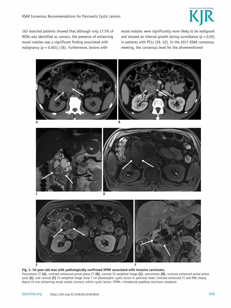
Fig. 3. 56-year-old man with pathologically confirmed IPMN associated with invasive carcinoma.G. Cut section of gross specimen shows solid mural nodules (arrows) within cyst. Histopathology confirmed IPMN with invasive carcinoma.IPMN = intraductal papillary mucinous neoplasm
G

551
KSAR Consensus Recommendations for Pancreatic Cystic Lesions
https://doi.org/10.3348/kjr.2018.0640kjronline.org
is greater than 10 mm, surgical resection is recommended as the malignancy rate has been reported to be as high as 62% (11, 58, 61). However, European consensus guidelines recommended that MD-IPMN greater than 6 mm should be considered for surgical resection (14) with a reference of a prior meta-analysis in which an MPD > 6 mm was associated with an increased risk with a pooled odds ratio of 7.27 (95% confidence interval, 3.0–17.4) for malignancy (48). Furthermore, other studies have proposed a cut-off MPD diameter of 5–7 mm for the prediction of malignancy (51, 52, 62). Therefore, further research regarding the cut-off value of MPD diameter in diagnosing and risk-stratifying MD-IPMN are strongly warranted.
Many studies reported a positive correlation between MPD dilatation and pathologic malignancy and the dilatation of the MPD as one of the independent predictors of malignancy in MD-IPMN (48, 50, 53-55, 57). For example, an MPD diameter of 5-9 mm is a potential predictor for malignancy in patients with IPMN and is regarded as a worrisome feature warranting a further diagnostic evaluation (11, 12, 59) and IPMN with a MPD > 5 mm has a substantial risk of malignancy (51, 59, 62). However, there are conflicting results. Other researchers have noted the significance of the MPD diameter to be a source of controversy (53, 56, 62). They suggested that the MPD diameter was not related to malignancy and that invasive carcinoma can also be found in patients with an MPD diameter smaller than 5 mm, without symptoms or mural nodules. Even though there has been various debates regarding the MPD diameter, we recommend that any MPD diameter greater than 5 mm without any obstructive causes or symptoms is required to be under active surveillance and could be subjected to surgical resection based on clinical findings and additional imaging studies. In the 2017 KSAR consensus meeting, the consensus level for this statement was 89.6%.
Section 2. Surveillance Tools and Follow-Up Intervals of Incidental Pancreatic Cystic Lesions
KQ 7. Should Contrast-Enhanced MRI Be Used for the Surveillance of Incidental Pancreatic Cystic Lesions?
Recommendation- We suggest that non-contrast MRI can be used for the
serial follow-up of incidental pancreatic cystic lesions, especially in patients with impaired renal function (agreement level: 90.9%).
Contrast-enhanced MRI with MRCP has a higher sensitivity for the detection of internal septa and mural nodules as well as for the assessment of communication with the MPD (26, 40, 41, 63, 64). It has a high accuracy when differentiating MCN from other PCLs (5, 63) and for preoperative characterization of IPMN (41, 65). Currently, MDCT with multiplanar reconstruction (MPR) provides an equivalent capability with MRCP for the evaluation of communication with the MPD (66). In terms of detecting malignant IPMNs, contrast-enhanced MDCT with MPR and contrast-enhanced MRI with MRCP showed similar diagnostic performances (21, 37, 62, 64). Therefore, we suggest that both contrast-enhanced MRI with MRCP and contrast-enhanced MDCT with MPR can be used as the follow-up imaging modality for incidental PCLs. However, in the 2017 KSAR consensus meeting, the consensus level for this statement was 70.3%. This statement was not adopted as a consensus recommendation due to its low agreement rate.
For the follow-up imaging of incidental PCLs, there is a limited added value of a contrast-enhanced MRI for management decisions regarding PCLs in comparison with a non-contrast MRI. Although we have a few reports regarding the use of MR contrast agent, the recommendations for incidental PCLs were concordant both with and without a contrast agent in 95.5% (107/112) of cases (67) and interobserver agreement both with and without MR contrast agent was excellent (0.86–0.97) (68). Moreover, an abbreviated MR protocol showed a similar performance to the standard MR protocol for the surveillance of incidental PCLs and provided sufficient information equivalent to the standard MR protocol (41). Therefore, non-contrast MRIs can be used for serial follow-up of incidental PCLs, particularly in patients with impaired renal function (67, 68). In the 2017 KSAR consensus meeting, the consensus level for this statement was 90.9%.
KQ 8. Which Sequences Should Be Included in Non-Contrast MRI for the Surveillance of Incidental Pancreatic Cystic Lesions?
Recommendation- We suggest that at least axial and coronal heavily T2-
weighted image and axial T1-weighted image should be included for the serial follow-up of incidental pancreatic cystic lesions (agreement level: 87.7%).
Non-contrast MRI can consist of various sequences

552
Lee et al.
https://doi.org/10.3348/kjr.2018.0640 kjronline.org
Fig. 4. 76-year-old man with incidental pancreatic cystic lesion.Initial two-dimensional MRCP (A), coronal T2-weighted image (B), and contrast-enhanced axial T1-weighted image (C) show 16 mm pleomorphic cystic lesion (arrows) in pancreas head without enhancing mural nodule. Follow-up three-dimensional MRCP (D), coronal T2-weighted image (E), and axial non-contrast T1-weighted image (F) obtained after 1 year demonstrate same cyst (arrows) without significant interval growth. There were no worrisome features on follow-up non-contrast MRI.
A B
C D
E F

553
KSAR Consensus Recommendations for Pancreatic Cystic Lesions
https://doi.org/10.3348/kjr.2018.0640kjronline.org
such as T1-weighed sequence, T2-weighted sequence, diffusion-weighed imaging, and MRCP. Among them, a T2-weighted sequence is essential with its excellent contrast resolution for the evaluation of PCLs. Since mural nodule and internal septation in incidental PCLs are easily depicted on a T2-weighted sequence, a change in these features is clearly seen when comparing the follow-up imaging with the initial MR examination (67, 68). An abbreviated MR protocol including an axial and coronal T2-weighted Half-Fourier-Acquired Single-shot Turbo spin Echo sequence and a non-contrast T1-weighted sequence showed a similar performance to the standard MR protocol for the surveillance of incidental PCLs (69). Therefore, we suggest that axial and coronal heavily T2-weighed sequences and axial T1-weighed sequences should be scanned for the serial follow-up of incidental PCLs (Fig. 4). In the 2017 KSAR consensus meeting, the consensus level for this statement was 87.7%.
Among patients with BD-IPMN, approximately 10% have an indication for surgery during the first year of follow-up after their diagnosis because of the occurrence of suspicious malignant findings (70). During the follow-up period, PCLs with a risk of malignancy or those that may require surgery showed higher growth rates, compared to cysts with no risk of malignancy or non-surgery (mean growth rate, 1.4–15 mm/year and 0.2–0.4 mm/year) (18, 19, 70, 71). Therefore, an intensive follow-up during the first year after diagnosis is recommended to closely monitor its stability and to determine its nature. If no change occurs during this time, the follow-up interval can be extended. However, because the risk of IPMN progression increases over time and the incidence steadily increases linearly with time (71, 72), follow-up can be conducted on an annual or biannual basis depending on the malignancy risk. Therefore, we recommend that an earlier follow-up for incidental PCLs should be done during the first year according to the risk of malignancy and subsequently followed-up with an extended time interval if they are stable. However, in the 2017 KSAR consensus meeting, the consensus level for this statement was only 57.1%. This statement was not adopted as a consensus recommendation due to its low agreement rate.
During the follow-up period, approximately 11% of PCLs exhibited a delayed growth after an initial first year period of stability and the growth rate was faster after 5 years (73, 74). Morphological changes suggestive of malignancy in PCLs may develop as late as 5–8 years after the initial diagnosis (71, 74, 75). There is still a lack of evidence for
long-term follow-up of more than 10 years for PCLs. The 5- and 10-year rates of development of pancreatic cancer during the follow-up of BD IPMNs were 2.4% and 20.0%, respectively (76). According to a recent systematic review and meta-analysis (72), even low-risk IPMNs had an almost 8% chance of progressing to pancreatic cancer at the 10-year follow-up mark. In addition, PCLs increase the risk of pancreatic adenocarcinoma throughout the entire pancreas as well as at the sites of existing cysts (77, 78). Therefore, we recommend that continuous monitoring for stable PCLs would be beneficial because some PCLs may show delayed growth. However, in the 2017 KSAR consensus meeting, the consensus level for this statement was 76.4%. This statement was not adopted as a consensus recommendation due to its low agreement rate. Since there is an insufficient evidence for long-term follow-up of more than 10 years for PCLs, more evidence is needed in the future to establish a follow-up strategy.
Section 3. Post-Operative Surveillance of Incidental Pancreatic Cystic Lesions
KQ 9. Should Patients with Pancreatic Cystic Lesions after Resection Undergo Surveillance?
Recommendation- We recommend a continuous surveillance for patients
with pancreatic cystic lesions after surgical resection because recurrence occurred in the remnant pancreas with a frequency of 17.0% (agreement level: 88.6%).
There have been scant previous reports for long-term results after pancreatectomy in patients with IPMN. Even after curative surgery with a negative resection margin, pancreatic remnants still harbor a risk of recurrence which requires long-term surveillance. After surgical resection, the recurrence rate in the remnant pancreas has been reported to be between 3% and 17%, regardless of the surgical margin status (47, 79-84). The histological type is a well-known risk factor for recurrence after surgical resection of IPMN (82): the frequency of recurrence is higher in invasive IPMN compared to non-invasive IPMN. The vast majority of recurrences occurred in patients with positive resection margins. Yogi et al. (79) retrospectively recruited 153 patients with IPMN who underwent surgical resection. They included wide ranges of histological subtypes such as low/intermediate-grade dysplasia (LGD/IGD) in 54.9%, high-

554
Lee et al.
https://doi.org/10.3348/kjr.2018.0640 kjronline.org
grade dysplasia (HGD) in 22.2%, T1a (stromal invasion ≤ 5 mm) in 4.6%, and IPMN associated invasive carcinoma in 18.3% of patients. During the median 46.4 (6.0–216.3) months follow-up period after surgery, the overall recurrence rate was 17.0%. Considering the non-negligible rate (up to 17.0%) of recurrence, we recommend continuous surveillance in patients with PCLs after resection. In the 2017 KSAR consensus meeting, the consensus level for this statement was 88.6%.
KQ 10. Should Patients with Pancreatic Cystic Lesions after Resection Undergo Surveillance according to Management Guideline of Pancreatic Cystic Lesions?
Recommendation- We suggest surveillance based on pathologic and clinical
findings according to management guideline using CT, MRI, and EUS (agreement level: 92.9%).
Risk factors associated with the recurrence of IPMNs include surgical margin status, invasiveness, histologic subtypes of IPMN, T stage, N stage, and carbohydrate antigen (CA) 19-9 level (79, 80, 82, 83). According to Park et al. (83), the recurrence rate was 12.6% in 103 patients with surgically resected IPMN. They found that the risk factors associated with the recurrence of IPMNs were invasive histology, elevated CA 19-9, and the location of the pancreatic head. Although the scientific evidence is not sufficient, some investigators have insisted that postoperative surveillance for patients with LGD to HGD (T1a) should be similar to non-resected IPMN and that surveillance for IPMN associated invasive carcinoma should be the same as for pancreatic ductal adenocarcinoma (79, 80). Therefore, we suggest that post-operative surveillance should be based on pathologic and clinical findings of IPMNs and should also follow the management guidelines using CT, MRI, and EUS. In the 2017 KSAR consensus meeting, the consensus level for this statement was 92.9%.
SUMMARY
Radiological examinations including CT and MRI with MRCP have been widely used as the main diagnostic and surveillance method for patients with incidental PCLs. Although most incidentally detected PCLs are considered benign, they have the potential to become malignant. Currently, we have several guidelines for the management of incidental PCLs.
However, there is still debate over proper management, in terms of accurate diagnosis, the optimal follow-up interval, and imaging tools. Because imaging studies play a crucial role in the management of incidental PCLs, the 2017 consensus recommendations of the KSAR for the diagnosis and surveillance of incidental PCLs approved several issues of debate from the radiologists’ point of view, based on routine clinical practices. Although several challenges remain in terms of optimization and standardization, these consensus recommendations might serve as useful tools to help provide a more standardized approach and to optimize care of patients with incidental PCLs.
Conflicts of InterestThe authors have no potential conflicts of interest to disclose.
ORCID iDsJung Hoon Kim
https://orcid.org/0000-0002-8090-7758 Eun Sun Lee
https://orcid.org/0000-0003-0780-7985
REFERENCES
1. Zhang XM, Mitchell DG, Dohke M, Holland GA, Parker L. Pancreatic cysts: depiction on single-shot fast spin-echo MR images. Radiology 2002;223:547-553
2. Lee KS, Sekhar A, Rofsky NM, Pedrosa I. Prevalence of incidental pancreatic cysts in the adult population on MR imaging. Am J Gastroenterol 2010;105:2079-2084
3. Kimura W, Nagai H, Kuroda A, Muto T, Esaki Y. Analysis of small cystic lesions of the pancreas. Int J Pancreato 1995;18:197-206
4. Laffan TA, Horton KM, Klein AP, Berlanstein B, Siegelman SS, Kawamoto S, et al. Prevalence of unsuspected pancreatic cysts on MDCT. AJR Am J Roentgenol 2008;191:802-807
5. de Jong K, van Hooft JE, Nio CY, Gouma DJ, Dijkgraaf MG, Bruno MJ, et al. Accuracy of preoperative workup in a prospective series of surgically resected cystic pancreatic lesions. Scand J Gastroenterol 2012;47:1056-1063
6. de Jong K, Nio CY, Hermans JJ, Dijkgraaf MG, Gouma DJ, Van Eijck CH, et al. High prevalence of pancreatic cysts detected by screening magnetic resonance imaging examinations. Clin Gastroenterol Hepatol 2010;8:806-811
7. Mella JM, Gómez EJ, Omodeo M, Manzotti M, Roel M, Pereyra L, et al. Prevalence of incidental clinically relevant pancreatic cysts at diagnosis based on current guidelines. Gastroenterol Hepatol (English Edition) 2018;41:293-301
8. Megibow AJ, Baker ME, Morgan DE, Kamel IR, Sahani DV,

555
KSAR Consensus Recommendations for Pancreatic Cystic Lesions
https://doi.org/10.3348/kjr.2018.0640kjronline.org
Newman E, et al. Management of incidental pancreatic cysts: a white paper of the ACR incidental findings committee. J Am Coll Radiol 2017;14:911-923
9. Vege SS, Ziring B, Jain R, Moayyedi P, Adams MA, Dorn SD, et al. American gastroenterological association institute guideline on the diagnosis and management of asymptomatic neoplastic pancreatic cysts. Gastroenterology 2015;148:819-822
10. Tanaka M, Fernández-del Castillo C, Kamisawa T, Jang JY, Levy P, Ohtsuka T, et al. Revisions of international consensus Fukuoka guidelines for the management of IPMN of the pancreas. Pancreatology 2017;17:738-753
11. Tanaka M, Fernández-del Castillo C, Adsay V, Chari S, Falconi M, Jang JY, et al. International consensus guidelines 2012 for the management of IPMN and MCN of the pancreas. Pancreatology 2012;12:183-197
12. Tanaka M, Chari S, Adsay V, Fernández-del Castillo C, Falconi M, Shimizu M, et al; International Association of Pancreatology. International consensus guidelines for management of intraductal papillary mucinous neoplasms and mucinous cystic neoplasms of the pancreas. Pancreatology 2006;6:17-32
13. American Gastroenterological Association. AGA Section. Managing pancreatic cysts: a patient guide. Gastroenterology 2015;149:498-499
14. Del Chiaro M, Verbeke C, Salvia R, Klöppel G, Werner J, McKay C, et al. European experts consensus statement on cystic tumours of the pancreas. Dig Liver Dis 2013;45:703-711
15. Italian Association of Hospital Gastroenterologists and Endoscopists; Italian Association for the Study of the Pancreas, Buscarini E, Pezzilli R, Cannizzaro R, De Angelis C, et al. Italian consensus guidelines for the diagnostic work-up and follow-up of cystic pancreatic neoplasms. Dig Liver Dis 2014;46:479-493
16. European Study Group on Cystic Tumours of the Pancreas. European evidence-based guidelines on pancreatic cystic neoplasms. Gut 2018;67:789-804
17. Berland LL, Silverman SG, Gore RM, Mayo-Smith WW, Megibow AJ, Yee J, et al. Managing incidental findings on abdominal CT: white paper of the ACR incidental findings committee. J Am Coll Radiol 2010;7:754-773
18. Kang MJ, Jang JY, Kim SJ, Lee KB, Ryu JK, Kim YT, et al. Cyst growth rate predicts malignancy in patients with branch duct intraductal papillary mucinous neoplasms. Clin Gastroenterol Hepatol 2011;9:87-93
19. Kwong WT, Lawson RD, Hunt G, Fehmi SM, Proudfoot JA, Xu R, et al. Rapid growth rates of suspected pancreatic cyst branch duct intraductal papillary mucinous neoplasms predict malignancy. Dig Dis Sci 2015;60:2800-2806
20. Kim SH, Lee JM, Lee ES, Baek JH, Kim JH, Han JK, et al. Intraductal papillary mucinous neoplasms of the pancreas: evaluation of malignant potential and surgical resectability by using MR imaging with MR cholangiography. Radiology 2014;274:723-733
21. Choi SY, Kim JH, Yu MH, Eun HW, Lee HK, Han JK. Diagnostic performance and imaging features for predicting the
malignant potential of intraductal papillary mucinous neoplasm of the pancreas: a comparison of EUS, contrast-enhanced CT and MRI. Abdom Radiol (NY) 2017;42:1449-1458
22. Boos J, Brook A, Chingkoe CM, Morrison T, Mortele K, Raptopoulos V, et al. MDCT vs. MRI for incidental pancreatic cysts: measurement variability and impact on clinical management. Abdom Radiol (NY) 2017;42:521-530
23. Gazelle GS, Mueller PR, Raafat N, Halpern EF, Cardenosa G, Warshaw AL. Cystic neoplasms of the pancreas: evaluation with endoscopic retrograde pancreatography. Radiology 1993;188:633-636
24. Koito K, Namieno T, Ichimura T, Yama N, Hareyama M, Morita K, et al. Mucin-producing pancreatic tumors: comparison of MR cholangiopancreatography with endoscopic retrograde cholangiopancreatography. Radiology 1998;208:231-237
25. Nordback I, Auvinen O, Airo I, Isolauri J, Teerenhovi O. ERCP in evaluating the mode of therapy in pancreatic pseudocyst. HPB Surg 1988;1:35-44
26. Kim JH, Eun HW, Park HJ, Hong SS, Kim YJ. Diagnostic performance of MRI and EUS in the differentiation of benign from malignant pancreatic cyst and cyst communication with the main duct. Eur J Radiol 2012;81:2927-2935
27. Kim YC, Choi JY, Chung YE, Bang S, Kim MJ, Park MS, et al. Comparison of MRI and endoscopic ultrasound in the characterization of pancreatic cystic lesions. AJR Am J Roentgenol 2010;195:947-952
28. Waters JA, Schmidt CM, Pinchot JW, White PB, Cummings OW, Pitt HA, et al. CT vs MRCP: optimal classification of IPMN type and extent. J Gastrointest Surg 2008;12:101-109
29. Song SJ, Lee JM, Kim YJ, Kim SH, Lee JY, Han JK, et al. Differentiation of intraductal papillary mucinous neoplasms from other pancreatic cystic masses: comparison of multirow-detector CT and MR imaging using ROC analysis. JJ Magn Reson Imaging 2007;26:86-93
30. Matthaei H, Norris AL, Tsiatis AC, Olino K, Hong SM, dal Molin M, et al. Clinicopathological characteristics and molecular analyses of multifocal intraductal papillary mucinous neoplasms of the pancreas. Ann Surg 2012;255:326-333
31. Rosenblatt R, Dorfman V, Epelboym I, Poneros JM, Sethi A, Lightdale C, et al. Demographic features and natural history of intermediate-risk multifocal versus unifocal intraductal papillary mucinous neoplasms. Pancreas 2015;44:478-483
32. Schmidt CM, White PB, Waters JA, Yiannoutsos CT, Cummings OW, Baker M, et al. Intraductal papillary mucinous neoplasms: predictors of malignant and invasive pathology. Ann Surg 2007;246:644-651; discussion 651-654
33. Mori Y, Ohtsuka T, Kono H, Ideno N, Aso T, Nagayoshi Y, et al. Management strategy for multifocal branch duct intraductal papillary mucinous neoplasms of the pancreas. Pancreas 2012;41:1008-1012
34. Arikawa S, Uchida M, Uozumi J, Sakoda J, Kaida H, Kunou Y, et al. Utility of multidetector row CT in diagnosing branch duct IPMNs of the pancreas compared with MR cholangiopancreatography and endoscopic ultrasonography.

556
Lee et al.
https://doi.org/10.3348/kjr.2018.0640 kjronline.org
Kurume Med J 2010;57:91-10035. Correa-Gallego C, Do R, LaFemina J, Gonen M, D’Angelica
MI, DeMatteo RP, et al. Predicting dysplasia and invasive carcinoma in intraductal papillary mucinous neoplasms of the pancreas: development of a preoperative nomogram. Ann Surg Oncol 2013;20:4348-4355
36. Crippa S, Salvia R, Warshaw AL, Domínguez I, Bassi C, Falconi M, et al. Mucinous cystic neoplasm of the pancreas is not an aggressive entity: lessons from 163 resected patients. Ann Surg 2008;247:571-579
37. Kang HJ, Lee JM, Joo I, Hur BY, Jeon JH, Jang JY, et al. Assessment of malignant potential in intraductal papillary mucinous neoplasms of the pancreas: comparison between multidetector CT and MR imaging with MR cholangiopancreatography. Radiology 2015;279:128-139
38. Kim TH, Song TJ, Hwang JH, Yoo KS, Lee WJ, Lee KH, et al. Predictors of malignancy in pure branch duct type intraductal papillary mucinous neoplasm of the pancreas: a nationwide multicenter study. Pancreatology 2015;15:405-410
39. Kirkpatrick ID, Desser TS, Nino-Murcia M, Jeffrey RB. Small cystic lesions of the pancreas: clinical significance and findings at follow-up. Abdom Imaging 2007;32:119-125
40. Manfredi R, Graziani R, Motton M, Mantovani W, Baltieri S, Tognolini A, et al. Main pancreatic duct intraductal papillary mucinous neoplasms: accuracy of MR imaging in differentiation between benign and malignant tumors compared with histopathologic analysis. Radiology 2009;253:106-115
41. Pilleul F, Rochette A, Partensky C, Scoazec JY, Bernard P, Valette PJ. Preoperative evaluation of intraductal papillary mucinous tumors performed by pancreatic magnetic resonance imaging and correlated with surgical and histopathologic findings. J Magn Reson Imaging 2005;21:237-244
42. Uehara H, Ishikawa O, Katayama K, Kawada N, Ikezawa K, Fukutake N, et al. Size of mural nodule as an indicator of surgery for branch duct intraductal papillary mucinous neoplasm of the pancreas during follow-up. J Gastroenterol 2011;46:657-663
43. Harima H, Kaino S, Shinoda S, Kawano M, Suenaga S, Sakaida I. Differential diagnosis of benign and malignant branch duct intraductal papillary mucinous neoplasm using contrast-enhanced endoscopic ultrasonography. World J Gastroenterol 2015;21:6252-6260
44. Castellano-Megías VM, Andrés CI, López-Alonso G, Colina-Ruizdelgado F. Pathological features and diagnosis of intraductal papillary mucinous neoplasm of the pancreas. World J Gastrointest Oncol 2014;6:311-324
45. Furukawa T, Klöppel G, Volkan Adsay N, Albores-Saavedra J, Fukushima N, Horii A, et al. Classification of types of intraductal papillary-mucinous neoplasm of the pancreas: a consensus study. Virchows Arch 2005;447:794-799
46. Kim KW, Park SH, Pyo J, Yoon SH, Byun JH, Lee MG, et al. Imaging features to distinguish malignant and benign branch-duct type intraductal papillary mucinous neoplasms of the
pancreas: a meta-analysis. Ann Surg 2014;259:72-8147. Salvia R, Fernández-del Castillo C, Bassi C, Thayer SP, Falconi
M, Mantovani W, et al. Main-duct intraductal papillary mucinous neoplasms of the pancreas: clinical predictors of malignancy and long-term survival following resection. Ann Surg 2004;239:678-685; discussion 685-687
48. Anand N, Sampath K, Wu BU. Cyst features and risk of malignancy in intraductal papillary mucinous neoplasms of the pancreas: a meta-analysis. Clin Gastroenterol Hepatol 2013;11:913-921
49. Procacci C, Megibow AJ, Carbognin G, Guarise A, Spoto E, Biasiutti C, et al. Intraductal papillary mucinous tumor of the pancreas: a pictorial essay. Radiographics 1999;19:1447-1463
50. Bournet B, Kirzin S, Carrère N, Portier G, Otal P, Selves J, et al. Clinical fate of branch duct and mixed forms of intraductal papillary mucinous neoplasia of the pancreas. J Gastroenterol Hepatol 2009;24:1211-1217
51. Hackert T, Fritz S, Klauss M, Bergmann F, Hinz U, Strobel O, et al. Main-duct intraductal papillary mucinous neoplasm: high cancer risk in duct diameter of 5 to 9 mm. Ann Surg 2015;262:875-881; discussion 880-881
52. Kang MJ, Jang JY, Lee S, Park T, Lee SY, Kim SW. Clinicopathological meaning of size of main-duct dilatation in intraductal papillary mucinous neoplasm of pancreas: proposal of a simplified morphological classification based on the investigation on the size of main pancreatic duct. World J Surg 2015;39:2006-2013
53. Lévy P, Jouannaud V, O’Toole D, Couvelard A, Vullierme MP, Palazzo L, et al. Natural history of intraductal papillary mucinous tumors of the pancreas: actuarial risk of malignancy. Clin Gastroenterol Hepatol 2006;4:460-468
54. Mimura T, Masuda A, Matsumoto I, Shiomi H, Yoshida S, Sugimoto M, et al. Predictors of malignant intraductal papillary mucinous neoplasm of the pancreas. J Clin Gastroenterol 2010;44:e224-e229
55. Nara S, Onaya H, Hiraoka N, Shimada K, Sano T, Sakamoto Y, et al. Preoperative evaluation of invasive and noninvasive intraductal papillary-mucinous neoplasms of the pancreas: clinical, radiological, and pathological analysis of 123 cases. Pancreas 2009;38:8-16
56. Serikawa M, Sasaki T, Fujimoto Y, Kuwahara K, Chayama K. Management of intraductal papillary-mucinous neoplasm of the pancreas: treatment strategy based on morphologic classification. J Clin Gastroenterol 2006;40:856-862
57. Shimizu Y, Yamaue H, Maguchi H, Yamao K, Hirono S, Osanai M, et al. Predictors of malignancy in intraductal papillary mucinous neoplasm of the pancreas: analysis of 310 pancreatic resection patients at multiple high-volume centers. Pancreas 2013;42:883-888
58. Waters JA, Schmidt CM. Intraductal papillary mucinous neoplasm-when to resect? Adv Surg 2008;42:87-108
59. Barron M, Roch A, Waters J, Parikh J, DeWitt J, Al-Haddad M, et al. Does preoperative cross-sectional imaging accurately predict main duct involvement in intraductal papillary

557
KSAR Consensus Recommendations for Pancreatic Cystic Lesions
https://doi.org/10.3348/kjr.2018.0640kjronline.org
mucinous neoplasm? J Gastrointest Surg 2014;18:447-456; discussion 5455-5456
60. Correa-Gallego C, Ferrone CR, Thayer SP, Wargo JA, Warshaw AL, Fernández-del Castillo C. Incidental pancreatic cysts: do we really know what we are watching? Pancreatology 2010;10:144-150
61. Ferrone CR, Correa-Gallego C, Warshaw AL, Brugge WR, Forcione DG, Thayer SP, et al. Current trends in pancreatic cystic neoplasms. Arch Surg 2009;144:448-454
62. Seo N, Byun JH, Kim JH, Kim HJ, Lee SS, Song KB, et al. Validation of the 2012 international consensus guidelines using computed tomography and magnetic resonance imaging: branch duct and main duct intraductal papillary mucinous neoplasms of the pancreas. Ann Surg 2016;263:557-564
63. Sainani NI, Saokar A, Deshpande V, Fernández-del Castillo C, Hahn P, Sahani DV. Comparative performance of MDCT and MRI with MR cholangiopancreatography in characterizing small pancreatic cysts. AJR Am J Roentgenol 2009;193:722-731
64. Walter TC, Steffen IG, Stelter LH, Maurer MH, Bahra M, Faber W, et al. Implications of imaging criteria for the management and treatment of intraductal papillary mucinous neoplasms-benign versus malignant findings. Eur Radiol 2015;25:1329-1338
65. Irie H, Yoshimitsu K, Aibe H, Tajima T, Nishie A, Nakayama T, et al. Natural history of pancreatic intraductal papillary mucinous tumor of branch duct type: follow-up study by magnetic resonance cholangiopancreatography. J Comput Assist Tomogr 2004;28:117-122
66. Sahani DV, Kadavigere R, Blake M, Fernandez-del Castillo C, Lauwers GY, Hahn PF. Intraductal papillary mucinous neoplasm of pancreas: multi-detector row CT with 2D curved reformations--correlation with MRCP. Radiology 2006;238:560-569
67. Macari M, Lee T, Kim S, Jacobs S, Megibow AJ, Hajdu C, et al. Is gadolinium necessary for MRI follow-up evaluation of cystic lesions in the pancreas? Preliminary results. AJR Am J Roentgenol 2009;192:159-164
68. Nougaret S, Reinhold C, Chong J, Escal L, Mercier G, Fabre JM, et al. Incidental pancreatic cysts: natural history and diagnostic accuracy of a limited serial pancreatic cyst MRI protocol. Eur Radiol 2014;24:1020-1029
69. Pozzi-Mucelli RM, Rinta-Kiikka I, Wünsche K, Laukkarinen J, Labori KJ, Ånonsen K, et al. Pancreatic MRI for the surveillance of cystic neoplasms: comparison of a short with a comprehensive imaging protocol. Eur Radiol 2017;27:41-50
70. Bae SY, Lee KT, Lee JH, Lee JK, Lee KH, Rhee JC. Proper management and follow-up strategy of branch duct intraductal papillary mucinous neoplasms of the pancreas. Dig Liver Dis 2012;44:257-260
71. Crippa S, Pezzilli R, Bissolati M, Capurso G, Romano L, Brunori MP, et al. Active surveillance beyond 5 years is required for presumed branch-duct intraductal papillary mucinous neoplasms undergoing non-operative management. Am J Gastroenterol 2017;112:1153-1161
72. Choi SH, Park SH, Kim KW, Lee JY, Lee SS. Progression of
unresected intraductal papillary mucinous neoplasms of the pancreas to cancer: a systematic review and meta-analysis. Clin Gastroenterol Hepatol 2017;15:1509-1520.e4
73. Brook OR, Beddy P, Pahade J, Couto C, Brennan I, Patel P, et al. Delayed growth in incidental pancreatic cysts: are the Current American College of Radiology recommendations for follow-up appropriate? Radiology 2015;278:752-761
74. Kayal M, Luk L, Hecht EM, Do C, Schrope BA, Chabot JA, et al. Long-term surveillance and timeline of progression of presumed low-risk intraductal papillary mucinous neoplasms. AJR Am J Roentgenol 2017;209:320-326
75. Tanno S, Nakano Y, Nishikawa T, Nakamura K, Sasajima J, Minoguchi M, et al. Natural history of branch duct intraductal papillary-mucinous neoplasms of the pancreas without mural nodules: long-term follow-up results. Gut 2008;57:339-343
76. Sawai Y, Yamao K, Bhatia V, Chiba T, Mizuno N, Sawaki A, et al. Development of pancreatic cancers during long-term follow-up of side-branch intraductal papillary mucinous neoplasms. Endoscopy 2010;42:1077-1084
77. Matsubara S, Tada M, Akahane M, Yagioka H, Kogure H, Sasaki T, et al. Incidental pancreatic cysts found by magnetic resonance imaging and their relationship with pancreatic cancer. Pancreas 2012;41:1241-1246
78. Tada M, Kawabe T, Arizumi M, Togawa O, Matsubara S, Yamamoto N, et al. Pancreatic cancer in patients with pancreatic cystic lesions: a prospective study in 197 patients. Clin Gastroenterol Hepatol 2006;4:1265-1270
79. Yogi T, Hijioka S, Imaoka H, Mizuno N, Hara K, Tajika M, et al. Risk factors for postoperative recurrence of intraductal papillary mucinous neoplasms of the pancreas based on a long-term follow-up study: proposals for follow-up strategies. J Hepatobiliary Pancreat Sci 2015;22:757-765
80. Winter JM, Jiang W, Basturk O, Mino-Kenudson M, Fong ZV, Tan WP, et al. Recurrence and survival after resection of small intraductal papillary mucinous neoplasm-associated carcinomas (≤ 20-mm invasive component): a multi-institutional analysis. Ann Surg 2016;263:793-801
81. Tamura K, Ohtsuka T, Ideno N, Aso T, Shindo K, Aishima S, et al. Treatment strategy for main duct intraductal papillary mucinous neoplasms of the pancreas based on the assessment of recurrence in the remnant pancreas after resection: a retrospective review. Ann Surg 2014;259:360-368
82. Passot G, Lebeau R, Hervieu V, Ponchon T, Pilleul F, Adham M. Recurrences after surgical resection of intraductal papillary mucinous neoplasm of the pancreas: a single-center study of recurrence predictive factors. Pancreas 2012;41:137-141
83. Park J, Lee KT, Jang TH, Seo YW, Lee KH, Lee JK, et al. Risk factors associated with the postoperative recurrence of intraductal papillary mucinous neoplasms of the pancreas. Pancreas 2011;40:46-51
84. Fujii T, Kato K, Kodera Y, Kanda M, Nagai S, Yamada S, et al. Prognostic impact of pancreatic margin status in the intraductal papillary mucinous neoplasms of the pancreas. Surgery 2010;148:285-290