Cystic Pancreatic Neoplasms Diagnosis of Cystic and Intraductal
33
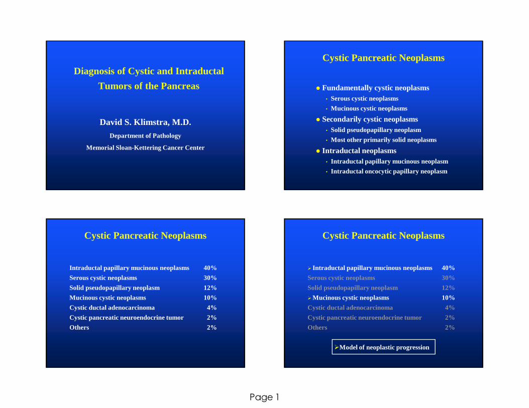
Page 1 Diagnosis of Cystic and Intraductal Tumors of the Pancreas David S. Klimstra, M.D. Department of Pathology Memorial Sloan-Kettering Cancer Center Cystic Pancreatic Neoplasms Fundamentally cystic neoplasms • Serous cystic neoplasms • Mucinous cystic neoplasms Secondarily cystic neoplasms • Solid pseudopapillary neoplasm • Most other primarily solid neoplasms Intraductal neoplasms • Intraductal papillary mucinous neoplasm • Intraductal oncocytic papillary neoplasm Cystic Pancreatic Neoplasms Intraductal papillary mucinous neoplasms 40% Serous cystic neoplasms 30% Solid pseudopapillary neoplasm 12% Mucinous cystic neoplasms 10% Cystic ductal adenocarcinoma 4% Cystic pancreatic neuroendocrine tumor 2% Others 2% Cystic Pancreatic Neoplasms Intraductal papillary mucinous neoplasms 40% Serous cystic neoplasms 30% Solid pseudopapillary neoplasm 12% Mucinous cystic neoplasms 10% Cystic ductal adenocarcinoma 4% Cystic pancreatic neuroendocrine tumor 2% Others 2% Model of neoplastic progression
Transcript of Cystic Pancreatic Neoplasms Diagnosis of Cystic and Intraductal
Page 1
Diagnosis of Cystic and Intraductal
Tumors of the Pancreas
David S. Klimstra, M.D.
Department of Pathology
Memorial Sloan-Kettering Cancer Center
Cystic Pancreatic Neoplasms
� Fundamentally cystic neoplasms• Serous cystic neoplasms
• Mucinous cystic neoplasms
� Secondarily cystic neoplasms• Solid pseudopapillary neoplasm
• Most other primarily solid neoplasms
� Intraductal neoplasms• Intraductal papillary mucinous neoplasm
• Intraductal oncocytic papillary neoplasm
Cystic Pancreatic Neoplasms
Intraductal papillary mucinous neoplasms 40%
Serous cystic neoplasms 30%
Solid pseudopapillary neoplasm 12%
Mucinous cystic neoplasms 10%
Cystic ductal adenocarcinoma 4%
Cystic pancreatic neuroendocrine tumor 2%
Others 2%
Cystic Pancreatic Neoplasms
� Intraductal papillary mucinous neoplasms 40%
Serous cystic neoplasms 30%
Solid pseudopapillary neoplasm 12%
�Mucinous cystic neoplasms 10%
Cystic ductal adenocarcinoma 4%
Cystic pancreatic neuroendocrine tumor 2%
Others 2%
�Model of neoplastic progression
Page 2
MONTHS
200180160140120100806040200
Pro
port
ion S
urvi
ving
1.0
.8
.6
.4
.2
0.0
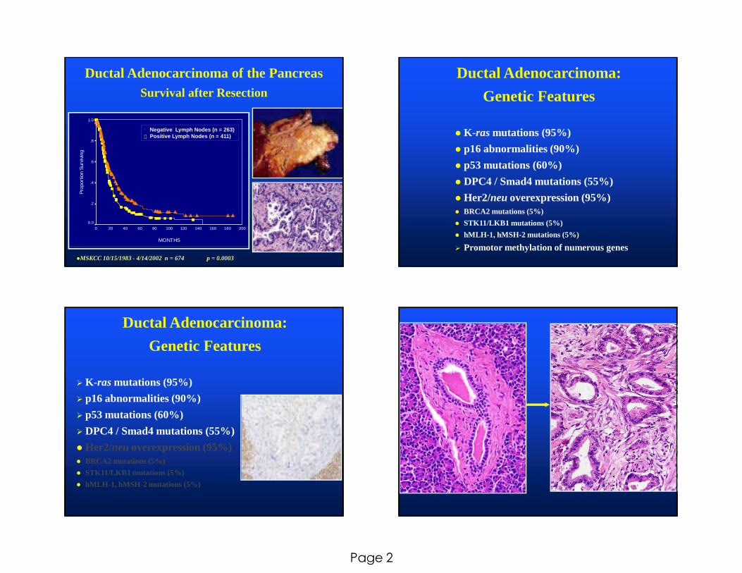
Ductal Adenocarcinoma of the Pancreas Survival after Resection
�MSKCC 10/15/1983 - 4/14/2002 n = 674 p = 0.0003
� Negative Lymph Nodes (n = 263)� Positive Lymph Nodes (n = 411)
Ductal Adenocarcinoma:
Genetic Features
� K- rasmutations (95%)
� p16 abnormalities (90%)
� p53 mutations (60%)
� DPC4 / Smad4 mutations (55%)
� Her2/neuoverexpression (95%)� BRCA2 mutations (5%)
� STK11/LKB1 mutations (5%)
� hMLH-1, hMSH-2 mutations (5%)
� Promotor methylation of numerous genes
Ductal Adenocarcinoma:
Genetic Features
� K- rasmutations (95%)
� p16 abnormalities (90%)
� p53 mutations (60%)
� DPC4 / Smad4 mutations (55%)
� Her2/neuoverexpression (95%)� BRCA2 mutations (5%)
� STK11/LKB1 mutations (5%)
� hMLH-1, hMSH-2 mutations (5%)
Page 3
Precursors to Invasive Ductal
Adenocarcinoma
• Pancreatic Intraepithelial Neoplasia (PanIN)
• Intraductal Papillary Mucinous Neoplasms
• Mucinous Cystic Neoplasms
Pancreatic Intraepithelial Neoplasia:
Background
• Metaplastic and proliferative lesions long recognized
• Some common, age-related, often incidental
• Others more associated with invasive ductal
adenocarcinomas
• Spectrum of intraepithelial lesions• Morphologic progression: metaplasia->hyperplasia->dysplasia
• Accumulation of genetic abnormalities
• “PanIN” terminology proposed, 1994
� Target for earlier detection of pancreatic carcinoma
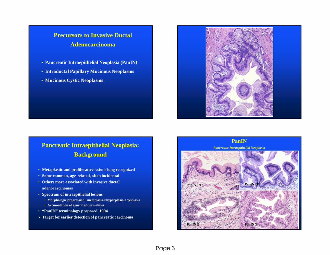
PanIN Pancreatic Intraepithelial Neoplasia
PanIN 1A
PanIN 3
PanIN 1B
PanIN 2
Page 4
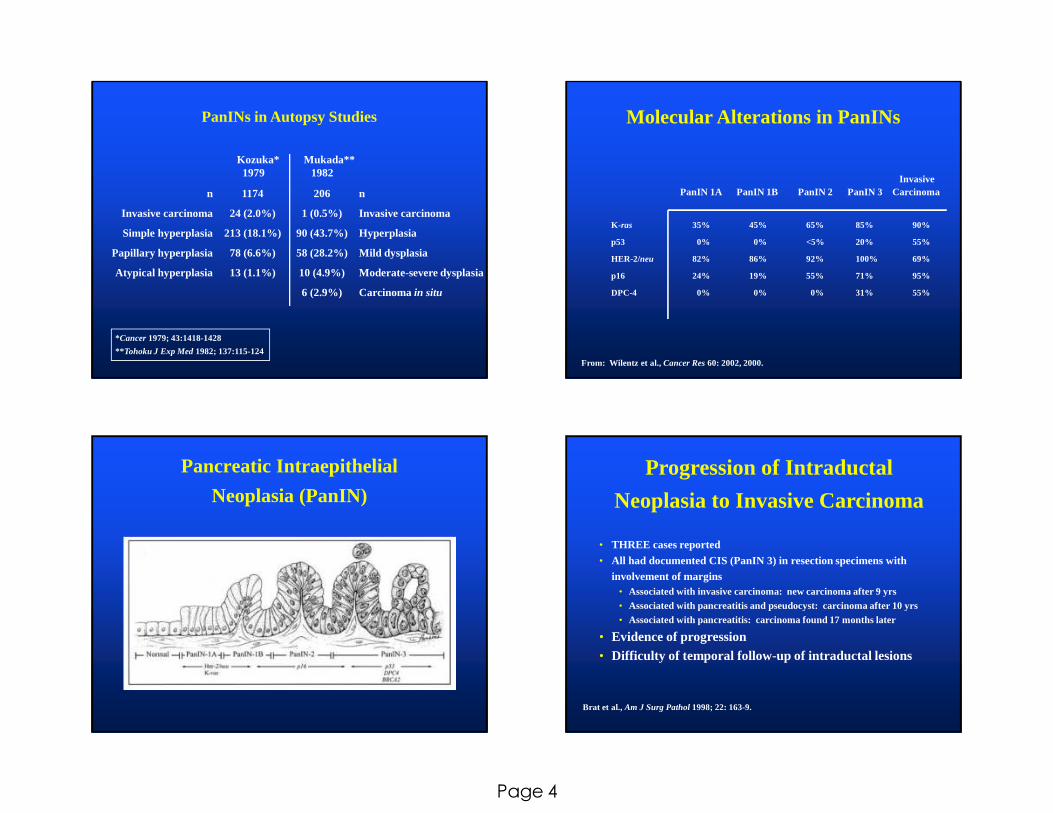
PanINs in Autopsy Studies
Kozuka* Mukada**1979 1982
*Cancer 1979; 43:1418-1428
** Tohoku J Exp Med1982; 137:115-124
n
Invasive carcinoma
Simple hyperplasia
Papillary hyperplasia
Atypical hyperplasia
n
Invasive carcinoma
Hyperplasia
Mild dysplasia
Moderate-severe dysplasia
Carcinoma in situ
1174
24 (2.0%)
213 (18.1%)
78 (6.6%)
13 (1.1%)
206
1 (0.5%)
90 (43.7%)
58 (28.2%)
10 (4.9%)
6 (2.9%)
Molecular Alterations in PanINs
K- ras 35% 45% 65% 85% 90%
p53 0% 0% <5% 20% 55%
HER-2/neu 82% 86% 92% 100% 69%
p16 24% 19% 55% 71% 95%
DPC-4 0% 0% 0% 31% 55%
InvasivePanIN 1A PanIN 1B PanIN 2 PanIN 3 Carcinoma
From: Wilentz et al., Cancer Res60: 2002, 2000.
Pancreatic Intraepithelial
Neoplasia (PanIN)Progression of Intraductal
Neoplasia to Invasive Carcinoma
• THREE cases reported
• All had documented CIS (PanIN 3) in resection specimens with involvement of margins
• Associated with invasive carcinoma: new carcinoma after 9 yrs• Associated with pancreatitis and pseudocyst: carcinoma after 10 yrs• Associated with pancreatitis: carcinoma found 17 months later
• Evidence of progression• Difficulty of temporal follow-up of intraductal les ions
Brat et al., Am J Surg Pathol1998; 22: 163-9.
Page 5
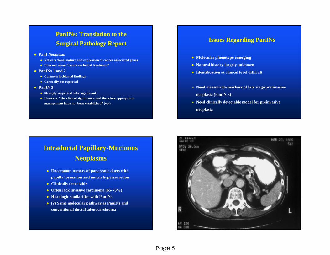
PanINs: Translation to the
Surgical Pathology Report
� PanI Neoplasm � Reflects clonal nature and expression of cancer associated genes
� Does not mean “requires clinical treatment”
� PanINs 1 and 2� Common incidental findings
� Generally not reported
� PanIN 3� Strongly suspected to be significant
� However, “the clinical significance and therefore appropriate
management have not been established” (yet)
Issues Regarding PanINs
� Molecular phenotype emerging
� Natural history largely unknown
� Identification at clinical level difficult
� Need measurable markers of late stage preinvasive
neoplasia (PanIN 3)
� Need clinically detectable model for preinvasive
neoplasia
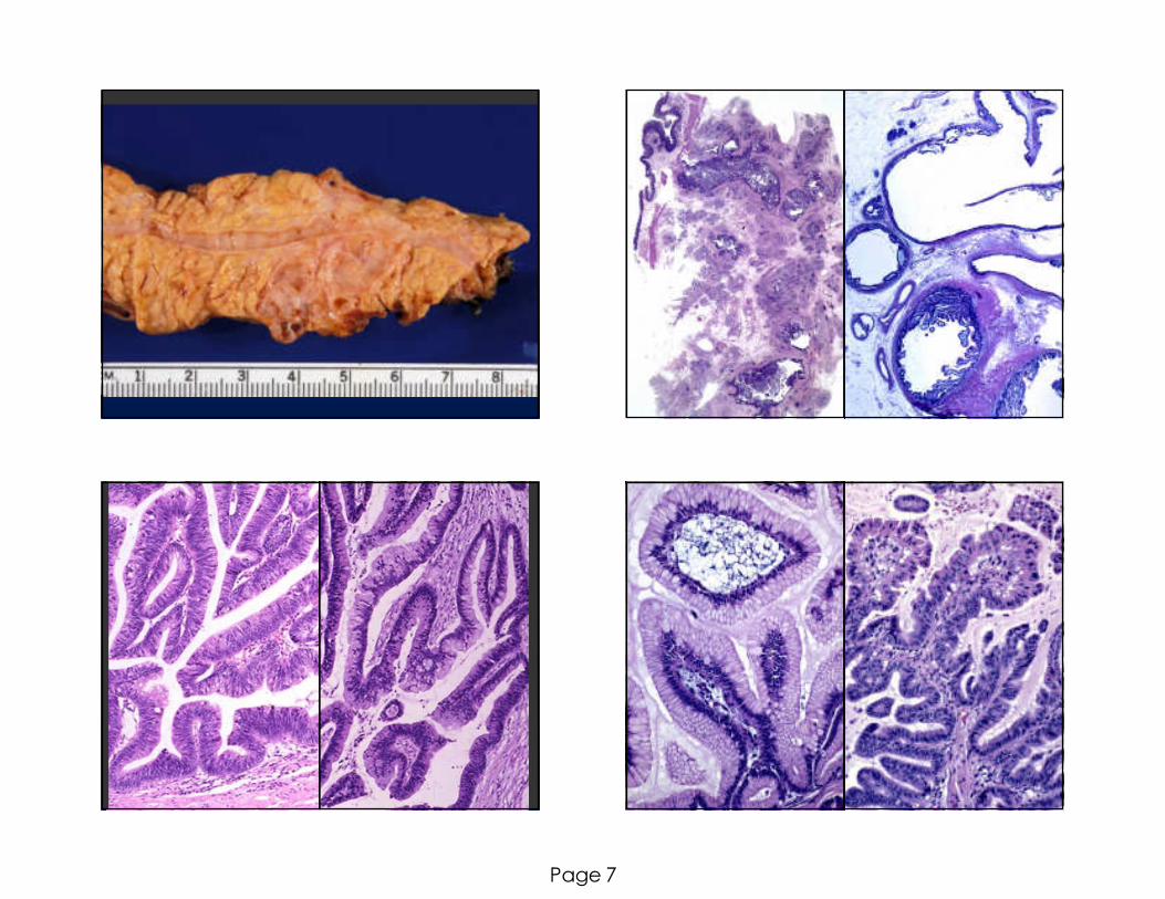
Intraductal Papillary-Mucinous
Neoplasms
� Uncommon tumors of pancreatic ducts with
papilla formation and mucin hypersecretion
� Clinically detectable
� Often lack invasive carcinoma (65-75%)
� Histologic similarities with PanINs
� (?) Same molecular pathway as PanINs and
conventional ductal adenocarcinoma

Page 6
Intraductal Papillary-Mucinous Neoplasms:Intraductal Ultrasound
Hara et al. Gastroenterology2002; 122: 34
Page 7
Page 8
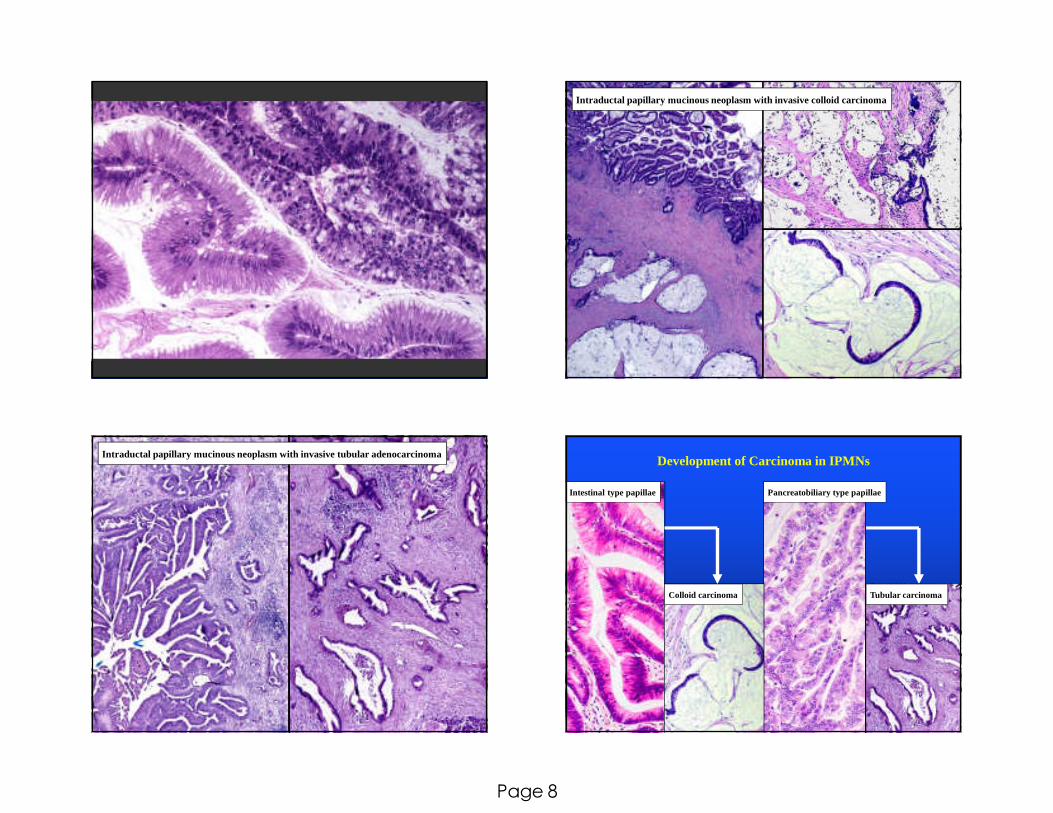
Intraductal papillary mucinous neoplasm with invasive colloid carcinoma
Intraductal papillary mucinous neoplasm with invasive tubular adenocarcinomaDevelopment of Carcinoma in IPMNs
Intestinal type papillae Pancreatobiliary type papillae
Colloid carcinoma Tubular carcinoma

Page 9
Time (months)
192168144120967248240
Cum
ula
tive
Sur
viva
l
1.0
.8
.6
.4
.2
0
Time (months)
192168144120967248240C
umul
ativ
e S
urvi
val
1.0
.8
.6
.4
.2
0
n = 32
n = 30
n = 32
n = 13
n = 17
p = 0.01 p = 0.008
non-invasive () invasive (- - - -)
non-invasive () invasive colloid carcinoma (- - - -) invasive tubular carcinoma ( - )
IPMN: Survival Intraductal Papillary Mucinous Neoplasms:Classification
IPMN with low grade dysplasia IPMN with low grade dysplasia
IPMN with intermediate grade IPMN with moderate dysplasia dysplasia
IPMN with high grade dysplasia IPMN with high grade dysplasia
IPMN with an associated IPMN with an associatedinvasive carcinoma invasive carcinoma
WHO 2010 AFIP Fascicle
Intraductal Papillary-Mucinous
Neoplasms: Main vs.Secondary Ducts
• 70% involve main duct, 30% confined to
secondary (branch) ducts
• Secondary duct type confined to head/neck
• Secondary duct type in younger patients
• Secondary duct type less aggressive• Main duct type: 20% CIS, 37% invasive carcinoma
• Secondary type: 15% CIS, 0% invasive carcinoma
Terris et al., Am J Surg Pathol2000; 24: 1372-7.
Gastric Intestinal Pancreatobiliary Oncocytic
Papilla Types in IPMNs

Page 10
Intraductal Oncocytic Papillary Neoplasm Intraductal Tubulopapillary Neoplasm
of the Pancreas
� Also reported as “Intraductal Tubular Carcinoma”
� Approximately 35 cases reported
� Mean age = 54 yrs (range = 25-72); F > M
� Symptoms: chronic pancreatitis
� Location: head > tail; 30% diffuse involvement
� Favorable outcome
Tajiri et al. Pancreas2004; 29: 116-122
Yamaguchi et al. Am J Surg Pathol 2009; 33: 1164-1172
Klimstra et al. Am J Surg Pathol2013; (in press)
P.D.
C.B.D.
Amp.

Page 11
Intraductal Neoplasms:
Immunohistochemistry
KeratinsCam5.2 100AE1:AE3 95CK7 70CK19 85CK20 30
Lineage MarkersChromogranin (35)Synaptophysin (35)Trypsin 0Chymotrypsin 0
GlycoproteinsCEA (m) 85CA19-9 90B72.3 50
Page 12
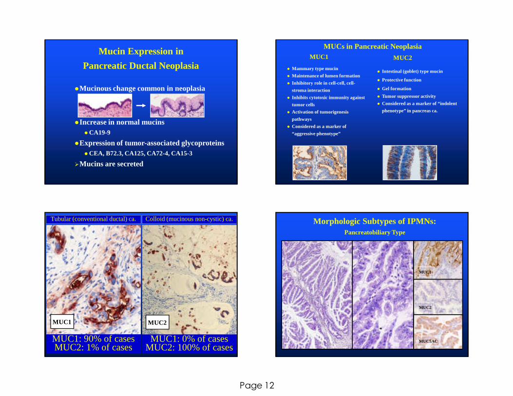
Mucin Expression in
Pancreatic Ductal Neoplasia
�Mucinous change common in neoplasia
�Increase in normal mucins� CA19-9
�Expression of tumor-associated glycoproteins� CEA, B72.3, CA125, CA72-4, CA15-3
�Mucins are secreted
MUCs in Pancreatic Neoplasia
� Mammary type mucin
� Maintenance of lumen formation
� Inhibitory role in cell-cell, cell-
stroma interaction
� Inhibits cytotoxic immunity against
tumor cells
� Activation of tumorigenesis
pathways
� Considered as a marker of
“aggressive phenotype”
MUC1 MUC2
� Intestinal (goblet) type mucin
� Protective function
� Gel formation
� Tumor suppressor activity
� Considered as a marker of “indolent
phenotype” in pancreas ca.
Tubular (conventional ductal) ca. Colloid (mucinous non-cystic) ca.
MUC1: 90% of cases MUC2: 1% of cases
MUC1: 0% of cases MUC2: 100% of cases
MUC1 MUC2
Morphologic Subtypes of IPMNs:Pancreatobiliary Type
MUC1
MUC5AC
MUC2
Page 13
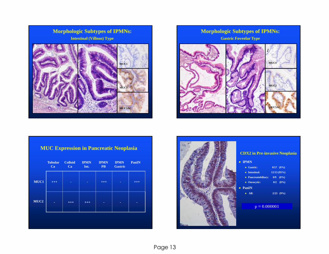
Morphologic Subtypes of IPMNs:Intestinal (Villous) Type
MUC1
MUC5AC
MUC2
Morphologic Subtypes of IPMNs:Gastric Foveolar Type
MUC1
MUC2
MUC5AC
MUC Expression in Pancreatic Neoplasia
Tubular Colloid IPMN IPMN IPMN PanINCa Ca Int. PB Gastric
MUC1
MUC2
+++ - - +++ - +++
- +++ +++ - - -
CDX2 in Pre-invasive Neoplasia
� IPMN
� Gastric: 0/17 (0%)
� Intestinal: 12/13 (95%)
� Pancreatobiliary: 0/9 (0%)
� Oncocytic: 0/2 (0%)
� PanIN
� All: 2/23 (9%)
p = 0.000001
Page 14
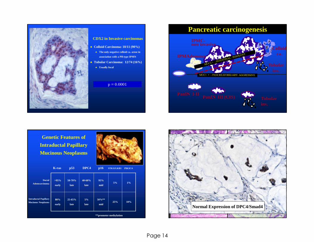
CDX2 in Invasive carcinomas
� Colloid Carcinoma: 10/11 (90%)
� The only negative colloid ca. arose in
association with a PB type IPMN
� Tubular Carcinoma: 12/74 (16%)
� Usually focal
p = 0.0001
.
.
. ..
IPMAdenoma
IPMC-non invasive
PanIN I-II
Colloid ca.
PanIN III (CIS) Tubular inv.
Tubular inv.
Pancreatic carcinogenesis
Genetic Features of
Intraductal Papillary
Mucinous Neoplasms
>95%
early
50-70%
late
40-60%
late
95%
mid5% 1%
80%
early
25-65%
late
5%
late
50%**
mid25% 10%
K-ras p53 DPC4 p16 STK11/LKB1 PIK3CA
Ductal
Adenocarcinoma
Intraductal Papillary
Mucinous Neoplasms
**promoter methylation
Invasive Colloid CarcinomaIPMN - Intestinal
Normal Expression of DPC4/Smad4
Page 15
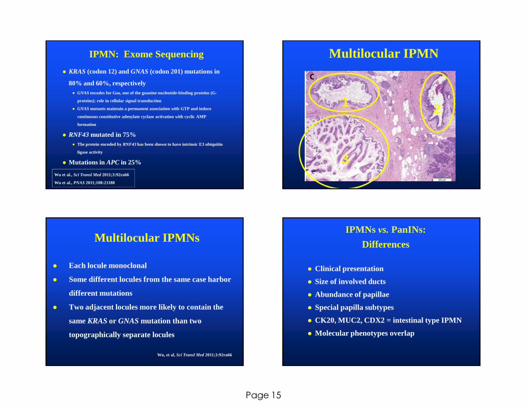
� KRAS (codon 12) and GNAS (codon 201) mutations in
80% and 60%, respectively� GNASencodes for Gsα, one of the guanine nucleotide-binding proteins (G-
proteins); role in cellular signal transduction
� GNASmutants maintain a permanent association with GTP and induce
continuous constitutive adenylate cyclase activation with cyclic AMP
formation
� RNF43 mutated in 75%� The protein encoded by RNF43 has been shown to have intrinsic E3 ubiquitin
ligase activity
� Mutations in APC in 25%
IPMN: Exome Sequencing
Wu et al., Sci Transl Med 2011;3:92ra66
Wu et al., PNAS2011;108:21188
Multilocular IPMN
1
3
2
Multilocular IPMNs
� Each locule monoclonal
� Some different locules from the same case harbor
different mutations
� Two adjacent locules more likely to contain the
same KRASor GNASmutation than two
topographically separate locules
Wu, et al, Sci Transl Med 2011;3:92ra66
IPMNs vs.PanINs:
Differences
� Clinical presentation
� Size of involved ducts
� Abundance of papillae
� Special papilla subtypes
� CK20, MUC2, CDX2 = intestinal type IPMN
� Molecular phenotypes overlap
Page 16
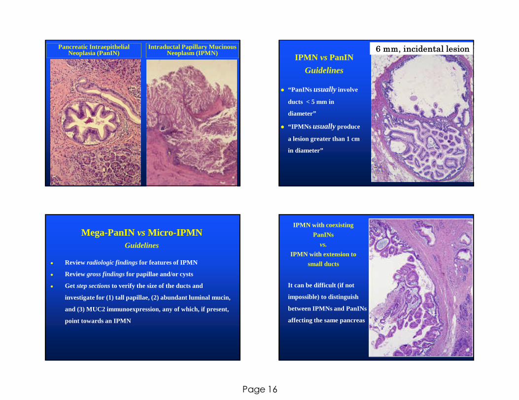
Pancreatic Intraepithelial Neoplasia (PanIN)
Intraductal Papillary Mucinous Neoplasm (IPMN)
IPMN vsPanIN
Guidelines
� “PanINs usually involve
ducts < 5 mm in
diameter”
� “IPMNs usuallyproduce
a lesion greater than 1 cm
in diameter”
6 mm, incidental lesion
Mega-PanIN vs Micro-IPMNGuidelines
● Review radiologic findingsfor features of IPMN
● Review gross findingsfor papillae and/or cysts
● Get step sectionsto verify the size of the ducts and
investigate for (1) tall papillae, (2) abundant luminal mucin,
and (3) MUC2 immunoexpression, any of which, if present,
point towards an IPMN
IPMN with coexisting
PanINs
vs.
IPMN with extension to
small ducts
It can be difficult (if not
impossible) to distinguish
between IPMNs and PanINs
affecting the same pancreas
Page 17
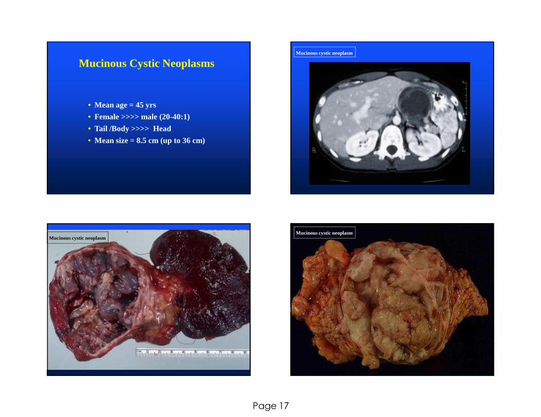
Mucinous Cystic Neoplasms
• Mean age = 45 yrs
• Female >>>> male (20-40:1)
• Tail /Body >>>> Head
• Mean size = 8.5 cm (up to 36 cm)
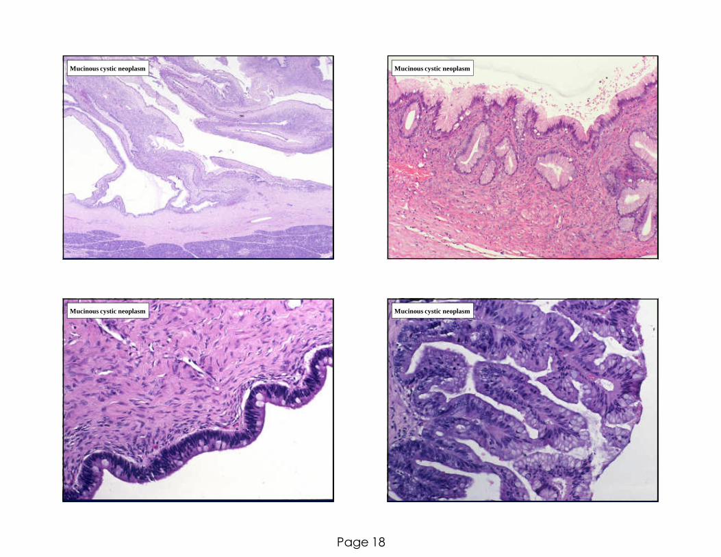
Mucinous cystic neoplasm
Mucinous cystic neoplasmMucinous cystic neoplasm
Page 18
Mucinous cystic neoplasm Mucinous cystic neoplasm
Mucinous cystic neoplasm Mucinous cystic neoplasm

Page 19
Mucinous cystic neoplasm
Estrogen receptors
Mucinous cystic neoplasm
Inhibin
A103
Mucinous cystic neoplasm Mucinous cystic neoplasm
Page 20
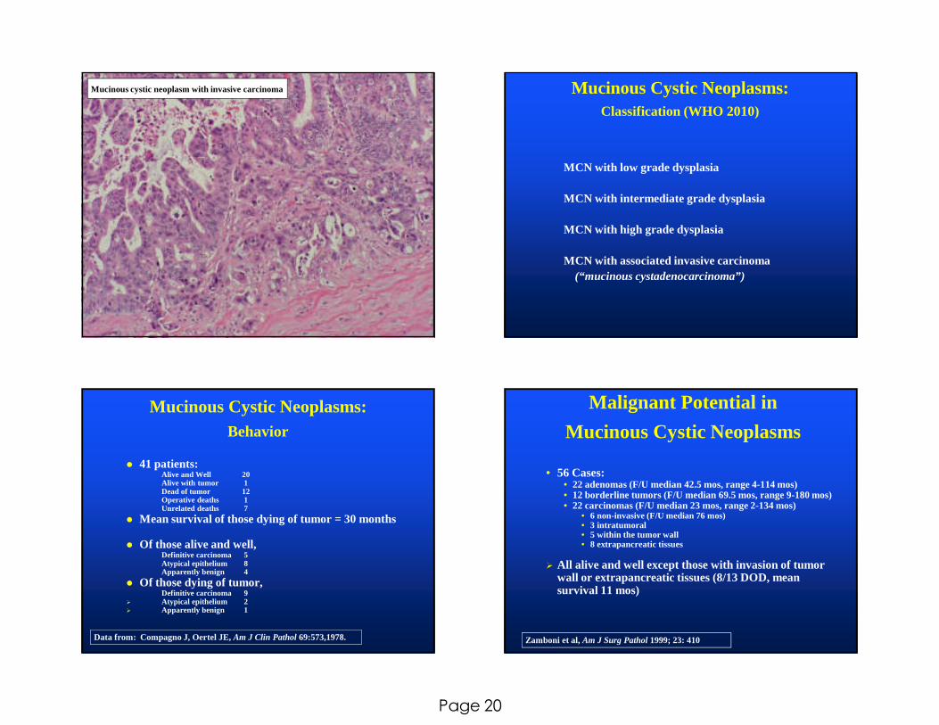
Mucinous cystic neoplasm with invasive carcinoma Mucinous Cystic Neoplasms:Classification (WHO 2010)
MCN with low grade dysplasia
MCN with intermediate grade dysplasia
MCN with high grade dysplasia
MCN with associated invasive carcinoma(“mucinous cystadenocarcinoma”)
Mucinous Cystic Neoplasms:Behavior
� 41 patients:Alive and Well 20Alive with tumor 1Dead of tumor 12Operative deaths 1Unrelated deaths 7
� Mean survival of those dying of tumor = 30 months
� Of those alive and well,Definitive carcinoma 5Atypical epithelium 8Apparently benign 4
� Of those dying of tumor,Definitive carcinoma 9
� Atypical epithelium 2� Apparently benign 1
Data from: Compagno J, Oertel JE, Am J Clin Pathol 69:573,1978.
Malignant Potential in
Mucinous Cystic Neoplasms
• 56 Cases:• 22 adenomas (F/U median 42.5 mos, range 4-114 mos)• 12 borderline tumors (F/U median 69.5 mos, range 9-180 mos)• 22 carcinomas (F/U median 23 mos, range 2-134 mos)
• 6 non-invasive (F/U median 76 mos)• 3 intratumoral• 5 within the tumor wall• 8 extrapancreatic tissues
� All alive and well except those with invasion of tumor wall or extrapancreatic tissues (8/13 DOD, mean survival 11 mos)
Zamboni et al, Am J Surg Pathol 1999; 23: 410
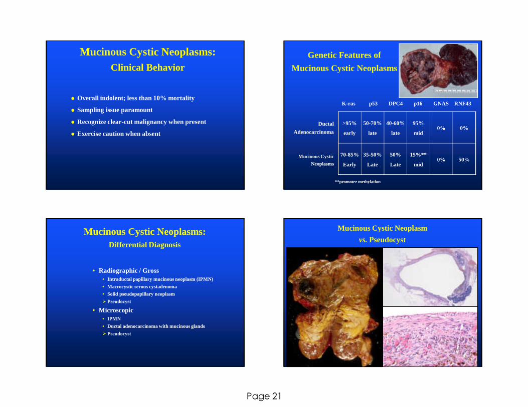
Page 21
Mucinous Cystic Neoplasms:Clinical Behavior
� Overall indolent; less than 10% mortality
� Sampling issue paramount
� Recognize clear-cut malignancy when present
� Exercise caution when absent
Genetic Features of
Mucinous Cystic Neoplasms
K-ras p53 DPC4 p16 GNAS RNF43
Ductal
Adenocarcinoma
Mucinous Cystic
Neoplasms
**promoter methylation
>95%
early
50-70%
late
40-60%
late
95%
mid0% 0%
70-85%
Early
35-50%
Late
50%
Late
15%**
mid0% 50%
Mucinous Cystic Neoplasms:Differential Diagnosis
• Radiographic / Gross• Intraductal papillary mucinous neoplasm (IPMN)
• Macrocystic serous cystadenoma
• Solid pseudopapillary neoplasm
� Pseudocyst
• Microscopic• IPMN
• Ductal adenocarcinoma with mucinous glands
� Pseudocyst
Mucinous Cystic Neoplasm
vs.Pseudocyst

Page 22
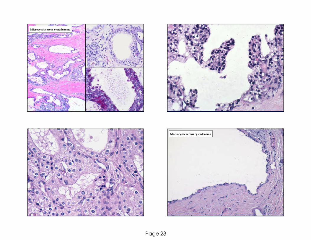
Serous Neoplasms
� Microcystic serous cystadenoma• Microcystic adenoma
• Glycogen-rich adenoma
� Macrocystic serous cystadenoma• Oligolocular ill-demarcated adenoma
� Solid serous adenoma
� Serous cystadenocarcinoma
Serous Cystic Neoplasms
� Mean age = 65 yrs
� Female > male (7:3)
� Associated with von Hippel Lindau
syndrome (vHL gene mutations)
� Head = Body / Tail
� Mean size = 6 cm (up to 30 cm)
Microcystic serous cystadenoma Macrocystic serous cystadenoma
Page 23
Microcystic serous cystadenoma
Macrocystic serous cystadenoma

Page 24
Solid serous adenoma
dPAS
PAS
Serous cystadenocarcinoma
Serous Cystic Neoplasms:Differential Diagnosis
� Radiographic / Gross� Microcystic
� Large: ????
� Small: any macrocystic lesion
� Macrocystic
� Branch duct IPMN
� Mucinous cystic neoplasm
� Retention cyst
� Microscopic� Microcystic
� Lymphangioma
� Renal cell carcinoma
� Macrocystic
� Lymphangioma
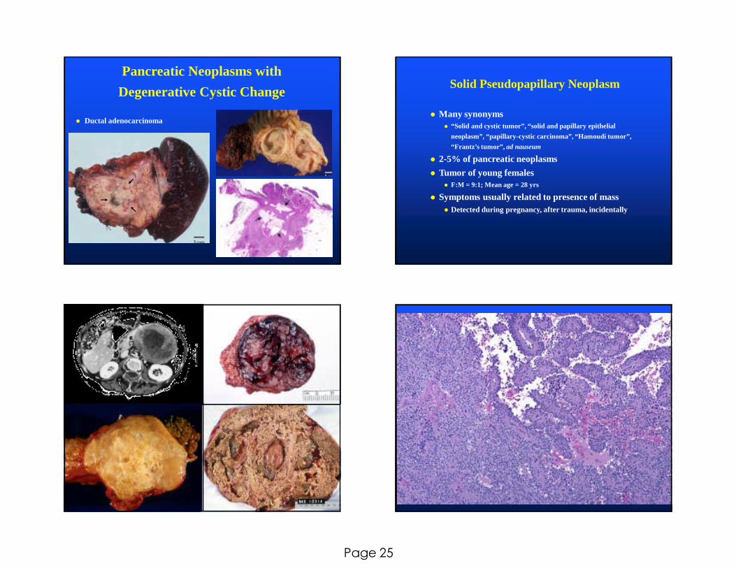
Pancreatic Neoplasms with
Degenerative Cystic Change
� Pancreatic endocrine
neoplasm
� Acinar cell carcinoma
Page 25
Pancreatic Neoplasms with
Degenerative Cystic Change
� Ductal adenocarcinoma
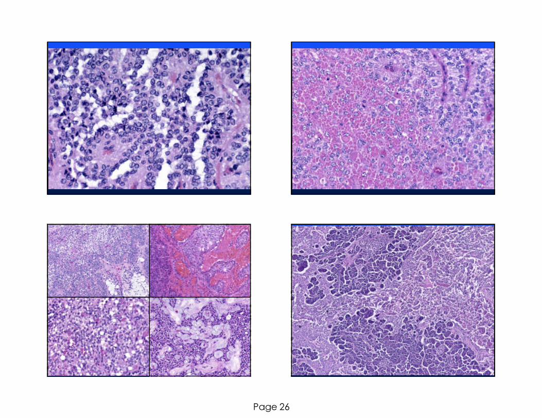
Solid Pseudopapillary Neoplasm
� Many synonyms� “Solid and cystic tumor”, “solid and papillary epithelial
neoplasm”, “papillary-cystic carcinoma”, “Hamoudi tumor”,
“Frantz’s tumor”, ad nauseum
� 2-5% of pancreatic neoplasms
� Tumor of young females � F:M = 9:1; Mean age = 28 yrs
� Symptoms usually related to presence of mass� Detected during pregnancy, after trauma, incidentally
Page 26
Page 27
β-catenin
Vimentin
α-1-antitrypsin
dPAS
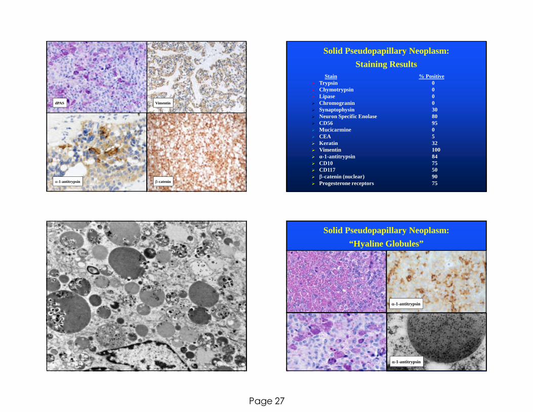
Solid Pseudopapillary Neoplasm:
Staining ResultsStain % Positive
� Trypsin 0� Chymotrypsin 0� Lipase 0� Chromogranin 0� Synaptophysin 30 � Neuron Specific Enolase 80� CD56 95� Mucicarmine 0� CEA 5� Keratin 32� Vimentin 100� α-1-antitrypsin 84� CD10 75� CD117 50� β-catenin (nuclear) 90� Progesterone receptors 75
Solid Pseudopapillary Neoplasm:
“Hyaline Globules”
α-1-antitrypsin
α-1-antitrypsin
Page 28
Solid Pseudopapillary Neoplasm:
Genetic Features
� APC / β-catenin pathway (90%)� β-cateninmutations
� Overexpression of cyclin D1
� Loss of membranous E-cadherin
� No abnormalities in “ductal adenocarcinoma genes”� KRAS
� TP53
� DPC4
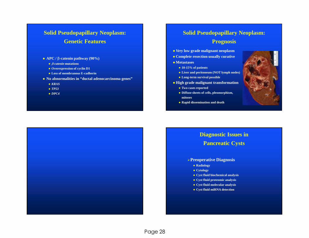
Solid Pseudopapillary Neoplasm:
Prognosis
� Very low grade malignant neoplasm
� Complete resection usually curative
� Metastases� 10-15% of patients
� Liver and peritoneum (NOT lymph nodes)
� Long-term survival possible
� High grade malignant transformation� Two cases reported
� Diffuse sheets of cells, pleomorphism,
mitoses
� Rapid dissemination and death
Diagnostic Issues in
Pancreatic Cysts
�Preoperative Diagnosis� Radiology
� Cytology
� Cyst fluid biochemical analysis
� Cyst fluid proteomic analysis
� Cyst fluid molecular analysis
� Cyst fluid miRNA detection
Page 29
Preoperative Diagnosis of
Pancreatic Cysts
�Distinguish Mucinous from Non-mucinous
Lesions � Pseudocyst
� Serous cystic neoplasm
� Cystic neuroendocrine neoplasm
�Distinguish Low Grade Dysplasia from High
Grade Dysplasia and Invasive Carcinoma� Resection vs. follow-up
� Optimum test: positive = high grade; negative = low grade
� Acceptable test: negative = low grade
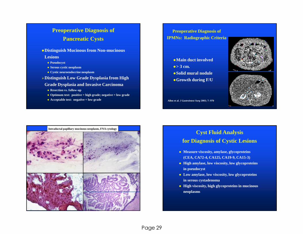
Preoperative Diagnosis of
IPMNs: Radiographic Criteria
�Main duct involved
�> 3 cm.
�Solid mural nodule
�Growth during F/U
Allen et al. J Gastrointest Surg2003; 7: 970
Intraductal papillary mucinous neoplasm, FNA cytology
Cyst Fluid Analysis
for Diagnosis of Cystic Lesions
� Measure viscosity, amylase, glycoproteins
(CEA, CA72-4, CA125, CA19-9, CA15-3)
� High amylase, low viscosity, low glycoproteins
in pseudocyst
� Low amylase, low viscosity, low glycoproteins
in serous cystadenoma
� High viscosity, high glycoproteins in mucinous
neoplasms
Page 30
Cyst Fluid Analysis
for Diagnosis of Cystic Lesions� CEA levels more sensitive than all other
glycoproteins (even in combination)
� Fluid CEA of 192 ng/ml separates mucinous vs.
nonmucinous cysts (79% accuracy)� Cytology accuracy = 59%
� EUS appearance accuracy = 51%
� Less sensitive to distinguish low grade vs.high
grade dysplasia (>2500 ng/ml = worrisome)
� Proteomic profiling: CEA, CA72.4 = mucinous
Brugge et al. Gastroenterology 2004; 126: 1330Pitman et al. Pancreatology2008; 8: 277Allen et al. Ann Surg2009; 250: 754
Wu et al., Sci Transl Med 2011;3:92ra66
Wu et al., PNAS2011;108:21188
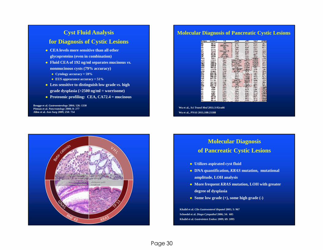
Molecular Diagnosis of Pancreatic Cystic Lesions
Molecular Diagnosis
of Pancreatic Cystic Lesions
� Utilizes aspirated cyst fluid
� DNA quantification, KRASmutation, mutational
amplitude, LOH analysis
� More frequent KRASmutation, LOH with greater
degree of dysplasia
� Some low grade (+), some high grade (-)
Khalid et al. Clin Gastroenterol Hepatol2005; 3: 967
Schoedel et al. Diagn Cytopathol2006; 34: 605
Khalid et al. Gastrointest Endosc 2009; 69: 1095
Page 31
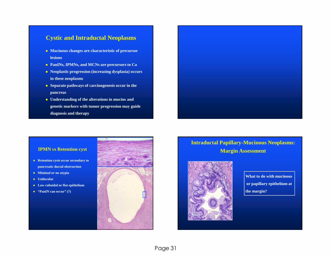
Cystic and Intraductal Neoplasms
� Mucinous changes are characteristic of precursor
lesions
� PanINs, IPMNs, and MCNs are precursors to Ca
� Neoplastic progression (increasing dysplasia) occurs
in these neoplasms
� Separate pathways of carcinogenesis occur in the
pancreas
� Understanding of the alterations in mucins and
genetic markers with tumor progression may guide
diagnosis and therapy
IPMN vsRetention cyst
� Retention cysts occur secondary to
pancreatic ductal obstruction
� Minimal or no atypia
� Unilocular
� Low cuboidal or flat epithelium
� “PanIN can occur” (?)
Intraductal Papillary-Mucinous Neoplasms:
Margin Assessment
What to do with mucinous
or papillary epithelium at
the margin?
Page 32
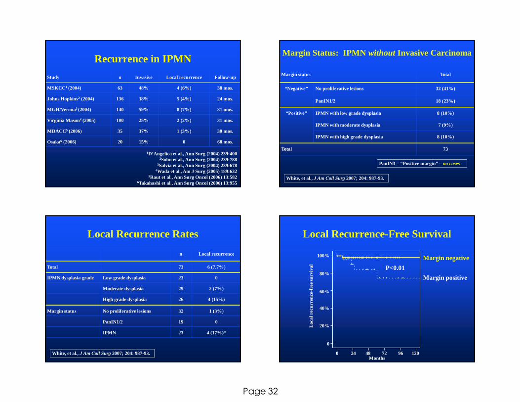
Recurrence in IPMN
Study n Invasive Local recurrence Follow-up
MSKCC 1 (2004) 63 48% 4 (6%) 38 mos.
Johns Hopkins2 (2004) 136 38% 5 (4%) 24 mos.
MGH/Verona3 (2004) 140 59% 8 (7%) 31 mos.
Virginia Mason4 (2005) 100 25% 2 (2%) 31 mos.
MDACC 5 (2006) 35 37% 1 (3%) 30 mos.
Osaka6 (2006) 20 15% 0 68 mos.
1D’Angelica et al., Ann Surg (2004) 239:4002Sohn et al., Ann Surg (2004) 239:788
3Salvia et al., Ann Surg (2004) 239:6784Wada et al., Am J Surg (2005) 189:632
5Raut et al., Ann Surg Oncol (2006) 13:5826Takahashi et al., Ann Surg Oncol (2006) 13:955
Margin Status: IPMN without Invasive Carcinoma
Margin status Total
“Negative” No proliferative lesions 32 (41%)
PanIN1/2 18 (23%)
“Positive” IPMN with low grade dysplasia 8 (10%)
IPMN with moderate dysplasia 7 (9%)
IPMN with high grade dysplasia 8 (10%)
Total 73
White, et al., J Am Coll Surg 2007; 204: 987-93.
PanIN3 = “Positive margin” – no cases
Local Recurrence Rates
n Local recurrence
Total 73 6 (7.7%)
IPMN dysplasia grade Low grade dysplasia 23 0
Moderate dysplasia 29 2 (7%)
High grade dysplasia 26 4 (15%)
Margin status No proliferative lesions 32 1 (3%)
PanIN1/2 19 0
IPMN 23 4 (17%)*
White, et al., J Am Coll Surg 2007; 204: 987-93.
Local Recurrence-Free Survival
0 24 48 72 96 120Months
Loca
l rec
urre
nce-
free
sur
viva
l
0
20%
40%
60%
80%
100% Margin negative
Margin positive
P<0.01
Page 33
Intraductal Papillary-Mucinous Neoplasms:
Margin Assessment
� Resect grossly evident disease
� If normal (not denuded!) or PanIN1/2 done
� If IPMN with moderate or high grade dysplasia (or
PanIN3) consider resecting more
� If IPMN with low grade dysplasia done?
� If cannot tell IPMN vs.PanIN grade the dysplasia
� When to do total pancreatectomy???
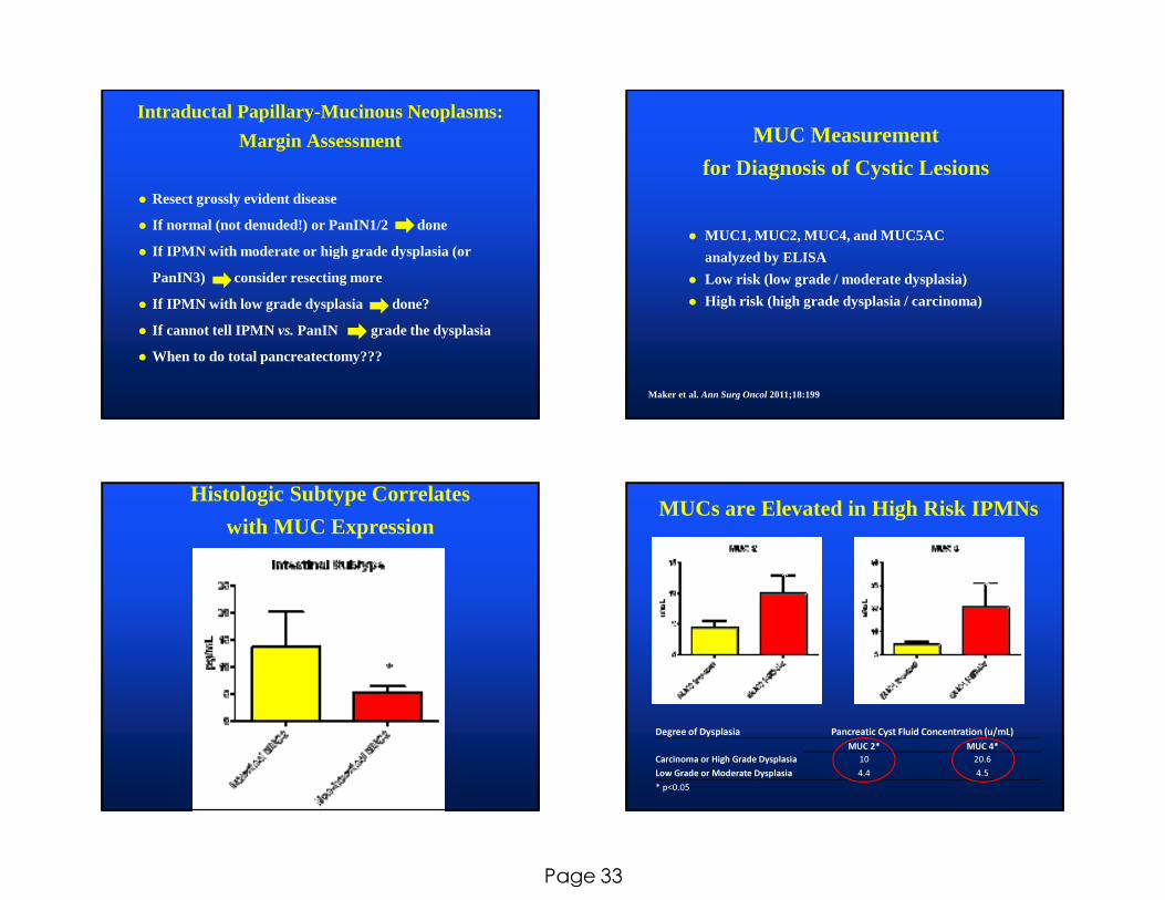
MUC Measurement
for Diagnosis of Cystic Lesions
� MUC1, MUC2, MUC4, and MUC5AC
analyzed by ELISA
� Low risk (low grade / moderate dysplasia)
� High risk (high grade dysplasia / carcinoma)
Maker et al. Ann Surg Oncol 2011;18:199
Histologic Subtype Correlates
with MUC ExpressionMUCs are Elevated in High Risk IPMNs
Degree of Dysplasia Pancreatic Cyst Fluid Concentration (u/mL)MUC 2* MUC 4*
Carcinoma or High Grade Dysplasia 10 20.6Low Grade or Moderate Dysplasia 4.4 4.5* p<0.05
**