Activating KRAS mutations are characteristic of oncocytic ...
626JOP. Journal of the Pancreas - http://pancreas.imedpub.com/ - Vol. 16 No. 6 – Nov 2015. [ISSN 1590-8577]
CASE REPORT
JOP. J Pancreas (Online) 2015 Nov 09; 16(6):626-632.
Pancreatic Oncocytic Intraductal Papillary Mucinous Neoplasms: Three Case Reports and Review of Cases in Literature
James X Wu1, Ramir Arcega2, Timothy R Donahue1,3, James S Tomlinson1,3,4, Howard A Reber1,3, Joe Hines1,3
1Department of Surgery, UCLA David Geffen School of Medicine, Los Angeles, California2Department of Pathology and Laboratory Medicine, UCLA David Geffen School of Medicine, Los
Angeles, California3UCLA Center for Pancreatic Diseases, Los Angeles, California
4Department of Surgery, VA Greater Los Angeles Healthcare System, Los Angeles, California
ABSTRACTObjectives Oncocytic intraductal papillary mucinous neoplasias represent a rare morphological subtype of intraductal papillary mucinous neoplasias, accounting for 1-8% of cases. Our aim was to characterize clinical factors associated with oncocytic intraductal papillary mucinous neoplasias, and factors that may predict invasive carcinoma. Methods Patient data for three consecutive cases of oncocytic intraductal papillary mucinous neoplasia were abstracted from electronic medical records. Literature search was performed using Pubmed and Google Scholar search engines using the keywords, “oncocytic intraductal papillary mucinous neoplasm,” “oncocytic intraductal papillary mucinous neoplasia,” “intraductal oncocytic papillary neoplasm,” and “intraductal oncocytic papillary neoplasms.” Studies that reported age, gender, invasiveness, and main versus branch duct involvement were included. Results We detailed three case reports and identified 77 previously published cases of oncocytic intraductal papillary mucinous neoplasia. The mean age at diagnosis was 61.6 years. There was a significant association of oncocytic intraductal papillary mucinous neoplasias with male gender (P=0.03), but not with main/mixed versus branch duct involvement (P=0.053). Invasive carcinoma was observed in 55.8% of oncocytic intraductal papillary mucinous neoplasias. In a subgroup of patients that underwent KRAS mutation testing (N=25), 7(28%) harbored a KRAS mutation; KRAS mutation did not predict presence of invasive carcinoma (P=1.00). Conclusions Clinical factors or KRAS mutation testing cannot currently predict the malignant potential of oncocytic intraductal papillary mucinous neoplasias. Management of oncocytic intraductal papillary mucinous neoplasias should still be based upon macroscopic criteria.
Received May 08th, 2015-Accepted July 28th, 2015Key words Oncocytic Intraductal Papillary Mucinous Neoplasm; Intraductal Oncocytic Papillary Neoplasm Abbreviations IPMN Intraductal papillary mucinous neoplasias; IOPN intraductal oncocytic papillary neoplasmsCorrespondence James X WuDepartment of Surgery10833 Le Conte AveLos Angeles, CA 90095 Phone +303 518-3186E-mail [email protected]
INTRODUCTION
Intraductal papillary mucinous neoplasms (IPMNs) are mucin-producing tumors of the pancreas that are precursors to pancreatic adenocarcinoma [1]. These lesions represent a significant proportion of surgical pancreatic disease: IPMNs now account for 25% of resected pancreatic neoplasms at major pancreatic referral centers [2].
IPMNs are currently classified macroscopically as main-duct versus branch-duct lesions, which dictates their surgical management [3]. However, IPMNs are
morphologically heterogeneous, comprised of four histological subtypes: gastric, intestinal, pancreatic-biliary, and oncocytic. These subtypes are genetically and phenotypically distinct [4], and carry differing levels of malignant potential [5-7]. Historically the histologic subtype could only be diagnosed following surgical resection and did not impact clinical management. However, recent small series have demonstrated that the histologic subtype can be predicted preoperatively from pancreatic secretions collected during ERCP or from fine needle aspiration performed during endoscopic ultrasound [8-11]. Thus, further characterization of each histologic subtype of IPMN is indicated to improve surgical management of IPMNs.
The oncocytic subtype of IPMNs is rare, accounting for only 1-8% of IPMNs [6, 12-14]. They are incompletely characterized, given the rarity of this disease. While some previous studies have reported oncocytic IPMNs have lower rates of invasive carcinoma [4], others have suggested oncocytic IPMN occur in relatively younger patients and are associated with a less favorable prognosis [5, 6, 15]. The aim of this study is to contribute three case

627JOP. Journal of the Pancreas - http://pancreas.imedpub.com/ - Vol. 16 No. 6 – Nov 2015. [ISSN 1590-8577]
JOP. J Pancreas (Online) 2015 Nov 09; 16(6):626-632.
reports from our institution, and collectively analyze cases of oncocytic IPMN reported in the medical literature.
METHODSCase Reports
Three consecutive case reports of oncocytic IPMN were identified at our institution. Patient history, laboratory values, radiology reports, pathology reports were gathered from electronic medical records.
Review of Literature
We systematically reviewed the literature for previously reported cases of oncocytic IPMN using Pubmed and Google Scholar search engines. A search was performed with the keywords “intraductal oncocytic papillary neoplasia,” “oncocytic intraductal papillary mucinous neoplasm,” “oncocytic IPMN,” and “IOPN.” We included studies of oncocytic IPMN that reported, at a minimum: age, gender, presence of low-grade dysplasia versus high-grade dysplasia versus invasive carcinoma, and main-duct versus branch duct vs mixed involvement. We excluded one study that only included invasive oncocytic IPMNs [16]. We extracted patient age, gender, presenting symptoms, tumor size, the presence of low-grade dysplasia/high-grade dysplasia/invasive carcinoma, lymph node involvement, presence of metastatic disease, location (head of pancreas/body-tail/diffuse), and main/mixed vs branch duct involvement. Additionally, if available, we tabulated preoperative serum CA 19-9 levels, as well as results of KRAS mutation testing on resected tumor. Studies that reported the IPMN as an “adenoma” were interpreted as “low-grade dysplasia” and “carcinoma-in-situ” was interpreted as “high-grade dysplasia.”
We also analyzed previously reported cases of oncocytic IPMN that provided individual-level data. This was done to facilitate statistical analysis to compare patient characteristics of main-duct versus branch-duct oncocytic IPMNs, as well as non-invasive versus invasive oncocytic IPMNs.
STATISTICAL ANALYSIS
Patient demographic data were reported as means ± standard deviations for continuous variables, and frequencies and percentages for categorical variables. Patient characteristics of main duct versus branch duct oncocytic IPMN, as well as oncocytic containing non-invasive lesions (low-grade dysplasia/high-grade dysplasia) versus invasive carcinoma was performed using unpaired t-test for continuous variables, and Fisher’s exact test and Chi-square test for categorical variables. Finally, the presence of invasive carcinoma was compared in all patients that underwent KRAS mutation testing using Fisher’s exact test. A p value of less than 0.05 was considered significant. Statistical analysis was performed using Microsoft Excel (Microsoft, Redmond, WA), GraphPad (GraphPad Software, La Jolla, CA), and Social Science Statistics calculator [17].
RESULTSCase ReportsPatient 1
The first patient was a 76 year-old female incidentally noted to have a 1 cm pancreatic cyst in the tail of pancreas on an abdominal CT scan, six years prior. Her past medical history was significant for ovarian cysts, pre-diabetes mellitus controlled with diet (A1c 6.0), and resected parathyroid adenoma. Patient had no significant family history of malignancy.
She was hospitalized five years later for acute pancreatitis and underwent another abdominal CT scan, which found that her pancreatic tail mass had increased in size to 1.5 cm. During the same hospitalization, she underwent an MRCP, which visualized a cystic lesion measuring 1.6 × 1.2 cm in the tail of the pancreas. This patient experienced another episode of pancreatitis one year later, and CT scan revealed the pancreatic tail lesion had further increased in size to 3.7 × 2 cm.
She was then referred to the surgery clinic for evaluation. Repeat CT of the abdomen demonstrated a 1.3 × 2 cm lesion involving the main duct. No significant findings were noted on her physical exam. Patient did not undergo preoperative endoscopic ultrasound (EUS). Preoperative laboratory testing did not include serum CA 19-9 or CEA. She underwent an elective laparoscopic distal pancreatectomy.
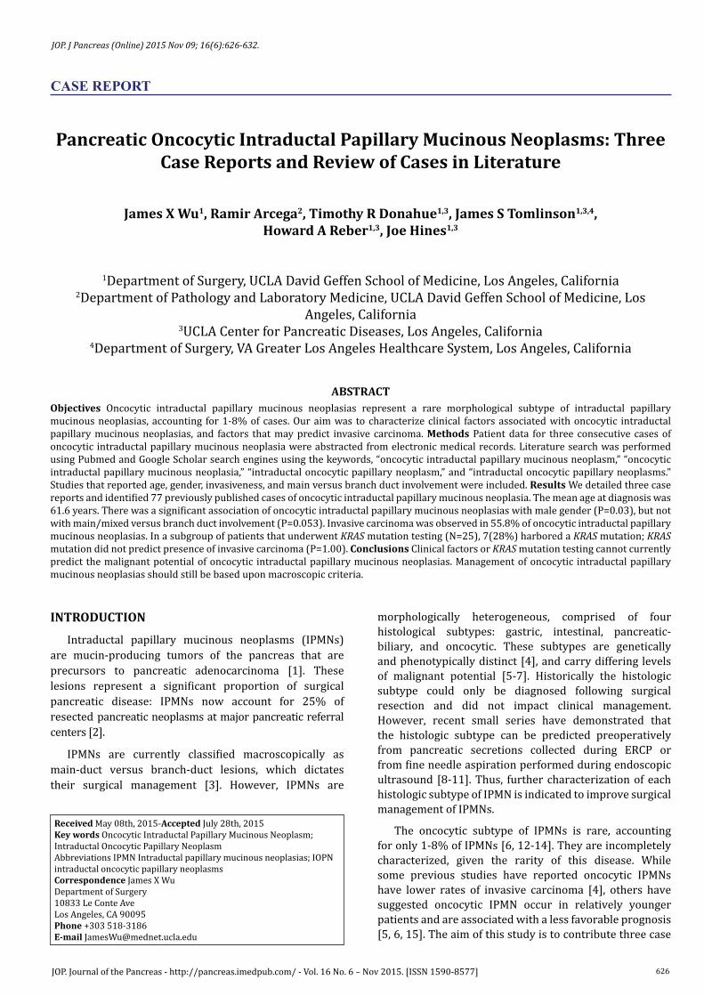
The distal pancreatectomy specimen contained a solid lesion with focal cystic areas measuring 3.8 × 1.7 × 1.2 cm. Two solid excrescences were noted within one of the cystic spaces, both measuring 0.8 × 0.5 × 0.5 cm. Microscopic examination revealed an intraductal neoplasm composed primarily of oncocytic cells with few scattered interspersed goblet cells, arranged in a complex architectural pattern, with mixed papillary and solid areas demonstrating cribriform architecture (Figure 1a). Mucinous material was identified within the inter- and intra-cellular luminal spaces. Cytologically, the cells had small, round, and basally oriented nuclei with occasional eccentrically placed nucleoli, and abundant eosinophilic and granular cytoplasm (Figure 1b). The adjacent uninvolved ducts were dilated and lined by low grade mucinous columnar cells, most consistent with low grade pancreatic intraepithelial neoplasia (PanIN) or small low grade IPMN. Ancillary studies demonstrated focal strong positivity with Hepar 1 immunostain, and no KRAS or BRAF mutations were identified by real-time PCR. KRAS gene mutation testing was performed using eight primer sets, which amplified the region that contained codons 12 and 13. BRAF gene mutation testing was performed using a single primer set, which amplifies the region of BRAF containing the V600E mutation site. Three benign lymph nodes were identified.
Her post-operative course was unremarkable. She was discharged on post-operative day 7 with no perioperative

628JOP. Journal of the Pancreas - http://pancreas.imedpub.com/ - Vol. 16 No. 6 – Nov 2015. [ISSN 1590-8577]
JOP. J Pancreas (Online) 2015 Nov 09; 16(6):626-632.
complications. She was seen in clinic 5 weeks after her surgery, and was recovering well.
Patient 2
The second patient was a 47 year-old man who presented to an outside emergency department with sudden onset flank pain, later diagnosed as symptomatic nephrolithiasis. CT scan of the abdomen noted an incidental mass in the distal pancreas. His past medical history was only significant for hypertension, controlled with Benicar/HCTZ. He had no history of pancreatitis. His family history was negative for malignancy among first-degree relatives.
Subsequent CT scans found that the pancreatic tail mass was increasing in size. To further evaluate this lesion, he underwent a EUS that noted a 2.2 cm pancreatic tail mass. An EUS-FNA was performed, and cytology from the FNA sample was determined to be benign. Surveillance CT performed one year later demonstrated a 1.9 × 2.9 cm septated cytic lesion in the tail of the pancreas without change in size. He was seen in clinic and decided to undergo surgical resection. He reported no abdominal pain, nausea, or changes in his bowel habits. Preoperative laboratory workup revealed CA 19-9 of <3 U/mL. He
underwent elective laparoscopic distal pancreatectomy and splenectomy.
The distal pancreatectomy specimen showed an encapsulated multicystic mass with no solid areas, measuring 3.1 × 2.2 × 2.9 cm. The contours of the thin cyst lining were smooth with no excrescences identified. Cyst contents were composed of approximately 5 cc of clear mucinous fluid with tenacious consistency. Microscopic examination revealed a complex papillary lesion with focal cribriform architecture composed primarily of oncocytic cells with occasional scattered goblet and mucinous cells (Figure 2a). The papillae project into the lumen of the dilated ducts. Focal branch duct involvement by tumor was identified. The cells demonstrated marked cytologic atypia with high N:C ratios, nuclear overlapping, and large bizarre nuclei (Figure 2b). The combined cytoarchitectural atypia of this lesion was sufficient to be considered a high-grade dysplasia; however, no invasive component was seen. Ancillary studies revealed positivity for both Hepar 1 and B72.3 immunohistochemical stains, in addition to KRAS Gly12Arg (GGT to CGT) mutation by real-time PCR. One benign lymph node was identified.
Figure 1. Patient 1 (a.). Oncocytic cells with scattered goblet cells are arranged in a complex papillary configuration with anastomosing fronds, with more solid areas displaying cribriform architecture. (b.). The cells have small, round, basally oriented nuclei with eccentrically placed nucleoli and granular eosinophilic cytoplasm. Hemotoxylin-eosin stain, original magnification 200x (a.); 400x (b).
a
b
Figure 2. Patient 2 (a.). Oncocytic cells with goblet and mucinous cells arranged in complex papillary projections and focal cribriform architecture. (b.). Neoplastic cells with high N:C ratio, large bizarre atypical nuclei, nuclear overlapping, prominent macronucleoli, with granular eosinophilic cytoplasm. A rare mitotic figure is noted. Hemotoxylin-eosin stain, original magnification 40x (a.); 400x (b.).
a
b

629JOP. Journal of the Pancreas - http://pancreas.imedpub.com/ - Vol. 16 No. 6 – Nov 2015. [ISSN 1590-8577]
JOP. J Pancreas (Online) 2015 Nov 09; 16(6):626-632.
His recovery was unremarkable. He was discharged on post-operative day 4. A surveillance MRI done at 1-year follow-up had no evidence of recurrence, and patient was doing well.
Patient 3
The third patient was a 49 year-old man with no significant past medical history that developed new onset of recurrent episodes of acute pancreatitis, requiring 8 hospitalizations over the course of 1 year. He had no evidence of gallstones on abdominal ultrasound, no history of alcohol use, and no significant family history. An abdominal CT identified a 1.8 cm low-density lesion in the head of the pancreas. The lesion was evaluated with EUS, which demonstrated a “salt and pepper” 1.7 cm cystic lesion without a mural nodule that did not appear to communicate with the pancreatic duct; it was diagnosed as a likely pancreatic pseudocyst and FNA was deferred. The EUS noted gallbladder sludge, prompting the patient to undergo laparoscopic cholecystectomy. Unfortunately, his symptoms continued to persist following this procedure.
Following the cholecystectomy, he underwent a series of abdominal MRIs to monitor the pancreatic cyst, which all found a stable, 2 cm cystic lesion in the head of the pancreas without decrease in size after six months. EUS was repeated, which showed a 1.8 cm cystic lesion that communicated with a non-dilated pancreatic duct, with a 0.9 × 1.0 cm hyperechoic lesion in the center of the cyst. FNA of the cyst revealed only extracellular mucin without definite epithelial cells.
At this time, the patient presented to general surgery clinic for evaluation. No significant findings were found on physical exam. Despite his recurring abdominal pain, he reported being otherwise well and tolerating a regular diet. Preoperative laboratory testing revealed a CA 19-9 level of <3 U/mL, with all other tests within normal limits. Patient underwent pylorus preserving pancreaticoduodenectomy for suspected IPMN without complications.
The pancreaticoduodenectomy specimen revealed a cystic lesion within the pancreatic head measuring 2.0 x 1.2 × 0.4 cm, with a soft tan-white excrescence measuring 0.8 × 0.8 × 0.3 cm attached to the cyst wall. The cystic lesion did not communicate with the main pancreatic duct, and was filled with serosanguinous fluid. Microscopic examination revealed bland oncocytic cells arranged in complex arborizing papillae which protrude into the cystically dilated ducts (Figure 3a). The cells displayed round nuclei and an evenly distributed chromatin pattern, single prominent eccentrically placed nucleoli, and abundant granular eosinophilic cytoplasm (Figure 3b). Focal areas of PanIN-1 (low grade) were also noted. Ancillary studies revealed positivity for both Hepar 1 and B72.3 immunohistochemical stains. The specimen was not tested for KRAS mutation. Twenty-four benign lymph nodes were identified.
The patient’s post-operative recovery was unremarkable. He was seen in clinic 4 weeks post-
Figure 3. Patient 3 (a.). Bland oncocytic cells are arranged in complex arborizing papillae. (b.). The cells display small round nuclei, evenly distributed chromatin, prominent eccentrically placed nucleoli, and granular eosinophilic cytoplasm. Hemotoxylin-eosin stain, original magnification 100x (a.); 400x (b.).
a
b
operatively, doing well. He will undergo follow-up imaging in 6-12 months.
Literature Review for Previously Reported Oncocytic IPMN
The literature review identified 23 studies reporting 77 cases of oncocytic IPMNs (Table 1), including the current study with three case reports above. The mean age of diagnosis was 61.6±12.3 years of age, ranging from 20 to 80 years of age. There was a significant association of oncocytic IPMN with male gender, with 62.3% (N=48) male, 37.7% (N=29) female patients (P=0.03). Main or mixed duct involvement was seen in 47 (61%) of oncocytic IPMNs, and branch-duct involvement was seen in 30 (39%) (P=0.053). Only a small percentage of oncocytic IPMNs contained low-grade dysplasia 6.5% (N=5), another 37.7% (N=29) contained high-grade dysplasia, and 55.8% (N=43) contained invasive carcinoma. Patient characteristics are summarized in Table 2.
Several of the 77 reported cases were missing data points regarding tumor location, tumor size, serum CA 19-9 levels, and presenting symptoms; for these disease

630JOP. Journal of the Pancreas - http://pancreas.imedpub.com/ - Vol. 16 No. 6 – Nov 2015. [ISSN 1590-8577]
JOP. J Pancreas (Online) 2015 Nov 09; 16(6):626-632.
Table 1. Included Studies.
Author Year N ReferenceJyotheeswaran et al. 1998 1 [25]Nobukawa et al. 1999 1 [16]Noji et al. 2002 1 [26]Patel et al. 2002 1 [27]Shima et al. 2005 1 [28]Oku et al. 2007 1 [29]Ishida et al. 2007 1 [30]Fischer et al. 2010 1 [31]Liszka et al. 2010 4 [24]Takasu et al. 2010 3 [23]Furukawa et al. 2011 24 [6]Kim et al. 2011 2 [15]Mohri et al. 2011 2 [32]Xiao et al. 2011 18 [4]Buxbaum et al. 2012 1 [33]Chiang et al. 2012 1 [34]Kato et al. 2012 2 [35]Distler et al. 2013 4 [12]Kang et al. 2013 2 [13]Wohlauer et al. 2013 1 [36]Murdock et al. 2014 1 [37]Yamada et al. 2014 1 [38]Wu et al. 2015 3 n/a
Table 2. Oncocytic IPMN Patient Characteristics.
Age, median(range) 61.6 (20-80)Gender
Male 47 (61.8%)Female 29 (38.2%)Ductal Involvement
Main/Mixed 47 (61%)Branch 30 (39%)Invasiveness
Low-grade Dysplasia 5 (6.5%)High-grade Dysplasia 29 (37.7%)Invasive Carcinoma 43 (55.8%)Tumor Location (50 reporting)
Head 26/50 (52%)Body/Tail 23/50 (46%)Diffuse 1/50 (2%)Tumor size, cm (44 reporting) 5.4 ± 3.7Serum 19-9, Units/mL (37 reporting) (Reference range 0-37 U/ml), median(range)
18.4 (0-75.6)
KRAS (25 reporting)
KRAS Mutation Present 7/25 (28%)KRAS Mutation Absent 18/25 (72%)Presenting Symptoms (27 reporting)
Abdominal Pain/Nausea 12/27 (44.4%)Incidental 10/27 (37%)Painless Jaundice 2/27 (7.4%)Pancreatitis 2/27 (7.4%)Other 1/27 (3.8%)
Continuous variables reported as pooled means ± standard deviation
characteristics, pooled means and standard deviation were calculated from subgroups. The mean tumor size was reported in 44 cases, with a pooled mean and standard deviation of 5.4±3.7 cm including the cystic portion of the
lesion. Serum 19-9 was reported in 37 cases, with a pooled mean and standard deviation of 18.4±24 U/mL. Presenting symptoms were reported in 27 cases; the majority of patients presented with abdominal discomfort or nausea (12/27, 44.4%), with a significant proportion found incidentally on abdominal imaging (10/27, 37%). Painless jaundice (2/27, 7.4%) and pancreatitis (2/27, 7.4%) were less commonly reported.
KRAS mutation testing was performed in 25 cases. Among these patients, 14/25 (56%) had invasive carcinoma. Only 7/25 (28%) harbored KRAS mutation. Among tumors containing a KRAS mutation, 4/7 (57.1%) also contained invasive carcinoma and 3/7 (42.9%) were non-invasive lesions, all with high-grade dysplasia. Oncocytic IPMNs without KRAS mutations were also divided evenly between invasive carcinoma (9/18, 50%) and non-invasive lesions (9/18, 50%). Of the 9 non-invasive lesions with wild-type KRAS, 8 contained high-grade dysplasia and 1 contained low-grade dysplasia. Thus, KRAS mutation did not predict invasiveness of oncocytic IPMNs (P=1.00).
Lastly, individual-level patient data was derived from 17 studies reporting 25 cases of oncocytic IPMN. Among these patients, 14 were main/mixed-duct and 10 were branch-duct oncocytic IPMN. The rate of invasive carcinoma was 78.5% (11/14) for main/mixed-duct lesions, and 30% (3/10) for branch-duct lesions. When patient characteristics were compared between main/mixed-duct versus branch-duct oncocytic IPMNs, there was no significant difference in age, gender, or presence of invasive carcinoma between groups. Similarly, comparison of non-invasive oncocytic IPMN versus oncocytic IPMN with invasive carcinoma found no significant differences between age, gender, main/mixed-duct versus branch-duct involvement between groups.
DISCUSSIONThis study described three previously unreported
cases of oncocytic IPMN, and is the largest review of cases of oncocytic IPMNs to our knowledge. Our findings indicate that 55.8% of oncocytic IPMNs contain invasive carcinoma. Main-duct involvement tends to increase the risk of malignancy among oncocytic IPMNs: a subset of 24 patients with individual-level data revealed that the rate of invasive carcinoma was 78.5% in main/mixed-duct oncocytic IPMN, and 30% in branch-duct oncocytic IPMN. Furthermore, KRAS mutation testing did not predict presence of invasive carcinoma. In a subgroup of patients that underwent KRAS mutation testing revealed that only a quarter of oncocytic IPMNs contained a KRAS mutation, even though 56% of the subgroup had invasive oncocytic IPMN. We also found that oncocytic IPMNs are associated with male gender, but do not have a predilection for main duct involvement.
The rate of invasion among oncocytic IPMNs are higher than IPMNs overall, with invasion seen in 78.5% of main/mixed-duct oncocytic IPMNs and 30% of branch-duct IPMNs. In comparison, the rates of invasive carcinoma in

631JOP. Journal of the Pancreas - http://pancreas.imedpub.com/ - Vol. 16 No. 6 – Nov 2015. [ISSN 1590-8577]
JOP. J Pancreas (Online) 2015 Nov 09; 16(6):626-632.
main-duct and branch-duct IPMNs (all subtypes) are 43 and 15%, respectively [18]. A few studies demonstrated that preoperative diagnosis of histologic subtype of IPMN might be feasible using pancreatic cyst fluid cytology [8, 11]. If oncocytic IPMN can be reliably diagnosed pre-operatively, resection could be considered even if does not meet current surgical criteria. Indeed, pre-operative diagnosis of IPMN subtype is an area in need of further research and development - none of the 77 cases in our review reported a pre-operative diagnosis of oncocytic IPMN. Furthermore, the higher rates of invasive carcinoma in our series may be due to selection or publication bias.
In our review, oncocytic IPMNs have an overall reduced proportion of lesions with KRAS mutation (28%) compared to IPMNs overall, which may have KRAS mutations in 50-80% of lesions [5]. Similarly, a previous series of 18 oncocytic IPMNs identified KRAS mutation in 17% of patients [4]. The relative higher rate of invasive carcinoma combined with a relatively lower rate of KRAS mutation in oncocytic IPMNs suggests they may have a distinct pathway to malignancy. While more studies are indicated, this data suggests KRAS testing has little influence on the prognosis of oncocytic IPMNs. GNAS and RNF43 mutations are also commonly found in IPMNs [19]. Recent studies have also identified the presence of activating GNAS mutations in up to 66% of IPMNs, 87.5% of adenocarcinomas associated with IPMNs, but not in pancreatic adenocarcinomas without a component of IPMN [20]. However, two recent series reported only one out of five total oncocytic IPMNs tested had a GNAS mutation, and out of three oncocytic IPMNs tested, none had RNF43 mutation [21, 22].
Our study has several limitations. In our three case reports, immunohistochemistry for mucin expression was not performed. Different patterns of mucin expression have been described for the histologic subtypes of IPMNs. While all of the studies that reported mucin expression of oncocytic IPMNs consistently reported MUC5AC expression, there is disagreement about the expression of MUC1 and MUC2; some describe oncocytic IPMNs as MUC1 positive and MUC2 negative [12, 15, 23], while others reported variable or focal MUC1 and MUC2 expression [4, 6, 24]. Since the study was retrospective and gathered cases reported in current literature, there may be a strong selection bias, as only severe cases would be submitted as case reports, and only included patients who underwent surgical resection. Furthermore, analysis of tumor location, tumor size, serum CA 19-9 levels, and presenting symptoms could only be analyzed in subgroups, since these data points were not found in all reported cases. In the future, a prospective, multi-institutional study would be ideal, but would require a significant length of time to identify an appropriate number of patients.
CONCLUSIONOncocytic IPMNs are observed more often in male
patients, and have equal rates of main- and branch-duct involvement. Despite a rate of invasive carcinoma of 55.8%, only a quarter of oncocytic IPMNs tested have KRAS mutations, and presence of KRAS mutation was not
associated with invasive carcinoma. Given that, at this time, no clinical factors can be used to predict invasive versus non-invasive oncocytic IPMNs, these lesions should still be managed by macroscopic criteria. Further characterization of oncocytic IPMNs is indicated.
Conflicting InterestThe authors had no conflicts of interest.
References1. Kloppel G, Basturk O, Schlitter AM, Konukiewitz B, Esposito I. Intraductal neoplasms of the pancreas. Semin Diagn Pathol 2014; 31:452-66. [PMID: 25282472]
2. Grützmann R, Niedergethmann M, Pilarsky C, Klöppel G, Saeger HD. Intraductal papillary mucinous tumors of the pancreas: biology, diagnosis, and treatment. Oncologist 2010; 15:1294-309. [PMID: 21147870]
3. Tanaka M, Fernández-del Castillo C, Adsay V, Chari S, Falconi M, Jang J-Y, et al. International consensus guidelines 2012 for the management of IPMN and MCN of the pancreas. Pancreatology 2012; 12:183-97. [PMID: 22687371]
4. Xiao HD, Yamaguchi H, Dias-Santagata D, Kuboki Y, Akhavanfard S, Hatori T, et al. Molecular characteristics and biological behaviours of the oncocytic and pancreatobiliary subtypes of intraductal papillary mucinous neoplasms. J Pathol 2011; 224:508-16. [PMID: 21547907]
5. Tanaka M. Intraductal Papillary Mucinous Neoplasm of the Pancreas: Springer Japan; 2013
6. Furukawa T, Hatori T, Fujita I, Yamamoto M, Kobayashi M, Ohike N, et al. Prognostic relevance of morphological types of intraductal papillary mucinous neoplasms of the pancreas. Gut 2011; 60:509-16.[PMID: 21193453]
7. Sadakari Y, Ohuchida K, Nakata K, Ohtsuka T, Aishima S, Takahata S, et al. Invasive carcinoma derived from the nonintestinal type intraductal papillary mucinous neoplasm of the pancreas has a poorer prognosis than that derived from the intestinal type. Surgery 2010; 147:812-7.[PMID: 20060146]
8. Hibi Y, Fukushima N, Tsuchida A, Sofuni A, Itoi T, Moriyasu F, et al. Pancreatic juice cytology and subclassification of intraductal papillary mucinous neoplasms of the pancreas. Pancreas 2007; 34:197-204. [PMID: 17312458]
9. Monzen M, Shimizu K, Hatori T, Furukawa T, Shiratori K. Usefulness of cell block cytology for preoperative grading and typing of intraductal papillary mucinous neoplasms. Pancreatology 2013; 13:369-78. [PMID: 23890135]
10. Hisaka T, Horiuchi H, Uchida S, Ishikawa H, Kawahara R, Kawashima Y, et al. Potential usefulness of mucin immunohistochemical staining of preoperative pancreatic biopsy or juice cytology specimens in the determination of treatment strategies for intraductal papillary mucinous neoplasm. Oncology reports 2013; 30:2035-41. [PMID: 24008495]
11. Hara T, Ikebe D, Odaka A, Sudo K, Nakamura K, Yamamoto H, et al. Preoperative histological subtype classification of intraductal papillary mucinous neoplasms (IPMN) by pancreatic juice cytology with MUC stain. Ann Surg 2013; 257:1103-11. [PMID: 23364699]
12. Distler M, Kersting S, Niedergethmann M, Aust DE, Franz M, Rückert F, et al. Pathohistological subtype predicts survival in patients with intraductal papillary mucinous neoplasm (IPMN) of the pancreas. Ann Surg 2013; 258:324-30. [PMID: 23532107]
13. Kang MJ, Lee KB, Jang JY, Han IW, Kim SW. Evaluation of clinical meaning of histological subtypes of intraductal papillary mucinous neoplasm of the pancreas. Pancreas 2013; 42:959-66. [PMID: 23462330]
14. Ideno N, Ohtsuka T, Kono H, Fujiwara K, Oda Y, Aishima S, et al. Intraductal papillary mucinous neoplasms of the pancreas with distinct pancreatic ductal adenocarcinomas are frequently of gastric subtype. Ann Surg 2013; 258:141-51. [PMID: 23532108]

632JOP. Journal of the Pancreas - http://pancreas.imedpub.com/ - Vol. 16 No. 6 – Nov 2015. [ISSN 1590-8577]
JOP. J Pancreas (Online) 2015 Nov 09; 16(6):626-632.
15. Kim J, Jang KT, Park SM, Lim SW, Kim JH, Lee KH, et al. Prognostic relevance of pathologic subtypes and minimal invasion in intraductal papillary mucinous neoplasms of the pancreas. Tumor Biology 2011; 32:535-42. [PMID: 21190101]
16. Nobukawa B, Suda K, Suyama M, Ariyama J, Beppu T, Futagawa S. Intraductal oncocytic papillary carcinoma with invasion arising from the accessory pancreatic duct. Gastrointest Endosc 1999; 50:864-6. [PMID: 10570358]
17. Statistics SS [Internet].[cited June 10th, 2014].
18. Bassi C, Sarr MG, Lillemoe KD, Reber HA. Natural history of intraductal papillary mucinous neoplasms (IPMN): current evidence and implications for management. J Gastrointest Surg 2008; 12:645-50. [PMID: 18097728]
19. Tan MC, Basturk O, Brannon AR, Bhanot U, Scott SN, Bouvier N, et al. GNAS and KRAS Mutations Define Separate Progression Pathways in Intraductal Papillary Mucinous Neoplasm-Associated Carcinoma. J Am Coll Surg 2015; 220:845-54. [PMID: 25840541]
20. Wu J, Matthaei H, Maitra A, Dal Molin M, Wood LD, Eshleman JR, et al. Recurrent GNAS mutations define an unexpected pathway for pancreatic cyst development. Sci Transl Med 2011; 3:92ra66. [PMID: 21775669]
21. Dal Molin M, Matthaei H, Wu J, Blackford A, Debeljak M, Rezaee N, et al. Clinicopathological correlates of activating GNAS mutations in intraductal papillary mucinous neoplasm (IPMN) of the pancreas. Ann Surg Oncol 2013; 20:3802-8. [PMID: 23846778]
22. Amato E, Molin Md, Mafficini A, Yu J, Malleo G, Rusev B, et al. Targeted next-generation sequencing of cancer genes dissects the molecular profiles of intraductal papillary neoplasms of the pancreas. J Pathol 2014; 233:217-27. [PMID: 24604757]
23. Takasu N, Kimura W, Moriya T, Hirai I, Takeshita A, Kamio Y, et al. Intraductal papillary-mucinous neoplasms of the gastric and intestinal types may have less malignant potential than the pancreatobiliary type. Pancreas 2010; 39:604-10. [PMID: 20124938]
24. Liszka L, Pająk J, Zielińska-Pająk E, Krzych L, Golka D, Mrowiec S, et al. Intraductal oncocytic papillary neoplasms of the pancreas and bile ducts: a description of five new cases and review based on a systematic survey of the literature. J Hepatobiliary Pancreat Sci. 2010; 17:246-61. [PMID: 20464560]
25. Jyotheeswaran S, Zotalis G, Penmetsa P, Levea CM, Schoeniger LO, Shah AN. A newly recognized entity: intraductal “oncocytic” papillary neoplasm of the pancreas. Am J Gastroenterol 1998; 93:2539-43. [PMID: 9860422]
26. Noji T, Kondo S, Hirano S, Ambo Y, Tanaka E, Katoh C, et al. Intraductal oncocytic papillary neoplasm of the pancreas shows strong positivity on FDG-PET. Int J Gastrointest Cancer 2002; 32:43-6. [PMID: 12630769]
27. Patel SA, Adams R, Goldstein M, Moskaluk CA. Genetic analysis of invasive carcinoma arising in intraductal oncocytic papillary neoplasm of the pancreas. Am J Surg Pathol 2002; 26:1071-7. [PMID: 12170096]
28. Shima Y, Yagi T, Inagaki M, Sadamori H, Tanaka N, Horimi T, et al. Intraductal oncocytic papillary neoplasm of the pancreas with celiac artery compression syndrome and a jejunal artery aneurysm: report of a case. Surg Today 2005; 35:86-90. [PMID: 15622472]
29. Oku T, Maeda M, Wada Y, Waga E, Ono K, Nagamachi Y, et al. Intraductal oncocytic papillary neoplasm having clinical characteristics of mucinous cystic neoplasm and a benign histology. Jop 2007; 8:206-13. [PMID: 17356245]
30. Ishida M, Egawa S, Aoki T, Sakata N, Mikami Y, Motoi F, et al. Characteristic clinicopathological features of the types of intraductal papillary-mucinous neoplasms of the pancreas. Pancreas 2007; 35:348-52. [PMID: 18090241]
31. Fischer MA, Donati O, Heinrich S, Weber A, Hany TF, Soldini D, et al. Intraductal oncocytic papillary neoplasm of the pancreas: a radio-pathological case study. Jop 2010; 11:49-54. [PMID: 20065553]
32. Mohri D, Asaoka Y, Ijichi H, Miyabayashi K, Kudo Y, Seto M, et al. Different subtypes of intraductal papillary mucinous neoplasm in the pancreas have distinct pathways to pancreatic cancer progression. J Gastroenterol 2012; 47:203-13. [PMID: 22041919]
33. Buxbaum JL, Jhala NC, Christein JD, Eloubeidi MA. Oncocytic intraductal papillary mucinous neoplasm with carcinomatous degeneration. Gastrointest Endosc 2012; 75:898-9. [PMID: 22440204]
34. Chiang KC, Yu CC, Chen JR, Huang YT, Huang CC, Yeh CN, et al. Oncocytic-type intraductal papillary mucinous neoplasm (IPMN)-derived invasive oncocytic pancreatic carcinoma with brain metastasis—A case report. World J Surg Oncol 2012; 10:138. [PMID: 22776211]
35. Kato T, Ikari S, Hirata K, Machida T, Nakamura H, Meguro T, et al. FDG-PET Findings of Intraductal Oncocytic Papillary Neoplasms of the Pancreas: Two Case Reports. Case reports in gastroenterology 2012; 6:415-24. [PMID: 22933987]
36. Wohlauer MV, McManus M, Fukami N, Gajdos C. Intraductal Oncocytic Papillary Neoplasm of the Pancreas: Report of a Case Requiring Completion Pancreatectomy. JOP 2013; 14:77-80. [PMID: 23306340]
37. Tricia Murdock MZ. Intraductal Oncocytic Papillary Neoplasm of the Pancreas. Annals of Clinical Pathology 2014; 2:1016-8.
38. Yamada S, Fujii T, Shimoyama Y, Kanda M, Nakayama G, Sugimoto H, et al. Clinical Implication of Morphological Subtypes in Management of Intraductal Papillary Mucinous Neoplasm. Ann Surg Oncol 2014; 21:2444-52. [PMID: 24562937]