Development/Plasticity/Repair...
13
Development/Plasticity/Repair Cell Migration along the Lateral Cortical Stream to the Developing Basal Telencephalic Limbic System Rosalind S. E. Carney, 1,2 Teresa B. Alfonso, 1 Daniela Cohen, 3 Haining Dai, 1 Susana Nery, 3 Bogdan Stoica, 1 Jonathan Slotkin, 1 Barbara S. Bregman, 1 Gord Fishell, 3 and Joshua G. Corbin 1,2 1 Department of Neuroscience, Georgetown University Medical Center, Washington, DC 20057, 2 Center for Neuroscience Research, Children’s Research Institute, Children’s National Medical Center, Washington, DC 20010, and 3 Developmental Genetics Program and the Department of Cell Biology, Skirball Institute of BioMolecular Medicine, New York University Medical Center, New York, New York 10016 During embryogenesis, the lateral cortical stream (LCS) emerges from the corticostriatal border (CSB), the boundary between the developing cerebral cortex and striatum. The LCS is comprised of a mix of pallial- and subpallial-derived neural progenitor cells that migrate to the developing structures of the basal telencephalon, most notably the piriform cortex and amygdala. Using a combination of in vitro and in vivo approaches, we analyzed the timing, composition, migratory modes, origin, and requirement of the homeodomain- containing transcription factor Gsh2 (genomic screened homeobox 2) in the development of this prominent migratory stream. We reveal that Pax6 (paired box gene 6)-positive pallial-derived and Dlx2 (distal-less homeobox 2)-positive subpallial-derived subpopulations of LCS cells are generated in distinct temporal windows during embryogenesis. Furthermore, our data indicate the CSB border not only is comprised of separate populations of pallial- and subpallial-derived progenitors that contribute to the LCS but also a subpopulation of cells coexpressing Pax6 and Dlx2. Moreover, despite migrating along a route outlined by a cascade of radial glia, the Dlx2-positive population appears to migrate primarily in an apparent chain-like manner, with LCS migratory cells being generated locally at the CSB with little contribution from other subpallial structures such as the medial, lateral, or caudal ganglionic eminences. We further demon- strate that the generation of the LCS is dependent on the homeodomain-containing gene Gsh2, revealing a novel requirement for Gsh2 in telencephalic development. Key words: amygdala; development; pallium; radial glia; specification; subpallium Introduction The basal limbic system of the mammalian telencephalon is com- prised of a number of complex structures that includes the piri- form cortex, amygdala, and nucleus accumbens. These regions, together with other forebrain limbic structures (prefrontal cor- tex, cingulate cortex, hippocampus, and hypothalamus) form a circuit that regulates a number of essential behaviors, including specific aspects of memory and emotion and the regulation of motivational stages involved with drives. Currently, the level of understanding of basal telencephalic limbic system development does not rival that of the cerebral cortex. In the cerebral cortex, regional neuronal cell diversity is established by the convergence of migratory progenitor cells from both pallial (dorsal telence- phalic) and subpallial (ventral telencephalic) compartments. Fu- ture excitatory neurons are locally generated in the pallial neuro- epithelium and migrate radially to their laminar destination (Rakic, 1974). In contrast, the majority of inhibitory neurons is derived from the subpallial progenitor zones, most notably the medial (MGE) and caudal (CGE) ganglionic eminences, and mi- grate tangentially to the developing cerebral cortex (for review, see Corbin et al., 2001; Marin and Rubenstein, 2001). Impor- tantly, these separate subpallial progenitor zones give rise to in- hibitory neurons with distinct spatial, neurochemical, morpho- logical, and electrophysiological characteristics (Nery et al., 2002; Xu et al., 2004; Butt et al., 2005). In contrast to the vast waves of dorsally directed migrating neural precursor cells from the subpallium, cells of the lateral cortical stream (LCS) migrate in the opposite direction from dor- sal to more ventral telencephalic regions. During embryogenesis, cells of the LCS emerge from the “molecular” corticostriatal bor- der (CSB), the region of the dorsal LGE (dLGE) in which pallial gene expression [e.g., Pax6 (paired box gene 6) and Ngn2 (neu- rogenin homolog 2)] abuts subpallial gene expression [e.g., Gsh2 (genomic screened homeobox 2) and Dlx1/2 (distal-less ho- meobox 2) (for review, see Corbin et al., 2001; Molna ´ r and Butler, 2002)]. This “molecular” CSB (herein referred to as CSB) is po- Received Jan. 31, 2006; revised Sept. 22, 2006; accepted Sept. 22, 2006. This work was supported by a grant from the National Alliance for Autism Research, MRDDRC award from the Children’s Research Institute, Washington, DC, National Institutes of Health Grants DA020140 (J.C.), NS 27054 (H.D. and B.S.B.), NS032993 (G.F.), MH071679 (G.F.), and MH068469 (G.F.), and National Institute of Child Health and Human Development Grant T32 HD 007459 (J.S. and B.S.B.). We thank Dr. Arturo Alvarez-Buylla for his input on early stages of this work, Dr. Molly Huntsman for sharing of equipment, Dr. Tarik Haydar and Randi Goodnight for technical assistance, and members of the Corbin, Fishell, and Haydar labs for their helpful discussions and insight. Correspondence should be addressed to Dr. Joshua Corbin, Center for Neuroscience Research, Children’s Research Institute, Suite 5347, Children’s National Medical Center, 111 Michigan Avenue NW, Washington, DC 20010. E-mail: [email protected]. S. Nery’s present addresses: London School of Hygiene and Tropical Medicine, Department of Infectious and Tropical Diseases, Unit of Pathogen Molecular Biology, Keppel Street, London WC1E 7HT, UK. J. Slotkin’s present address: Department of Neurosurgery, The Brigham and Women’s Hospital, Children’s Hos- pital Boston, Harvard Medical School, Boston, MA 02115. DOI:10.1523/JNEUROSCI.3092-06.2006 Copyright © 2006 Society for Neuroscience 0270-6474/06/2611562-13$15.00/0 11562 • The Journal of Neuroscience, November 8, 2006 • 26(45):11562–11574
-
Upload
trinhduong -
Category
Documents
-
view
221 -
download
0
Transcript of Development/Plasticity/Repair...
Development/Plasticity/Repair
Cell Migration along the Lateral Cortical Stream to theDeveloping Basal Telencephalic Limbic System
Rosalind S. E. Carney,1,2 Teresa B. Alfonso,1 Daniela Cohen,3 Haining Dai,1 Susana Nery,3 Bogdan Stoica,1
Jonathan Slotkin,1 Barbara S. Bregman,1 Gord Fishell,3 and Joshua G. Corbin1,2
1Department of Neuroscience, Georgetown University Medical Center, Washington, DC 20057, 2Center for Neuroscience Research, Children’s ResearchInstitute, Children’s National Medical Center, Washington, DC 20010, and 3Developmental Genetics Program and the Department of Cell Biology, SkirballInstitute of BioMolecular Medicine, New York University Medical Center, New York, New York 10016
During embryogenesis, the lateral cortical stream (LCS) emerges from the corticostriatal border (CSB), the boundary between thedeveloping cerebral cortex and striatum. The LCS is comprised of a mix of pallial- and subpallial-derived neural progenitor cells thatmigrate to the developing structures of the basal telencephalon, most notably the piriform cortex and amygdala. Using a combination ofin vitro and in vivo approaches, we analyzed the timing, composition, migratory modes, origin, and requirement of the homeodomain-containing transcription factor Gsh2 (genomic screened homeobox 2) in the development of this prominent migratory stream. We revealthat Pax6 (paired box gene 6)-positive pallial-derived and Dlx2 (distal-less homeobox 2)-positive subpallial-derived subpopulations ofLCS cells are generated in distinct temporal windows during embryogenesis. Furthermore, our data indicate the CSB border not only iscomprised of separate populations of pallial- and subpallial-derived progenitors that contribute to the LCS but also a subpopulation ofcells coexpressing Pax6 and Dlx2. Moreover, despite migrating along a route outlined by a cascade of radial glia, the Dlx2-positivepopulation appears to migrate primarily in an apparent chain-like manner, with LCS migratory cells being generated locally at the CSBwith little contribution from other subpallial structures such as the medial, lateral, or caudal ganglionic eminences. We further demon-strate that the generation of the LCS is dependent on the homeodomain-containing gene Gsh2, revealing a novel requirement for Gsh2 intelencephalic development.
Key words: amygdala; development; pallium; radial glia; specification; subpallium
IntroductionThe basal limbic system of the mammalian telencephalon is com-prised of a number of complex structures that includes the piri-form cortex, amygdala, and nucleus accumbens. These regions,together with other forebrain limbic structures (prefrontal cor-tex, cingulate cortex, hippocampus, and hypothalamus) form acircuit that regulates a number of essential behaviors, includingspecific aspects of memory and emotion and the regulation ofmotivational stages involved with drives. Currently, the level ofunderstanding of basal telencephalic limbic system developmentdoes not rival that of the cerebral cortex. In the cerebral cortex,
regional neuronal cell diversity is established by the convergenceof migratory progenitor cells from both pallial (dorsal telence-phalic) and subpallial (ventral telencephalic) compartments. Fu-ture excitatory neurons are locally generated in the pallial neuro-epithelium and migrate radially to their laminar destination(Rakic, 1974). In contrast, the majority of inhibitory neurons isderived from the subpallial progenitor zones, most notably themedial (MGE) and caudal (CGE) ganglionic eminences, and mi-grate tangentially to the developing cerebral cortex (for review,see Corbin et al., 2001; Marin and Rubenstein, 2001). Impor-tantly, these separate subpallial progenitor zones give rise to in-hibitory neurons with distinct spatial, neurochemical, morpho-logical, and electrophysiological characteristics (Nery et al., 2002;Xu et al., 2004; Butt et al., 2005).
In contrast to the vast waves of dorsally directed migratingneural precursor cells from the subpallium, cells of the lateralcortical stream (LCS) migrate in the opposite direction from dor-sal to more ventral telencephalic regions. During embryogenesis,cells of the LCS emerge from the “molecular” corticostriatal bor-der (CSB), the region of the dorsal LGE (dLGE) in which pallialgene expression [e.g., Pax6 (paired box gene 6) and Ngn2 (neu-rogenin homolog 2)] abuts subpallial gene expression [e.g., Gsh2(genomic screened homeobox 2) and Dlx1/2 (distal-less ho-meobox 2) (for review, see Corbin et al., 2001; Molnar and Butler,2002)]. This “molecular” CSB (herein referred to as CSB) is po-
Received Jan. 31, 2006; revised Sept. 22, 2006; accepted Sept. 22, 2006.This work was supported by a grant from the National Alliance for Autism Research, MRDDRC award from the
Children’s Research Institute, Washington, DC, National Institutes of Health Grants DA020140 (J.C.), NS 27054 (H.D.and B.S.B.), NS032993 (G.F.), MH071679 (G.F.), and MH068469 (G.F.), and National Institute of Child Health andHuman Development Grant T32 HD 007459 (J.S. and B.S.B.). We thank Dr. Arturo Alvarez-Buylla for his input on earlystages of this work, Dr. Molly Huntsman for sharing of equipment, Dr. Tarik Haydar and Randi Goodnight fortechnical assistance, and members of the Corbin, Fishell, and Haydar labs for their helpful discussions and insight.
Correspondence should be addressed to Dr. Joshua Corbin, Center for Neuroscience Research, Children’s ResearchInstitute, Suite 5347, Children’s National Medical Center, 111 Michigan Avenue NW, Washington, DC 20010. E-mail:[email protected].
S. Nery’s present addresses: London School of Hygiene and Tropical Medicine, Department of Infectious andTropical Diseases, Unit of Pathogen Molecular Biology, Keppel Street, London WC1E 7HT, UK.
J. Slotkin’s present address: Department of Neurosurgery, The Brigham and Women’s Hospital, Children’s Hos-pital Boston, Harvard Medical School, Boston, MA 02115.
DOI:10.1523/JNEUROSCI.3092-06.2006Copyright © 2006 Society for Neuroscience 0270-6474/06/2611562-13$15.00/0
11562 • The Journal of Neuroscience, November 8, 2006 • 26(45):11562–11574
sitioned just ventral to the corticostriatal sulcus and also is the siteof expression of a variety of specific intrinsic [e.g., Dbx1 (devel-oping brain homeobox 1), Er81 (Ets transcription factor ER81),Sp8 (a member of the Sp1 zinc finger transcription factor genefamily), and Tsh1 (Teashirt 1)] and extrinsic [e.g., Sfrp2 (secretedfrizzled-related protein 2), Tgf-�, and Fgf7] factors (Lu et al.,1996; Kim et al., 2001; Assimacopoulos et al., 2003; Stenman etal., 2003a; Caubit et al., 2005; Waclaw et al., 2006). Furthermore,gene expression studies have indicated that the LCS consists of atleast two progenitor populations that arise from both the pallialand subpallial compartments (Stoykova et al., 1996; Puelles et al.,2000; Toresson et al., 2000; Yun et al., 2001; Medina et al., 2004;Tole et al., 2005).
The first evidence that the CSB region contributes cells to thebasal telencephalic limbic system came from early birthdatingstudies (Hicks and D’Amato, 1968; Bayer and Altman, 1991) (forreview, see Molnar and Butler, 2002), with more recent studiesusing DiI (de Carlos et al., 1996) or labeled retroviruses (Suzukiand Goldman, 2003), to reveal complex dynamic patterns of mi-gration along the LCS to the basal telencephalon. A number ofgenetic studies are also consistent with the LCS being a significantsource of cells for the subregions of the basal telencephalic limbicsystem, including the amygdala and piriform cortices (Stoykovaet al., 2000; Toresson et al., 2000; Yun et al., 2001). Interestingly,it appears the CSB (dLGE) may also be the embryonic source ofprogenitor cells that migrate along the rostral migratory stream(RMS) to give rise to olfactory bulb interneurons, as well as toprecursor cells in the postnatal subventricular zone (SVZ) (Mar-shall and Goldman, 2002; Stenman et al., 2003a, Caubit et al.,2005; Waclaw et al., 2006).
Despite the evidence that the LCS contributes neural progen-itor cells to the basal telencephalic limbic system, little is currentlyknown regarding the embryonic development of this complexmigratory stream. In this study, we examined a number of im-portant characteristics of the LCS, including the timing of thegeneration of migratory cells, cellular composition, pathway andmode of migration, embryonic origin, and the genetic require-ment of the homeodomain-containing gene Gsh2 in itsdevelopment.
Materials and MethodsAnimal use. Swiss Webster (Taconic Farms, Albany, NY) and CD1 (Har-lan Sprague Dawley, Indianapolis, IN) mice used in these studies weremaintained according to protocols approved by New York UniversitySchool of Medicine and Georgetown University Medical Center. Forstaging of the embryos, midday of the day of vaginal plug detection wasconsidered as embryonic day 0.5 (E0.5). For postnatal animals, the day ofbirth was considered as postnatal day 0. The generation and genotypingof Gsh2�/�;Dlx2�/tauLacZ and Nkx2.1�/�;Dlx2�/tauLacZ embryos wereperformed as described previously (Corbin et al., 2000; Nery et al., 2003).For Gsh2 and Nkx2.1 [NK2 transcription factor-related; also known asTTF1 (thyroid transcription factor 1)] genotypes, both littermate hetero-zygote and wild-type mice were used as controls. For cell and tissuetransplantation experiments, transgenic embryos constitutively express-ing either placental alkaline phosphatase (PLAP) (DePrimo et al., 1996)or green fluorescent protein (GFP) (Okabe et al., 1997) were used asdonors as described previously (Nery et al., 2002; Butt et al., 2005). Fur-thermore, because the Gsh2�/� and Nkx2.1�/� mutant phenotypes arefully penetrant, in all cases the number of animals in which the pheno-type was observed is equal to the n of animals examined.
Tissue preparation and histology. For immunofluorescence, in situ hy-bridization, and 5-bromo-4-chloro-3-indolyl-�-D-galactopyranoside(X-gal) staining, at E17.5 and younger, heads of embryos were postfixedin 4% paraformaldehyde (PFA) in 0.1 M PBS, pH 7.4, at 4°C for 4 – 6 h.For embryos older than E17.5 and postnatal mice, fixation was achieved
by transcardial perfusion and postfixation in 4% PFA. All brains werecryoprotected by sucrose immersion before embedding in HistoPrep(Fisher Scientific, Pittsburgh, PA) and frozen. Serial coronal sections ofembedded tissue were cut (20 – 40 �m thickness, depending on the age)using a cryostat. X-gal staining on frozen sections was preformed asdescribed previously (Corbin et al., 2000).
Immunohistochemistry. Cryocut mounted sections were air dried andunderwent rinses in PBS before blocking of nonspecific binding sitesusing 10% normal serum diluted in PBS with 0.02% Triton X-100 to aidpermeabilization. Primary antibody mixtures were made up in PBS(0.2% Triton X-100 with 1% normal serum) and incubated overnight at4°C. The primary antibodies used were as follows: mouse-anti RC2 (1:5;Developmental Hybridoma Studies Bank, University of Iowa, Iowa City,IA), mouse anti-neuronal class III �-tubulin (TUJ1) (1:2000; CovanceResearch Products, Berkeley, CA), goat anti-�-galactosidase (1:200; Bio-gen, Cambridge, MA), rat anti-5-bromo-2�-deoxyuridine (BrdU)(1:250; Serotec, Oxford, UK), rabbit anti-brain lipid binding protein(BLBP) (1:1500; gift from Dr. Todd Anthony, Rockefeller University,New York, NY), rabbit anti-Pax6 (1:1000; Covance Research Products),rabbit anti-Ki67 (1:500; Novocastra, Newcastle, UK), and rabbit-anti-calbindin (1:400; Calbiochem, La Jolla, CA). For primary antibody de-tection, secondary antibodies against goat, mouse, rat, and rabbit IgG ormouse IgM (RC2) were used (cyanine 3 and 5, 1:200; FITC, 1:50; all fromJackson ImmunoResearch, West Grove, PA) and diluted as for the pri-mary antibodies. Nuclear counterstaining was performed by incubationin To-Pro-3 iodide (1:100; Invitrogen, Carlsbad, CA) for 10 min at roomtemperature. Final rinses in PBS were performed before coverslipping.
BrdU labeling. To label S-phase cells, an E15.5 Dlx2�/tauLacZ pregnantdam was administered BrdU (Sigma, St. Louis, MO) by intraperitonealinjection (dissolved in PBS) at a dose of 40 mg/kg. To achieve short-termlabeling, the dam was killed after 2 h, and the embryos were harvested,fixed in PFA, and frozen embedded as described above. Cryostat sectionswere pretreated with 2N HCl for 45 min at 37°C for DNA denaturation,before standard immunohistochemical procedures.
Electron microscopy. Whole-mount E15.5 Dlx2�/tauLacZ brains werefixed with 1% PFA/2% glutaraldehyde in 0.1 M phosphate buffer (PB) for2 h, rinsed four times (5 min each) in PBS, and X-gal stained, and the CSBregion from LacZ-positive (LacZ �) brains was dissected using LumsdenBioScissors (Bio-Rad, Hercules, CA). This was followed by postfixationwith 1% osmium tetroxide in PB for 1 h. After additional rinsing (fourtimes for 5 min each), the specimens were stained with 2% uranyl acetatefor 20 min in the dark, rinsed (four times for 5 min each with dH2O), anddehydrated through a series of graded ethyl alcohols (50, 70, 95, 95, 100,and 100%) for 10 min each. The specimens were transferred into 100%propylene oxide (two times for 15 min each) and finally into a 50:50mixture of propylene oxide and the embedding resin (Embed 812; Elec-tron Microscopy Sciences, Fort Washington, PA) overnight. Specimenswere then transferred to fresh 100% embedding media for 6 h, changedinto fresh 100% embedding media, and kept in a 62°C oven for 12–18 h.The blocks were cut at a thickness of 1 �m or 85 nm for light microscopicor electron microscopic (H-7600; Hitachi, Pleasanton, CA) analysis, re-spectively. Slot copper grids (2 � 1) coated with Formvar/carbon filmwere used for electron microscopy.
Matrigel experiments. E13.5 embryos from Gsh2�/�;Dlx2�/tauLacZ �Gsh2�/� crosses were harvested by cesarean section of dams and rapidlyplaced in chilled L-15 medium (Invitrogen). Heads were dissected, em-bryo bodies were immediately processed for X-gal staining, and onlybrains from LacZ � embryos were used. Embryos were genotyped asdescribed previously (Corbin et al., 2000). Coronal explants containingthe CSB region were dissected along the rostrocaudal axis of the brain byhand using Lumsden BioScissors. The ventralmost two-thirds of the LGEwas cut away to permit cell migration on the Matrigel surface along theputative LCS. The explants were cultured in Neurobasal medium (In-vitrogen), supplemented with penicillin/streptomycin (1:100; Invitro-gen) B27 supplement (1:50; Invitrogen) and glutamine (1:100; Invitro-gen) for 3– 4 d in vitro (DIV). The explants were fixed in 4% PFA for 20min, rinsed briefly in PBS, and then processed for X-gal staining.Whether Dlx2� cell migration occurred along the putative LCS and RMS
Carney et al. • Cell Migration along the Lateral Cortical Stream J. Neurosci., November 8, 2006 • 26(45):11562–11574 • 11563
in mutant and control explants was assayedblindly by two independent observers.
In utero transplantation. Cells dissected fromdifferent progenitor zones from E15.5 GFP �
(CSB) or E13.5 PLAP � (LGE, MGE, and CGE)transgenic mice were homotypically trans-planted into age-matched wild-type embryos,guided by ultrasound biomicroscopy (UBM)microscopy (E-Technologies, Bettendorf, IA)as described previously (Liu et al., 1998; Wich-terle et al., 2001; Nery et al., 2002; Butt et al.,2005). The transplanted embryos were har-vested 2– 4 d after transplantation by fixation ofheads (younger than E17.5) or transcardial per-fusion. Postfixed brains were cryoprotected bysucrose immersion before embedding in His-toPrep and sectioned coronally at 50 �m on acryostat. Sections from GFP � transplantedbrains were rinsed in PBS, coverslipped, andanalyzed using epifluorescence microscopy.Sections from PLAP � transplanted brains wereincubated in PBS at 65°C for 30 min, rinsed inalkaline phosphatase buffer (in mM: 100 Tris,pH 9.5, 50 MgCl2, and 10 NaCl), and stained foralkaline phosphatase with nitroblue-tetra-zolium-chloride/5-bromo-4-chlor-indolyl-phos-phate (Roche, Indianapolis, IN).
Organotypic cultures. Time-mated wild-typeCD1 dams at E13.5 gestation were killed, andharvested embryos were placed in chilled sterileHBSS (Cellgro, Herndon, VA). The brains weredissected in ice-cold HBSS and embedded inlow-melting agarose (3% in HBSS), and 300-�m-thick coronal sections were cut using a Vi-bratome (VT1000S; Leica, Nussloch, Ger-many). The sections were transferred ontoblack-gridded 0.45 �m, 13 mm nitrocellulosemembranes (Millipore, Bedford, MA) in mediacontaining MEM supplemented with 10% fetalbovine serum, L-glutamine, and penicillin/streptomycin, and placed inan incubator (37°C, 5% CO2) for 1–2 h for recovery. Explants from theCSB, LGE, MGE, and CGE of GFP � age-matched embryos were thengrafted homotypically onto the wild-type cultures. After grafting GFP �
explants, the cultures were transferred into Neurobasal media containingB27 supplement and penicillin/streptomycin and were further incubatedfor 2 DIV. The cultures were fixed in 4% PFA for 30 min, mounted oncavity slides, and coverslipped for analysis. Process orientation was quan-tified by counting the number of cells in which the leading process couldbe clearly identified. Based on the orientation of the leading process,migratory direction was assigned to one of four bins each representing aquadrant of a circle.
Data analysis. Digital photographs of X-gal-stained brains were ob-tained using bright-field microscopy (Axioplan 2; Zeiss, Oberkochen,Germany). For fluorescence, digital photographs were obtained fromepifluorescence microscopy (Axiophot; Zeiss) and selected for additionalanalysis performed by confocal microscopy (LSM510 META; Zeiss). Forsome panels, each fluorophore was scanned sequentially, and Z-stacks ofthe images obtained were collapsed into a single projection image usingthe LSM510 software. These images were reconstructed, and the imagesobtained with different filters were merged using Adobe Photoshop 6.0software (Adobe Systems, San Jose, CA). Adjustments to color and/orcontrast were applied across each image as a whole and equally to controland mutant brains. For anatomical descriptions, the Paxinos and Frank-lin (2000) adult mouse brain atlas was used. To determine the extent ofcolocalization of immunofluorescent markers, the following criteriawere applied: (1) in all cases, we also stained the sections with To-Pro-3iodide as a nuclear marker to visualize individual cell nuclei, and colo-calized cells were only scored as double labeled if an immunopositive cellbody was clearly surrounded by an immunopositive nucleus that also was
To-Pro-3 iodide positive; (2) images used for quantification were indi-vidual optical confocal sections as opposed to a collapsed projectionimage; and (3) in all cases, quantification was undertaken by two inde-pendent observers.
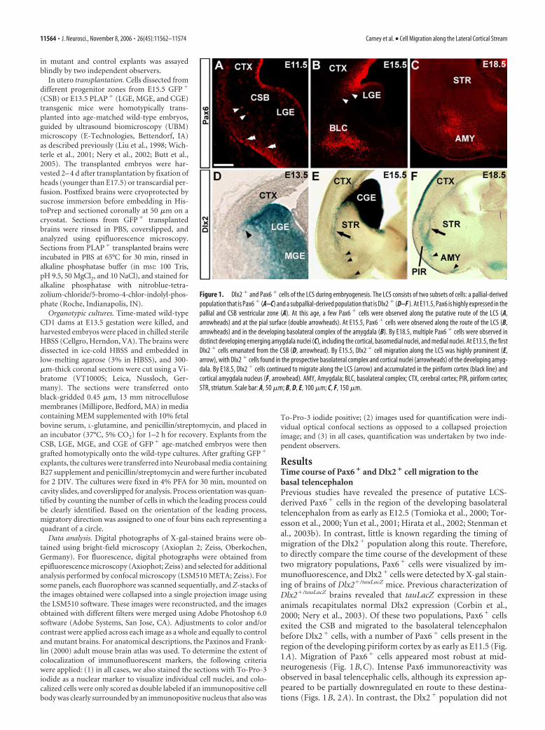
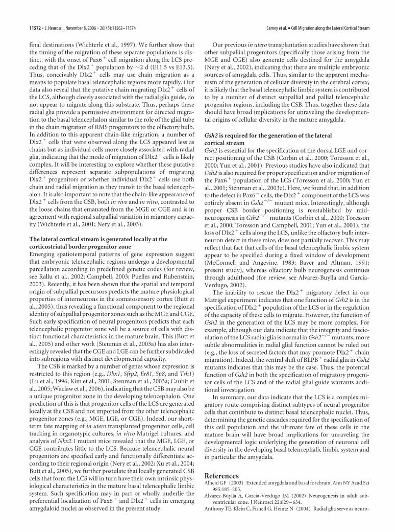
ResultsTime course of Pax6 � and Dlx2 � cell migration to thebasal telencephalonPrevious studies have revealed the presence of putative LCS-derived Pax6� cells in the region of the developing basolateraltelencephalon from as early as E12.5 (Tomioka et al., 2000; Tor-esson et al., 2000; Yun et al., 2001; Hirata et al., 2002; Stenman etal., 2003b). In contrast, little is known regarding the timing ofmigration of the Dlx2� population along this route. Therefore,to directly compare the time course of the development of thesetwo migratory populations, Pax6� cells were visualized by im-munofluorescence, and Dlx2� cells were detected by X-gal stain-ing of brains of Dlx2�/tauLacZ mice. Previous characterization ofDlx2�/tauLacZ brains revealed that tauLacZ expression in theseanimals recapitulates normal Dlx2 expression (Corbin et al.,2000; Nery et al., 2003). Of these two populations, Pax6� cellsexited the CSB and migrated to the basolateral telencephalonbefore Dlx2� cells, with a number of Pax6� cells present in theregion of the developing piriform cortex by as early as E11.5 (Fig.1A). Migration of Pax6� cells appeared most robust at mid-neurogenesis (Fig. 1B,C). Intense Pax6 immunoreactivity wasobserved in basal telencephalic cells, although its expression ap-peared to be partially downregulated en route to these destina-tions (Figs. 1B, 2A). In contrast, the Dlx2� population did not
Figure 1. Dlx2 � and Pax6 � cells of the LCS during embryogenesis. The LCS consists of two subsets of cells: a pallial-derivedpopulation that is Pax6 � (A–C) and a subpallial-derived population that is Dlx2 � (D–F ). At E11.5, Pax6 is highly expressed in thepallial and CSB ventricular zone (A). At this age, a few Pax6 � cells were observed along the putative route of the LCS (A,arrowheads) and at the pial surface (double arrowheads). At E15.5, Pax6 � cells were observed along the route of the LCS (B,arrowheads) and in the developing basolateral complex of the amygdala (B). By E18.5, multiple Pax6 � cells were observed indistinct developing emerging amygdala nuclei (C), including the cortical, basomedial nuclei, and medial nuclei. At E13.5, the firstDlx2 � cells emanated from the CSB (D, arrowhead). By E15.5, Dlx2 � cell migration along the LCS was highly prominent (E,arrow), with Dlx2 � cells found in the prospective basolateral complex and cortical nuclei (arrowheads) of the developing amyg-dala. By E18.5, Dlx2 � cells continued to migrate along the LCS (arrow) and accumulated in the piriform cortex (black line) andcortical amygdala nucleus (F, arrowhead). AMY, Amygdala; BLC, basolateral complex; CTX, cerebral cortex; PIR, piriform cortex;STR, striatum. Scale bar: A, 50 �m; B, D, E, 100 �m; C, F, 150 �m.
11564 • J. Neurosci., November 8, 2006 • 26(45):11562–11574 Carney et al. • Cell Migration along the Lateral Cortical Stream
begin to emerge from the CSB until �2 d later at E13.5 (Figs. 1D,2A). Similar to the Pax6� population, a robust stream of Dlx2�
migratory cells was observed along the LCS by mid-neurogenesis(Fig. 1E), with migration of both populations completed by earlypostnatal ages (data not shown). Both Pax6� and Dlx2� popu-lations appeared to migrate to specific emerging amygdala nucleiand other basal telencephalic structures. By E15.5, Pax6� cellswere observed accumulating in the prospective basolateral com-plex, cortical and basomedial nuclei of the amygdala, as well asthe olfactory tubercle and ventral pallidum, with a few cells alsopresent in the piriform cortex (Fig. 1B,C and data not shown).Interestingly, similar to the Pax6� cells, Dlx2� cells putativelyarising from the LCS were also observed accumulating in thebasolateral complex, basomedial and cortical nuclei of the amyg-dala, and the piriform cortex (Figs. 1E,F, 2A) (see Figs. 4 I, 8Eand data not shown).
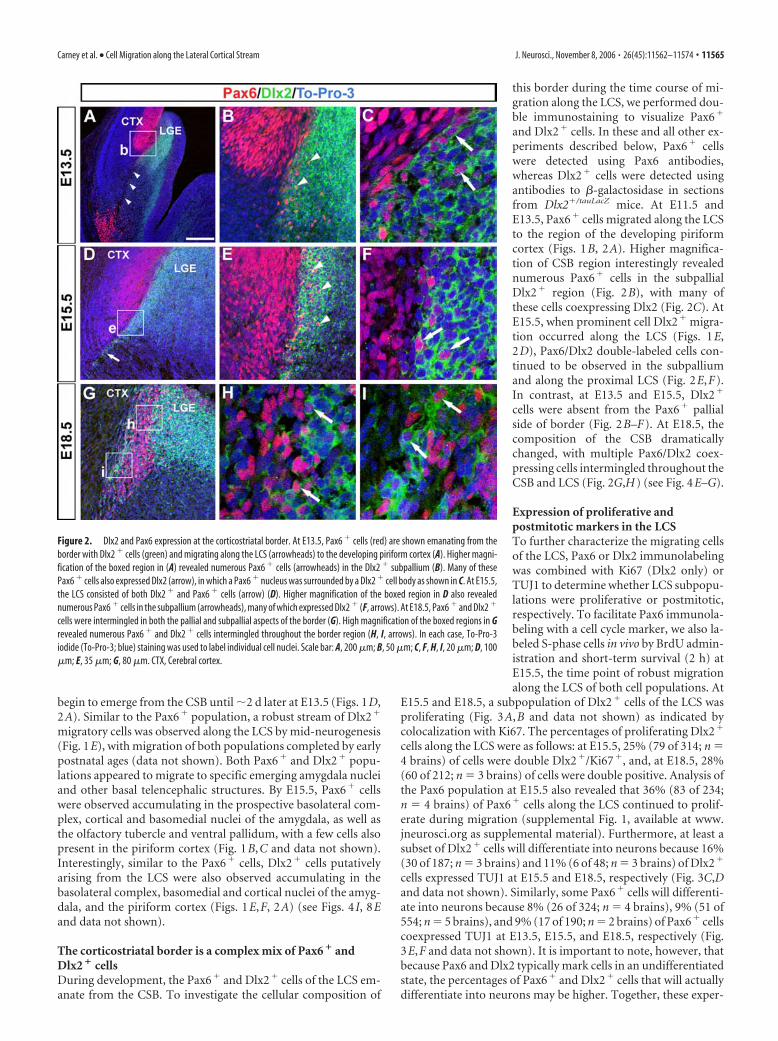
The corticostriatal border is a complex mix of Pax6 � andDlx2 � cellsDuring development, the Pax6� and Dlx2� cells of the LCS em-anate from the CSB. To investigate the cellular composition of
this border during the time course of mi-gration along the LCS, we performed dou-ble immunostaining to visualize Pax6�
and Dlx2� cells. In these and all other ex-periments described below, Pax6� cellswere detected using Pax6 antibodies,whereas Dlx2� cells were detected usingantibodies to �-galactosidase in sectionsfrom Dlx2�/tauLacZ mice. At E11.5 andE13.5, Pax6� cells migrated along the LCSto the region of the developing piriformcortex (Figs. 1B, 2A). Higher magnifica-tion of CSB region interestingly revealednumerous Pax6� cells in the subpallialDlx2� region (Fig. 2B), with many ofthese cells coexpressing Dlx2 (Fig. 2C). AtE15.5, when prominent cell Dlx2� migra-tion occurred along the LCS (Figs. 1E,2D), Pax6/Dlx2 double-labeled cells con-tinued to be observed in the subpalliumand along the proximal LCS (Fig. 2E,F).In contrast, at E13.5 and E15.5, Dlx2�
cells were absent from the Pax6� pallialside of border (Fig. 2B–F). At E18.5, thecomposition of the CSB dramaticallychanged, with multiple Pax6/Dlx2 coex-pressing cells intermingled throughout theCSB and LCS (Fig. 2G,H) (see Fig. 4E–G).
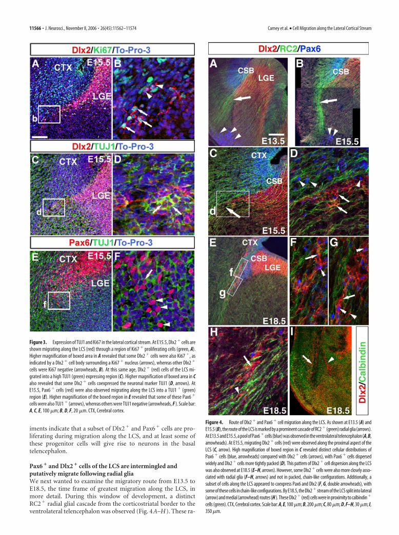
Expression of proliferative andpostmitotic markers in the LCSTo further characterize the migrating cellsof the LCS, Pax6 or Dlx2 immunolabelingwas combined with Ki67 (Dlx2 only) orTUJ1 to determine whether LCS subpopu-lations were proliferative or postmitotic,respectively. To facilitate Pax6 immunola-beling with a cell cycle marker, we also la-beled S-phase cells in vivo by BrdU admin-istration and short-term survival (2 h) atE15.5, the time point of robust migrationalong the LCS of both cell populations. At
E15.5 and E18.5, a subpopulation of Dlx2� cells of the LCS wasproliferating (Fig. 3A,B and data not shown) as indicated bycolocalization with Ki67. The percentages of proliferating Dlx2�
cells along the LCS were as follows: at E15.5, 25% (79 of 314; n �4 brains) of cells were double Dlx2�/Ki67�, and, at E18.5, 28%(60 of 212; n � 3 brains) of cells were double positive. Analysis ofthe Pax6 population at E15.5 also revealed that 36% (83 of 234;n � 4 brains) of Pax6� cells along the LCS continued to prolif-erate during migration (supplemental Fig. 1, available at www.jneurosci.org as supplemental material). Furthermore, at least asubset of Dlx2� cells will differentiate into neurons because 16%(30 of 187; n � 3 brains) and 11% (6 of 48; n � 3 brains) of Dlx2�
cells expressed TUJ1 at E15.5 and E18.5, respectively (Fig. 3C,Dand data not shown). Similarly, some Pax6� cells will differenti-ate into neurons because 8% (26 of 324; n � 4 brains), 9% (51 of554; n � 5 brains), and 9% (17 of 190; n � 2 brains) of Pax6� cellscoexpressed TUJ1 at E13.5, E15.5, and E18.5, respectively (Fig.3E,F and data not shown). It is important to note, however, thatbecause Pax6 and Dlx2 typically mark cells in an undifferentiatedstate, the percentages of Pax6� and Dlx2� cells that will actuallydifferentiate into neurons may be higher. Together, these exper-
Figure 2. Dlx2 and Pax6 expression at the corticostriatal border. At E13.5, Pax6 � cells (red) are shown emanating from theborder with Dlx2 � cells (green) and migrating along the LCS (arrowheads) to the developing piriform cortex (A). Higher magni-fication of the boxed region in (A) revealed numerous Pax6 � cells (arrowheads) in the Dlx2 � subpallium (B). Many of thesePax6 � cells also expressed Dlx2 (arrow), in which a Pax6 � nucleus was surrounded by a Dlx2 � cell body as shown in C. At E15.5,the LCS consisted of both Dlx2 � and Pax6 � cells (arrow) (D). Higher magnification of the boxed region in D also revealednumerous Pax6 � cells in the subpallium (arrowheads), many of which expressed Dlx2 � (F, arrows). At E18.5, Pax6 � and Dlx2 �
cells were intermingled in both the pallial and subpallial aspects of the border (G). High magnification of the boxed regions in Grevealed numerous Pax6 � and Dlx2 � cells intermingled throughout the border region (H, I, arrows). In each case, To-Pro-3iodide (To-Pro-3; blue) staining was used to label individual cell nuclei. Scale bar: A, 200 �m; B, 50 �m; C, F, H, I, 20 �m; D, 100�m; E, 35 �m; G, 80 �m. CTX, Cerebral cortex.
Carney et al. • Cell Migration along the Lateral Cortical Stream J. Neurosci., November 8, 2006 • 26(45):11562–11574 • 11565
iments indicate that a subset of Dlx2� and Pax6� cells are pro-liferating during migration along the LCS, and at least some ofthese progenitor cells will give rise to neurons in the basaltelencephalon.
Pax6 � and Dlx2 � cells of the LCS are intermingled andputatively migrate following radial gliaWe next wanted to examine the migratory route from E13.5 toE18.5, the time frame of greatest migration along the LCS, inmore detail. During this window of development, a distinctRC2� radial glial cascade from the corticostriatal border to theventrolateral telencephalon was observed (Fig. 4A–H). These ra-
Figure 3. Expression of TUJ1 and Ki67 in the lateral cortical stream. At E15.5, Dlx2 � cells areshown migrating along the LCS (red) through a region of Ki67 � proliferating cells (green, A).Higher magnification of boxed area in A revealed that some Dlx2 � cells were also Ki67 �, asindicated by a Dlx2 � cell body surrounding a Ki67 � nucleus (arrows), whereas other Dlx2 �
cells were Ki67 negative (arrowheads, B). At this same age, Dlx2 � (red) cells of the LCS mi-grated into a high TUJ1 (green) expressing region (C). Higher magnification of boxed area in Calso revealed that some Dlx2 � cells coexpressed the neuronal marker TUJ1 (D, arrows). AtE15.5, Pax6 � cells (red) were also observed migrating along the LCS into a TUJ1 � (green)region (E). Higher magnification of the boxed region in E revealed that some of these Pax6 �
cells were also TUJ1 � (arrows), whereas others were TUJ1 negative (arrowheads, F ). Scale bar:A, C, E, 100 �m; B, D, F, 20 �m. CTX, Cerebral cortex.
Figure 4. Route of Dlx2 � and Pax6 � cell migration along the LCS. As shown at E13.5 (A) andE15.5 (B), the route of the LCS is marked by a prominent cascade of RC2 � (green) radial glia (arrows).AtE13.5andE15.5,apoolofPax6 �cells(blue)wasobservedintheventrolateraltelencephalon(A, B,arrowheads). At E15.5, migrating Dlx2 � cells (red) were observed along the proximal aspect of theLCS (C, arrow). High magnification of boxed region in C revealed distinct cellular distributions ofPax6 � cells (blue, arrowheads) compared with Dlx2 � cells (arrows), with Pax6 � cells dispersedwidely and Dlx2 � cells more tightly packed (D). This pattern of Dlx2 � cell dispersion along the LCSwas also observed at E18.5 (E–H, arrows). However, some Dlx2 � cells were also more closely asso-ciated with radial glia (F–H, arrows) and not in packed, chain-like configurations. Additionally, asubset of cells along the LCS appeared to coexpress Pax6 and Dlx2 (F, G, double arrowheads), withsome of these cells in chain-like configurations. By E18.5, the Dlx2 � stream of the LCS split into lateral(arrow) and medial (arrowhead) routes (H ). These Dlx2 � (red) cells were in proximity to calbindin �
cells (green). CTX, Cerebral cortex. Scale bar: A, E, 100 �m; B, 200 �m; C, 80 �m; D, F–H, 30 �m; I,350 �m.
11566 • J. Neurosci., November 8, 2006 • 26(45):11562–11574 Carney et al. • Cell Migration along the Lateral Cortical Stream
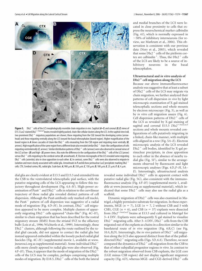
dial glia are clearly evident at E13.5 and E15.5 and extended fromthe CSB to the ventrolateral telencephalic pial surface, with theputative migrating cells of the LCS appearing to follow this tra-jectory throughout development (Fig. 4A–H). High-power ex-amination of Pax6� and Dlx2� cells in relation to the curvilineartrajectory of these radial glia revealed distinct patterns of celldispersion. Although the Pax6 antibody only marked cell nuclei,the Pax6� pattern of cell dispersion was suggestive of a radialmode of migration (Fig. 4D–H). In contrast, Dlx2� cell migra-tion appeared to be more complex. Multiple clusters of appar-ently migrating Dlx2� cells appeared “chain-like” (Fig. 4C–H),similar to chain migration that has been described for the rostralmigratory stream (RMS) from the striatal SVZ to the olfactorybulb (Lois et al., 1996; Wichterle et al., 1999). Interestingly, theseDlx2� clusters, although following the route outlined by the ra-dial glial cascade, did not appear to contact the radial glia butinstead appeared embedded within the loosely packed radial glialfascicles (Fig. 4C–H) (supplemental movie 1, available at www.jneurosci.org as supplemental material). Some individual Dlx2�
cells more closely apposed to radial glia were also observed (Fig.4F–H). Thus, it appears that the mode of migration of the Dlx2�
cells of the LCS may be complex, perhaps comprising multiplemodes of migration. By E18.5, Dlx2� cells of the both the lateral
and medial branches of the LCS were lo-cated in close proximity to cells that ex-press the neurochemical marker calbindin(Fig. 4 I), which is normally expressed in�50% of inhibitory interneurons (for re-view, see Markram et al., 2004). This ob-servation is consistent with our previousdata (Nery et al., 2003), which revealedthat some Dlx2� cells of the piriform cor-tex are calbindin�. Thus, the Dlx2� cellsof the LCS are likely to be a source of in-hibitory neurons in the basaltelencephalon.
Ultrastructural and in vitro analysis ofDlx2 � cell migration along the LCSBecause our above immunofluorescenceanalysis was suggestive that at least a subsetof Dlx2� cells of the LCS may migrate viachain migration, we further analyzed theirpatterns of cell dispersion in vivo by lightmicroscopic examination of X-gal-stainedtelencephalic sections and whole mountsby electron microscopy (Fig. 5), as well asby in vitro cell migration assays (Fig. 6).Cell dispersion patterns of Dlx2� cells ofthe LCS as revealed by X-gal staining ofsagittal and coronal E15.5 Dlx2�/tauLacZ
sections and whole mounts revealed con-figurations of cells putatively migrating ina linked, chain-like manner, with multiplecells aligned in a row (Fig. 5A–E). Electronmicroscopic analysis of the LCS revealedDlx2� cell bodies, identified by X-gal pe-rinuclear precipitate, in close appositionto each other in the absence of nearby ra-dial glia (Fig. 5F), similar to the arrange-ments observed by fluorescent and lightmicroscopic analyses (Figs. 4D,F–H, 5A–E). Interestingly, ultrastructural analysis
revealed some individual Dlx2� cells in apparent contact withputative radial glia (Fig. 5G), also consistent with the immuno-fluorescence analysis (Fig. 4F–H) (supplemental movie 1, avail-able at www.jneurosci.org as supplemental material), which in-dicated that some Dlx2� cells may also use the radial glia as ascaffold.
Dynamic migration of Dlx2� cells was also examined in Ma-trigel, a highly permissive substrate for migration. In these exper-iments, MGE (n � 7), LGE (n � 7, 2 without CSB and 5 withCSB), CGE (n � 6), and CSB (n � 37) explants were dissectedfrom Dlx2�/tauLacZ brains at E13.5 and cultured in Matrigel for3– 4 DIV. Explants were subsequently X-gal stained to visualizeDlx2� migrating cells. After 3– 4 DIV, Dlx2� cells from the CSBmigrated out of the explants as chains in a direction similar to thebasolateral route of in vivo migration (Fig. 6B,C) (see Fig.10A,B,D). Interestingly, the in vivo pattern of Dlx2� cell migra-tion along the LCS also appeared distinct from that of the pallial-directed Dlx2� migrating cells (Figs. 5D, 6A). Therefore, we nextcompared the dynamics of Dlx2� cell migration from the CSB tothat of other subpallial progenitor regions in vitro. In contrast tothe robust migration observed from CSB explants, the LGE core(LGE minus CSB region) did not display significant migratorycapacity (Fig. 4D), whereas MGE- and CGE-derived Dlx2� cells
Figure 5. Dlx2 � cells of the LCS morphologically resemble chain migration in vivo. Sagittal (A–C) and coronal (D, E) views ofE15.5 LacZ-stained Dlx2�/tauLacZ brains revealed tightly packed, chain-like cellular clusters along the LCS. In the sagittal view in A,two prominent Dlx2 � migratory populations are shown; those migrating from the CGE toward the developing cortex (arrow-head) and those migrating ventrally along the LCS toward the basal telencephalon (boxed region). Higher magnification of theboxed region in A shows cascades of chain-like Dlx2 � cells emanating from the CSB region and migrating more ventrally (B,arrows). High magnification of the same region from a different brain also revealed similar Dlx2 � chain-like configurations of cellsmigrating ventrolaterally (C, arrows). Similar distribution patterns of Dlx2 � cells (arrows) were also observed in coronal views ofthe LCS at low- (D) and high- (E) power views. Also note the difference in the configuration of the Dlx2 � cells of the LCS (arrow)versus Dlx2 � cells migrating to the cerebral cortex (D, arrowheads). F, Electron micrographs of the LCS revealed some migratingDlx2 � cells (asterisks) also in close apposition to each other. G, In contrast, some Dlx2 � cells were also observed to migrate inisolation and more closely associated with radial glia. Arrowheads in F and G show perinuclear LacZ precipitate marking Dlx2 �
cells. CTX, Cerebral cortex; RG, radial glia. Scale bars: A, 900 �m; B, 350 �m; C, 150 �m; D, 100 �m; E, 25 �m; F, G, 4 �m.
Carney et al. • Cell Migration along the Lateral Cortical Stream J. Neurosci., November 8, 2006 • 26(45):11562–11574 • 11567
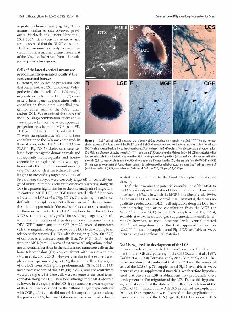
migrated as loose chains (Fig. 4E,F) in amanner similar to that observed previ-ously (Wichterle et al., 1999; Nery et al.,2002, 2003). Thus, these in vivo and in vitroresults revealed that the Dlx2� cells of theLCS have an innate capacity to migrate aschains and in a manner distinct from thatof the Dlx2� cells derived from other sub-pallial progenitor regions.
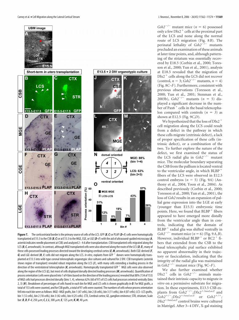
Cells of the lateral cortical stream arepredominantly generated locally at thecorticostriatal borderCurrently, the source of progenitor cellsthat comprise the LCS is unknown. We hy-pothesized that the cells of the LCS may (1)originate solely from the CSB or (2) com-prise a heterogeneous population with acontribution from other subpallial pro-genitor zones such as the MGE, LGE,and/or CGE. We examined the source ofthe LCS using a combination in vivo and invitro approaches. For the in vivo approach,progenitor cells from the MGE (n � 25),LGE (n � 5), CGE (n � 10), and CSB (n �7) were transplanted in utero, and theircontribution to the LCS was compared. Inthese studies, either GFP� (Fig. 7B,C) orPLAP� (Fig. 7D–I) labeled cells were iso-lated from transgenic donor animals andsubsequently homotopically and homo-chronically transplanted into wild-typebrains with the aid of ultrasound imaging(Fig. 7A). Although it was technically chal-lenging to successfully target the CSB (7 of56 surviving embryos were correctly targeted), in correctly tar-geted brains, numerous cells were observed migrating along theLCS in a pattern highly similar to their normal path of migration.In contrast, MGE, LGE, or CGE transplanted cells did not con-tribute to the LCS in vivo (Fig. 7D–I). Considering the technicaldifficulty in transplanting CSB cells in vivo, we further examinedthe migratory potential of these cells in slice culture preparations.In these experiments, E13.5 GFP� explants from the CSB orMGE were homotypically grafted into wild-type organotypic cul-tures, and the location of migratory cells was examined after 2DIV. GFP� transplants to the CSB (n � 22) revealed numerouscells that migrated along the route of the LCS to developing basaltelencephalic regions (Fig. 7J), with the majority (62%, 60 of 97)of cell processes oriented ventrally (Fig. 7K,N,O). GFP� graftsfrom the MGE (n � 17) revealed extensive cell migration, includ-ing tangential migration to the pallium and numerous cells in thebasal telencephalon (Fig. 7L), consistent with previous studies(Marin et al., 2001, 2003). However, similar to the in vivo trans-plantation experiment (Fig. 7D,E), the GFP� cells in the regionof the LCS from MGE grafts predominantly (88%, 134 of 153)had processes oriented dorsally (Fig. 7M–O) and not ventrally aswould be expected if these cells were en route to the basal telen-cephalon along the LCS. Therefore, although these MGE-derivedcells were in the region of the LCS, it appeared that a vast majorityof these cells were destined for the pallium. Organotypic cultureswith CGE grafts (n � 6) did not exhibit any cell migration alongthe posterior LCS, because CGE-derived cells assumed a direct,
ventral migratory route to the basal telencephalon (data notshown).
To further examine the potential contribution of the MGE tothe LCS, we analyzed the status of Dlx2� migration in knock-outmice lacking Nkx2.1 in which the MGE is lost (Sussel et al., 1999).As shown at E14.5 (n � 6 control, n � 6 mutants), there was noqualitative reduction in Dlx2� cell migration along the LCS, fur-ther indicating little or no contribution from the MGE (orNkx2.1� anterior CGE) to the LCS (supplemental Fig. 2A,B,available at www.jneurosci.org as supplemental material). Inter-estingly however, at more posterior levels, ventral-directedDlx2� cell migration from the CGE appeared reduced inNkx2.1�/� mutants (supplemental Fig. 2C,D, available at www.jneurosci.org as supplemental material).
Gsh2 is required for development of the LCSPrevious studies have revealed that Gsh2 is required for develop-ment of the LGE and pattering of the CSB (Szucsik et al., 1997;Corbin et al., 2000; Toresson et al., 2000; Yun et al., 2001). Be-cause our above data indicated that the CSB was the source ofcells of the LCS (Fig. 7) (supplemental Fig. 2, available at www.jneurosci.org as supplemental material), we therefore hypothe-sized that defects in CSB establishment may profoundly affectdevelopment and/or migration of the LCS. To test this hypothe-sis, we first examined the status of the Dlx2� population of theLCS in Gsh2�/� mutant mice. At E15.5, in control telencephalons(n � 9), Dlx2 expression was observed in the ganglionic emi-nences and in cells of the LCS (Figs. 1E, 8A). In contrast, E15.5
Figure 6. Dlx2 � cells of the LCS migrate as chains in vitro. �-Galactosidase immunostaining of Dlx2�/tauLacZ coronal telence-phalic sections at E16.5 also showed that Dlx2 � cells of the LCS (A, arrow) appeared to migrate in a manner distinct from that ofDlx2 � cells tangentially migrating to the cerebral cortex (A, arrowheads). In B–F, explants from the corticostriatal border region,LGE, MGE, and CGE were dissected from Dlx2�/tauLacZ animals at E13.5 and cultured in Matrigel for 3– 4 d. CSB explants stained forLacZ revealed cells that migrated away from the CSB in tightly packed configurations (arrow in B and a higher magnificationshown in C). In contrast, explants from the LGE did not display significant migration (D), whereas cells from the MGE (E) and CGE(F ) migrated as loose chains (E, F, arrowheads), similar to that observed for pallial-directed migrating Dlx2 � cells as shown in A(and shown in Fig. 5D). CTX, Cerebral cortex. Scale bar: A, 100 �m; B, D, 250 �m; C, E, F, 75 �m.
11568 • J. Neurosci., November 8, 2006 • 26(45):11562–11574 Carney et al. • Cell Migration along the Lateral Cortical Stream
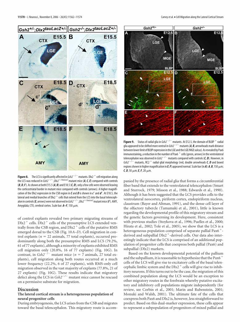
Gsh2�/� mutant mice (n � 6) possessedonly a few Dlx2� cells at the proximal partof the LCS and none along the normalroute of LCS migration (Fig. 8B). Theperinatal lethality of Gsh2�/� mutantsprecluded an examination of these animalsat later time points, and, although pattern-ing of the striatum was essentially recov-ered by E18.5 (Corbin et al., 2000; Tores-son et al., 2000; Yun et al., 2001), analysisat E18.5 revealed that the migration ofDlx2� cells along the LCS did not recover(control, n � 3; Gsh2�/� mutants, n � 4)(Fig. 8C–F). Furthermore, consistent withprevious observations (Toresson et al.,2000; Yun et al., 2001; Stenman et al.,2003b), Gsh2�/� mutants (n � 5) dis-played a significant decrease in the num-ber of Pax6� cells in the basal telencepha-lon compared with controls (n � 3) asshown at E12.5 (Fig. 9C,D).
We hypothesized that the loss of Dlx2�
cell migration along the LCS could resultfrom a defect in the pathway in whichthese cells migrate (extrinsic defect), a lackof proper specification of these cells (in-trinsic defect), or a combination of thetwo. To further explore the nature of thedefect, we first examined the status ofthe LCS radial glia in Gsh2�/� mutantmice. The molecular boundary separatingthe CSB from the pallium is located ventralto the ventricular angle, in which BLBP�
fibers of the LCS were observed in E12.5control embryos (n � 5) (Fig. 9A) (An-thony et al., 2004; Yoon et al., 2004). Asdescribed previously (Corbin et al., 2000;Toresson et al., 2000; Yun et al., 2001), theloss of Gsh2 results in an expansion of pal-lial gene expression into the LGE at early(younger than E15.5) embryonic timepoints. Here, we found that BLBP� fibersappeared to have emerged more distallyfrom the ventricular angle than in con-trols, indicating that the domain ofBLBP� radial glia was shifted ventrally inGsh2�/� mutant mice (n � 6) (Fig. 9A,B).However, individual BLBP� or RC2� fi-bers that extended from the CSB to thebasal telencephalic pial surface exhibitedno apparent abnormality in their trajec-tory or fasciculation, indicating that theintegrity of the radial glia was maintainedin Gsh2�/� mutant mice (Fig. 9C–F).
We also further examined whetherDlx2� cells in Gsh2�/� animals main-tained their intrinsic capacity to migrate invitro on a permissive substrate for migra-tion. In these experiments, E13.5 CSB ex-plants from Gsh2�/�;Dlx2�/tauLacZ andGsh2�/�;Dlx2�/tauLacZ or Gsh2�/�;Dlx2�/tauLacZ control brains were culturedin Matrigel. After 3– 4 DIV, X-gal staining
Figure 7. The corticostriatal border is the primary source of cells of the LCS. GFP (B, C) or PLAP (D–I ) cells were homotopicallytransplanted at E15.5 to the CSB (B, C) or at E13.5 to the MGE, CGE, or LGE (D–I ) with the aid of ultrasound-guided microscopy (A,asterisk indicates needle placement at CSB) and analyzed 2– 4 d after transplantation. CSB transplanted cells migrated along theLCS (B, C, arrowheads). In contrast, although MGE transplanted cells were also observed along the route of the LCS (D, E), many ofthese cells possessed leading processes directed toward the developing cerebral cortex (E, M, arrowheads). Both CGE-derived (F,G) and LGE-derived (H, I ) cells did not migrate along the LCS. In vitro, explants from GFP � donors were homotopically trans-planted at E13.5 into wild-type coronal telencephalic organotypic slice cultures and cultured for 2 DIV. CSB transplants (asteriskshows region of transplant) revealed robust migration along the LCS (J ), with many cells extending a leading process in thedirection of the ventrolateral telencephalon (K, arrowheads). Homotopically transplanted GFP � MGE cells were also observedalong the region of the LCS (L), but most of cells displayed dorsally directed leading processes (M, arrowheads). Quantification ofprocess orientation (cells were placed into 1 of 4 bins based on the direction of the leading process) revealed that 88% (134 of 153)of MGE cells had processes directed dorsally (bins 1, 4), whereas 62% (60 of 97) of LCS cells had processes oriented ventrally (bins2, 3) (N ). Breakdown of percentages of cells found in each bin for MGE and LCS cells is shown graphically in O. For MGE grafts, atotal of 153 cells were counted, and for CSB grafts, a total of 97 cells were counted. The numbers of cells whose process orientationfell into each bin were as follows: MGE–MGE grafts, bin 1 (47 cells), bin 2 (8 cells), bin 3 (11 cells), bin 4 (87 cells); LCS–LCS grafts,bin 1 (12 cells), bin 2 (18 cells), bin 3 (42 cells), bin 4 (25 cells). CTX, Cerebral cortex; GE, ganglion eminence; STR, striatum; Scalebar: B, D, F–I, 250 �m; C, J, L, 300 �m; E, 125 �m; K, M, 40 �m.
Carney et al. • Cell Migration along the Lateral Cortical Stream J. Neurosci., November 8, 2006 • 26(45):11562–11574 • 11569
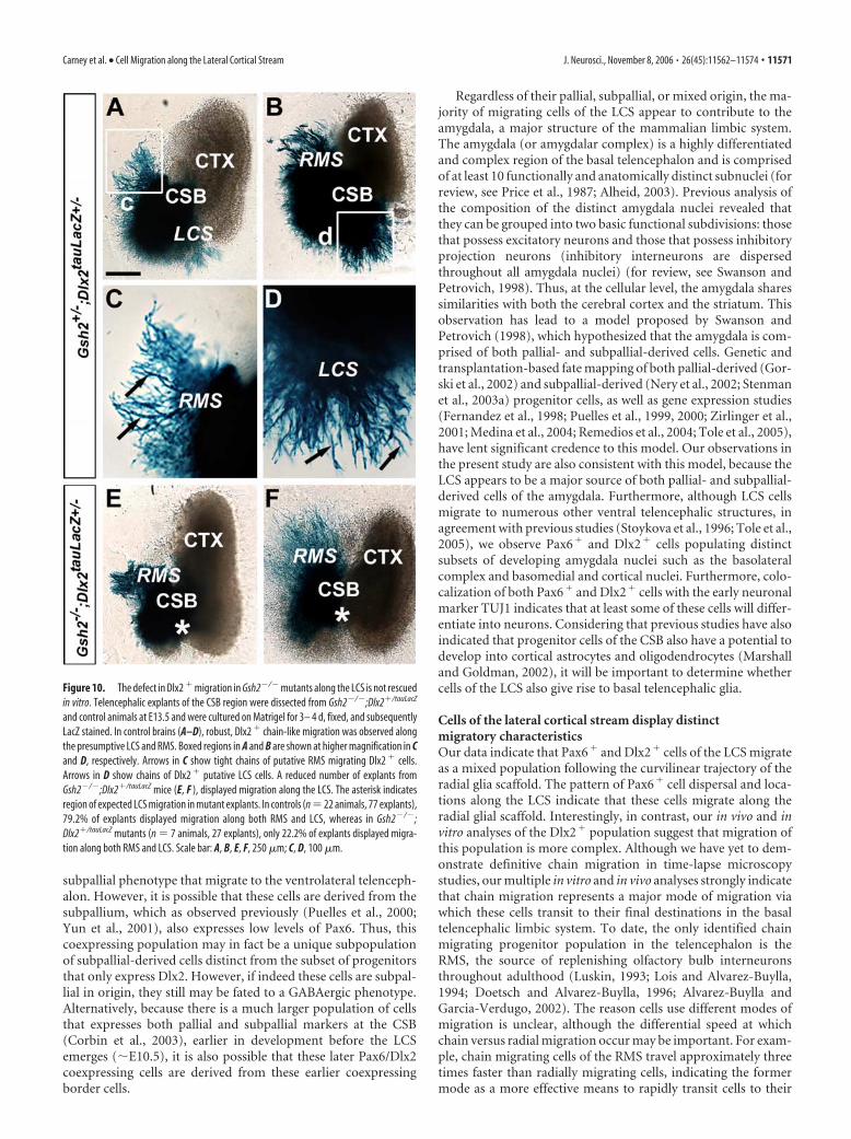
of control explants revealed two primary migrating streams ofDlx2� cells. Dlx2� cells of the presumptive LCS extended ven-trally from the CSB region, and Dlx2� cells of the putative RMSemerged dorsal to the CSB (Fig. 10A–D). Cell migration in con-trol explants (n � 22 animals, 77 total explants), occurred pre-dominantly along both the presumptive RMS and LCS (79.2%,61 of 77 explants), although a minority of explants exhibited RMScell migration only (20.8%, 16 of 77 explants) (Fig. 10G). Incontrast, in Gsh2�/� mutant mice (n � 7 animals, 27 total ex-plants), cell migration along both routes occurred at a muchlower frequency (22.2%, 6 of 27 explants), with RMS-only cellmigration observed in the vast majority of explants (77.8%, 21 of27 explants) (Fig. 10G). These results indicate that migratorydefect along the LCS in Gsh2�/� mutant mice cannot be rescuedon a permissive substrate for migration.
DiscussionThe lateral cortical stream is a heterogeneous population ofneural progenitor cellsDuring embryogenesis, the LCS arises from the CSB and migratestoward the basal telencephalon. This migratory route is accom-
panied by the presence of radial glia that forms a circumferentialfiber band that extends to the ventrolateral telencephalon (Smartand Sturrock, 1979; Misson et al., 1988; Edwards et al., 1990).Although it has been suggested that the LCS provides cells to theventrolateral neocortex, piriform cortex, endopiriform nucleus,claustrum (Bayer and Altman, 1991), and the dense cell layer ofthe olfactory tubercle (Tamamaki et al., 2001), little is knownregarding the developmental profile of this migratory stream andthe genetic factors governing its development. Here, consistentwith previous studies (Stoykova et al., 1996; Puelles et al., 2000;Hirata et al., 2002; Tole et al., 2005), we show that the LCS is aheterogeneous population comprised of separate pallial Pax6�-derived and subpallial Dlx2�-derived cells. Our data also inter-estingly indicate that the LCS is comprised of an additional pop-ulation of progenitor cells that coexpress both pallial (Pax6) andsubpallial (Dlx2) markers.
Based on the known developmental potential of the palliumand the subpallium, it is reasonable to hypothesize that the Pax6�
cells of the LCS will give rise to excitatory cells of the basal telen-cephalic limbic system and the Dlx2� cells will give rise to inhib-itory neurons. If this turns out to be the case, the migration of thiscombined population along the LCS would be an exception toother migratory routes in the forebrain whereby putative excita-tory and inhibitory cell populations migrate independently (forreview, see Corbin et al., 2001; Marin and Rubenstein, 2001;Monuki and Walsh, 2001). The ultimate fate of the cells thatcoexpress both Pax6 and Dlx2 is, however, less straightforward topredict. Based on this dual-marker expression, these cells appearto represent a subpopulation of progenitors of mixed pallial and
Figure 8. The LCS is significantly affected in Gsh2�/� mutants. Dlx2 � cell migration alongthe LCS was reduced in Gsh2�/�;Dlx2�/tauLacZ mutant mice (A, C, E) compared with controls(B, D, F ). As shown at both E15.5 (A, B) and E18.5 (C, D), only a few cells were observed leavingthe corticostriatal border in mutant mice compared with controls (arrows). A higher magnifi-cation of the Dlx2 expression in the CSB region in C and D is shown in c� and d�. At E18.5, thelateral and medial branches of Dlx2 � cells that extend from the LCS into the basal telenceph-alon in controls (E, arrows) were not observed in Gsh2�/�;Dlx2�/tauLacZ mutant mice (F ). AMY,Amygdala; CTX, cerebral cortex. Scale bar: A–F, 150 �m.
Figure 9. Status of radial glia in Gsh2�/� mutants. At E12.5, the domain of BLBP � radialglia appeared to be shifted more ventral in Gsh2�/� mutants (A, B, arrowheads mark distancebetween lower limit of BLBP expression in the LGE and the LGE/MGE sulcus). As revealed by Pax6immunostaining, a reduction in the number of Pax6 � cells (green, arrows) in the ventrolateraltelencephalon was observed in Gsh2�/� mutants compared with controls (C, D). However, inGsh2�/� mutants, RC2 � radial glial morphology (red, double arrowheads C, D and boxedregions shown in higher magnification in E, F) appeared normal. Scale bar (in A): A, B, 150 �m;C, D, 50 �m; E, F, 20 �m.
11570 • J. Neurosci., November 8, 2006 • 26(45):11562–11574 Carney et al. • Cell Migration along the Lateral Cortical Stream
subpallial phenotype that migrate to the ventrolateral telenceph-alon. However, it is possible that these cells are derived from thesubpallium, which as observed previously (Puelles et al., 2000;Yun et al., 2001), also expresses low levels of Pax6. Thus, thiscoexpressing population may in fact be a unique subpopulationof subpallial-derived cells distinct from the subset of progenitorsthat only express Dlx2. However, if indeed these cells are subpal-lial in origin, they still may be fated to a GABAergic phenotype.Alternatively, because there is a much larger population of cellsthat expresses both pallial and subpallial markers at the CSB(Corbin et al., 2003), earlier in development before the LCSemerges (�E10.5), it is also possible that these later Pax6/Dlx2coexpressing cells are derived from these earlier coexpressingborder cells.
Regardless of their pallial, subpallial, or mixed origin, the ma-jority of migrating cells of the LCS appear to contribute to theamygdala, a major structure of the mammalian limbic system.The amygdala (or amygdalar complex) is a highly differentiatedand complex region of the basal telencephalon and is comprisedof at least 10 functionally and anatomically distinct subnuclei (forreview, see Price et al., 1987; Alheid, 2003). Previous analysis ofthe composition of the distinct amygdala nuclei revealed thatthey can be grouped into two basic functional subdivisions: thosethat possess excitatory neurons and those that possess inhibitoryprojection neurons (inhibitory interneurons are dispersedthroughout all amygdala nuclei) (for review, see Swanson andPetrovich, 1998). Thus, at the cellular level, the amygdala sharessimilarities with both the cerebral cortex and the striatum. Thisobservation has lead to a model proposed by Swanson andPetrovich (1998), which hypothesized that the amygdala is com-prised of both pallial- and subpallial-derived cells. Genetic andtransplantation-based fate mapping of both pallial-derived (Gor-ski et al., 2002) and subpallial-derived (Nery et al., 2002; Stenmanet al., 2003a) progenitor cells, as well as gene expression studies(Fernandez et al., 1998; Puelles et al., 1999, 2000; Zirlinger et al.,2001; Medina et al., 2004; Remedios et al., 2004; Tole et al., 2005),have lent significant credence to this model. Our observations inthe present study are also consistent with this model, because theLCS appears to be a major source of both pallial- and subpallial-derived cells of the amygdala. Furthermore, although LCS cellsmigrate to numerous other ventral telencephalic structures, inagreement with previous studies (Stoykova et al., 1996; Tole et al.,2005), we observe Pax6� and Dlx2� cells populating distinctsubsets of developing amygdala nuclei such as the basolateralcomplex and basomedial and cortical nuclei. Furthermore, colo-calization of both Pax6� and Dlx2� cells with the early neuronalmarker TUJ1 indicates that at least some of these cells will differ-entiate into neurons. Considering that previous studies have alsoindicated that progenitor cells of the CSB also have a potential todevelop into cortical astrocytes and oligodendrocytes (Marshalland Goldman, 2002), it will be important to determine whethercells of the LCS also give rise to basal telencephalic glia.
Cells of the lateral cortical stream display distinctmigratory characteristicsOur data indicate that Pax6� and Dlx2� cells of the LCS migrateas a mixed population following the curvilinear trajectory of theradial glia scaffold. The pattern of Pax6� cell dispersal and loca-tions along the LCS indicate that these cells migrate along theradial glial scaffold. Interestingly, in contrast, our in vivo and invitro analyses of the Dlx2� population suggest that migration ofthis population is more complex. Although we have yet to dem-onstrate definitive chain migration in time-lapse microscopystudies, our multiple in vitro and in vivo analyses strongly indicatethat chain migration represents a major mode of migration viawhich these cells transit to their final destinations in the basaltelencephalic limbic system. To date, the only identified chainmigrating progenitor population in the telencephalon is theRMS, the source of replenishing olfactory bulb interneuronsthroughout adulthood (Luskin, 1993; Lois and Alvarez-Buylla,1994; Doetsch and Alvarez-Buylla, 1996; Alvarez-Buylla andGarcia-Verdugo, 2002). The reason cells use different modes ofmigration is unclear, although the differential speed at whichchain versus radial migration occur may be important. For exam-ple, chain migrating cells of the RMS travel approximately threetimes faster than radially migrating cells, indicating the formermode as a more effective means to rapidly transit cells to their
Figure 10. The defect in Dlx2 � migration in Gsh2�/� mutants along the LCS is not rescuedin vitro. Telencephalic explants of the CSB region were dissected from Gsh2�/�;Dlx2�/tauLacZ
and control animals at E13.5 and were cultured on Matrigel for 3– 4 d, fixed, and subsequentlyLacZ stained. In control brains (A–D), robust, Dlx2 � chain-like migration was observed alongthe presumptive LCS and RMS. Boxed regions in A and B are shown at higher magnification in Cand D, respectively. Arrows in C show tight chains of putative RMS migrating Dlx2 � cells.Arrows in D show chains of Dlx2 � putative LCS cells. A reduced number of explants fromGsh2�/�;Dlx2�/tauLacZ mice (E, F ), displayed migration along the LCS. The asterisk indicatesregion of expected LCS migration in mutant explants. In controls (n � 22 animals, 77 explants),79.2% of explants displayed migration along both RMS and LCS, whereas in Gsh2�/�;Dlx2�/tauLacZ mutants (n � 7 animals, 27 explants), only 22.2% of explants displayed migra-tion along both RMS and LCS. Scale bar: A, B, E, F, 250 �m; C, D, 100 �m.
Carney et al. • Cell Migration along the Lateral Cortical Stream J. Neurosci., November 8, 2006 • 26(45):11562–11574 • 11571
final destinations (Wichterle et al., 1997). We further show thatthe timing of the migration of these separate populations is dis-tinct, with the onset of Pax6� cell migration along the LCS pre-ceding that of the Dlx2� population by �2 d (E11.5 vs E13.5).Thus, conceivably Dlx2� cells may use chain migration as ameans to populate basal telencephalic regions more rapidly. Ourdata also reveal that the putative chain migrating Dlx2� cells ofthe LCS, although closely associated with the radial glia guide, donot appear to migrate along this substrate. Thus, perhaps theseradial glia provide a permissive environment for directed migra-tion to the basal telencephalon similar to the role of the glial tubein the chain migration of RMS progenitors to the olfactory bulb.In addition to this apparent chain-like migration, a number ofDlx2� cells that were observed along the LCS appeared less aschains but as individual cells more closely associated with radialglia, indicating that the mode of migration of Dlx2� cells is likelycomplex. It will be interesting to explore whether these putativedifferences represent separate subpopulations of migratingDlx2� progenitors or whether individual Dlx2� cells use bothchain and radial migration as they transit to the basal telenceph-alon. It is also important to note that the chain-like appearance ofDlx2� cells from the CSB, both in vivo and in vitro, contrasted tothe loose chains that emanated from the MGE or CGE and is inagreement with regional subpallial variation in migratory capac-ity (Wichterle et al., 2001; Nery et al., 2003).
The lateral cortical stream is generated locally at thecorticostriatal border progenitor zoneEmerging spatiotemporal patterns of gene expression suggestthat embryonic telencephalic regions undergo a developmentalparcellation according to predefined genetic codes (for review,see Rallu et al., 2002; Campbell, 2003; Puelles and Rubenstein,2003). Recently, it has been shown that the spatial and temporalorigin of subpallial precursors predicts the mature physiologicalproperties of interneurons in the somatosensory cortex (Butt etal., 2005), thus revealing a functional component to the regionalidentity of subpallial progenitor zones such as the MGE and CGE.Such early specification of neural progenitors predicts that eachtelencephalic progenitor zone will be a source of cells with dis-tinct functional characteristics in the mature brain. This (Butt etal., 2005) and other work (Stenman et al., 2003a) has also inter-estingly revealed that the CGE and LGE can be further subdividedinto subregions with distinct developmental capacity.
The CSB is marked by a number of genes whose expression isrestricted to this region (e.g., Dbx1, Sfrp2, Er81, Sp8, and Tsh1)(Lu et al., 1996; Kim et al., 2001; Stenman et al., 2003a; Caubit etal., 2005; Waclaw et al., 2006), indicating that the CSB may also bea unique progenitor zone in the developing telencephalon. Oneprediction of this is that progenitor cells of the LCS are generatedlocally at the CSB and not imported from the other telencephalicprogenitor zones (e.g., MGE, LGE, or CGE). Indeed, our short-term fate mapping of in utero transplanted progenitor cells, celltracking in organotypic cultures, in vitro Matrigel cultures, andanalysis of Nkx2.1 mutant mice revealed that the MGE, LGE, orCGE contributes little to the LCS. Because telencephalic neuralprogenitors are specified early and functionally differentiate ac-cording to their regional origin (Nery et al., 2002; Xu et al., 2004;Butt et al., 2005), we further postulate that locally generated CSBcells that form the LCS will in turn have their own intrinsic phys-iological characteristics in the mature basal telencephalic limbicsystem. Such specification may in part or wholly underlie thepreferential localization of Pax6� and Dlx2� cells in emergingamygdaloid nuclei as observed in the present study.
Our previous in utero transplantation studies have shown thatother subpallial progenitors (specifically those arising from theMGE and CGE) also generate cells destined for the amygdala(Nery et al., 2002), indicating that there are multiple embryonicsources of amygdala cells. Thus, similar to the apparent mecha-nism of the generation of cellular diversity in the cerebral cortex,it is likely that the basal telencephalic limbic system is contributedto by a number of distinct subpallial and pallial telencephalicprogenitor regions, including the CSB. Thus, together these datashould have broad implications for unraveling the developmen-tal origins of cellular diversity in the mature amygdala.
Gsh2 is required for the generation of the lateralcortical streamGsh2 is essential for the specification of the dorsal LGE and cor-rect positioning of the CSB (Corbin et al., 2000; Toresson et al.,2000; Yun et al., 2001). Previous studies have also indicated thatGsh2 is also required for proper specification and/or migration ofthe Pax6� population of the LCS (Toresson et al., 2000; Yun etal., 2001; Stenman et al., 2003c). Here, we found that, in additionto the defect in Pax6� cells, the Dlx2� component of the LCS wasentirely absent in Gsh2�/� mutant mice. Interestingly, althoughproper CSB border positioning is reestablished by mid-neurogenesis in Gsh2�/� mutants (Corbin et al., 2000; Toressonet al., 2000; Toresson and Campbell, 2001; Yun et al., 2001), theloss of Dlx2� cells along the LCS, unlike the olfactory bulb inter-neuron defect in these mice, does not partially recover. This mayreflect that fact that cells of the basal telencephalic limbic systemappear to be specified during a fixed window of development(McConnell and Angevine, 1983; Bayer and Altman, 1991;present study), whereas olfactory bulb neurogenesis continuesthrough adulthood (for review, see Alvarez-Buylla and Garcia-Verdugo, 2002).
The inability to rescue the Dlx2� migratory defect in ourMatrigel experiment indicates that one function of Gsh2 is in thespecification of Dlx2� population of the LCS or in the regulationof the capacity of these cells to migrate. However, the function ofGsh2 in the generation of the LCS may be more complex. Forexample, although our data indicate that the integrity and fascic-ulation of the LCS radial glia is normal in Gsh2�/� mutants, moresubtle abnormalities in radial glial function cannot be ruled out(e.g., the loss of secreted factors that may promote Dlx2� chainmigration). Indeed, the ventral shift of BLPB� radial glia in Gsh2mutants indicates that this may be the case. Thus, the potentialfunction of Gsh2 in both the specification of migratory progeni-tor cells of the LCS and of the radial glial guide warrants addi-tional investigation.
In summary, our data indicate that the LCS is a complex mi-gratory route comprising distinct subtypes of neural progenitorcells that contribute to distinct basal telencephalic nuclei. Thus,determining the genetic cascades required for the specification ofthis cell population and the ultimate fate of these cells in themature brain will have broad implications for unraveling thedevelopmental logic underlying the generation of neuronal celldiversity in the developing basal telencephalic limbic system andin particular the amygdala.
ReferencesAlheid GF (2003) Extended amygdala and basal forebrain. Ann NY Acad Sci
985:185–205.Alvarez-Buylla A, Garcia-Verdugo JM (2002) Neurogenesis in adult sub-
ventricular zone. J Neurosci 22:629 – 634.Anthony TE, Klein C, Fishell G, Heintz N (2004) Radial glia serve as neuro-
11572 • J. Neurosci., November 8, 2006 • 26(45):11562–11574 Carney et al. • Cell Migration along the Lateral Cortical Stream
nal progenitors in all regions of the central nervous system. Neuron41:881– 890.
Assimacopoulos S, Grove EA, Ragsdale CW (2003) Identification of a Pax6-dependent epidermal growth factor family signaling source at the lateraledge of the embryonic cerebral cortex. J Neurosci 23:6399 – 6403.
Bayer S, Altman J (1991) Neocortical development. New York: Raven.Butt SJ, Fuccillo M, Nery S, Noctor S, Kriegstein A, Corbin JG, Fishell G
(2005) The temporal and spatial origins of cortical interneurons predicttheir physiological subtype. Neuron 48:591– 604.
Campbell K (2003) Dorsal-ventral patterning in the mammalian telenceph-alon. Curr Opin Neurobiol 13:50 –56.
Caubit X, Tiveron MC, Cremer H, Fasano L (2005) Expression patterns ofthe three Teashirt-related genes define specific boundaries in the devel-oping and postnatal mouse forebrain. J Comp Neurol 486:76 – 88.
Corbin JG, Gaiano N, Machold RP, Langston A, Fishell G (2000) The Gsh2homeodomain gene controls multiple aspects of telencephalic develop-ment. Development 127:5007–5020.
Corbin JG, Nery S, Fishell G (2001) Telencephalic cells take a tangent: non-radial migration in the mammalian forebrain. Nat Neurosci [Suppl]4:1177–1182.
Corbin JG, Rutlin M, Gaiano N, Fishell G (2003) Combinatorial function ofthe homeodomain proteins Nkx2.1 and Gsh2 in ventral telencephalicpatterning. Development 20:4895– 4906.
de Carlos JA, Lopez-Mascaraque L, Valverde F (1996) Dynamics of cell mi-gration from the lateral ganglionic eminence in the rat. J Neurosci16:6146 – 6156.
DePrimo SE, Stambrook PJ, Stringer JR (1996) Human placental alkalinephosphatase as a histochemical marker of gene expression in transgenicmice. Transgenic Res 5:459 – 466.
Doetsch F, Alvarez-Buylla A (1996) Network of tangential pathways forneuronal migration in adult mammalian brain. Proc Natl Acad Sci USA93:14895–14900.
Edwards MA, Yamamoto M, Caviness Jr VS (1990) Organization of radialglia and related cells in the developing murine CNS. An analysis basedupon a new monoclonal antibody marker. Neuroscience 36:121–144.
Fernandez AS, Pieau C, Reperant J, Boncinelli E, Wassef M (1998) Expres-sion of the Emx-1 and Dlx-1 homeobox genes define three molecularlydistinct domains in the telencephalon of mouse, chick, turtle and frogembryos: implications for the evolution of telencephalic subdivisions inamniotes. Development 125:2099 –2111.
Gorski JA, Talley T, Qiu M, Puelles L, Rubenstein JL, Jones KR (2002) Cor-tical excitatory neurons and glia, but not GABAergic neurons, are pro-duced in the Emx1-expressing lineage. J Neurosci 22:6309 – 6314.
Hicks SP, D’Amato CJ (1968) Cell migrations to the isocortex in the rat.Anat Rec 160:619 – 634.
Hirata T, Nomura T, Takagi Y, Sato Y, Tomioka N, Fujisawa H, Osumi N(2002) Mosaic development of the olfactory cortex with Pax6-dependentand -independent components. Brain Res Dev Brain Res 136:17–26.
Kim AS, Anderson SA, Rubenstein JL, Lowenstein DH, Pleasure SJ (2001)Pax-6 regulates expression of SFRP-2 and Wnt-7b in the developing CNS.J Neurosci 21:RC132(1–5).
Liu A, Joyner AL, Turnbull DH (1998) Alteration of limb and brain pattern-ing in early mouse embryos by ultrasound-guided injection of Shh-expressing cells. Mech Dev 75:107–115.
Lois C, Alvarez-Buylla A (1994) Long-distance neuronal migration in theadult mammalian brain. Science 264:1145–1148.
Lois C, Garcia-Verdugo JM, Alvarez-Buylla A (1996) Chain migration ofneuronal precursors. Science 271:978 –981.
Lu S, Shashikant CS, Ruddle FH (1996) Separate cis-acting elements deter-mine the expression of mouse Dbx gene in multiple spatial domains of thecentral nervous system. Mech Dev 58:193–202.
Luskin MB (1993) Restricted proliferation and migration of postnatallygenerated neurons derived from the forebrain subventricular zone. Neu-ron 11:173–189.
Marin O, Rubenstein JL (2001) A long, remarkable journey: tangential mi-gration in the telencephalon. Nat Rev Neurosci 2:780 –790.
Marin O, Yaron A, Bagri A, Tessier-Lavigne M, Rubenstein JL (2001) Sort-ing of striatal and cortical interneurons regulated by semaphorin-neuropilin interactions. Science 293:872– 875.
Marin O, Plump AS, Flames N, Sanchez-Camacho C, Tessier-Lavigne M,Rubenstein JL (2003) Directional guidance of interneuron migration to
the cerebral cortex relies on subcortical Slit1/2-independent repulsionand cortical attraction. Development 130:1889 –1901.
Markram H, Toledo-Rodriguez M, Wang Y, Gupta A, Silberberg G, Wu C(2004) Interneurons of the neocortical inhibitory system. Nat Rev Neu-rosci 5:793– 807.
Marshall CA, Goldman JE (2002) Subpallial dlx2-expressing cells give riseto astrocytes and oligodendrocytes in the cerebral cortex and white mat-ter. J Neurosci 22:9821–9830.
McConnell J, Angevine Jr JB (1983) Time of neuron origin in the amygda-loid complex of the mouse. Brain Res 272:150 –156.
Medina L, Legaz I, Gonzalez G, De Castro F, Rubenstein JL, Puelles L (2004)Expression of Dbx1, Neurogenin 2, Semaphorin 5A, Cadherin 8, andEmx1 distinguish ventral and lateral pallial histogenetic divisions in thedeveloping mouse claustroamygdaloid complex. J Comp Neurol474:504 –523.
Misson JP, Edwards MA, Yamamoto M, Caviness Jr VS (1988) Identifica-tion of radial glial cells within the developing murine central nervoussystem: studies based upon a new immunohistochemical marker. BrainRes Dev Brain Res 44:95–108.
Molnar Z, Butler AB (2002) The corticostriatal junction: a crucial region forforebrain development and evolution. BioEssays 24:530 –541.
Monuki ES, Walsh CA (2001) Mechanisms of cerebral cortical patterning inmice and humans. Nat Neurosci [Suppl] 4:1199 –1206.
Nery S, Fishell G, Corbin JG (2002) The caudal ganglionic eminence is asource of distinct cortical and subcortical cell populations. Nat Neurosci5:1279 –1287.
Nery S, Corbin JG, Fishell G (2003) Dlx2 progenitor migration in wild typeand Nkx2.1 mutant telencephalon. Cereb Cortex 13:895–903.
Okabe M, Ikawa M, Kominami K, Nakanishi T, Nishimune Y (1997)“Green mice” as a source of ubiquitous green cells. FEBS Lett407:313–319.
Paxinos G, Franklin KBJ (2000) The mouse brain in stereotaxic coordinates,Ed 2. San Diego: Academic.
Price JL, Russchen FT, Amaral DG (1987) Integrated systems of the CNS. In:Handbook of clinical neuroanatomy, Pt I (Bjorkland A, Hokfelt T, Swan-son LW, eds), pp 279 –387. San Diego: Elsevier Science.
Puelles L, Rubenstein JL (2003) Forebrain gene expression domains and theevolving prosomeric model. Trends Neurosci 26:469 – 476.
Puelles L, Kuwana E, Puelles E, Rubenstein JL (1999) Comparison of themammalian and avian telencephalon from the perspective of gene expres-sion data. Eur J Morphol 37:139 –150.
Puelles L, Kuwana E, Puelles E, Bulfone A, Shimamura K, Keleher J, Smiga S,Rubenstein JL (2000) Pallial and subpallial derivatives in the embryonicchick and mouse telencephalon, traced by the expression of the genesDlx-2, Emx-1, Nkx-2.1, Pax-6, and Tbr-1. J Comp Neurol 424:409 – 438.
Rakic P (1974) Neurons in rhesus monkey visual cortex: systematic relationbetween time of origin and eventual disposition. Science 183:425– 427.
Rallu M, Corbin JG, Fishell G (2002) Parsing the prosencephalon. Nat RevNeurosci 3:943–951.
Remedios R, Subramanian L, Tole S (2004) LIM genes parcellate the embry-onic amygdala and regulate its development. J Neurosci 24:6986 – 6990.
Smart IHM, Sturrock RR (1979) Ontogeny of the neostriatum. In: Theneostriatum (Divac I, Oberg RGE, eds). Oxford: Pergamon.
Stenman J, Toresson H, Campbell K (2003a) Identification of two distinctprogenitor populations in the lateral ganglionic eminence: implicationsfor striatal and olfactory bulb neurogenesis. J Neurosci 23:167–174.
Stenman J, Yu RT, Evans RM, Campbell K (2003b) Tlx and Pax6 co-operategenetically to establish the pallio-subpallial boundary in the embryonicmouse telencephalon. Development 130:1113–1122.
Stenman JM, Wang B, Campbell K (2003c) Tlx controls proliferation andpatterning of lateral telencephalic progenitor domains. J Neurosci23:10568 –10576.
Stoykova A, Fritsch R, Walther C, Gruss P (1996) Forebrain patterning de-fects in Small eye mutant mice. Development 122:3453–3465.
Stoykova A, Treichel D, Hallonet M, Gruss P (2000) Pax6 modulates thedorsoventral patterning of the mammalian telencephalon. J Neurosci20:8042– 8050.
Sussel L, Marin O, Kimura S, Rubenstein JL (1999) Loss of Nkx2.1 ho-meobox gene function results in a ventral to dorsal molecular respecifi-cation within the basal telencephalon: evidence for a transformation ofthe pallidum into the striatum. Development 126:3359 –3370.
Suzuki SO, Goldman JE (2003) Multiple cell populations in the early post-
Carney et al. • Cell Migration along the Lateral Cortical Stream J. Neurosci., November 8, 2006 • 26(45):11562–11574 • 11573
natal subventricular zone take distinct migratory pathways: a dynamicstudy of glial and neuronal progenitor migration. J Neurosci23:4240 – 4250.
Swanson LW, Petrovich GD (1998) What is the amygdala? Trends Neurosci21:323–331.
Szucsik JC, Witte DP, Li H, Pixley SK, Small KM, Potter SS (1997) Alteredforebrain and hindbrain development in mice mutant for the Gsh-2 ho-meobox gene. Dev Biol 191:230 –242.
Tamamaki N, Nakamura K, Kaneko T (2001) Cell migration from the cor-ticostriatal angle to the basal telencephalon in rat embryos. NeuroReport12:775–780.
Tole S, Remedios R, Saha B, Stoykova A (2005) Selective requirement ofPax6, but not Emx2, in the specification and development of several nu-clei of the amygdaloid complex. J Neurosci 25:2753–2760.
Tomioka N, Osumi N, Sato Y, Inoue T, Nakamura S, Fujisawa H, Hirata T(2000) Neocortical origin and tangential migration of guidepost neuronsin the lateral olfactory tract. J Neurosci 20:5802–5812.
Toresson H, Campbell K (2001) A role for Gsh1 in the developing striatumand olfactory bulb of Gsh2 mutant mice. Development 128:4769 – 4780.
Toresson H, Potter SS, Campbell K (2000) Genetic control of dorsal-ventralidentity in the telencephalon: opposing roles for Pax6 and Gsh2. Devel-opment 127:4361– 4371.
Waclaw RR, Allen II ZJ, Bell SM, Erdelyi F, Szabo G, Potter SS, Campbell K
(2006) The zinc finger transcription factor Sp8 regulates the generationand diversity of olfactory bulb interneurons. Neuron 49:503–516.
Wichterle H, Garcia-Verdugo JM, Alvarez-Buylla A (1997) Direct evidencefor homotypic, glia-independent neuronal migration. Neuron18:779 –791.
Wichterle H, Garcia-Verdugo JM, Herrera DG, Alvarez-Buylla A (1999)Young neurons from medial ganglionic eminence disperse in adult andembryonic brain. Nat Neurosci 2:461– 466.
Wichterle H, Turnbull DH, Nery S, Fishell G, Alvarez-Buylla A (2001) Inutero fate mapping reveals distinct migratory pathways and fates of neu-rons born in the mammalian basal forebrain. Development128:3759 –3771.
Xu Q, Cobos I, De La Cruz E, Rubenstein JL, Anderson SA (2004) Origins ofcortical interneuron subtypes. J Neurosci 24:2612–2622.
Yoon K, Nery S, Rutlin ML, Radtke F, Fishell G, Gaiano N (2004) Fibroblastgrowth factor receptor signaling promotes radial glial identity and inter-acts with Notch1 signaling in telencephalic progenitors. J Neurosci24:9497–9506.
Yun K, Potter S, Rubenstein JL (2001) Gsh2 and Pax6 play complementaryroles in dorsoventral patterning of the mammalian telencephalon. Devel-opment 128:193–205.
Zirlinger M, Kreiman G, Anderson DJ (2001) Amygdala-enriched genesidentified by microarray technology are restricted to specific amygdaloidsubnuclei. Proc Natl Acad Sci USA 98:5270 –5275.
11574 • J. Neurosci., November 8, 2006 • 26(45):11562–11574 Carney et al. • Cell Migration along the Lateral Cortical Stream