Development/Plasticity/Repair ...
10
Development/Plasticity/Repair High Levels of Cre Expression in Neuronal Progenitors Cause Defects in Brain Development Leading to Microencephaly and Hydrocephaly Paolo E. Forni, 1,4,5 Claudio Scuoppo, 1,4 Itaru Imayoshi, 6 Riccardo Taulli, 1,4 Walter Dastru `, 2,3 Valentina Sala, 1,4 Ulrich A. K. Betz, 7 Patrizia Muzzi, 1 Daniela Martinuzzi, 1 Alessandro E. Vercelli, 1 Ryoichiro Kageyama, 6 and Carola Ponzetto 1,4,5 1 Department of Anatomy, Pharmacology, and Forensic Medicine, 2 Department of Inorganic, Physical, and Materials Chemistry, 3 Center of Molecular Imaging, 4 Center for Experimental Research and Medical Studies, 5 Rita Levi-Montalcini Center for Brain Repair, University of Turin, 10126 Turin, Italy, 6 Institute for Virus Research, Kyoto University, Sakyo-ku, Kyoto 606-8507, Japan, and 7 Merck KGaA, Global Preclinical Research and Development, D- 64293 Darmstadt, Germany Hydrocephalus is a common and variegated pathology often emerging in newborn children after genotoxic insults during pregnancy (Hicks and D’Amato, 1980). Cre recombinase is known to have possible toxic effects that can compromise normal cell cycle and survival. Here we show, by using three independent nestin Cre transgenic lines, that high levels of Cre recombinase expression into the nucleus of neuronal progenitors can compromise normal brain development. The transgenics analyzed are the nestin Cre Balancer (Bal1) line, expressing the Cre recombinase with a nuclear localization signal, and two nestin CreER T2 (Cre recombinase fused with a truncated estrogen receptor) mice lines with different levels of expression of a hybrid CreER T2 recombinase that translocates into the nucleus after tamoxifen treatment. All homozygous Bal1 nestin Cre embryos displayed reduced neuronal proliferation, increased aneuploidy and cell death, as well as defects in ependymal lining and lamination of the cortex, leading to microencephaly and to a form of communicating hydrocephalus. An essentially overlapping phenotype was observed in the two nestin CreER T2 transgenic lines after tamoxifen mediated- CreER T2 translocation into the nucleus. Neither tamoxifen-treated wild-type nor nestin CreER T2 oil-treated control mice displayed these defects. These results indicate that some forms of hydrocephalus may derive from a defect in neuronal precursors proliferation. Further- more, they underscore the potential risks for developmental studies of high levels of nuclear Cre in neurogenic cells. Key words: Cre recombinase; nestin Cre; Cre genotoxicity; hydrocephalus; cortex; neuronal progenitor cell Introduction Hydrocephalus is a multifactorial brain pathology characterized by increased volume of CSF associated with enlargement of the head, prominence of the forehead, and progressive brain atrophy, affecting approximately one child in 1000 worldwide (Davy and Robinson, 2003). Hydrocephalus is traditionally classified as communicating or noncommunicating. Different procedures have been used to obtain both forms of hydrocephalus in exper- imental animals: radiation, infection, gene removal, and trans- gene expression (Aolad et al., 1998; Davy and Robinson, 2003; Crews et al., 2004). However, by and large, the etiology of com- municating hydrocephalus is still poorly understood. The use of Cre/lox recombination allows to define the role of genes in specific tissues at a specific time, bypassing lethal or severe developmental defects resulting from their systemic abla- tion (Kos, 2004). Studies on cultured cells lacking exogenous loxP sites have shown that Cre recombinase can cause decreased growth, cytopathic effects, and chromosomal aberrations (Loon- stra et al., 2001; Pfeifer et al., 2001). However, no evidence has been provided for a Cre-induced phenotype in vivo, besides a recent report showing a Cre-related phenotype in plants (Baba et al., 2005) and a transgenic mouse line in which male sterility was shown to arise from high level of Cre expression in spermatids (Schmidt et al., 2000). Nestin is an intermediate filament protein abundantly expressed by neuronal precursors, radial glia, and ependymal cells of the CNS (Mignone et al., 2004). The 5 region of the nestin gene has been well characterized, and different in- tronic elements have been identified (Mignone et al., 2004). Con- structs based on the nestin promoter and its second intronic enhancer to direct Cre transcription have been used to generate transgenic mice expressing a form of Cre, including a nuclear translocation signal (NLSCre) in the CNS (Betz et al., 1996). It was reported that two of the resulting mouse lines, defined as balancer 2 (Bal2) and Bal3, occasionally developed a severe form of hydrocephalus, lethal within the first month of life (Betz et al., 1996). This phenotype did not seem to arise in animals of an Received May 27, 2005; revised Aug. 3, 2006; accepted Aug. 3, 2006. We thank Dr. P. Chambon for the Cre-ER T2 cDNA, Dr. G. Enikolopov for the nestin promoter/enhancer cassette, Dr. Ferdinando Rossi for critical reading of this manuscript, Dr. Silvio Aime for sharing the NMR facility, Dr. Marco Forni for technical assistance, and Giuseppe Lattanzio for his intellectual help. Correspondence should be addressed to Paolo Forni, Department of Anatomy, Pharmacology, and Forensic Medicine, University of Turin, Corso Massimo D’Azeglio 52, 10126 Turin, Italy. E-mail: [email protected]. DOI:10.1523/JNEUROSCI.2815-06.2006 Copyright © 2006 Society for Neuroscience 0270-6474/06/269593-10$15.00/0 The Journal of Neuroscience, September 13, 2006 • 26(37):9593–9602 • 9593
Transcript of Development/Plasticity/Repair ...

Development/Plasticity/Repair
High Levels of Cre Expression in Neuronal Progenitors CauseDefects in Brain Development Leading to Microencephalyand Hydrocephaly
Paolo E. Forni,1,4,5 Claudio Scuoppo,1,4 Itaru Imayoshi,6 Riccardo Taulli,1,4 Walter Dastru,2,3 Valentina Sala,1,4
Ulrich A. K. Betz,7 Patrizia Muzzi,1 Daniela Martinuzzi,1 Alessandro E. Vercelli,1 Ryoichiro Kageyama,6 andCarola Ponzetto1,4,5
1Department of Anatomy, Pharmacology, and Forensic Medicine, 2Department of Inorganic, Physical, and Materials Chemistry, 3Center of MolecularImaging, 4Center for Experimental Research and Medical Studies, 5Rita Levi-Montalcini Center for Brain Repair, University of Turin, 10126 Turin, Italy,6Institute for Virus Research, Kyoto University, Sakyo-ku, Kyoto 606-8507, Japan, and 7Merck KGaA, Global Preclinical Research and Development, D-64293 Darmstadt, Germany
Hydrocephalus is a common and variegated pathology often emerging in newborn children after genotoxic insults during pregnancy(Hicks and D’Amato, 1980). Cre recombinase is known to have possible toxic effects that can compromise normal cell cycle and survival.Here we show, by using three independent nestin Cre transgenic lines, that high levels of Cre recombinase expression into the nucleus ofneuronal progenitors can compromise normal brain development. The transgenics analyzed are the nestin Cre Balancer (Bal1) line,expressing the Cre recombinase with a nuclear localization signal, and two nestin CreER T2 (Cre recombinase fused with a truncatedestrogen receptor) mice lines with different levels of expression of a hybrid CreER T2 recombinase that translocates into the nucleus aftertamoxifen treatment. All homozygous Bal1 nestin Cre embryos displayed reduced neuronal proliferation, increased aneuploidy and celldeath, as well as defects in ependymal lining and lamination of the cortex, leading to microencephaly and to a form of communicatinghydrocephalus. An essentially overlapping phenotype was observed in the two nestin CreER T2 transgenic lines after tamoxifen mediated-CreER T2 translocation into the nucleus. Neither tamoxifen-treated wild-type nor nestin CreER T2 oil-treated control mice displayed thesedefects. These results indicate that some forms of hydrocephalus may derive from a defect in neuronal precursors proliferation. Further-more, they underscore the potential risks for developmental studies of high levels of nuclear Cre in neurogenic cells.
Key words: Cre recombinase; nestin Cre; Cre genotoxicity; hydrocephalus; cortex; neuronal progenitor cell
IntroductionHydrocephalus is a multifactorial brain pathology characterizedby increased volume of CSF associated with enlargement of thehead, prominence of the forehead, and progressive brain atrophy,affecting approximately one child in 1000 worldwide (Davy andRobinson, 2003). Hydrocephalus is traditionally classified ascommunicating or noncommunicating. Different procedureshave been used to obtain both forms of hydrocephalus in exper-imental animals: radiation, infection, gene removal, and trans-gene expression (Aolad et al., 1998; Davy and Robinson, 2003;Crews et al., 2004). However, by and large, the etiology of com-municating hydrocephalus is still poorly understood.
The use of Cre/lox recombination allows to define the role ofgenes in specific tissues at a specific time, bypassing lethal or
severe developmental defects resulting from their systemic abla-tion (Kos, 2004). Studies on cultured cells lacking exogenous loxPsites have shown that Cre recombinase can cause decreasedgrowth, cytopathic effects, and chromosomal aberrations (Loon-stra et al., 2001; Pfeifer et al., 2001). However, no evidence hasbeen provided for a Cre-induced phenotype in vivo, besides arecent report showing a Cre-related phenotype in plants (Baba etal., 2005) and a transgenic mouse line in which male sterility wasshown to arise from high level of Cre expression in spermatids(Schmidt et al., 2000). Nestin is an intermediate filament proteinabundantly expressed by neuronal precursors, radial glia, andependymal cells of the CNS (Mignone et al., 2004). The 5� regionof the nestin gene has been well characterized, and different in-tronic elements have been identified (Mignone et al., 2004). Con-structs based on the nestin promoter and its second intronicenhancer to direct Cre transcription have been used to generatetransgenic mice expressing a form of Cre, including a nucleartranslocation signal (NLSCre) in the CNS (Betz et al., 1996). Itwas reported that two of the resulting mouse lines, defined asbalancer 2 (Bal2) and Bal3, occasionally developed a severe formof hydrocephalus, lethal within the first month of life (Betz et al.,1996). This phenotype did not seem to arise in animals of an
Received May 27, 2005; revised Aug. 3, 2006; accepted Aug. 3, 2006.We thank Dr. P. Chambon for the Cre-ER T2 cDNA, Dr. G. Enikolopov for the nestin promoter/enhancer cassette, Dr.
Ferdinando Rossi for critical reading of this manuscript, Dr. Silvio Aime for sharing the NMR facility, Dr. Marco Fornifor technical assistance, and Giuseppe Lattanzio for his intellectual help.
Correspondence should be addressed to Paolo Forni, Department of Anatomy, Pharmacology, and ForensicMedicine, University of Turin, Corso Massimo D’Azeglio 52, 10126 Turin, Italy. E-mail: [email protected].
DOI:10.1523/JNEUROSCI.2815-06.2006Copyright © 2006 Society for Neuroscience 0270-6474/06/269593-10$15.00/0
The Journal of Neuroscience, September 13, 2006 • 26(37):9593–9602 • 9593

independent line, named Bal1, obtained by using a different con-struct (Betz et al., 1996).
Here we show that bringing Bal1 nestin NLSCre mice in thehomozygous state (and thus doubling the level of Cre in thenucleus of nestin-expressing cells) results in a highly penetrantform of communicating hydrocephalus. This phenotype is attrib-utable to impaired proliferation and increased death of neuronalprogenitors caused by Cre genotoxicity.
To strengthen our conclusion, we extended our analysis totwo additional transgenic lines in which the nestin promoterdrives the expression of a Cre recombinase fused with a truncatedestrogen receptor (CreER T2). In this model, CreER translocatesinto the nucleus during tamoxifen treatment (Indra et al., 1999).The two independent lines differed in the level of CreER expres-sion. In both models, hydrocephaly appeared in heterozygousembryos, only after tamoxifen treatment albeit at different dos-ages. Furthermore, the CNS morphological and cellular lesionsinduced by the treatment were essentially overlapping with thoseof homozygous Bal1 embryos. Together, the results obtainedwith these three models unequivocally link development of hy-drocephalus to high levels of nuclear Cre activity in neurogeniccells.
Materials and MethodsHistology. Animals were anesthetized and transcardially perfused with4% paraformaldehyde in PBS. Brains were postfixed overnight at 4°C,washed in PBS, dehydrated, and paraffin embedded according to stan-dard protocols. Samples were sliced at 12 �m on a sliding microtome(Leica, Nussloch, Germany) and mounted on Superfrost Plus slides.
Area calculations. Areas of the brains and ventricles were calculatedusing the public NIH ImageJ 1.36b software on comparable 12 �m sec-tions taken at the same anteroposterior level photographed at 5� mag-nitude. The ratio between ventricles and total brain areas was expressedin percentage. The ventricular area was the sum of the areas of the twolateral and the third ventricles. The value for each brain was calculated asthe mean of the values obtained from three serial sections. Student’s t testwas used to asses statistical significance between groups.
BrdU. Timed-pregnant females were given intraperitoneal injectionsof 5-bromo-2-deoxyuridine (BrdU) in sterile PBS (100 �g/g body
weight) at embryonic day 19.5 (E19.5). At 4.5 d after injection, pups werekilled and the brains were processed for immunostaining.
Immunohistochemistry and immunofluorescence. Sections were rehy-drated and, for all staining, processed, except for BrdU staining, with fourantigen retrieval cycles of 5� each in a microwave oven (800 W) in citratebuffer, pH 6. After blocking for 1 h at room temperature (RT) with 0.1%Triton X-100 and 10% normal serum in PBS, the sections were incubatedwith primary antibodies in the same solution at RT overnight. Primaryantibodies were added at a dilution of 1:300 for anti-Cre recombinase(Novagen, Madison, WI), 1:200 for anti-nestin (Chemicon, Temecula,CA), 1:500 for anti-Ki-67 (Novocastra, Newcastle, UK), and anti-MBP(Immunological Sciences). For BrdU immunolabeling, rehydrated sec-tions were incubated in 2N HCl for 30 min at 37°C, rinsed in 0.1 M
sodium borate, pH 8.3, and incubated overnight with anti-BrdU mono-clonal antibody (Sigma, St. Louis, MO) at a dilution of 1:100. For immu-nofluorescence, secondary cyanine 3- or FITC-conjugated anti-rabbit oranti-mouse (Sigma) were used at a dilution of 1:300.
For immunohistochemistry, the sections were further processed withbiotinylated secondary antibodies (1:500) and the avidin– biotin–peroxi-dase complex (ABC; Vector Laboratories, Burlingame, CA) and finallyvisualized with DAB (Roche, Mannheim, Germany).
Quantifications. To quantitate differences between the mutant andcontrol littermate brains, comparable transverse brain sections were ex-amined with a Leica DMR light microscope. BrdU-positive cells in cor-tical plate were counted on digital pictures (20� magnitude) through thewhole cortical margin in a grid spanning 150 �m under the pial surface,using NIH ImageJ 1.36b software. The value of BrdU-positive cells foreach section was the sum of all BrdU-positive cells counted (three sec-tions per animal). Values are given as the mean of the values obtainedfrom three animals for each group. Counts of cleaved caspase-3-positivecells in the cortical plate and pyknotic nuclei in cerebellum were manu-
Figure 1. Bal1 nestin Cre homozygous mice (at P14 in the picture) are smaller than their wild-type littermates (A), have an enlarged head, and display postural defects (B). C, Whole brains of E19.5wild-type (WT) (1), Bal1 nestin Cre heterozygous (2), and Bal1 nestin Cre homozygous (3) embryos. D, Brains of Bal1 nestin Cre homozygous mice at the indicated postnatal age (bottom row) aresignificantly smaller with respect to those of control wild-type littermates (top row) and display progressive ventricular enlargement. Brains are shown in normal light (dark field) and transillumi-nated (bright field). E, The extent of ventricular dilation at E19.5 is expressed as percentage of brain area occupied by the ventricles. Measurements were done as indicated in Materials and Methods.Error bars represent mean � SD; n � 5 for each genotype. *p � 0.001, t test.
Table 1. Frequency of microencephaly/hydrocephalus in the progeny of Bal1heterozygous crosses
Genotype Microencephaly/hydrocephalus n
Bal1/Bal1 100% 54Bal1/WT 0% 116WT 0% 56
Microencephaly was determined by gross observation within the first month of age. The presence of hydrocephaluswas verified by transillumination of whole brains or by histological sections. WT, Wild type.�./�
9594 • J. Neurosci., September 13, 2006 • 26(37):9593–9602 Forni et al. • Cre Causes Defects in Brain Development

ally performed with a Leica DMR light microscope at 20� magnitude onthree serial sections per brain. Values were given as the total of pyknoticnuclei or Ki-67-positive cells per section (three animal for each genotype/treatment). For Ki-67 quantification, 20 areas (2900 �m 2) adjacent theventricular zone were counted on digital pictures (40� magnitude)spanning the subventricular zone (SVZ). Counts were performed onthree serial sections for each brain (three animals for each genotype/treatment). Student’s t test was used to asses statistical significance foreach group.
Western blot analysis. Tissue samples were homogenized using aDounce homogenizer, on ice, in radioimmunoprecipitation assay buffer(150 mM NaCl, 1% Nonidet P-40, 0.1% SDS, 0.5% deoxycholate, 50 mM
Tris HCl, pH 8.0, 2 mM EDTA, pH 8.0, 0.2 mM PMSF, and 0.5 mM DTT)containing protease inhibitor cocktail (Sigma). Protein concentrationwas determined using DcProtein assay (Bio-Rad, Hercules, CA). Equalamounts (15 �g) of protein were separated on 12% SDS-PAGE andtransferred to polyvinylidene difluoride membranes. Blots were deco-rated with anti-Cre rabbit polyclonal antibody (Novagen), monoclonalanti-tubulin (Sigma), and monoclonal anti-p-21 (Santa Cruz Biotech-nology, Santa Cruz, CA) according to standard methods. Visualizationwas performed by using Super Signal Pico enhanced chemiluminescence(Pierce, Rockford, IL).
Semi-quantitative PCR. PCR was performed according to standardprocedures. Briefly, DNA was extracted from mouse tails and digestedovernight with 100 �g/ml Proteinase K (Sigma) at 56°C in lysis buffercontaining 1.5 mM MgCl2, 10 mM Tris, pH 8.5, 50 mM KCl, 0.01% gelatin,0.45% Tween 20, and 0.45% Nonidet P-40. A 645 bp Cre fragment and a336 bp �-actin fragment were amplified using the following primers: Cresense, AGGCGTTTTCTGAGCATACC; Cre antisense, TAGCTGGCTG-GTGGCAGATG for Cre fragment; actin sense, TGAGAAGCTGGC-CAAAGAGAAGGGTTAC; and actin antisense, GTGACCTGTTACTT-TGGGAGTGGCAAGC for �-actin fragment. Semiquantitative PCR
reaction was performed using the following pa-rameters: 94°C for 1 min, 67°C for 30 s, and72°C for 1 min for 28 cycles using Red-Taq(Sigma). In these conditions, the intensity ofCre versus �-actin fragment was indicative ofhomozygous or heterozygous Cre state. Geno-typing results were further confirmed by dot-blot analysis (data not shown).
Magnetic resonance imaging. Magnetic reso-nance imaging (MRI) was performed on post-natal day 6 (P6) animals on a vertical 7.05 Tsystem (Avance 300 Wide Bore; Bruker, New-ark, DE) equipped with an in vivo microimag-ing probe head and animal handling unit.
Multislice RARE images were acquired usingthe following settings: repetition time, 7.7 s; echotime, 3.2 ms; RARE factor, 128; n �32 repetitions;128 � 128 matrix; slice thickness, 0.5 mm; andfield of view, 20 � 20 mm.
Cell preparation and karyotype analysis.Timed-pregnant females were killed by cervicaldislocation, and the embryos were removed atE12.5. Dissected hemispheres were incubatedin 10% FBS high-glucose DMEM supple-mented with 50 ng/ml fibroblast growthfactor-2 (Invitrogen, Carlsbad, CA),100 ng/mlIGF-1, and 100 ng/ml Colcemid (Invitrogen)for 2 h. The hemispheres were then dissociatedin single-cell resuspension, subjected to hypo-tonic swelling in 75 mM KCl solution, and fixedin methanol/acetic acid (3:1). Cells resus-pended in a drop of fixative were dropped ontohumidified, clean glass slides. The slides werestained with 4,6-diamidino-2-phenylindole(DAPI) (0.3 g/ml; Sigma) and coverslipped withMowiol or stained with conventional GiemsaStain (5% Giemsa; Sigma), pH 6.88, for 10 min.Individual metaphases were determined from mi-
crographs captured using a 100� oil-immersion lens (Leica).Tamoxifen treatment. Pregnant mothers, at E10.5, were subjected to a
single intraperitoneal injection of 2 or 8 mg of tamoxifen (T-5648;Sigma) dissolved in corn oil (C-8267; Sigma), whereas controls wereinjected with corn oil only.
ResultsBal1 nestin Cre homozygous mice exhibit growth delay,hydrocephalus, tremors, ataxia, and a reduced brain sizeWhile breeding nestin Cre Bal1 mice to bring Cre recombinase ona defined genetic background, we observed that some pups ofmixed background, originating from fortuitous crosses amongheterozygotes, developed severe hydrocephalus (Fig. 1). Addi-tional crosses among Bal1 heterozygotes were set up to determinewhether the phenotype was linked to homozygosity for nestin Creand thus to increased level of expression of the Cre transgene.Hydrocephaly in the progeny indeed followed a Mendelian ratio(Table 1). Homozygosity was confirmed by dot blot, test breed-ing, and semiquantitative PCR (data not shown).
At birth, homozygous pups were easily identifiable because oftheir small size, tremors, and balance problems. In the first fewweeks of life, homozygous Bal1 nestin Cre mice showed growthdelay and increasing disproportion of the head size relative to thewhole body, suggestive of hydrocephalus (Fig. 1A,B). Further-more, they displayed progressive signs of spasticity and severelocomotor defects, and all died within the fifth week. These ani-mals showed microencephaly, and their brain ventricles were en-larged (Fig. 1C,D). In E19.5 Bal1 homozygotes, the mean increase
Figure 2. A, B, Cresyl violet staining of coronal sections trough the superior colliculi from wild-type (A) and homozygous (B)Bal1 nestin Cre mice at P6. In homozygous Bal1 nestin Cre mice, the Sylvius aqueduct is pervious but displays defects in ependymallining (arrowhead; B). Cresyl violet staining of coronal brain sections from wild-type (A�) and homozygous (B�) Bal1 nestin Cremice at P6 show defects in ependymal lining (arrowhead) and in the third ventricle (III). MRI analysis (horizontal sections) ofwild-type (C, E, G, I ) and homozygous (D, F, H, L) Bal1 nestin Cre littermates at P6 shows enlargement of the lateral (LV), third (III),and fourth (IV) ventricles. Although there are no obvious signs of obstruction, the Sylvius aqueduct (SA; F ) appears dilated. Scalebars: A, B, 50 �m; A�, B�, 100 �m.
Forni et al. • Cre Causes Defects in Brain Development J. Neurosci., September 13, 2006 • 26(37):9593–9602 • 9595

in the brain area occupied by the ventricles was close to eightfoldthe controls (Fig. 1E).
The hydrocephalus of Bal1 nestin Cre homozygous mice is ofthe communicant (nonobstructive) typeTo find out whether the hydrocephalus observed in homozygousBal1 mice resulted from a CSF drainage block, brains of affectedand control littermates were serially sectioned. Detailed histolog-ical analysis revealed persistence of the Sylvius aqueduct, whichwas pervious but defective in ependymal lining (Fig. 2A,B).Ependymal denudation is known to be a key event in the etiologyof nonobstructive forms of hydrocephalus (Grondona et al.,1996; Jimenez et al., 2001; Wagner et al., 2003; Dominguez-Pinoset al., 2005). Our histological analysis revealed ependymal liningdefects at various levels of the aqueduct and in the third ventricle(Fig. 2A�,B�). MRI analysis on Bal1 pups confirmed enlargementof the four ventricles and the nonobstructive nature of the pa-thology (Fig. 2C–L).
Brain defects in Bal1 nestin Cre homozygous miceAtrophy of the periventricular white matter, including the corpuscallosum, is a constant finding in hydrocephalic mice (Dobkin,1978). In P8 homozygous Bal1 nestin Cre transgenics, the thick-ness of the cortex was severely reduced, and the cortical layerswere less organized than in controls (Fig. 3A–D). In all homozy-gous Bal1 mice, the dentate gyrus of the hippocampus wassmaller and disorganized (Fig. 3E,F). The cerebellum was se-verely underdeveloped as well (Fig. 3G,H). No obvious defects orabnormalities were detected in the choroid plexus (data notshown). At P18, the loss and disorganization of myelinated axonsand the decrease in neuronal cells of the cortex could be clearlyvisualized in hydrocephalic mice by, respectively, MBP or DAPIstaining (supplemental Fig. 1, available at www.jneurosci.org assupplemental material).
A reduced number of neuronal progenitors reaches thecortical plate in Bal1 nestin Cre homozygous miceIn some late embryos and newborn Bal1 nestin Cre homozygousmice, in which the lateral ventricles enlargement was still modest,the decrease in brain size and cortex thickness was already signif-icant (Fig. 4A,B). This suggested that the ensuing development ofhydrocephalus could be secondary to a reduction in the numberof progenitors reaching the cortical plate (Xie et al., 2002; Assadiet al., 2003).
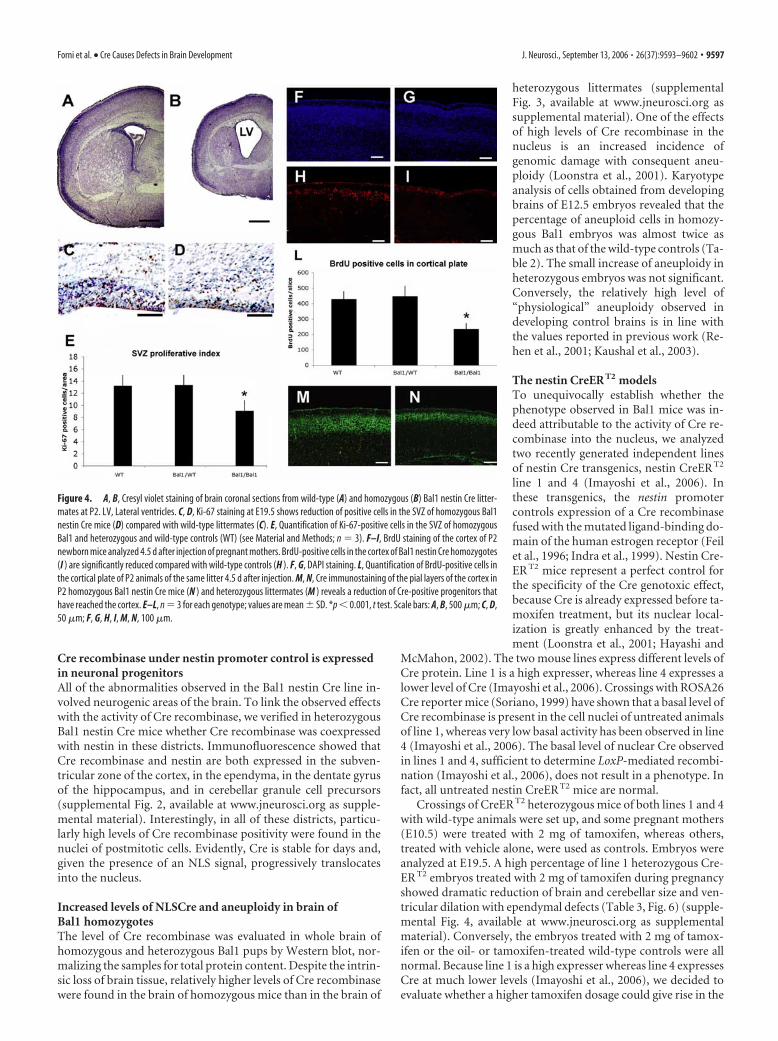
To evaluate the possible differences in proliferation rate of theprogenitors in the SVZ, we decorated E19.5 brain sections for theproliferative antigen Ki-67 (Fig. 4C,D). Quantitative analysis re-vealed an �30% reduction in proliferating SVZ precursors inhomozygous Bal1 embryos (Fig. 4E). We then injected BrdU atthe same gestational age (E19.5) but analyzed the brains at P2.This allowed us to evaluate the number of labeled progenitorsthat reach the cortical plate in 4.5 d (Fig. 4F–I). Counts of BrdU-positive cells revealed a reduction of �50% in homozygous Bal1nestin Cre mice compared with controls (Fig. 4L). Immunoflu-orescence for Cre protein confirmed that the neuronal progeni-tors were indeed Cre positive and that their number was reducedin Bal1 nestin Cre homozygous mice with respect to heterozygouslittermates (Fig. 4M,N). The lower number of progenitors thatreach the cortical plate does not seem to be attributable to a defectin migration because cresyl violet, BrdU, or Cre labeling did notshow abnormal accumulation of progenitors along the migratorypath.
Apoptosis is increased in the developing cortex andcerebellum of Bal1 nestin Cre homozygous miceBecause the reduction in neuronal progenitors seemed to bemore dramatic in the cortical plate than in the SVZ, we verifiedwhether apoptosis occurred during migration. Cleaved caspase 3staining of E19.5 brain sections revealed an increase in apoptoticcells in the intermediate zone and in the cortical plate of homozy-gous Bal1 nestin Cre embryos compared with controls (Fig.5A,B). Cleaved caspase-3-positive cell counts in the cortical plateshowed a 35% increase of apoptosis, indicating that a significantnumber of cells undergoes cell death after leaving the SVZ. Ac-cordingly, analysis of postnatal sections of cerebellum at P2 alsorevealed an increase in cleaved caspase-3-positive cells (Fig.5D,E) and a 39% increase of pyknotic nuclei compared withcontrols (Fig. 5D–F).
Figure 3. Cresyl violet staining of brain sagittal sections from wild-type (A, C, E, G) andhomozygous (B, D, F, H ) Bal1 nestin Cre mice at P8. A, B, The thickness of the parieto-occipitalcerebral cortex (ctx) is reduced in homozygous Bal1 nestin Cre mice. C, D, Higher magnificationshows reduced cellularity and reduced organization of the cortical layers (I–VI) in the occipitalcortex. SP, Subplate. The dentate gyrus (DG) of the hippocampus is reduced and less organized(E, F ) and the cerebellum is underdeveloped (G, H ) in homozygous Bal1 nestin Cre mice. Scalebars: A, B, G, H, 500 �m; C–F, 100 �m.
9596 • J. Neurosci., September 13, 2006 • 26(37):9593–9602 Forni et al. • Cre Causes Defects in Brain Development
Cre recombinase under nestin promoter control is expressedin neuronal progenitorsAll of the abnormalities observed in the Bal1 nestin Cre line in-volved neurogenic areas of the brain. To link the observed effectswith the activity of Cre recombinase, we verified in heterozygousBal1 nestin Cre mice whether Cre recombinase was coexpressedwith nestin in these districts. Immunofluorescence showed thatCre recombinase and nestin are both expressed in the subven-tricular zone of the cortex, in the ependyma, in the dentate gyrusof the hippocampus, and in cerebellar granule cell precursors(supplemental Fig. 2, available at www.jneurosci.org as supple-mental material). Interestingly, in all of these districts, particu-larly high levels of Cre recombinase positivity were found in thenuclei of postmitotic cells. Evidently, Cre is stable for days and,given the presence of an NLS signal, progressively translocatesinto the nucleus.
Increased levels of NLSCre and aneuploidy in brain ofBal1 homozygotesThe level of Cre recombinase was evaluated in whole brain ofhomozygous and heterozygous Bal1 pups by Western blot, nor-malizing the samples for total protein content. Despite the intrin-sic loss of brain tissue, relatively higher levels of Cre recombinasewere found in the brain of homozygous mice than in the brain of
heterozygous littermates (supplementalFig. 3, available at www.jneurosci.org assupplemental material). One of the effectsof high levels of Cre recombinase in thenucleus is an increased incidence ofgenomic damage with consequent aneu-ploidy (Loonstra et al., 2001). Karyotypeanalysis of cells obtained from developingbrains of E12.5 embryos revealed that thepercentage of aneuploid cells in homozy-gous Bal1 embryos was almost twice asmuch as that of the wild-type controls (Ta-ble 2). The small increase of aneuploidy inheterozygous embryos was not significant.Conversely, the relatively high level of“physiological” aneuploidy observed indeveloping control brains is in line withthe values reported in previous work (Re-hen et al., 2001; Kaushal et al., 2003).
The nestin CreER T2 modelsTo unequivocally establish whether thephenotype observed in Bal1 mice was in-deed attributable to the activity of Cre re-combinase into the nucleus, we analyzedtwo recently generated independent linesof nestin Cre transgenics, nestin CreER T2
line 1 and 4 (Imayoshi et al., 2006). Inthese transgenics, the nestin promotercontrols expression of a Cre recombinasefused with the mutated ligand-binding do-main of the human estrogen receptor (Feilet al., 1996; Indra et al., 1999). Nestin Cre-ER T2 mice represent a perfect control forthe specificity of the Cre genotoxic effect,because Cre is already expressed before ta-moxifen treatment, but its nuclear local-ization is greatly enhanced by the treat-ment (Loonstra et al., 2001; Hayashi and
McMahon, 2002). The two mouse lines express different levels ofCre protein. Line 1 is a high expresser, whereas line 4 expresses alower level of Cre (Imayoshi et al., 2006). Crossings with ROSA26Cre reporter mice (Soriano, 1999) have shown that a basal level ofCre recombinase is present in the cell nuclei of untreated animalsof line 1, whereas very low basal activity has been observed in line4 (Imayoshi et al., 2006). The basal level of nuclear Cre observedin lines 1 and 4, sufficient to determine LoxP-mediated recombi-nation (Imayoshi et al., 2006), does not result in a phenotype. Infact, all untreated nestin CreER T2 mice are normal.
Crossings of CreER T2 heterozygous mice of both lines 1 and 4with wild-type animals were set up, and some pregnant mothers(E10.5) were treated with 2 mg of tamoxifen, whereas others,treated with vehicle alone, were used as controls. Embryos wereanalyzed at E19.5. A high percentage of line 1 heterozygous Cre-ER T2 embryos treated with 2 mg of tamoxifen during pregnancyshowed dramatic reduction of brain and cerebellar size and ven-tricular dilation with ependymal defects (Table 3, Fig. 6) (supple-mental Fig. 4, available at www.jneurosci.org as supplementalmaterial). Conversely, the embryos treated with 2 mg of tamox-ifen or the oil- or tamoxifen-treated wild-type controls were allnormal. Because line 1 is a high expresser whereas line 4 expressesCre at much lower levels (Imayoshi et al., 2006), we decided toevaluate whether a higher tamoxifen dosage could give rise in the
Figure 4. A, B, Cresyl violet staining of brain coronal sections from wild-type (A) and homozygous (B) Bal1 nestin Cre litter-mates at P2. LV, Lateral ventricles. C, D, Ki-67 staining at E19.5 shows reduction of positive cells in the SVZ of homozygous Bal1nestin Cre mice (D) compared with wild-type littermates (C). E, Quantification of Ki-67-positive cells in the SVZ of homozygousBal1 and heterozygous and wild-type controls (WT) (see Material and Methods; n � 3). F–I, BrdU staining of the cortex of P2newborn mice analyzed 4.5 d after injection of pregnant mothers. BrdU-positive cells in the cortex of Bal1 nestin Cre homozygotes(I ) are significantly reduced compared with wild-type controls (H ). F, G, DAPI staining. L, Quantification of BrdU-positive cells inthe cortical plate of P2 animals of the same litter 4.5 d after injection. M, N, Cre immunostaining of the pial layers of the cortex inP2 homozygous Bal1 nestin Cre mice (N ) and heterozygous littermates (M ) reveals a reduction of Cre-positive progenitors thathave reached the cortex. E–L, n � 3 for each genotype; values are mean � SD. *p � 0.001, t test. Scale bars: A, B, 500 �m; C, D,50 �m; F, G, H, I, M, N, 100 �m.
Forni et al. • Cre Causes Defects in Brain Development J. Neurosci., September 13, 2006 • 26(37):9593–9602 • 9597

latter to the phenotype observed in line 1. For this purpose, wetreated heterozygous line 4 pregnant mothers (E10.5) with a sin-gle injection of 8 mg of tamoxifen. As reported by others, weobserved an increase in the incidence of abortions at this dosage(Hayashi and McMahon, 2002). However, analysis of the surviv-ing embryos at E19.5 revealed no brain defects in tamoxifen-treated wild-type controls. Conversely, in heterozygous embryosof the CreER T2 line 4, 8 mg of tamoxifen produced a phenotypeessentially overlapping with that described previously for ho-mozygous Bal1 mice and tamoxifen-treated (2 mg) heterozygousCreER T2 line 1 animals (Fig. 7, Table 3) (supplemental Fig. 4,available at www.jneurosci.org as supplemental material). Thesomehow lower frequency of hydrocephalus observed in the twoinducible CreER T2 lines with respect to homozygous Bal1 mice islikely to be attributable to variability in the efficacy of the tamox-ifen treatment.
Defects in neuronal precursors after tamoxifen treatmentTo verify whether the cellular defects underlying the develop-ment of the brain phenotype in homozygous Bal1 mice weredetectable also in the tamoxifen-treated heterozygous CreER T2
lines, we duplicated the analysis of proliferation and apoptosis inthe new animal models. Visualization of proliferating cells byKi-67 staining in tamoxifen-treated E19.5 CreER T2 brain slicesshowed that the number of proliferating cells in the SVZ andintermediate zone was dramatically reduced (Fig. 8A,B). Count-ing of Ki-67-labeled cells showed that the reduction of cyclingcells in the SVZ/intermediate zone of tamoxifen-treated embryoscompared with controls was of the order of 30% for line 1 and50% for line 4 (when treated at the effective dosage of tamoxifen)(Fig. 8C). Surprisingly, at variance with respect to the homozy-gous Bal1 model, staining with cleaved caspase-3 of E19.5 brainslices did not reveal a significant increase in apoptotic cells (datanot shown). Because this model is based on transient rather thanconstitutive nuclear Cre translocation, it is possible that the peakof Cre-induced cell death may occur with a different timing.
DiscussionSeveral reports indicate that Cre recombinase might exert toxiceffects attributable to recombination of genomic DNA at sitesthat share only a limited homology with the loxP sequence ofbacteriophage P1. These ‘‘illegitimate“ recombination events canresult in DNA strand breaks, chromosome rearrangements, an-euploidy, and alteration in the normal proliferation rate attribut-able to an accumulation in the G2–M phase (Thyagarajan et al.,2000; Loonstra et al., 2001; Pfeifer et al., 2001; Silver and Living-ston, 2001).
The present study describes a Cre-induced phenotype charac-terized by severe reduction of the brain and hydrocephaly. This
Figure 5. Immunostaining for cleaved caspase-3 on E19.5 wild-type (A) and homozygous(B) Bal1 nestin Cre brain sections reveals an increased number of cells undergoing apoptosis inthe intermediate zone (IZ), throughout the pre-plate (PP), and in the cortical plate (CP). C,Quantification of cleaved caspase-3 in developing cortex (E19.5). Immunostaining for cleavedcaspase-3 on sections of P3 wild-type (WT; D) and homozygous (E) Bal1 nestin Cre cerebellumsections shows an increased number of cleaved caspase-3-positive cells in the inner granulelayer (IGL) of homozygous Bal1 nestin Cre mice (E). F, Quantification of pyknotic nuclei incerebellum after cresyl violet staining on P2 pups of the same litter. n � 3 for each genotype;values are mean � SD.*p � 0.001, t test (C, F ). Scale bars, 50 �m.
Table 2. Chromosomal aneuploidy in isolated embryonic (E12.5) neuroblastspreads of control (WT/WT), heterozygous (Bal1/WT), and homozygous (Bal1/Bal1)embryos revealed by Giemsa staining
Genotype Number of spread examined % Aneuploid
WT 153 35 � 4Bal1/WT 156 42 � 9*Bal1/Bal1 173 69 � 7**
n � 3 for each genotype; *p � 0.1, �2 test; **p � 10�10, �2 test. WT, Wild type.
Table 3. Frequency of microencephaly/hydrocephalus in CreERT2 embryos (line 1and line 4) after indicated treatment at E10.5
Mouse line Treatment at E10.5 Microencephaly/hydrocephalus n
CreERT2 line 1/� 2 mg of tamoxifen 77% 22CreERT2 line 1/� Vehicle 0% 40CreERT2 line 4/� 8 mg of tamoxifen 83% 12CreERT2 line 4/� 2 mg of tamoxifen 0% 3CreERT2 line 4/� Vehicle 0% 6WT 8 mg of tamoxifen 0% 8WT 2 mg of tamoxifen 0% 40WT Vehicle 0% 48
Microencephaly was determined by gross observation at E19.5. The presence of hydrocephalus was verified bytransillumination of whole brains or by histological sections. WT, Wild type.
9598 • J. Neurosci., September 13, 2006 • 26(37):9593–9602 Forni et al. • Cre Causes Defects in Brain Development

phenotype was observed in three distinct nestin Cre transgeniclines, thus ruling out the possibility that it could be attributable tothe insertion site or to cosegregating mutations. In the first model(Bal1 line), microencephaly and hydrocephaly were found in100% of homozygous animals. In this mouse line, the Cre pro-tein, because of the presence of a nuclear localization signal(NLSCre), is constantly localized in the nucleus. Conversely, inthe second and third model (CreER T2 lines 1 and 4), the devel-opment of hydrocephaly was dependent on tamoxifen, whichcontrols the translocation of the enzyme into the nucleus. Thus,the described effects were not attributable to aspecific toxicity ofthe Cre protein (ineffective when stored in the cytoplasm) but diddepend on its nuclear localization. The requirement for a thresh-old of Cre in the nucleus was confirmed by the observation thatan increase of aneuploidy was observed only in homozygous Bal1brains.
The causal link between development of the phenotype andCre activity was further strengthened by the observation of itsdependence on tamoxifen dosage. Whereas in the CreER T2 line 1,
a single treatment with 2 mg of tamoxifenat E10.5 was sufficient to induce hydro-cephaly, this dosage was without effect inline 4, which expresses CreER recombi-nase at a lower level (Imayoshi et al., 2006).This, together with the lack of phenotypein tamoxifen-treated wild-type controls,proves that hydrocephaly is not an artifactof the treatment. The fact that hydroceph-aly appeared only in homozygosity in theNLSCre Bal1 line or when increasing thedosage of tamoxifen to 8 mg in the lowexpresser CreER T2 line 4 indicates that, toexert its genotoxic effect, the level of nu-clear Cre must overcome a critical thresh-old. It is important to note that, in ourmodels, chronic or transient translocationof Cre in the nucleus of neuronal progen-itors was facilitated, via either an NLS orfusion with the ER. It is known that both ofthese modifications strongly increase nu-clear translocation and recombination ef-ficiency but may also cause higher geno-toxicity (Loonstra et al., 2001; Baba et al.,2005). This, however, has been observed incell lines only when the concentration ofCre in the nucleus exceeds a certainthreshold (Loonstra et al., 2001; Baba etal., 2005). Accordingly, in NLSCre Bal1mice, the phenotype appeared only in ho-mozygosity, whereas in the CreER T2 lines,it was induced only on adequate tamoxifendosage (Hayashi and McMahon, 2002).Cre genotoxicity may also critically de-pend on when the cells are exposed to Creactivity. In particular, Cre recombinase ismore likely to yield toxic effects duringphases of the cell cycle when chromatin isuncondensed and probably more vulnera-ble to its illegitimate action (Loonstra etal., 2001). Ki-67 staining of brain sectionsderived from E19.5 NLSCre Bal1 andtamoxifen-treated CreER T2 embryosshowed a significant reduction of the cy-
cling cells in the SVZ. However, heavy Cre labeling in the piallayers of the developing cortex in Bal1 mice indicates that, oncepostmitotic, neural cells seem to be able to endure prolongedexposure to Cre without loss of viability.
A plethora of nestin Cre models exists and have been success-fully used in the heterozygous state to investigate the role of spe-cific proteins during development. Heterozygous nestin NLSCreBalancer mice have themselves been successfully used to over-come embryonic lethality (Betz et al., 1996; Alonzi et al., 2001;Uno et al., 2003). These models do not generally result in anincrease rate of idrocephaly (Tronche et al., 1999; Trumpp et al.,1999; Treutelaar et al., 2003; Thuret et al., 2004; Liu et al., 2005).It should be noted, however, that (except for Bal1 mice, whichwere always used in heterozygosity) the constructs described inthese works did not include an element to facilitate Cre nucleartranslocation.
Nestin Cre-mediated gene inactivation has been successfullyapplied to genes having key roles in proliferation, survival, ormigration (Isaka et al., 1999; Pirvola et al., 2002; Haigh et al.,
Figure 6. Top row, Wild-type and heterozygous CreER T2 line 1 whole brains at E19.5 after the indicated treatments. Cresylviolet staining of coronal brain sections (A–D), sagittal cerebellar sections (E–H ), coronal sections of the third ventricle (III) (I–N ),and coronal sections of the Sylvius aqueduct (O–R). Heterozygous nestin CreER T2 embryos treated with vehicle alone (oil) displaynormal brain (B) and normal cerebellar development (F ) and are thus indistinguishable from wild-type (WT) controls treated witheither oil (A, E) or 2 mg of tamoxifen (C, G; tam). Tamoxifen-treated heterozygous nestin CreER T2 display severe signs of microen-cephaly and ventricular dilation (D), reduced development of the cerebellum (H ), ventricular dilation (D, N, R), and defects inependymal lining of the third ventricle (III) and Sylvius aqueduct (N, R, arrows). Scale bars: A–D, 500 �m; E–H, 200 �m; I–R,100 �m.
Forni et al. • Cre Causes Defects in Brain Development J. Neurosci., September 13, 2006 • 26(37):9593–9602 • 9599

2003; Lin et al., 2003; Tomita et al., 2003; Treutelaar et al., 2003;Klezovitch et al., 2004; Raab et al., 2004; Berube et al., 2005).Interestingly, all of the features described in these models as wellas in other animal models of impaired neurogenesis, such as spe-cific ablation of radial glia (Xie et al., 2002) or loss-of-function ofgenes involved in neuronal migration or adhesion (Ramanathanet al., 1996; Fransen et al., 1998; Bearer, 2001; Assadi et al., 2003;Sakata-Haga et al., 2004), were recapitulated at different levels inour homozygous NLSCre Bal1 mice or in the heterozygous Cre-ER T2 models. This suggests that impairment of nestin-positivecell populations either by specific genes removal or through Cre-mediated toxicity may lead to similar defects.
In conclusion, while taking advantage of the Cre/LoxP system,it should be kept in mind that the presence of elements facilitatingnuclear translocation or the use of transgenics with high levels ofCre expression can, on one side, ensure high level of recombina-tion, but, on the other, could yield enhanced phenotypes, attrib-utable not only to target gene ablation but also to Cre-mediatedgenotoxicity. This could be especially true in the CNS, which is acompartment naturally prone to genomic instability (Kim et al.,2001; Rehen et al., 2001; Kaushal et al., 2003; Yang et al., 2003). It
is therefore essential to evaluate the phenotypes observed in Cre/loxP conditional knock-outs against Cre transgenic controls.
ReferencesAlonzi T, Middleton G, Wyatt S, Buchman V, Betz UA, Muller W, Musiani P,
Poli V, Davies AM (2001) Role of STAT3 and PI 3-kinase/Akt in medi-ating the survival actions of cytokines on sensory neurons. Mol Cell Neu-rosci 18:270 –282.
Aolad HM, Inouye M, Hayasaka S, Darmanto W, Murata Y (1998) Congen-ital hydrocephalus caused by exposure to low level X-radiation at earlygestational stage in mice. Biol Sci Space 12:256 –257.
Assadi AH, Zhang G, Beffert U, McNeil RS, Renfro AL, Niu S, QuattrocchiCC, Antalffy BA, Sheldon M, Armstrong DD, Wynshaw-Boris A, Herz J,D’Arcangelo G, Clark GD (2003) Interaction of reelin signaling and Lis1in brain development. Nat Genet 35:270 –276.
Baba Y, Nakano M, Yamada Y, Saito I, Kanegae Y (2005) Practical range ofeffective dose for Cre recombinase-expressing recombinant adenoviruswithout cell toxicity in mammalian cells. Microbiol Immunol49:559 –570.
Bearer CF (2001) L1 cell adhesion molecule signal cascades: targets for eth-anol developmental neurotoxicity. Neurotoxicology 22:625– 633.
Berube NG, Mangelsdorf M, Jagla M, Vanderluit J, Garrick D, Gibbons RJ,Higgs DR, Slack RS, Picketts DJ (2005) The chromatin-remodeling pro-
Figure 7. Top row, Wild-type and heterozygous CreER T2 line 4 whole brains at E19.5 after the indicated treatments. Cresyl violet staining of coronal brain sections (A–F ), sagittal cerebellarsections (G–L), coronal sections of the third ventricle (III) (M–R), and Sylvius aqueduct (S–X ). Heterozygous nestin CreER T2 embryos treated with vehicle alone (oil) (B, H, N, T ) or with 2 mg oftamoxifen (tam) (D, J, P, V ) are indistinguishable from oil-treated (A, G, M, S) and 2 mg tamoxifen-treated (C, I, O, U ) wild-type (WT) animals. Tamoxifen-treated (8 mg) wild-type embryos areindistinguishable from the previous controls (E, K, Q, W ), whereas 8 mg tamoxifen-treated heterozygous nestin CreER T2 (F, L, R, X ) display severe signs of microencephaly and ventricular dilation(F ), reduced development of the cerebellum (L), defects in ependymal lining in the third ventricle (R) and defects in Sylvius aqueduct ependymal lining (X ) (arrowheads). Scale bars: A–F, 500 �m;G–L, 200 �m; M–X, 100 �m.
9600 • J. Neurosci., September 13, 2006 • 26(37):9593–9602 Forni et al. • Cre Causes Defects in Brain Development

tein ATRX is critical for neuronal survival during corticogenesis. J ClinInvest 115:258 –267.
Betz UA, Vosshenrich CA, Rajewsky K, Muller W (1996) Bypass of lethalitywith mosaic mice generated by Cre-loxP-mediated recombination. CurrBiol 6:1307–1316.
Crews L, Wyss-Coray T, Masliah E (2004) Insights into the pathogenesis ofhydrocephalus from transgenic and experimental animal models. BrainPathol 14:312–316.
Davy BE, Robinson ML (2003) Congenital hydrocephalus in hy3 mice iscaused by a frameshift mutation in Hydin, a large novel gene. Hum MolGenet 12:1163–1170.
Dobkin BH (1978) Syncope in the adult Chiari anomaly. Neurology28:718 –720.
Dominguez-Pinos MD, Paez P, Jimenez AJ, Weil B, Arraez MA, Perez-FigaresJM, Rodriguez EM (2005) Ependymal denudation and alterations of thesubventricular zone occur in human fetuses with a moderate communi-cating hydrocephalus. J Neuropathol Exp Neurol 64:595– 604.
Feil R, Brocard J, Mascrez B, LeMeur M, Metzger D, Chambon P (1996)Ligand-activated site-specific recombination in mice. Proc Natl Acad SciUSA 93:10887–10890.
Fransen E, D’Hooge R, Van Camp G, Verhoye M, Sijbers J, Reyniers E, Sori-ano P, Kamiguchi H, Willemsen R, Koekkoek SK, De Zeeuw CI, De DeynPP, Van der Linden A, Lemmon V, Kooy RF, Willems PJ (1998) L1knockout mice show dilated ventricles, vermis hypoplasia and impairedexploration patterns. Hum Mol Genet 7:999 –1009.
Grondona JM, Perez-Martin M, Cifuentes M,Perez J, Jimenez AJ, Perez-Figares JM,Fernandez-Llebrez P (1996) Ependymal de-nudation, aqueductal obliteration and hydro-cephalus after a single injection of neuramini-dase into the lateral ventricle of adult rats.J Neuropathol Exp Neurol 55:999 –1008.
Haigh JJ, Morelli PI, Gerhardt H, Haigh K, Tsien J,Damert A, Miquerol L, Muhlner U, Klein R,Ferrara N, Wagner EF, Betsholtz C, Nagy A(2003) Cortical and retinal defects caused bydosage-dependent reductions in VEGF-Aparacrine signaling. Dev Biol 262:225–241.
Hayashi S, McMahon AP (2002) Efficient re-combination in diverse tissues by a tamoxifen-inducible form of Cre: a tool for temporallyregulated gene activation/inactivation in themouse. Dev Biol 244:305–318.
Hicks SP, D’Amato CJ (1980) Effects of radiationon development, especially of the nervous sys-tem. Am J Forensic Med Pathol 1:309 –317.
Imayoshi I, Ohtsuka T, Metzger D, Chambon P,Kageyama R (2006) Temporal regulation ofCre recombinase activity in neural stem cells.Genesis 44:233–238.
Indra AK, Warot X, Brocard J, Bornert JM, XiaoJH, Chambon P, Metzger D (1999)Temporally-controlled site-specific mutagen-esis in the basal layer of the epidermis: compar-ison of the recombinase activity of thetamoxifen-inducible Cre-ER(T) and Cre-ER(T2) recombinases. Nucleic Acids Res27:4324 – 4327.
Isaka F, Ishibashi M, Taki W, Hashimoto N, Na-kanishi S, Kageyama R (1999) Ectopic ex-pression of the bHLH gene Math1 disturbsneural development. Eur J Neurosci11:2582–2588.
Jimenez AJ, Tome M, Paez P, Wagner C, Rodri-guez S, Fernandez-Llebrez P, Rodriguez EM,Perez-Figares JM (2001) A programmedependymal denudation precedes congenitalhydrocephalus in the hyh mutant mouse.J Neuropathol Exp Neurol 60:1105–1119.
Kaushal D, Contos JJ, Treuner K, Yang AH, Kings-bury MA, Rehen SK, McConnell MJ, Okabe M,Barlow C, Chun J (2003) Alteration of geneexpression by chromosome loss in the postna-
tal mouse brain. J Neurosci 23:5599 –5606.Kim GJ, Park SY, Kim H, Chun YH, Park SH (2001) Chromosomal aberra-
tions in neuroblastoma cell lines identified by cross species color bandingand chromosome painting. Cancer Genet Cytogenet 129:10 –16.
Klezovitch O, Fernandez TE, Tapscott SJ, Vasioukhin V (2004) Loss of cellpolarity causes severe brain dysplasia in Lgl1 knockout mice. Genes Dev18:559 –571.
Kos CH (2004) Cre/loxP system for generating tissue-specific knockoutmouse models. Nutr Rev 62:243–246.
Lin T, Sandusky SB, Xue H, Fishbein KW, Spencer RG, Rao MS, FrancomanoCA (2003) A central nervous system specific mouse model for thanato-phoric dysplasia type II. Hum Mol Genet 12:2863–2871.
Liu W, Sun X, Braut A, Mishina Y, Behringer RR, Mina M, Martin JF (2005)Distinct functions for Bmp signaling in lip and palate fusion in mice.Development 132:1453–1461.
Loonstra A, Vooijs M, Beverloo HB, Allak BA, van Drunen E, Kanaar R, BernsA, Jonkers J (2001) Growth inhibition and DNA damage induced by Crerecombinase in mammalian cells. Proc Natl Acad Sci USA 98:9209 –9214.
Mignone JL, Kukekov V, Chiang AS, Steindler D, Enikolopov G (2004)Neural stem and progenitor cells in nestin-GFP transgenic mice. J CompNeurol 469:311–324.
Pfeifer A, Brandon EP, Kootstra N, Gage FH, Verma IM (2001) Delivery ofthe Cre recombinase by a self-deleting lentiviral vector: efficient genetargeting in vivo. Proc Natl Acad Sci USA 98:11450 –11455.
Figure 8. Ki-67 labeling (dark stain) at E19.5 on coronal sections from oil-treated (A) and 2 mg tamoxifen-treated (TAM) (B)nestin CreER T2 line 1 heterozygous embryos reveals a reduction of the number of proliferating cells in the SVZ and in theintermediate zone (IZ) of tamoxifen-treated embryos (B) with respect to oil-treated control (A). LV, Lateral ventricle. C, Quanti-fication of Ki-67-positive cells in the SVZ at E19.5 in heterozygous CreER T2 line 1 and line 4 and wild-type controls embryos afterthe indicated treatment performed at E10.5. Error bars represent mean � SD; n � 3 for each genotype. *p � 0.001, t test.
Forni et al. • Cre Causes Defects in Brain Development J. Neurosci., September 13, 2006 • 26(37):9593–9602 • 9601

Pirvola U, Ylikoski J, Trokovic R, Hebert JM, McConnell SK, Partanen J(2002) FGFR1 is required for the development of the auditory sensoryepithelium. Neuron 35:671– 680.
Raab S, Beck H, Gaumann A, Yuce A, Gerber HP, Plate K, Hammes HP,Ferrara N, Breier G (2004) Impaired brain angiogenesis and neuronalapoptosis induced by conditional homozygous inactivation of vascularendothelial growth factor. Thromb Haemost 91:595– 605.
Ramanathan R, Wilkemeyer MF, Mittal B, Perides G, Charness ME (1996)Alcohol inhibits cell-cell adhesion mediated by human L1. J Cell Biol133:381–390.
Rehen SK, McConnell MJ, Kaushal D, Kingsbury MA, Yang AH, Chun J(2001) Chromosomal variation in neurons of the developing and adultmammalian nervous system. Proc Natl Acad Sci USA 98:13361–13366.
Sakata-Haga H, Sawada K, Ohnishi T, Fukui Y (2004) Hydrocephalus fol-lowing prenatal exposure to ethanol. Acta Neuropathol (Berl)108:393–398.
Schmidt EE, Taylor DS, Prigge JR, Barnett S, Capecchi MR (2000) Illegiti-mate Cre-dependent chromosome rearrangements in transgenic mousespermatids. Proc Natl Acad Sci USA 97:13702–13707.
Silver DP, Livingston DM (2001) Self-excising retroviral vectors encodingthe Cre recombinase overcome Cre-mediated cellular toxicity. Mol Cell8:233–243.
Soriano P (1999) Generalized lacZ expression with the ROSA26 Cre re-porter strain. Nat Genet 21:70 –71.
Thuret S, Alavian KN, Gassmann M, Lloyd CK, Smits SM, Smidt MP, Klein R,Dyck RH, Simon HH (2004) The neuregulin receptor, ErbB4, is notrequired for normal development and adult maintenance of the substan-tia nigra pars compacta. J Neurochem 91:1302–1311.
Thyagarajan B, Guimaraes MJ, Groth AC, Calos MP (2000) Mammaliangenomes contain active recombinase recognition sites. Gene 244:47–54.
Tomita S, Ueno M, Sakamoto M, Kitahama Y, Ueki M, Maekawa N, Saka-moto H, Gassmann M, Kageyama R, Ueda N, Gonzalez FJ, Takahama Y(2003) Defective brain development in mice lacking the Hif-1alpha genein neural cells. Mol Cell Biol 23:6739 – 6749.
Treutelaar MK, Skidmore JM, Dias-Leme CL, Hara M, Zhang L, Simeone D,Martin DM, Burant CF (2003) Nestin-lineage cells contribute to themicrovasculature but not endocrine cells of the islet. Diabetes52:2503–2512.
Tronche F, Kellendonk C, Kretz O, Gass P, Anlag K, Orban PC, Bock R, KleinR, Schutz G (1999) Disruption of the glucocorticoid receptor gene in thenervous system results in reduced anxiety. Nat Genet 23:99 –103.
Trumpp A, Depew MJ, Rubenstein JL, Bishop JM, Martin GR (1999) Cre-mediated gene inactivation demonstrates that FGF8 is required for cellsurvival and patterning of the first branchial arch. Genes Dev13:3136 –3148.
Uno S, Wang B, Shertzer HG, Nebert DW, Dalton TP (2003) Balancer-Cretransgenic mouse germ cells direct the incomplete resolution of a tri-loxP-targeted Cyp1a1 allele, producing a conditional knockout allele.Biochem Biophys Res Commun 312:494 – 499.
Wagner C, Batiz LF, Rodriguez S, Jimenez AJ, Paez P, Tome M, Perez-FigaresJM, Rodriguez EM (2003) Cellular mechanisms involved in the stenosisand obliteration of the cerebral aqueduct of hyh mutant mice developingcongenital hydrocephalus. J Neuropathol Exp Neurol 62:1019 –1040.
Xie Y, Skinner E, Landry C, Handley V, Schonmann V, Jacobs E, Fisher R,Campagnoni A (2002) Influence of the embryonic preplate on the orga-nization of the cerebral cortex: a targeted ablation model. J Neurosci22:8981– 8991.
Yang AH, Kaushal D, Rehen SK, Kriedt K, Kingsbury MA, McConnell MJ,Chun J (2003) Chromosome segregation defects contribute to aneu-ploidy in normal neural progenitor cells. J Neurosci 23:10454 –10462.
9602 • J. Neurosci., September 13, 2006 • 26(37):9593–9602 Forni et al. • Cre Causes Defects in Brain Development