Development/Plasticity/Repair TheRelationshipbetweenPSD ...
12
Development/Plasticity/Repair The Relationship between PSD-95 Clustering and Spine Stability In Vivo Michele Cane, 1,2 Bohumil Maco, 3,4 Graham Knott, 3 and Anthony Holtmaat 1 1 Department of Basic Neurosciences, Faculty of Medicine and the Geneva Neuroscience Center, University of Geneva, CH 1211, Switzerland, and 2 Lemanic Neuroscience Doctoral School, 3 Biological Electron Microscopy Facility, Centre of Electron Microscopy, and 4 Computer Vision Laboratory, École Polytechnique Fédérale de Lausanne, Lausanne, CH 1015, Switzerland The appearance and disappearance of dendritic spines, accompanied by synapse formation and elimination may underlie the experience- dependent reorganization of cortical circuits. The exact temporal relationship between spine and synapse formation in vivo remains unclear, as does the extent to which synapse formation enhances the stability of newly formed spines and whether transient spines produce synapses. We used in utero electroporation of DsRedExpress- and eGFP-tagged postsynaptic density protein 95 (PSD-95) to investigate the relationship between spine and PSD stability in mouse neocortical L2/3 pyramidal cells in vivo. Similar to previous studies, spines and synapses appeared and disappeared, even in naive animals. Cytosolic spine volumes and PSD-95-eGFP levels in spines covaried over time, suggesting that the strength of many individual synapses continuously changes in the adult neocortex. The minority of newly formed spines acquired PSD-95-eGFP puncta. Spines that failed to acquire a PSD rarely survived for more than a day. Although PSD-95-eGFP accumulation was associated with increased spine lifetimes, most new spines with a PSD did not convert into persistent spines. This indicates that transient spines may serve to produce short-lived synaptic contacts. Persistent spines that were destined to disappear showed, on average, reduced PSD-95-eGFP levels well before the actual pruning event. Altogether, our data indicate that the PSD size relates to spine stability in vivo. Introduction The formation of new synapses at the expense of old synapses may underlie the experience-dependent and functional reorgani- zation of cortical circuits in adults (Holtmaat and Svoboda, 2009; Fu and Zuo, 2011). Imaging studies in vivo have shown that prox- ies for synapses, such as dendritic spines and axonal boutons, appear and disappear over days, even in naive mice. Under base- line conditions, new dendritic spines rarely transform into per- sistent spines and usually retract within hours and days after they are formed (Holtmaat et al., 2005; Zuo et al., 2005). These tran- sient spines were hypothesized to generate short-lived synaptic contacts. Indeed, in vitro studies have shown that new spines can display transmitter-evoked Ca 2 transients within hours (Zito et al., 2009) and often rapidly incorporate morphologically mature synapses (Na ¨gerl et al., 2007; Zito et al., 2009). However, retro- spective electron microscopy (EM) on dendrites that were im- aged in vivo has shown that a large fraction of new spines do not bear a morphologically defined synapse within the first day (Knott et al., 2006); but they always contain synapses after several days and their sizes correlate with the spines’ lifetimes. This sug- gests that synapse growth in vivo is slow and that spine stabiliza- tion and synapse growth are closely linked. It also implies that transient spines are new spines that fail to form a synapse. How- ever, because the findings were based on end-point measure- ments, it remains uncertain whether synapse formation in vivo is a unique signature of new persistent spines. Neither do we know whether new spine survival and pruning in vivo is associated with synapse growth or shrinkage, respectively. To answer these questions, we expressed PSD-95-eGFP in adult L2/3 pyramidal cells of the barrel cortex using in utero elec- troporation and imaged dendritic spines in vivo. PSD-95 is a postsynaptic scaffold protein that is present in the majority of excitatory synapses and interlinks a variety of proteins (Craven et al., 1999; Chen et al., 2005; Sheng and Hoogenraad, 2007). PSD-95 modulates postsynaptic function and maturation (El- Husseini et al., 2000; Ehrlich et al., 2007). Expression of PSD-95- eGFP allows the visualization of postsynaptic densities (PSDs) in real time in vitro (Okabe et al., 1999; Sala et al., 2003; Sharma et al., 2006; Woods et al., 2011) and in vivo (Niell et al., 2004; Gray et al., 2006; Kelsch et al., 2008; Livneh et al., 2009) without disturb- ing gross synaptic network dynamics (Gray et al., 2006). Such experiments have indicated that the appearance and disappear- ance of fluorescently labeled PSD-95 puncta is likely to represent Received Aug. 6, 2013; revised Nov. 21, 2013; accepted Dec. 20, 2013. Author contributions: M.C., G.K., and A.H. designed research; M.C., B.M., G.K., and A.H. performed research; M.C., B.M., G.K., and A.H. analyzed data; M.C. and A.H. wrote the paper. This work was supported by the Swiss National Science Foundation (Grants 31003A_120685 (AH), 31003A_135631 (AH), CRSI33_127289 (AH), and CRF II313470/1 to G.K.); the International Foundation for Research in Paraplegia; and the Hans Wilsdorf Foundation (Dr Alain Rossier Chair funding to A.H.). We thank Simon Borgeaud, Laurant Brodier, and Alberto Bisco for help with image analysis; Daniel Lebrecht and Aurelie Pala for advise on single-cell electroporation experiments; Marco Cantoni for help with the FIBSEM imaging; Michael Patterson and Ste ´phane Page `s for comments on our manuscript; Roby Weimer, Noah Gray, and Karel Svoboda for initial help with the experiments and for sharing the PSD-95-eGFP and DsRedExpress plasmids; and Karel Svoboda, Sen Song, Vijay Iyer, and Tim O’Connor for sharing image analysis software. The authors declare no competing financial interests. Correspondence should be addressed to Anthony Holtmaat, Department of Basic Neurosciences, CMU, 1 rue Michel Servet, 1211 Geneva-4, Switzerland. E-mail: [email protected]. DOI:10.1523/JNEUROSCI.3353-13.2014 Copyright © 2014 the authors 0270-6474/14/342075-12$15.00/0 The Journal of Neuroscience, February 5, 2014 • 34(6):2075–2086 • 2075
Transcript of Development/Plasticity/Repair TheRelationshipbetweenPSD ...

Development/Plasticity/Repair
The Relationship between PSD-95 Clustering and SpineStability In Vivo
Michele Cane,1,2 Bohumil Maco,3,4 Graham Knott,3 and Anthony Holtmaat1
1Department of Basic Neurosciences, Faculty of Medicine and the Geneva Neuroscience Center, University of Geneva, CH 1211, Switzerland, and 2LemanicNeuroscience Doctoral School, 3Biological Electron Microscopy Facility, Centre of Electron Microscopy, and 4Computer Vision Laboratory, ÉcolePolytechnique Fédérale de Lausanne, Lausanne, CH 1015, Switzerland
The appearance and disappearance of dendritic spines, accompanied by synapse formation and elimination may underlie the experience-dependent reorganization of cortical circuits. The exact temporal relationship between spine and synapse formation in vivo remainsunclear, as does the extent to which synapse formation enhances the stability of newly formed spines and whether transient spinesproduce synapses. We used in utero electroporation of DsRedExpress- and eGFP-tagged postsynaptic density protein 95 (PSD-95) toinvestigate the relationship between spine and PSD stability in mouse neocortical L2/3 pyramidal cells in vivo. Similar to previous studies,spines and synapses appeared and disappeared, even in naive animals. Cytosolic spine volumes and PSD-95-eGFP levels in spinescovaried over time, suggesting that the strength of many individual synapses continuously changes in the adult neocortex. The minorityof newly formed spines acquired PSD-95-eGFP puncta. Spines that failed to acquire a PSD rarely survived for more than a day. AlthoughPSD-95-eGFP accumulation was associated with increased spine lifetimes, most new spines with a PSD did not convert into persistentspines. This indicates that transient spines may serve to produce short-lived synaptic contacts. Persistent spines that were destined todisappear showed, on average, reduced PSD-95-eGFP levels well before the actual pruning event. Altogether, our data indicate that thePSD size relates to spine stability in vivo.
IntroductionThe formation of new synapses at the expense of old synapsesmay underlie the experience-dependent and functional reorgani-zation of cortical circuits in adults (Holtmaat and Svoboda, 2009;Fu and Zuo, 2011). Imaging studies in vivo have shown that prox-ies for synapses, such as dendritic spines and axonal boutons,appear and disappear over days, even in naive mice. Under base-line conditions, new dendritic spines rarely transform into per-sistent spines and usually retract within hours and days after theyare formed (Holtmaat et al., 2005; Zuo et al., 2005). These tran-sient spines were hypothesized to generate short-lived synapticcontacts. Indeed, in vitro studies have shown that new spines candisplay transmitter-evoked Ca 2� transients within hours (Zito etal., 2009) and often rapidly incorporate morphologically mature
synapses (Nagerl et al., 2007; Zito et al., 2009). However, retro-spective electron microscopy (EM) on dendrites that were im-aged in vivo has shown that a large fraction of new spines do notbear a morphologically defined synapse within the first day(Knott et al., 2006); but they always contain synapses after severaldays and their sizes correlate with the spines’ lifetimes. This sug-gests that synapse growth in vivo is slow and that spine stabiliza-tion and synapse growth are closely linked. It also implies thattransient spines are new spines that fail to form a synapse. How-ever, because the findings were based on end-point measure-ments, it remains uncertain whether synapse formation in vivo isa unique signature of new persistent spines. Neither do we knowwhether new spine survival and pruning in vivo is associated withsynapse growth or shrinkage, respectively.
To answer these questions, we expressed PSD-95-eGFP inadult L2/3 pyramidal cells of the barrel cortex using in utero elec-troporation and imaged dendritic spines in vivo. PSD-95 is apostsynaptic scaffold protein that is present in the majority ofexcitatory synapses and interlinks a variety of proteins (Craven etal., 1999; Chen et al., 2005; Sheng and Hoogenraad, 2007).PSD-95 modulates postsynaptic function and maturation (El-Husseini et al., 2000; Ehrlich et al., 2007). Expression of PSD-95-eGFP allows the visualization of postsynaptic densities (PSDs) inreal time in vitro (Okabe et al., 1999; Sala et al., 2003; Sharma etal., 2006; Woods et al., 2011) and in vivo (Niell et al., 2004; Gray etal., 2006; Kelsch et al., 2008; Livneh et al., 2009) without disturb-ing gross synaptic network dynamics (Gray et al., 2006). Suchexperiments have indicated that the appearance and disappear-ance of fluorescently labeled PSD-95 puncta is likely to represent
Received Aug. 6, 2013; revised Nov. 21, 2013; accepted Dec. 20, 2013.Author contributions: M.C., G.K., and A.H. designed research; M.C., B.M., G.K., and A.H. performed research; M.C.,
B.M., G.K., and A.H. analyzed data; M.C. and A.H. wrote the paper.This work was supported by the Swiss National Science Foundation (Grants 31003A_120685 (AH),
31003A_135631 (AH), CRSI33_127289 (AH), and CRF II313470/1 to G.K.); the International Foundation for Researchin Paraplegia; and the Hans Wilsdorf Foundation (Dr Alain Rossier Chair funding to A.H.). We thank Simon Borgeaud,Laurant Brodier, and Alberto Bisco for help with image analysis; Daniel Lebrecht and Aurelie Pala for advise onsingle-cell electroporation experiments; Marco Cantoni for help with the FIBSEM imaging; Michael Patterson andStephane Pages for comments on our manuscript; Roby Weimer, Noah Gray, and Karel Svoboda for initial help withthe experiments and for sharing the PSD-95-eGFP and DsRedExpress plasmids; and Karel Svoboda, Sen Song, VijayIyer, and Tim O’Connor for sharing image analysis software.
The authors declare no competing financial interests.Correspondence should be addressed to Anthony Holtmaat, Department of Basic Neurosciences, CMU, 1 rue
Michel Servet, 1211 Geneva-4, Switzerland. E-mail: [email protected]:10.1523/JNEUROSCI.3353-13.2014
Copyright © 2014 the authors 0270-6474/14/342075-12$15.00/0
The Journal of Neuroscience, February 5, 2014 • 34(6):2075–2086 • 2075

synapse formation and synaptic pruning (Prange and Murphy,2001; De Roo et al., 2008; Chen et al., 2011).
Our data confirm that PSDs are dynamic in vivo and implythat spine maintenance is associated with the presence of a PSDand may even depend on its size. The accumulation of PSD-95 isassociated with (new) spine survival. However, the presence of aPSD is not an exclusive attribute of persistent spines; a smallfraction of transient spines may serve to produce short-lived syn-aptic contacts.
Materials and MethodsDNA constructsThe pCAG-PSD-95-eGFP-WPRE and pCAG-DsRedExpress-WPREplasmids were obtained from K. Svoboda, Janelia Research Farms (Grayet al., 2006). In the PSD-95-eGFP plasmid, the eGFP open reading framewas placed in frame at the 3� translated region of PSD-95, which codes forthe c-terminal part of the PSD-95 protein (Okabe et al., 1999). In bothplasmids, expression was driven by the CMV-enhanced chicken �-actinpromoter (CAG). The 3� untranslated regions contained the woodchuckhepatitis virus posttranslational regulatory element (WPRE) and the bo-vine growth hormone polyadenylation site.
ElectroporationThis study was performed according to the guidelines of the Swiss FederalAct on Animal Protection and Swiss Animal Protection Ordinance. Allexperiments were approved by the ethics committee of the University ofGeneva and the Cantonal Veterinary Office (Geneva, Switzerland).
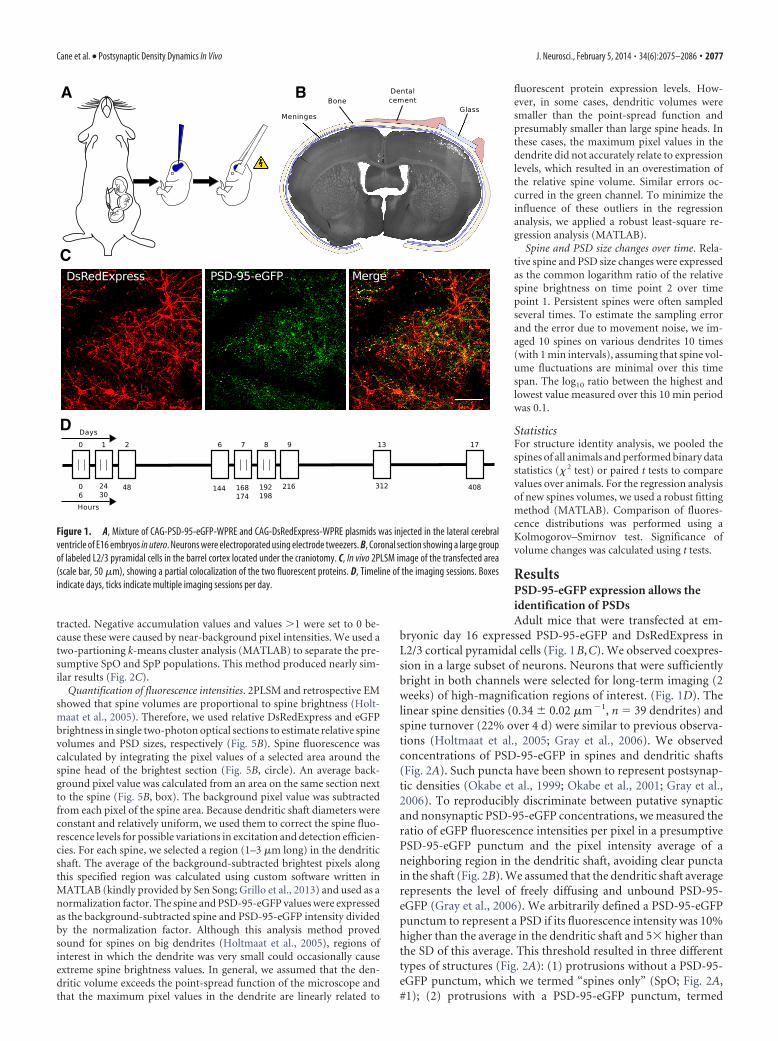
In utero electroporations were performed as described previously(Saito and Nakatsuji, 2001; Tabata and Nakajima, 2001; Fig. 1A). Inshort, E16 timed-pregnant C57BL/6J mice were anesthetized using anisoflurane– oxygen mixture (2% vol isoflurane/vol O2). The uterinehorns were exposed through a �1 cm incision on the abdominal wall.Approximately 1 �l of buffer solution containing �2 �g/�l pCAG-PSD-95-eGFP-WPRE plasmid, a molar equivalent of pCAG-DsRedExpress-WPRE plasmid, and a trace of Fast Green (Sigma) was injected using acustom-made intracellular microinjection dispense system that was con-trolled by a pulse stimulator (Master-8; A.M.P.I.). The fluid was injectedinto the right lateral ventricle of each embryo through a pulled-glasspipette. Subsequently, the head of each embryo was placed betweencustom-made tweezer electrodes with the positive electrode contactingthe right side of the head. Electroporation into the right cerebral cortexwas achieved using 5 square pulses (duration � 50 ms, frequency � 1 Hz,40 V).
The single cell electroporation was performed as described previously(Judkewitz et al., 2009). The procedure was performed on a 6-week-oldC57BL/6 male. An intracellular solution containing �100 ng/�l pCAG-PSD-95-eGFP-WPRE plasmid and a molar equivalent of pCAG-DsRedExpress-WPRE plasmid was used.
Cranial windowAt postnatal day 42, mice were anesthetized with a ketamine (0.10 mg/gbody weight) and xylazine (0.01 mg/g body weight) mixture. The cranialwindows were implanted as described previously (Holtmaat et al., 2009;Fig. 1B). Before the surgery, dexamethasone (�2 �g/g body weight) wasinjected intramuscularly to reduce cerebral edema during the craniot-omy. The craniotomy was located above the right somatosensory cortex(�1.5 mm caudal and �3 mm lateral of bregma). The craniotomy wascovered with a 5-mm-diameter cover glass, which was permanentlyglued to the skull using dental acrylic cement. The dura remainedintact. After surgery, the animals received buprenorphine (0.1 �g/gbody weight, s.c.) and carprofen (5 �g/g body weight, i.p.) to reducepain and inflammation. Imaging was started 2 weeks after cranialwindow implantation.
ImagingIn vivo images were acquired under isoflurane anesthesia (1% vol isoflu-rane/vol O2) using a custom-built, two-photon laser-scanning micro-scope (2PLSM; Holtmaat et al., 2009) controlled by custom softwarewritten in MATLAB (Scanimage; Janelia Research Farm; Pologruto et al.,
2003). As a light source, we used a tunable Ti:sapphire laser (Chameleonultra II; Coherent) running at 940 nm for simultaneous excitation ofeGFP and DsRedExpress (typically 80 –120 mW at the back focal plane ofthe objective). The microscope was equipped with a 40�, 0.8 numericalaperture water-immersion objective (Olympus) and high-quantum-efficiency photomultiplier tubes (R3896; Hamamatsu). Emitted lightwas spectrally separated using a 565 nm dichroic mirror (Chroma) andtwo band-pass filters (510/50 nm and 620/60 nm; Chroma). Typically,the mice coexpressed PSD-95-eGFP and DsRedExpress in a large subsetof L2/3 pyramidal cells under the cranial window (Fig. 1 B, C). Dendriteswere imaged for 2 weeks over various time intervals (6, 18, and 96 h; Fig.1D). Images were acquired at 2 ms/line (image size, 512 � 512 pixels;pixel size, 0.06 � 0.06 �m).
Focused ion beam scanning electron microscopyEM of an in vivo imaged dendrite was performed using the same methodas described previously (Maco et al., 2013). The mouse was perfused viathe heart immediately after the live imaging using a buffered mix of 2%paraformaldehyde and 2.5% glutaraldehyde in PB (0.1 M, pH 7.4). Twohours later, the brain was removed and 60-�m-thick Vibratome sectionscut tangentially to the imaged region. The section containing the den-drites of interest was then imaged again in the two-photon microscope.Using the laser, fiducial marks were burnt into the fixed tissue around theportion of dendrite required for EM analysis.
The section was then washed in cacodylate buffer (0.1 M, pH 7.4) andpostfixed and stained in 1.5% potassium ferrocyanide with 1% osmiumtetroxide, followed by 1% osmium tetroxide alone and then finally 1%aqueous uranyl acetate. Sections were dehydrated in graded alcohol se-ries and embedded in Durcupan resin.
Once the resin had hardened, the region containing the laser markswas mounted onto a flat, blank resin slab and trimmed with a glass knifeand ultramicrotome ready for the electron microscope. The portion ofthe block containing the fiducial marks was imaged inside an NVision 40FIBSEM microscope (Carl Zeiss) using an acceleration voltage of 1.5 kV,current of 350 pA, and dwell time of 10 �s/pixel. The images were col-lected at a magnification of 6 nm/pixel. The milling depth between im-ages was 12 nm. The entire image series was aligned in the Fiji software(http://fiji.sc/wiki/index.php/Fiji) and the dendrites of interest manuallysegmented using TrakEM2 program in Fiji software (Cardona et al.,2012). The Blender software (www.blender.org) was used for the finalvisualization and measurements of the synaptic surface areas.
Image analysisScoring of spines and PSD-95-eGFP-puncta. A custom-written MATLABprogram (Holtmaat et al., 2009) was used to analyze dendritic spines andPSD-95 puncta dynamics in three dimensions over the 13 imaging ses-sions. We collected data from 7 C57BL/6J mice (6 males and 1 female),including 1583 spines or PSD-95 puncta (a total number of �10,000spines or puncta were detected over the whole time course of the exper-iment, including gained and lost elements), with the number of struc-tures per animal ranging from 122 to 480. Only spines that wereemanating laterally from dendritic shaft were included. Protrusions orPSD-95 puncta were scored manually using strict criteria. To assess rap-idly during the scoring procedure whether a spine contained a PSD-95-eGFP punctum, a region smaller than the spine head was selected in thegreen channel (SpG; Fig. 2B, gray arrowhead). Several (3– 4) regions ofthe same size were selected in the dendritic shaft just below the spine (Fig.2B, black arrowhead), carefully avoiding unambiguous PSD-95-eGFPconcentrations (DG; Fig. 2B, open arrowhead), and averaged. Presump-tive PSD-95 puncta within the shaft were analyzed similarly. We denoteda punctum if the intensity of SpG was 10% brighter than the average of DG
and 5� higher than the SD of this average. This analysis revealed threedifferent groups: spines with a PSD-95-eGFP punctum, spines without aPSD-95-eGFP punctum, and PSD-95-eGFP puncta without a discerniblespine. In a subset of spines (n � 217), we measured the fraction ofdiffusible PSD-95-eGFP that was bound in the spine head using a methoddescribed previously (Otmakhov et al., 2004). The fluorescence intensi-ties were taken the same regions as described above, including bothchannels ([SpG �DG�{SpR/DR}]/SpG). Background pixel values were sub-
2076 • J. Neurosci., February 5, 2014 • 34(6):2075–2086 Cane et al. • Postsynaptic Density Dynamics In Vivo
tracted. Negative accumulation values and values �1 were set to 0 be-cause these were caused by near-background pixel intensities. We used atwo-partioning k-means cluster analysis (MATLAB) to separate the pre-sumptive SpO and SpP populations. This method produced nearly sim-ilar results (Fig. 2C).
Quantification of fluorescence intensities. 2PLSM and retrospective EMshowed that spine volumes are proportional to spine brightness (Holt-maat et al., 2005). Therefore, we used relative DsRedExpress and eGFPbrightness in single two-photon optical sections to estimate relative spinevolumes and PSD sizes, respectively (Fig. 5B). Spine fluorescence wascalculated by integrating the pixel values of a selected area around thespine head of the brightest section (Fig. 5B, circle). An average back-ground pixel value was calculated from an area on the same section nextto the spine (Fig. 5B, box). The background pixel value was subtractedfrom each pixel of the spine area. Because dendritic shaft diameters wereconstant and relatively uniform, we used them to correct the spine fluo-rescence levels for possible variations in excitation and detection efficien-cies. For each spine, we selected a region (1–3 �m long) in the dendriticshaft. The average of the background-subtracted brightest pixels alongthis specified region was calculated using custom software written inMATLAB (kindly provided by Sen Song; Grillo et al., 2013) and used as anormalization factor. The spine and PSD-95-eGFP values were expressedas the background-subtracted spine and PSD-95-eGFP intensity dividedby the normalization factor. Although this analysis method provedsound for spines on big dendrites (Holtmaat et al., 2005), regions ofinterest in which the dendrite was very small could occasionally causeextreme spine brightness values. In general, we assumed that the den-dritic volume exceeds the point-spread function of the microscope andthat the maximum pixel values in the dendrite are linearly related to
fluorescent protein expression levels. How-ever, in some cases, dendritic volumes weresmaller than the point-spread function andpresumably smaller than large spine heads. Inthese cases, the maximum pixel values in thedendrite did not accurately relate to expressionlevels, which resulted in an overestimation ofthe relative spine volume. Similar errors oc-curred in the green channel. To minimize theinfluence of these outliers in the regressionanalysis, we applied a robust least-square re-gression analysis (MATLAB).
Spine and PSD size changes over time. Rela-tive spine and PSD size changes were expressedas the common logarithm ratio of the relativespine brightness on time point 2 over timepoint 1. Persistent spines were often sampledseveral times. To estimate the sampling errorand the error due to movement noise, we im-aged 10 spines on various dendrites 10 times(with 1 min intervals), assuming that spine vol-ume fluctuations are minimal over this timespan. The log10 ratio between the highest andlowest value measured over this 10 min periodwas 0.1.
StatisticsFor structure identity analysis, we pooled thespines of all animals and performed binary datastatistics (� 2 test) or paired t tests to comparevalues over animals. For the regression analysisof new spines volumes, we used a robust fittingmethod (MATLAB). Comparison of fluores-cence distributions was performed using aKolmogorov–Smirnov test. Significance ofvolume changes was calculated using t tests.
ResultsPSD-95-eGFP expression allows theidentification of PSDsAdult mice that were transfected at em-
bryonic day 16 expressed PSD-95-eGFP and DsRedExpress inL2/3 cortical pyramidal cells (Fig. 1B,C). We observed coexpres-sion in a large subset of neurons. Neurons that were sufficientlybright in both channels were selected for long-term imaging (2weeks) of high-magnification regions of interest. (Fig. 1D). Thelinear spine densities (0.34 0.02 �m�1, n � 39 dendrites) andspine turnover (22% over 4 d) were similar to previous observa-tions (Holtmaat et al., 2005; Gray et al., 2006). We observedconcentrations of PSD-95-eGFP in spines and dendritic shafts(Fig. 2A). Such puncta have been shown to represent postsynap-tic densities (Okabe et al., 1999; Okabe et al., 2001; Gray et al.,2006). To reproducibly discriminate between putative synapticand nonsynaptic PSD-95-eGFP concentrations, we measured theratio of eGFP fluorescence intensities per pixel in a presumptivePSD-95-eGFP punctum and the pixel intensity average of aneighboring region in the dendritic shaft, avoiding clear punctain the shaft (Fig. 2B). We assumed that the dendritic shaft averagerepresents the level of freely diffusing and unbound PSD-95-eGFP (Gray et al., 2006). We arbitrarily defined a PSD-95-eGFPpunctum to represent a PSD if its fluorescence intensity was 10%higher than the average in the dendritic shaft and 5� higher thanthe SD of this average. This threshold resulted in three differenttypes of structures (Fig. 2A): (1) protrusions without a PSD-95-eGFP punctum, which we termed “spines only” (SpO; Fig. 2A,#1); (2) protrusions with a PSD-95-eGFP punctum, termed
A
C
D
B
Figure 1. A, Mixture of CAG-PSD-95-eGFP-WPRE and CAG-DsRedExpress-WPRE plasmids was injected in the lateral cerebralventricle of E16 embryos in utero. Neurons were electroporated using electrode tweezers. B, Coronal section showing a large groupof labeled L2/3 pyramidal cells in the barrel cortex located under the craniotomy. C, In vivo 2PLSM image of the transfected area(scale bar, 50 �m), showing a partial colocalization of the two fluorescent proteins. D, Timeline of the imaging sessions. Boxesindicate days, ticks indicate multiple imaging sessions per day.
Cane et al. • Postsynaptic Density Dynamics In Vivo J. Neurosci., February 5, 2014 • 34(6):2075–2086 • 2077

“spines with PSD” (SpP; Fig. 2A, #2 and #3); and (3) puncta in thedendritic shaft, termed “PSD only” (PO; Fig. 2A, #4). It should benoted that the latter structures may represent shaft synapses andsynapses on spines that could not be resolved along the opticalaxis of the microscope. At the start of the experiment, 17.4% of allstructures were classified as SpO, 62.5% as SpP, and 20.1% as PO(total n � 690 structures; Fig. 4A,B).
To determine whether this method reliably distinguishesthe different classes, we also measured the fraction of boundPSD-95-eGFP in 217 randomly selected spines using a methodused previously to index the accumulation of synaptic proteins(Otmakhov et al., 2004). In the majority of spines (76%), thefraction of PSD-95-eGFP bound within the spine head equaled0.75 or higher. The distribution of bound PSD-95-eGFP fractionsdisplayed a local minimum of �0.55, suggesting that the spineswith and without PSDs may separate around this value (Fig. 2C,left). Indeed, a partitioning of the data in 2 clusters (k-meanscluster analysis) revealed a similar separation value (centroids:cluster 1, fraction bound 0.19 0.19, n � 41, and cluster 2,fraction bound 0.88 0.08, n � 176), which nearly matched theSpO (n � 40) and SpP (n � 177) scores, respectively (Fig. 2C,right). One SpP fell into cluster 1 and two SpOs fell into cluster 2.The ambiguity in some of the spines (�5% of the SpOs) wasprobably due to the variability in the PSD-95-GFP and dsRedEx-press levels in different dendrites, which was used to normalizespine fluorescence. Therefore, the rapid puncta detection maycause a slight overrepresention of the number of SpOs. Nonethe-less, the results suggest that in vivo PSD punctum detection meth-ods distinguish the vast majority of PSD-containing spines fromthe PSD-less structures.
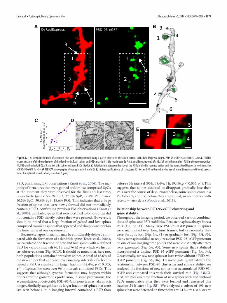
To further investigate the accuracy of the scoring criteria, weused single-cell electroporation of PSD-95-eGFP combined withfocused ion beam scanning electron microscopy (FIBSEM; Macoet al., 2013) to reconstruct a dendritic branch of an imaged PSD-95-GFP-expressing neuron that had PSD-95-eGFP-less protru-sions (Fig. 3A,E, #5 and #6) and small PSD-95-eGFP puncta justabove the detection threshold (Fig. 3A,E, #4). All protrusionsthat were seen in the 2PLSM image were found in the EMreconstruction (compare Fig. 3A,B), even thin spines with necksthinner than 85 nm (Fig. 3A,B, #5 and #6). Similarly, all PSD-95-eGFP clusters that we had defined as puncta represented postsyn-aptic densities in the electron micrographs (Fig. 3B–D), with thesmallest PSD-95-eGFP punctum being among the smallest PSDsthat can be observed in FIBSEM. The three protrusions that wehad defined as SpOs did not contain a visible postsynaptic densityin the EM. The brightness of the puncta displayed a linearrelationship with the size of the PSDs, as measured in the EMreconstruction (Fig. 3C). This indicates that, in vivo, 2PLSM ofPSD-95-eGFP reliably detects the smallest PSDs.
PSD-95-eGFP puncta appear and disappearTime-lapse imaging revealed many protrusions and PSD-95-eGFP puncta that were present over the whole experiment (n �302). However, as expected and similar to previous in vivo imagingstudies, some structures appeared or disappeared during the im-aging period. A total of 1–5% of the structures that were presentfrom the start until the end were classified as SpOs (1.7% at 0 h,5.0% at d17; Fig. 4A,B), whereas 78 – 82% were SpPs (81.8% at0 h, 78.4% at d17) and 16% were POs (16.6% at 0 h, 16.6% atd17). This indicates that persistent spines nearly always contain a
A B
C
Figure 2. A, Dendritic branch imaged at high magnification (scale bar, 5 �m). Three types of structure are visible: #1, a long thin spine without a PSD-95-eGFP punctum (SpO); #2, a long thin spinewith a PSD-95-GFP punctum (SpP); #3, a mushroom-like spine with a PSD-95-eGFP punctum (SpP); and #4, a PSD-95-eGFP punctum in the dendritic shaft (PO). For the sake of visualization, bothchannels are presented with different lookup tables. B, Procedure to determine the presence of PSD-95-eGFP puncta. PSD-95-eGFP fluorescence (right) in the spine head (gray arrowhead) wasdetermined by integrating pixel values over an area smaller than the spine head (SpG). This value was divided by the average of the integrated pixel values of several (3– 4) equally sized areas (DG)in the dendritic shaft (black arrowhead), carefully avoiding apparent PSD-95-eGFP puncta (open arrowhead) in the shaft. C, Left, Distribution of the fraction of PSD-95-eGFP bound in spine heads(n � 217) as measured according to Otmakhov et al. (2004). Right, Result of a k-means cluster analysis (k � 2) of the spine population from the left graph. Rapid puncta-based scoring results shownin color. Note that our scoring results nearly match the clustering. Arrowheads, two SpOs from Figure 3; arrow, smallest SpP from Figure 3.
2078 • J. Neurosci., February 5, 2014 • 34(6):2075–2086 Cane et al. • Postsynaptic Density Dynamics In Vivo
PSD, confirming EM observations (Knott et al., 2006). The ma-jority of structures that were gained and/or lost comprised SpOsat the moment they were observed for the first and last time,respectively (gains: 55.0% SpO, 27.2% SpP, 17.8% PO; losses:50.5% SpO, 30.9% SpP, 18.6% PO). This indicates that a largefraction of spines that were newly formed did not immediatelycontain a PSD, confirming previous EM observations (Knott etal., 2006). Similarly, spines that were destined to be lost often didnot contain a PSD shortly before they were pruned. However, itshould be noted that a large fraction of gained and lost spinescomprised transient spines that appeared and disappeared withinthe time frame of our experiment.
Because synapse formation may be considerably delayed com-pared with the formation of a dendritic spine (Knott et al., 2006),we calculated the fraction of new and lost spines with a definedPSD for various intervals (6, 18, and 96 h) over which we first orlast observed them (Fig. 4C,D). It should be noted that, here too,both populations contained transient spines. A total of 18.6% ofthe new spines that appeared over imaging intervals of 6 h con-tained a PSD. A significantly larger fraction (42.4%, p 0.005,� 2) of spines first seen over 96 h intervals contained PSDs. Thissuggests that although synapse formation may happen withinhours after the growth of a protrusion, in some protrusions, theaccumulation of detectable PSD-95-GFP puncta may take muchlonger. Similarly, a significantly larger fraction of spines that werelast seen before a 96 h imaging interval contained a PSD than
before a 6 h interval (96 h, 48.4%; 6 h, 19.4%, p 0.005, � 2). Thissuggests that spines destined to disappear gradually lose theirPSD over the course of days. Nonetheless, some spines contain aPSD shortly (hours) before they are pruned, in accordance withrecent in vitro data (Woods et al., 2011).
Relationship between PSD-95-eGFP clustering andspine stabilityThroughout the imaging period, we observed various combina-tions of spine and PSD stabilities. Persistent spines always bore aPSD (Fig. 5A, #1). Many large PSD-95-eGFP puncta in spineswere maintained over long time frames, but occasionally theywere abruptly lost (Fig. 5A, #1) or gradually lost (Fig. 5H, #5).Many new spines failed to acquire a clear PSD-95-eGFP punctumon one of our imaging time points and were lost shortly after theywere generated (Fig. 5A, #3). Some new spines that stabilizedincorporated a distinct PSD-95-eGFP punctum (Fig. 5A, #4).Occasionally, we saw new spines at least twice without a PSD-95-eGFP punctum (Fig. 5G, #6). To investigate quantitatively therelationship between PSD-95 clustering and spine stability, weanalyzed the fractions of new spines that accumulated PSD-95-eGFP and compared this with their survival rate (Fig. 5B,C).First, we measured the fraction of new spines with and withoutPSDs immediately after they were formed and reassessed thisfraction 24 h later (Fig. 5B). We analyzed a subset of 193 newspines that were detected on time point t � 24 h, t � 168 h, or t �
A B
C D
E
Figure 3. A, Dendritic branch of a neuron that was electroporated using a patch pipette in the adult cortex. Left, dsRedExpress. Right, PSD-95-eGFP (scale bar, 5 �m) B, FIBSEMreconstruction of the boxed region of the dendrite in A. All spines and PSDs match; #1, big mushroom SpP; #2, small mushroom SpP; #3, SpP with the smallest PSD in the reconstruction;#4, PSD on the shaft (PO); #5 and #6, thin spines without PSDs (SpOs). C, Relationship between the size of the PSDs in the EM reconstruction and the normalized fluorescence intensitiesof PSD-95-eGFP in vivo. D, FIBSEM micrographs of two spines (#1 and #2). E, High magnifications of structures #3, #4, and #5 in the red and green channel (images are filtered severaltimes for optimal visualization; scale bar, 1 �m).
Cane et al. • Postsynaptic Density Dynamics In Vivo J. Neurosci., February 5, 2014 • 34(6):2075–2086 • 2079

192 h and did not exist 24 h before these points (t � 0 h, 144 h,and 168 h, respectively). A total of 70.5% (136) of spines weredevoid of a detectable PSD-95-eGFP punctum. Out of this poolof spines, 17.6% (24 spines) were still present after 24 h. In thissubset of new spines, none had accumulated PSD-95-eGFP (butsee Fig. 5A, spine #4, for an example of a new spine with a delayedacquisition of a PSD). Conversely, 29.5% (57 spines) of the newspines contained a PSD-95-GFP punctum and 59.6% (34) ofthose were detected again 24 h later. All had maintained theirsynapses. As a result, the fraction of new spines with a PSD hadsignificantly increased over 24 h (59% at t � 24 h, 30% at t � 0 h,p 0.005, � 2). When we tracked new spines that were only up to6 h old (n � 161; Fig. 5C), we found that an even smaller fractioncontained a PSD-95-eGFP punctum (18.6%, n � 30), of whichonly a small portion survived over the next 18 h (33.3%, n � 10).A total of 14.5% of spines that did not contain a PSD-95-eGFPpunctum survived over the next 18 h (n � 19); 31.6% of these(n � 6) had incorporated a PSD. Therefore, of all of the new SpPsthat were observed at the second time point (n � 16), 37.5% hadincorporated PSD over the 18 h interval. This indicates that PSD-
95-eGFP accumulation in a new spine is not always immediateand may occur with a delay of �6 h. To assess the relationshipbetween the presence of PSD-95-eGFP and spine survival overlonger time frames, we grouped new spines that were seen in atleast two imaging sessions into two categories: those that wereseen over an interval 96 h (transient spines; Holtmaat et al.,2005) and those with a lifespan of �96 h (Fig. 5D). Out of thetotal number of new spines (721) that had appeared during theexperiment, the majority was seen only once (71.1%); 126(24.6%) of these spines had a PSD when they were detected. Asmall fraction of the new spines survived for �96 h (14.0%) andthe majority of those displayed a PSD at least once (83.2%; 84 of101). Of the remaining spines (14.9%; i.e., those that were seen atleast twice but survived for 96 h), a significantly lower fraction(64.5%; 69 of 107; p 0.005, � 2) had displayed a PSD punctumat least once. This shows that the presence of a PSD indicates ahigher probability for a new spine to stabilize. However, the ac-quisition of a PSD does not per se predict the stabilization of aspine. In fact, the majority of new spines bearing a PSD at leastonce disappear before they are 4 d old (69.9%).
Figure 4. A, D, Fraction of spines with a PSD-95-eGFP punctum (SpP, dark gray), spines without a PSD-95-eGFP punctum (SpO, light gray), and PSD-95-eGFP puncta in the dendriticshaft (PO, white). C, D, Fraction of new spines with a PSD-95-eGFP punctum (SpP, dark gray) and new spines without a PSD-95-eGFP punctum (SpO, light gray) for spines appearing over6, 18, and 96 h intervals (left) and spines disappearing over 6, 18, and 96 h intervals (right). A, C, Averages over mice (*p 0.05; ***p 0.005; paired t test). B, D, Structures from allmice pooled (***p 0.005, � 2).
2080 • J. Neurosci., February 5, 2014 • 34(6):2075–2086 Cane et al. • Postsynaptic Density Dynamics In Vivo

To investigate the interaction between the presence of PSD-95-eGFP puncta and spine loss, we selected a population of SpPsthat were present on the first imaging time point (t � 0 h) yetwere lost over the course of the experiment (after t � 6 h). Thelost spines were grouped into three categories: those last seenbefore an interval of 6, 18, and 96 h. Although the majority ofspines that disappeared still had a PSD-95-eGFP punctum on thelast time point (6 h, 65%; Fig. 5E), a substantial fraction had losttheir puncta before their disappearance (6 h, 35%; Fig. 5H, #5).The fraction of spines that had lost their PSD-95-eGFP punctumbefore their disappearance tended to decrease gradually with anincrease of the interval time, but we did not detect a significantdifference (Fig. 5E). This suggests that, in general, spine and syn-
apse disappearance happens concomitantly (over the course ofhours). However, some spines may persist for some time, evenafter they have lost their PSD (Fig. 5H). A total of 20% of thedisappearing spines survived for �96 h after they had lost theirPSD-95-eGFP punctum (Fig. 5F). This fraction is significantlyhigher compared with new spines that never accumulate a PSD-95-eGFP punctum (5%, p 0.005 � 2; Fig. 5F). This suggests that,once spines have acquired a PSD, their survival rate increaseseven when they lose their synapse.
Spine volume and PSD-95 size are linearly relatedEM studies have indicated that spine and synapse size are posi-tively correlated (Harris and Stevens, 1989; Nusser et al., 1998;
A
B C D G
E F H
Figure 5. A, Time-lapse image of a dendritic region containing the following: a SpP disappearing after day 13 (#1); a PO disappearing after the second imaging session on day 0 (#2); a transientSpO appearing and disappearing within 24 h after the first imaging session (#3); and a new spine appearing as a SpO on day 6 and turning into a SpP on day 7 (#4). Scale bar, 1 �m. B, Fraction ofnew spines (24 h old) that survives over the next 24 h. The fraction of new SpPs that is still present after 24 h is larger than the fraction of SpOs (***p 0.005). C, Fraction of new spines (6 hold) that survives over the next 18 h. The fraction of new SpPs that is still present after 18 h is larger than the fraction of SpOs (***p 0.005). D, New spines that show a PSD-95-eGFP punctum atleast once during their lifetime have a higher probability of surviving for �96 h (***p 0.005). E, Fractions of lost SpPs that maintain or lose their PSD-95-eGFP punctum before disappearance. F,Left, Fraction of new SpOs with lifetimes of more or 96 h. Right, Fraction of SpPs that continue as SpOs for more or 96 h before disappearing (***p 0.005). G, New SpO that is present overtwo time points (#6). Scale bar, 1 �m. H, Disappearing spine that loses its PSD-95-eGFP punctum before pruning (#5). Scale bar, 1 �m.
Cane et al. • Postsynaptic Density Dynamics In Vivo J. Neurosci., February 5, 2014 • 34(6):2075–2086 • 2081

Takumi et al., 1999; Holtmaat et al., 2005;Knott et al., 2006). Electrophysiology andimaging studies in vitro have confirmedthis finding (Matsuzaki et al., 2001; Zito etal., 2004). The FIBSEM reconstructionsshow that PSD size is proportional to thebrightness of PSD-95-eGFP in spines (Fig.3C). This suggests that spine brightness invivo is proportional to PSD-95-eGFPbrightness. In general, we observed thatbright spines in the red channel also con-tained high green fluorescence intensitylevels (Figs. 5A, 6A). Conversely, dimspines in the red channel contained lowgreen fluorescence intensities. To investi-gate quantitatively the relationship be-tween spine size and putative PSD size, weselected a random population of SpPs(n � 108) that were seen on the first im-aging session. It was shown previouslythat spine brightness, as measured in sin-gle 2PLSM optical sections, is monotoni-cally related to spine volume (Holtmaat etal., 2005; Mostany et al., 2013). Here, weassumed that the brightness of PSD-95-eGFP reliably represents the PSD size. Be-cause the absolute fluorescence levelsdiffered between animals and differentcells, we normalized the integrated redand green fluorescence intensity in the spine head to the mean ofthe maximum pixel values that we encountered in a 1 to 3 �mstretch of parent dendrite just below the spine (Fig. 6B). Bothgreen and red fluorescence intensities in spines followed a log-normal distribution (data not shown; Loewenstein et al., 2011). Aregression analysis revealed a linear relationship between red andgreen intensities in the spine head (R 2 � 0.79, r � 7.12; Fig. 6C).These data corroborate previous EM studies (Harris and Stevens,1989; Knott et al., 2006) and indicate that the expression of PSD-95-eGFP does not disturb the relationship between spine andPSD size.
Relationship between spine formation and PSD sizeTo investigate the interaction between PSD size and new spineformation, we measured the levels of PSD-95-eGFP and newspine volumes. We selected a random population of new spinesthat had appeared at any time point and analyzed their normal-ized spine head volume and PSD sizes on the first time point ofobservation (Fig. 6D,E). The population of new spines was di-vided into those that were classified as SpP (Fig. 6D, #2 and #3;Fig. 6E, blue dots) and SpO (Fig. 5D, #1; Fig. 6E, red dots). Asexpected, in the SpO subpopulation, the PSD-95-eGFP intensitymodestly increased with spine volume. In new SpPs, the slope ofthe interaction was steeper (rSpP � 2.38, range 2.31–2.46; rSpO �1.18, range 1.14 –1.22; robust regression analysis), but still shal-lower than for randomly selected SpPs. Therefore, the cytosolicvolumes and the PSD-95-eGFP levels of new spines that had ac-quired a PSD were significantly larger than the volumes of newspines without a PSD and their respective normalized cumulativedistributions were significantly different (Fig. 6F; dotted lines vsdotted lines, p 0.005; solid lines vs solid lines, p 0.005; KStest). These data indicate that, on average, young new spinescontain smaller PSDs relative to their head volume compared
with persistent spines. This may be indicative of a synaptic mat-uration process that lags spine enlargement.
PSD size changes correlate with spine volume changesSpine head volumes have been shown to fluctuate in vivo, even inpersistent spines (Holtmaat et al., 2005; Holtmaat et al., 2006;Hofer et al., 2009; Loewenstein et al., 2011). Although spine vol-ume and synapse size are tightly coupled, it could not be deducedfrom these experiments whether PSD sizes closely follow spinevolume changes. We estimated spine head volume and PSD sizealterations of persistent spines by measuring the ratio of fluores-cence intensities between two consecutive time points (intervalsof 6, 18, and 96 h) in both the green and red channel. This analysisconfirmed that spine head volumes fluctuate continuously. PSDsize changes and spine head volume changes could be fit with alinear function (r � 0.6; n � 1224; Fig. 6A), but the correlationbetween the magnitudes of the changes was poor (R 2 � 0.26). Ina small fraction (162 of 1224, 13.2%) of sampled spines, the PSDsize and spine head volume dynamics were anisometric; that is,the spine grew whereas the PSD shrunk or vice versa (Fig. 7A,quadrant II and IV). These types may represent spines that hadundergone synaptic weakening or strengthening shortly beforethey were imaged, because the initial phase (5–10 min) of long-term potentiation (LTP) induction shows relatively poor interac-tions between spine head size and synapse strength (Matsuzaki etal., 2004; Lee et al., 2009).
To investigate the relationship between spine stability andPSD size and spine head volume changes, we compared thechanges over two time points for new spines and lost spines withpersistent spines (Fig. 7B). A random set of new SpPs was selectedat various time points (from t � 6 h onward) and their size wasmeasured on the first and second time point on which they weredetected (Fig. 7D). Lost spines were defined as those that weredetected with a PSD-95-eGFP punctum on the first imaging day
Figure 6. A, Examples of SpPs with small (left) and large (right) volumes. B, Procedure for measurement of DsRedExpress andPSD-95-eGFP fluorescence intensities. In brightest optical section of each channel, background-corrected pixel intensities areintegrated over an area spanning the spine head (circles). Background pixel values are taken from nearby regions (rectangles). Ineach region of interest, spine values are normalized to the average maximum pixel values of neighboring areas in the dendriticshaft (jagged lines). C, Linear correlation between green and red fluorescence intensities in a random set of SpPs at t � 0 h. D, NewSpO (#1) and a new SpPs (#2 and #3). E, Linear fits of red and green fluorescence for new SpPs (blue), new SpOs (red), and spinesfrom C (black). Inset shows the smallest data points. F, Cumulative distribution of red (continuous lines) and green (dotted lines)fluorescence intensities for all spines from E and C. Scale bars, 1 �m.
2082 • J. Neurosci., February 5, 2014 • 34(6):2075–2086 Cane et al. • Postsynaptic Density Dynamics In Vivo

(t � 0 h) and were subsequently lost over the time course of theexperiment. Their sizes were measured on the last two timepoints at which they were detected. Despite numerous large fluc-tuations (e.g., �3-fold changes), on average, growth and shrink-age canceled out one another in persistent spines (PSD-95-eGFP,�0.048 0.015; DsRedExpress, �0.023 0.013, n � 1224; Fig.7B). This may be indicative of homeostatic processes that keepsynapse strength, and thereby PSD size, equal over time (Turri-giano and Nelson, 2004). Conversely, the log10 ratios were slightlyskewed toward positive values for new spines and toward nega-tive values for lost spines (new: PSD-95-eGFP, 0.064 0.042;DsRedExpress, 0.065 0.033; n � 386, p 0.005, t test betweenpersistent and new; lost: PSD-95-eGFP, �0.366 0.093; DsRe-dExpress, �0.298 0.068; n � 117, p 0.005, t test betweenpersistent and lost). To determine whether those types of spinesdisplayed different structural dynamics over their whole lifetime,we measured fluorescence changes in a randomly selected set ofimaging time points and intervals (Fig. 7E) and took the absolutevalue of the ratios between the two time points to assess thevariance in the changes. New and lost spines displayed increasedPSD size and spine head volume fluctuations, indicating thattheir synapses are generally less stable than those of persistentspines (persistent: PSD-95-eGFP, 0.393 0.010; DsRedExpress,0.311 0.008, n � 1224; new: PSD-95-eGFP, 0.533 0.058;DsRedExpress, 0.488 0.047; n � 37, p 0.005, t test; lost:PSD-95-eGFP, 0.481 0.027; DsRedExpress, 0.379 0.024; n �277, p 0.005, t test; Fig. 7C). The increased PSD dynamics maybe a signature of spines that are still in a growth process or aredestined to disappear.
DiscussionWe have characterized the relationship between PSD dynamicsand spine stability in real time in vivo. Our data show that theexpression of PSD-95-eGFP in vivo allows the identification ofthe smallest PSDs seen in EM (Fig. 3). Persistent spines almostalways bear a PSD (Figs. 4, 5). Two-thirds of the new spines andspines that are destined to be lost lack a clear PSD (Figs. 4, 5). Newspines that acquire a PSD are more likely to become persistent(Fig. 5). However, most PSD-bearing new spines are transient.Persistent spines that lose a PSD are likely to be pruned (Fig. 5).Furthermore, PSD size is proportional to spine head volume (Fig.
6). The PSD to head size ratio of new spines is lower than inpersistent spines, suggesting that they slowly increase their PSDcontent. Spines that are destined to be lost on average lose PSDcontent over hours to days before they are pruned (Fig. 7).
Auxiliary expression of PSD-95 and scoring ofPSD-95-eGFP punctaPSD-95 overexpression has been shown to increase LTP-likeplasticity and synapse maturation in neuronal cultures and orga-notypic brain slices (El-Husseini et al., 2000; Ehrlich and Mali-now, 2004; Ehrlich et al., 2007). Therefore, in our study, theauxiliary expression of PSD-95 may have altered synaptic physi-ology. However, several observations indicate that the disturbingeffects were minimal. We expressed PSD-95-eGFP in a similarway to Gray et al. (2006), in which spine densities and synapticconnectivity were not affected. Spine densities were similar toother in vivo imaging studies (Holtmaat et al., 2005; Gray et al.,2006; Hofer et al., 2009), and spine turnover rates were compa-rable to those in GFP-transgenic mice (Holtmaat et al., 2005;Wilbrecht et al., 2010). Furthermore, spine sizes and PSD sizesfollowed a log-normal distribution, similar to a recent imagingstudy in vivo (Loewenstein et al., 2011), and individual spines andPSDs alternately grew and shrunk over time. Many new spinesnever acquired PSDs or only expressed them transiently. Manypreviously persistent spines lost their PSDs before being pruned.Altogether, this confirms that the auxiliary expression of PSD-95did not cause a continuous growth of PSDs, did not massivelystabilize spines, and still allowed continuous spine and synapsepruning. This also suggests that the synaptic incorporation ofPSD-95 is a regulated process that is not readily inflated by aux-iliary expression, which holds true for various other synaptic pro-teins (Kessels et al., 2009).
In our study, the number of spines without a PSD was com-parable but slightly higher compared with some in vitro (De Rooet al., 2008) and EM studies (Knott et al., 2006; Arellano et al.,2007). Spine characteristics vary between cell and dendrite types(Nimchinsky et al., 2002). Therefore, a likely explanation is thedifference in neuron and dendrite populations that were analyzedin those studies. We are confident that our scoring criteria de-tected the smallest PSDs. The results were similar when we basedscoring criteria on a cluster analysis of the fraction of PSD-95-
A B C D
E
Figure 7. A, Relationship between changes in red and green fluorescence intensities. Dotted lines represent the measurement error. B, New spines display an increase in both green and redfluorescence over the first two time points after their appearance compared with persistent spines (***p 0.005). Lost spines display a decrease in both green and red fluorescence over the last twotime points before their disappearance compared with persistent spines (***p 0.005). C, Absolute value of fluorescence intensity ratios over a random set of two consecutive time points forpersistent, new, and preexisting disappearing spines. New and lost spines display increased fluctuations (***p 0.005). D, Example of a time interval used in B. E, Example of timeintervals used in C.
Cane et al. • Postsynaptic Density Dynamics In Vivo J. Neurosci., February 5, 2014 • 34(6):2075–2086 • 2083

eGFP that was bound in spine heads (Otmakhov et al., 2004).Furthermore, the EM reconstruction showed that the lowest flu-orescence intensities that we classified as puncta corresponded tothe smallest PSDs that can be seen at the ultrastructural level. Inaddition, it should also be noted that a large proportion of thespine population without a PSD represented very thin structures,which may often be overlooked in serial section EM. For example,the widths of some thin spines in our 2PLSM images that werereconstructed using FIBSEM were as little as 80 nm. These couldhave remained undetected without the information from the liveimaging that indicated their presence.
Relationship between spine and PSD formationThe majority of gained spines did not stabilize and did not ac-quire a distinct PSD. This corroborates EM studies that havedetected a lack of PSDs and/or synapses in a large fraction ofnewly formed spines (Knott et al., 2006; Nagerl et al., 2007). NewSpOs rarely acquired a PSD later on. However, we also observedan increasing fraction of new SpPs with increasing time intervals.This indicates that PSD-95 clustering is not always immediate,but can occur over hours and days after the growth of the parentspine. PSD growth showed similarities to in vitro studies. How-ever, they were quantitatively different from the fast and func-tional synapse formation seen in those experiments (Nagerl et al.,2007; Zito et al., 2009). There are several explanations. First, ourPSD scoring criteria may have resulted in a slight underestima-tion of synapse formation (see Results). Second, synaptic-likeactivity may occur even when PSDs are small or absent throughnonclustered NMDA and AMPA receptors that are juxtaposed topresynaptic boutons. Third, PSD formation may be slower in vivocompared with organotypic cultures. This could be due to differ-ences in circuit activity, synaptic inhibition, and neuromodula-tion. In our experiments, the dendrites were positioned in L1, aregion of the cortex that receives many modulatory and long-range connections, whereas most slice experiments focused onhippocampal pyramidal cells that lacked long-range modulatoryinputs and hormonal regulation. Hormonal oscillations andmodulatory inputs can have profound effects on synaptic plastic-ity and spine maturation (Jones et al., 2009; Liston et al., 2013).
Some new spines displayed a PSD yet did not stabilize for longperiods of time. However, they still persisted longer than spineswithout PSDs. This suggests that, in addition to the PSD, otherfactors determine the stabilization of the spine. This is not incon-ceivable because spine morphology is regulated through a vastsignaling network (Patterson and Yasuda, 2011). Because we didnot have insight into axonal bouton dynamics, we can also notexclude the possibility that some PSDs lacked a presynaptic coun-terpart. Spine and PSD formation might be induced by largequantities of presynaptic glutamate release or spillover (Richardset al., 2005; Kwon and Sabatini, 2011). However, these spines maynot always be able to contact the presynaptic site from whichglutamate was released. Alternatively, synapses in those spinesmay not be potentiated and therefore fail to stabilize, which maycause the spines to retract (Matsuzaki et al., 2004; Kopec et al.,2006; Harvey and Svoboda, 2007; Hill and Zito, 2013; Oh et al.,2013). Nevertheless, our data imply that transient SpPs may serveto provide short-term synaptic connections and could therebytemporarily influence network activity. This may be importantfor learning and downstream neuronal network plasticity (Xu etal., 2009; Yang et al., 2009).
Relationship between spine loss and PSD dynamicsPSD-95-eGFP levels in persistent spines fluctuated. We found aconcomitant decrease in spine volume and PSD-95-eGFP levelsbefore spines were pruned. This indicates that spine loss is asso-ciated with a gradual loss of PSD components. A gradual reduc-tion of spine volumes before spine pruning was also observedduring the experience dependent loss of persistent spines (Holt-maat et al., 2006). These observations fit well with intrinsic spinevolume changes in organotypic brain slices (Yasumatsu et al.,2008) and multiplicative dynamics in vivo (Loewenstein et al.,2011). The results may indicate that the maintenance of a spinerelies on PSD size. New spines that do not acquire enough PSDmolecules may disappear and old spines that lose too many maybe pruned. The loss of PSD components likely depends on long-term depression (Nagerl et al., 2004; Zhou et al., 2004; Oh et al.,2013). Synaptic adhesion molecules that are linked to PSDs maybe key in the maintenance of a synapse (Sheng and Hoogenraad,2007; Han and Kim, 2008). Once the PSD size is reduced below acritical point, adhesion to the presynaptic bouton may becomeinsufficient to maintain the synaptic connection. Spines withouta synapse may spontaneously disappear due to a lack of filamen-tous actin-anchoring points (Sekino et al., 2007; Soria Fregozoand Perez Vega, 2012). Conversely, we did observe persistentspines that temporarily existed without a PSD before being lost(Fig. 5H). We cannot exclude the possibility that, in some cases,the spine was lost and replaced by a new spine that had not (yet)acquired a PSD. Nonetheless, the result may indicate that some,perhaps nonsynaptic, adhesion molecules temporarily keep thespine head linked to the presynaptic bouton after the completeremoval of the PSD.
Similar to Woods et al. (2011), we found that the loss of PSDenrichment is not a prerequisite for spine loss. Many spines dis-played normal PSD-95-GFP levels at the last time point of obser-vation. This may indicate that spines can be very rapidly prunedafter the loss of a synapse. An interesting alternative is that activespine pruning in some cases strips the synaptic contact, a processthat could be modulated by nonsynaptic mechanisms. However,it should be noted that most of our time intervals were muchlonger than in the in vitro studies and we may not have capturedrapid PSD decline before spine pruning.
ReferencesArellano JI, Espinosa A, Fairen A, Yuste R, DeFelipe J (2007) Non-synaptic
dendritic spines in neocortex. Neuroscience 145:464 – 469. CrossRefMedline
Cardona A, Saalfeld S, Schindelin J, Arganda-Carreras I, Preibisch S, LongairM, Tomancak P, Hartenstein V, Douglas RJ (2012) TrakEM2 software forneural circuit reconstruction. PLoS One 7:e38011. CrossRef Medline
Chen X, Vinade L, Leapman RD, Petersen JD, Nakagawa T, Phillips TM,Sheng M, Reese TS (2005) Mass of the postsynaptic density and enu-meration of three key molecules. Proc Natl Acad Sci U S A 102:11551–11556. CrossRef Medline
Chen X, Nelson CD, Li X, Winters CA, Azzam R, Sousa AA, Leapman RD,Gainer H, Sheng M, Reese TS (2011) PSD-95 is required to sustain themolecular organization of the postsynaptic density. J Neurosci 31:6329 –6338. CrossRef Medline
Craven SE, El-Husseini AE, Bredt DS (1999) Synaptic targeting of the post-synaptic density protein PSD-95 mediated by lipid and protein motifs.Neuron 22:497–509. CrossRef Medline
De Roo M, Klauser P, Mendez P, Poglia L, Muller D (2008) Activity-dependent PSD formation and stabilization of newly formed spines inhippocampal slice cultures. Cereb Cortex 18:151–161. CrossRef Medline
Ehrlich I, Malinow R (2004) Postsynaptic density 95 controls AMPA recep-tor incorporation during long-term potentiation and experience-drivensynaptic plasticity. J Neurosci 24:916 –927. Medline
Ehrlich I, Klein M, Rumpel S, Malinow R (2007) PSD-95 is required for
2084 • J. Neurosci., February 5, 2014 • 34(6):2075–2086 Cane et al. • Postsynaptic Density Dynamics In Vivo

activity-driven synapse stabilization. Proc Natl Acad Sci U S A 104:4176 –4181. CrossRef Medline
El-Husseini AE, Schnell E, Chetkovich DM, Nicoll RA, Bredt DS (2000)PSD-95 involvement in maturation of excitatory synapses. Science 290:1364 –1368. CrossRef Medline
Fu M, Zuo Y (2011) Experience-dependent structural plasticity in the cor-tex. Trends Neurosci 34:177–187. CrossRef Medline
Gray NW, Weimer RM, Bureau I, Svoboda K (2006) Rapid redistribution ofsynaptic PSD-95 in the neocortex in vivo. PLoS Biol 4:e370. CrossRefMedline
Grillo FW, Song S, Teles-Grilo Ruivo LM, Huang L, Gao G, Knott GW, MacoB, Ferretti V, Thompson D, Little GE, De Paola V (2013) Increased ax-onal bouton dynamics in the aging mouse cortex. Proc Natl Acad SciU S A 110:E1514 –1523. CrossRef Medline
Han K, Kim E (2008) Synaptic adhesion molecules and PSD-95. Progress inneurobiology 84:263–283. CrossRef Medline
Harris KM, Stevens JK (1989) Dendritic spines of CA1 pyramidal cells in therat hippocampus: serial electron microscopy with reference to their bio-physical characterisitcs. J Neurosci 9:2982–2997. Medline
Harvey CD, Svoboda K (2007) Locally dynamic synaptic learning rules inpyramidal neuron dendrites. Nature 450:1195–1200. CrossRef Medline
Hill TC, Zito K (2013) LTP-induced long-term stabilization of individualnascent dendritic spines. J Neurosci 33:678 – 686. CrossRef Medline
Hofer SB, Mrsic-Flogel TD, Bonhoeffer T, Hubener M (2009) Experienceleaves a lasting structural trace in cortical circuits. Nature 457:313–317.CrossRef Medline
Holtmaat A, Svoboda K (2009) Experience-dependent structural synapticplasticity in the mammalian brain. Nat Rev Neurosci 10:647– 658.CrossRef Medline
Holtmaat AJ, Trachtenberg JT, Wilbrecht L, Shepherd GM, Zhang X, KnottGW, Svoboda K (2005) Transient and persistent dendritic spines in theneocortex in vivo. Neuron 45:279 –291. CrossRef Medline
Holtmaat A, Wilbrecht L, Knott GW, Welker E, Svoboda K (2006)Experience-dependent and cell-type-specific spine growth in the neocor-tex. Nature 441:979 –983. CrossRef Medline
Holtmaat A, Bonhoeffer T, Chow DK, Chuckowree J, De Paola V, Hofer SB,Hubener M, Keck T, Knott G, Lee WC, Mostany R, Mrsic-Flogel TD,Nedivi E, Portera-Cailliau C, Svoboda K, Trachtenberg JT, Wilbrecht L(2009) Long-term, high-resolution imaging in the mouse neocortexthrough a chronic cranial window. Nat Protoc 4:1128 –1144. CrossRefMedline
Jones KA, Srivastava DP, Allen JA, Strachan RT, Roth BL, Penzes P (2009)Rapid modulation of spine morphology by the 5-HT2A serotonin recep-tor through kalirin-7 signaling. Proc Natl Acad Sci U S A 106:19575–19580. CrossRef Medline
Judkewitz B, Rizzi M, Kitamura K, Hausser M (2009) Targeted single-cellelectroporation of mammalian neurons in vivo. Nat Protoc 4:862– 869.CrossRef Medline
Kelsch W, Lin CW, Lois C (2008) Sequential development of synapses indendritic domains during adult neurogenesis. Proc Natl Acad Sci U S A105:16803–16808. CrossRef Medline
Kessels HW, Kopec CD, Klein ME, Malinow R (2009) Roles of stargazin andphosphorylation in the control of AMPA receptor subcellular distribu-tion. Nat Neurosci 12:888 – 896. CrossRef Medline
Knott GW, Holtmaat A, Wilbrecht L, Welker E, Svoboda K (2006) Spinegrowth precedes synapse formation in the adult neocortex in vivo. NatNeurosci 9:1117–1124. CrossRef Medline
Kopec CD, Li B, Wei W, Boehm J, Malinow R (2006) Glutamate receptorexocytosis and spine enlargement during chemically induced long-termpotentiation. J Neurosci 26:2000 –2009. CrossRef Medline
Kwon HB, Sabatini BL (2011) Glutamate induces de novo growth of func-tional spines in developing cortex. Nature 474:100 –104. CrossRefMedline
Lee SJ, Escobedo-Lozoya Y, Szatmari EM, Yasuda R (2009) Activation ofCaMKII in single dendritic spines during long-term potentiation. Nature458:299 –304. CrossRef Medline
Liston C, Cichon JM, Jeanneteau F, Jia Z, Chao MV, Gan WB (2013) Circa-dian glucocorticoid oscillations promote learning-dependent synapseformation and maintenance. Nat Neurosci 16:698 –705. CrossRefMedline
Livneh Y, Feinstein N, Klein M, Mizrahi A (2009) Sensory input enhances
synaptogenesis of adult-born neurons. J Neurosci 29:86 –97. CrossRefMedline
Loewenstein Y, Kuras A, Rumpel S (2011) Multiplicative dynamics underliethe emergence of the log-normal distribution of spine sizes in the neocor-tex in vivo. J Neurosci 31:9481–9488. CrossRef Medline
Maco B, Holtmaat A, Cantoni M, Kreshuk A, Straehle CN, Hamprecht FA,Knott GW (2013) Correlative in vivo 2 photon and focused ion beamscanning electron microscopy of cortical neurons. PloS One 8:e57405.CrossRef Medline
Matsuzaki M, Ellis-Davies GC, Nemoto T, Miyashita Y, Iino M, Kasai H(2001) Dendritic spine geometry is critical for AMPA receptor expres-sion in hippocampal CA1 pyramidal neurons. Nat Neurosci 4:1086 –1092. CrossRef Medline
Matsuzaki M, Honkura N, Ellis-Davies GC, Kasai H (2004) Structural basisof long-term potentiation in single dendritic spines. Nature 429:761–766.CrossRef Medline
Mostany R, Anstey JE, Crump KL, Maco B, Knott G, Portera-Cailliau C(2013) Altered synaptic dynamics during normal brain aging. J Neurosci33:4094 – 4104. CrossRef Medline
Nagerl UV, Eberhorn N, Cambridge SB, Bonhoeffer T (2004) Bidirectionalactivity-dependent morphological plasticity in hippocampal neurons.Neuron 44:759 –767. CrossRef Medline
Nagerl UV, Kostinger G, Anderson JC, Martin KA, Bonhoeffer T (2007)Protracted synaptogenesis after activity-dependent spinogenesis in hip-pocampal neurons. J Neurosci 27:8149 – 8156. CrossRef Medline
Niell CM, Meyer MP, Smith SJ (2004) In vivo imaging of synapse formationon a growing dendritic arbor. Nat Neurosci 7:254 –260. CrossRef Medline
Nimchinsky EA, Sabatini BL, Svoboda K (2002) Structure and function ofdendritic spines. Annu Rev Physiol 64:313–353. CrossRef Medline
Nusser Z, Lujan R, Laube G, Roberts JD, Molnar E, Somogyi P (1998) Celltype and pathway dependence of synaptic AMPA receptor number andvariability in the hippocampus. Neuron 21:545–559. CrossRef Medline
Oh WC, Hill TC, Zito K (2013) Synapse-specific and size-dependent mech-anisms of spine structural plasticity accompanying synaptic weakening.Proc Natl Acad Sci U S A 110:E305–312. CrossRef Medline
Okabe S, Kim HD, Miwa A, Kuriu T, Okado H (1999) Continual remodel-ing of postsynaptic density and its regulation by synaptic activity. NatNeurosci 2:804 – 811. CrossRef Medline
Okabe S, Miwa A, Okado H (2001) Spine formation and correlated assem-bly of presynaptic and postsynaptic molecules. J Neurosci 21:6105– 6114.Medline
Otmakhov N, Tao-Cheng JH, Carpenter S, Asrican B, Dosemeci A, Reese TS,Lisman J (2004) Persistent accumulation of calcium/calmodulin-dependent protein kinase II in dendritic spines after induction of NMDAreceptor-dependent chemical long-term potentiation. J Neurosci 24:9324 –9331. Medline
Patterson M, Yasuda R (2011) Signalling pathways underlying structuralplasticity of dendritic spines. Br J Pharmacol 163:1626 –1638. CrossRefMedline
Pologruto TA, Sabatini BL, Svoboda K (2003) ScanImage: Flexible softwarefor operating laser-scanning microscopes. BioMedical Engineering On-Line 2:13. Medline
Prange O, Murphy TH (2001) Modular transport of postsynapticdensity-95 clusters and association with stable spine precursors duringearly development of cortical neurons. J Neurosci 21:9325–9333.Medline
Richards DA, Mateos JM, Hugel S, de Paola V, Caroni P, Gahwiler BH,McKinney RA (2005) Glutamate induces the rapid formation of spinehead protrusions in hippocampal slice cultures. Proc Natl Acad Sci U S A102:6166 – 6171. CrossRef Medline
Saito T, Nakatsuji N (2001) Efficient gene transfer into the embryonicmouse brain using in vivo electroporation. Dev Biol 240:237–246.CrossRef Medline
Sala C, Futai K, Yamamoto K, Worley PF, Hayashi Y, Sheng M (2003)Inhibition of dendritic spine morphogenesis and synaptic transmis-sion by activity-inducible protein Homer1a. J Neurosci 23:6327– 6337.Medline
Sekino Y, Kojima N, Shirao T (2007) Role of actin cytoskeleton in den-dritic spine morphogenesis. Neurochem Int 51:92–104. CrossRefMedline
Sharma K, Fong DK, Craig AM (2006) Postsynaptic protein mobility in
Cane et al. • Postsynaptic Density Dynamics In Vivo J. Neurosci., February 5, 2014 • 34(6):2075–2086 • 2085

dendritic spines: long-term regulation by synaptic NMDA receptor acti-vation. Mol Cell Neurosci 31:702–712. CrossRef Medline
Sheng M, Hoogenraad CC (2007) The postsynaptic architecture of excit-atory synapses: a more quantitative view. Annu Rev Biochem 76:823– 847.CrossRef Medline
Soria Fregozo C, Perez Vega MI (2012) Actin-binding proteins and signal-ling pathways associated with the formation and maintenance of den-dritic spines. Neurologia 27:421– 431. Medline
Tabata H, Nakajima K (2001) Efficient in utero gene transfer system tothe developing mouse brain using electroporation: visualization ofneuronal migration in the developing cortex. Neuroscience 103:865–872. CrossRef Medline
Takumi Y, Ramírez-Leon V, Laake P, Rinvik E, Ottersen OP (1999) Differ-ent modes of expression of AMPA and NMDA receptors in hippocampalsynapses. Nat Neurosci 2:618 – 624. CrossRef Medline
Turrigiano GG, Nelson SB (2004) Homeostatic plasticity in the developingnervous system. Nat Rev Neurosci 5:97–107. CrossRef Medline
Wilbrecht L, Holtmaat A, Wright N, Fox K, Svoboda K (2010) Structuralplasticity underlies experience-dependent functional plasticity of corticalcircuits. J Neurosci 30:4927– 4932. CrossRef Medline
Woods GF, Oh WC, Boudewyn LC, Mikula SK, Zito K (2011) Loss of
PSD-95 enrichment is not a prerequisite for spine retraction. J Neurosci31:12129 –12138. CrossRef Medline
Xu T, Yu X, Perlik AJ, Tobin WF, Zweig JA, Tennant K, Jones T, Zuo Y(2009) Rapid formation and selective stabilization of synapses for endur-ing motor memories. Nature 462:915–919. CrossRef Medline
Yang G, Pan F, Gan WB (2009) Stably maintained dendritic spines are as-sociated with lifelong memories. Nature 462:920 –924. CrossRef Medline
Yasumatsu N, Matsuzaki M, Miyazaki T, Noguchi J, Kasai H (2008) Princi-ples of long-term dynamics of dendritic spines. J Neurosci 28:13592–13608. CrossRef Medline
Zhou Q, Homma KJ, Poo MM (2004) Shrinkage of dendritic spines associ-ated with long-term depression of hippocampal synapses. Neuron 44:749 –757. CrossRef Medline
Zito K, Knott G, Shepherd GM, Shenolikar S, Svoboda K (2004) Inductionof spine growth and synapse formation by regulation of the spine actincytoskeleton. Neuron 44:321–334. CrossRef Medline
Zito K, Scheuss V, Knott G, Hill T, Svoboda K (2009) Rapid functionalmaturation of nascent dendritic spines. Neuron 61:247–258. CrossRefMedline
Zuo Y, Lin A, Chang P, Gan WB (2005) Development of long-term den-dritic spine stability in diverse regions of cerebral cortex. Neuron 46:181–189. CrossRef Medline
2086 • J. Neurosci., February 5, 2014 • 34(6):2075–2086 Cane et al. • Postsynaptic Density Dynamics In Vivo