Spike timing dependent plasticity Homeostatic regulation of synaptic plasticity.
Development/Plasticity/Repair
Perineurial Glia Are Essential for Motor Axon Regrowthfollowing Nerve Injury
Gwendolyn M. Lewis and X Sarah KucenasDepartment of Biology, University of Virginia, Charlottesville, Virginia 22904
Development and maintenance of the peripheral nervous system (PNS) are essential for an organism to survive and reproduce, anddamage to the PNS by disease or injury is often debilitating. Remarkably, the nerves of the PNS are capable of regenerating after trauma.However, full functional recovery after nerve injuries remains poor. Peripheral nerve regeneration has been studied extensively, withparticular emphasis on elucidating the roles of Schwann cells and macrophages during degeneration and subsequent regeneration. Incontrast, the roles of other essential nerve components, including perineurial glia, are poorly understood. Here, we use laser nervetransection and in vivo, time-lapse imaging in zebrafish to investigate the role and requirement of perineurial glia after nerve injury. Weshow that perineurial glia respond rapidly and dynamically to nerve transections by extending processes into injury sites and phagocy-tizing debris. Perineurial glia also bridge injury gaps before Schwann cells and axons, and we demonstrate that these bridges are essentialfor axon regrowth. Additionally, we show that perineurial glia and macrophages spatially coordinate early debris clearance and thatperineurial glia require Schwann cells for their attraction to injury sites. This work highlights the complex nature of cell– cell interactionsafter injury and introduces perineurial glia as integral players in the regenerative process.
Key words: motor nerve; nerve injury; perineurial glia; regeneration; Schwann cells; zebrafish
IntroductionIn all vertebrate peripheral nerves, axon–Schwann cell complexesare bundled into fascicles by the perineurium (see Fig. 1A). Themature perineurium is a cellular sheath formed by concentricallyarranged layers of flattened, interdigitated perineurial cells thatoriginate in the CNS as glia (Akert et al., 1976; Schmidt et al.,1997; Kucenas et al., 2008; Clark et al., 2014). These cells functionto protect the nerve and regulate the transport of material be-tween the interior and exterior of the fascicle (Burkel, 1967;Bourne, 1968; Kristensson and Olsson, 1971; Akert et al., 1976).Previously, we showed that perineurial glia play an essential rolein nerve development (Binari et al., 2013; Kucenas et al., 2008;Clark et al., 2014). However, how they respond to nerve injuryand participate in regeneration is poorly understood.
Immediately after a nerve transection, proximal and distalstump axons begin to fragment and degenerate back from theinjury site in a process called acute axonal degeneration (AAD)(Kerschensteiner et al., 2005). Distal stump axons then begin
Wallerian degeneration, which is characterized by a latent periodfollowed by distal axon fragmentation (Waller, 1850; George etal., 1995; Villegas et al., 2012). During these degenerative pro-cesses, Schwann cells rapidly transdifferentiate, phagocytize theirown myelin, and recruit macrophages to clear debris (Perry et al.,1995; Arthur-Farraj et al., 2012). As regeneration begins, a tissuebridge forms to connect the proximal and distal stumps.Schwann cells then infiltrate this bridge, organize into cellularcolumns, and help guide newly regrowing axons across the gapand back to their original targets (Nguyen et al., 2002; McDonaldand Zochodne, 2003; McDonald et al., 2006; Parrinello et al.,2010; for review, see Chen et al., 2007; Vargas and Barres, 2007;Wang et al., 2012). The role of the perineurium throughout theseprocesses is poorly understood, although several studies have hy-pothesized that the initial tissue bridge could be perineurial inorigin (Scaravilli, 1984; Schroder et al., 1993). In support of thishypothesis, electron microscopy studies show that axons regrowthrough tracts containing both Schwann cell and perineurial la-mellae (Thomas and Jones, 1967; Morris et al., 1972). However,no study has ever focused on perineurial glial behavior after in-jury, and it is not known how these cells coordinate with other celltypes or whether they are required for axon regrowth.
Here we use laser axotomy and in vivo, time-lapse confocalimaging to transect spinal motor nerves in live transgenic ze-brafish and visualize perineurial glial responses after injury. Weshow that perineurial glia respond immediately to nerve transec-tion by extending processes toward the injury site and phagocy-tizing debris. Perineurial processes bridge the proximal and distalstumps; and in the absence of these bridges, axon regrowth isimpaired. Additionally, we show that debris clearance appears tobe coordinated between perineurial glia, macrophages, and
Received May 12, 2014; revised July 25, 2014; accepted Aug. 11, 2014.Author contributions: G.M.L. and S.K. designed research; G.M.L. performed research; G.M.L. contributed unpub-
lished reagents/analytic tools; G.M.L. and S.K. analyzed data; G.M.L. and S.K. wrote the paper.This work was supported by the National Institutes of Health Grant NS072212 to S.K., Grant NS051140 to G.M.L.,
and the University of Virginia Fund for Excellence in Science and Technology to S.K. We thank members of the S.K.laboratory for valuable discussions; Lori Tocke for zebrafish care; Travis Lewis for help with diagrammatic represen-tations; and Dr. Michael Granato for sharing the following fish lines: Tg(NBT:dsred)zf148, colourless (clstw11), Tg(mnx1:Wlds-GFP)p160, and Tg(spi1:Gal4,UAS:EGFP)zf149.
The authors declare no competing financial interests.Correspondence should be addressed to Dr. Sarah Kucenas, Department of Biology, Physical & Life Sciences
Building, Room 312, PO Box 400328, University of Virginia, Charlottesville, VA 22904-4328. E-mail:[email protected].
DOI:10.1523/JNEUROSCI.1906-14.2014Copyright © 2014 the authors 0270-6474/14/3412762-16$15.00/0
12762 • The Journal of Neuroscience, September 17, 2014 • 34(38):12762–12777

Schwann cells and that perineurial glial attraction to injury sites isdependent on Schwann cells. These data reveal an essential rolefor perineurial glia during regeneration and demonstrate the im-portance of Schwann cell–perineurial glial interactions afterinjury.
Materials and MethodsFish husbandry. All animal studies were approved by the Universityof Virginia Institutional Animal Care and Use Committee. Zebrafishstrains used in this study included Tg(nkx2.2a:megfp)vu17 (Kirby et al.,2006; Kucenas et al., 2008), Tg(olig2: dsred2)vu19 (Kucenas et al., 2008),Tg(mpeg1:EGFP) (Ellett et al., 2011), Tg(sox10:eos) (Prendergast et al.,2012), Tg(NBT:DsRed) (Peri and Nusslein-Volhard, 2008), Tg(spi1:Gal4,UAS:EGFP)zf149 (Peri and Nusslein-Volhard, 2008), Tg(mnx1:Mmu.Wlds-GFP) (Rosenberg et al., 2012), and colourlesstw11 (sox10!/!)(Kelsh et al., 1996). Table 1 describes the expression and abbreviations ofall of these lines. Embryos were produced by pairwise matings, raised at28.5°C in egg water, staged according to hours or days post fertilization(hpf and dpf, respectively) and embryos of either sex were used for allexperiments described below (Kimmel et al., 1995). Embryos used forimmunohistochemistry and microscopy were treated with 0.003% phe-nylthiourea (PTU) in egg water to reduce pigmentation.
In vivo imaging. At 24 hpf, all embryos used for live imaging weremanually dechorionated and transferred to egg water containing PTU toblock pigment formation. At specified stages, embryos were anesthetizedusing 3-aminobenzoic acid ester (Tricaine), immersed in 0.8% low-melting point agarose, and mounted on their sides in glass-bottomed 35mm Petri dishes (Electron Microscopy Sciences). Images were capturedusing either 40" (NA 1.2) or 63" (NA 1.2) water-immersion objectivesmounted on a motorized Zeiss AxioObserver Z1 microscope equippedwith a Quorum WaveFX-X1 spinning disc confocal system (QuorumTechnologies). For time-lapse imaging, z-image stacks were collectedevery 5 min for 3– 4 h, and 3D datasets were compiled using Sorenson 3video compression (Sorenson Media) and exported to QuickTime (Ap-ple) to create movies. Image adjustments were limited to contrast en-hancement and level settings using MetaMorph software and ImageJ.
DAPT treatment. Stock solutions for DAPT (565784; N-[(3,5-difluorophenyl)acetyl]-L-alanyl-2-phenyl]glycine-1,1-dimethylethyl es-ter; EMD Chemicals) were made in DMSO and used at 100 !M aspreviously described (Binari et al., 2013). DAPT was diluted in PTU eggwater plus DMSO to a final concentration of 1% DMSO and mixed byvortexing. Larvae were placed in 3 ml of PTU egg water containing DAPTin 35 mm Petri dishes from 48 to 72 hpf. Larvae were then washed in PTUegg water and moved to fresh PTU until 6 dpf for experiments. Controllarvae were placed in 3 ml of PTU egg water containing 1% DMSO alonefrom 48 to 72 hpf, washed in PTU egg water, and then moved to freshPTU until 6 dpf for experiments.
LysoTracker staining. To label organelles with low pH, 4 dpf larvae wereimmersed in 20 !M LysoTracker Red DND-99 (Invitrogen)/2% DMSO
or 10 !M LysoTracker Deep Red (Invitrogen)/1% DMSO in egg water for2 h at 23°C. Larvae were then rinsed once with egg water and immediatelymounted for imaging.
Immunohistochemistry. Larvae were fixed in AB Fix (4% PFA, 0.1%Triton X-100, 1" PBS) for 3 h at 23°C, followed by a 5 min wash withPBSTx (1% Triton X-100, 1" PBS), a 5 min wash with DWTx (1%Triton X-100 in distilled water), a 5 min wash with acetone at 23°C, anda 10 min wash with acetone at !20°C. Larvae were preblocked in 5% goatserum/PBSTx for 1 h, then incubated in primary antibody for 1 h at 23°Cand overnight at 4°C. The primary antibodies used included mouse anti-acetylated tubulin (1:5000, Sigma), rabbit antibody to Sox10 (1:5000)(Binari et al., 2013), mouse antibody to ZO-1 (zona occludins 1; 1:200,Invitrogen), and rabbit antibody to MBP (1:250) (Kucenas et al., 2009).Larvae were washed extensively with 1" PBSTx and incubated in sec-ondary antibody for 1 h at 23°C and overnight at 4°C. Secondary anti-bodies used were AlexaFluor-488 goat anti-rabbit, AlexaFluor-568 goatanti-rabbit, or AlexaFluor-568 goat anti-mouse, AlexaFluor-647 goatanti-rabbit and AlexaFluor-647 goat anti-mouse (Invitrogen). Larvaewere washed extensively with 1" PBSTx and stored in 50% glycerol/PBSat 4°C until imaging. Larvae were mounted on their sides in 0.8% low-melting point agarose on glass-bottomed 35 mm Petri dishes and imagedusing the confocal microscope described above. Image adjustments werelimited to contrast enhancement and level settings using MetaMorphsoftware and ImageJ.
Data quantification and statistical analysis. All graphically presenteddata represent the mean of the analyzed data. Statistical analyses andgraphing were performed with GraphPad Prism software. The level ofsignificance was determined by using an unpaired t test using a confi-dence interval of 95%. For Figure 4E, we ran a z-test to compare twoproportions using independent samples and compared the proportion ofnerves exhibiting “no regrowth” between each group. We again used aconfidence interval of 95% to determine significance.
Nerve transection. Nerve transections were performed using a Micro-Point Laser (Andor Technology) as previously published (Rosenberg etal., 2012; Banerjee et al., 2013; Binari et al., 2013; Lewis and Kucenas,2013), attached to a spinning disk confocal system (Quorum Technolo-gies) consisting of a nitrogen-pumped dye laser (wavelength 435 nm)controlled by MetaMorph Version 7.7. Ablation laser settings rangedfrom power 15 to 20 depending on the age of the larvae and nerve posi-tion. One or two motor nerves per larva in hemisegments 10 –16 weretransected in all experiments. To transect nerves, a thin elliptical ROI wasdrawn digitally in MetaMorph over the image of the nerve, and the nervewas laser pulsed precisely within the ROI until the nerve appeared tran-sected, whereby fluorescence did not refill the ROI in #10 s. Successfultransections with this protocol were independently confirmed by identi-fying the presence of axonal degeneration in transgenic lines that labelmotor axons (data not shown). In vivo imaging of transected nerves wasperformed as described above. For multiday experiments, larvae weredemounted immediately after injury, allowed to recover in PTU, andremounted for imaging at 24 and/or 48 h after injury.
Transmission electron microscopy. Larvae were prepared for electronmicroscopy using microwave fixation (Panasonic Model #NNSD967S)and were kept on ice at $15°C throughout all microwave steps to preventsample heating. The 6 dpf larvae were killed with Tricaine and fixed in 2%glutaraldehyde/4% PFA in 0.1 M PB. Samples were pulsed in the micro-wave, then kept in fixative for 2 h at 23°C. Samples were washed in 0.1 M
PB for 30 min, transferred to 1% OsO4 in 0.1 M PB, pulsed in the micro-wave, then left for 1 h at 23°C. Samples were washed in 0.1 M PB for 30min, followed by 5 min rinses in 50% and 70% EtOH. Preparations werestained en bloc with 4% uranyl acetate in 70% EtOH and pulsed in themicrowave. Samples were then subjected to ethanol dehydration andpulsed in the microwave after each change in solution: 70% ethanol, 95%ethanol, and 100% ethanol twice. Preparations were transferred to 100%acetone, pulsed in the microwave, changed to new 100% acetone,and pulsed in the microwave again. Samples were put in a 1:1acetone/EMBED mix and left overnight at 23°C, then changed to 100%EMBED and left uncapped for 6 h to allow evaporation of any remainingacetone. Finally, samples were placed into fresh EMBED and left to cureovernight in a 60°C oven. Ultrathin sections (70 nm) were obtained on a
Table 1. Descriptions and abbreviations of transgenic lines used in this studya
Transgene name Abbreviation Description of expression
Tg(nkx2.2a:megfp)vu1 nkx2.2a:gfp Membrane EGFP in nkx2.2a% cells (perineur-ial glia, floorplate)
Tg(olig2: dsred2)vu19 olig2:dsred Cytosolic DsRed in olig2% cells (motor neu-rons/axons)
Tg(mpeg1:EGFP) mpeg1:gfp Cytosolic EGFP in mpeg1% cells (macro-phages)
Tg(sox10:Eos) sox10:eos Cytosolic photoconvertible Eos in sox10% cells(Schwann cells)
Tg(NBT:DsRed) nbt:dsred Cytosolic DsRed in nbt% cells (neurons/axons)Tg(spi1:Gal4,UAS:EGFP)zf149 spi1:gfp Cytosolic EGFP in spi1% cells (leukocytes, micro-
glia)Tg(mnx1:Mmu.Wlds-GFP) mnx1:wlds-gfp Wld s and nuclear GFP in mnx% cells (motor
neurons/axons)aAll lines used were stable, germline transgenics. Cell types listed for each transgene are only those pertinent to thisstudy.
Lewis and Kucenas • Perineurial Glia Are Required for Nerve Regeneration J. Neurosci., September 17, 2014 • 34(38):12762–12777 • 12763

Leica UCT ultramicrotome, transferred to copper grids, and counter-stained with uranyl acetate. Sections were visualized on a JEOL 1010Transmission electron microscope and imaged with a 16 megapixel SIA-12C camera (Scientific Instruments and Applications).
Morpholino injections. The spi1b translation blocking morpholinooligonucleotide (MO) (5&-GATATACTGATACTCCATTGGTGGT-3&)(Rhodes et al., 2005; Villegas et al., 2012) was purchased from GeneTools. The MO was dissolved in water to create a stock solution of 2 mM
and diluted in water and Phenol red to create a working injection con-centration of 0.5 mM. We injected 2– 4 nl into the yolk just below thesingle cell of fertilized embryos. All MO-injected embryos were raised inPTU embryo medium at 28.5°C.
ResultsThe zebrafish spinal motor nerve as a model for studying glialbehaviors during regenerationTo investigate the role and requirement of perineurial glia duringdegeneration and subsequent regeneration, we used the spinalmotor nerve of 6 dpf zebrafish larvae as a model. Zebrafish haverecently become a common model system in which to studynerve degeneration and regeneration (O’Brien et al., 2009; Mar-tin et al., 2010; Rosenberg et al., 2012; Villegas et al., 2012;O’Donnell et al., 2013), and previous work has shown that, by5– 6 dpf, both motor axons and their associated glia are differen-tiating and forming mature spinal motor nerves (Rosenberg etal., 2012; Binari et al., 2013). This makes regeneration studiesfeasible in a relatively mature, yet tractable, system.
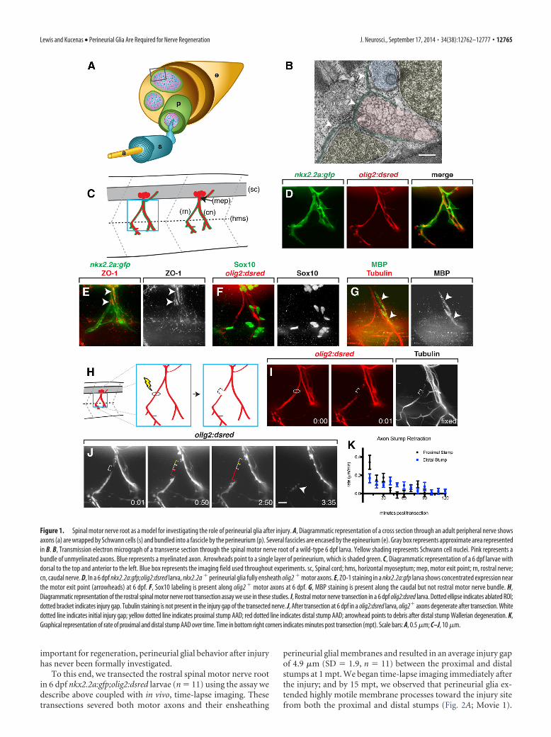
Before performing nerve transections, we sought to furthercharacterize spinal motor nerve ultrastructure in 6 dpf zebrafishlarvae. Using electron microscopy, we observed that, by thisstage, motor axons were sorted with large-caliber axons associ-ated with myelinating Schwann cells and small-diameter axonsencased within immature Schwann cells, and both were bundledinto a fascicle by a single layer of perineurial cells (Fig. 1B). Usingnkx2.2a:gfp;olig2:dsred larvae, where nkx2.2a regulatory se-quences drive expression of membrane tethered EGFP inperineurial glia and olig2 regulatory sequences drive expression ofcytosolic DsRed in motor neurons/axons, we assayed the extentof perineurial ensheathment along motor nerves. At 6 dpf, spinalmotor root axons were fully ensheathed by nkx2.2a% perineurialglia from the motor exit point to the horizontal myoseptum, andperineurial processes frequently extended along axons well be-yond the horizontal myoseptum (Fig. 1C,D). In the matureperineurium, differentiated perineurial cells express tight junc-tion proteins, including zona occludins 1 (ZO-1), which connectindividual cells together and give this nerve component its blood-nerve-barrier function. To investigate the extent of perineurialglial maturity and differentiation at 6 dpf, we labeled larvae withan antibody specific to ZO-1. Consistent with previous findings(Binari et al., 2013), we observed ZO-1 labeling along nkx2.2a%
perineurial sheaths with the highest concentration near the mo-tor exit point (Fig. 1E). To confirm that Schwann cells were pres-ent along motor nerves and at a similar stage of differentiation,we labeled 6 dpf larvae with antibodies specific to Sox10, a tran-scription factor expressed by all Schwann cells, and MBP, to labelmature, myelinating glia. At this stage, we observed Sox10% cellsalong both rostral and caudal nerve roots, and MBP immunore-activity was present along the caudal motor root and absent fromthe rostral motor root (Fig. 1F,G). This MBP profile persisted outto $20 dpf (data not shown); therefore, we concluded that rostralmotor root axons are associated with nonmyelinating Sox10%
glia, whereas caudal motor root axons are ensheathed by myeli-nating glia. Together, these data show that Sox10% glia andperineurial glia are differentiating and forming mature structures
along spinal motor nerve roots at 6 dpf. Therefore, these nervesare an ideal model to study the role of perineurial glia duringnerve degeneration and regeneration.
To investigate the role of perineurial glia during motor nervedegeneration and subsequent regeneration, we developed aninjury-response assay in live, 6 dpf larvae. For this assay, we useda nitrogen pumped dye laser (MicroPoint, Andor Technology) totransect the rostral spinal motor nerve root, resulting in a com-plete axotomy of this nerve bundle (Lewis and Kucenas, 2013)(Fig. 1H, I). This assay is highly reproducible and similar to otherpublished nerve transection assays (Rosenberg et al., 2012).Given that this method of transection relies on the use of a UVlaser, we sought to confirm that motor axons were fully cut andnot simply photobleached after injury. To investigate this possi-bility, we fixed 6 dpf larvae after transection and labeled themwith an antibody specific to acetylated tubulin to label all spinalmotor axons. In these larvae, we consistently observed clearbreaks in tubulin expression along rostral motor nerves where wehad previously transected the nerve, but differential interferencecontrast imaging did not show any obvious tissue damage sur-rounding the lesions (Fig. 1I; and data not shown). From thesedata, we are confident that our nerve injury assay produces fulland reproducible transections with no obvious damage to neigh-boring tissue.
To determine whether our injury assay produced axonal de-generation dynamics similar to previously published studies(Martin et al., 2010; Rosenberg et al., 2012; Villegas et al., 2012),we sought to characterize these events more carefully. Followingtransection of the rostral motor nerve, we observed that proximaland distal stump axons appeared to die back, gradually causing awidening of the injury gap (Fig. 1J). The phenomenon of axondie back, or AAD, is well established in mammalian systems (Ker-schensteiner et al., 2005; Wang et al., 2012; Zochodne, 2012) andhas been reported in zebrafish as well (Villegas et al., 2012). In ourassay, we observed that AAD began along both stumps immedi-ately after injury, increasing the distance between stumps by anaverage of 16.6 !m over the first 2 hours post transection (hpt)(SD ' 6.0, n ' 10). Interestingly, during our temporal analysis,we noticed that proximal stump AAD occurred earlier and at afaster rate than distal stump AAD (Fig. 1K). Ultimately, within afew hours, AAD along the distal stump gave way to Walleriandegeneration of distal axons, with individual axons fragmentingall at once, usually between 151 and 180 min post transection(mpt) (data not shown), which is consistent with previous find-ings in zebrafish (Martin et al., 2010; Rosenberg et al., 2012; Vil-legas et al., 2012). From these studies, we conclude that our nerveinjury assay is highly reproducible with axon degeneration dy-namics similar to previously reported models of axon injury inzebrafish (Martin et al., 2010; Rosenberg et al., 2012; Villegas etal., 2012), and therefore, provides an ideal model to investigate therole of perineurial glia in nerve degeneration and regeneration.
Perineurial glia respond to spinal motor nerve root injuryAlthough many studies have investigated the mechanisms thatgovern peripheral axon degeneration and regeneration, the roleof perineurial glia during these processes is still poorly under-stood. Studies on fixed tissue preparations have hypothesizedthat: (1) perineurial cells may be the first cellular component totraverse injury gaps and connect proximal and distal stumps afternerve transection (Scaravilli, 1984; Schroder et al., 1993), and (2)axons regrow through tracks that contain both Schwann cell andperineurial lamella (Thomas and Jones, 1967; Morris et al., 1972).However, despite these indications that perineurial glia may be
12764 • J. Neurosci., September 17, 2014 • 34(38):12762–12777 Lewis and Kucenas • Perineurial Glia Are Required for Nerve Regeneration
important for regeneration, perineurial glial behavior after injuryhas never been formally investigated.
To this end, we transected the rostral spinal motor nerve rootin 6 dpf nkx2.2a:gfp;olig2:dsred larvae (n ' 11) using the assay wedescribe above coupled with in vivo, time-lapse imaging. Thesetransections severed both motor axons and their ensheathing
perineurial glial membranes and resulted in an average injury gapof 4.9 !m (SD ' 1.9, n ' 11) between the proximal and distalstumps at 1 mpt. We began time-lapse imaging immediately afterthe injury; and by 15 mpt, we observed that perineurial glia ex-tended highly motile membrane processes toward the injury sitefrom both the proximal and distal stumps (Fig. 2A; Movie 1).
Figure 1. Spinal motor nerve root as a model for investigating the role of perineurial glia after injury. A, Diagrammatic representation of a cross section through an adult peripheral nerve showsaxons (a) are wrapped by Schwann cells (s) and bundled into a fascicle by the perineurium (p). Several fascicles are encased by the epineurium (e). Gray box represents approximate area representedin B. B, Transmission electron micrograph of a transverse section through the spinal motor nerve root of a wild-type 6 dpf larva. Yellow shading represents Schwann cell nuclei. Pink represents abundle of unmyelinated axons. Blue represents a myelinated axon. Arrowheads point to a single layer of perineurium, which is shaded green. C, Diagrammatic representation of a 6 dpf larvae withdorsal to the top and anterior to the left. Blue box represents the imaging field used throughout experiments. sc, Spinal cord; hms, horizontal myoseptum; mep, motor exit point; rn, rostral nerve;cn, caudal nerve. D, In a 6 dpf nkx2.2a:gfp;olig2:dsred larva, nkx2.2a % perineurial glia fully ensheath olig2 % motor axons. E, ZO-1 staining in a nkx2.2a:gfp larva shows concentrated expression nearthe motor exit point (arrowheads) at 6 dpf. F, Sox10 labeling is present along olig2 % motor axons at 6 dpf. G, MBP staining is present along the caudal but not rostral motor nerve bundle. H,Diagrammatic representation of the rostral spinal motor nerve root transection assay we use in these studies. I, Rostral motor nerve transection in a 6 dpf olig2:dsred larva. Dotted ellipse indicates ablated ROI;dotted bracket indicates injury gap. Tubulin staining is not present in the injury gap of the transected nerve. J, After transection at 6 dpf in a olig2:dsred larva, olig2% axons degenerate after transection. Whitedotted line indicates initial injury gap; yellow dotted line indicates proximal stump AAD; red dotted line indicates distal stump AAD; arrowhead points to debris after distal stump Wallerian degeneration. K,Graphical representation of rate of proximal and distal stump AAD over time. Time in bottom right corners indicates minutes post transection (mpt). Scale bars: A, 0.5 !m; C–J, 10 !m.
Lewis and Kucenas • Perineurial Glia Are Required for Nerve Regeneration J. Neurosci., September 17, 2014 • 34(38):12762–12777 • 12765

Around this same time, vesicles formed in perineurial glialmembranes near the transection site (Fig. 2A; Movie 1). Inter-estingly, these vesicle-like structures appeared along bothstumps during the period of AAD and were particularly nu-merous along the proximal stump, which degenerates morerapidly (Fig. 2A; Movie 1). During the imaging window (3 h)after nerve injury, perineurial glia eventually bridged the in-jury site, connecting the proximal and distal stumps, and re-mained along the distal stump after distal axons degeneratedand were cleared (Fig. 2A; Movie 1).
To determine whether the size of the initial injury gap wascorrelated to the time it took perineurial glia to form bridges, we
measured the size of the injury gap at 1 mpt and plotted it againstthe time it took perineurial cells to extend processes into theinjury site and bridge the gap. We found that, regardless of thesize of the injury, perineurial glia always extended highly motilemembrane processes toward the injury gap within 20 mpt (Fig.2B). However, as injury size increased, the time to bridge forma-tion increased linearly, indicating that larger injuries takeperineurial glia, respectively, longer to bridge (Fig. 2B). Interest-ingly, perineurial glia did not routinely bridge large injuries (#10!m) during the imaging window, which led us to hypothesizethat the bridging behavior may be mediated by perineurial–perineurial interactions.
Figure 2. Perineurial glia respond to rostral motor nerve injury. Frames captured from a 3 h time-lapse movie of a 6 dpf nkx2.2a:gfp;olig2:dsred larva beginning at 1 mpt with dorsal to the top andanterior to the left. mpt is indicated in bottom right corner of each image. A, Perineurial glia respond to rostral spinal motor nerve root transection by extending processes into the injury site, formingvesicles (insets and arrowheads), and bridging the injury gap (bracket). B, Graphical representation of the time it took perineurial glia to bridge the injury gap plotted against the initial injury size.C, D, Perineurial glial membranes are stable in both (C) the absence of injury and (D) after injury to tissue adjacent to the nerve. Dotted ellipse indicates ablated ROI. Scale bars, 10 !m.
12766 • J. Neurosci., September 17, 2014 • 34(38):12762–12777 Lewis and Kucenas • Perineurial Glia Are Required for Nerve Regeneration

To verify that the perineurial glial behaviors we observed werea response to nerve injury and not general damage or heat createdby the laser, we created focal lesions in the muscle tissue (10 !mfrom rostral motor axons (n ' 3) and assayed the perineurial glialresponse with time-lapse imaging. In the 3 h after injury, this typeof damage did not elicit any changes in nearby perineurial gliacompared with uninjured control larvae (Fig. 2C,D). Together,these data are consistent with the hypothesis that cues derivedspecifically from nerve damage induce a rapid change inperineurial glial behavior that results in perineurial glia extendingprocesses toward the injury site, developing vesicles, and ulti-mately bridging the injury gap.
Perineurial glia phagocytize debris after nerve injuryWhile characterizing the response of perineurial glia to spinalmotor nerve root injury, we noticed that the formation and tem-poral dynamics of perineurial glial vesicles coincided with peri-ods of axonal degeneration (Figs. 1 and 2; Movie 1). Manyvesicles formed along the proximal stump as it was rapidly degen-erating during the first 45 mpt, and vesicles frequently formedalong the distal stump during acute and Wallerian degeneration(Fig. 2A; Movie 1). However, we never observed perineurial ves-icles along uninjured nerves or during normal nerve develop-ment before 6 dpf (Fig. 2C; and data not shown). This led us tohypothesize that perineurial glia may be phagocytic and aid indebris clearance after injury. To investigate this hypothesis, wetransected nerves in 6 dpf nkx2.2a:gfp;olig2:dsred larvae andlooked for the presence of axonal fluorescence within perineurialglial vesicles. Single z-planes taken from time-lapse imagesshowed that DsRed% axonal fluorescence was frequently presentinside GFP% perineurial membrane vesicles during both AADand Wallerian degeneration (Fig. 3A–C). Additionally, orthogo-nal views demonstrated that the GFP% perineurial vesicles wereindeed spherical in nature and, therefore, morphologically con-
sistent with phagocytic vesicles (Fig. 3D). Notably, not all vesiclescontained axonal fluorescence, and it is possible that these vesi-cles contained debris derived from other nerve components, in-cluding myelin, Schwann cells, or extracellular matrix.
As an independent method of testing the phagocytic ability ofperineurial glia, we used LysoTracker dye. LysoTracker is amembrane-permeable dye that fluoresces at acidic pH, and thisdye has previously been used in zebrafish to label phagosomes asthey fuse with lysosomes and acidify (Peri and Nusslein-Volhard,2008). To confirm that the vesicle-like structures we observedafter injury represent phagosomes or phagolysosomes, we treated 4dpf nkx2.2a:gfp larvae with 20 !M LysoTracker Red, transectednerves and followed individual perineurial glial vesicles with time-lapse imaging. After injury, we observed that perineurial vesiclesbecame LysoTracker-positive over time (Fig. 3E), suggesting thatthese vesicles were acidifying. LysoTracker staining was presentbefore injury and in uninjured controls; however, this stainingnever occurred within clearly defined perineurial vesicles and waslikely the result of staining of lysosomes (data not shown)(Chazotte, 2011). These data demonstrate that after nerve dam-age, perineurial glia form phagocytic vesicles, many of whichcontain axonal debris. In our previous time-lapse imaging (Fig.2A), we noticed that perineurial vesicles were most numerousalong the proximal stump as those axons underwent AAD.Therefore, we conclude that perineurial glia initially focus theirphagocytic activity along the proximal stump. This sets forth anintriguing hypothesis, where debris clearance may be spatiallycoordinated between different cell types after nerve injury.
Perineurial glial bridges are required for axon regrowthAbove, we show that perineurial glia bridge the injury gap be-tween proximal and distal nerve stumps after transection, withtiming that is dependent on the size of the injury (Fig. 2). Impor-tantly, these bridges formed before new axons began to regener-ate and remained intact after distal axons degenerated. Previousstudies of sciatic nerve regeneration in rats showed that a tissuebridge forms to connect the proximal and distal stumps shortlyafter transection and that this bridge precedes the ingrowth ofaxons and Schwann cells (Schroder et al., 1993; McDonald andZochodne, 2003; McDonald et al., 2006; Parrinello et al., 2010).Collectively, these data are consistent with the hypothesis thatperineurial glia provide an essential tissue bridge that helps guideregenerating axons across the injury gap and back to their origi-nal targets.
To investigate this hypothesis, we assessed the ability of axonsto regrow in the absence of perineurial glial bridges. Using 6 dpfnkx2.2a:gfp;olig2:dsred larvae, we deliberately created rostral spi-nal motor nerve transections that resulted in gaps of varying sizes,ranging from 3.5 to 20 !m. We then let the larvae recover andimaged them at 48 h hpt to assess the presence or absence ofperineurial bridges and the extent of axon regrowth. Previousstudies in zebrafish demonstrate that motor axons begin to re-grow after injury by 24 hpt, with full functional recovery achievedby 48 hpt (Rosenberg et al., 2012), rendering 48 hpt a suitabletime point to assay axon regrowth.
In our studies, we defined full regrowth as the presence of anaxon bundle at 48 hpt that was approximately the same thicknessand followed a similar path as the original, uninjured nerve (Fig.4A). Moderate regrowth was defined as the presence of an axonbundle that was significantly thinner than the original nerve butfollowed a similar path (Fig. 4B). Finally, no regrowth was char-acterized as exhibiting either no axon regrowth from the proxi-mal stump or sprouting that did not extend beyond the
Movie 1. Perineurial glia respond to motor nerve transection. Four hour time-lapse movie ofa 6 dpf nkx2.2a:gfp;olig2:dsred larva beginning at 1 mpt with dorsal to the top and anterior tothe left. Images were captured in 5 min intervals, and mpt is indicated in bottom right corner ofeach frame. White circle represents the approximate ablated ROI. Perineurial glia extend pro-cesses toward the injury site, form vesicles within their membranes, and bridge the injury gap.
Lewis and Kucenas • Perineurial Glia Are Required for Nerve Regeneration J. Neurosci., September 17, 2014 • 34(38):12762–12777 • 12767

horizontal myoseptum (Fig. 4C). Whenwe created transections that resulted in in-jury gaps that were (7 !m (n ' 9) andimaged at 48 hpt, perineurial glia alwaysbridged injury sites and axons successfullyregrew along these bridges following theoriginal path (Fig. 4A,B). Of these nerves,33% exhibited full regrowth and 66% ex-hibited moderate regrowth (Fig. 4E),which is consistent with clinical data de-scribing that peripheral regeneration re-sults in variable and often imperfectoutcomes (Witzel et al., 2005; Zochodne,2012). In contrast, when injury gapsranged between 7 and 10 !m (n ' 6), only17% of nerves exhibited full axon re-growth at 48 hpt, 50% exhibited moderateregrowth, and 33% exhibited no regrowth(Fig. 4E). In the cases of full and moderateregrowth, perineurial glia bridged the in-jury site in all but one instance. In thissituation, axons appeared to reroutearound the injury site and make their wayback to a perineurial glial cell along thedistal stump. Once meeting with the distalperineurial glia, axons then followed theglia along what appeared to be their orig-inal path (data not shown). In all cases ofno regrowth, perineurial bridges werenever present; and although we often ob-served axon sprouts from the proximalstump, they did not follow along any de-fined path and perineurial ensheathmentof these sprouts was variable (Fig. 4C). Fi-nally, when we created transections thatresulted in injury gaps that were #10 !m(n ' 6), we never observed full regrowth,33% of cases had moderate regrowth withperineurial bridges, and 67% had no re-growth and no perineurial bridges (Fig.4C,E). Importantly, regardless of injurysize, in 20 of 21 cases, the presence of aperineurial bridge was associated withaxon regrowth, and the absence of abridge was associated with no regrowth.We interpret this to mean that, as the sizeof the injury increased, the ability ofperineurial glia to bridge became im-paired; and when bridges did not form,axons were unable to regrow. Together,these data are consistent with the hypoth-esis that perineurial glial bridge formationis required for axon regrowth after injury.
To more directly test the requirementof perineurial glia for axonal regenera-tion, we assessed the ability of axons toregrow in the absence of perineurial glia.In a previous study, we show that Notch signaling is requiredduring a distinct temporal window (48–72 hpf) for perineurial glialdevelopment (Binari et al., 2013). During this window, treatingembryos with either DAPT, a gamma secretase inhibitor, or ge-netically blocking Notch signaling via a dominant-negative formof the Notch intraceullar domain (NICD) cofactor, suppressor of
hairless (Su(H)), led to a complete failure of perineurial glialmigration into the PNS (Binari et al., 2013). Importantly, eventhough perineurial glia failed to exit the spinal cord, Schwanncells were still present along motor nerves after Notch perturba-tion, although they failed to myelinate axons (Binari et al., 2013).Therefore, this experimental paradigm allowed us to remove
Figure 3. Perineurial glia phagocytize debris after nerve injury. Frames captured from a time-lapse movie of a 6 dpf nkx2.2a:gfp;olig2:dsred larva beginning at 1 mpt with dorsal to the top and anterior to the left. mpt is indicated in bottom right corner ofeach image; dotted ellipses indicate ablated ROIs. Magnified regions (white boxes) are single z-planes where indicated. A, B,Rostral nerve transections in 6 dpf nkx2.2a:gfp;olig2:dsred larvae show olig2 % axon fragments (arrowheads) within nkx2.2a %
perineurial membranes along the proximal stump during AAD (A) and distal stump during Wallerian degeneration (B). C, Fluores-cence intensity of nkx2.2a:gfp and olig2:dsred along the white dashed line ROI drawn in A shows an increase in olig2:dsredfluorescence within the nkx2.2a:gfp fluorescing vesicle. D, A perineurial vesicle shown with accompanying orthogonal views. E,Rostral and caudal nerve transections of a 4 dpf nkx2.2a:gfp larva stained with LysoTracker. At 55 mpt, Lysotracker staining is visiblewithin an nkx2.2a % perineurial vesicle (white arrowhead); and by 60 mpt, staining is visible within a second, adjacent vesicle(yellow arrowhead). Scale bars: full images, 10 !m; magnified insets, 1.5 !m.
12768 • J. Neurosci., September 17, 2014 • 34(38):12762–12777 Lewis and Kucenas • Perineurial Glia Are Required for Nerve Regeneration
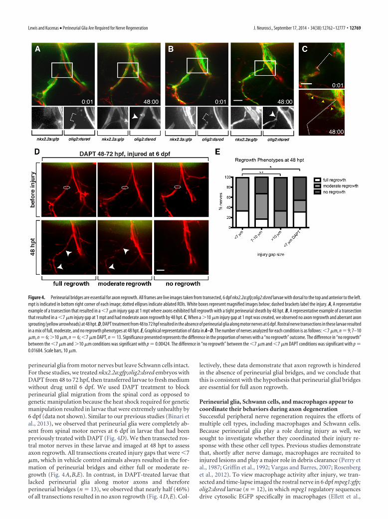
perineurial glia from motor nerves but leave Schwann cells intact.For these studies, we treated nkx2.2a:gfp;olig2:dsred embryos withDAPT from 48 to 72 hpf, then transferred larvae to fresh mediumwithout drug until 6 dpf. We used DAPT treatment to blockperineurial glial migration from the spinal cord as opposed togenetic manipulation because the heat shock required for geneticmanipulation resulted in larvae that were extremely unhealthy by6 dpf (data not shown). Similar to our previous studies (Binari etal., 2013), we observed that perineurial glia were completely ab-sent from spinal motor nerves at 6 dpf in larvae that had beenpreviously treated with DAPT (Fig. 4D). We then transected ros-tral motor nerves in these larvae and imaged at 48 hpt to assessaxon regrowth. All transections created injury gaps that were (7!m, which in vehicle control animals always resulted in the for-mation of perineurial bridges and either full or moderate re-growth (Fig. 4A,B,E). In contrast, in DAPT-treated larvae thatlacked perineurial glia along motor axons and thereforeperineurial bridges (n ' 13), we observed that nearly half (46%)of all transections resulted in no axon regrowth (Fig. 4D,E). Col-
lectively, these data demonstrate that axon regrowth is hinderedin the absence of perineurial glial bridges, and we conclude thatthis is consistent with the hypothesis that perineurial glial bridgesare essential for full axon regrowth.
Perineurial glia, Schwann cells, and macrophages appear tocoordinate their behaviors during axon degenerationSuccessful peripheral nerve regeneration requires the efforts ofmultiple cell types, including macrophages and Schwann cells.Because perineurial glia play a role during injury as well, wesought to investigate whether they coordinated their injury re-sponse with these other cell types. Previous studies demonstratethat, shortly after nerve damage, macrophages are recruited toinjured lesions and play a major role in debris clearance (Perry etal., 1987; Griffin et al., 1992; Vargas and Barres, 2007; Rosenberget al., 2012). To view macrophage activity after injury, we tran-sected and time-lapse imaged the rostral nerve in 6 dpf mpeg1:gfp;olig2:dsred larvae (n ' 12), in which mpeg1 regulatory sequencesdrive cytosolic EGFP specifically in macrophages (Ellett et al.,
Figure 4. Perineurial bridges are essential for axon regrowth. All frames are live images taken from transected, 6 dpf nkx2.2a:gfp;olig2:dsred larvae with dorsal to the top and anterior to the left.mpt is indicated in bottom right corner of each image; dotted ellipses indicate ablated ROIs. White boxes represent magnified images below; dashed brackets label the injury. A, A representativeexample of a transection that resulted in a (7 !m injury gap at 1 mpt where axons exhibited full regrowth with a tight perineurial sheath by 48 hpt. B, A representative example of a transectionthat resulted in a (7 !m injury gap at 1 mpt and had moderate axon regrowth by 48 hpt. C, When a #10 !m injury gap at 1 mpt was created, we observed no axon regrowth and aberrant axonsprouting (yellow arrowheads) at 48 hpt. D, DAPT treatment from 48 to 72 hpf resulted in the absence of perineurial glia along motor nerves at 6 dpf. Rostral nerve transections in these larvae resultedin a mix of full, moderate, and no regrowth phenotypes at 48 hpt. E, Graphical representation of data in A–D. The number of nerves analyzed for each condition is as follows: (7 !m, n ' 9; 7–10!m, n ' 6; #10 !m, n ' 6; (7 !m DAPT, n ' 13. Significance presented represents the difference in the proportion of nerves with a “no regrowth” outcome. The difference in “no regrowth”between the (7 !m and #10 !m conditions was significant with p ' 0.00424. The difference in “no regrowth” between the (7 !m and (7 !m DAPT conditions was significant with p '0.01684. Scale bars, 10 !m.
Lewis and Kucenas • Perineurial Glia Are Required for Nerve Regeneration J. Neurosci., September 17, 2014 • 34(38):12762–12777 • 12769
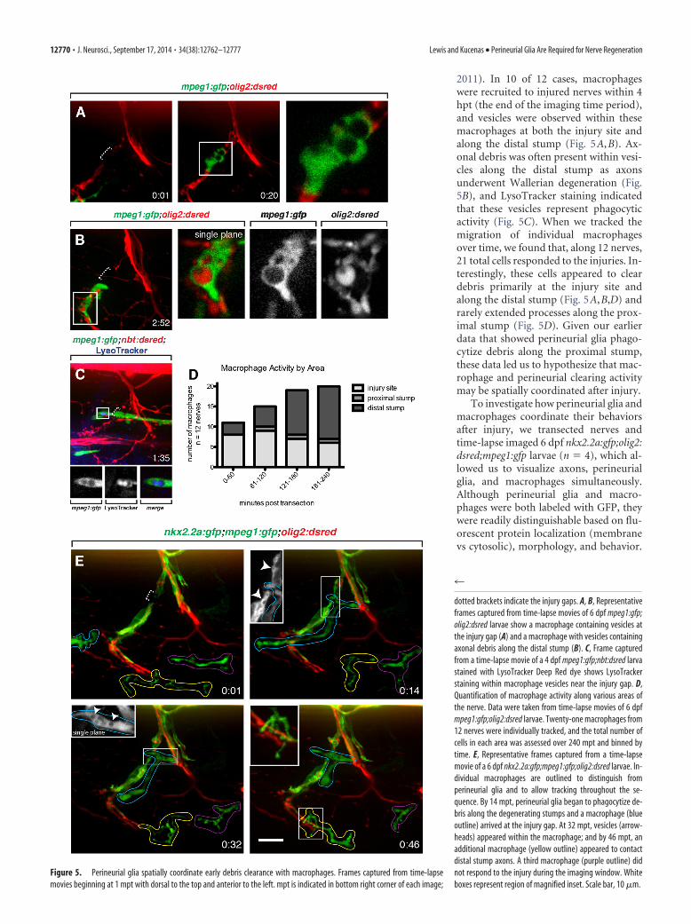
2011). In 10 of 12 cases, macrophageswere recruited to injured nerves within 4hpt (the end of the imaging time period),and vesicles were observed within thesemacrophages at both the injury site andalong the distal stump (Fig. 5A,B). Ax-onal debris was often present within vesi-cles along the distal stump as axonsunderwent Wallerian degeneration (Fig.5B), and LysoTracker staining indicatedthat these vesicles represent phagocyticactivity (Fig. 5C). When we tracked themigration of individual macrophagesover time, we found that, along 12 nerves,21 total cells responded to the injuries. In-terestingly, these cells appeared to cleardebris primarily at the injury site andalong the distal stump (Fig. 5A,B,D) andrarely extended processes along the prox-imal stump (Fig. 5D). Given our earlierdata that showed perineurial glia phago-cytize debris along the proximal stump,these data led us to hypothesize that mac-rophage and perineurial clearing activitymay be spatially coordinated after injury.
To investigate how perineurial glia andmacrophages coordinate their behaviorsafter injury, we transected nerves andtime-lapse imaged 6 dpf nkx2.2a:gfp;olig2:dsred;mpeg1:gfp larvae (n ' 4), which al-lowed us to visualize axons, perineurialglia, and macrophages simultaneously.Although perineurial glia and macro-phages were both labeled with GFP, theywere readily distinguishable based on flu-orescent protein localization (membranevs cytosolic), morphology, and behavior.
Figure 5. Perineurial glia spatially coordinate early debris clearance with macrophages. Frames captured from time-lapsemovies beginning at 1 mpt with dorsal to the top and anterior to the left. mpt is indicated in bottom right corner of each image;
4
dotted brackets indicate the injury gaps. A, B, Representativeframes captured from time-lapse movies of 6 dpf mpeg1:gfp;olig2:dsred larvae show a macrophage containing vesicles atthe injury gap (A) and a macrophage with vesicles containingaxonal debris along the distal stump (B). C, Frame capturedfrom a time-lapse movie of a 4 dpf mpeg1:gfp;nbt:dsred larvastained with LysoTracker Deep Red dye shows LysoTrackerstaining within macrophage vesicles near the injury gap. D,Quantification of macrophage activity along various areas ofthe nerve. Data were taken from time-lapse movies of 6 dpfmpeg1:gfp;olig2:dsred larvae. Twenty-one macrophages from12 nerves were individually tracked, and the total number ofcells in each area was assessed over 240 mpt and binned bytime. E, Representative frames captured from a time-lapsemovie of a 6 dpf nkx2.2a:gfp;mpeg1:gfp;olig2:dsred larvae. In-dividual macrophages are outlined to distinguish fromperineurial glia and to allow tracking throughout the se-quence. By 14 mpt, perineurial glia began to phagocytize de-bris along the degenerating stumps and a macrophage (blueoutline) arrived at the injury gap. At 32 mpt, vesicles (arrow-heads) appeared within the macrophage; and by 46 mpt, anadditional macrophage (yellow outline) appeared to contactdistal stump axons. A third macrophage (purple outline) didnot respond to the injury during the imaging window. Whiteboxes represent region of magnified inset. Scale bar, 10 !m.
12770 • J. Neurosci., September 17, 2014 • 34(38):12762–12777 Lewis and Kucenas • Perineurial Glia Are Required for Nerve Regeneration

As described above (Figs. 2 and 3; Movie 1), our time-lapse im-aging data revealed that transection induced perineurial glia toextend highly motile membrane processes toward injury sites andform phagocytic vesicles (Fig. 5E). These phagocytic vesicles pop-ulated perineurial glial membranes along both stumps immedi-ately adjacent to the injury site and were particularly numerousalong the proximal stump, which undergoes rapid AAD imme-diately after injury (Fig. 5E). Interestingly, these perineurial re-sponses were usually observed before the arrival of macrophages,and the mean response time for perineurial glia was significantlyshorter and less variable than for macrophages (perineurial glialmean response time: 8.636 ) 0.09749 mpt, n ' 11; macrophagemean arrival time: 45.0 ) 14.43 mpt, n ' 9, p ' 0.0120). Whereasperineurial glial vesicles appeared along the stumps undergoingAAD, macrophages were usually attracted directly to the injurygap, and vesicles within these macrophages indicated they wereclearing debris primarily in the gap zone (Fig. 5E). We also ob-served that additional macrophages were often recruited to thedistal stump as axons fragmented during Wallerian degeneration(Fig. 5E), and we frequently observed vesicles in perineurial gliaalong the distal stump around this time as well (Fig. 3). These datademonstrate that perineurial glia respond to transections andbegin debris clearance more quickly than macrophages. Thesedata also show that these two cell types initially focus on clearingdebris within distinct zones, with perineurial glia focusing onthe degenerating stumps and macrophages focusing on theinjury gap, but work together in similar areas during laterstages of degeneration. This is consistent with the hypothesisthat perineurial glia and macrophages spatially coordinate debrisclearance.
In addition to macrophages, Schwann cells also play an im-portant role in peripheral degeneration and regeneration byphagocytizing myelin and forming cellular columns that spaninjury gaps to help guide regenerating axons (Perry et al., 1995;Parrinello et al., 2010; Webber and Zochodne, 2010; Gaudet et al.,2011; Brosius Lutz and Barres, 2014). Because Schwann cells andperineurial glia coordinate their behaviors and require each otherduring development (Kucenas et al., 2008; Binari et al., 2013;Clark et al., 2014), we hypothesized that these cell types maycoordinate again after nerve injury. To test this hypothesis, wetransected nerves and time-lapse imaged nkx2.2a:gfp;sox10:eoslarvae, which allowed visualization of perineurial glia andSchwann cells simultaneously. The sox10:eos transgene uses reg-ulatory elements from the sox10 gene to drive expression a cyto-solic, photo-convertible Eos protein in Schwann cells. MatureEos protein, when exposed to UV light, shifts its emission from agreen fluorescent state (516 nm) to a red fluorescent state (581nm). By exposing these larvae to UV light, we can easily distin-guish Schwann cells (red after UV light exposure) from perineur-ial glia (green after UV light exposure). For these studies, weexposed larvae to UV light before time-lapse imaging and subse-quently reexposed the larvae every 20 min throughout the courseof imaging to convert any newly produced green Eos so thatSchwann cells would remain completely red. Similar to our re-sults above (Fig. 2), we observed several phagocytic vesicleswithin perineurial membranes along the proximal stump within15–20 mpt (data not shown). We also observed vesicles withinsox10% cells on the proximal stump (data not shown), and lateralong the distal stump (Fig. 6A). Importantly, these vesicles la-beled with LysoTracker, suggesting they represent phagocytic ac-tivity (Fig. 6A). These data indicate that Schwann cellsphagocytize debris with similar temporal and spatial dynamics asperineurial glia. As we continued imaging, perineurial glia
bridged the injury gap during the 4 h imaging window we usedafter transection (Fig. 6B,C). Schwann cells, however, extendedprocesses slightly toward the injury site but never bridged theinjury gap during the imaging time-period (n ' 4) (Fig. 6B,C).These data demonstrate that perineurial glia and Schwann cellsboth phagocytize debris after injury and that perineurial gliabridge injury gaps before Schwann cells. This is consistent withthe idea that perineurial glia form an initial tissue bridge acrossthe injury gap, with Schwann cells and axons traversing the gaplater.
Perineurial glia require Schwann cells after nerve injuryInteractions between cell types are critical for ultrastructural andfunctional nerve regeneration (Parrinello et al., 2010; Napoli etal., 2012). Therefore, we sought to identify which cell types, if any,were required for the perineurial glial response to injury. To in-vestigate this, we used a combination of time-lapse imaging, mu-tant analysis, and cell-type specific perturbation. We firstconsidered the possibility that perineurial glia respond to signalsreleased from neighboring, damaged perineurial glial cells. Totest this hypothesis, we transected motor axon tracts in nkx2.2a:gfp;ntb:dsred larvae that were not ensheathed by perineurial gliaand assessed the response of nearby perineurial glial cells. Theseexperiments were performed in 4 dpf larvae, as not all spinalmotor axon tracts have become fully ensheathed by nkx2.2a%
perineurial glia yet at this stage, providing an ideal situation totest this hypothesis. Importantly, perineurial glia respond tonerve transection by phagocytizing debris and bridging injurygaps in a manner similar to what we describe at 6 dpf (compareFig. 7A with Fig. 2). Therefore, using 4 dpf larvae, we transectedunensheathed axons $10 –15 !m away from neighboringperineurial glia and assessed the response of these neighbors tothe injury (n ' 7). Immediately after injury, we observed thatperineurial cells along neighboring, uninjured axon tracts rapidlyextended processes toward the injury site and formed phagocyticvesicles by 15 mpt (Fig. 7B; Movie 2). This response time wassimilar to our previous findings along fully ensheathed tracts in 6dpf larvae (mean response time for 4 dpf, 9.286 ) 2.296 mpt, n '7; 6 dpf, 8.636 ) 0.9749 mpt, n ' 11), suggesting that perineurialglia respond to injuries with similar temporal dynamics regard-less of ensheathment. To independently test the possibility thatperineurial glia do not simply respond to axotomies because theysense damage in neighboring cells, we transected dorsal root gan-glia sensory axons in 6 dpf nkx2.2a:gfp;ntb:dsred larvae, as they arenot ensheathed by nkx2.2a% perineurial glia (unpublished data).In these instances, we again observed neighboring perineurial gliaextend processes onto the unensheathed sensory axons andphagocytize debris (Fig. 7C). Collectively, these data are consis-tent with the hypothesis that the perineurial glia do not respondto nerve injury using an autocrine mechanism. Instead, we hy-pothesize that perineurial glial activation after injury requiresnon–perineurial-derived cues. Interestingly, this is distinct fromtheir bridging behavior, which we hypothesize is mediated byperineurial–perineurial interactions.
Another potential source of factors that could elicit perineur-ial behavior after injury is macrophages. Macrophages that re-spond to injury sites release a variety of cytokines (Leskovar et al.,2000), which could play a role in recruiting perineurial glia toinjury sites or stimulating their phagocytic activity. To test thishypothesis, we transected nerves in larvae that lacked macro-phages and assessed the perineurial glial response. To createlarvae that completely lacked macrophages, we prevented mac-rophage specification by injecting single-cell embryos with
Lewis and Kucenas • Perineurial Glia Are Required for Nerve Regeneration J. Neurosci., September 17, 2014 • 34(38):12762–12777 • 12771
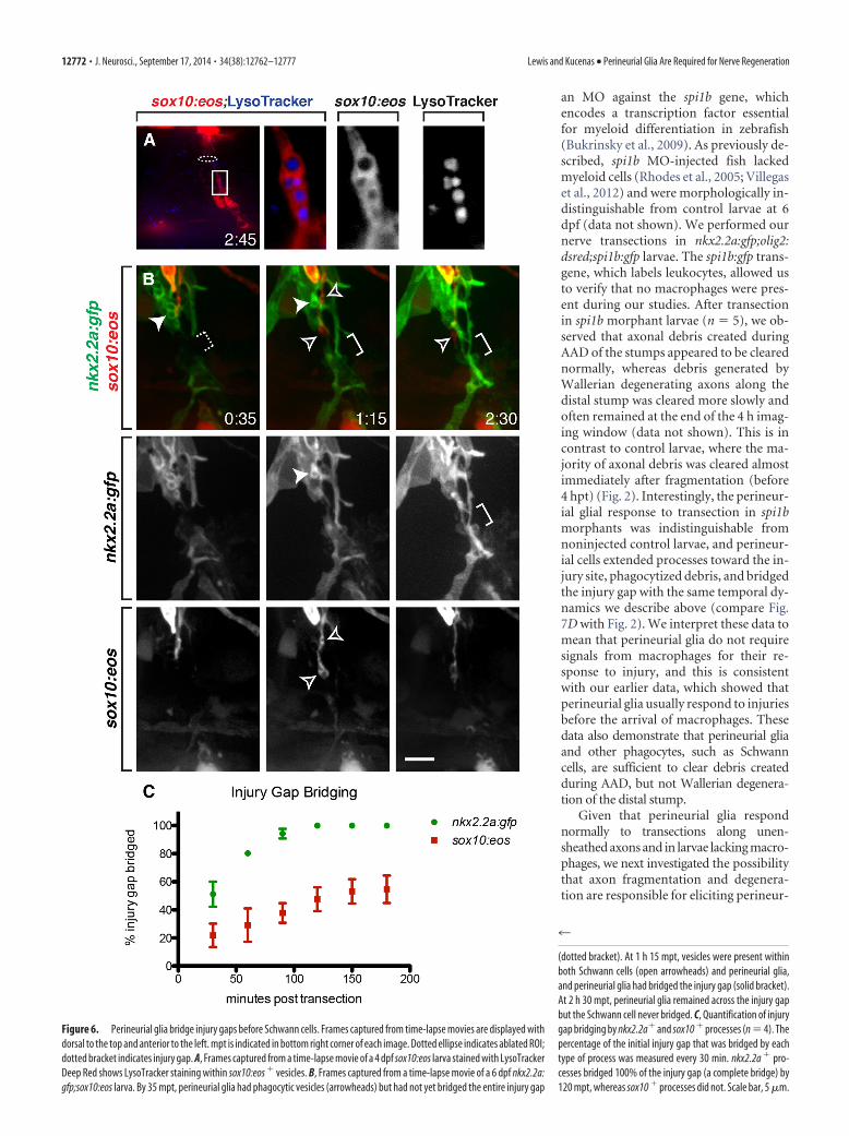
an MO against the spi1b gene, whichencodes a transcription factor essentialfor myeloid differentiation in zebrafish(Bukrinsky et al., 2009). As previously de-scribed, spi1b MO-injected fish lackedmyeloid cells (Rhodes et al., 2005; Villegaset al., 2012) and were morphologically in-distinguishable from control larvae at 6dpf (data not shown). We performed ournerve transections in nkx2.2a:gfp;olig2:dsred;spi1b:gfp larvae. The spi1b:gfp trans-gene, which labels leukocytes, allowed usto verify that no macrophages were pres-ent during our studies. After transectionin spi1b morphant larvae (n ' 5), we ob-served that axonal debris created duringAAD of the stumps appeared to be clearednormally, whereas debris generated byWallerian degenerating axons along thedistal stump was cleared more slowly andoften remained at the end of the 4 h imag-ing window (data not shown). This is incontrast to control larvae, where the ma-jority of axonal debris was cleared almostimmediately after fragmentation (before4 hpt) (Fig. 2). Interestingly, the perineur-ial glial response to transection in spi1bmorphants was indistinguishable fromnoninjected control larvae, and perineur-ial cells extended processes toward the in-jury site, phagocytized debris, and bridgedthe injury gap with the same temporal dy-namics we describe above (compare Fig.7D with Fig. 2). We interpret these data tomean that perineurial glia do not requiresignals from macrophages for their re-sponse to injury, and this is consistentwith our earlier data, which showed thatperineurial glia usually respond to injuriesbefore the arrival of macrophages. Thesedata also demonstrate that perineurial gliaand other phagocytes, such as Schwanncells, are sufficient to clear debris createdduring AAD, but not Wallerian degenera-tion of the distal stump.
Given that perineurial glia respondnormally to transections along unen-sheathed axons and in larvae lacking macro-phages, we next investigated the possibilitythat axon fragmentation and degenera-tion are responsible for eliciting perineur-
Figure 6. Perineurial glia bridge injury gaps before Schwann cells. Frames captured from time-lapse movies are displayed withdorsal to the top and anterior to the left. mpt is indicated in bottom right corner of each image. Dotted ellipse indicates ablated ROI;dotted bracket indicates injury gap. A, Frames captured from a time-lapse movie of a 4 dpf sox10:eos larva stained with LysoTrackerDeep Red shows LysoTracker staining within sox10:eos % vesicles. B, Frames captured from a time-lapse movie of a 6 dpf nkx2.2a:gfp;sox10:eos larva. By 35 mpt, perineurial glia had phagocytic vesicles (arrowheads) but had not yet bridged the entire injury gap
4
(dotted bracket). At 1 h 15 mpt, vesicles were present withinboth Schwann cells (open arrowheads) and perineurial glia,and perineurial glia had bridged the injury gap (solid bracket).At 2 h 30 mpt, perineurial glia remained across the injury gapbut the Schwann cell never bridged. C, Quantification of injurygap bridging by nkx2.2a% and sox10 % processes (n ' 4). Thepercentage of the initial injury gap that was bridged by eachtype of process was measured every 30 min. nkx2.2a % pro-cesses bridged 100% of the injury gap (a complete bridge) by120 mpt, whereas sox10 % processes did not. Scale bar, 5 !m.
12772 • J. Neurosci., September 17, 2014 • 34(38):12762–12777 Lewis and Kucenas • Perineurial Glia Are Required for Nerve Regeneration

ial glial activity after injury. To test this hypothesis, we transectednerves in mnx:Wlds-gfp larvae, in which motor neurons expressthe Wallerian degeneration Slow (Wld s) protein (Rosenberg etal., 2012). Expression of Wld s significantly delays Wallerian de-generation through a conserved axon-intrinsic mechanism
(Lunn et al., 1989; Adalbert et al., 2005; MacDonald et al., 2006;Martin et al., 2010; Rosenberg et al., 2012) and has been shown tosuppress aspects of glial activation in mouse and Drosophila(Lunn et al., 1989; MacDonald et al., 2006). Surprisingly, afteraxotomy in these mutants (n ' 6), perineurial glia phagocytized
Figure 7. Perineurial glia require Schwann cells, but not macrophages or Wallerian degeneration, for their response to injury. All images are frames captured from time-lapse movies of larvaebeginning at 1 mpt with dorsal to the top and anterior to the left. mpt is indicated in bottom right corner of each image. Dotted ellipses indicate ablated ROIs. A, The rostral motor nerve was transectedin 4 dpf nkx2.2a:gfp;olig2:dsred larvae. Perineurial glia responded by phagocytizing debris (arrowhead) and bridging the injury gap (bracket) similarly as in 6 dpf larvae. B, C, Unensheathed nervetracts were transected in 4 and 6 dpf nkx2.2a:gfp;nbt:dsred larvae, respectively. Perineurial glia phagocytized debris (arrowheads) and extended processes toward injury sites along (B) unen-sheathed motor nerves and (C) sensory nerves in a manner indistinguishable from fully ensheathed nerves. D, Rostral motor nerves were transected in a 6 dpf nkx2.2a:gfp;olig2:dsred;spi1b:gfp spi1bMO-injected larva that lacked macrophages. Perineurial glia responded similarly to noninjected controls by phagocytizing debris (arrowhead) and bridging the injury gap (bracket). E, Rostral motornerves were transected in a 6 dpf nkx2.2a:gfp;olig2:dsred;mnx1:wlds-gfp larva where Wallerian degeneration was significantly delayed. Again, perineurial glia responded similarly to controls byphagocytizing debris (arrowhead) and bridging the injury gap (bracket). F, Perineurial glia phagocytized debris (arrowhead) but did not extend processes toward the injury site in a 4 dpfnkx2.2a:gfp;nbt:dsred;colourless mutant larva (compare with B). G, Graphical representation of the perineurial response time in wild-type and cls larvae shows that there is no relationship betweenthe distance of glial cells from the transection site and response time in these experiments. The number of minutes post transection it took perineurial glia to begin extending processes towardinjuries or phagocytize debris was plotted by the initial distance between the glial cells and the transection site. H, Quantification of the extension of perineurial processes toward transection sitesin wild-type and cls larvae shows that perineurial glia extend processes significantly further in wild-type. Because the initial distance between the injury site and glial cells varied slightly with eachtrial, membrane extension was plotted as a percentage of the initial injury distance traveled, where values #1 represent processes that have extended beyond the initial injury site. p values for eachtime point assessed are as follows: 15 mpt, p ' 0.43966 (ns, not significant); 30 mpt, p ' 0.01204 (*); 45 mpt, p ' 0.02885 (*); 60 mpt, p ' 0.00159 (**); 120 mpt, p ' 0.00037 (***); 180 mpt,p ' 0.00268 (**). Scale bar, 5 !m.
Lewis and Kucenas • Perineurial Glia Are Required for Nerve Regeneration J. Neurosci., September 17, 2014 • 34(38):12762–12777 • 12773

debris and bridged injuries in a manner that was indistinguish-able from control larvae (compare Fig. 7E with Fig. 2), suggestingthat Wallerian degeneration is not necessary for perineurial gliato respond to injuries in our assay.
Finally, we investigated the possibility that Schwann cells arerequired for the perineurial glial injury response. Nerve injuryinduces Schwann cells to release cytokines and growth factorsthat are responsible for recruiting macrophages and promotingaxon regrowth (Chen et al., 2007; Vargas and Barres, 2007; Na-poli et al., 2012; Brosius Lutz and Barres, 2014). Additionally,previous studies demonstrate that Schwann cells communicatereciprocally with perineurial glia during development and thatdevelopment of either cell type is disrupted in the absence of theother (Kucenas et al., 2008; Binari et al., 2013). This raised theintriguing possibility that signals released by Schwann cells couldbe responsible for eliciting perineurial glial responses after injury.To investigate this hypothesis, we transected nerves in 4 dpfnkx2.2a:gfp;ntb:dsred;colourlesstw11 (cls) mutant larvae, which aredeficient for sox10 and lack motor nerve-associated Schwanncells (Dutton et al., 2001). Although oligodendrocyte precursorcells exit the spinal cord in cls larvae, they do not ensheath axonsand die before 4 dpf (Kucenas et al., 2009). However, even in theabsence of all other peripheral glia, we found that some perineur-ial glial cells still migrated from the spinal cord into the peripheryand remained associated with motor nerves after oligodendro-cyte precursor cells died (Fig. 7F). Although perineurial glia werepresent, they were sparse and did not form contiguous sheaths(Fig. 7F). Therefore, we transected unensheathed axons to testthe ability of neighboring perineurial glial cells in 4 dpf cls larvaeto respond to injury (n ' 8). As in wild-type, transections weremade $10 –15 !m away from the nearest perineurial glial cell(average distance between perineurial glia and transection sitewas 12.43 ) 0.9436, n ' 7 in wild-type and 10.89 ) 1.144, n ' 8for cls, not significant), and there was no relationship between the
distance from transection site and the perineurial response timefor either genotype (Fig. 7G). Unlike wild-type larvae (Fig. 7B;Movie 2), we observed that transection in cls larvae induced onlyminimal extension of perineurial processes toward injury sites(Fig. 7F,H; Movie 3). However, we still observed phagocytic ves-icles along the proximal stump as it degenerated in all cases forboth cls (n ' 8) and wild-type larvae (n ' 7) (Fig. 7B,F; Movie2,3). These data demonstrate that perineurial glia are significantlyless attracted to injury sites in the absence of Schwann cells butremain competent to phagocytize debris. Together, these studiesare consistent with the hypothesis that perineurial glial behaviorsafter nerve injury are not dependent on signals from macro-phages or degenerating axons. Instead, they appear to requireinteractions with Schwann cells, much like they do duringdevelopment.
DiscussionPeripheral nerve regeneration has long been a subject of intensestudy. However, the role of perineurial glia in this process hasremained somewhat elusive. Some have hypothesized thatperineurial cells may be responsible for creating the first bridgeacross the injury gap (Scaravilli, 1984; Schroder et al., 1993), andothers have shown that the perineurium dramatically changes itsstructure along proximal and distal stumps after transection(Thomas and Jones, 1967; Morris et al., 1972). Here, we providethe first detailed account of dynamic perineurial glial behaviorsafter nerve transection. We show that spinal motor root axontransection induces rapid changes in the membrane activity ofperineurial glia, with cells immediately adjacent to the injuryextending highly motile processes toward the transected area andphagocytizing debris. Perineurial glial membranes then bridgeinjury gaps, and we demonstrate that these bridges are essentialfor guiding axon regrowth (Fig. 8). We also show that perineurialglia do not require signals from other perineurial glia or macro-
Movie 2. Perineurial glia respond to transections along unensheathed axons. Three hourtime-lapse movie of a 4 dpf nkx2.2a:gfp;nbt:dsred larva beginning at 1 mpt with dorsal to thetop and anterior to the left. Images were captured in 5 min intervals, and mpt is indicated inbottom right corner of each frame. White circle represents the approximate ablated ROI.Perineurial glial processes are attracted to the injury site and phagocytize debris.
Movie 3. Perineurial processes are not attracted to injury sites in colourlesstw11 mutants.Three hour time-lapse movie of a 4 dpf nkx2.2a:gfp;nbt:dsred;colourlesstw11 larva beginning at1 mpt with dorsal to the top and anterior to the left. Images were captured in 5 min intervals,and mpt is indicated in bottom right corner of each frame. White circle represents the approx-imate ablated ROI. Perineurial glia do not extend significant processes toward the injury site butdo form phagocytic vesicles.
12774 • J. Neurosci., September 17, 2014 • 34(38):12762–12777 Lewis and Kucenas • Perineurial Glia Are Required for Nerve Regeneration
phages for this response, and they respond normally even in theabsence of Wallerian degeneration. Interestingly, perineurial gliado require Schwann cells for specific aspects of their injury re-sponse, much like they do during development. These data shedlight on novel behaviors of perineurial glia after motor nerveinjury and raise the intriguing possibility that they may be in-volved in other degenerative diseases of the PNS as well.
Perineurial glial phagocytic behaviorMacrophages and Schwann cells are the primary cells involved indebris clearance after nerve injury in the PNS. Schwann cellsbegin phagocytosis of myelin immediately after injury, and mac-rophages infiltrate nerves soon after (Liu et al., 1995; Perry et al.,1995; Hirata and Kawabuchi, 2002; Vargas and Barres, 2007).Our data support these findings and demonstrate that perineurialglia are phagocytic and aid in debris clearance as well. These dataare fitting with several electron microscopy studies that noted thepresence of vesicles within perineurial cells along the proximalstump of transected mouse sciatic nerves (Morris et al., 1972;Roytta et al., 1987) and within the cells that form the initial bridgebetween transected stumps (Scaravilli, 1984). However, despitethese observations, the authors stopped short of concluding thatperineurial glia are phagocytic, as it is very difficult to determinethe identity and origin of individual cells by electron microscopyafter transection. Using in vivo imaging in a live system that al-lows for the specific and continuous visualization of perineurialglia, we demonstrate that perineurial glia are phagocytic.
Temporally, our data reveal that perineurial glial phagocyticvesicles are particularly abundant along the proximal stump im-mediately after injury, which is consistent with our data showingthat proximal stump AAD occurs more rapidly than distal stumpAAD. This is in contrast to mammalian systems, where AAD isreported to occur symmetrically along both stumps (Kerschen-steiner et al., 2005). Spatially, our studies reveal that, whereasperineurial glia focus on clearing debris along the degeneratingstumps immediately after injury, macrophages tend to focus ini-tially on the injury gap. These data are consistent with the hy-pothesis that macrophages and perineurial glia may coordinatedebris clearance in different areas and raise the intriguing possi-bility that distinct phagocytic populations may selectively clearspecific debris. In contrast, our studies showed that Schwann cellvesicles formed along the proximal stump shortly after injury,and later along the distal stump, similar to perineurial glia. How-ever, it is still possible that perineurial glia and Schwann cellsphagocytize different types of debris in these regions. In the future,investigations into (1) how debris clearance is coordinated betweenperineurial glia, Schwann cells, and macrophages and (2) the molec-ular mechanisms that drive perineurial glial phagocytosis will pro-duce a more complete picture of nerve degeneration.
Perineurial glial bridges are required for regenerationPrevious studies in mammalian systems demonstrate that, aftersciatic nerve transection, a tissue bridge forms connecting theproximal and distal stumps, which axons and Schwann cells usewhile regenerating (McDonald and Zochodne, 2003; McDonaldet al., 2006). The precise importance of this initial tissue bridgeand the origin of the cells that construct it are not well under-stood, although several studies have proposed that this bridge isformed by perineurial cells (Scaravilli, 1984; Schroder et al.,1993). Parrinello et al. (2010) described that fibroblasts withinthis bridge are required to direct the formation of Schwann cellbands and therefore affect axon regrowth. In a recent study(Clark et al., 2014), we demonstrate that a subset of mammalian
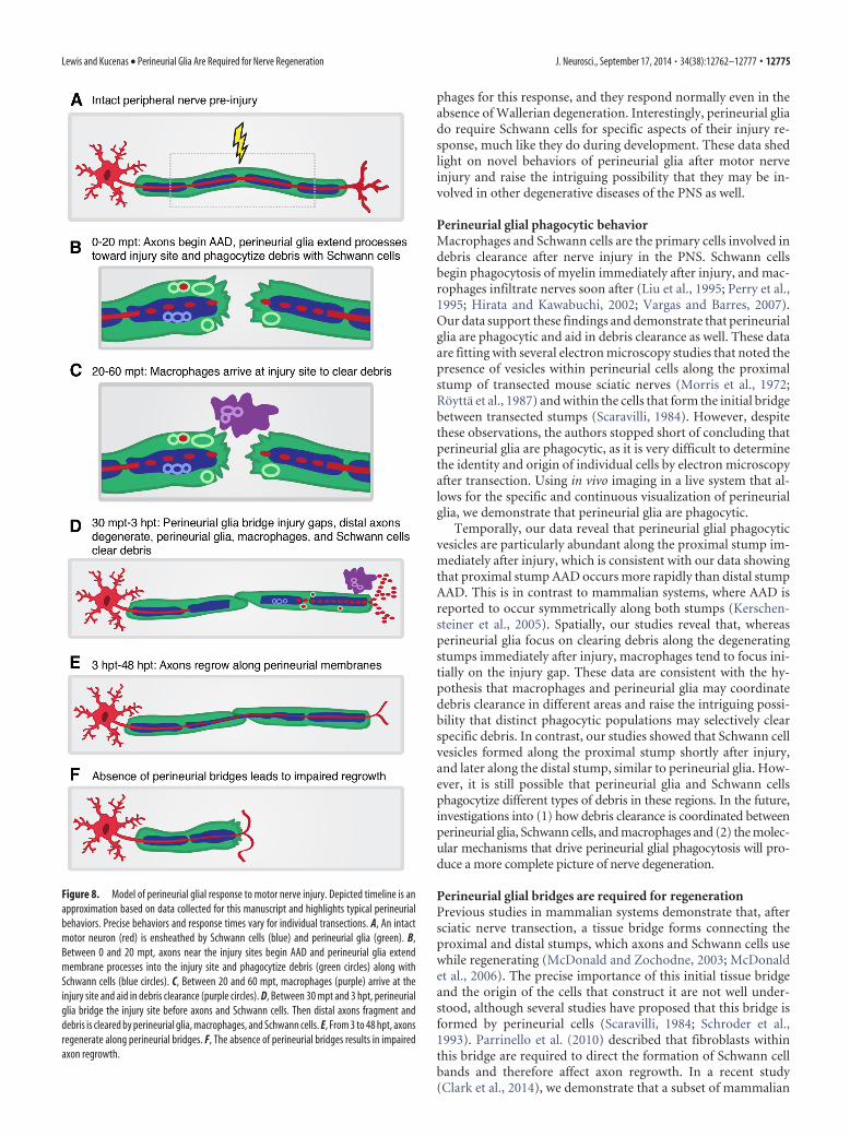
Figure 8. Model of perineurial glial response to motor nerve injury. Depicted timeline is anapproximation based on data collected for this manuscript and highlights typical perineurialbehaviors. Precise behaviors and response times vary for individual transections. A, An intactmotor neuron (red) is ensheathed by Schwann cells (blue) and perineurial glia (green). B,Between 0 and 20 mpt, axons near the injury sites begin AAD and perineurial glia extendmembrane processes into the injury site and phagocytize debris (green circles) along withSchwann cells (blue circles). C, Between 20 and 60 mpt, macrophages (purple) arrive at theinjury site and aid in debris clearance (purple circles). D, Between 30 mpt and 3 hpt, perineurialglia bridge the injury site before axons and Schwann cells. Then distal axons fragment anddebris is cleared by perineurial glia, macrophages, and Schwann cells. E, From 3 to 48 hpt, axonsregenerate along perineurial bridges. F, The absence of perineurial bridges results in impairedaxon regrowth.
Lewis and Kucenas • Perineurial Glia Are Required for Nerve Regeneration J. Neurosci., September 17, 2014 • 34(38):12762–12777 • 12775

perineurial cells express Nkx2.2 and are CNS-derived, similar tozebrafish, raising the intriguing possibility that the fibroblastsdescribed by Parrinello et al. (2010) may be perineurial glia.
Our data unequivocally show that perineurial glia bridge in-jury gaps within the first few hours after transection, and thisbehavior occurs well before regenerating axons or Schwann cellsare found in this region. When we tested the requirement ofperineurial glial bridges by either eliminating perineurial glia orcreating injuries too large to be bridged, axon regrowth was im-paired, frequently resulting in no regrowth and aberrant sprout-ing. Collectively, these data are consistent with the hypothesisthat axons regrow along perineurial glial bridges and that thesebridges are required for efficient regeneration of the nerve. In ourDAPT-treatment studies, we did observe some cases where axonsregrew in the absence of perineurial bridges, and it is possible thatSchwann cells and/or remaining axon guidance cues were suffi-cient to direct axon regrowth. We did not assay Schwann cells inthese studies because previous work in the laboratory has dem-onstrated that a lack of perineurial glia due to Notch perturbationresults in a deficit of peripheral myelin along spinal motor nerves(Binari et al., 2013). Although we cannot rule out that reduced orimproperly differentiated Schwann cells are a factor in our con-clusions, there is currently no method available to eliminateperineurial glia without affecting Schwann cell development. To-gether, our data demonstrate that axon regrowth is most success-ful when perineurial bridges are formed.
What are the signals that drive perineurial glial behavior afternerve injury?Nerve regeneration is a precisely coordinated process requiringcommunication between multiple cell types, and previous studieshave shown that axons, Schwann cells, and macrophages are allcapable of releasing factors that affect neighboring cells (Chen etal., 2007; Zochodne, 2012; Klimaschewski et al., 2013). Becausewe never observed extensive motility in perineurial processes orphagocytosis in the absence of nerve injury, we reasoned thatperineurial activation had to be a result of changes to the nerveenvironment due to transection. Our studies reveal that the ini-tial perineurial glial response to injury is independent of macro-phages, degenerating axons, and other perineurial glia. However,although distal axon fragmentation is not required for perineur-ial responses, we cannot rule out the possibility that signals re-leased by fragmenting axons undergoing AAD, or damaged axonsat the injury site, may elicit perineurial behavior.
We have previously demonstrated that perineurial glia andSchwann cells communicate reciprocally during development(Kucenas et al., 2008; Binari et al., 2013), and Schwann cells areknown to release a variety of factors in response to injury thataffect surrounding cells, including NGF, BDNF, LIF, MCP-1, andTNF" (Vargas and Barres, 2007; Jessen and Mirsky, 2008).Therefore, we hypothesized that these cell populations may com-municate after nerve injury. Using mutants where all motorSchwann cells are absent, we observed that perineurial glia wereless attracted to injury sites. Although we cannot rule out thepossibility that changes in perineurial behavior are the result ofdisrupted perineurial glial differentiation in these mutants, thereis currently no alternative approach to eliminate Schwann cellswithout perturbing perineurial glia.
Our data are consistent with the hypothesis that Schwann cellsmay be involved in either attracting perineurial glia to injury sitesor that their damage during injury releases factors that lead toperineurial invasion into the injury gap. Interestingly, perineurialglia did form phagocytic vesicles in these studies, suggesting that
perineurial attraction and phagocytic behavior are independentactivities. This is reminiscent of what is seen in Drosophila, wheredifferent signaling pathways mediate glial activation and phago-cytosis of debris (Ziegenfuss et al., 2012). This is also consistentwith our data showing perineurial glia do not routinely bridgelarge injury gaps, which suggests that, although perineurial gliacan be activated in the absence of perineurial-derived cues, theymay still coordinate during bridging. Future studies will focus onelucidating the precise mechanisms by which perineurial gliacommunicate with other cell types after nerve injury, and deter-mine whether Schwann cell–perineurial glial signaling is recipro-cal as it is during development.
In conclusion, we demonstrate that perineurial glia are essen-tial for motor nerve regeneration. Immediately after injury,perineurial processes are rapidly attracted to nerve injuries andaid Schwann cells and macrophages in phagocytizing debris.Perineurial glia then bridge injury gaps before axons andSchwann cells, and these bridges are essential for axon regrowth.Collectively, our work highlights the essential function ofperineurial glia in nerve regeneration and provides insight intohow these cells can be used to promote better regeneration innerve injury patients.
ReferencesAdalbert R, Gillingwater TH, Haley JE, Bridge K, Beirowski B, Berek L, Wag-
ner D, Grumme D, Thomson D, Celik A, Addicks K, Ribchester RR,Coleman MP (2005) A rat model of slow Wallerian degeneration(WldS) with improved preservation of neuromuscular synapses. EurJ Neurosci 21:271–277. CrossRef Medline
Akert K, Sandri C, Weibel ER, Peper K, Moor H (1976) The fine structure ofthe perineural endothelium. Cell Tissue Res 165:281–295. Medline
Arthur-Farraj PJ, Latouche M, Wilton DK, Quintes S, Chabrol E, Banerjee A,Woodhoo A, Jenkins B, Rahman M, Turmaine M, Wicher GK, Mitter R,Greensmith L, Behrens A, Raivich G, Mirsky R, Jessen KR (2012) c-Junreprograms Schwann cells of injured nerves to generate a repair cell es-sential for regeneration. Neuron 75:633– 647. CrossRef Medline
Banerjee S, Isaacman-Beck J, Schneider VA, Granato M (2013) A novel rolefor Lh3 dependent ECM modifications during neural crest cell migrationin zebrafish. PLoS One 8:e54609. CrossRef Medline
Binari LA, Lewis GM, Kucenas S (2013) Perineurial glia require notch sig-naling during motor nerve development but not regeneration. J Neurosci33:4241– 4252. CrossRef Medline
Bourne GH (1968) The structure and function of nervous tissue. New York:Academic.
Brosius Lutz AB, Barres BA (2014) Contrasting the glial response to axoninjury in the central and peripheral nervous systems. Dev Cell 28:7–17.CrossRef Medline
Bukrinsky A, Griffin KJ, Zhao Y, Lin S, Banerjee U (2009) Essential role ofspi-1-like (spi-1l) in zebrafish myeloid cell differentiation. Blood 113:2038 –2046. CrossRef Medline
Burkel WE (1967) The histological fine structure of perineurium. Anat Rec158:177–189. CrossRef Medline
Chazotte B (2011) Labeling lysosomes in live cells with LysoTracker. ColdSpring Harbor Protoc 2011:pdb.prot5571. CrossRef Medline
Chen ZL, Yu WM, Strickland S (2007) Peripheral regeneration. Annu RevNeurosci 30:209 –233. CrossRef Medline
Clark JK, O’Keefe A, Mastracci TL, Sussel L, Matise MP, Kucenas S (2014)Mammalian Nkx2.2% perineurial glia are essential for motor nerve de-velopment. Dev Dyn. Advance online publication. Retrieved Jun 30, 2014.doi: 10.1002/dvdy.24158. CrossRef Medline
Dutton KA, Pauliny A, Lopes SS, Elworthy S, Carney TJ, Rauch J, Geisler R,Haffter P, Kelsh RN (2001) Zebrafish colourless encodes sox10 andspecifies non-ectomesenchymal neural crest fates. Development 128:4113– 4125. Medline
Ellett F, Pase L, Hayman JW, Andrianopoulos A, Lieschke GJ (2011) mpeg1promoter transgenes direct macrophage-lineage expression in zebrafish.Blood 117:e49 – e56. CrossRef Medline
Gaudet AD, Popovich PG, Ramer MS (2011) Wallerian degeneration: gain-
12776 • J. Neurosci., September 17, 2014 • 34(38):12762–12777 Lewis and Kucenas • Perineurial Glia Are Required for Nerve Regeneration
ing perspective on inflammatory events after peripheral nerve injury.J Neuroinflammation 8:110. CrossRef Medline
George EB, Glass JD, Griffin JW (1995) Axotomy-induced axonal degener-ation is mediated by calcium influx through ion-specific channels. J Neu-rosci 15:6445– 6452. Medline
Griffin JW, George R, Lobato C, Tyor WR, Yan LC, Glass JD (1992) Macro-phage responses and myelin clearance during Wallerian degeneration:relevance to immune-mediated demyelination. J Neuroimmunol 40:156 –166. Medline
Hirata K, Kawabuchi M (2002) Myelin phagocytosis by macrophages andnonmacrophages during Wallerian degeneration. Microsc Res Tech 57:541–547. CrossRef Medline
Jessen KR, Mirsky R (2008) Negative regulation of myelination: relevancefor development, injury, and demyelinating disease. Glia 56:1552–1565.CrossRef Medline
Kelsh RN, Brand M, Jiang YJ, Heisenberg CP, Lin S, Haffter P, Odenthal J,Mullins MC, van Eeden FJ, Furutani-Seiki M, Granato M, Hammer-schmidt M, Kane DA, Warga RM, Beuchle D, Vogelsang L, Nusslein-Volhard C (1996) Zebrafish pigmentation mutations and the processesof neural crest development. Development 123:369 –389. Medline
Kerschensteiner M, Schwab ME, Lichtman JW, Misgeld T (2005) In vivoimaging of axonal degeneration and regeneration in the injured spinalcord. Nat Med 11:572–577. CrossRef Medline
Kimmel CB, Ballard WW, Kimmel SR, Ullmann B, Schilling TF (1995) Stagesof embryonic development of the zebrafish. Dev Dyn 203:253–310.CrossRef Medline
Kirby BB, Takada N, Latimer AJ, Shin J, Carney TJ, Kelsh RN, Appel B (2006)In vivo time-lapse imaging shows dynamic oligodendrocyte progenitorbehavior during zebrafish development. Nat Neurosci 9:1506 –1511.CrossRef Medline
Klimaschewski L, Hausott B, Angelov DN (2013) The pros and cons ofgrowth factors and cytokines in peripheral axon regeneration, Ed 1. NewYork: Elsevier.
Kristensson K, Olsson Y (1971) The perineurium as a diffusion barrier toprotein tracers. Acta Neuropathol 17:127–138. CrossRef Medline
Kucenas S, Takada N, Park HC, Woodruff E, Broadie K, Appel B (2008)CNS-derived glia ensheath peripheral nerves and mediate motor rootdevelopment. Nat Neurosci 11:143–151. CrossRef Medline
Kucenas S, Wang WD, Knapik EW, Appel B (2009) A selective glial barrierat motor axon exit points prevents oligodendrocyte migration from thespinal cord. J Neurosci 29:15187–15194. CrossRef Medline
Leskovar A, Moriarty LJ, Turek JJ, Schoenlein IA, Borgens RB (2000) Themacrophage in acute neuronal injury: changes in cell numbers over timeand levels of cytokine production in mammalian central and peripheralnervous systems. J Exp Biol 203:1783–1795. Medline
Lewis GM, Kucenas S (2013) Motor nerve transection and time-lapse imag-ing of glial cell behaviors in live zebrafish. J Vis Exp 76.
Liu HM, Yang LH, Yang YJ (1995) Schwann cell properties: 3. C-Fos expres-sion, bFGF production, phagocytosis and proliferation during Walleriandegeneration. J Neuropathol Exp Neurol 54:487– 496. Medline
Lunn ER, Perry VH, Brown MC, Rosen H, Gordon S (1989) Absence ofWallerian degeneration does not hinder regeneration in peripheral nerve.Eur J Neurosci 1:27–33. CrossRef Medline
MacDonald JM, Beach MG, Porpiglia E, Sheehan AE, Watts RJ, Freeman MR(2006) The Drosophila cell corpse engulfment receptor Draper mediatesglial clearance of severed axons. Neuron 50:869 – 881. CrossRef Medline
Martin SM, O’Brien GS, Portera-Cailliau C, Sagasti A (2010) Wallerian de-generation of zebrafish trigeminal axons in the skin is required for regen-eration and developmental pruning. Development 137:3985–3994.CrossRef Medline
McDonald DS, Zochodne DW (2003) An injectable nerve regenerationchamber for studies of unstable soluble growth factors. J Neurosci Meth-ods 122:171–178. CrossRef Medline
McDonald D, Cheng C, Chen Y, Zochodne D (2006) Early events of periph-eral nerve regeneration. Neuron Glia Biol 2:139 –147. CrossRef Medline
Morris JH, Hudson AR, Weddell G (1972) A study of degeneration andregeneration in the divided rat sciatic nerve based on electron micros-copy. Z Zellforsch Mikrosk Anat 124:165–203. CrossRef Medline
Napoli I, Noon LA, Ribeiro S, Kerai AP, Parrinello S, Rosenberg LH, CollinsMJ, Harrisingh MC, White IJ, Woodhoo A, Lloyd AC (2012) A centralrole for the ERK-signaling pathway in controlling Schwann cell plasticity
and peripheral nerve regeneration in vivo. Neuron 73:729 –742. CrossRefMedline
Nguyen QT, Sanes JR, Lichtman JW (2002) Pre-existing pathways promoteprecise projection patterns. Nat Neurosci 5:861– 867. CrossRef Medline
O’Brien GS, Martin SM, Sollner C, Wright GJ, Becker CG, Portera-Cailliau C,Sagasti A (2009) Developmentally regulated impediments to skin rein-nervation by injured peripheral sensory axon terminals. Curr Biol 19:2086 –2090. CrossRef Medline
O’Donnell KC, Vargas ME, Sagasti A (2013) WldS and PGC-1 regulate mi-tochondrial transport and oxidation state after axonal injury. J Neurosci33:14778 –14790. CrossRef Medline
Parrinello S, Napoli I, Ribeiro S, Wingfield Digby P, Fedorova M, ParkinsonDB, Doddrell RD, Nakayama M, Adams RH, Lloyd AC (2010) EphBsignaling directs peripheral nerve regeneration through Sox2-dependentSchwann cell sorting. Cell 143:145–155. CrossRef Medline
Peri F, Nusslein-Volhard C (2008) Live imaging of neuronal degradation bymicroglia reveals a role for v0-ATPase a1 in phagosomal fusion in vivo.Cell 133:916 –927. CrossRef Medline
Perry VH, Brown MC, Gordon S (1987) The macrophage response to cen-tral and peripheral nerve injury. J Exp Med 165:1218 –1223. CrossRefMedline
Perry VH, Tsao JW, Fearn S, Brown MC (1995) Radiation-induced reductionsin macrophage recruitment have only slight effects on myelin degeneration insectioned peripheral nerves of mice. Eur J Neurosci 7:271–280. CrossRefMedline
Prendergast A, Linbo TH, Swarts T, Ungos JM, McGraw HF, Krispin S, Wein-stein BM, Raible DW (2012) The metalloproteinase inhibitor Reck isessential for zebrafish DRG development. Development 139:1141–1152.CrossRef Medline
Rhodes J, Hagen A, Hsu K, Deng M, Liu TX, Look AT, Kanki JP (2005)Interplay of Pu.1 and Gata1 determines myelo-erythroid progenitor cellfate in zebrafish. Dev Cell 8:97–108. CrossRef Medline
Rosenberg AF, Wolman MA, Franzini-Armstrong C, Granato M (2012) Invivo nerve-macrophage interactions following peripheral nerve injury.J Neurosci 32:3898 –3909. CrossRef Medline
Roytta M, Salonen V, Peltonen J (1987) Reversible endoneurial changes af-ter nerve injury. Acta Neuropathol 73:323–329. CrossRef Medline
Scaravilli F (1984) Regeneration of the perineurum across a surgically in-duced gap in a nerve encased in a plastic tube. J Anat 139:411– 424.Medline
Schmidt H, Rickert C, Bossing T, Vef O, Urban J, Technau GM (1997) Theembryonic central nervous system lineages of Drosophila melanogaster.Dev Biol 189:186 –204. CrossRef Medline
Schroder J, May R, Weis J (1993) Perineurial cells are the first to traversegaps of peripheral nerves in silicone tubes. Clin Neurol Neurosurg 95[Suppl]:S78 –S83.
Thomas PK, Jones DG (1967) The cellular response to nerve injury: II. Re-generation of the perineurium after nerve section. J Anat 101:45–55.Medline
Vargas ME, Barres BA (2007) Why Is Wallerian degeneration in the CNS soslow? Annu Rev Neurosci 30:153–179. CrossRef Medline
Villegas R, Martin SM, Donnell KC, Carrillo SA, Sagasti A, Allende ML(2012) Dynamics of degeneration and regeneration in developing ze-brafish peripheral axons reveals a requirement for extrinsic cell types.Neural Dev 7:19. CrossRef Medline
Waller A (1850) Experiments on the section of the glossopharyngeal andhypoglossal nerves of the frog, and observations of the alterations pro-duced thereby in the structure of their primitive fibres. Philos Trans R SocLond B Biol Sci 140:423– 429.
Wang JT, Medress ZA, Barres BA (2012) Axon degeneration: molecularmechanisms of a self-destruction pathway. J Cell Biol 196:7–18. CrossRefMedline
Webber C, Zochodne D (2010) The nerve regenerative microenvironment:early behavior and partnership of axons and Schwann cells. Exp Neurol223:51–59. CrossRef Medline
Witzel C, Rohde C, Brushart TM (2005) Pathway sampling by regeneratingperipheral axons. J Comp Neurol 485:183–190. CrossRef Medline
Ziegenfuss JS, Doherty J, Freeman MR (2012) Distinct molecular pathwaysmediate glial activation and engulfment of axonal debris after axotomy.Nat Neurosci 15:979 –987. CrossRef Medline
Zochodne DW (2012) The challenges and beauty of peripheral nerve re-growth. J Peripher Nerv Syst 17:1–18. CrossRef Medline
Lewis and Kucenas • Perineurial Glia Are Required for Nerve Regeneration J. Neurosci., September 17, 2014 • 34(38):12762–12777 • 12777