Development of Bioabsorbable Materials Based on ...
100
DEVELOPMENT OF BIOABSORBABLE MATERIALS BASED ON POLYCAPROLACTONE (PCL) WITH CONTROLLED DELIVERY OF ACTIVE COMPOUNDS 2015 THESIS MASTER ELENA TORRES ROCA UPV | Universitat Politécnica de ValenciaCampus de Alcoy
Transcript of Development of Bioabsorbable Materials Based on ...
DEVELOPMENT OF
BIOABSORBABLE MATERIALS
BASED ON
POLYCAPROLACTONE (PCL)
WITH CONTROLLED DELIVERY
OF ACTIVE COMPOUNDS
2015
THESIS MASTER ELENA TORRES ROCA
UPV | Universitat Politécnica de ValenciaCampus de Alcoy
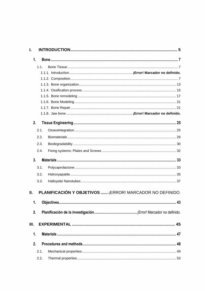
I. INTRODUCTION ............................................................................................ 5
1. Bone ...................................................................................................................................... 7
1.1. Bone Tissue .................................................................................................................... 7
1.1.1. Introduction .................................................................. ¡Error! Marcador no definido.
1.1.2. Composition ................................................................................................................. 7
1.1.3. Bone organization ...................................................................................................... 13
1.1.4. Ossification process .................................................................................................. 15
1.1.5. Bone remodeling ....................................................................................................... 17
1.1.6. Bone Modeling ........................................................................................................... 21
1.1.7. Bone Repair ............................................................................................................... 21
1.1.8. Jaw bone ..................................................................... ¡Error! Marcador no definido.
2. Tissue Engineering ............................................................................................................ 25
2.1. Osseointegration ........................................................................................................... 25
2.2. Biomaterials .................................................................................................................. 26
2.3. Biodegradability ............................................................................................................ 30
2.4. Fixing systems: Plates and Screws .............................................................................. 32
3. Materials ............................................................................................................................. 33
3.1. Polycaprolactone .......................................................................................................... 33
3.2. Hidroxyapatite ............................................................................................................... 35
3.3. Halloysite Nanotubes .................................................................................................... 37
II. PLANIFICACIÓN Y OBJETIVOS ...... ¡ERROR! MARCADOR NO DEFINIDO.
1. Objectives ........................................................................................................................... 43
2. Planificación de la investigación ............................................. ¡Error! Marcador no definido.
III. EXPERIMENTAL ......................................................................................... 45
1. Materials ............................................................................................................................. 47
2. Procedures and methods .................................................................................................. 48
2.1. Mechanical properties ................................................................................................... 49
2.2. Thermal properties ........................................................................................................ 53

2.3. Dynamical Mechanical Analysis (DMA) ........................................................................ 55
2.4. Scanning electron microscope (SEM) .......................................................................... 55
IV. DISCUSSION ............................................................................................... 57
1. Mechanical properties ....................................................................................................... 59
2. Thermal properties ............................................................................................................ 67
3. Dynamical Mechanical Analysis (DMA) ........................................................................... 72
V. CONCLUSIONS ........................................................................................... 74
VI. FUTURE WORKS ........................................................................................ 76
VII. REFERENCE ............................................................................................... 80
VIII. APPENDIX ................................................................................................... 91


I. Summary

Within medical research, efforts are needed in the investigation of the materials
employed in bond fracture applications. Metal prostheses currently used are heavy and
expensive. These metal alloys are biocompatible and highly resistant but do not
promote the bone regeneration and neither are degraded over time. Controlled delivery
of osteogenic biomaterials is an alternative to traditional bone repair techniques.
Some applications such as fixing and screws would have huge advantages if they were
biodegradable in physiologic environment and furthermore it promoted the bound
regeneration. This effect could avoid a second surgical intervention in order to remove
this fixing and screws. Moreover this would entail a faster recuperation of the involved
joint. Such materials capable to fulfill these functions are the biodegradable polymers,
which present also an easy processability and have a lower cost.
The bound regeneration would be accelerated if the fixing and screws were
functionalized with bioactive molecules which promote the mesenchymal stem cell
proliferation in the osteoblast and the calcium deposition.
In short, the treatment of complex bone fractures with fixing and screws is promising.
Specifically if the materials used were osteoconductive and osteoinductive, which are
able to entail the osteogenesis. This is the reason why the present project research
focuses in the intention to study and develop fixings and screws based on
polycaprolactone (PCL) strengthened with Halloysite nanotubes (HNTs) and
hydroxyapatite (HA) in order to enhance the mechanical strength and also to obtain
osteoconductive properties. At the same time, the second step of this project is to
functionalize the Halloysite nanotubes with curcumin in order to give to these fixings
and screws antioxidant, anti-inflammatory and potentially chemotherapeutic properties.


II. Introduction

I. Master Thesis 7
1. Bone
Bone can be considered as man-made engineering materials. However, due to the
nature of its synthesis as an organic material, it is likely to show more variation in
measured properties than typical engineering materials.
1.1. Bone Tissue
The bone tissue is referred to specialize connective tissue which form the skeleton
which has different functions, such as:
• Body support to provide a rigid structure to the muscles.
• Movement, muscles are inserted to the bone through the tendons in order to
allow the movement.
• Protection, since the cavities formed by the skeleton protect different inters
organs.
• Mineral homeostasis because bones has the function to store several ores such
as Calcium (Ca) and Phosphorous (P). [1]
The main benefit of the bone tissue resides in bone regeneration property; this feature
gives the peculiarity of being the only tissue which is able to repair itself without scarfs.
1.1.1. Composition
The bone is a composite material form of diverse cells and an extracellular matrix,
where the principal components are collagen type I, Hydroxyapatite and water [2].
Although the bone composition depends of several factors (age, genetic variability,
environmental effects and skeleton localization of the bone), on average of 70% of its
components correspond to the mineral phase and the remainder corresponds to the
organic phase besides the water (Figure I.1).

8 Capítulo
Inorganic components
The inorganic component is the most abundant component of the bone, composed of a
95% of apatite crystals, which is analogous to Hidroxyapatite ore present in the nature
(HAP, (PO4)6(OH)2 Ca10). This bone apatite (hydroxyapatite) is a deficient in calcium
and hydroxide, containing 5% of impurities such as sodium, potassium, fluorides,
citrates, carbonates, pyrophosphates and chlorides among others [3-6] . These
Prot. Extracell85% Prot. Cell
15%
Figure II.1 Bone composition
I. Master Thesis 9
crystals, produced by osteoblasts, have approximately 30nm in size and form an
imperfect crystalline network, facilitating the absorption of ions and their dissolution by
the osteoclast. These apatite crystals are deposited between the collagen fibers, giving
stiffness to the bone.
Organic components
Collagen Type I is the major organic component of the bone. Collagen is synthetized
by osteoblast; this protein gives tensile strength, elasticity and flexibility to the bone.
Moreover, it offers a perfect place to produce nucleation and hydroxiapatite crystals
growth [7-9]. There are different collagen types, but the most abundant is collagen type
I with an average of 95%, while the remaining 5% corresponds to collagen type V.
Collagen molecules are composed by a triplex, with a diameter of approximately 1.5
nm in size with a twist to the right, integrated for three polypeptide chains namely α-
chain.
Every α-chain is composed by a repetitive characteristic structure through all the three
amino acids chain, “glycine-X-Y, where X and Y mainly are proline and hydroxyproline
respectively [7].
In addition to collagen type I and HPA, others proteins form the extracellular bone
matrix, such as proteoglycans, osteocalcin, osteonectin, osteopotina, sialoproteins,
fibronectin, thrombospondin, vitronectin and growth factors [10-12].

10 Capítulo
Frotein (Chromosome) Function
Collagen
type I (17q21.23, 7q22.1) The most abundant protein in the
extracellular bone matrix.
type X (6q21) Found in hypertrophic cartilage
type III (2q31) Small amounts in bone, can regulate
the diameter of fibrillar collagen
type V (9q34.2-34.3; 2q24.3-31;
19q13.2)
Small amounts in bone, can regulate
the diameter of fibrillar collagen
Whey proteins in the bone matrix (4q11-13) Decrease the growth of HAP crystals
Proteins which
contain
glicoaminoglicano
and proteins
which contain
leucine rich
regions
Aggrecan (15q26.1) Matrix organization; retention of
calcium and phosphorus
Versican (5q14.3) Defines the space destined to become
bone
Decorin (12q21.3) regulates the diameter of collagen
fibers; union to TGF-β
Biglycan (Xq28)
Union to collagen, union to TGF-β;
genetic determinant of maximum
bone mass
Hyaluronan
(multigenic complex)
Can work together with versican to
define the space designated to
became bone
Glycoproteins
Alkaline phosphatase (1p36.1-
p34)
hydrolyze mineral deposition
inhibitors
Osteonectin (5q31.3-32) controls the collagen fibers diameter
SIBLINGS proteins Osteopontin (4q21)
Inhibits bone mineralization and
remodeling
Bone sialoprotein (4q21) Mineralization initiator
MEPE (4q21.1) Phosphate metabolism regulator
glycoproteins
containing RGD
Thrombospondin (15q15, 6q27,
1q21, 5q13, 19p13,1) Cell adhesion
Fibronectin (2q34) Cell union
I. Master Thesis 11
Vitronectin (17q11) Cell adhesion
Fibrillin 1 and 2 (15q21.1, 5q23-
31)
Regulates the formation of elastic
fibers
Proteins which
contain γ-
carboxyglutamic
acid
Matrix Gla proteins (12q13.1-
p21.3 Mineralization inhibitor
Osteocalcin (1q25-q31) Osteoclast regulator; mineralization
inhibitor
S Protein (3p11.2) Liver product, can be synthesized by
osteoblasts
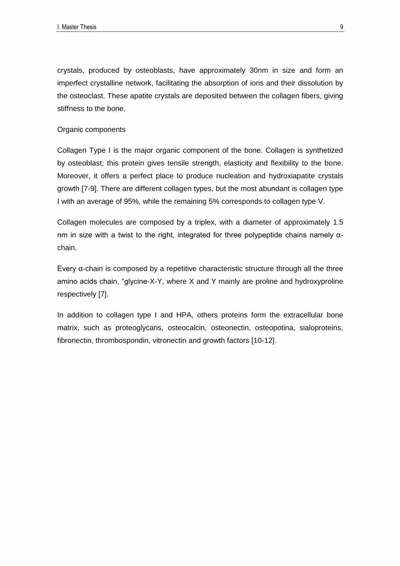
Table II-1 Main proteins constituents of the bone extracellular matrix and functions.
Adapted from Clarke, 2008 [10]
Bone cells
Cells implicated in the bone metabolism are: mesenchymal stem cells which are
multipotent stromal cells that can be differentiated into a variety of cell types.
Osteoblasts are cells responsible for the formation of the bone tissue. Osteocytes and
Osteoclast (from the monocytic line) are responsible for the bone resorption.
o Mesenchymal stem Cells
Constitute a kind of stems cells namely as well progenitor cells. Each of these cells are
defined to their functional features, which are: undifferentiated, self-maintenance,
undifferentiated progeny production and tissue regeneration [13].
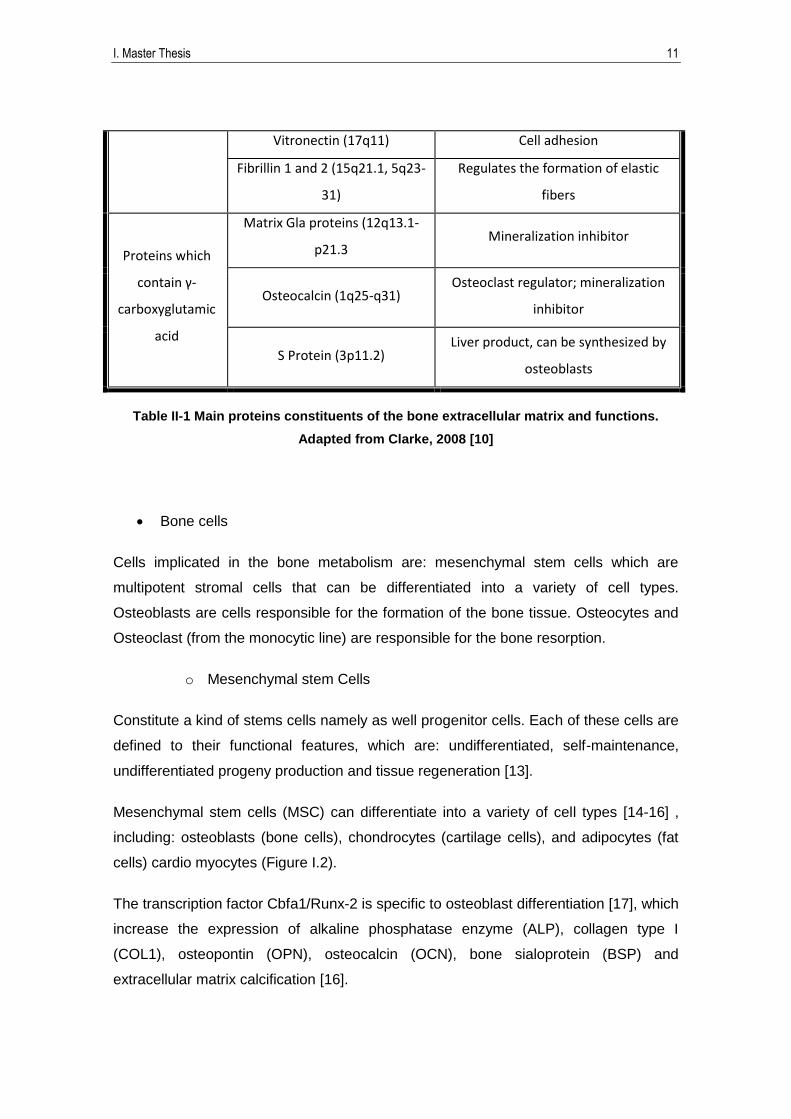
Mesenchymal stem cells (MSC) can differentiate into a variety of cell types [14-16] ,
including: osteoblasts (bone cells), chondrocytes (cartilage cells), and adipocytes (fat
cells) cardio myocytes (Figure I.2).
The transcription factor Cbfa1/Runx-2 is specific to osteoblast differentiation [17], which
increase the expression of alkaline phosphatase enzyme (ALP), collagen type I
(COL1), osteopontin (OPN), osteocalcin (OCN), bone sialoprotein (BSP) and
extracellular matrix calcification [16].
12 Capítulo
o Osteoblast
Osteoblasts are cells located on the surface of bone tissue. Their function is to produce
bone tissue, forming collagen type I and calcifying it [18]. Osteoblasts are produced by
MSCs differentiations, they have a diameter average size of 25µm and a polyhedral
shape. Osteoblasts have a highly basophilic cytoplasm due to high ARN production, an
extensive endoplasmic reticulum and a highly developed Golgi apparatus, as evidence
of its high synthetic activity [1, 19].
Moreover, osteoblasts show great alkaline phosphatase enzyme activity in the
cytoplasmic membrane. This enzyme performs a key role in the bone mineralization
[18].
o Osteocytes
After bone formation, between 10% and 20% of osteoblast are housed in the bosom of
bone and gradually are differentiated in osteocytes [1, 20].
Osteocytes are the most abundant cells in bone tissue. Its population may reach 90%
of total cellular constituents [21, 22]. Located in the osteocytic gaps, osteocytes are
more elongated than osteoblast, with a bigger nucleus and a less developed
Figure II.2 Cell differentiation scheme
I. Master Thesis 13
endoplasmic reticulum. Its cytoplasm is basophilic and has cytoplasmic prolongations,
which are inserted into bone canaliculi and kept in touch with others osteocites and
osteoblast neighbors. This disposition enables to form an extracellular network and lets
cellular communication among bone bosom cells and bone cells surface [21]. The
nutrition of these cells depends on the connections through the canaliculis.
Osteocytes can detect mechanical changes and translate them into biochemical
signals to act directly on the bone. Thus, the osteocyte network can address the bone
remodeling and fix micro fractures [21, 23].
o Osteoclast
Osteoclasts are the only known cells able to break down the bone. Osteoclasts are
formed by the fusion of monocytes which should have abandoned the circulating blood;
hence, osteoclasts are derived from hematopoietic stem cells instead of mesenchymal
cells. Osteoclasts are cells which have several cytological features such as:
Large quantity of mitochondria and lysosomes
Are multinuclear
Are highly polarized
Their mission is to dissolve HAP crystals and digest the organic matrix by secreting
diverse enzymes, highlighting proteases as the most important whose task is focused
on bone breakdown and bone reabsorption [1].
1.1.2. Bone organization
Bone organization can be classified in different ways. Regarding its ripeness, it can be
distinguish between immature bundle bone (also called woven bone) and mature
lamellar bone.
Immature bundle bone is present in fetal development, newborns, fractured healing
and growing metaphysis. This kind of bone is characterized by exhibiting great number
of cells, its bulkiness and having thick non oriented collagen fibers type I, which
provides mechanical isotropy to the tissue.

14 Capítulo
However, mature lamellar bone is formed in immature bundle bone. Primary bone
contains interlaced collagen fibers. The immature primary bone is present in the
developing skeletal system and bone regeneration, being subsequently replaced by
lamellar bone tissue. Mature lamellar bone can be found in the entire mature skeleton,
including both cancellous bone (spongy bone) and cortical bone. All of its collagen
fibers are organized and oriented with the bone major axis. This fact confers
mechanical anisotropy to the tissue which shows deformation resistance when the
strength is parallel to the bone major axis.
Another classification could be granted regarding its structure, distinguishing between
spongy bone (trabecular bone) and compact bone (cortical bone) (Figure I.3).
The ratio (trabecular:cortical) bone is stablishes as 1:4, although depending on the
location it could exhibit different ratio [10].
• Trabecular bone (spongy bone) has a highly porous variable structure between
50 and 90% with interconnected pores. It forms series of trabeculae by plates and rods
where is located the bone marrow.
• Cortical bone (compact bone) is denser than trabecular bone, with a 5% porous
structure. Cortical bone is set out on bone surface, varying its thickness depending on
its location. It is distributed in parallel disposition following the bone major axis forming
cylindrical structures denominated osteons or Haversian system, which are wrapped
Figure II.3 Bone structure scheme
I. Master Thesis 15
forming concentric layers (Figure I.3). Cortical bone external surface is limited by the
periosteum and the internal surface by the endosteum. The cellular activity of
periosteum surface is important regarding to appositional growth and fracture repair.
Endosteum surface exhibits higher remodeling action than periosteum surface, likely,
because of a high exposition of cytokines, due to bone marrow proximity [10].
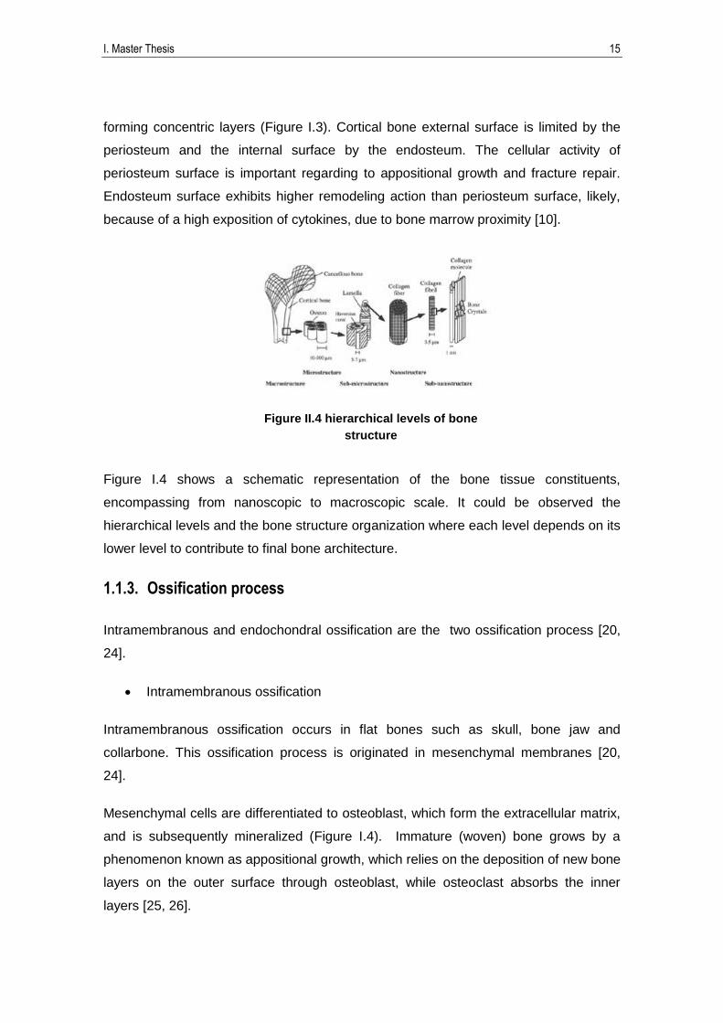
Figure I.4 shows a schematic representation of the bone tissue constituents,
encompassing from nanoscopic to macroscopic scale. It could be observed the
hierarchical levels and the bone structure organization where each level depends on its
lower level to contribute to final bone architecture.
1.1.3. Ossification process
Intramembranous and endochondral ossification are the two ossification process [20,
24].
Intramembranous ossification
Intramembranous ossification occurs in flat bones such as skull, bone jaw and
collarbone. This ossification process is originated in mesenchymal membranes [20,
24].
Mesenchymal cells are differentiated to osteoblast, which form the extracellular matrix,
and is subsequently mineralized (Figure I.4). Immature (woven) bone grows by a
phenomenon known as appositional growth, which relies on the deposition of new bone
layers on the outer surface through osteoblast, while osteoclast absorbs the inner
layers [25, 26].
Figure II.4 hierarchical levels of bone
structure
16 Capítulo
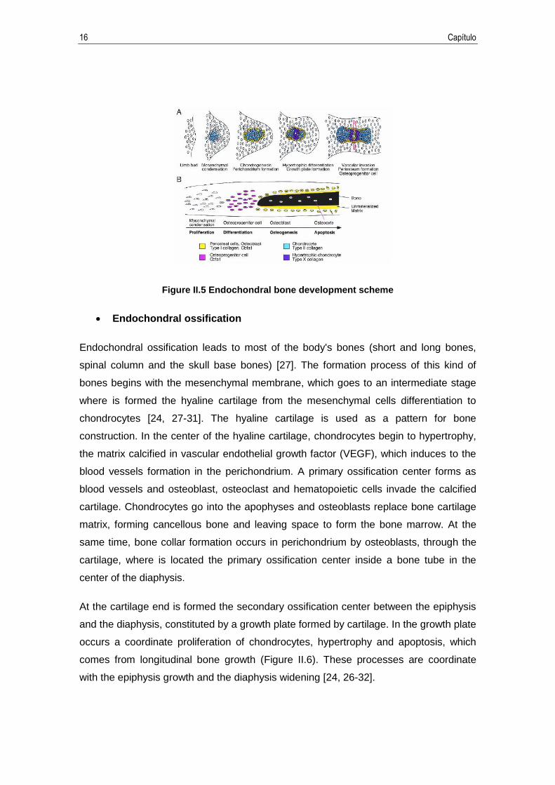
Figure II.5 Endochondral bone development scheme
Endochondral ossification
Endochondral ossification leads to most of the body's bones (short and long bones,
spinal column and the skull base bones) [27]. The formation process of this kind of
bones begins with the mesenchymal membrane, which goes to an intermediate stage
where is formed the hyaline cartilage from the mesenchymal cells differentiation to
chondrocytes [24, 27-31]. The hyaline cartilage is used as a pattern for bone
construction. In the center of the hyaline cartilage, chondrocytes begin to hypertrophy,
the matrix calcified in vascular endothelial growth factor (VEGF), which induces to the
blood vessels formation in the perichondrium. A primary ossification center forms as
blood vessels and osteoblast, osteoclast and hematopoietic cells invade the calcified
cartilage. Chondrocytes go into the apophyses and osteoblasts replace bone cartilage
matrix, forming cancellous bone and leaving space to form the bone marrow. At the
same time, bone collar formation occurs in perichondrium by osteoblasts, through the
cartilage, where is located the primary ossification center inside a bone tube in the
center of the diaphysis.
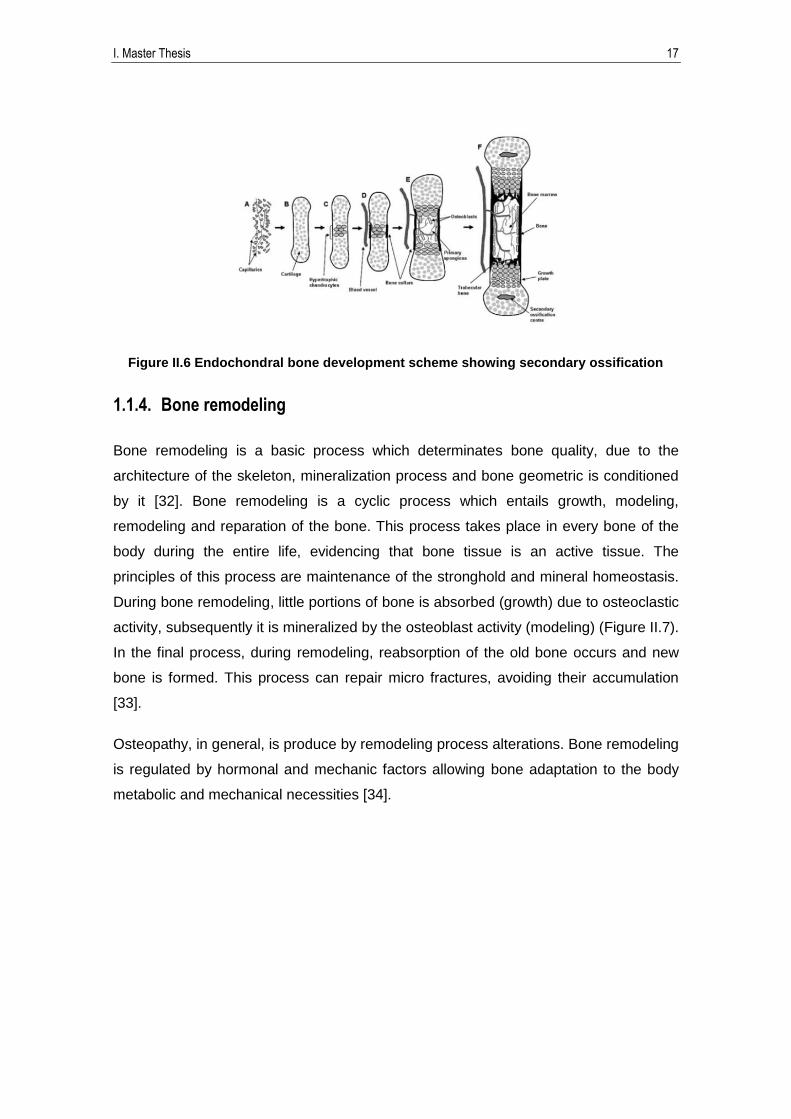
At the cartilage end is formed the secondary ossification center between the epiphysis
and the diaphysis, constituted by a growth plate formed by cartilage. In the growth plate
occurs a coordinate proliferation of chondrocytes, hypertrophy and apoptosis, which
comes from longitudinal bone growth (Figure II.6). These processes are coordinate
with the epiphysis growth and the diaphysis widening [24, 26-32].
I. Master Thesis 17
Figure II.6 Endochondral bone development scheme showing secondary ossification
1.1.4. Bone remodeling
Bone remodeling is a basic process which determinates bone quality, due to the
architecture of the skeleton, mineralization process and bone geometric is conditioned
by it [32]. Bone remodeling is a cyclic process which entails growth, modeling,
remodeling and reparation of the bone. This process takes place in every bone of the
body during the entire life, evidencing that bone tissue is an active tissue. The
principles of this process are maintenance of the stronghold and mineral homeostasis.
During bone remodeling, little portions of bone is absorbed (growth) due to osteoclastic
activity, subsequently it is mineralized by the osteoblast activity (modeling) (Figure II.7).
In the final process, during remodeling, reabsorption of the old bone occurs and new
bone is formed. This process can repair micro fractures, avoiding their accumulation
[33].
Osteopathy, in general, is produce by remodeling process alterations. Bone remodeling
is regulated by hormonal and mechanic factors allowing bone adaptation to the body
metabolic and mechanical necessities [34].
18 Capítulo
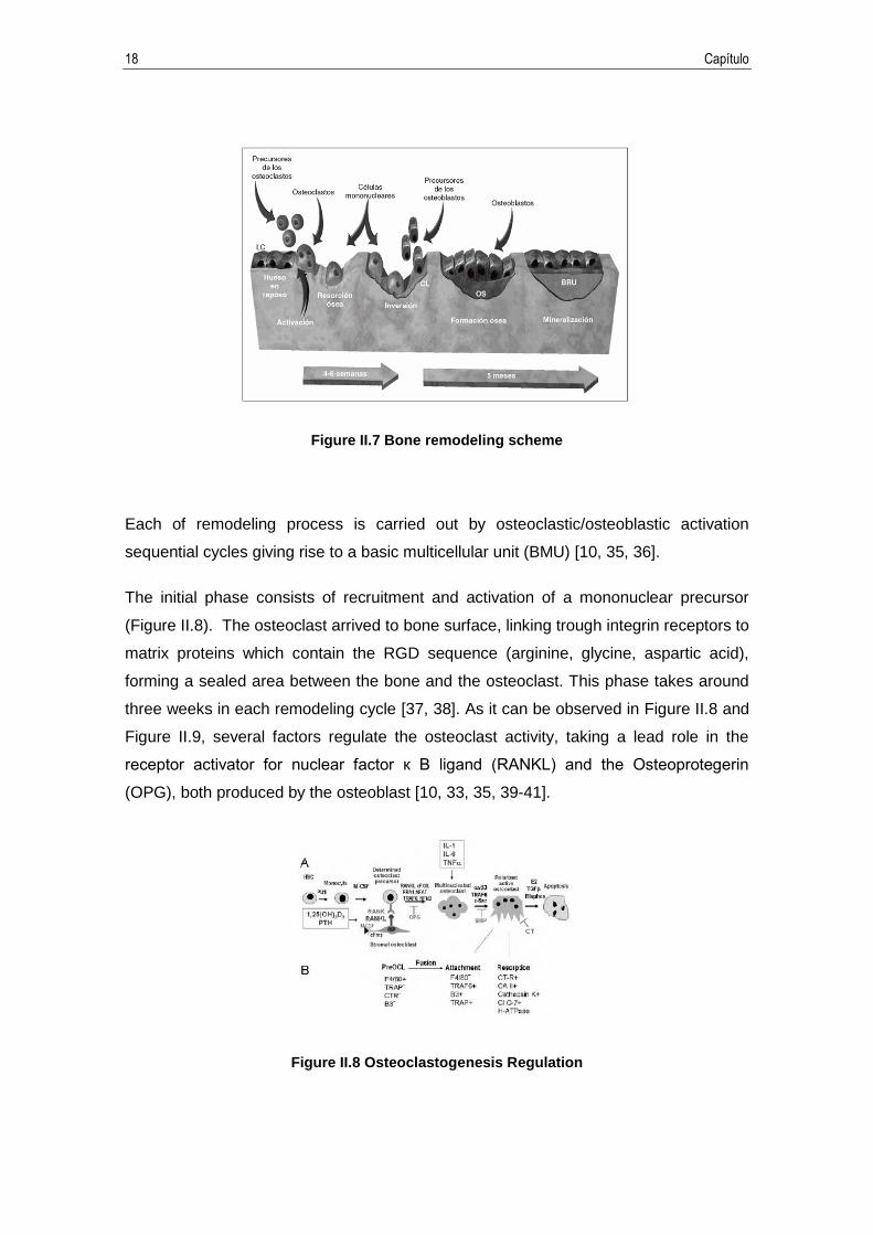
Figure II.7 Bone remodeling scheme
Each of remodeling process is carried out by osteoclastic/osteoblastic activation
sequential cycles giving rise to a basic multicellular unit (BMU) [10, 35, 36].
The initial phase consists of recruitment and activation of a mononuclear precursor
(Figure II.8). The osteoclast arrived to bone surface, linking trough integrin receptors to
matrix proteins which contain the RGD sequence (arginine, glycine, aspartic acid),
forming a sealed area between the bone and the osteoclast. This phase takes around
three weeks in each remodeling cycle [37, 38]. As it can be observed in Figure II.8 and
Figure II.9, several factors regulate the osteoclast activity, taking a lead role in the
receptor activator for nuclear factor к B ligand (RANKL) and the Osteoprotegerin
(OPG), both produced by the osteoblast [10, 33, 35, 39-41].
Figure II.8 Osteoclastogenesis Regulation

I. Master Thesis 19
Figure II.9 Interaction among the osteocytes, osteoblasts and osteoclasts in bone
remodeling
Once the osteoclast is fixed in the bone, tissue erosion begins, to perform it osteoclasts
release H+ through ATP-dependent proton pumps (H+ ATPasa) with the propose of
getting a constant PH of 4.5 inside the resorption chamber to make easier the HAP
removal. Moreover, degradation of extracellular matrix by osteoclasts release enzyme,
such as acid phosphatase, tartrate resistant, cathepsin K, metalloproteinase matrix
and cytoplasmic gelatin lysosomes; results in resorption of the Howship´s lacuna with
approximately 50µm in length in trabecular bone and 100µm in cortical bone [18, 38]
(Figure II.9). This process takes around 2-3 weeks. Once resorption is finished,
osteoclasts are eliminated by the apoptosis.
After resorption phase, remodeling entails a formation process, in which osteoblasts
differ from their precursors (Figure II.10)[42]. It is presume that final resorption process
and beginning of formation process involve different factors such as transforming
growth factor beta (TGF-β), insulin-like growth factor 1 and 2 (IGF-1 and IGF-2), bone
morphogenetic proteins (BMPs) and fibliblast growth factor (PDGF). The coupling of
these two processes takes 1-2 weeks [18, 33, 39, 41, 42].
Bone formation takes between 4 or 5 months[10, 38]. During this time, osteoblastic
cells secrete extracellular matrix and vesicles containing high amount of Ca2+ and
phosphates concentrations, in order to get collage mineralization. Formation process
has two phases: matrix synthesis and mineralization. Once osteoblasts are in contact
with the surface; osteoid (formed by collagen I fibers, which are alienated before
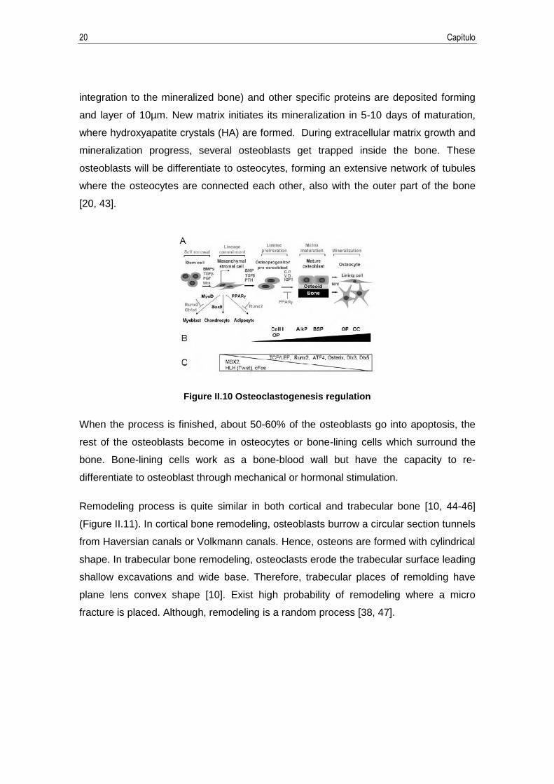
20 Capítulo
integration to the mineralized bone) and other specific proteins are deposited forming
and layer of 10µm. New matrix initiates its mineralization in 5-10 days of maturation,
where hydroxyapatite crystals (HA) are formed. During extracellular matrix growth and
mineralization progress, several osteoblasts get trapped inside the bone. These
osteoblasts will be differentiate to osteocytes, forming an extensive network of tubules
where the osteocytes are connected each other, also with the outer part of the bone
[20, 43].
Figure II.10 Osteoclastogenesis regulation
When the process is finished, about 50-60% of the osteoblasts go into apoptosis, the
rest of the osteoblasts become in osteocytes or bone-lining cells which surround the
bone. Bone-lining cells work as a bone-blood wall but have the capacity to re-
differentiate to osteoblast through mechanical or hormonal stimulation.
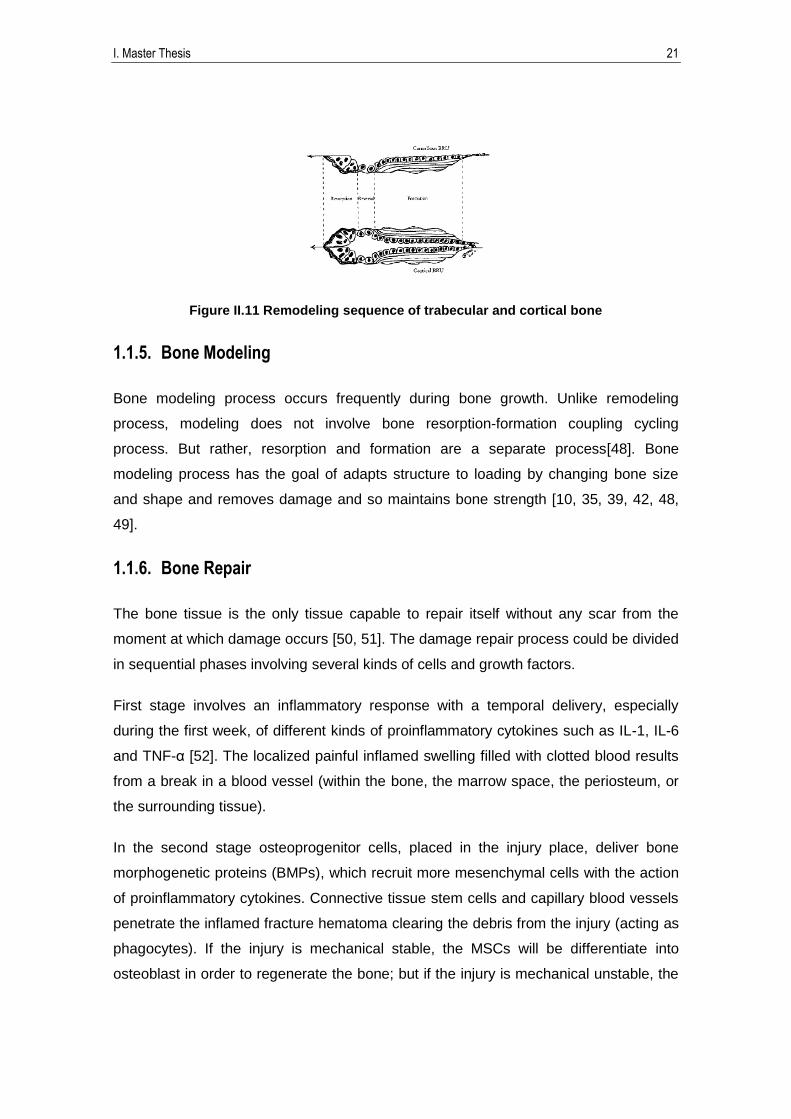
Remodeling process is quite similar in both cortical and trabecular bone [10, 44-46]
(Figure II.11). In cortical bone remodeling, osteoblasts burrow a circular section tunnels
from Haversian canals or Volkmann canals. Hence, osteons are formed with cylindrical
shape. In trabecular bone remodeling, osteoclasts erode the trabecular surface leading
shallow excavations and wide base. Therefore, trabecular places of remolding have
plane lens convex shape [10]. Exist high probability of remodeling where a micro
fracture is placed. Although, remodeling is a random process [38, 47].
I. Master Thesis 21
Figure II.11 Remodeling sequence of trabecular and cortical bone
1.1.5. Bone Modeling
Bone modeling process occurs frequently during bone growth. Unlike remodeling
process, modeling does not involve bone resorption-formation coupling cycling
process. But rather, resorption and formation are a separate process[48]. Bone
modeling process has the goal of adapts structure to loading by changing bone size
and shape and removes damage and so maintains bone strength [10, 35, 39, 42, 48,
49].
1.1.6. Bone Repair
The bone tissue is the only tissue capable to repair itself without any scar from the
moment at which damage occurs [50, 51]. The damage repair process could be divided
in sequential phases involving several kinds of cells and growth factors.
First stage involves an inflammatory response with a temporal delivery, especially
during the first week, of different kinds of proinflammatory cytokines such as IL-1, IL-6
and TNF-α [52]. The localized painful inflamed swelling filled with clotted blood results
from a break in a blood vessel (within the bone, the marrow space, the periosteum, or
the surrounding tissue).
In the second stage osteoprogenitor cells, placed in the injury place, deliver bone
morphogenetic proteins (BMPs), which recruit more mesenchymal cells with the action
of proinflammatory cytokines. Connective tissue stem cells and capillary blood vessels
penetrate the inflamed fracture hematoma clearing the debris from the injury (acting as
phagocytes). If the injury is mechanical stable, the MSCs will be differentiate into
osteoblast in order to regenerate the bone; but if the injury is mechanical unstable, the

22 Capítulo
MSCs will be differentiate into chondrocytes to form a collagen layer acting as a bridge
between both fracture ends; resulting in fibrocartilaginous callus (soft callus) (Figure
II.12). Procallus material usually extends beyond the volume previously occupied by
the uninjured bone.
In the final stage (several weeks to months in duration) the soft callus is consequently
calcified and forms bony callus (hard callus), which will be remodeling to form spongy
bone. In the healing of a bone fracture; osteoclasts continue to dissolve away the
fibrous and cartilaginous components of the injury site, while osteoblasts continue to
replace the material with new bone matrix. Bone remodeling will continue until normal
dimensions and composition of the bone are recreated; it represents the third and final
stage in repair of a bone fracture, though some additional bone remodelling will often
follow over time [50, 51, 53, 54].
Figure II.12 Scheme of a bone callus during regeneration
1.1.7. Craniofacial injuries
Skull injures are the most frequent trauma seen in urban trauma centers [55, 56],
leading causes of death and disability worldwide [57]. Accordingly big efforts are
focused on the research of new materials capable to fulfill the requirements for bone
fracture remodeling.
I. Master Thesis 23
Elderly people is the most affected collective of craniofacial injuries, due to with normal
aging, loss of estrogen increases and incurs bone loss and osteoporosis, which is
especially prominent in women after menopause [58]. According to National Institute on
Aging (US Department of Health and Human Services), in 2014, an estimated 8.1%
percent of the world’s population was over 61 years old. By 2050, this number is
expected to triplicate.
Considering that compositions of cranial bone change with the age, physical properties
are affected by this differences. A thin and non-homogeneous cortical bone layer
compose fetal cranial bones, whereas mature adult cranial bones consist in a stiff inner
and outer strata of cortical bone and a lightweight trabecular layer between them [59].
Care should be taken when both mechanical properties are compared. According the
literature present for now, the most studies found related to cranial bone mechanical
properties examine fetal or infant cranial bone [60-62]. Nevertheless, few studies have
reported adult cranial bone [59, 63, 64]. Table II-1 shown below summarized a review
of the literature comparing the reported average elastic moduli and maximum energy
for failure for human cranial bone. It can be noticed a high degree of inter-individual
variation between values. It must be considered that some factors influenced stiffer and
stronger values of each skull, for example:
Impact speed influences elastic modulus and maximum energy for failure,
higher average speed increases those values.
Morphological differences between individual cranial vaults play an important
role, implying porosity, thickness and radius curvature.
Bone site entails different composition and morphology of the bone between
parietal and frontal bond. Parietal bone tends to be less thick, more porous and
also has a lower percentage of bone volume compared to frontal bone [59]. All
these facts entail that parietal bone resist less forces at fracture and also
absorbs less energy before fracture.

24 Capítulo
Gestation Ref Child Ref Adult Ref
Elastic
modulus
25 weeks
0.071 GPa [60]
6 months
3.58 GPa [60]
9.56 GPa [65] 1 year
0.461 GPa [61]
40weeks
3.88 GPa [66] 6 years
7.38 GPa [66]
Maximum
energy
for failure
30 weeks
0.0031 GPa [60]
6 months
0.0446 GPa [60]
0.12784 GPa [59] 1 year
0.03023 GPa [61]
Table II-2 a review of the literature comparing the reported average elastic moduli and
maximum energy for failure for human cranial bone
Focusing our attention in adult cranial bone, different mechanical properties can be
noticed depending on the specimen. As it was aforementioned, several factors
influence the morphology and consequently the mechanical properties. Motherway et
al. [59] state the maximum force of failure significantly affect depending of the position
of the bond regarding to parietal and frontal bond. Also, the energy absorbed until
failure is higher for frontal bound. In that study, it was noticed that elastic modulus and
maximum bending stress increase with the increase in the percentage bone volume.
Moreover, the author states that parietal bone tends to be less thick, more porous and
also has a lower percentage of bone volume compared to frontal bone. All these facts
entail that parietal bone resist less forces at fracture and also absorbs less energy
before fracture.
From the statistical analysis, it can be noticed that mechanical properties show a
dependence with the loading rate. It was found that higher speed entails noticeably
higher maximum forces, 7.46±5.39 GPa (0.5 m/s), 10.77±9.38 GPa (1.0 m/s) and
15.54±10.29 GPa (2.5 m/s). Similar results were obtained to maximum bending stress,
which was significantly larger at the highest speed. Nevertheless, the correlation
between strain rate and elastic modulus is not significant.
I. Master Thesis 25
2. Tissue Engineering
Tissue engineering relies on development of biomaterials combining scaffolds, cells,
and biologically active molecules into functional tissues. The goal of tissue engineering
is to assemble functional structures that restore, maintain, or improve damaged tissues
or whole organs.
2.1. Osseointegration
Osseointegration is defined as “formation of a direct interface between an implant and
the bone, without intervening soft tissue” [67]. This term refers to direct structural
adaptation and functional connection between living bone and the surface of a load-
bearing artificial implant. Process success depends on both previous processes
function: osteoinduction and osteoconduction [68].
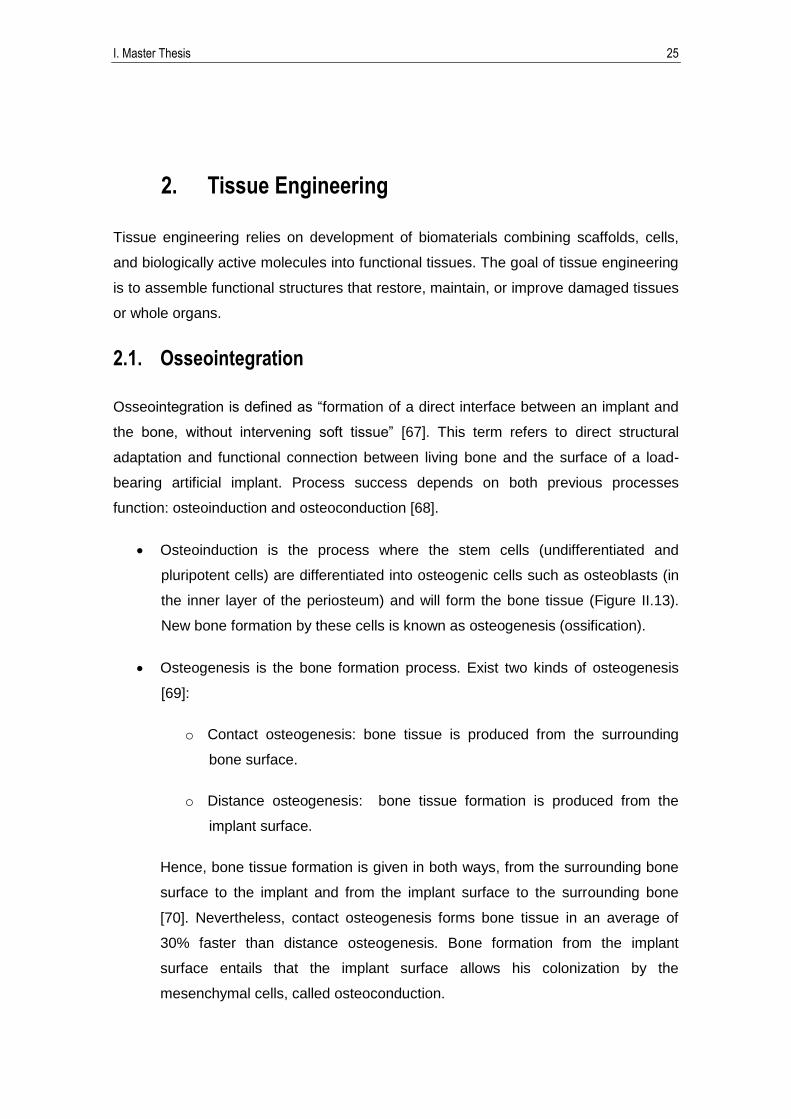
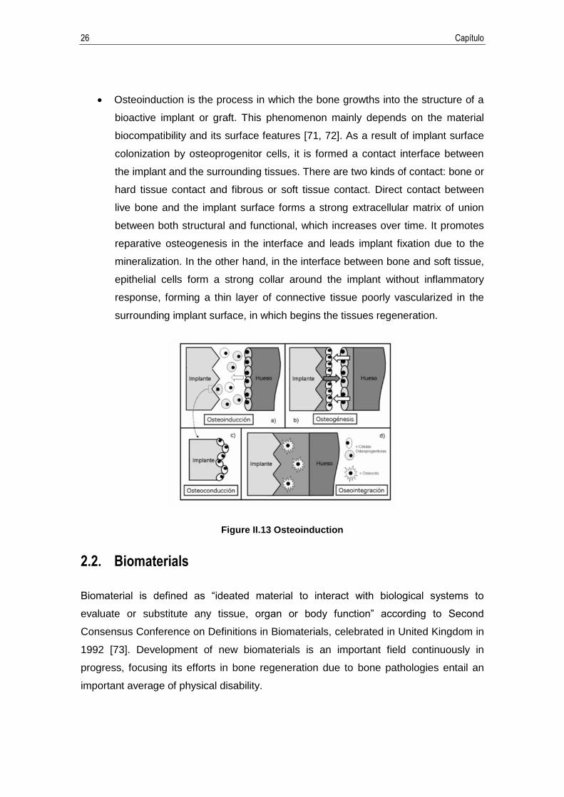
Osteoinduction is the process where the stem cells (undifferentiated and
pluripotent cells) are differentiated into osteogenic cells such as osteoblasts (in
the inner layer of the periosteum) and will form the bone tissue (Figure II.13).
New bone formation by these cells is known as osteogenesis (ossification).
Osteogenesis is the bone formation process. Exist two kinds of osteogenesis
[69]:
o Contact osteogenesis: bone tissue is produced from the surrounding
bone surface.
o Distance osteogenesis: bone tissue formation is produced from the
implant surface.
Hence, bone tissue formation is given in both ways, from the surrounding bone
surface to the implant and from the implant surface to the surrounding bone
[70]. Nevertheless, contact osteogenesis forms bone tissue in an average of
30% faster than distance osteogenesis. Bone formation from the implant
surface entails that the implant surface allows his colonization by the
mesenchymal cells, called osteoconduction.
26 Capítulo
Osteoinduction is the process in which the bone growths into the structure of a
bioactive implant or graft. This phenomenon mainly depends on the material
biocompatibility and its surface features [71, 72]. As a result of implant surface
colonization by osteoprogenitor cells, it is formed a contact interface between
the implant and the surrounding tissues. There are two kinds of contact: bone or
hard tissue contact and fibrous or soft tissue contact. Direct contact between
live bone and the implant surface forms a strong extracellular matrix of union
between both structural and functional, which increases over time. It promotes
reparative osteogenesis in the interface and leads implant fixation due to the
mineralization. In the other hand, in the interface between bone and soft tissue,
epithelial cells form a strong collar around the implant without inflammatory
response, forming a thin layer of connective tissue poorly vascularized in the
surrounding implant surface, in which begins the tissues regeneration.
Figure II.13 Osteoinduction
2.2. Biomaterials
Biomaterial is defined as “ideated material to interact with biological systems to
evaluate or substitute any tissue, organ or body function” according to Second
Consensus Conference on Definitions in Biomaterials, celebrated in United Kingdom in
1992 [73]. Development of new biomaterials is an important field continuously in
progress, focusing its efforts in bone regeneration due to bone pathologies entail an
important average of physical disability.
I. Master Thesis 27
Originally, bone implants approach only focused on achieving development of materials
with the ability of supporting mechanic efforts in the fractured bone. Furthermore,
materials should be biocompatible. This category includes metallic materials such as
stainless steel and titanium alloys. Nevertheless, these systems need a second
chirurgical intervention to remove the devise; entailing higher costs and promoting
infections. Moreover, all material introduced in the body produce a response, albeit
minimal.
In recent years, several authors such as Ikada [74], Meyer [75] and Barrere [76]
describe bone implant materials with the following properties:
Biocompatibility: integration in the organism without immune response, cytotoxic
or genotoxic effects. This is a fundamental biomaterial property.
Biodegradability: decomposition process (by hydrolysis) at rates that are as
close as possible to the rates of new bone formation. This entails a challenge to
the biocompatibility, due to degradation products should not be toxic.
Strength and mechanical compatibility: Resisting mechanical stress as the
position of the bone tissue it replaces. Mechanical properties, for example
elasticity modulus, should be the most closer possible to the replaced tissue to
prevent bone loss associated with the use of bone grafts or stress shielding.
Osteoinductivity: promoting fixation and specific cells formation of the bone
tissue. This is achieved with the recruit mesenchymal stem cells and
osteoprogenitor with subsequent proliferation and differentiation into osteogenic
line.
Osteoconductivity: acting as a structural support in the formation and growth of
new bone. This property is combined with biodegradability because the implant
material should be absorbed to provide space to the new tissue.
Radiolucency: radiographically distinguishable with regard to the tissue where it
is implanted.
Development in tissue engineering regarding biomaterials require materials which
fulfills these properties in different extents, with an appropriate response of the host.

28 Capítulo
There are material families with features that confer to the materials specific clinical
applications. The biomaterial selection depends on the application.
Organic materials
Natural materials are those which use bone tissue from the same individual (autograft),
from different individual of the same species (allograft) or from different species
(xenograft). Autografts usually fulfill the three desirable properties of an implant;
osteoinductivity, osteoconductivity and osteogenicity. The disadvantages of this are the
high cost and complexity of the chirurgical process. Furthermore, invasive surgeries
present postoperative complications. Allografts require treatments such as freeze
drying, irradiation, acid wash, etc. to prevent rejection by the recipient and eliminate
possible infection in the implantable tissue. Xerografts, such as bovine bone also
present some disadvantages from contagious diseases.
Ceramic materials
Ceramics are inorganic not metallic materials with a crystalline structure, obtained from
high temperature and pressure process [77]. In orthopedic applications are generally
used two types of ceramic materials: metallic oxides and calcium phosphates.
Metallic oxides such as Alumina (Al2O3) or Zirconia (ZrO2) are known as inert ceramic
materials, which are highly resistant to corrosion and are broadly used as prosthetic
joins surfaces [77].
Calcium Phosphates are known as bioactive ceramics due to their chemical fixation to
the bone, involving bone regeneration. The two most important are Hydroxyapatite
(HA) and Calcium Triphosphate (Ca3(PO4)2); those are characterized in general for
showing similar chemical composition with the bone mineral composition and exhibit
similar mechanic properties compared with the bone.
In this category, the most widely used is hydroxyapatite (HA) which is a crystalline
calcium phosphate (Ca10) (PO4)6 (OH)2). This material, is the principal bone
compounding of mammals, around a 60-70% of dry residue from the bone. Depending
on the source, hydroxyapatite structure could show similitudes with the bone tissue.
Regarding osteoconductive properties, Hidroxyapatite enables the penetration of the
surrounding connective bone tissue and performs a ossification process [78, 79].
I. Master Thesis 29
According to Osrborn [80], Hidroxyapatite is the only material capable to set a primary
union with the bone.
In order to understand how the osteoconductivity works, several researches were
carried out. Okumura [81] research showed how osteogenesis begins on the porous
surface of the implant and grows among the available space of the porous, process
which was deeply studied by Trecant [82] to establish the proper density to optimize
the process. Implant mechanical properties in vivo were also studied by Trecant et
al.[82], who in 1994 designed a study to attest how the calcium phosphate implants
improve their properties due to bone tissue growing and the formation of collagen and
biologic apatite.
Even though, different types of calcium phosphate and variations of HA could show
different biodegradability grades, Kitsugi et al. [83] demonstrated that there are not
significant changes in material osteoconductivity properties. In 1996 Zongjian et al. [84]
studied implants in diverse animals and demonstrated that hydroxyapatite and calcium
phosphate could generate ontogenesis process; even though the implant was placed
intramuscularly or subcutaneously instead of in the tissue bone. Depending on the
animal, the tissue formation rate varies. So, in dogs was demonstrate an increase in
vascular connective tissue in 15 days, mesenchymal cell accommodation in 30 days,
matrix bone generation in 45 days and bone tissue remodeling in 60 days.
Superficial implant reactivity was studied by Ducheyne and Qiu [85], fact which affects
fixation, proliferation, differentiation and mineralization of bone tissue cells. Cerroni et
al. [86] carried out studies based on synthetic hydroxyapatite and exposed how bone
tissue was generated inside the synthetic ceramic porous.
Polymers
Concerning restorable polymers, the vast majority of researches are focused on
polyglycolic acid (PGA) and polylactic acid (PLA). The first use of these polymers was
reported in 1960 with the development of biodegradables sutures. The most widely
studied material is PLA, a thermoplastic, amorphous and semi crystalline polymer,
broadly used as a suture, also as a control drug deliver and bone implants. Hasegawa
et al. [87] states that the implant degradation ratio is related with the place where is
30 Capítulo
implanted, hence the mechanical loads which should hold the tissue, influence the
formation rate of it.
Compounds
Currently, development of scaffolds based on polymer /ceramic compounds shows a
great trending of study. This is due to, ceramic materials such as calcium phosphates
have excellent osteoinductive properties despite of showing low biodegradability,
mechanical strength and difficult process of conformation due to its geometry and
physics properties. On the other hand, several polymers such as polylactic acid shows
low osteoconductivity but better mechanical and degradable properties. Moreover, PLA
has an easy conformation properties by diverse process which enables better control of
its geometrical properties. Development of biodegradable polymer/ceramic compounds
allows to obtain materials which fulfill properties as great mechanical strength,
osteoconductive, osteoinductive and easy conformability. The table showed below
(¡Error! No se encuentra el origen de la referencia.) grades the applications and
properties of biomaterials.
In 1999 Shikinami [88] published his studies based on HA and polylactic acid
compounds formed by pressure. These compounds showed closely mechanical
properties to cortical bone; also it showed resorbability, bioactivity and
osteoconductivity properties. In 2004 Ignjatovic y Uskokovic [89] developed HA and
polylactic acid compound by pressure with heat. It was determinate that the variation in
pressure entails different porosity. These materials were implanted in rats showing
good compatibility and tissue adhesion of the implant. Weir et al [90] states that
crystallinity changes and molecular weight are generated by sterilization process
altering significantly mechanical properties.
2.3. Biodegradability
Biodegradability refers to polymer transformation and degradation due to enzymes
activity or microorganism actuation such as bacteria and fungi. Biodegradation could
be partial or total. Partial degradation consists in chemical structure alteration of the
material entailing loose of specific properties.
I. Master Thesis 31
Physiological environment satisfies proper conditions to carry out the hydrolytic
process. For this purpose, the polymer must have hydrolytically unstable linkages and
has to be hydrophilic to make the biodegradable process happen in a reasonable time.
Hydrolysis process should be performed under PH physiological conditions (PH
between 7 and 7.4).
In general terms, biodegradation occurs in the organism through via hydrolytic and is
complete usually with an enzyme process. Accordingly, in new materials development
should be studied different environment resistance. The most common biodegradability
agents in the body organism are: water, inorganic salts (anions and cations),
physiologic environment PH and enzymatic agents.
Biodegradable materials in medical applications should maintain their mechanic
properties until they have complete their function. From that moment, they can be
absorbed and excreted from the body without leaving traces. Chemical hydrolysis in
weak links occurs in two steps. In the first step, water molecules penetrate the material,
attacking the chemical links of the amorphous phase breaking the chains in smaller
soluble water fragments. Therefore, this process occurs in the amorphous phase
resulting in molecular weight loss without losing physical properties since the material
matrix is supported by crystal phase. Once the amorphous phase is consumed, begins
crystalline phase degradation. This selectivity can be attributed to a less ordered
arrangement of amorphous phase, enabling the enzymes penetration into the polymer.
The crosslinking factor of polycaprolactone also decreases degradation velocity. Due to
limited chains mobility, enzymes have a difficult accessibility.
During the second step of the biodegradation process, named erosion bulk, the
enzymatic attack produces fragments, leading to a faster polymer mass loss.
Degradation velocity is influenced by different factors:
Environmental conditions: temperature, moisture, PH…
Polymer features: presence of hydrolytically unstable linkages,
stereochemistry, chain mobility, molecular weight, specific surface area, glass
transition temperature and melting point, presence of residual monomer or
additives, sequence distribution, presence of phenolic, hydroxyl or carboxylic

32 Capítulo
lateral groups (because several proteolytic enzymes specifically catalyze the
hydrolysis of peptide bonds adjacent substituent groups), ...
Microorganism features: quantity, variety, source, activity…
2.4. Fixing systems: Plates and Screws
Currently, the most popular method for rigid internal fixation of fracture bone is plates
and screw, also called osteosynthesis plates. Conventionally, these rigid fixations were
made of stainless steel, Cr-Co and Ti alloys and are designed to provide high axial
pressures (dynamic compression) in the bone fracture, in order to facilities the bone
heating and avoiding the formation of external callus. After one or two years from the
operation, the plate and the screw are removed when a complete bone healing has
been accomplished. However, it was reported that rigid fixation results in bone atrophy
beneath the plate, and could cause refracture of the bone after plate removal [91-93].
This is attributed to stress shielding effect. Such effect occurs because the applied
stress is lower than the normal physiological load, due to load is supported largely by
the plate instead of the bone, giving rise to bone mass decrease and bone weakening.
Hence, the bone placed underneath the plates adapts to the low stress and becomes
less dense and weal. Several researches [94-96] have shown that degree of stiffness is
proportional to the degree of stress protection mismatch. The stress-shielding affects
bone remodeling and healing process leading to increase bone porosity (bone atrophy)
[97-99]. It has been reported that a similar stiffness values between the implant and
the host tissue, produces desired tissue remodeling. Taking this into account, the use
of low-modulus polymers for these applications appears interesting.
According to that, it may be noted that the modulus of stainless steel is much higher
than bone modulus (210-230 GPa vs 1-18 GPa). Analyzing this values, it can be
understood that the plate transmit the majority of the stress [100]. The most similar
stiffness enhance fracture healing mechanism. Due to the higher strain at the fracture
sites, the primary healing is no longer possible and is replaced by physiological bone
healing process, leading to the formation of an external callus and bridging the fracture.
Hence, the cross-section of the newly formed bone is increased by the callus, prevents
refracture.
I. Master Thesis 33
3. Materials
Skull injures are the most frequent trauma seen in urban trauma centers [55, 56],
leading causes of death and disability worldwide [57]. Accordingly big efforts are
focused on the research of new materials capable to fulfill the requirements for bone
fracture remodeling. Metal prostheses currently used are biocompatible and highly
resistant but are heavy, expensive, do not promote the bone regeneration and neither
are degraded over time. Consequently, a second intervention should be practiced in
order to remove the metal prosthesis once the fractured bone is healed, entailing a
slower recuperation process and possible infections. For the purpose of preventing the
aforementioned stress shielding effect, several studies [101-104] focus their efforts in
development of low-modulus biodegradable polymers in order to get similar stiffness
values between the implant and the host tissue, resulting in a desirable tissue
remodeling, avoiding the stress-shield effect.
Controlled delivery of osteogenic biomaterials is an alternative to traditional bone repair
techniques, leading also a faster recuperation of the involved joint. Materials used for
this kind of purposes must be able to overcome important medical issues e.g. implant
rejection, chronic inflammatory reaction, infections, corrosion, metal toxicity, commonly
present in typical metallic prostheses [103, 105]. Natural and synthetic polymers have
been broadly studied as possible use in fixations, being bio absorbable synthetic
polymers the most used, for instance Polylactic acid (PLA), polyglicol acid (PGA) or
polycaprolactone (PCL) [101, 102, 104].
3.1. Polycaprolactone
Polycaprolactone (PCL) is an aliphatic polyester and degradable biomaterial. A great
number of studies regarding in vitro and in vivo biocompatibility have been performed,
accomplishing PCL as a suitable material for medical and drug delivery devices by US
Food and Drug Administration approval [106-109]. The glass transition temperature of
the PCL is around -60ºC, thus, it can be degraded under physiologic conditions as the
same way as if it were a poly(α hydroxi acids) [110]. However, its applications are
limited due to its low mechanical properties. For this reason it is necessary in many
34 Capítulo
cases to modify the PCL by adding fillers or blends [111-115]. The use of bio
absorbable polycaprolactone for medical implants entail two significant challenge. First
of all, it should be controlled the resorption degradation process. Secondly, mechanical
properties will change with the rate of resorption, so the material should fulfill
sufficiently high modulus until the regeneration of the broken bone. Comparing
resorption kinetics degradation of others aliphatic polyesters (PLA, PGA), PCL shows
considerably slower rate of resorption due to its hydrophobic character and high
crystallinity [116], fact that encourages us to study PCL efficiency as a bioabsorbable
plates. Craniofacial fractures might require implants with structural properties
maintained for between 6 and 52 weeks [117]. Polycaprolactone is known to persist in
vivo for more than 1 year [118, 119]. However, Polyglicol acid (PGA) loses practically
all its strength in 6 weeks, being an hydrophilic polymer with a degradation rate within
3-12 months [120], notwithstanding its elevate degree of crystallinity, around 45-55%,
thus resulting in insolubility in water [121]. Polylactic acid (PLA) remains intact longer
than PGA, around 6 weeks due to its hydrophobicity and its isomers tend to form
crystals during degradation process [122], with a complete resorption time for pure PLA
between 1-6 years [120].
Marra et al. [123] reported that PCL is a suitable substrate for supporting cell growth
promoting the regeneration of bone tissue.
Pego et al. [124] studied the degradation and the tissue response of polycaprolactone
copolymers by immersing the explants in PBS containing 1 vol % of
penicillin/streptomycin and kept at 4°C until further evaluation eight and number
average molecular weight (M w and M n, respectively), polydispersity index (PDI), and
intrinsic viscosity ([η]) were determined by gel permeation chromatography (GPC)
using chloroform as eluent at a flow rate of 1.5 mL/min. Up to wee s the M n shown
that degradation was slow espite the decrease in molecular weight observed in the
-year implantation period, M n was still >90,000. One can expect that after this time
the implants still possess suitable mechanical properties. Pego states that copolymers
of polycaprolactone (90 mol %) had a constant strength during the first 50 weeks of
degradation Nevertheless, it was observed a linear decrease in σmax during the second
year, agreeing with the point in time that M n reached values below After
wee s, when the samples had values of M n of , it presented fragility when were
I. Master Thesis 35
tested At this point, the samples seem to have a negligible tensile strength when it
reaches those values of M n.
The degradation mechanisms of polycaprolactone have been studied for several
groups, although it is still unclear. The products generated are either metabolized via
the tricarboxylic acid (TCA) cycle or eliminated by direct renal secretion. It is known
that the increase in polymer crystallinity reduce the accessibility of the polymer bulk,
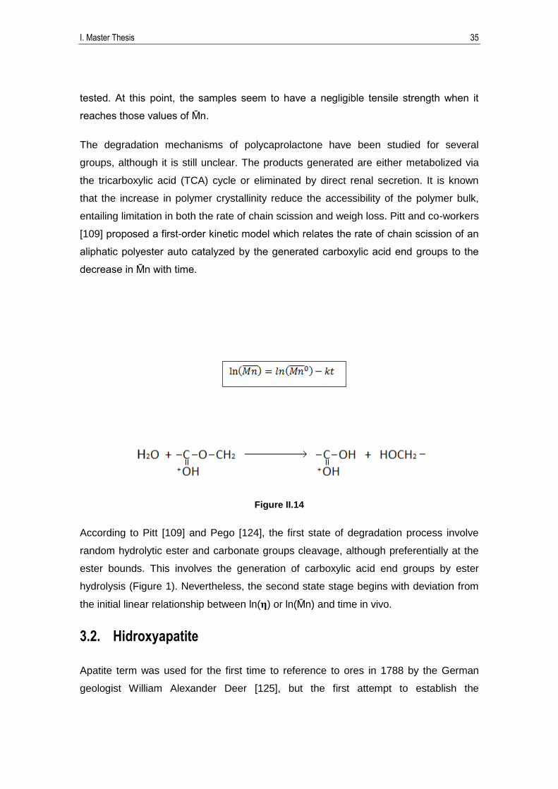
entailing limitation in both the rate of chain scission and weigh loss. Pitt and co-workers
[109] proposed a first-order inetic model which relates the rate of chain scission of an
aliphatic polyester auto catalyzed by the generated carboxylic acid end groups to the
decrease in M n with time.
Figure II.14
According to Pitt [109] and Pego [124], the first state of degradation process involve
random hydrolytic ester and carbonate groups cleavage, although preferentially at the
ester bounds. This involves the generation of carboxylic acid end groups by ester
hydrolysis (Figure 1). Nevertheless, the second state stage begins with deviation from
the initial linear relationship between ln( ) or ln(M n) and time in vivo.
3.2. Hidroxyapatite
Apatite term was used for the first time to reference to ores in 1788 by the German
geologist William Alexander Deer [125], but the first attempt to establish the
36 Capítulo
hydroxyapatite´s composition by chemical analysis began in the second half of the 18th
century by the Swedish chemist Jöns Jacob Berzelius (1779-1848) [126]. Currently this
term is referred to a crystal family of materials related to the formula M10(RO4)6X2
where M refers to calcium, R to Phosphorus and X to an hydroxide or another halogen
compound such as fluorine. The inorganic phase of the bone was determinate by X ray
diffraction technique exposing that it was apatite in 1926 [127].
Hydroxyapatite is an apatite composed essentially with phosphorous and calcium with
the chemical formula Ca10(PO4)6(OH)2. The atomic ratio CA/P is 1.67 with 39% by
weight of Ca, 18.5% P and 3.38% of OH [128, 129].
Although several materials have been designed as hydroxyapatite by diverse
researchers, actually, the Ca/P rate shown could be variable with a range of 1.3 to 2.0.
These materials are seldom properly characterized, for this reason is though compare
results from different sources of study. Nevertheless, the American Society for Testing
and Materials (1987) determined the reference patterns both for Hydroxyapatite to
tricalcium phosphates, in order to control and determinate the purity of these
compounds[127] (Jaffe and Scott 1996). According to the explained before, the rate
Ca/P should be 1.67, while the rate of tricalcium phosphates should be 1.5 [127].
Contribution of hydroxyapatite in the bone
Bone composition varies according the position, age, food background and illness.
Usually, the mineral phase represents between 60-70% of the bone tissue, water 5-8%,
and organic matrix constitutes the rest (20%), which the 85-90% of this is collagen and
15-5% constitutes to non-collagenous proteins [130].
The mineral phase of the human bone includes different kinds of hydrated calcium
phosphates; the most common is hydroxyapatite. This apatite is presented in bone as a
flat crystal with a length of 20-80 nm and a thickness of 2-5 nm [130, 131]. It is
calculated that an average of 65% of mineral phase in human bone is composed by
hydroxyapatite [130].
Hydroxyapatites produced biologically does not contain only ions and radicals of the
HAp but also traces of CO3, Mg, Na, F and Cl because phosphate(PO3) and hydroxide
groups (OH) could be replaced by carbonate (CO3) or chlorine(Cl) and fluorine(F)
respectively, influencing physics properties of the ore. Impurity amounts vary according
I. Master Thesis 37
to food background or the specific type of tissue, afecting properties and bioactivity of
the bone [131].
The most highlight mechanic property of hydroxyapatite is the stiffness and toughness,
which are combined with collagen elasticity. Collagen is the mainly component in bone
organic phase, and provides particularity properties to the bone. Hydroxyapatite
crystals improve bone matrix stiffness, in such a way that without them, bone could
bend in an extremely easy way [132].
Improvement of PCL mechanical properties depending of HA loading
percentage.
Following the experimental data carried out by Biqiong Chen and Kang Sun [133] with
the study of different amount of hydroxyapatite reinforced poly ε-caprolactone, they
reported that the optimum percentage of hydroxyapatite is 20%. Is for that reason that
we decided to introduce a load of 20% hydroxyapatite in our material. They studied a
load of 0%, 20%, 40% and 60%, concluding that a load of 20% enhance the
mechanical and thermal properties of the compound. Regarding the thermal properties,
there are not significant, nevertheless the melting (Tm) and crystallization (Tc)
temperature of the composite were increased by the presence of HA, suggesting slight
interactions where HA facilitates the crystallization of PCL under cooling, acting as a
nucleating agent.
On the other hand, it was shown an enhancement of mechanical properties. The
strength reaches its greatest values when the concentration of HA was 20% and as the
content of HA increased, the strength decreased significantly. It can be explain
because when a matrix is loaded with certain particles, them limit the movement of the
molecular chains in the matrix, when the amount is high the particles tend to
agglomerate.
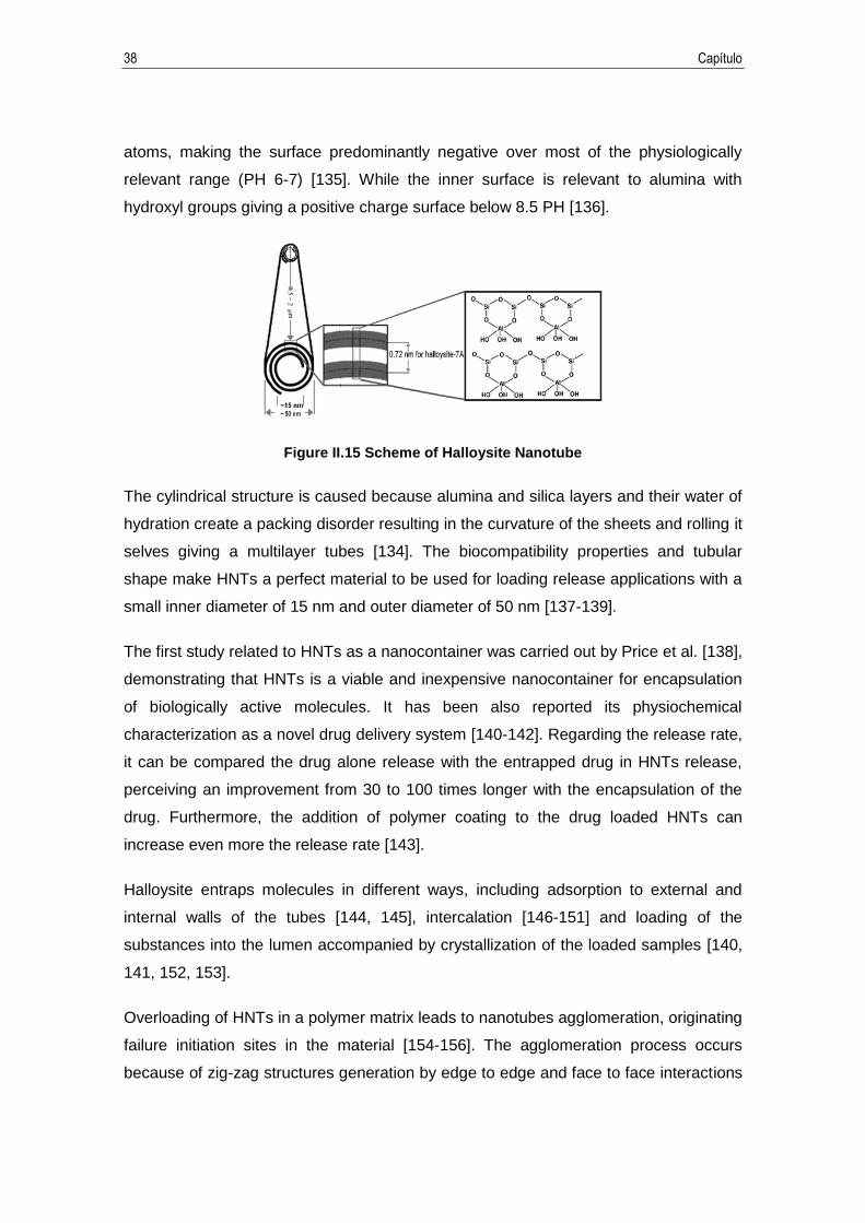
3.3. Halloysite Nanotubes
Halloysite Nanotubes (HNTs) is a clay material found as a raw mineral from natural
deposits, fact that makes halloysite economically viable [134]. HNTs presents
nanotubular structure where the outer surface can be compared to silica with oxygen
38 Capítulo
atoms, making the surface predominantly negative over most of the physiologically
relevant range (PH 6-7) [135]. While the inner surface is relevant to alumina with
hydroxyl groups giving a positive charge surface below 8.5 PH [136].
Figure II.15 Scheme of Halloysite Nanotube
The cylindrical structure is caused because alumina and silica layers and their water of
hydration create a packing disorder resulting in the curvature of the sheets and rolling it
selves giving a multilayer tubes [134]. The biocompatibility properties and tubular
shape make HNTs a perfect material to be used for loading release applications with a
small inner diameter of 15 nm and outer diameter of 50 nm [137-139].
The first study related to HNTs as a nanocontainer was carried out by Price et al. [138],
demonstrating that HNTs is a viable and inexpensive nanocontainer for encapsulation
of biologically active molecules. It has been also reported its physiochemical
characterization as a novel drug delivery system [140-142]. Regarding the release rate,
it can be compared the drug alone release with the entrapped drug in HNTs release,
perceiving an improvement from 30 to 100 times longer with the encapsulation of the
drug. Furthermore, the addition of polymer coating to the drug loaded HNTs can
increase even more the release rate [143].
Halloysite entraps molecules in different ways, including adsorption to external and
internal walls of the tubes [144, 145], intercalation [146-151] and loading of the
substances into the lumen accompanied by crystallization of the loaded samples [140,
141, 152, 153].
Overloading of HNTs in a polymer matrix leads to nanotubes agglomeration, originating
failure initiation sites in the material [154-156]. The agglomeration process occurs
because of zig-zag structures generation by edge to edge and face to face interactions
I. Master Thesis 39
between HNTs due to the chemical composition of HNTs [157]. The shape of
nanotubes plus charge distribution (inner and outer face of tubule walls carry net
negative charges and the edges are amphoteric with positive charge at low PH) favors
face to end attachment [152]. Formation of aggregates can be possibly avoided by a
surface modification of HNTs. Nevertheless, agglomeration effect can be minimized by
a previous extrusion process to injection molding to improve the HNTs dispersion in the
polymeric matrix, enhancing the mechanical properties. Moreover, it was observed by
[158] that higher rotation speed of extruder, results in better nanotubes dispersion.


I. Master Thesis 41
III. Objectives


I. Master Thesis 43
1. Objectives
1.1. General objective
Improve mechanical and biologic properties of bio absorbable polymeric fixing systems
to use in craniofacial injuries.
1.2. Specific objectives
1. Design blend formulations and manufacturing test specimens of bio absorbable
materials based on Polycaprolactone additive with Hydroxyapatite and
Halloysite Nanotubes.
2. Determine the optimum additive percentage.
3. Mechanical and thermal analysis of the different additived materials.


I. Master Thesis 45
IV. Experimental

I. Master Thesis 47
1. Materials
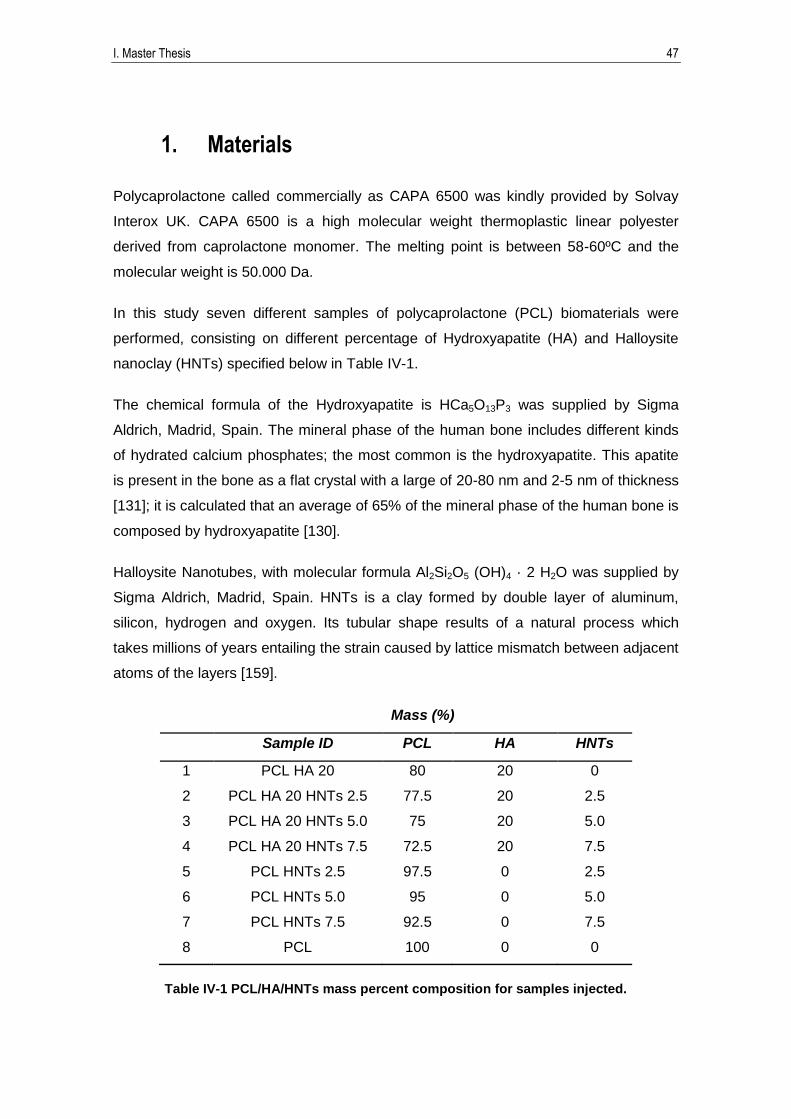
Polycaprolactone called commercially as CAPA 6500 was kindly provided by Solvay
Interox UK. CAPA 6500 is a high molecular weight thermoplastic linear polyester
derived from caprolactone monomer. The melting point is between 58-60ºC and the
molecular weight is 50.000 Da.
In this study seven different samples of polycaprolactone (PCL) biomaterials were
performed, consisting on different percentage of Hydroxyapatite (HA) and Halloysite
nanoclay (HNTs) specified below in Table IV-1.
The chemical formula of the Hydroxyapatite is HCa5O13P3 was supplied by Sigma
Aldrich, Madrid, Spain. The mineral phase of the human bone includes different kinds
of hydrated calcium phosphates; the most common is the hydroxyapatite. This apatite
is present in the bone as a flat crystal with a large of 20-80 nm and 2-5 nm of thickness
[131]; it is calculated that an average of 65% of the mineral phase of the human bone is
composed by hydroxyapatite [130].
Halloysite Nanotubes, with molecular formula Al2Si2O5 (OH)4 · 2 H2O was supplied by
Sigma Aldrich, Madrid, Spain. HNTs is a clay formed by double layer of aluminum,
silicon, hydrogen and oxygen. Its tubular shape results of a natural process which
takes millions of years entailing the strain caused by lattice mismatch between adjacent
atoms of the layers [159].
Mass (%)
Sample ID PCL HA HNTs
1 PCL HA 20 80 20 0
2 PCL HA 20 HNTs 2.5 77.5 20 2.5
3 PCL HA 20 HNTs 5.0 75 20 5.0
4 PCL HA 20 HNTs 7.5 72.5 20 7.5
5 PCL HNTs 2.5 97.5 0 2.5
6 PCL HNTs 5.0 95 0 5.0
7 PCL HNTs 7.5 92.5 0 7.5
8 PCL 100 0 0
Table IV-1 PCL/HA/HNTs mass percent composition for samples injected.
48 Capítulo
2. Procedures and methods
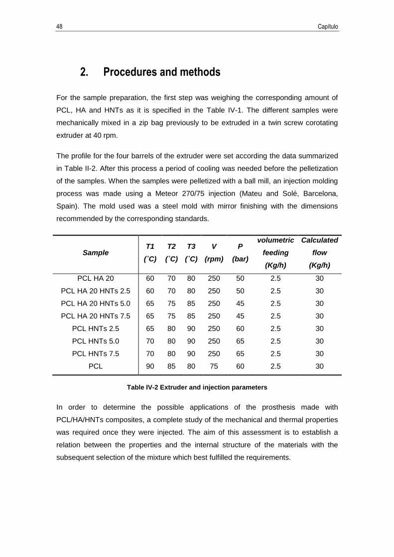
For the sample preparation, the first step was weighing the corresponding amount of
PCL, HA and HNTs as it is specified in the Table IV-1. The different samples were
mechanically mixed in a zip bag previously to be extruded in a twin screw corotating
extruder at 40 rpm.
The profile for the four barrels of the extruder were set according the data summarized
in Table II-2. After this process a period of cooling was needed before the pelletization
of the samples. When the samples were pelletized with a ball mill, an injection molding
process was made using a Meteor 270/75 injection (Mateu and Solé, Barcelona,
Spain). The mold used was a steel mold with mirror finishing with the dimensions
recommended by the corresponding standards.
Sample T1
(˚C)
T2
(˚C)
T3
(˚C)
V
(rpm)
P
(bar)
volumetric
feeding
(Kg/h)
Calculated
flow
(Kg/h)
PCL HA 20 60 70 80 250 50 2.5 30
PCL HA 20 HNTs 2.5 60 70 80 250 50 2.5 30
PCL HA 20 HNTs 5.0 65 75 85 250 45 2.5 30
PCL HA 20 HNTs 7.5 65 75 85 250 45 2.5 30
PCL HNTs 2.5 65 80 90 250 60 2.5 30
PCL HNTs 5.0 70 80 90 250 65 2.5 30
PCL HNTs 7.5 70 80 90 250 65 2.5 30
PCL 90 85 80 75 60 2.5 30
Table IV-2 Extruder and injection parameters
In order to determine the possible applications of the prosthesis made with
PCL/HA/HNTs composites, a complete study of the mechanical and thermal properties
was required once they were injected. The aim of this assessment is to establish a
relation between the properties and the internal structure of the materials with the
subsequent selection of the mixture which best fulfilled the requirements.
I. Master Thesis 49
2.1. Mechanical properties
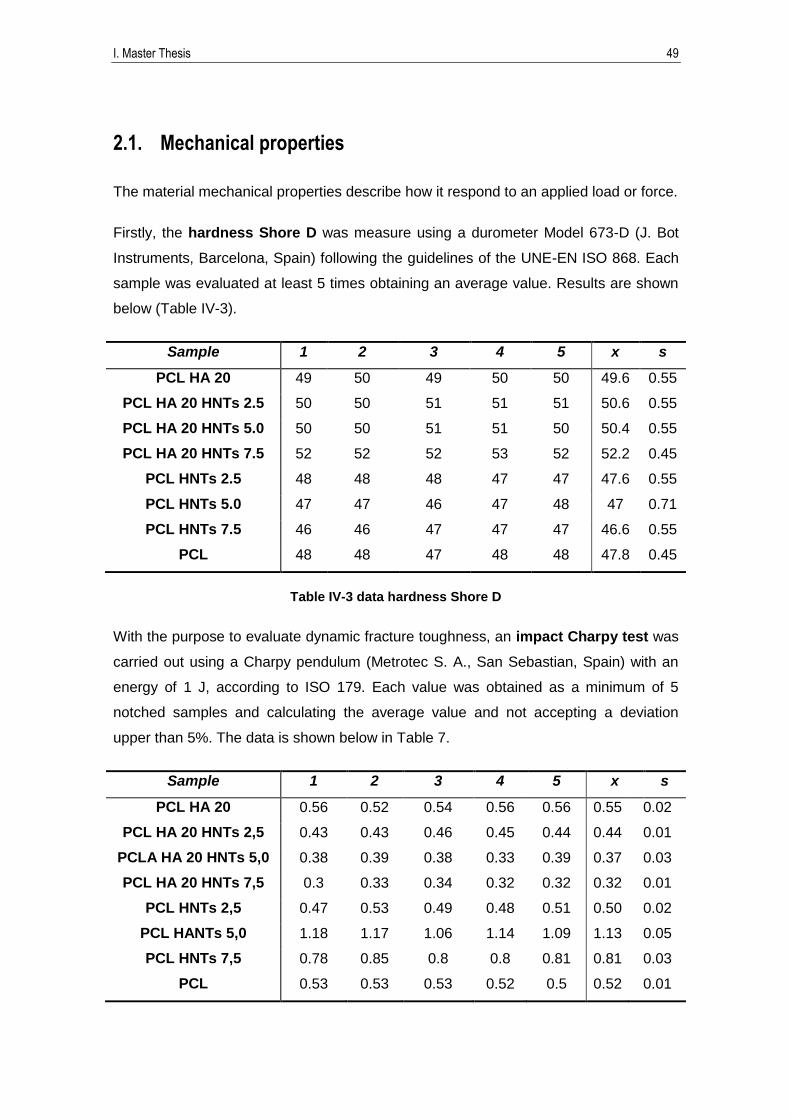
The material mechanical properties describe how it respond to an applied load or force.
Firstly, the hardness Shore D was measure using a durometer Model 673-D (J. Bot
Instruments, Barcelona, Spain) following the guidelines of the UNE-EN ISO 868. Each
sample was evaluated at least 5 times obtaining an average value. Results are shown
below (Table IV-3).
Sample 1 2 3 4 5 x s
PCL HA 20 49 50 49 50 50 49.6 0.55
PCL HA 20 HNTs 2.5 50 50 51 51 51 50.6 0.55
PCL HA 20 HNTs 5.0 50 50 51 51 50 50.4 0.55
PCL HA 20 HNTs 7.5 52 52 52 53 52 52.2 0.45
PCL HNTs 2.5 48 48 48 47 47 47.6 0.55
PCL HNTs 5.0 47 47 46 47 48 47 0.71
PCL HNTs 7.5 46 46 47 47 47 46.6 0.55
PCL 48 48 47 48 48 47.8 0.45
Table IV-3 data hardness Shore D
With the purpose to evaluate dynamic fracture toughness, an impact Charpy test was
carried out using a Charpy pendulum (Metrotec S. A., San Sebastian, Spain) with an
energy of 1 J, according to ISO 179. Each value was obtained as a minimum of 5
notched samples and calculating the average value and not accepting a deviation
upper than 5%. The data is shown below in Table 7.
Sample 1 2 3 4 5 x s
PCL HA 20 0.56 0.52 0.54 0.56 0.56 0.55 0.02
PCL HA 20 HNTs 2,5 0.43 0.43 0.46 0.45 0.44 0.44 0.01
PCLA HA 20 HNTs 5,0 0.38 0.39 0.38 0.33 0.39 0.37 0.03
PCL HA 20 HNTs 7,5 0.3 0.33 0.34 0.32 0.32 0.32 0.01
PCL HNTs 2,5 0.47 0.53 0.49 0.48 0.51 0.50 0.02
PCL HANTs 5,0 1.18 1.17 1.06 1.14 1.09 1.13 0.05
PCL HNTs 7,5 0.78 0.85 0.8 0.8 0.81 0.81 0.03
PCL 0.53 0.53 0.53 0.52 0.5 0.52 0.01
50 Capítulo
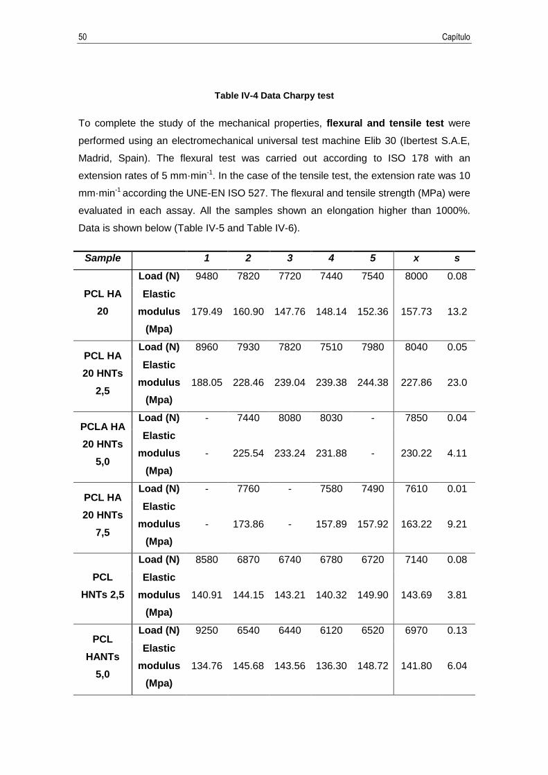
Table IV-4 Data Charpy test
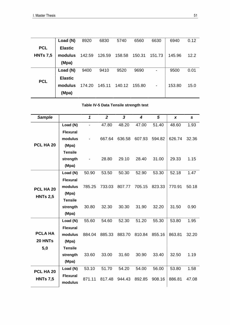
To complete the study of the mechanical properties, flexural and tensile test were
performed using an electromechanical universal test machine Elib 30 (Ibertest S.A.E,
Madrid, Spain). The flexural test was carried out according to ISO 178 with an
extension rates of 5 mm·min-1. In the case of the tensile test, the extension rate was 10
mm·min-1 according the UNE-EN ISO 527. The flexural and tensile strength (MPa) were
evaluated in each assay. All the samples shown an elongation higher than 1000%.
Data is shown below (Table IV-5 and Table IV-6).
Sample
1 2 3 4 5 x s
PCL HA
20
Load (N) 9480 7820 7720 7440 7540 8000 0.08
Elastic
modulus
(Mpa)
179.49 160.90 147.76 148.14 152.36 157.73 13.2
PCL HA
20 HNTs
2,5
Load (N) 8960 7930 7820 7510 7980 8040 0.05
Elastic
modulus
(Mpa)
188.05 228.46 239.04 239.38 244.38 227.86 23.0
PCLA HA
20 HNTs
5,0
Load (N) - 7440 8080 8030 - 7850 0.04
Elastic
modulus
(Mpa)
- 225.54 233.24 231.88 - 230.22 4.11
PCL HA
20 HNTs
7,5
Load (N) - 7760 - 7580 7490 7610 0.01
Elastic
modulus
(Mpa)
- 173.86 - 157.89 157.92 163.22 9.21
PCL
HNTs 2,5
Load (N) 8580 6870 6740 6780 6720 7140 0.08
Elastic
modulus
(Mpa)
140.91 144.15 143.21 140.32 149.90 143.69 3.81
PCL
HANTs
5,0
Load (N) 9250 6540 6440 6120 6520 6970 0.13
Elastic
modulus
(Mpa)
134.76 145.68 143.56 136.30 148.72 141.80 6.04
I. Master Thesis 51
PCL
HNTs 7,5
Load (N) 8920 6830 5740 6560 6630 6940 0.12
Elastic
modulus
(Mpa)
142.59 126.59 158.58 150.31 151.73 145.96 12.2
PCL
Load (N) 9400 9410 9520 9690 - 9500 0.01
Elastic
modulus
(Mpa)
174.20 145.11 140.12 155.80 - 153.80 15.0
Table IV-5 Data Tensile strength test
Sample
1 2 3 4 5 x s
PCL HA 20
Load (N) - 47.80 48.20 47.00 51.40 48.60 1.93
Flexural
modulus
(Mpa)
- 667.64 636.58 607.93 594.82 626.74 32.36
Tensile
strength
(Mpa)
- 28.80 29.10 28.40 31.00 29.33 1.15
PCL HA 20
HNTs 2,5
Load (N) 50.90 53.50 50.30 52.90 53.30 52.18 1.47
Flexural
modulus
(Mpa)
785.25 733.03 807.77 705.15 823.33 770.91 50.18
Tensile
strength
(Mpa)
30.80 32.30 30.30 31.90 32.20 31.50 0.90
PCLA HA
20 HNTs
5,0
Load (N) 55.60 54.60 52.30 51.20 55.30 53.80 1.95
Flexural
modulus
(Mpa)
884.04 885.33 883.70 810.84 855.16 863.81 32.20
Tensile
strength
(Mpa)
33.60 33.00 31.60 30.90 33.40 32.50 1.19
PCL HA 20
HNTs 7,5
Load (N) 53.10 51.70 54.20 54.00 56.00 53.80 1.58
Flexural
modulus 871.11 817.48 944.43 892.85 908.16 886.81 47.08

52 Capítulo
(Mpa)
Tensile
strength
(Mpa)
32.10 31.20 32.70 38.22 33.80 33.60 2.75
PCL HNTs
2,5
Load (N) 40.20 42.40 41.50 40.60 40.20 40.98 0.95
Flexural
modulus
(Mpa)
391.91 463.86 447.52 446.79 445.90 439.20 27.46
Tensile
strength
(Mpa)
24.30 25.60 25.10 24.50 24.30 24.76 0.57
PCL HANTs
5,0
Load (N) 41.60 39.50 39.30 39.00 39.30 39.74 1.05
Flexural
modulus
(Mpa)
471.70 437.59 444.76 463.21 401.73 443.80 27.22
Tensile
strength
(Mpa)
25.10 23.80 23.70 23.50 23.73 23.97 0.64
PCL HNTs
7,5
Load (N) 41.00 42.70 41.80 40.00 43.80 41.86 1.47
Flexural
modulus
(Mpa)
483.66 463.60 540.53 404.95 525.19 483.59 53.75
Tensile
strength
(Mpa)
24.80 25.80 25.20 24.20 26.50 25.30 0.89
PCL
Load (N) 41.80 41.90 38.00 40.40 42.00 40.82 1.71
Flexural
modulus
(Mpa)
430.15 430.28 387.98 387.64 452.24 417.66 28.69
Tensile
strength
(Mpa)
25.20 23.60 21.40 22.70 23.60 23.30 1.39
Table IV-6 Data Flexural strength test
I. Master Thesis 53
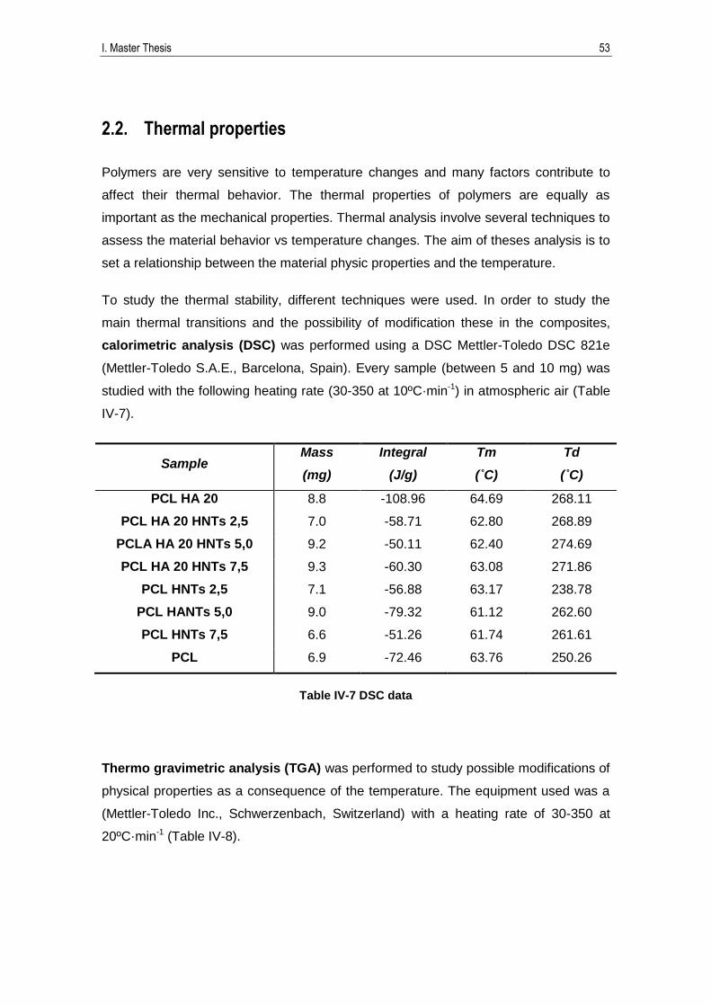
2.2. Thermal properties
Polymers are very sensitive to temperature changes and many factors contribute to
affect their thermal behavior. The thermal properties of polymers are equally as
important as the mechanical properties. Thermal analysis involve several techniques to
assess the material behavior vs temperature changes. The aim of theses analysis is to
set a relationship between the material physic properties and the temperature.
To study the thermal stability, different techniques were used. In order to study the
main thermal transitions and the possibility of modification these in the composites,
calorimetric analysis (DSC) was performed using a DSC Mettler-Toledo DSC 821e
(Mettler-Toledo S.A.E., Barcelona, Spain). Every sample (between 5 and 10 mg) was
studied with the following heating rate (30-350 at 10ºC·min-1) in atmospheric air (Table
IV-7).
Sample Mass
(mg)
Integral
(J/g)
Tm
(˚C)
Td
(˚C)
PCL HA 20 8.8 -108.96 64.69 268.11
PCL HA 20 HNTs 2,5 7.0 -58.71 62.80 268.89
PCLA HA 20 HNTs 5,0 9.2 -50.11 62.40 274.69
PCL HA 20 HNTs 7,5 9.3 -60.30 63.08 271.86
PCL HNTs 2,5 7.1 -56.88 63.17 238.78
PCL HANTs 5,0 9.0 -79.32 61.12 262.60
PCL HNTs 7,5 6.6 -51.26 61.74 261.61
PCL 6.9 -72.46 63.76 250.26
Table IV-7 DSC data
Thermo gravimetric analysis (TGA) was performed to study possible modifications of
physical properties as a consequence of the temperature. The equipment used was a
(Mettler-Toledo Inc., Schwerzenbach, Switzerland) with a heating rate of 30-350 at
20ºC·min-1 (Table IV-8).
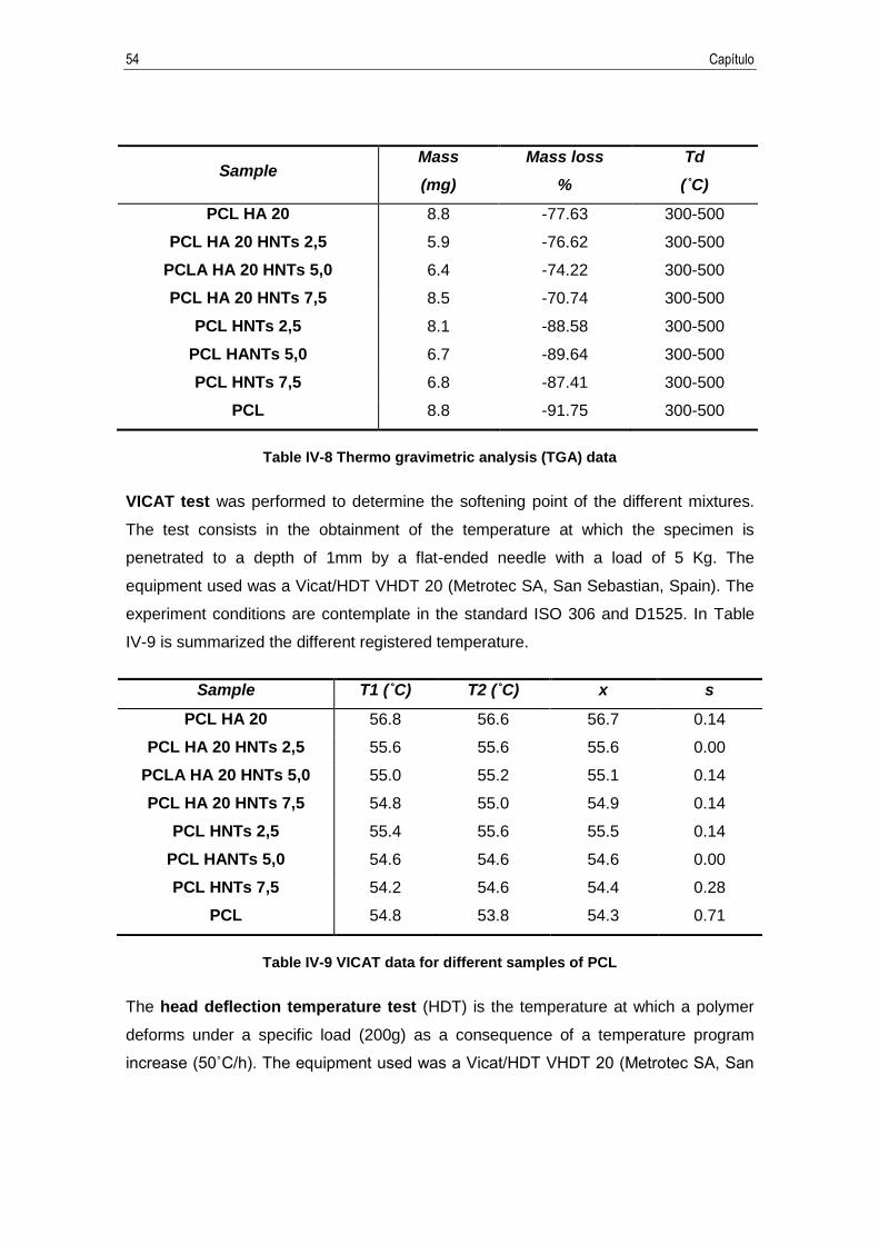
54 Capítulo
Sample Mass
(mg)
Mass loss
%
Td
(˚C)
PCL HA 20 8.8 -77.63 300-500
PCL HA 20 HNTs 2,5 5.9 -76.62 300-500
PCLA HA 20 HNTs 5,0 6.4 -74.22 300-500
PCL HA 20 HNTs 7,5 8.5 -70.74 300-500
PCL HNTs 2,5 8.1 -88.58 300-500
PCL HANTs 5,0 6.7 -89.64 300-500
PCL HNTs 7,5 6.8 -87.41 300-500
PCL 8.8 -91.75 300-500
Table IV-8 Thermo gravimetric analysis (TGA) data
VICAT test was performed to determine the softening point of the different mixtures.
The test consists in the obtainment of the temperature at which the specimen is
penetrated to a depth of 1mm by a flat-ended needle with a load of 5 Kg. The
equipment used was a Vicat/HDT VHDT 20 (Metrotec SA, San Sebastian, Spain). The
experiment conditions are contemplate in the standard ISO 306 and D1525. In Table
IV-9 is summarized the different registered temperature.
Sample T1 (˚C) T2 (˚C) x s
PCL HA 20 56.8 56.6 56.7 0.14
PCL HA 20 HNTs 2,5 55.6 55.6 55.6 0.00
PCLA HA 20 HNTs 5,0 55.0 55.2 55.1 0.14
PCL HA 20 HNTs 7,5 54.8 55.0 54.9 0.14
PCL HNTs 2,5 55.4 55.6 55.5 0.14
PCL HANTs 5,0 54.6 54.6 54.6 0.00
PCL HNTs 7,5 54.2 54.6 54.4 0.28
PCL 54.8 53.8 54.3 0.71
Table IV-9 VICAT data for different samples of PCL
The head deflection temperature test (HDT) is the temperature at which a polymer
deforms under a specific load (200g) as a consequence of a temperature program
increase ( ˚C/h) The equipment used was a Vicat/H T VH T (Metrotec SA, San
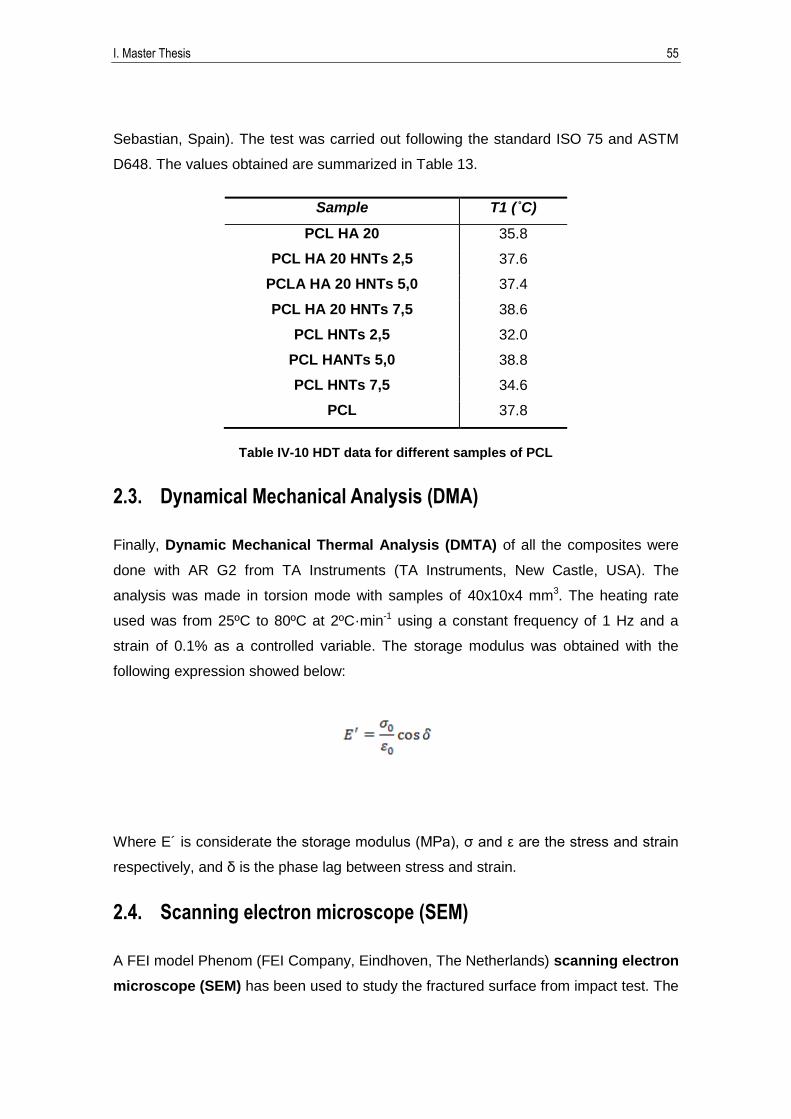
I. Master Thesis 55
Sebastian, Spain). The test was carried out following the standard ISO 75 and ASTM
D648. The values obtained are summarized in Table 13.
Sample T1 (˚C)
PCL HA 20 35.8
PCL HA 20 HNTs 2,5 37.6
PCLA HA 20 HNTs 5,0 37.4
PCL HA 20 HNTs 7,5 38.6
PCL HNTs 2,5 32.0
PCL HANTs 5,0 38.8
PCL HNTs 7,5 34.6
PCL 37.8
Table IV-10 HDT data for different samples of PCL
2.3. Dynamical Mechanical Analysis (DMA)
Finally, Dynamic Mechanical Thermal Analysis (DMTA) of all the composites were
done with AR G2 from TA Instruments (TA Instruments, New Castle, USA). The
analysis was made in torsion mode with samples of 40x10x4 mm3. The heating rate
used was from 25ºC to 80ºC at 2ºC·min-1 using a constant frequency of 1 Hz and a
strain of 0.1% as a controlled variable. The storage modulus was obtained with the
following expression showed below:
Where E´ is considerate the storage modulus (MPa), σ and ε are the stress and strain
respectively, and δ is the phase lag between stress and strain.
2.4. Scanning electron microscope (SEM)
A FEI model Phenom (FEI Company, Eindhoven, The Netherlands) scanning electron
microscope (SEM) has been used to study the fractured surface from impact test. The

56 Capítulo
accelerating voltage was 5 kV. Previously, samples were metallized with a gold-
palladium alloy in a sputter coater EMITECH mod. SC7620 (Quorum Technologies
Ltd., East Sussex, United Kingdom).

I. Master Thesis 57
V. Discussion

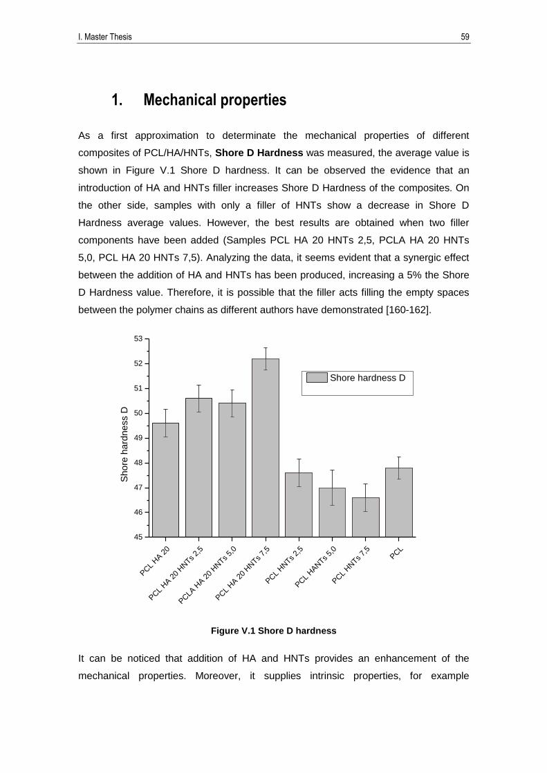
I. Master Thesis 59
1. Mechanical properties
As a first approximation to determinate the mechanical properties of different
composites of PCL/HA/HNTs, Shore D Hardness was measured, the average value is
shown in Figure V.1 Shore D hardness. It can be observed the evidence that an
introduction of HA and HNTs filler increases Shore D Hardness of the composites. On
the other side, samples with only a filler of HNTs show a decrease in Shore D
Hardness average values. However, the best results are obtained when two filler
components have been added (Samples PCL HA 20 HNTs 2,5, PCLA HA 20 HNTs
5,0, PCL HA 20 HNTs 7,5). Analyzing the data, it seems evident that a synergic effect
between the addition of HA and HNTs has been produced, increasing a 5% the Shore
D Hardness value. Therefore, it is possible that the filler acts filling the empty spaces
between the polymer chains as different authors have demonstrated [160-162].
PCL
HA 2
0
PCL
HA 2
0 HNTs
2,5
PCLA
HA 2
0 HNTs
5,0
PCL
HA 2
0 HNTs
7,5
PCL
HNTs
2,5
PCL
HANTs
5,0
PCL
HNTs
7,5
PCL
45
46
47
48
49
50
51
52
53
Sho
re h
ard
ne
ss D
Shore hardness D
Figure V.1 Shore D hardness
It can be noticed that addition of HA and HNTs provides an enhancement of the
mechanical properties. Moreover, it supplies intrinsic properties, for example
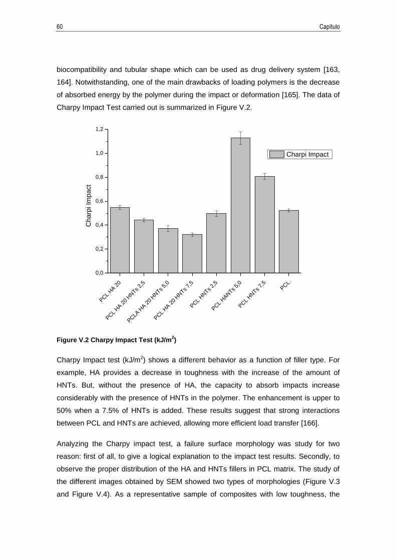
60 Capítulo
biocompatibility and tubular shape which can be used as drug delivery system [163,
164]. Notwithstanding, one of the main drawbacks of loading polymers is the decrease
of absorbed energy by the polymer during the impact or deformation [165]. The data of
Charpy Impact Test carried out is summarized in Figure V.2.
PCL
HA 2
0
PCL
HA 2
0 HNTs
2,5
PCLA
HA 2
0 HNTs
5,0
PCL
HA 2
0 HNTs
7,5
PCL
HNTs
2,5
PCL
HANTs
5,0
PCL
HNTs
7,5
PCL
0,0
0,2
0,4
0,6
0,8
1,0
1,2
Ch
arp
i Im
pa
ct
Charpi Impact
Figure V.2 Charpy Impact Test (kJ/m2)
Charpy Impact test (kJ/m2) shows a different behavior as a function of filler type. For
example, HA provides a decrease in toughness with the increase of the amount of
HNTs. But, without the presence of HA, the capacity to absorb impacts increase
considerably with the presence of HNTs in the polymer. The enhancement is upper to
50% when a 7.5% of HNTs is added. These results suggest that strong interactions
between PCL and HNTs are achieved, allowing more efficient load transfer [166].
Analyzing the Charpy impact test, a failure surface morphology was study for two
reason: first of all, to give a logical explanation to the impact test results. Secondly, to
observe the proper distribution of the HA and HNTs fillers in PCL matrix. The study of
the different images obtained by SEM showed two types of morphologies (Figure V.3
and Figure V.4). As a representative sample of composites with low toughness, the
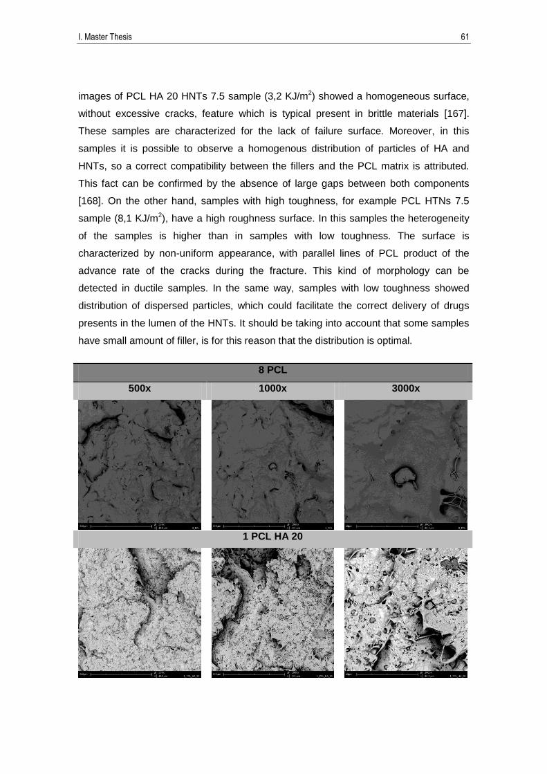
I. Master Thesis 61
images of PCL HA 20 HNTs 7.5 sample (3,2 KJ/m2) showed a homogeneous surface,
without excessive cracks, feature which is typical present in brittle materials [167].
These samples are characterized for the lack of failure surface. Moreover, in this
samples it is possible to observe a homogenous distribution of particles of HA and
HNTs, so a correct compatibility between the fillers and the PCL matrix is attributed.
This fact can be confirmed by the absence of large gaps between both components
[168]. On the other hand, samples with high toughness, for example PCL HTNs 7.5
sample (8,1 KJ/m2), have a high roughness surface. In this samples the heterogeneity
of the samples is higher than in samples with low toughness. The surface is
characterized by non-uniform appearance, with parallel lines of PCL product of the
advance rate of the cracks during the fracture. This kind of morphology can be
detected in ductile samples. In the same way, samples with low toughness showed
distribution of dispersed particles, which could facilitate the correct delivery of drugs
presents in the lumen of the HNTs. It should be taking into account that some samples
have small amount of filler, is for this reason that the distribution is optimal.
8 PCL
500x 1000x 3000x
1 PCL HA 20
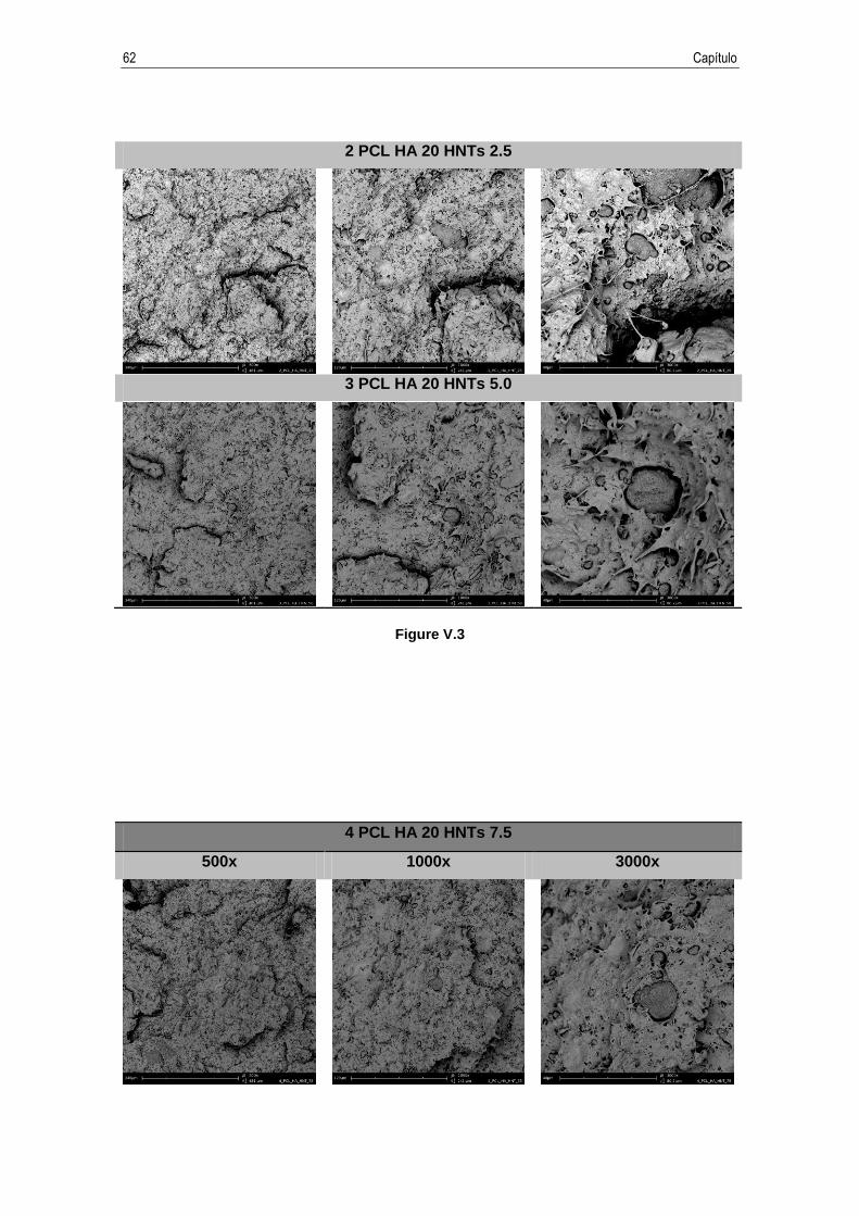
62 Capítulo
2 PCL HA 20 HNTs 2.5
3 PCL HA 20 HNTs 5.0
Figure V.3
4 PCL HA 20 HNTs 7.5
500x 1000x 3000x
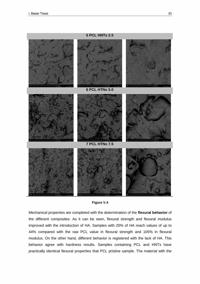
I. Master Thesis 63
5 PCL HNTs 2.5
6 PCL HTNs 5.0
7 PCL HTNs 7.5
Figure V.4
Mechanical properties are completed with the determination of the flexural behavior of
the different composites. As it can be seen, flexural strength and flexural modulus
improved with the introduction of HA. Samples with 20% of HA reach values of up to
44% compared with the raw PCL value in flexural strength and 105% in flexural
modulus. On the other hand, different behavior is registered with the lack of HA. This
behavior agree with hardness results. Samples containing PCL and HNTs have
practically identical flexural properties that PCL pristine sample. The material with the
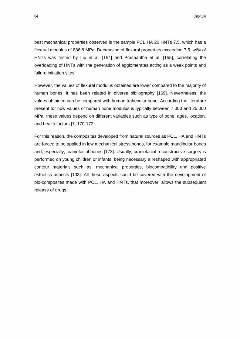
64 Capítulo
best mechanical properties observed is the sample PCL HA 20 HNTs 7.5, which has a
flexural modulus of 886.8 MPa. Decreasing of flexural properties exceeding 7.5 wt% of
HNTs was tested by Liu et al. [154] and Prashantha et al. [155], correlating the
overloading of HNTs with the generation of agglomerates acting as a weak points and
failure initiation sites.
However, the values of flexural modulus obtained are lower compared to the majority of
human bones, it has been related in diverse bibliography [169]. Nevertheless, the
values obtained can be compared with human trabecular bone. According the literature
present for now values of human bone modulus is typically between 7.000 and 25.000
MPa, these values depend on different variables such as type of bone, ages, location,
and health factors [7, 170-172].
For this reason, the composites developed from natural sources as PCL, HA and HNTs
are forced to be applied in low mechanical stress bones, for example mandibular bones
and, especially, craniofacial bones [173]. Usually, craniofacial reconstructive surgery is
performed on young children or infants, being necessary a reshaped with appropriated
contour materials such as, mechanical properties, biocompatibility and positive
esthetics aspects [103]. All these aspects could be covered with the development of
bio-composites made with PCL, HA and HNTs, that moreover, allows the subsequent
release of drugs.
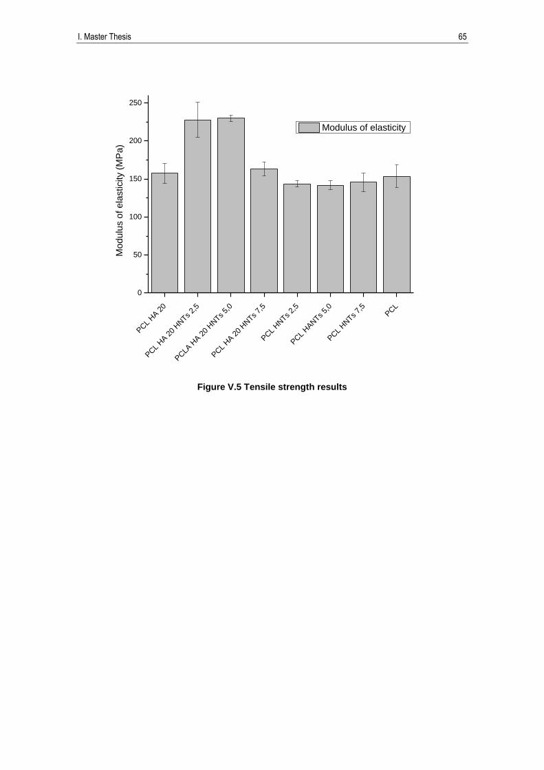
I. Master Thesis 65
PCL
HA 2
0
PCL
HA 2
0 HNTs
2,5
PCLA
HA 2
0 HNTs
5,0
PCL
HA 2
0 HNTs
7,5
PCL
HNTs
2,5
PCL
HANTs
5,0
PCL
HNTs
7,5
PCL
0
50
100
150
200
250
Mo
du
lus o
f e
lasticity (
MP
a)
trials
Modulus of elasticity
Figure V.5 Tensile strength results
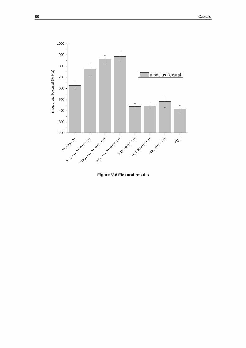
66 Capítulo
PCL
HA 2
0
PCL
HA 2
0 HNTs
2,5
PCLA
HA 2
0 HNTs
5,0
PCL
HA 2
0 HNTs
7,5
PCL
HNTs
2,5
PCL
HANTs
5,0
PCL
HNTs
7,5
PCL
200
300
400
500
600
700
800
900
1000
mo
du
lus fle
xu
ral (M
Pa
)
modulus flexural
Figure V.6 Flexural results
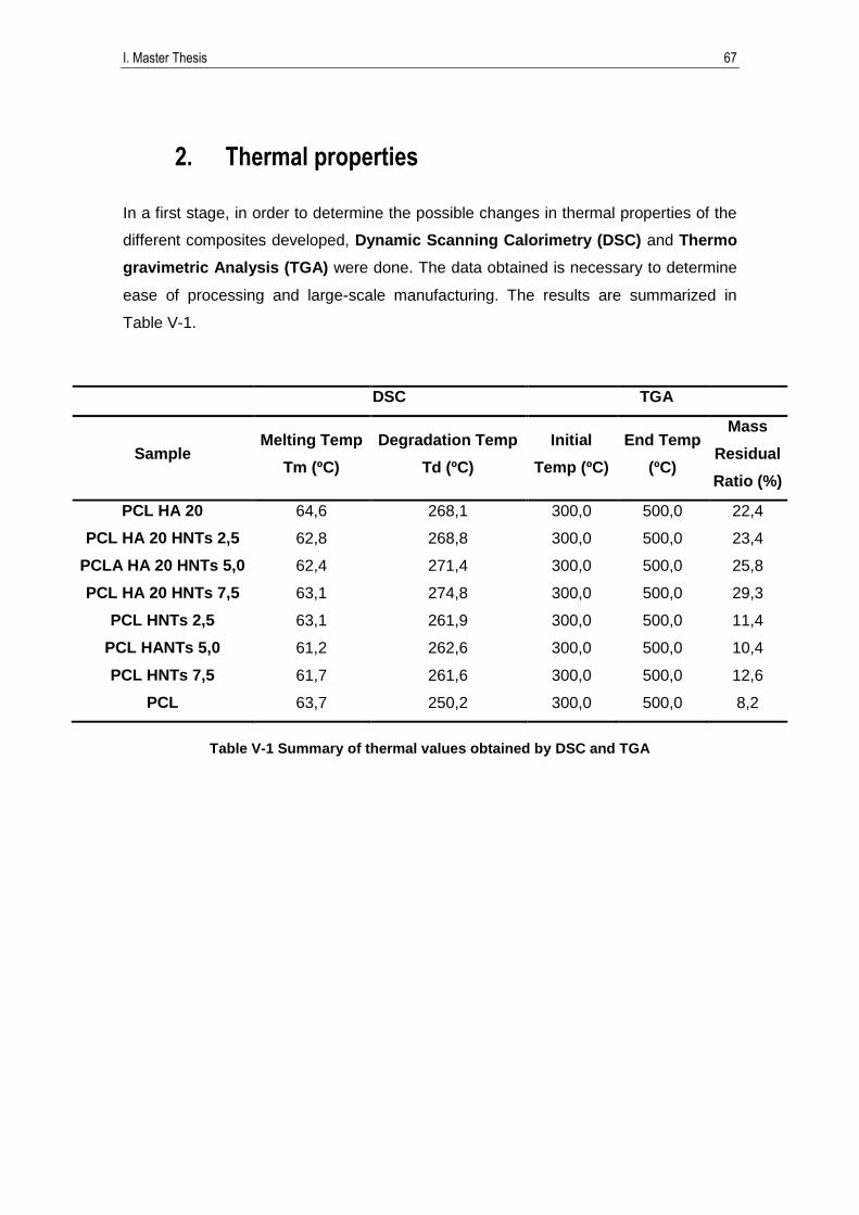
I. Master Thesis 67
2. Thermal properties
In a first stage, in order to determine the possible changes in thermal properties of the
different composites developed, Dynamic Scanning Calorimetry (DSC) and Thermo
gravimetric Analysis (TGA) were done. The data obtained is necessary to determine
ease of processing and large-scale manufacturing. The results are summarized in
Table V-1.
DSC TGA
Sample Melting Temp
Tm (ºC)
Degradation Temp
Td (ºC)
Initial
Temp (ºC)
End Temp
(ºC)
Mass
Residual
Ratio (%)
PCL HA 20 64,6 268,1 300,0 500,0 22,4
PCL HA 20 HNTs 2,5 62,8 268,8 300,0 500,0 23,4
PCLA HA 20 HNTs 5,0 62,4 271,4 300,0 500,0 25,8
PCL HA 20 HNTs 7,5 63,1 274,8 300,0 500,0 29,3
PCL HNTs 2,5 63,1 261,9 300,0 500,0 11,4
PCL HANTs 5,0 61,2 262,6 300,0 500,0 10,4
PCL HNTs 7,5 61,7 261,6 300,0 500,0 12,6
PCL 63,7 250,2 300,0 500,0 8,2
Table V-1 Summary of thermal values obtained by DSC and TGA
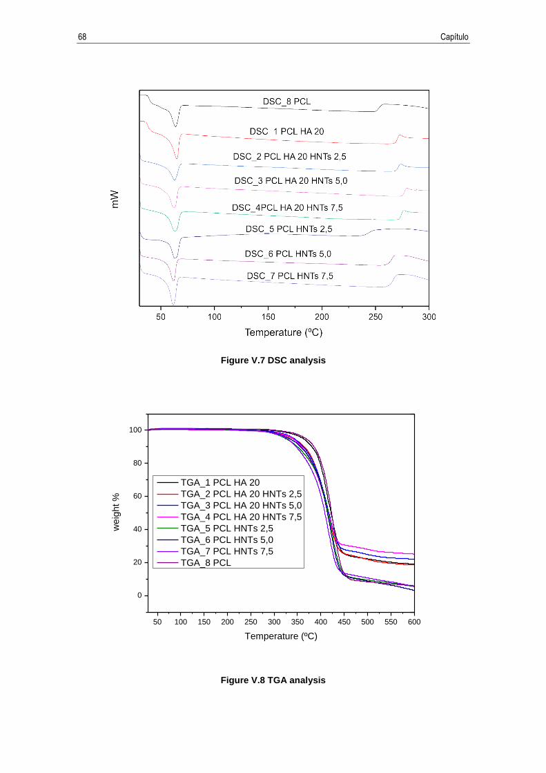
68 Capítulo
Figure V.7 DSC analysis
50 100 150 200 250 300 350 400 450 500 550 600
0
20
40
60
80
100
we
igh
t %
Temperature (ºC)
TGA_1 PCL HA 20
TGA_2 PCL HA 20 HNTs 2,5
TGA_3 PCL HA 20 HNTs 5,0
TGA_4 PCL HA 20 HNTs 7,5
TGA_5 PCL HNTs 2,5
TGA_6 PCL HNTs 5,0
TGA_7 PCL HNTs 7,5
TGA_8 PCL
Figure V.8 TGA analysis
I. Master Thesis 69
Similar values melting temperatures were obtained for all the composites, only a
maximum difference of 2ºC respect raw PCL was detected. This slight difference is a
positive aspect for processability by techniques as injection molding, extrusion, etc.
because it does not affect an increase in operating temperature, with the consequent
energy saving [174]. Moreover, the degradation temperature obtained in the seven
composites developed has high value than the PCL pristine sample. The degradation
temperature of PCL pristine sample is around 250 ºC, while in the different composites
presented this temperature is higher than 274ºC. This difference, bigger than 20ºC,
provides a greater range of thermal stability. This process could be justified by the
introduction of high contents of filler that affects increasing the degradation mechanism
by a complex two-step nucleation-drive reaction [175]. On the other hand, in PCL
pristine sample this mechanism is only based on a chain scission process [176].
Thermo gravimetric analysis (TGA) shows a higher residual mass ratio when the filler
content increases, reaching almost 30% corresponding to the sum of the content of HA
and HNTs. This behavior is due to the high thermal stability supplied by calcium apatite
present in the HA. Only a 4% of weight loss of HA is attributed to the absorbed water
and CO2 registered from 600 to 1000ºC [177]. Therefore, the incorporation of HA and
HNTs as fillers in PCL matrix has not drawbacks in usual manufacturing polymers
process. If the viscosity of the mix is increased in any case, only some adjusts in
pressure or time cycle would be necessary.
To complete the thermal analysis VICAT and HDT test were performed in order to
determine heat resistance characteristics of the material and to define precisely the
thermal behavior. These properties should be determine in order to define the
processability. In injection moulding process, the removal of the piece from the mold
has to be neart or below the HDT temperature, this means that part deformation will be
held withing acceptable limits after removal.
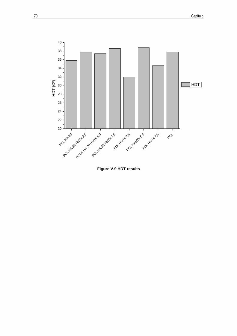
70 Capítulo
PCL
HA 2
0
PCL
HA 2
0 HNTs
2,5
PCLA
HA 2
0 HNTs
5,0
PCL
HA 2
0 HNTs
7,5
PCL
HNTs
2,5
PCL
HANTs
5,0
PCL
HNTs
7,5
PCL
20
22
24
26
28
30
32
34
36
38
40
HD
T (
Cº)
HDT
Figure V.9 HDT results

I. Master Thesis 71
PCL
HA 2
0
PCL
HA 2
0 HNTs
2,5
PCLA
HA 2
0 HNTs
5,0
PCL
HA 2
0 HNTs
7,5
PCL
HNTs
2,5
PCL
HANTs
5,0
PCL
HNTs
7,5
PCL
30
35
40
45
50
55
60V
ICA
T (
Cº)
A
VICAT
Figure V.10 VICAT results
If we assess the data from our test, show above in Table IV-9 and Table IV-10, it can
be noticed that temperatures between samples have a variance not more than ˚C in
Vicat and 3˚C in H T The increase in temperature with the addition of HNTs should be
related with the hydrogen bounding interactions between the hydroxyl group present in
HNTs surface and the carboxyl present in ester linkage of PCL molecular chain [154].
Results obtained don’t provide substantial information, however we obtained the
required information to perform an optimum injection process for the material.
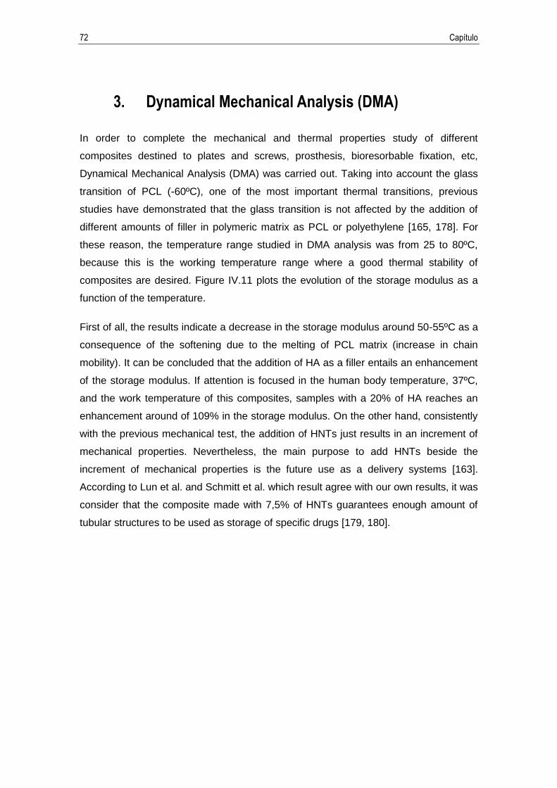
72 Capítulo
3. Dynamical Mechanical Analysis (DMA)
In order to complete the mechanical and thermal properties study of different
composites destined to plates and screws, prosthesis, bioresorbable fixation, etc,
Dynamical Mechanical Analysis (DMA) was carried out. Taking into account the glass
transition of PCL (-60ºC), one of the most important thermal transitions, previous
studies have demonstrated that the glass transition is not affected by the addition of
different amounts of filler in polymeric matrix as PCL or polyethylene [165, 178]. For
these reason, the temperature range studied in DMA analysis was from 25 to 80ºC,
because this is the working temperature range where a good thermal stability of
composites are desired. Figure IV.11 plots the evolution of the storage modulus as a
function of the temperature.
First of all, the results indicate a decrease in the storage modulus around 50-55ºC as a
consequence of the softening due to the melting of PCL matrix (increase in chain
mobility). It can be concluded that the addition of HA as a filler entails an enhancement
of the storage modulus. If attention is focused in the human body temperature, 37ºC,
and the work temperature of this composites, samples with a 20% of HA reaches an
enhancement around of 109% in the storage modulus. On the other hand, consistently
with the previous mechanical test, the addition of HNTs just results in an increment of
mechanical properties. Nevertheless, the main purpose to add HNTs beside the
increment of mechanical properties is the future use as a delivery systems [163].
According to Lun et al. and Schmitt et al. which result agree with our own results, it was
consider that the composite made with 7,5% of HNTs guarantees enough amount of
tubular structures to be used as storage of specific drugs [179, 180].
I. Master Thesis 73
Figure V.11 Dynamical Mechanical Analysis results

74 Capítulo
VI. Conclusions
I. Master Thesis 75
The main objective of this study is to improve mechanical and biologic properties of bio
absorbable polymeric fixing systems to use in craniofacial injuries. Taking into account
that facial trauma is one of the most common injuries seen in urban trauma centers,
development of new bio absorbable materials capable to fulfill the requirements of
bone fracture remodeling is a promising alternative to current metal prostheses.
Commonly, metal prostheses are heavy, expensive, do not promote bone regeneration,
neither are degraded over time and could lead to bone refracture after the required
removal of the plates attributed to the stress shielding effect.
Controlled delivery of osteogenic biomaterials is an alternative to traditional bone repair
techniques, leading also a faster recuperation of the involved joint. Polycaprolactone
(PCL) is a degradable biomaterial under physiologic conditions, persisting in vivo for
more than one year, fulfilling sufficient high modulus until the regeneration of the
broken bone. However, its applications are limited due to its low mechanical properties.
For this reason two fillers are used to modify the PCL strength. Hydroxyapatite (HA)
provides osteoconductive properties, without inflammatory response, neither toxic.
Halloysite nanotubes (HNTs) is biocompatible, with high mechanical strength and
abundant in the nature.
Designing blend formulations and manufacturing test specimens is the first specific
objective of this work. Supporting our design blend formulations after considering
studies already done, a 20 wt% of HA is kept constant while the percentage of HNTs
varies from 2.5 wt% to 7.5 wt%.
Once design blend formulations are established, determination of the optimum HNTs
percentage is mechanically and thermally tested. After evaluating the results, the
materials which show the best properties compared to the bone properties are “3 PLC
HA NTHs ” and “4 PLC HA NTHs ” with a 5 wt% of HNTs and 7.5 wt% of
HNTs respectively. Although, both materials show similar mechanical properties, a 7.5
wt% of HNTs is established as the optimum percentage due to the amount of Halloysite
Nanotubes loaded to be functionalized in futures works is higher. These results agree
with the studies carried out by Liu et al. [154] and Prashantha et al. [155], who tested
the decrease of flexural properties exceeding 7.5 wt% of HNTs, correlating the
overloading of HNTs with the generation of agglomerates acting as a weak points and
failure initiation sites.

76 Capítulo
VII. Future works
I. Master Thesis 77
The present study exhibits promising results capable to compete with current
researches based on osteoconductive biocompatible polymers to be applied in
complex bone fractures treatment as fixing and screws.
Professor Turng Lih-Sheng, Chair of BIONATES Laboratory at UW-Madison and a
recognized authority in tissue engineering, proposed the possibility of a future
collaboration to me, extending this study to my PhD Thesis. Fact that encouraged me
to further explore this area of research with the promising study of seeding, growth and
culture of cells on the developed bio material. This potential field of study supplies the
desirable value to my research work thanks to the recognized experience on the
discipline by the research group led by Professor Turng lih-Sheng. Two different
assignments are proposed bellow in order to provide continuity to the study.
Considering the improvement of Polycaprolactone mechanical properties with the
addition of 20 wt% of HA and 7.5 wt% of HNTs, a future work will focus on the
implementation of this devices with curcumin in order to give them antioxidant, anti-
inflammatory and potentially chemotherapeutic properties. To achieve this propose,
Halloysite Nanotubes will be functionalized with the target compound and subsequently
mixed with Polycaprolactone (PCL) and hydroxyapatite (HA). Different goals can be
established:
First of all loading procedure of Halloysite Nanotube with curcumin should be
designed using different solvents to compeer the process efficiency (ethanol,
acetone and water). Following the previous procedure carried out by Abdullayev
et al. [134, 136].
The introduction of curcumin in Halloysite Nanotubes lumen has to be proved
(Scanning Electron Microscopy SEM, Transmission Electron Microscopy TEM,
and Thermo Gravimetric Analysis TGA).
Determination of the best solvent performance.
Determination of amount of functionalized Halloysite Nanotubes to be loaded in
Polycaprolactone.
78 Capítulo
To verify the new material as capable to be use in biomedical applications, Cytotoxicity
and Cell Viability test should be carried on to examine the interaction of the new
material with fibroblast cells (NIH 3T3), enabling the drug screening and prediction of
potential toxic effects of chemicals. Proliferation and viability analysis is crucial for cell
growth and differentiation studies. Different tasks can be proposed:
Cell seeding and culture of Mesenchymal stem cell (MSCs cells), following the
protocol approved by University of Wisconsin-Madison IRB [181]. In order to
characterize cell attachment and cell morphology on the surface of the plates,
Scanning Electron Microscopy (SEM) will be use.
Cell proliferation will be carried out following the procedure MTS assay
(CellTiter 96® AQ One Solution Cell Proliferation Assay, Pomega, Madison, WI)
[182].
In Vitro degradation studies using a phosphate buffer saline (PBS) will be
performed according to the procedure proposed by Cui et al. [183].

I. Master Thesis 79

80 Capítulo
VIII. Reference
I. Master Thesis 81
[1] L. Duplomb, M. Dagouassat, P. Jourdon, and D. Heymann, "Differentiation of osteoblasts from mouse embryonic stem cells without generation of embryoid body," In Vitro Cellular & Developmental Biology-Animal, vol. 43, pp. 21-24, Jan 2007.
[2] R. Rizzoli, Atlas of Postmenopausal Osteoporosis: SpringerLink, 2010. [3] A. Bigi, G. Cojazzi, S. Panzavolta, A. Ripamonti, N. Roveri, M. Romanello, et al.,
"Chemical and structural characterization of the mineral phase from cortical and trabecular bone," Journal of Inorganic Biochemistry, vol. 68, pp. 45-51, Oct 1997.
[4] S. P. R. Markus J. Seibel and J. P. Bilezikian, Dynamics of Bone and Cartilage Metabolism, Second Edition: Principles and Clinical Applications. Hardcover, 2006.
[5] J. L. Holden, J. G. Clement, and P. P. Phakey, "AGE AND TEMPERATURE-RELATED CHANGES TO THE ULTRASTRUCTURE AND COMPOSITION OF HUMAN BONE-MINERAL," Journal of Bone and Mineral Research, vol. 10, pp. 1400-1409, Sep 1995.
[6] C. K. Loong, C. Rey, L. T. Kuhn, C. Combes, Y. Wu, S. H. Chen, et al., "Evidence of hydroxyl-ion deficiency in bone apatites: An inelastic neutron-scattering study," Bone, vol. 26, pp. 599-602, Jun 2000.
[7] J. Y. Rho, L. Kuhn-Spearing, and P. Zioupos, "Mechanical properties and the hierarchical structure of bone," Medical Engineering & Physics, vol. 20, pp. 92-102, Mar 1998.
[8] W. J. Landis, M. J. Song, A. Leith, L. McEwen, and B. F. McEwen, "MINERAL AND ORGANIC MATRIX INTERACTION IN NORMALLY CALCIFYING TENDON VISUALIZED IN 3 DIMENSIONS BY HIGH-VOLTAGE ELECTRON-MICROSCOPIC TOMOGRAPHY AND GRAPHIC IMAGE-RECONSTRUCTION," Journal of Structural Biology, vol. 110, pp. 39-54, Jan-Feb 1993.
[9] L. F. Cooper, "Biologic determinants of bone formation for osseointegration: Clues for future clinical improvements," Journal of Prosthetic Dentistry, vol. 80, pp. 439-449, Oct 1998.
[10] B. Clarke, "Normal Bone Anatomy and Physiology," Clinical Journal of the American Society of Nephrology, vol. 3, pp. S131-S139, Nov 2008.
[11] C. M. Gundberg, "Matrix proteins," Osteoporosis International, vol. 14, pp. S37-S40, Sep 2003.
[12] A. H. Reddi, "Bone morphogenesis and modeling: Soluble signals sculpt osteosomes in the solid state," Cell, vol. 89, pp. 159-161, Apr 18 1997.
[13] S. J. Morrison and J. Kimble, "Asymmetric and symmetric stem-cell divisions in development and cancer," Nature, vol. 441, pp. 1068-1074, Jun 28 2006.
[14] J. E. Aubin, "Bone stem cells," Journal of cellular biochemistry. Supplement, vol. 30-31, pp. 73-82, 1998 1998.
[15] T. Yin and L. H. Li, "The stem cell niches in bone," Journal of Clinical Investigation, vol. 116, pp. 1195-1201, May 2006.
[16] E. Potier, J. Noailly, and K. Ito, "Directing bone marrow-derived stromal cell function with mechanics," Journal of Biomechanics, vol. 43, pp. 807-817, Mar 22 2010.
[17] T. Komori, "Runx2, a multifunctional transcription factor in skeletal development," Journal of Cellular Biochemistry, vol. 87, pp. 1-8, 2002 2002.
[18] H. C. Anderson, R. Garimella, and S. E. Tague, "The role of matrix vesicles in growth plate development and biomineralization," Frontiers in Bioscience-Landmark, vol. 10, pp. 822-837, Jan 1 2005.
[19] R. Bielby, E. Jones, and D. McGonagle, "The role of mesenchymal stem cells in maintenance and repair of bone," Injury-International Journal of the Care of the Injured, vol. 38, pp. S26-S32, Mar 2007.
[20] T. A. Franz-Odendaal, B. K. Hall, and P. E. Witten, "Buried alive: How osteoblasts become osteocytes," Developmental Dynamics, vol. 235, pp. 176-190, Jan 2006.
[21] L. F. Bonewald and M. L. Johnson, "Osteocytes, mechanosensing and Wnt signaling," Bone, vol. 42, pp. 606-615, Apr 2008.
[22] L. E. Lanyon, Rubin, Raisz, Marotti, and Lees, "OSTEOCYTES, STRAIN DETECTION, BONE MODELING AND REMODELING," Calcified Tissue International, vol. 53, pp. S102-S107, 1993 1993.
82 Capítulo
[23] D. B. Burr, A. G. Robling, and C. H. Turner, "Effects of biomechanical stress on bones in animals," Bone, vol. 30, pp. 781-786, May 2002.
[24] F. Shapiro, "Bone development and its relation to fracture repair. The role of mesenchymal osteoblasts and surface osteoblasts," European Cells & Materials, vol. 15, pp. 53-76, Jan-Jun 2008.
[25] G. M. Morriss-Kay, "Derivation of the mammalian skull vault," 1-2, 2001. [26] D. M. Ornitz and P. J. Marie, "FGF signaling pathways in endochondral and
intramembranous bone development and human genetic disease," Genes & Development, vol. 16, pp. 1446-1465, Jun 15 2002.
[27] C. C. Teixeira, H. Agoston, and F. Beier, "Nitric oxide, C-type natriuretic peptide and cGMP as regulators of endochondral ossification," Developmental Biology, vol. 319, pp. 171-178, Jul 15 2008.
[28] J. M. Kanczler and R. O. C. Oreffo, "Osteogenesis and angiogenesis: The potential for engineering bone," European Cells & Materials, vol. 15, pp. 100-114, Jan-Jun 2008.
[29] B. R. Olsen, A. M. Reginato, and W. F. Wang, "Bone development," Annual Review of Cell and Developmental Biology, vol. 16, pp. 191-220, 2000 2000.
[30] N. Ortega, D. J. Behonick, and Z. Werb, "Matrix remodeling during endochondral ossification," Trends in Cell Biology, vol. 14, pp. 86-93, Feb 2004.
[31] L. Shum and G. Nuckolls, "The life cycle of chondrocytes in the developing skeleton," Arthritis Research, vol. 4, pp. 94-106, 2002 2002.
[32] I. Villemure and I. A. F. Stokes, "Growth plate mechanics and mechanobiology. A survey of present understanding," Journal of Biomechanics, vol. 42, pp. 1793-1803, Aug 25 2009.
[33] U. H. Lerner, "Inflammation-induced bone remodeling in periodontal disease and the influence of post-menopausal osteoporosis," Journal of Dental Research, vol. 85, pp. 596-607, Jul 2006.
[34] A. M. Parfitt, M. K. Drezner, F. H. Glorieux, J. A. Kanis, H. Malluche, P. J. Meunier, et al., "BONE HISTOMORPHOMETRY - STANDARDIZATION OF NOMENCLATURE, SYMBOLS, AND UNITS," Journal of Bone and Mineral Research, vol. 2, pp. 595-610, Dec 1987.
[35] S. D. Bain and B. A. Watkins, "LOCAL MODULATION OF SKELETAL GROWTH AND BONE MODELING IN POULTRY," Journal of Nutrition, vol. 123, pp. 317-322, Feb 1993.
[36] S. M. Krane, "Identifying genes that regulate bone remodeling as potential therapeutic targets," Journal of Experimental Medicine, vol. 201, pp. 841-843, Mar 21 2005.
[37] X. Feng and J. M. McDonald, "Disorders of Bone Remodeling," Annual Review of Pathology: Mechanisms of Disease, Vol 6, vol. 6, pp. 121-145, 2011 2011.
[38] R. Zernicke, C. MacKay, and C. Lorincz, "Mechanisms of bone remodeling during weight-bearing exercise," Applied Physiology Nutrition and Metabolism-Physiologie Appliquee Nutrition Et Metabolisme, vol. 31, pp. 655-660, Dec 2006.
[39] B. F. Boyce and L. Xing, "Functions of RANKL/RANK/OPG in bone modeling and remodeling," Archives of Biochemistry and Biophysics, vol. 473, pp. 139-146, May 15 2008.
[40] J. C. Gallagher and A. J. Sai, "Molecular biology of bone remodeling: Implications for new therapeutic targets for osteoporosis," Maturitas, vol. 65, pp. 301-307, Apr 2010.
[41] T. J. Martin and N. Udagawa, "Hormonal regulation of osteoclast function," Trends in Endocrinology and Metabolism, vol. 9, pp. 6-12, Jan-Feb 1998.
[42] J. Lian and G. Stain, Dynamics of Bone and Cartilage Metabolism, Second Edition: Principles and Clinical Applications. Hardcover, 2006.
[43] N. D. Escudero, C. B. Bozal, V. H. Tomasi, M. Lacave, A. M. Ubios, and P. M. Mandalunis, "Effect of olpadronate on osteoclasts: Histochemical study," Bone, vol. 38, pp. S5-S5, May 2006.
[44] D. W. Dempster and H. Zhou, Dynamics of Bone and Cartilage Metabolism: New concepts in bone remodeling. Hardcover, 2006.
[45] E. F. Eriksen, "Cellular mechanisms of bone remodeling," Reviews in Endocrine & Metabolic Disorders, vol. 11, pp. 219-227, Dec 2010.
[46] T. Steiniche and E. M. Hauge, Handbook of Histology Methods for Bone and Cartilague. Normal Structure and Function of Bone, 2003.
I. Master Thesis 83
[47] R. B. Martin, "Toward a unifying theory of bone remodeling," Bone, vol. 26, pp. 1-6, Jan 2000.
[48] B. L. Specker, "Influence of rapid growth on skeletal adaptation to exercise," Journal of musculoskeletal & neuronal interactions, vol. 6, pp. 147-53, 2006 2006.
[49] K. Ksiezopolska-Orlowska, "Changes in bone mechanical strength in response to physical therapy," Polskie Archiwum Medycyny Wewnetrznej-Polish Archives of Internal Medicine, vol. 120, pp. 368-373, Sep 2010.
[50] C. T. Brighton and R. M. Hunt, "EARLY HISTOLOGICAL AND ULTRASTRUCTURAL-CHANGES IN MEDULLARY FRACTURE CALLUS," Journal of Bone and Joint Surgery-American Volume, vol. 73A, pp. 832-847, Jul 1991.
[51] B. McKibbin, "BIOLOGY OF FRACTURE HEALING IN LONG BONES," Journal of Bone and Joint Surgery-British Volume, vol. 60, pp. 150-162, 1978 1978.
[52] P. M. Mountziaris and A. G. Mikos, "Modulation of the inflammatory response for enhanced bone tissue regeneration," Tissue Engineering Part B-Reviews, vol. 14, pp. 179-186, Jun 2008.
[53] R. Liu, A. Schindeler, and D. G. Little, "The potential role of muscle in bone repair," Journal of Musculoskeletal & Neuronal Interactions, vol. 10, pp. 71-76, Mar 2010.
[54] V. I. Sikavitsas, J. S. Temenoff, and A. G. Mikos, "Biomaterials and bone mechanotransduction," Biomaterials, vol. 22, pp. 2581-2593, Oct 2001.
[55] P. Kelley, M. Crawford, S. Higuera, and L. H. Hollier, "Two Hundred Ninety-Four Consecutive Facial Fractures in an Urban Trauma Center: Lessons Learned," Plastic and Reconstructive Surgery, vol. 116, pp. 42E-49E, Sep 1 2005.
[56] P. Ko and C. Dang, Manual of forensic emergency medicine—a guide for clinicians. London, United Kingdom, 2010.
[57] N. Malli, T. Ehammer, K. Yen, and E. Scheurer, "Detection and characterization of traumatic scalp injuries for forensic evaluation using computed tomography," International Journal of Legal Medicine, vol. 127, pp. 195-200, Jan 2013.
[58] S. Khosla, L. J. Melton, and B. L. Riggs, "Clinical review 144 - Estrogen and the male skeleton," Journal of Clinical Endocrinology & Metabolism, vol. 87, pp. 1443-1450, Apr 2002.
[59] J. A. Motherway, P. Verschueren, G. Van der Perre, J. V. Sloten, and M. D. Gilchrist, "The mechanical properties of cranial bone: The effect of loading rate and cranial sampling position," Journal of Biomechanics, vol. 42, pp. 2129-2135, Sep 18 2009.
[60] S. S. Margulies and K. H. Thibault, "Infant skull and suture properties: Measurements and implications for mechanisms of pediatric brain injury," Journal of Biomechanical Engineering-Transactions of the Asme, vol. 122, pp. 364-371, Aug 2000.
[61] B. Coats and S. S. Margulies, "Material properties of human infant skull and suture at high rates," Journal of Neurotrauma, vol. 23, pp. 1222-1232, Aug 2006.
[62] M. T. Davis, A. M. Loyd, H.-y. H. Shen, M. H. Mulroy, R. W. Nightingale, B. S. Myers, et al., "The mechanical and morphological properties of 6 year-old cranial bone," Journal of Biomechanics, vol. 45, pp. 2493-2498, Oct 11 2012.
[63] R. Delille, D. Lesueur, P. Potier, P. Drazetic, and E. Markiewicz, "Experimental study of the bone behaviour of the human skull bone for the development of a physical head model," International Journal of Crashworthiness, vol. 12, pp. 101-108, 2007 2007.
[64] Y. Delannoy, A. Becart, T. Colard, R. Delille, G. Tournel, V. Hedouin, et al., "Skull wounds linked with blunt trauma (hammer example). A report of two depressed skull fractures - Elements of biomechanical explanation," Legal Medicine, vol. 14, pp. 258-262, Sep 2012.
[65] R. P. Hubbard, "FLEXURE OF LAYERED CRANIAL BONE," Journal of Biomechanics, vol. 4, pp. 251-&, 1971 1971.
[66] G. K. McPherson and T. J. Kriewall, "ELASTIC-MODULUS OF FETAL CRANIAL BONE - 1ST STEP TOWARDS AN UNDERSTANDING OF THE BIOMECHANICS OF FETAL HEAD MOLDING," Journal of Biomechanics, vol. 13, pp. 9-16, 1980 1980.
[67] C. Callan, "ENCYCLOPEDIA AND DICTIONARY OF MEDICINE, NURSING, AND ALLIED HEALTH, 3RD EDITION - MILLER,B, KEANE,CB," American Journal of Occupational Therapy, vol. 37, pp. 707-707, 1983 1983.
[68] T. Albrektsson and C. Johansson, "Osteoinduction, osteoconduction and osseointegration," European Spine Journal, vol. 10, pp. S96-S101, Oct 2001.
84 Capítulo
[69] [70] D. A. Puleo and A. Nanci, "Understanding and controlling the bone-implant interface,"
Biomaterials, vol. 20, pp. 2311-2321, Dec 1999. [71] Y. H. Huang, A. V. Xiropaidis, R. G. Sorensen, J. M. Albandar, J. Hall, and U. M. E.
Wikesjo, "Bone formation at titanium porous oxide (TiUnite (TM)) oral implants in type IV bone," Clinical Oral Implants Research, vol. 16, pp. 105-111, Feb 2005.
[72] W. A., T. Albrektsson, and L. J., Clinical Periodontology and Implant Dentistry. Surface topography of titanium implants. . Oxford, UK: Blackwell Munksgaard., 2003.
[73] F. J. Gil, M. P. Ginebra, and J. A. Planell, "Surface hardening by anodizing and heat treatments of Ti6Al4V alloys for articular prostheses," Bio-Medical Materials and Engineering, vol. 12, pp. 271-281, 2002 2002.
[74] Y. Ikada, "Challenges in tissue engineering," Journal of the Royal Society Interface, vol. 3, pp. 589-601, Oct 22 2006.
[75] U. M. Meyer, Th., J. Handschel, and H. P. Wiesmann, Fundamentals of Tissue Engineering and Regenerative Medicine, 2009.
[76] F. Barrere, T. A. Mahmood, K. de Groot, and C. A. van Blitterswijk, "Advanced biomaterials for skeletal tissue regeneration: Instructive and smart functions," Materials Science & Engineering R-Reports, vol. 59, pp. 38-71, Feb 29 2008.
[77] R. J. Friedman, "Advances in biomaterials and factors affecting implant fixation," Instructional course lectures, vol. 41, pp. 127-36, 1992 1992.
[78] K. Degroot, "BIOCERAMICS CONSISTING OF CALCIUM-PHOSPHATE SALTS," Biomaterials, vol. 1, pp. 47-50, 1980 1980.
[79] C. Klein, J. M. A. Deblieckhogervorst, J. G. C. Wolke, and K. Degroot, "STUDIES OF THE SOLUBILITY OF DIFFERENT CALCIUM-PHOSPHATE CERAMIC PARTICLES INVITRO," Biomaterials, vol. 11, pp. 509-512, Sep 1990.
[80] J. F. Osborn, "THE BIOLOGICAL BEHAVIOR OF THE HYDROXYAPATITE CERAMIC COATING ON A TITANIUM STEM OF A HIP-PROSTHESIS - THE 1ST HISTOLOGICAL EVALUATING OF HUMAN AUTOPSY MATERIAL," Biomedizinische Technik, vol. 32, pp. 177-183, Jul-Aug 1987.
[81] M. Okumura, H. Ohgushi, and S. Tamai, "BONDING OSTEOGENESIS IN CORALLINE HYDROXYAPATITE COMBINED WITH BONE-MARROW CELLS," Biomaterials, vol. 12, pp. 411-416, May 1991.
[82] M. Trecant, J. Delecrin, J. Royer, E. Goyenvalle, and G. Daculsi, "MECHANICAL CHANGES IN MACROPOROUS CALCIUM-PHOSPHATE CERAMICS AFTER IMPLANTATION IN BONE," Clinical Materials, vol. 15, pp. 233-240, 1994 1994.
[83] T. Kitsugi, T. Yamamuro, T. Nakamura, and O. Masanori, "TRANSMISSION ELECTRON-MICROSCOPY OBSERVATIONS AT THE INTERFACE OF BONE AND 4 TYPES OF CALCIUM-PHOSPHATE CERAMICS WITH DIFFERENT CALCIUM PHOSPHORUS MOLAR RATIOS," Biomaterials, vol. 16, pp. 1101-1107, Sep 1995.
[84] Z. J. Yang, H. P. Yuan, W. D. Tong, P. Zou, W. Q. Chen, and X. D. Zhang, "Osteogenesis in extraskeletally implanted porous calcium phosphate ceramics: Variability among different kinds of animals," Biomaterials, vol. 17, pp. 2131-2137, Nov 1996.
[85] P. Ducheyne and Q. Qiu, "Bioactive ceramics: the effect of surface reactivity on bone formation and bone cell function," Biomaterials, vol. 20, pp. 2287-2303, Dec 1999.
[86] L. Cerroni, R. Filocamo, M. Fabbri, C. Piconi, S. Caropreso, and S. G. Condo, "Growth of osteoblast-like cells on porous hydroxyapatite ceramics: an in vitro study," Biomolecular Engineering, vol. 19, pp. 119-124, Aug 2002.
[87] S. Hasegawa, S. Ishii, J. Tamura, T. Furukawa, M. Neo, Y. Matsusue, et al., "A 5-7 year in vivo study of high-strength hydroxyapatite/poly(L-lactide) composite rods for the internal fixation of bone fractures," Biomaterials, vol. 27, pp. 1327-1332, Mar 2006.
[88] Y. Shikinami and M. Okuno, "Bioresorbable devices made of forged composites of hydroxyapatite (HA) particles and poly-L-lactide (PLLA): Part I. Basic characteristics," Biomaterials, vol. 20, pp. 859-877, May 1999.
[89] N. Ignjatovic and D. Uskokovic, "Synthesis and application of hydroxyapatite/polylactide composite biomaterial," Applied Surface Science, vol. 238, pp. 314-319, Nov 15 2004.
I. Master Thesis 85
[90] N. A. Weir, F. J. Buchanan, J. F. Orr, D. F. Farrar, and A. Boyd, "Processing, annealing and sterilisation of poly-L-lactide," Biomaterials, vol. 25, pp. 3939-3949, Aug 2004.
[91] P. A. Deluca, R. W. Lindsey, and P. A. Ruwe, "REFRACTURE OF BONES OF THE FOREARM AFTER THE REMOVAL OF COMPRESSION PLATES," Journal of Bone and Joint Surgery-American Volume, vol. 70A, pp. 1372-1376, Oct 1988.
[92] S. Hidaka and R. B. Gustilo, "REFRACTURE OF BONES OF THE FOREARM AFTER PLATE REMOVAL," Journal of Bone and Joint Surgery-American Volume, vol. 66A, pp. 1241-1243, 1984 1984.
[93] S. L. Y. Woo, W. H. Akeson, R. D. Coutts, L. Rutherford, D. Doty, G. F. Jemmott, et al., "COMPARISON OF CORTICAL BONE ATROPHY SECONDARY TO FIXATION WITH PLATES WITH LARGE DIFFERENCES IN BENDING STIFFNESS," Journal of Bone and Joint Surgery-American Volume, vol. 58, pp. 190-195, 1976 1976.
[94] P. Christel, V. Travers, and J. Witvoet, "TRANSPLANTAR FIXATION OF COMPLEX AND UNSTABLE ANKLE FRACTURES - REPORT OF 23 CASES," Annales De Chirurgie, vol. 40, pp. 343-350, Sep 1986.
[95] B. J. L. Moyen, P. J. Lahey, E. H. Weinberg, and W. H. Harris, "EFFECTS ON INTACT FEMORA OF DOGS OF APPLICATION AND REMOVAL OF METAL PLATES - METABOLIC AND STRUCTURAL STUDY COMPARING STIFFER AND MORE FLEXIBLE PLATES," Journal of Bone and Joint Surgery-American Volume, vol. 60, pp. 940-947, 1978 1978.
[96] H. K. Uhthoff and M. Finnegan, "THE EFFECTS OF METAL PLATES ON POST-TRAUMATIC REMODELING AND BONE MASS," Journal of Bone and Joint Surgery-British Volume, vol. 65, pp. 66-71, 1983 1983.
[97] R. Huiskes, "Some fundamental aspects of human joint replacement. Analyses of stresses and heat conduction in bone-prosthesis structures," Acta orthopaedica Scandinavica. Supplementum, vol. 185, pp. 1-208, 1980 1980.
[98] E. Schneider, C. Kinast, J. Eulenberger, D. Wyder, G. Eskilsson, and S. M. Perren, "A COMPARATIVE-STUDY OF THE INITIAL STABILITY OF CEMENTLESS HIP PROSTHESES," Clinical Orthopaedics and Related Research, pp. 200-209, Nov 1989.
[99] L. A. Whiteside and J. Pafford, "LOAD-TRANSFER CHARACTERISTICS OF A NONCEMENTED TOTAL KNEE ARTHROPLASTY," Clinical Orthopaedics and Related Research, pp. 168-177, Feb 1989.
[100] A. J. Tonino, C. L. Davidson, P. J. Klopper, and L. A. Linclau, "PROTECTION FROM STRESS IN BONE AND ITS EFFECTS - EXPERIMENTS WITH STAINLESS-STEEL AND PLASTIC PLATES IN DOGS," Journal of Bone and Joint Surgery-British Volume, vol. 58, pp. 107-113, 1976 1976.
[101] S. Bose, M. Roy, and A. Bandyopadhyay, "Recent advances in bone tissue engineering scaffolds," Trends in Biotechnology, vol. 30, pp. 546-554, Oct 2012.
[102] K. Tuzlakoglu, N. Bolgen, A. J. Salgado, M. E. Gomes, E. Piskin, and R. L. Reis, "Nano- and micro-fiber combined scaffolds: A new architecture for bone tissue engineering," Journal of Materials Science-Materials in Medicine, vol. 16, pp. 1099-1104, Dec 2005.
[103] S. N. Kurpad, J. A. Goldstein, and A. R. Cohen, "Bioresorbable fixation for congenital pediatric craniofacial surgery: A 2-year follow-up," Pediatric Neurosurgery, vol. 33, pp. 306-310, Dec 2000.
[104] S. Liao, W. Wang, M. Uo, S. Ohkawa, T. Akasaka, K. Tamura, et al., "A three-layered nano-carbonated hydroxyapatite/collagen/PLGA composite membrane for guided tissue regeneration," Biomaterials, vol. 26, pp. 7564-7571, Dec 2005.
[105] M. B. Habal, "Triad of system applications for absorbable rigid fixation of the craniofacial skeleton," Journal of Craniofacial Surgery, vol. 7, pp. 394-398, Sep 1996.
[106] R. S. Bezwada, D. D. Jamiolkowski, I. Y. Lee, V. Agarwal, J. Persivale, S. Trenkabenthin, et al., "MONOCRYL(R) SUTURE, A NEW ULTRA-PLIABLE ABSORBABLE MONOFILAMENT SUTURE," Biomaterials, vol. 16, pp. 1141-1148, Oct 1995.
[107] P. D. Darney, S. E. Monroe, C. M. Klaisle, and A. Alvarado, "CLINICAL-EVALUATION OF THE CAPRONOR CONTRACEPTIVE IMPLANT - PRELIMINARY-REPORT," American Journal of Obstetrics and Gynecology, vol. 160, pp. 1292-1295, May 1989.
86 Capítulo
[108] S. C. Woodward, P. S. Brewer, F. Moatamed, A. Schindler, and C. G. Pitt, "THE INTRACELLULAR DEGRADATION OF POLY(EPSILON-CAPROLACTONE)," Journal of Biomedical Materials Research, vol. 19, pp. 437-444, 1985 1985.
[109] C. G. Pitt, M. M. Gratzl, G. L. Kimmel, J. Surles, and A. Schindler, "ALIPHATIC POLYESTERS .2. THE DEGRADATION OF POLY(DL-LACTIDE), POLY(EPSILON-CAPROLACTONE), AND THEIR COPOLYMERS INVIVO," Biomaterials, vol. 2, pp. 215-220, 1981 1981.
[110] K. H. Lee, H. Y. Kim, M. S. Khil, Y. M. Ra, and D. R. Lee, "Characterization of nano-structured poly(epsilon-caprolactone) nonwoven mats via electrospinning," Polymer, vol. 44, pp. 1287-1294, Feb 2003.
[111] Z. He, J. Liu, J. Jiao, G. Wu, and X. Xiao, "Fabrication and Characterization of Poly(L-lactic acid)-polycaprolactone Composite Nanofiber Scaffolds," Chemical Journal of Chinese Universities-Chinese, vol. 35, pp. 2265-2271, Oct 10 2014.
[112] S. U. Maheshwari, S. V. Kumar, N. Nagiah, and T. S. Uma, "Electrospinning of polyvinylalcohol-polycaprolactone composite scaffolds for tissue engineering applications," Polymer Bulletin, vol. 70, pp. 2995-3010, Nov 2013.
[113] M. Mattioli-Belmonte, G. Vozzi, Y. Whulanza, M. Seggiani, V. Fantauzzi, G. Orsini, et al., "Tuning polycaprolactone-carbon nanotube composites for bone tissue engineering scaffolds," Materials Science & Engineering C-Materials for Biological Applications, vol. 32, pp. 152-159, Mar 1 2012.
[114] C. R. Reed, L. Han, A. Andrady, M. Caballero, M. C. Jack, J. B. Collins, et al., "Composite Tissue Engineering on Polycaprolactone Nanofiber Scaffolds," Annals of Plastic Surgery, vol. 62, pp. 505-512, May 2009.
[115] Q. Yao, P. Nooeaid, R. Detsch, J. A. Roether, Y. Dong, O.-M. Goudouri, et al., "Bioglass (R)/chitosan-polycaprolactone bilayered composite scaffolds intended for osteochondral tissue engineering," Journal of Biomedical Materials Research Part A, vol. 102, pp. 4510-4518, Dec 2014.
[116] H. Kweon, M. K. Yoo, I. K. Park, T. H. Kim, H. C. Lee, H. S. Lee, et al., "A novel degradable polycaprolactone networks for tissue engineering," Biomaterials, vol. 24, pp. 801-808, Feb 2003.
[117] L. Onal, S. Cozien-Cazuc, I. A. Jones, and C. D. Rudd, "Water absorption properties of phosphate glass fiber-reinforced poly-epsilon-caprolactone composites for craniofacial bone repair," Journal of Applied Polymer Science, vol. 107, pp. 3750-3755, Mar 15 2008.
[118] A. P. Pego, A. A. Poot, D. W. Grijpma, and J. Feijen, "In vitro degradation of trimethylene carbonate based (Co)polymers," Macromolecular Bioscience, vol. 2, pp. 411-419, Dec 30 2002.
[119] A. P. Pego, A. A. Poot, D. W. Grijpma, and J. Feijen, "Copolymers of trimethylene carbonate and epsilon-caprolactone for porous nerve guides: Synthesis and properties," Journal of Biomaterials Science-Polymer Edition, vol. 12, pp. 35-53, 2001 2001.
[120] M. J. Imola, D. D. Hamlar, W. Shao, K. Chowdhury, and S. Tatum, "Resorbable plate fixation in pediatric craniofacial surgery: long-term outcome," Archives of facial plastic surgery, vol. 3, pp. 79-90, 2001 2001.
[121] A. C. Vieira, J. C. Vieira, R. M. Guedes, and A. T. Marques, "Degradation and Viscoelastic Properties of PLA-PCL, PGA-PCL, PDO and PGA Fibres," 1-2, 2010.
[122] H. Pistner, D. R. Bendix, J. Muhling, and J. F. Reuther, "POLY(L-LACTIDE) - A LONG-TERM DEGRADATION STUDY INVIVO .3. ANALYTICAL CHARACTERIZATION," Biomaterials, vol. 14, pp. 291-298, Mar 1993.
[123] K. G. Marra, J. W. Szem, P. N. Kumta, P. A. DiMilla, and L. E. Weiss, "In vitro analysis of biodegradable polymer blend/hydroxyapatite composites for bone tissue engineering," Journal of Biomedical Materials Research, vol. 47, pp. 324-335, Dec 5 1999.
[124] A. P. Pego, M. J. A. Van Luyn, L. A. Brouwer, P. B. van Wachem, A. A. Poot, D. W. Grijpma, et al., "In vivo behavior of poly(1,3-trimethylene carbonate) and copolymers of 1,3-trimethylene carbonate with D,L-lactide or epsilon-caprolactone: Degradation and tissue response," Journal of Biomedical Materials Research Part A, vol. 67A, pp. 1044-1054, Dec 1 2003.
[125] A. Ospovat, "Dictionary of Scientific Biography," in Dictionary of Scientific Biography, A. G. Werner, Gillespie, and CC., Eds., ed, 1970-1990.

I. Master Thesis 87
[126] J. J. Berzelius, "Ueber basische phosphorsaure kalkerde," Ann Chem Pharmac vol. 53, p. 286, 1845.
[127] D. F. Scott and W. L. Jaffe, "Host-bone response to porous-coated cobalt-chrome and hydroxyapatite-coated titanium femoral components in hip arthroplasty - Dual-energy x-ray absorptiometry analysis of paired bilateral cases at 5 to 7 years," Journal of Arthroplasty, vol. 11, pp. 429-437, Jun 1996.
[128] H. M. Kim, T. Himeno, M. Kawashita, T. Kokubo, and T. Nakamura, "The mechanism of biomineralization of bone-like apatite on synthetic hydroxyapatite: an in vitro assessment," Journal of the Royal Society Interface, vol. 1, pp. 17-22, Nov 22 2004.
[129] W. A. H. Deer, R. A.Zussman, J., An Introduction to the Rock-Forming Minerals. Prentice Hall: London, 1962.
[130] K. J. MacKelvie, H. A. McKay, M. A. Petit, and R. W. Schutz, "Familial relationship in proximal femur bone mineral density is related to child's stage of maturity," Journal of Bone and Mineral Research, vol. 14, pp. S228-S228, Sep 1999.
[131] E. E. Lawson, B. W. Barry, A. C. Williams, and H. G. M. Edwards, "Biomedical applications of Raman spectroscopy," Journal of Raman Spectroscopy, vol. 28, pp. 111-117, Feb-Mar 1997.
[132] S. H. Allen, M. A. Minor, L. S. Hillman, and D. R. Kay, "EFFECT OF EXERCISE ON THE BONE-MINERAL DENSITY AND BONE REMODELING INDEXES IN WOMEN WITH RHEUMATOID-ARTHRITIS - 2 CASE-STUDIES," Journal of Rheumatology, vol. 20, pp. 1247-1249, Jul 1993.
[133] B. Q. Chen and K. Sun, "Mechanical and dynamic viscoelastic properties of hydroxyapatite reinforced poly(epsilon-caprolactone)," Polymer Testing, vol. 24, pp. 978-982, Dec 2005.
[134] Y. M. Lvov, D. G. Shchukin, H. Mohwald, and R. R. Price, "Halloysite clay nanotubes for controlled release of protective agents," Acs Nano, vol. 2, pp. 814-820, May 2008.
[135] Y. Lvov, R. Price, B. Gaber, and I. Ichinose, "Thin film nanofabrication via layer-by-layer adsorption of tubule halloysite, spherical silica, proteins and polycations," Colloids and Surfaces a-Physicochemical and Engineering Aspects, vol. 198, pp. 375-382, Feb 18 2002.
[136] E. Abdullayev and Y. Lvov, "Halloysite clay nanotubes as a ceramic "skeleton" for functional biopolymer composites with sustained drug release," Journal of Materials Chemistry B, vol. 1, pp. 2894-2903, 2013 2013.
[137] Ruiz-Hitzky, A. E., and L. K., Y.,, Halloysite Nanotubes, a novel substrate for the Controlled Delivery of Bioactive molecules. in Bio-Inorganic Hybrid Nanomaterials. Berlin, 2007.
[138] R. Price and B. Gaber, "Controlled Release of Active Agents Using Inorganic Tubules," United States of America Patent, 1997.
[139] R. R. Price, B. P. Gaber, and Y. Lvov, "In-vitro release characteristics of tetracycline HCl, khellin and nicotinamide adenine dineculeotide from halloysite; a cylindrical mineral," Journal of Microencapsulation, vol. 18, pp. 713-722, Nov-Dec 2001.
[140] S. R. Levis and P. B. Deasy, "Use of coated microtubular halloysite for the sustained release of diltiazem hydrochloride and propranolol hydrochloride," International Journal of Pharmaceutics, vol. 253, pp. 145-157, Mar 6 2003.
[141] H. M. Kelly, P. B. Deasy, E. Ziaka, and N. Claffey, "Formulation and preliminary in vivo dog studies of a novel drug delivery system for the treatment of periodontitis," International Journal of Pharmaceutics, vol. 274, pp. 167-183, Apr 15 2004.
[142] N. G. Veerabadran, R. R. Price, and Y. M. Lvov, "Clay nanotubes for encapsulation and sustained release of drugs," Nano, vol. 2, pp. 115-120, Apr 2007.
[143] D. G. Shchukin, G. B. Sukhorukov, R. R. Price, and Y. M. Lvov, "Halloysite nanotubes as biomimetic nanoreactors," Small, vol. 1, pp. 510-513, May 2005.
[144] S. Y. Lee and S. J. Kim, "Adsorption of naphthalene by HDTMA modified kaolinite and halloysite," Applied Clay Science, vol. 22, pp. 55-63, Nov 2002.
[145] C. Viseras, C. Aguzzi, P. Cerezo, and M. C. Bedmar, "Biopolymer-clay nanocomposites for controlled drug delivery," Materials Science and Technology, vol. 24, pp. 1020-1026, Sep 2008.
88 Capítulo
[146] S. Mellouk, S. Cherifi, M. Sassi, K. Marouf-Khelifa, A. Bengueddach, J. Schott, et al., "Intercalation of halloysite from Djebel Debagh (Algeria) and adsorption of copper ions," Applied Clay Science, vol. 44, pp. 230-236, May 2009.
[147] E. Joussein, S. Petit, J. Churchman, B. Theng, D. Righi, and B. Delvaux, "Halloysite clay minerals - A review," Clay Minerals, vol. 40, pp. 383-426, Dec 2005.
[148] R. M. Carr, N. Chaikum, and N. Patterson, "INTERCALATION OF SALTS IN HALLOYSITE," Clays and Clay Minerals, vol. 26, pp. 144-152, 1978 1978.
[149] R. L. Frost and J. Kristof, "Intercalation of halloysite: A Raman spectroscopic study," Clays and Clay Minerals, vol. 45, pp. 551-563, Aug 1997.
[150] E. Joussein, S. Petit, and B. Delvaux, "Behavior of halloysite clay under formamide treatment," Applied Clay Science, vol. 35, pp. 17-24, Jan 2007.
[151] S. Hillier and P. C. Ryan, "Identification of halloysite (7 angstrom) by ethylene glycol solvation: the 'MacEwan effect'," Clay Minerals, vol. 37, pp. 487-496, Sep 2002.
[152] S. R. Levis and P. B. Deasy, "Characterisation of halloysite for use as a microtubular drug delivery system," International Journal of Pharmaceutics, vol. 243, pp. 125-134, Aug 28 2002.
[153] S. J. Antill, "Halloysite: A low-cost alternative nanotube," Australian Journal of Chemistry, vol. 56, pp. 723-723, 2003 2003.
[154] M. Liu, B. Guo, M. Du, Y. Lei, and D. Jia, "Natural inorganic nanotubes reinforced epoxy resin nanocomposites," Journal of Polymer Research, vol. 15, pp. 205-212, Jun 2008.
[155] K. Prashantha, M. F. Lacrampe, and P. Krawczak, "Processing and characterization of halloysite nanotubes filled polypropylene nanocomposites based on a masterbatch route: effect of halloysites treatment on structural and mechanical properties," Express Polymer Letters, vol. 5, pp. 295-307, Apr 2011.
[156] U. A. Handge, K. Hedicke-Hoechstoetter, and V. Altstaedt, "Composites of polyamide 6 and silicate nanotubes of the mineral halloysite: Influence of molecular weight on thermal, mechanical and rheological properties," Polymer, vol. 51, pp. 2690-2699, May 28 2010.
[157] H. Ismail, P. Pasbakhsh, M. N. A. Fauzi, and A. Abu Bakar, "Morphological, thermal and tensile properties of halloysite nanotubes filled ethylene propylene diene monomer (EPDM) nanocomposites," Polymer Testing, vol. 27, pp. 841-850, Oct 2008.
[158] A. Fernandez, M. Muniesa, and J. Gonzalez, "Characterisation and Processing of Reinforced PA 6 with Halloysite Nanotubes (HNT) for Injection Molding," Strojniski Vestnik-Journal of Mechanical Engineering, vol. 59, pp. 183-192, Mar 2013.
[159] E. Abdullayev and Y. Lvov, "Clay nanotubes for corrosion inhibitor encapsulation: release control with end stoppers," Journal of Materials Chemistry, vol. 20, pp. 6681-6687, 2010 2010.
[160] A. Kamoun, A. Jelidi, and M. Chaabouni, "Evaluation of the performance of sulfonated esparto grass lignin as a plasticizer-water reducer for cement," Cement and Concrete Research, vol. 33, pp. 995-1003, Jul 2003.
[161] J. Mohammadi-Rovshandeh, P. Pouresmaeel-Selakjani, S. M. Davachi, B. Kaffashi, A. Hassani, and A. Bahmeyi, "Effect of Lignin Removal on Mechanical, Thermal, and Morphological Properties of Polylactide/Starch/Rice Husk Blend Used in Food Packaging," Journal of Applied Polymer Science, vol. 131, Nov 15 2014.
[162] M. R. Rahman, M. N. Islam, M. M. Huque, S. Hamdan, and A. S. Ahmed, "EFFECT OF CHEMICAL TREATMENT ON RICE HUSK (RH) REINFORCED POLYETHYLENE (PE) COMPOSITES," Bioresources, vol. 5, pp. 854-869, 2010 2010.
[163] D. Rawtani and Y. K. Agrawal, "MULTIFARIOUS APPLICATIONS OF HALLOYSITE NANOTUBES: A REVIEW," Reviews on Advanced Materials Science, vol. 30, pp. 282-295, Jun 2012.
[164] A. K. Gosain, P. A. Riordan, L. S. Song, M. T. Amarante, B. Kalantarian, P. G. Nagy, et al., "A 1-year study of osteoinduction in hydroxyapatite-derived Biomaterials in an adult sheep model: Part II. Bioengineering implants to optimize bone replacement in reconstruction of cranial defects," Plastic and Reconstructive Surgery, vol. 114, pp. 1155-1163, Oct 2004.
[165] B. Ferrero, T. Boronat, V. Fontbuena, R. Moriana, O. Fenollar, and R. Balart. (2014, Development of Natural Fiber-Reinforced Plastics (NFRP) Based on Biobased Polyethylene and Waste Fibers from Posidonia oceanica Seaweed.
I. Master Thesis 89
[166] K. Prashantha, B. Lecouvet, M. Sclavons, M. F. Lacrampe, and P. Krawczak, "Poly(lactic acid)/halloysite nanotubes nanocomposites: Structure, thermal, and mechanical properties as a function of halloysite treatment," Journal of Applied Polymer Science, vol. 128, pp. 1895-1903, May 5 2013.
[167] F. I. Altuna, L. H. Esposito, R. A. Ruseckaite, and P. M. Stefani, "Thermal and Mechanical Properties of Anhydride-Cured Epoxy Resins with Different Contents of Biobased Epoxidized Soybean Oil," Journal of Applied Polymer Science, vol. 120, pp. 789-798, Apr 15 2011.
[168] [169] L. F. Charles, M. T. Shaw, J. R. Olson, and M. Wei, "Fabrication and mechanical
properties of PLLA/PCL/HA composites via a biomimetic, dip coating, and hot compression procedure," Journal of Materials Science-Materials in Medicine, vol. 21, pp. 1845-1854, Jun 2010.
[170] R. B. Ashman and J. Y. Rho, "ELASTIC-MODULUS OF TRABECULAR BONE MATERIAL," Journal of Biomechanics, vol. 21, pp. 177-181, 1988 1988.
[171] D. T. Reilly, A. H. Burstein, and V. H. Frankel, "ELASTIC-MODULUS FOR BONE," Journal of Biomechanics, vol. 7, pp. 271-&, 1974 1974.
[172] P. K. Zysset, X. E. Guo, C. E. Hoffler, K. E. Moore, and S. A. Goldstein, "Elastic modulus and hardness of cortical and trabecular bone lamellae measured by nanoindentation in the human femur," Journal of Biomechanics, vol. 32, pp. 1005-1012, Oct 1999.
[173] Y. Ducic, "Titanium mesh and hydroxyapatite cement cranioplasty: a report of 20 cases," Journal of oral and maxillofacial surgery : official journal of the American Association of Oral and Maxillofacial Surgeons, vol. 60, pp. 272-6, 2002-Mar 2002.
[174] M. Ranjbar-Mohammadi and S. H. Bahrami, "Development of nanofibrous scaffolds containing gum tragacanth/poly (epsilon-caprolactone) for application as skin scaffolds," Materials Science & Engineering C-Materials for Biological Applications, vol. 48, pp. 71-79, Mar 1 2015.
[175] P. E. Sanchez-Jimenez, L. A. Perez-Maqueda, A. Perejon, and J. M. Criado, "Nanoclay Nucleation Effect in the Thermal Stabilization of a Polymer Nanocomposite: A Kinetic Mechanism Change," Journal of Physical Chemistry C, vol. 116, pp. 11797-11807, May 31 2012.
[176] R. Khiari, Z. Marrakchi, M. N. Belgacem, E. Mauret, and F. Mhenni, "New lignocellulosic fibres-reinforced composite materials: A stepforward in the valorisation of the Posidonia oceanica balls," Composites Science and Technology, vol. 71, pp. 1867-1872, Nov 14 2011.
[177] D. Milovac, G. Gallego Ferrer, M. Ivankovic, and H. Ivankovic, "PCL-coated hydroxyapatite scaffold derived from cuttlefish bone: Morphology, mechanical properties and bioactivity," Materials Science & Engineering C-Materials for Biological Applications, vol. 34, pp. 437-445, Jan 1 2014.
[178] F. Yahiaoui, F. Benhacine, H. Ferfera-Harrar, A. Habi, A. S. Hadj-Hamou, and Y. Grohens, "Development of antimicrobial PCL/nanoclay nanocomposite films with enhanced mechanical and water vapor barrier properties for packaging applications," Polymer Bulletin, vol. 72, pp. 235-254, Feb 2015.
[179] H. Lun, J. Ouyang, and H. Yang, "Natural halloysite nanotubes modified as an aspirin carrier," Rsc Advances, vol. 4, pp. 44197-44202, 2014 2014.
[180] H. Schmitt, N. Creton, K. Prashantha, J. Soulestin, M. F. Lacrampe, and P. Krawczak, "Melt-Blended Halloysite Nanotubes/Wheat Starch Nanocomposites as Drug Delivery System," Polymer Engineering and Science, vol. 55, pp. 573-580, Mar 2015.
[181] Z. Cui, B. Nelson, Y. Peng, K. Li, S. Pilla, W.-J. Li, et al., "Fabrication and characterization of injection molded poly (epsilon-caprolactone) and poly (epsilon-caprolactone)/hydroxyapatite scaffolds for tissue engineering," Materials Science & Engineering C-Materials for Biological Applications, vol. 32, pp. 1674-1681, Aug 1 2012.
[182] J. M. Oliveira, M. T. Rodrigues, S. S. Silva, P. B. Malafaya, M. E. Gomes, C. A. Viegas, et al., "Novel hydroxyapatite/chitosan bilayered scaffold for osteochondral tissue-engineering applications: Scaffold design and its performance when seeded with goat bone marrow stromal cells," Biomaterials, vol. 27, pp. 6123-6137, Dec 2006.

90 Capítulo
[183] Z. Cui, Y. Peng, K. Li, J. Peng, H. Zhao, L.-S. Turng, et al., "The degradation rate of polyanhydride (poly(sebacic acid), diacetoxy terminated, PSADT)," Journal of Wuhan University of Technology-Materials Science Edition, vol. 28, pp. 793-797, Aug 2013.

I. Master Thesis 91
IX. Appendix

I. Master Thesis 93
Tables list
Table I-1 Main proteins constituents of the bone extracellular matrix and functions. Adapted
from Clarke, 2008 [10] ................................................................................................................. 11
Table I-2 Materiales para implantes óseos. Extractado y adaptado de Ikada [62]65. ......... ¡Error!
Marcador no definido.
Table III-1 PCL/HA/HNTs mass percent composition for samples injected. ............................... 47
Table III-2 Extruder and injection parameters ............................................................................. 48
Table III-3 data hardness Shore D .............................................................................................. 49
Table III-4 Data Charpy test ........................................................................................................ 50
Table III-5 Data Tensile strength test .......................................................................................... 51
Table III-6 Data Flexural strength test ......................................................................................... 52
Table III-7 DSC data .................................................................................................................... 53
Table III-8 Thermogravimetric analysis (TGA) data .................................................................... 54
Table III-9 VICAT data for different samples of PCL ................................................................... 54
Table III-10 HDT data for different samples of PCL .................................................................... 55
Table IV-1 Summary of thermal values obtained by DSC and TGA ........................................... 67

I. Master Thesis 95
Figures list
Figure I.1 Bone composition .......................................................................................................... 8
Figure I.2 ..................................................................................................................................... 12
Figure I.3 ..................................................................................................................................... 14
Figure I.4 ..................................................................................................................................... 15
Figure I.5 ..................................................................................................................................... 16
Figure I.6 ..................................................................................................................................... 17
Figure I.7 ..................................................................................................................................... 18
Figure I.8 ..................................................................................................................................... 18
Figure I.9 ..................................................................................................................................... 19
Figure I.10 ................................................................................................................................... 20
Figure I.11 ................................................................................................................................... 21
Figure I.12 ................................................................................................................................... 22
Figure I.13 ..................................................................................... ¡Error! Marcador no definido.
Figure I.14 ................................................................................................................................... 26
Figure I.15 ................................................................................................................................... 35
Figure I.16 Scheme of Halloysite Nanotube ................................................................................ 38
Figure IV.1 Shore D hardness ..................................................................................................... 59
Figure IV.2 Charpy Impact Test (kJ/m2) ...................................................................................... 60
Figure IV.3 ................................................................................................................................... 62
Figure IV.4 ................................................................................................................................... 63
Figure IV.5 Tensile strength results............................................................................................. 65
Figure IV.6 Flexural results ......................................................................................................... 66

96 Capítulo
Figure IV.7 DSC analysis ............................................................................................................ 68
Figure IV.8 TGA analysis ............................................................................................................ 68
Figure IV.9 HDT results ............................................................................................................... 70
Figure IV.10 VICAT results .......................................................................................................... 71
Figure IV.11 Dynamical Mechanical Analysis results.................................................................. 73