Dengue and Bedside Ultrasound
35
Dengue and Bedside US: What Could Help You More Than Blood Test Rathachai Kaewlai, MD Division of Emergency Radiology, Dept of Radiology Ramathibodi Hospital, Bangkok, Thailand WINFOCUS Ultrasound Enhanced Life Support, Chulalongkorn Hospital University, 10 Jan 2016
-
Upload
rathachai-kaewlai -
Category
Health & Medicine
-
view
1.436 -
download
2
Transcript of Dengue and Bedside Ultrasound

Dengue and Bedside US: ���What Could Help You More Than Blood Test
Rathachai Kaewlai, MD Division of Emergency Radiology, Dept of Radiology Ramathibodi Hospital, Bangkok, Thailand WINFOCUS Ultrasound Enhanced Life Support, Chulalongkorn Hospital University, 10 Jan 2016

Outline
Dengue infection
Traditional means of diagnosis Bedside ultrasound – evidence so far

Dengue: Major Public Health
3.9 billion people in 128 countries at risk 390 million dengue infections per year

Dengue: Major Public Health
Average annual number of DF and DHF cases reported to WHO, and of countries reporting dengue, 1955-2007
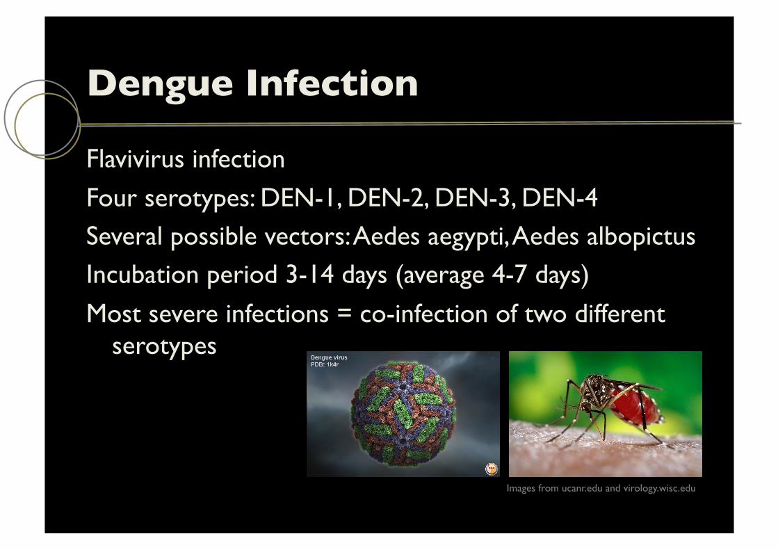
Dengue Infection
Flavivirus infection
Four serotypes: DEN-1, DEN-2, DEN-3, DEN-4 Several possible vectors: Aedes aegypti, Aedes albopictus Incubation period 3-14 days (average 4-7 days)
Most severe infections = co-infection of two different serotypes
Images from ucanr.edu and virology.wisc.edu
Dengue Infection: ���Primary or Secondary Primary: infection with any serotype inducing immune
response protecting re-infection by that serotype Mostly children
Secondary: infection by another serotype Mostly adults or elderly A/W significant morbidity and occasional death
Dengue Infection
Wide spectrum of clinical presentations
Often unpredictable clinical evolution and outcome Infection may be asymptomatic, self-limiting febrile
illness or
Small number of cases – severe/life-threatening characterized by plasma leakage w/wo hemorrhage
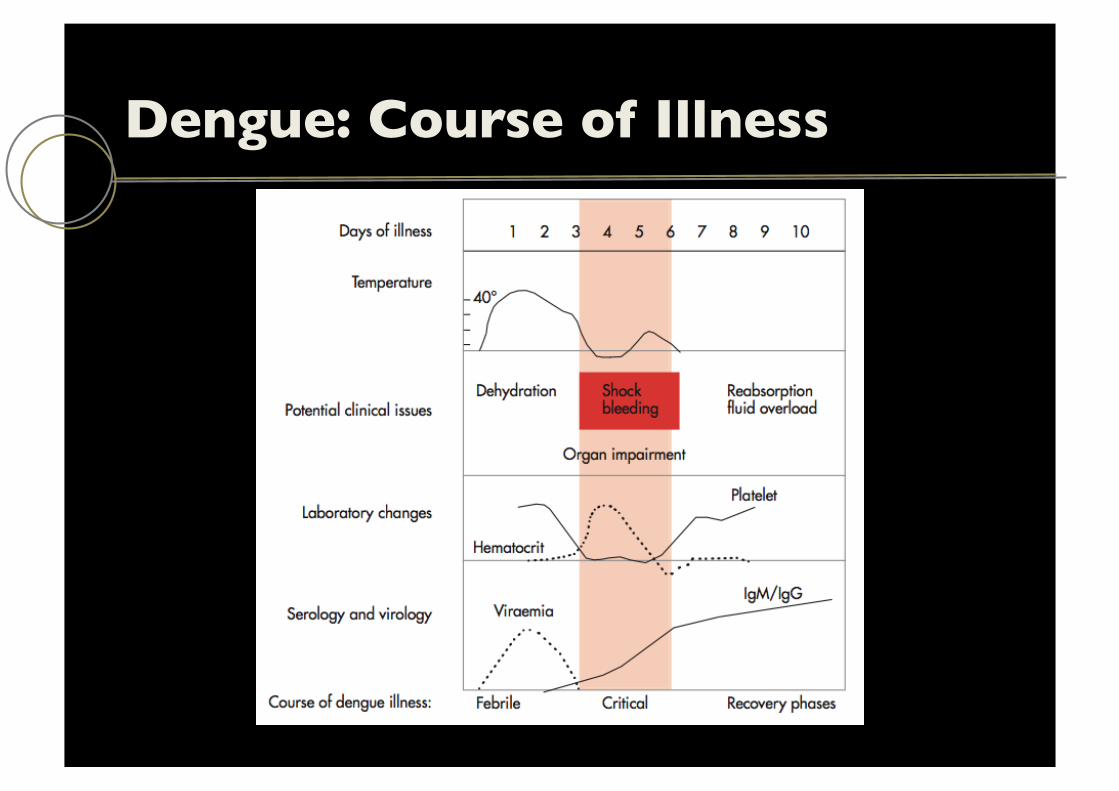
Dengue: Course of Illness

WHO: ���Focus on First Level of Care Recognize that the febrile patient could have dengue
Notify public health authorities Manage patients in early febrile phase Recognize early stage of plasma leakage or critical phase
and initiate fluid therapy Recognize patients with warning signs needing referral
or admission
Recognize and manage severe plasma leakage and shock
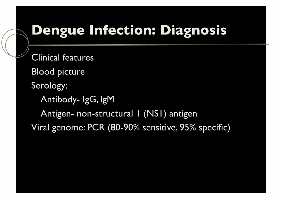
Dengue Infection: Diagnosis
Clinical features
Blood picture Serology: Antibody- IgG, IgM
Antigen- non-structural 1 (NS1) antigen Viral genome: PCR (80-90% sensitive, 95% specific)
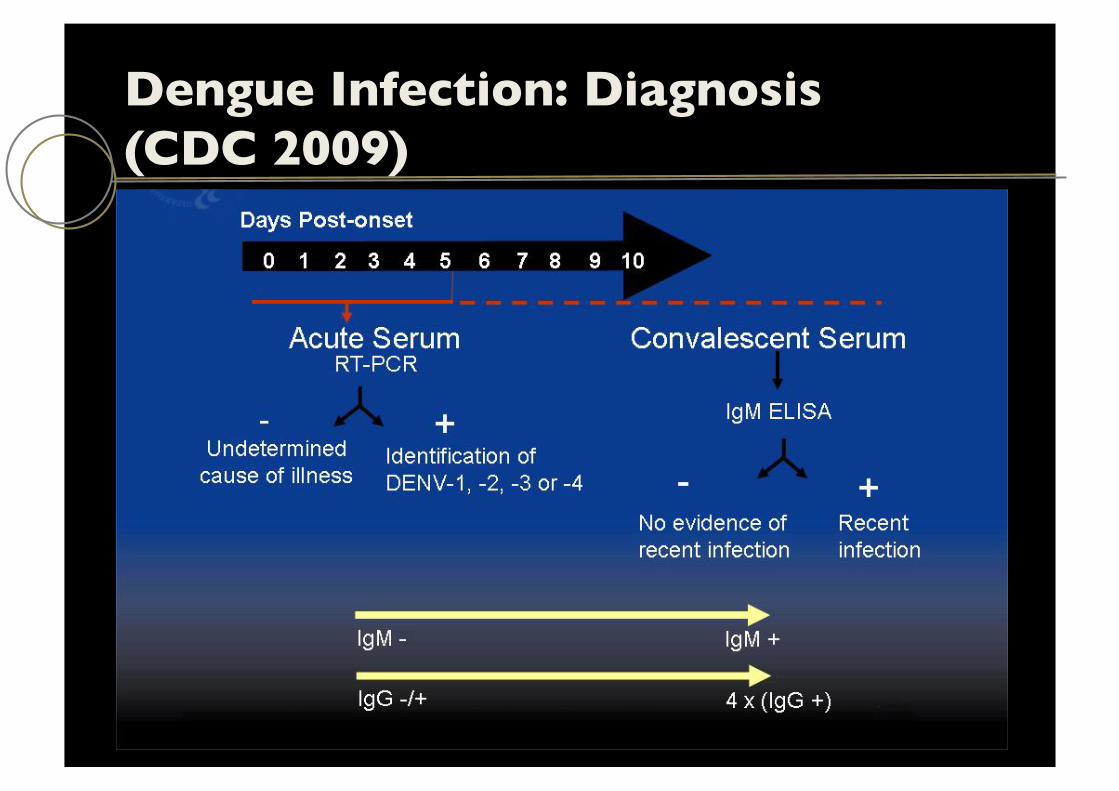
Dengue Infection: Diagnosis ���(CDC 2009)

Dengue Infection Classification: WHO 2009

Dengue Infection Classification:���WHO SEA (2010)
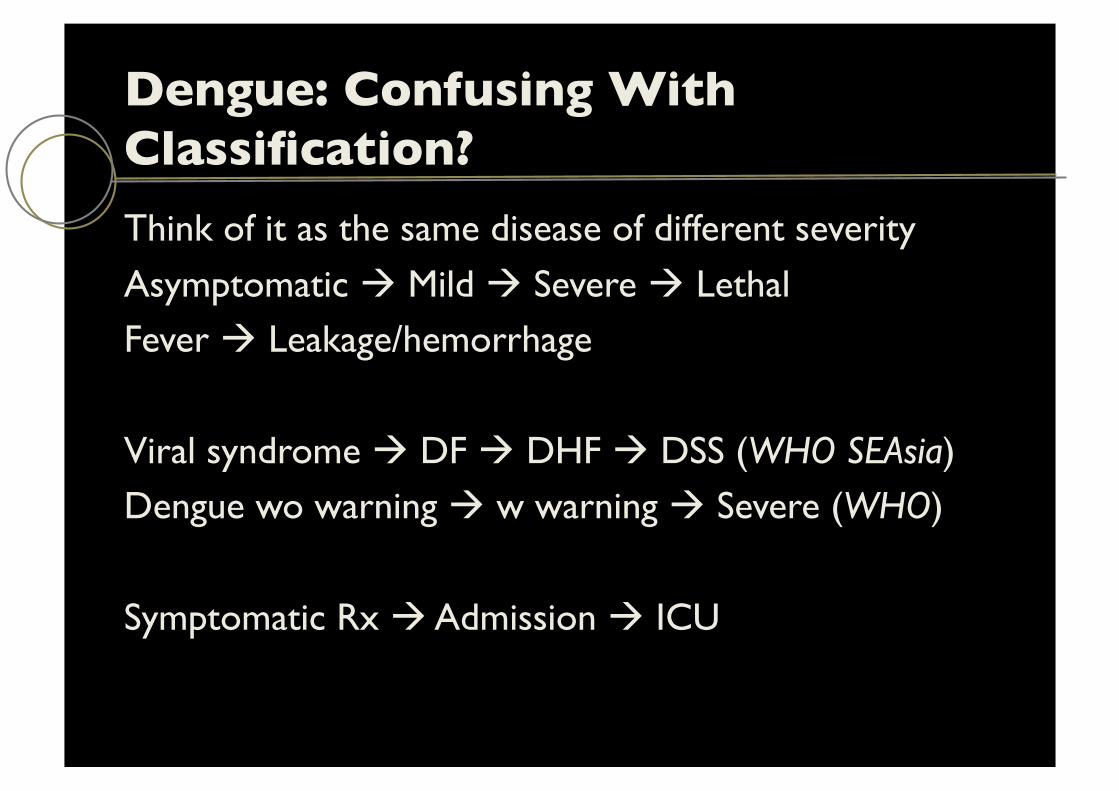
Dengue: Confusing With Classification? Think of it as the same disease of different severity
Asymptomatic ! Mild ! Severe ! Lethal Fever ! Leakage/hemorrhage
Viral syndrome ! DF ! DHF ! DSS (WHO SEAsia) Dengue wo warning ! w warning ! Severe (WHO)
Symptomatic Rx ! Admission ! ICU
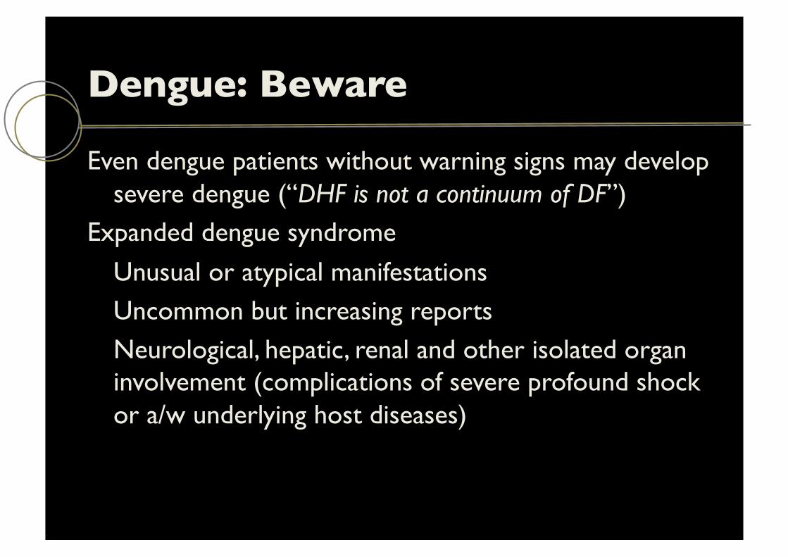
Dengue: Beware
Even dengue patients without warning signs may develop severe dengue (“DHF is not a continuum of DF”)
Expanded dengue syndrome
Unusual or atypical manifestations Uncommon but increasing reports Neurological, hepatic, renal and other isolated organ involvement (complications of severe profound shock or a/w underlying host diseases)
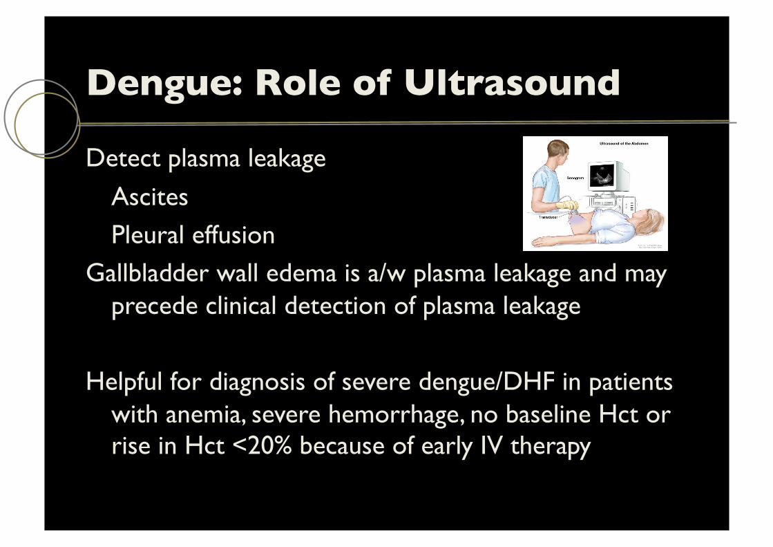
Dengue: Role of Ultrasound
Detect plasma leakage
Ascites Pleural effusion
Gallbladder wall edema is a/w plasma leakage and may precede clinical detection of plasma leakage
Helpful for diagnosis of severe dengue/DHF in patients with anemia, severe hemorrhage, no baseline Hct or rise in Hct <20% because of early IV therapy
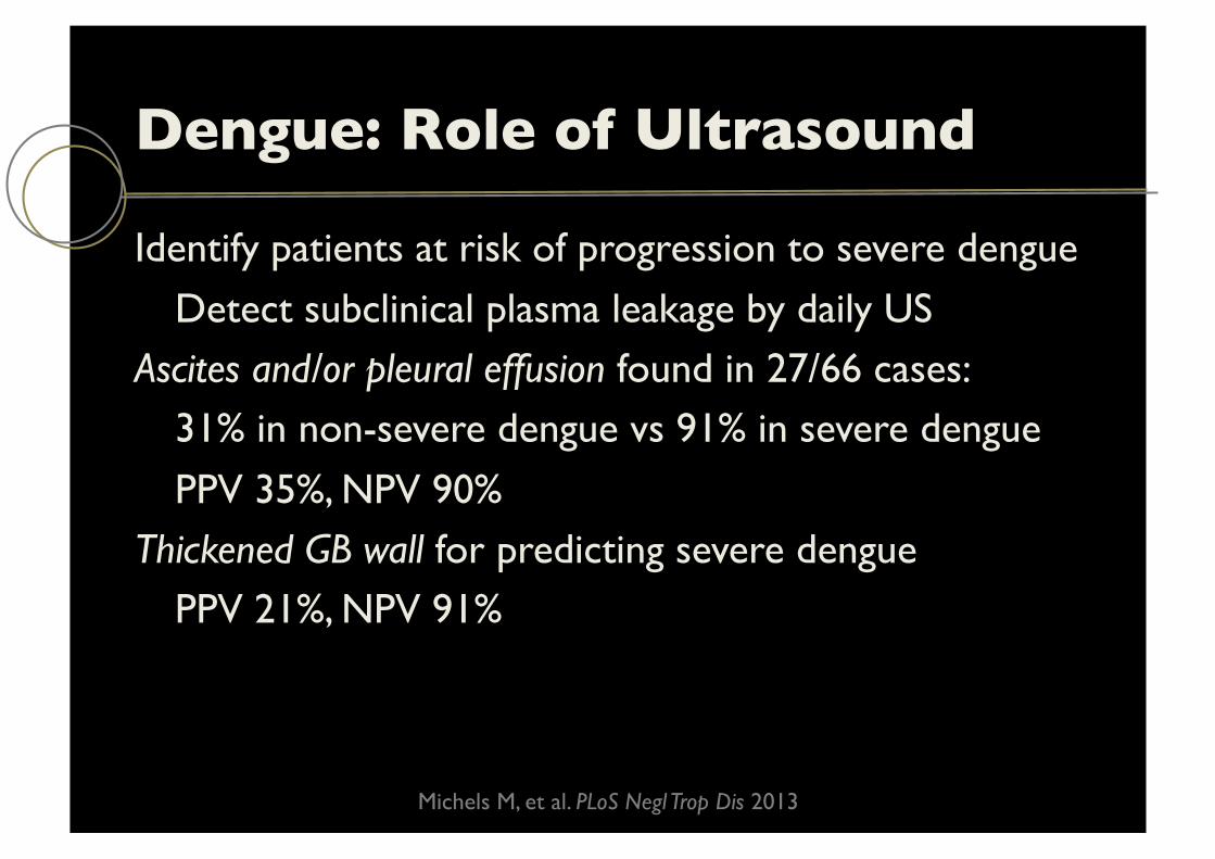
Dengue: Role of Ultrasound
Identify patients at risk of progression to severe dengue
Detect subclinical plasma leakage by daily US Ascites and/or pleural effusion found in 27/66 cases: 31% in non-severe dengue vs 91% in severe dengue
PPV 35%, NPV 90% Thickened GB wall for predicting severe dengue PPV 21%, NPV 91%
Michels M, et al. PLoS Negl Trop Dis 2013

Dengue: Role of Ultrasound
Raise a possibility of this diagnosis in unsuspected cases:
Atypical presentation Limited resources in lab testing
Bertfish.com
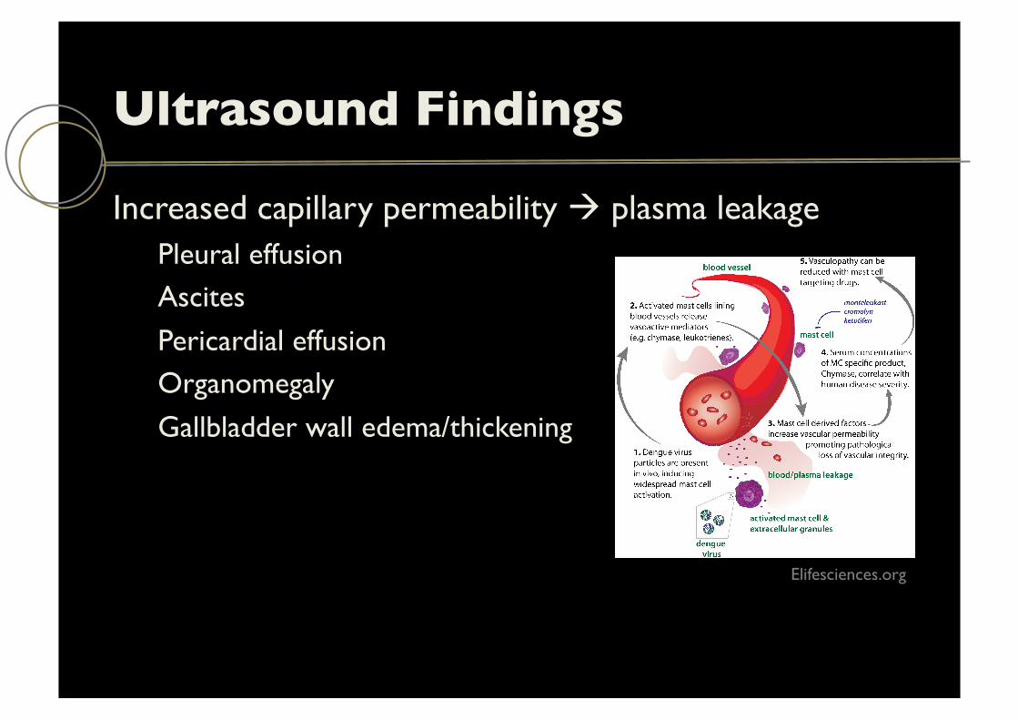
Ultrasound Findings
Increased capillary permeability ! plasma leakage Pleural effusion Ascites
Pericardial effusion Organomegaly
Gallbladder wall edema/thickening
Elifesciences.org
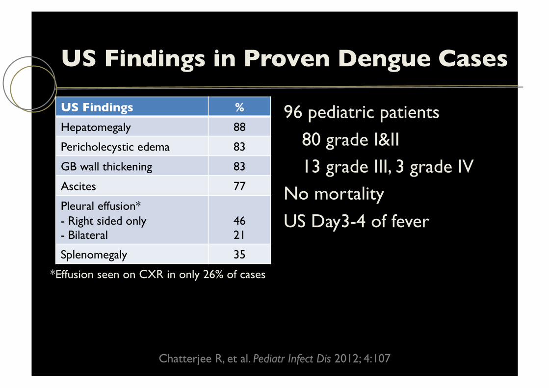
US Findings in Proven Dengue Cases
US Findings % Hepatomegaly 88
Pericholecystic edema 83
GB wall thickening 83
Ascites 77
Pleural effusion* - Right sided only - Bilateral
46 21
Splenomegaly 35
96 pediatric patients
80 grade I&II 13 grade III, 3 grade IV
No mortality
US Day3-4 of fever
*Effusion seen on CXR in only 26% of cases
Chatterjee R, et al. Pediatr Infect Dis 2012; 4:107
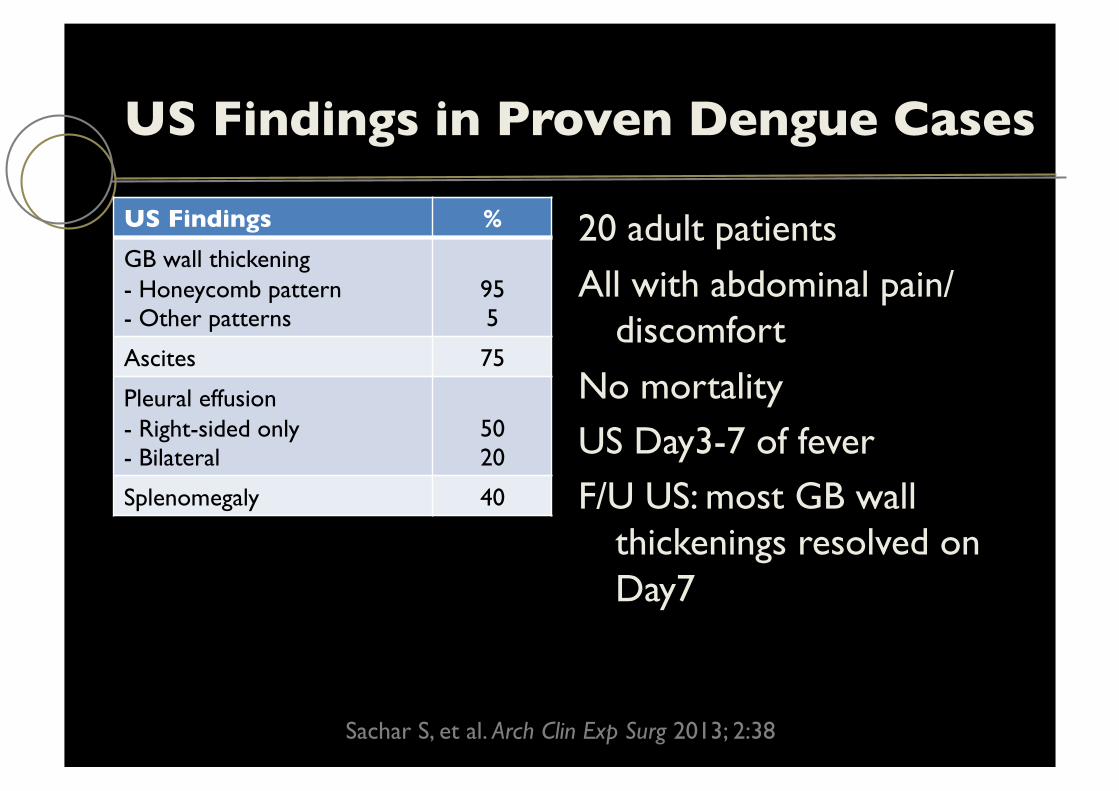
US Findings in Proven Dengue Cases
US Findings % GB wall thickening - Honeycomb pattern - Other patterns
95 5
Ascites 75
Pleural effusion - Right-sided only - Bilateral
50 20
Splenomegaly 40
20 adult patients
All with abdominal pain/discomfort
No mortality US Day3-7 of fever F/U US: most GB wall
thickenings resolved on Day7
Sachar S, et al. Arch Clin Exp Surg 2013; 2:38

US Findings in Proven Dengue Cases
US Findings Non-severe (%)
Severe (%)
GB wall thickening 87 100
Pericholecystic fluid 44 60
Hepatomegaly 27 60
Splenomegaly 22 40
Effusion - Right - Left
9 -
60 20
Ascites 18 60
Pericardial effusion 4 20
50 patients (6-59 years)
45 non-severe 5 severe (2 deaths)
GB wall > 5 mm
No Murphy sign
Mehdi SA, et al. Ann Punjab Med Coll 2012; 6:32
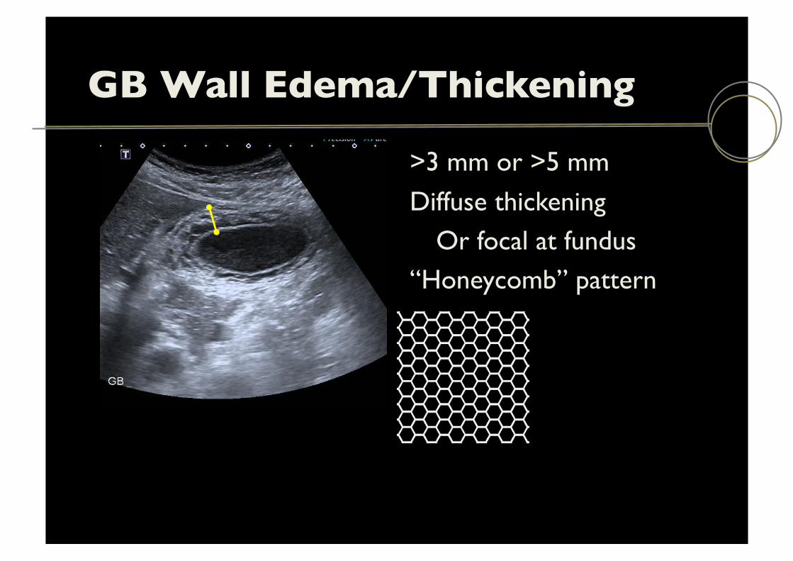
GB Wall Edema/Thickening
>3 mm or >5 mm
Diffuse thickening Or focal at fundus
“Honeycomb” pattern
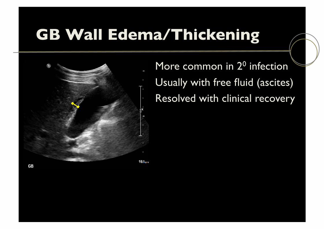
GB Wall Edema/Thickening
More common in 20 infection
Usually with free fluid (ascites) Resolved with clinical recovery
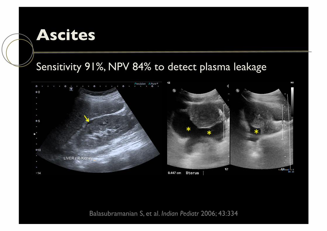
Ascites
Sensitivity 91%, NPV 84% to detect plasma leakage
Balasubramanian S, et al. Indian Pediatr 2006; 43:334
* * *
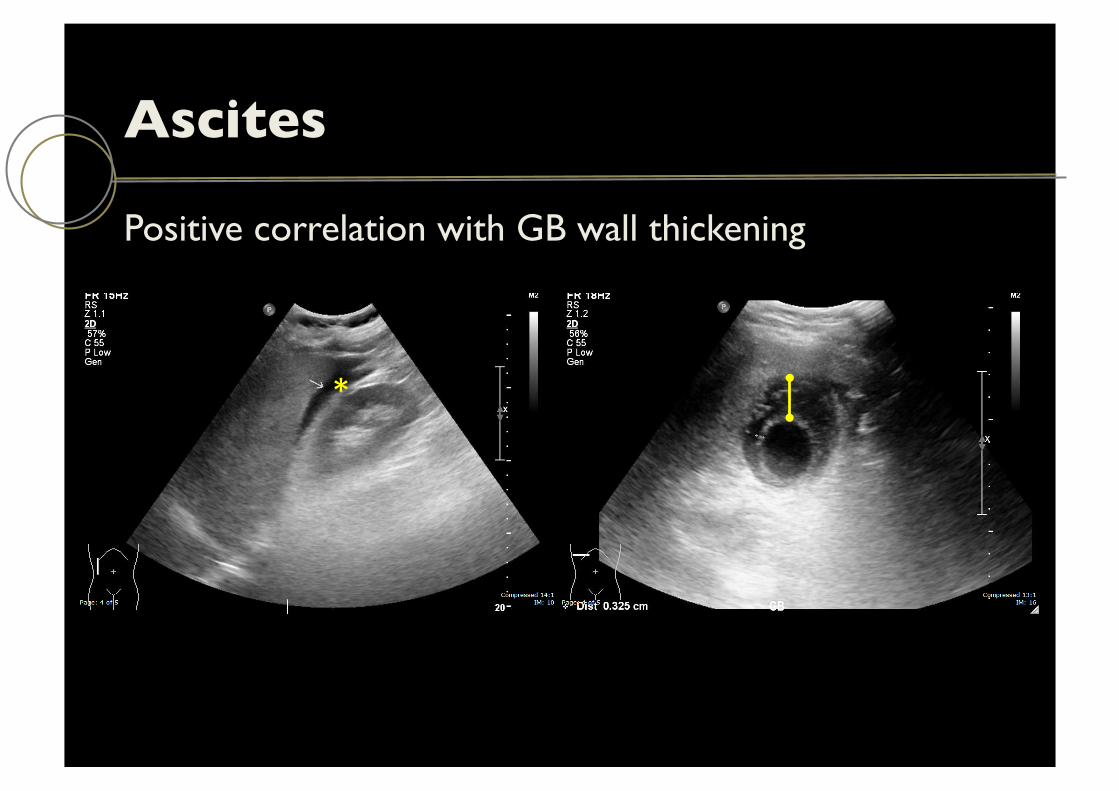
Ascites
Positive correlation with GB wall thickening
*

Pleural Effusion
Right or bilateral effusions
Very rarely (or non-existent?) isolated left effusion
Srikiatkhachorn, et al. Pediatr Infect Dis 2007; 26:283
*

Pleural Effusion
Time of performance of US may influence its presence
Common US evidence of plasma leakage, starting 2 days before defervescence (decrease of body temp)
Srikiatkhachorn, et al. Pediatr Infect Dis 2007; 26:283
Day 3 of fever
*
Pericardial Effusion
8% of patients with DHF had small pericardial effusion*
US at Day5-8 from onset of fever ! 28% had this** 0 out of 32 patients US Day2-3 had this** 3 out of 12 volunteers inoculated with dengue virus
developed small pericardial effusion btw Day10-20 Cardiac tamponade possible but very rare (case reports)
*Setiawan MW, et al. J Clin Ultrasound 1998; 23:357 **Venkata Sai PM, et al. Br J Radiol 2005; 78:416

Severity of Disease
Possible early prediction of disease severity
Mild disease – less % of US abnormalities Severe disease – US abnormalities very common

US Differential Diagnosis
Acute cholecystitis:
GB distension Gallstones (acalculous – uncommon in ER) Wall thickening not marked
Ascites, pleural effusion, pericardial eff not common Murphy may not be that helpful

US: Limitations
Findings of plasma leakage are seen in both mild and severe disease, primary as well as secondary infections
Findings possibly related to time of onset Can findings be reflective of treatment? Nonspecific US findings when stand-alone
Summary (I)
Spectrum of plasma leakage a/w dengue is broad. With improved techniques of identification – minor leakage will become more apparent
Ascites, pleural effusion, pericardial effusion, GB wall thickening and hepatosplenomegaly are US signs of plasma leakage

Summary (II)
Unsuspected case – US performed for other reasons
Suspected case, limited hospital resources – US helps making diagnosis or narrowing DDx
Suspected/confirm case – US helps detecting subclinical plasma leakage
Serial US may help identify patients whom disease might progress
Suggested Readings
2010