
Cardiac Limited Ultrasound Examination Techniques to Augment the Bedside … · ·...
8
Cardiac Limited Ultrasound Examination Techniques to Augment the Bedside Cardiac Physical Examination or centuries, physicians have taught examination of the heart and lungs, as the physical findings of these organs are often critical in formulating medical diagnoses and manag- ing the course of both cardiac and noncardiac disease. The recent advent of pocket-sized ultrasound devices has made the application of ultrasound to enhance the accuracy of the physical examination not only possible but convenient. However, in current cost-conscious and evidence-driven practice, any future expansion of bedside examina- tions must be time-efficient, demonstrate value in patient care or in the prediction of outcomes, and be feasibly taught. 1–4 In this article, we address the novel concept of adding specific, evidence-based 2-dimensional “quick-look” ultrasound techniques to conventional bedside assessment of the cardiovascular system. A simplified cardiac limited ultrasound examination (CLUE) can be created, which searches for only a few ultrasound “signs” indicating the presence of left ventricular (LV) systolic dysfunction, pulmonary edema, pleural and pericardial fluid, right ventricular (RV) enlarge- ment, and elevation of central venous pressure: traditional targets of the cardiac physical examination that have been firmly embedded in bedside formulative reasoning. The CLUE imaging procedure was structured in accordance with practical requirements that the exam- ination can be completed within a few minutes, can be routinely applied to all patients by all physicians, and requires only basic skills and equipment such that training can be broadly incorporated into medical school or postgraduate curricula on physical examination. Bruce J. Kimura, MD, David J. Shaw, MD, Stan A. Amundson, MD, James N. Phan, RDCS, RVT, Daniel G. Blanchard, MD, Anthony N. DeMaria, MD Received September 3, 2014, from Scripps Mercy Hospital, San Diego, California USA (B.J.K., D.J.S., S.A.A., J.N.P.); and University of California, San Diego Medical Center, San Diego, California USA (D.G.B., A.N.D.). Revision requested October 14, 2014. Revised manuscript accepted for publication November 29, 2014. We thank Dudie Keane for administrative support, Tanya Wolfson for statistical support, and the cardiac sonographers at Scripps Mercy Hospital for their work. Address correspondence to Bruce J. Kimura, MD, Department of Noninvasive Cardiology, Scripps Mercy Hospital, 4060 Fourth Ave, 206, San Diego, CA 92103 USA. E-email: [email protected] Abbreviations CHF, congestive heart failure; CLUE, cardiac limited ultrasound examination; IVC, inferior vena cava; LA, left atrium; LV, left ventricu- lar; RA, right atrial; RV, right ventricular F ©2015 by the American Institute of Ultrasound in Medicine | J Ultrasound Med 2015; 34:1683–1690 | 0278-4297 | www.aium.org TECHNICAL INNOVATION The current practice of physical diagnosis is dependent on physician skills and biases, inductive reasoning, and time efficiency. Although the clinical utility of echocardio- graphy is well known, few data exist on how to integrate 2-dimensional screening “quick- look” ultrasound applications into a novel, modernized cardiac physical examination. We discuss the evidence basis behind ultrasound “signs” pertinent to the cardiovascular system and elemental in synthesis of bedside diagnoses and propose the application of a brief cardiac limited ultrasound examination based on these signs. An ultrasound- augmented cardiac physical examination can be taught in traditional medical educa- tion and has the potential to improve bedside diagnosis and patient care. Key Words—continuing medical education; graduate medical education; hand-carried ultrasound; echocardiography; physical examination; physician training; point-of-care ultra- sound; screening; stethoscope; vascular ultrasound doi:10.7863/ultra.15.14.09002
Transcript of Cardiac Limited Ultrasound Examination Techniques to Augment the Bedside … · ·...
Cardiac Limited Ultrasound ExaminationTechniques to Augment the BedsideCardiac Physical Examination
or centuries, physicians have taught examination of theheart and lungs, as the physical findings of these organs areoften critical in formulating medical diagnoses and manag-
ing the course of both cardiac and noncardiac disease. The recentadvent of pocket-sized ultrasound devices has made the application ofultrasound to enhance the accuracy of the physical examination notonly possible but convenient. However, in current cost-conscious andevidence-driven practice, any future expansion of bedside examina-tions must be time-efficient, demonstrate value in patient care or in theprediction of outcomes, and be feasibly taught.1–4
In this article, we address the novel concept of adding specific,evidence-based 2-dimensional “quick-look” ultrasound techniquesto conventional bedside assessment of the cardiovascular system.A simplified cardiac limited ultrasound examination (CLUE) can becreated, which searches for only a few ultrasound “signs” indicatingthe presence of left ventricular (LV) systolic dysfunction, pulmonaryedema, pleural and pericardial fluid, right ventricular (RV) enlarge-ment, and elevation of central venous pressure: traditional targets ofthe cardiac physical examination that have been firmly embedded inbedside formulative reasoning. The CLUE imaging procedure wasstructured in accordance with practical requirements that the exam-ination can be completed within a few minutes, can be routinelyapplied to all patients by all physicians, and requires only basic skillsand equipment such that training can be broadly incorporated intomedical school or postgraduate curricula on physical examination.
Bruce J. Kimura, MD, David J. Shaw, MD, Stan A. Amundson, MD, James N. Phan, RDCS, RVT, Daniel G. Blanchard, MD, Anthony N. DeMaria, MD
Received September 3, 2014, from Scripps MercyHospital, San Diego, California USA (B.J.K.,D.J.S., S.A.A., J.N.P.); and University of California,San Diego Medical Center, San Diego, CaliforniaUSA (D.G.B., A.N.D.). Revision requestedOctober 14, 2014. Revised manuscript acceptedfor publication November 29, 2014.
We thank Dudie Keane for administrativesupport, Tanya Wolfson for statistical support,and the cardiac sonographers at Scripps MercyHospital for their work.
Address correspondence to Bruce J. Kimura,MD, Department of Noninvasive Cardiology,Scripps Mercy Hospital, 4060 Fourth Ave, 206,San Diego, CA 92103 USA.
E-email: [email protected]
AbbreviationsCHF, congestive heart failure; CLUE, cardiaclimited ultrasound examination; IVC, inferiorvena cava; LA, left atrium; LV, left ventricu-lar; RA, right atrial; RV, right ventricular
F
©2015 by the American Institute of Ultrasound in Medicine | J Ultrasound Med 2015; 34:1683–1690 | 0278-4297 | www.aium.org
TECHNICAL INNOVATION
The current practice of physical diagnosis is dependent on physician skills and biases,inductive reasoning, and time efficiency. Although the clinical utility of echocardio-graphy is well known, few data exist on how to integrate 2-dimensional screening “quick-look” ultrasound applications into a novel, modernized cardiac physical examination.We discuss the evidence basis behind ultrasound “signs” pertinent to the cardiovascularsystem and elemental in synthesis of bedside diagnoses and propose the application ofa brief cardiac limited ultrasound examination based on these signs. An ultrasound- augmented cardiac physical examination can be taught in traditional medical educa-tion and has the potential to improve bedside diagnosis and patient care.
Key Words—continuing medical education; graduate medical education; hand-carriedultrasound; echocardiography; physical examination; physician training; point-of-care ultra-sound; screening; stethoscope; vascular ultrasound
doi:10.7863/ultra.15.14.09002
3409jum1663-1700_Layout 1 8/24/15 1:42 PM Page 1683
CLUE: 6 Ultrasound Signs of Cardiac Disease
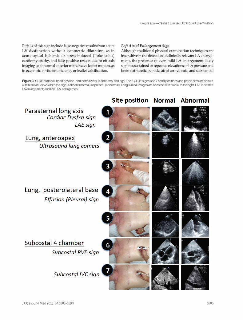
In CLUE, the basic 2-dimensional views are designed tointegrate physiologic assessment with the acquisition ofstructural data using a standard 2–3-MHz ultrasoundtransducer. Sequential imaging is performed from the LVto the left atrium (LA) to the lungs to the RV and finally tothe inferior vena cava (IVC). By working backward againstthe flow of blood, the user can deduce the extent of diseaseencountered (Table 1) and more easily remember theexamination (Figure 1).
Cardiac (LV) Systolic Dysfunction SignThe detection of LV systolic dysfunction has long been agoal of the cardiac examination, as its presence has prog-nostic, diagnostic, and therapeutic implications in bothacute and chronic settings. In CLUE, the cardiac systolicdysfunction sign is present when the anterior leaflet of themitral valve fails to approach the ventricular septum towithin 1 cm by subjective estimation of its opening motionfrom the parasternal long-axis view (Figure 1, site 1). Thisso-called E-point septal separation distance has been justi-fied by echocardiographic5 and magnetic resonance imag-ing6 studies with sensitivity of 65% and specificity of 92%for detection of systolic LV dysfunction. Teaching this sin-gle-view parameter to novice users improves their diag-
nostic accuracy for LV dysfunction beyond the physicalexamination alone7 and elevates their bedside estimates ofLV function to that of the visually estimated LV ejection frac-tion by experts.8
In the CLUE protocol, the parasternal long-axis view,a view obtained by placing the transducer against the leftside of the patient’s sternum in the third or fourth inter-costal space and aiming its directional marker toward theright shoulder, was chosen over other windows due to itsrelative ease of acquisition and display of multiple cardiacdisorders. The parasternal long-axis view can be obtainedon supine, restrained, or intubated patients and preservesmodesty by exposing only the sternal region during out-patient examinations.
In a “real-world” assessment of 1016 consecutiveechocardiograms, the cardiac dysfunction sign had sensi-tivity of 69% and specificity of 91% for an LV ejection frac-tion 40% or lower and showed a univariate relationshipwith in-hospital mortality.3 As with the detection of an S3gallop, the clinician must recognize that the presence ofthis ultrasound sign needs confirmation by standardechocardiography and a search for its cause. Furthermore,as is true when any abnormal finding is discovered, earlytermination of the diagnostic process should be resisted, asthe presence of LV dysfunction does not necessarily imply asole cardiac etiology in an episode of shock or dyspnea.
Kimura et al—Cardiac Limited Ultrasound Examination
J Ultrasound Med 2015; 34:1683–16901684
Table 1. Diagnostic Patterns in CLUE
Disorder C L U E SR SI
LV systolic dysfunction, compensated ++/−
Diastolic dysfunction, compensated Atrial fibrillation (paroxysmal or chronic) Severe mitral regurgitation/stenosis, compensated Severe multivessel CAD, compensated +/− Symptomatic aortic valve disease, compensated CHF exacerbation, HFpEF ++/− CHF exacerbation, HFrEF ++/− Cardiogenic pulmonary edema ++/− ++/− ++/−
ARDS, noncardiogenic pulmonary edema +/− − +/−
Interstitial lung disease (acute or chronic) ++/− +/−
COPD with cor pulmonale +/− − Pneumonia or small pulmonary embolism +/− − +/−
Submassive pulmonary embolism +/− − +/− Cardiogenic shock ++/− +/− Tamponade RV myocardial infarction Chronic right heart failure or severe TR Septic or hypovolemic shock
ARDS indicates adult respiratory distress syndrome sign; C, cardiac dysfunction; CAD, coronary artery disease; COPD, chronic obstructive pul-
monary disease; E, pleural (or pericardial) effusion sign; HFpEF, heart failure with preserved ejection fraction; HFrEF, heart failure with reduced
ejection fraction; L, LA enlargement sign; SI, subcostal IVC plethora sign; SR, Subcostal RV enlargement sign; TR, tricuspid regurgitation;
U, ultrasound lung comet tail sign; , present; ++/–, frequently present; +/–, commonly present; and +/– –, occasionally present.
3409jum1663-1700_Layout 1 8/24/15 1:42 PM Page 1684
Pitfalls of this sign include false-negative results from acuteLV dysfunction without symmetric dilatation, as inacute apical ischemia or stress-induced (Takotsubo) cardiomyopathy, and false-positive results due to off-axisimaging or abnormal anterior mitral valve leaflet motion, asin eccentric aortic insufficiency or leaflet calcification.
Left Atrial Enlargement SignAlthough traditional physical examination techniques areinsensitive in the detection of clinically relevant LA enlarge-ment, the presence of even mild LA enlargement likelysignifies sustained or repeated elevations of LA pressure andbrain natriuretic peptide, atrial arrhythmia, and substantial
J Ultrasound Med 2015; 34:1683–1690 1685
Kimura et al—Cardiac Limited Ultrasound Examination
Figure 1. CLUE protocol, hand position, and normal versus abnormal findings. The 6 CLUE signs and 7 hand positions and probe sites are shown
with resultant views when the sign is absent (normal) or present (abnormal). Longitudinal images are oriented with cranial to the right. LAE indicates
LA enlargement; and RVE, RV enlargement.
3409jum1663-1700_Layout 1 8/24/15 1:42 PM Page 1685
structural findings on echocardiography9,10 and has out-patient prognostic value.11 The detection of LA enlarge-ment can have utility in the assessment of unexplaineddyspnea or palpitations, improve referral for echocardiog-raphy, and be recognized by novice users.9 Despite itsimportance in both acute and chronic cardiac disease, rel-atively few data exist regarding evaluation of LA enlarge-ment by bedside ultrasound imaging.
The easily recognized LA enlargement sign is definedfrom the same parasternal long-axis view as the cardiac dys-function sign after assessment of the LV ejection fractionand is present when the anteroposterior diameter of theLA appears larger than the overlying aorta throughout the cardiac cycle (Figure 1, site 1).10 The reported sensi-tivity and specificity of this sign for LA enlargement as indi-cated by an LA volume index of greater than 28 mL/m2
are 59% and 79%, respectively10 and 75% and 72% whencompared to the reporting of any LA enlargement on stan-dard echocardiography.3 The absence of the LA enlarge-ment sign in the setting of the cardiac dysfunction sign cansuggest a well-compensated or well-diuresed state, whereasits presence in the setting of normal LV systolic functioncan suggest diastolic dysfunction, substantial left-sidedheart valve disease, or atrial fibrillation (Table 1). Pitfalls ofthe LA enlargement sign include interpretation errors dueto a bright far-field side-lobe artifact generated from the nor-mal right atrial (RA)–pericardial border, asymmetric LAelongation caused by space limitations within the antero-posterior dimension of the chest cavity due to right heartenlargement or, less commonly, skeletal deformity (eg,pectus excavatum or ankylosing spondylitis), and thepresence of an aortic root aneurysm.
Ultrasound Lung Comet Tail SignExamination of the lung for rales or effusions remains avital component in the cardiac physical examination.Ultrasound imaging of the lung parenchyma can identify aB-line artifact manifest as a linear full-field reverberationartifact generated from subpleural tissue or interlobularsepta that has been thickened by extravascular lung water,inflammation, or fibrosis. The appearance of at least 3 B-lines in a single view constitutes the ultrasound lung comettail sign.3 Although there is limited standardization andpathologic confirmatory data, the ultrasound lung comettail artifact has become an accepted finding12 akin to asonographic form of rales, showing similar clinical behav-ior and implications.
In CLUE, after detection of LA enlargement, the lungsare examined for evidence of elevated LA pressure fromcardiac decompensation, manifesting as pulmonary edema
or pleural effusions. In the absence of LA enlargement, theultrasound lung comet tail sign can also be attributed to acapillary leak in adult respiratory distress syndrome, local-ized lung edema or inflammation, or diffuse primary inter-stitial lung disease (Table 1). B-lines, either when tabulatedas a total score by mapping the lung13 or when grouped inultrasound lung comet tail signs and found in the lungapices,3 are independent predictors of mortality that aremore powerful than the LV ejection fraction. Althoughmultiple imaging protocols exist to evaluate the chest for B-lines, a recent study confirmed high specificity of 89%(99% confidence interval, 74%–97%), albeit low sensitiv-ity of 40% (99% confidence interval, 21%–61%) for con-gestive heart failure (CHF) of 2 anteroapical views.14 InCLUE, the antero apices of each lung are interrogated as asagittal image in the midclavicular line in approximatelythe third intercostal space (Figure 1, sites 2 and 3). Thespecificity of these 2 views for CHF can be made particu-larly effective when complemented with the 85% to 90%sensitivity afforded by elevated N-terminal pro–brain natri-uretic peptide measurement or the presence of effusionson basal lung imaging.15 Imaging for ultrasound lung comettails is generally easy to learn16,17 and likely has device vari-ability and minimal acquisition errors. Ultrasound lungcomet tails, like rales, can vary by patient position and aregravity and lung volume dependent, can be present in thelung bases in healthy individuals, and can disappear rapidlywith diuresis or dialysis.18 In addition, the presence of ultra-sound lung comet tails under the probe virtually excludesthe presence of pneumothorax at that site, as does the pres-ence of pleural sliding,19 a normal motion best seen with ahigher-frequency (5–7-MHz) probe. An important clinicalpitfall can occur when ultrasound lung comet tails arechronic and due to fibrosis but are mistakenly attributedto acute edema or inflammation, an occurrence that canbe common in chronic lung disease and could be avoided byprior documentation of the finding.
Effusion (Pleural) SignUltrasound imaging has the ability to show as little as a fewmilliliters of fluid present in the costophrenic angle and hasmore sensitivity than physical examination or standardanteroposterior chest radiography,15 which typically requirean effusion volume of at least 200 mL before detection.20
Commonly, bilateral effusions with right-sided predomi-nance suggest CHF,20 whereas the presence of a unilateraleffusion, particularly when complicated by septation orparticulate matter, suggests the possibility of parapneu-monic causes, pulmonary embolism, or malignancy.
Kimura et al—Cardiac Limited Ultrasound Examination
J Ultrasound Med 2015; 34:1683–16901686
3409jum1663-1700_Layout 1 8/24/15 1:42 PM Page 1686
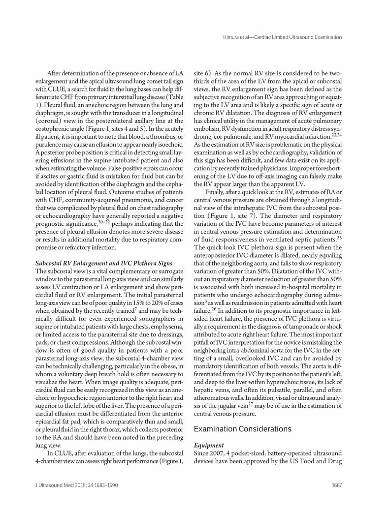
After determination of the presence or absence of LAenlargement and the apical ultrasound lung comet tail signwith CLUE, a search for fluid in the lung bases can help dif-ferentiate CHF from primary interstitial lung disease (Table1). Pleural fluid, an anechoic region between the lung anddiaphragm, is sought with the transducer in a longitudinal(coronal) view in the posterolateral axillary line at thecostophrenic angle (Figure 1, sites 4 and 5). In the acutelyill patient, it is important to note that blood, a thrombus, orpurulence may cause an effusion to appear nearly isoechoic.A posterior probe position is critical in detecting small lay-ering effusions in the supine intubated patient and alsowhen estimating the volume. False-positive errors can occurif ascites or gastric fluid is mistaken for fluid but can beavoided by identification of the diaphragm and the cepha-lad location of pleural fluid. Outcome studies of patientswith CHF, community-acquired pneumonia, and cancerthat was complicated by pleural fluid on chest radiographyor echocardiography have generally reported a negativeprognostic significance,20–22 perhaps indicating that thepresence of pleural effusion denotes more severe diseaseor results in additional mortality due to respiratory com-promise or refractory infection.
Subcostal RV Enlargement and IVC Plethora SignsThe subcostal view is a vital complementary or surrogatewindow to the parasternal long-axis view and can similarlyassess LV contraction or LA enlargement and show peri-cardial fluid or RV enlargement. The initial parasternallong-axis view can be of poor quality in 15% to 20% of caseswhen obtained by the recently trained7 and may be tech-nically difficult for even experienced sonographers insupine or intubated patients with large chests, emphysema,or limited access to the parasternal site due to dressings,pads, or chest compressions. Although the subcostal win-dow is often of good quality in patients with a poorparasternal long-axis view, the subcostal 4-chamber viewcan be technically challenging, particularly in the obese, inwhom a voluntary deep breath hold is often necessary tovisualize the heart. When image quality is adequate, peri-cardial fluid can be easily recognized in this view as an ane-choic or hypoechoic region anterior to the right heart andsuperior to the left lobe of the liver. The presence of a peri-cardial effusion must be differentiated from the anteriorepicardial fat pad, which is comparatively thin and small,or pleural fluid in the right thorax, which collects posteriorto the RA and should have been noted in the precedinglung view.
In CLUE, after evaluation of the lungs, the subcostal4-chamber view can assess right heart performance (Figure 1,
site 6). As the normal RV size is considered to be two-thirds of the area of the LV from the apical or subcostalviews, the RV enlargement sign has been defined as thesubjective recognition of an RV area approaching or equat-ing to the LV area and is likely a specific sign of acute orchronic RV dilatation. The diagnosis of RV enlargementhas clinical utility in the management of acute pulmonaryembolism, RV dysfunction in adult respiratory distress syn-drome, cor pulmonale, and RV myocardial infarction.23,24
As the estimation of RV size is problematic on the physicalexamination as well as by echocardiography, validation ofthis sign has been difficult, and few data exist on its appli-cation by recently trained physicians. Improper foreshort-ening of the LV due to off-axis imaging can falsely makethe RV appear larger than the apparent LV.
Finally, after a quick look at the RV, estimates of RA orcentral venous pressure are obtained through a longitudi-nal view of the intrahepatic IVC from the subcostal posi-tion (Figure 1, site 7). The diameter and respiratoryvariation of the IVC have become parameters of interestin central venous pressure estimation and determinationof fluid responsiveness in ventilated septic patients.25
The quick-look IVC plethora sign is present when theanteroposterior IVC diameter is dilated, nearly equalingthat of the neighboring aorta, and fails to show respiratoryvariation of greater than 50%. Dilatation of the IVC with-out an inspiratory diameter reduction of greater than 50%is associated with both increased in-hospital mortality inpatients who undergo echocardiography during admis-sion3 as well as readmission in patients admitted with heartfailure.26 In addition to its prognostic importance in left-sided heart failure, the presence of IVC plethora is virtu-ally a requirement in the diagnosis of tamponade or shockattributed to acute right heart failure. The most importantpitfall of IVC interpretation for the novice is mistaking theneighboring intra-abdominal aorta for the IVC in the set-ting of a small, overlooked IVC and can be avoided bymandatory identification of both vessels. The aorta is dif-ferentiated from the IVC by its position to the patient's left,and deep to the liver within hyperechoic tissue, its lack ofhepatic veins, and often its pulsatile, parallel, and oftenatheromatous walls. In addition, visual or ultrasound analy-sis of the jugular vein27 may be of use in the estimation ofcentral venous pressure.
Examination Considerations
Equipment Since 2007, 4 pocket-sized, battery-operated ultrasounddevices have been approved by the US Food and Drug
J Ultrasound Med 2015; 34:1683–1690 1687
Kimura et al—Cardiac Limited Ultrasound Examination
3409jum1663-1700_Layout 1 8/24/15 1:42 PM Page 1687
Kimura et al—Cardiac Limited Ultrasound Examination
J Ultrasound Med 2015; 34:1683–16901688
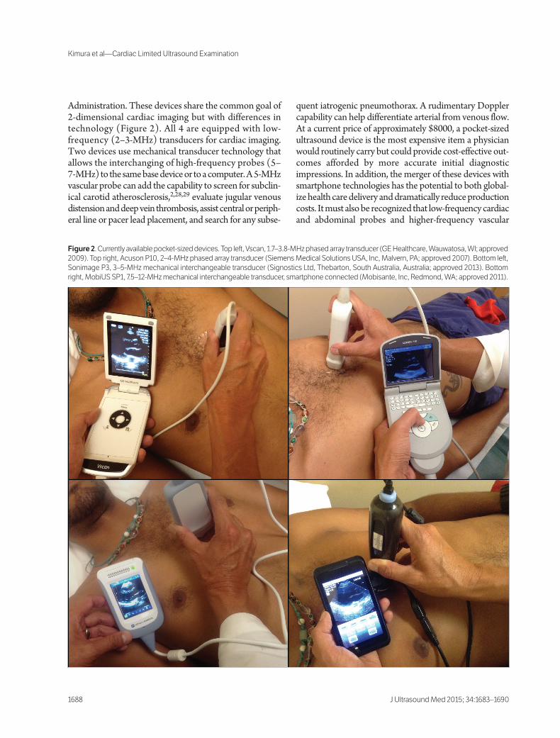
Administration. These devices share the common goal of2-dimensional cardiac imaging but with differences intechnology (Figure 2). All 4 are equipped with low-frequency (2–3-MHz) transducers for cardiac imaging.Two devices use mechanical transducer technology thatallows the interchanging of high-frequency probes (5–7-MHz) to the same base device or to a computer. A 5-MHzvascular probe can add the capability to screen for subclin-ical carotid atherosclerosis,2,28,29 evaluate jugular venousdistension and deep vein thrombosis, assist central or periph-eral line or pacer lead placement, and search for any subse-
quent iatrogenic pneumothorax. A rudimentary Dopplercapability can help differentiate arterial from venous flow.At a current price of approximately $8000, a pocket-sizedultrasound device is the most expensive item a physicianwould routinely carry but could provide cost-effective out-comes afforded by more accurate initial diagnosticimpressions. In addition, the merger of these devices withsmartphone technologies has the potential to both global-ize health care delivery and dramatically reduce productioncosts. It must also be recognized that low-frequency cardiacand abdominal probes and higher-frequency vascular
Figure 2. Currently available pocket-sized devices. Top left, Vscan, 1.7–3.8-MHz phased array transducer (GE Healthcare, Wauwatosa, WI; approved
2009). Top right, Acuson P10, 2–4-MHz phased array transducer (Siemens Medical Solutions USA, Inc, Malvern, PA; approved 2007). Bottom left,
Sonimage P3, 3–5-MHz mechanical interchangeable transducer (Signostics Ltd, Thebarton, South Australia, Australia; approved 2013). Bottom
right, MobiUS SP1, 7.5–12-MHz mechanical interchangeable transducer, smartphone connected (Mobisante, Inc, Redmond, WA; approved 2011).
3409jum1663-1700_Layout 1 8/24/15 1:42 PM Page 1688
probes for carotid or deep venous thrombosis imaging arealready available on small laptop ultrasound machine plat-forms, as well as many older cart-based platforms. There-fore, a simplified 2-dimensional examination such as CLUEcan be practiced on machines that are already available atmost hospitals, emergency departments, and clinics anddoes not require the newer, more convenient, technologies.
ProcedureIn contrast to standard echocardiographic practice, thebasic 6-site CLUE can be performed in less than 2 minutes.The ultrasound device is explained to the patient as a “modern stethoscope,” to avoid a patient expectation ofa formal ultrasound examination. During the examination, ageneral attempt is made to minimize gel reapplications andmaintain control and cleanliness of the device by not plac-ing the unit down. The physician starts at the parasternallong-axis view and proceeds to interpret “on the spot,”moving from transducer site to transducer site (Figure 1)around the lung views and finishing in the subcostal viewof the IVC. Image or video loop recordings are not manda-tory and left to physician discretion, but they can be usefulat times to later compare with prior examinations or toshow the findings to the patient. Afterward, the probe, itscord, and the patient are wiped clean.
The presence of abnormal CLUE signs can be recordedas a part of the cardiac physical examination for ease ofreference. Using the acronym CLUE, as listed in this arti-cle’s subheadings, where cardiac dysfunction, left atrialenlargement, ultrasound lung comets, effusions (pleural orpericardial), and subcostal (RV and IVC) signs maintainthe “working backward” mnemonic, charting of the pres-ence or absence of the CLUE signs is simplified and easilycategorized. Alternatively, the reporting of ultrasound andtraditional physical signs can be comingled in the tradi-tional charting of the examination by organ system.
Current Terminology and PracticeThe act of incorporating ultrasound into the physicalexamination remains poorly defined as a medical proce-dure and is a source of controversy in current practice.Similar to the physical examination, CLUE is a search forspecific signs to form an initial diagnostic impression orfollow the patient serially and does not require measure-ments, calculations, or Doppler recordings. Compared torecent definitions, CLUE is not a form of limited echocar-diography but does share one characteristic of a limited orfocused cardiac ultrasound examination30 in that the useris not interpreting all data manifested in each view.However, distinct from the practice of a focused cardiac
ultrasound examination, CLUE is not a specific medicaltest based on a “focused” intent to answer a clinical questionthat arose after the initial evaluation. As in the performanceof a traditional physical examination, the practice and accu-racy of quick-look ultrasound imaging are subjective anddependent on physician skill at eliciting signs by the propertechnique. Diagnostic biases during the bedside physicalexamination, some brought forward from the patient his-tory and some due to the physician’s own convictions aboutthe data acquired by his or her own technique, are unavoid-able, have a variable effect on subsequent decision making,and are an integral part of the responsible practice of bed-side examinations.
TrainingThe acquisition of the skills in the performance, interpre-tation, and integration of CLUE into the physical exami-nation has been reported in the 10-year experience of aninternal medicine residency program.4 Over the 3-yearresidency, attainment of the simplified CLUE skills occurredin 80% of residents, required 50 hours of study and 60 casesof imaging, and was not related to the resident’s academicperformance. Ultrasound methods such as CLUE enhancethe detection of entities that manifest time-honoredphysical signs, such as S3 and S4 gallops, rales, egophony,parasternal heaves, and jugular venous distension, andtherefore can be easily integrated into the physical exam-ination curriculum in traditional medical education.Ultrasound methods that enhance bedside detection oftraditional pathophysiologic observations but do not dis-rupt bedside thinking could be considered fundamentaltraining for all clinicians and could bridge recent genera-tions of bedside practice by linking both traditional andultrasound physical examination methods in a natural con-tinuum of learning.
References
1. Kimura BJ, Willis CL, Blanchard DG, DeMaria AN. Limited cardiac ultra-sound examination for cost-effective echo referral. J Am Soc Echocardiogr2002; 15:640–646.
2. Kimura BJ, Shaw DJ, Agan DL, Amundson SA, Ping AC, DeMaria AN.Value of a cardiovascular limited ultrasound examination using a hand-carried ultrasound device on clinical management in an outpatient med-ical clinic. Am J Cardiol 2007; 100:321–325.
3. Kimura BJ, Yogo N, O’Connell CW, Phan JN, Showalter BK, Wolfson T.Cardiopulmonary limited ultrasound examination for “quick-look” bed-side application. Am J Cardiol 2011; 108:586–590.
4. Kimura BJ, Amundson SA, Phan JN, Agan DL, Shaw DJ. Observationsduring development of an internal medicine residency training program
J Ultrasound Med 2015; 34:1683–1690 1689
Kimura et al—Cardiac Limited Ultrasound Examination
3409jum1663-1700_Layout 1 8/24/15 1:42 PM Page 1689

in cardiovascular limited ultrasound examination. J Hosp Med 2012;7:537–542.
5. Lew W, Henning H, Schelbert H, Karliner JS. Assessment of mitral valveE-point septal separation as an index of left ventricular performance inpatients with acute and previous myocardial infarction. Am J Cardiol 1978;41:836–845.
6. Silverstein JR, Laffely NH, Ritkin RD. Quantitative estimation of left ven-tricular ejection fraction from mitral valve E-point septal separation andcomparison to magnetic resonance imaging. Am J Cardiol 2006; 97:137–140.
7. Kimura BJ, Amundson SA, Willis CL, Gilpin EA, DeMaria AN. Useful-ness of a hand-held ultrasound device for the bedside examination of leftventricular function. Am J Cardiol 2002; 90:1038–1039.
8. Secko MA, Lazar JM, Salciccioli LA, Stone MB. Can junior emergencyphysicians use E-point septal separation to accurately estimate left ven-tricular function in acutely dyspneic patients? Acad Emerg Med 2011;18:1223–1226.
9. Kimura BJ, Fowler SJ, Fergus TS, et al. Detection of left atrial enlargementusing hand-carried ultrasound devices to screen for cardiac abnormalities.Am J Med 2005; 118:912–916.
10. Kimura BJ, Kedar E, Weiss DE, Wahlstrom CL, Agan DL. A hand-car-ried ultrasound sign of cardiac disease: The left atrium-to-aorta diastolicratio. Am J Emerg Med 2010; 28:203–207.
11. Bouzas-Mosquera A, Broullón FJ, Álvarez-García N, et al. Left atrial sizeand risk for all-cause mortality and ischemic stroke. CMAJ 2011;183:E657–E664.
12. Volpicelli G, Elbarbary M, Blaivas M, et al. International evidence-basedrecommendations for point-of-care lung ultrasound. Intensive Care Med2012; 38:577–591.
13. Frassi F, Gargani L, Tesorio P, Raciti M, Mottola G, Picano E. Prognos-tic value of extravascular lung water assessed with ultrasound lung cometsby chest sonography in patients with dyspnea and/or chest pain. J CardFail 2007; 13:830–835.
14. Liteplo AS, Marill KA, Villen T, et al. Emergency thoracic ultrasound in thedifferentiation of the etiology of shortness of breath (ETUDES): sono-graphic B-lines and N-terminal pro-brain-type natriuretic peptide in diag-nosing congestive heart failure. Acad Emerg Med 2009; 16:201–210.
15. Kataoka H, Takada S. The role of thoracic ultrasonography for evaluationof patients with decompensated chronic heart failure. J Am Coll Cardiol2000; 35:1638–1646.
16. Bedetti G, Gargani L, Corbisiero A, Frassi F, Poggianti E, Mottola G. Eval-uation of ultrasound lung comets by hand-held echocardiography.Cardiovasc Ultrasound 2006; 4:34.
17. Mai TV, Shaw DJ, Amundson SA, Agan DL, Kimura BJ. Learning to applythe pocket ultrasound device on the critically ill: comparing six “quicklook” signs for quality and prognostic values during initial use by novices.Crit Care 2013; 17:448.
18. Noble VE, Murray AF, Capp R, Sylvia-Reardon MH, Steele DJ, LiteploA. Ultrasound assessment for extravascular lung water in patients under-going hemodialysis: time course for resolution. Chest 2009; 135:1433–1439.
19. Alrajhi K, Woo MY, Vaillancourt C. Test characteristics of ultrasonogra-phy for the detection of pneumothorax: a systematic review and meta-analysis. Chest 2012; 141:703–708.
20. Wong CL, Holroyd-Leduc J, Straus SE. Does this patient have a pleuraleffusion? JAMA 2009; 301:309–317.
21. Roguin A, Behar D, Ben Ami H, et al. Long-term prognosis of acute pul-monary oedema: an ominous outcome. Eur J Heart Fail 2000; 2:137–144.
22. Ercan S, Davutoglu V, Altunbas G, et al. Prognostic role of incidental pleu-ral effusion diagnosed during echocardiographic evaluation. Clin Cardiol2014; 37:115–118.
23. Frémont B, Pacouret G, Jacobi D, Puglisi R, Charbonnier B, de LabriolleA. Prognostic value of echocardiographic right/left ventricular end-dias-tolic diameter ration in patients with acute pulmonary embolism: resultsfrom a monocenter registry of 1416 patients. Chest 2008; 133:358–362.
24. Lainscak M, Pernat A. Importance of bedside echocardiography for detec-tion of unsuspected isolated right ventricular infarction as a cause of car-diovascular collapse. Am J Emerg Med 2007; 25:110–114.
25. Barbier C, Loubières Y, Schmit C, et al. Respiratory changes in inferiorvena cava diameter are helpful in predicting fluid responsiveness in venti-lated septic patients. Intensive Care Med 2004; 30:1740–1746.
26. Goonewardena SN, Gemignani A, Ronan A, et al. Comparison of hand-carried ultrasound assessment of the inferior vena cava and N-terminalpro-brain natriuretic peptide for predicting readmission after hospitaliza-tion for acute decompensated heart failure. JACC Cardiovasc Imaging2008; 1:595–601.
27. Siva B, Hunt A, Boudville N. The sensitivity and specificity of ultrasoundestimation of central venous pressure using the internal jugular vein. J CritCare 2012; 27:315.e7–315.e11.
28. Spence JD, Eliasziw M, DiCicco M, Hackam DG, Galil R, Lohmann T.Carotid plaque area: a tool for targeting and evaluating vascular preven-tive therapy. Stroke 2002; 33:2916–2922.
29. Kimura BJ, Fowler SJ, Nguyen DT, Amundson SA, DeMaria AN. Detec-tion of early carotid arterial atherosclerosis by briefly trained physiciansusing a hand-held ultrasound device. Am J Cardiol 2003; 92:239–240.
30. Spencer KT, Kimura BJ, Korcarz CE, Pellikka PA, Rahko PS, Siegel RJ.Focused cardiac ultrasound: recommendations from the American Societyof Echocardiography. J Am Soc Echocardiogr 2013; 26:567–581.
Kimura et al—Cardiac Limited Ultrasound Examination
J Ultrasound Med 2015; 34:1683–16901690
3409jum1663-1700_Layout 1 8/24/15 1:42 PM Page 1690


![Ultrasound Open Platforms for Next-Generation Imaging ...Ultrasound imaging has enjoyed tremendous success as a real-time imaging modality for bedside diagnostics [1]. This success](https://static.fdocuments.net/doc/165x107/61295b306c6a976af85c16ad/ultrasound-open-platforms-for-next-generation-imaging-ultrasound-imaging-has.jpg)

