Diagnostic Bedside Ultrasound for the Hospitalist
23
10/26/2015 1 Diagnostic Bedside Ultrasound for the Hospitalist Trevor Jensen MD MS Assistant Professor, UCSF Nima Afshar MD Associate Professor, UCSF Diagnostic Bedside Ultrasound AKA Point-of-Care Ultrasound (POCUS) Less common names: AKA Emergency Ultrasound AKA Focused Diagnostic Ultrasound AKA Clinician-performed Ultrasound
Transcript of Diagnostic Bedside Ultrasound for the Hospitalist
10/26/2015
1
Diagnostic Bedside Ultrasound for the
HospitalistTrevor Jensen MD MS
Assistant Professor, UCSF
Nima Afshar MDAssociate Professor, UCSF
Diagnostic Bedside Ultrasound
AKA Point-of-Care Ultrasound (POCUS)
Less common names:AKA Emergency Ultrasound
AKA Focused Diagnostic Ultrasound
AKA Clinician-performed Ultrasound

10/26/2015
2
Objectives
To understand how and why POCUS is being used in hospital medicine
To stimulate further study/training
NOT to teach you how to use US in your practice (yet)Requires more in-depth training
Which best describes your practice environment?
1. University Hospital
2. County/General Hospital
3. Veterans Hospital
4. Large HMO (ie. Kaiser)
5. Other nonprofit hospital
6. For Profit Hospital
7. Other
10/26/2015
3
What best describes your experience with POCUS?
1. Extensive experience with diagnostic POCUS • Significant training, regular use in clinical practice
2. Limited experience with diagnostic POCUS • Limited training, occasional use in clinical practice
3. Experience with procedural POCUS only
4. No experience
Overview
Basics & History
Diagnostic Bedside Ultrasound for the Hospitalist
How to integrate Ultrasound into Clinical CareCase 1: Leg Swelling
Case 2: Hypotension
Case 3: AKI
Challenges & how to learn more

10/26/2015
4
What POCUS is…
Attributes
Bedside
Focused
Goal Directed
Easy to learn
Quick to perform
Done by MD
NOT
Uses
Organomegaly
SOB
Hypotension
Flank Pain
Leg Pain/swelling
Chest Pain
What POCUS isn’t…
A substitute for a comprehensive formal US exam
10/26/2015
5
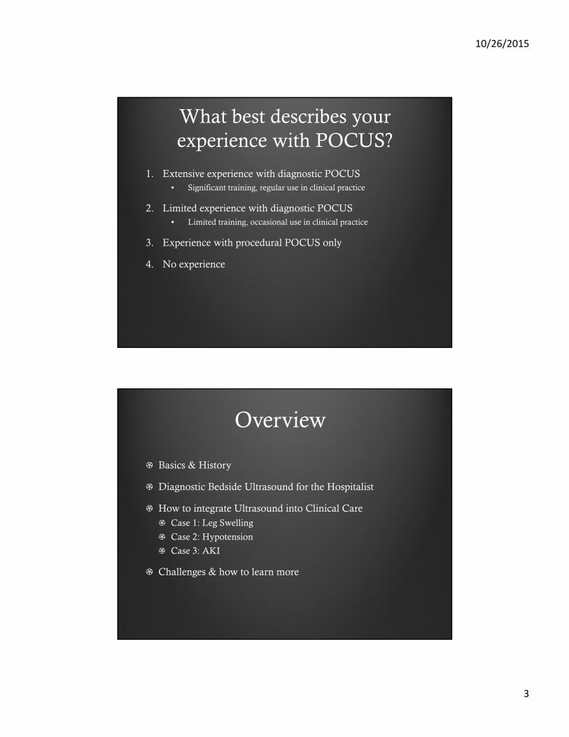
History of US
Ultrasound in M
edicine
1794 Echolocation
1877 Piezoelectric effect
1915 Sonar (WWI & Titanic)
1920s Soccer Physical Therapy
1940s Brain and Breast Tumors
1953 First echocardiogram
1956 Doppler
1958 First use in OB/GYN
1960 standard in radiology, OB/GYN, cardiology, GI
1990s POC US
History of POCUS
Ultrasound in M
edicine
1989 First use in ICU & ED
1990s US guided procedures
~1994 First EM US curriculum
~2005 First med school US curricula
2008 Radiology/EM statement on limited cardiac US
~2010 First formal IM US residency curriculum
EM program “near boston” circa 1995 ???
10/26/2015
6
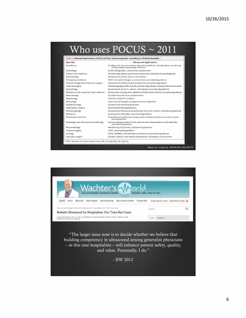
Who uses POCUS ~ 2011
Moore CL, Copel JA. NEJM 2011;364:749-757.
“The larger issue now is to decide whether we believe that building competency in ultrasound among generalist physicians – in this case hospitalists – will enhance patient safety, quality,
and value. Personally, I do.”
- BW 2012
10/26/2015
7
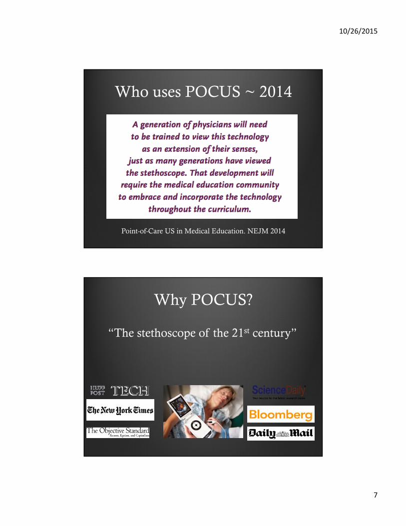
Who uses POCUS ~ 2014
Point-of-Care US in Medical Education. NEJM 2014
Why POCUS?
“The stethoscope of the 21st century”

10/26/2015
8
Why POCUS… really?
Allows earlier diagnosis and treatment
Reduces iatrogenic complications (procedures)
Reduces radiation exposure
Reduces length of stay
Reduces cost of stay
Increases patient satisfaction (hands-on)
Pleural effusion
Pulmonary edema
Pneumonia
Pneumothorax
Volume status
DVT
Ascites
Aortic aneurysm
Hydronephrosis
Organomegaly
LV systolic function
Pericardial effusion
* Chamber size
* Valvular disease
* Advanced uses

10/26/2015
9
How to use POCUS
Case 1
70 year old woman with immobility due to osteoarthritis, breast CA, chronic venous stasis presenting with L>R LE swelling, erythema, tenderness
+ fever, tachypnea, malaise
Ddx: cellulitis > other infection + asymmetric edema > DVT

10/26/2015
10
Why use DVT POCUS?
1. Pomero F et al. Accuracy of emergency physician-performed ultrasonography in the diagnosis of deep-vein thrombosis: a systematic review and meta-analysis. Thromb Haemost. 2013
• Many common clinical scenarios: • unilateral leg swelling, SOB/hypoxia
• Quick, noninvasive
• Physicians can achieve proficiency with brief, focused training
• POCUS compression DVT exam is highly accurate• Sensitivity of 96% and specificity of 96%
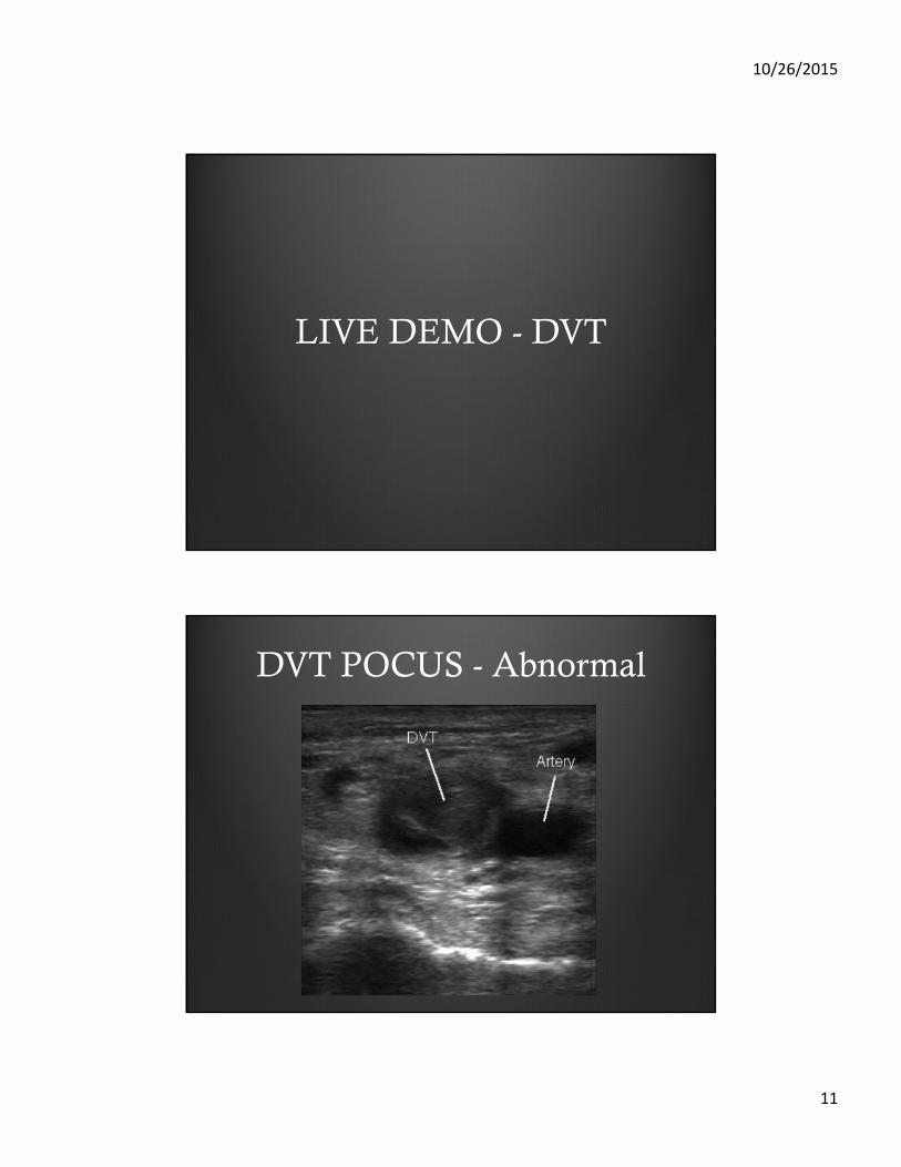
DVT POCUS
10/26/2015
11
LIVE DEMO - DVT
DVT POCUS - Abnormal

10/26/2015
12
Case 2
54 year old man with COPD, CHF presenting with hypotension
+ sputum, SOB, subjective fevers, missed lasix dose x 4 days
CXR, BNP relatively equivocal
Ddx: Sepsis from pulmonary source > CHF exacerbation
POCUS for Undifferentiated Shock
Many ProtocolsCLUE
RUSH
Major ComponentsIVC
LV systolic function
Lung Ultrasound

10/26/2015
13
Why use IVC POCUS?
1. Brennan et al. Handcarried ultrasound measurement of the inferior vena cava for assessment of intravascular volume status in the outpatient hemodialysis clinic. Clin J Am Soc Nephrol. 2006
2. DeCara et al. The use of small personal ultrasound devices by internists without formal training in echocardiography. Eur J Echocardiogr. 2003
3. Brennan et al. A comparison by medicine residents of physical examination versus hand-carried ultrasound for estimation of right atrial pressure. Am J Cardiol. 2007 10.
• Many common clinical scenarios: • hypotension, hypoxia, diuresis
• Quick, noninvasive, bedside
• Physicians can achieve proficiency with brief, focused training
• Moderate utility if used independently (better if combined)• Diameter ROC 0.55, Collapsibility ROC = 0.84
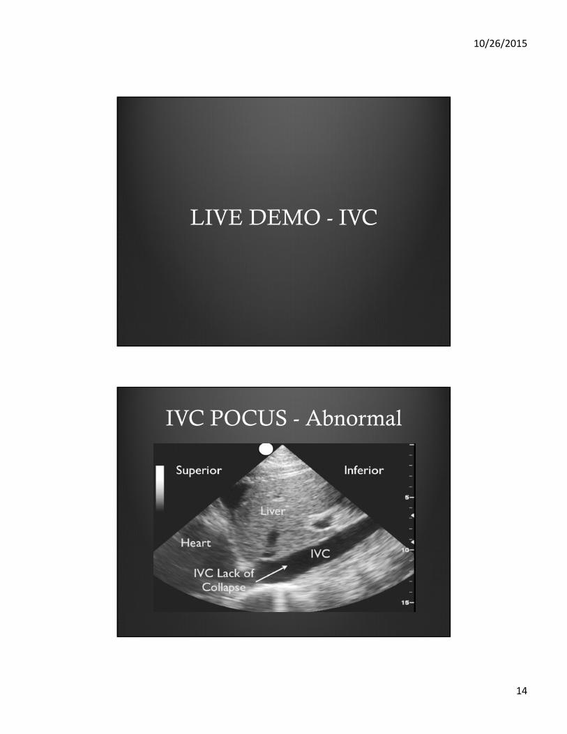
IVC POCUS
10/26/2015
14
LIVE DEMO - IVC
IVC POCUS - Abnormal
10/26/2015
15
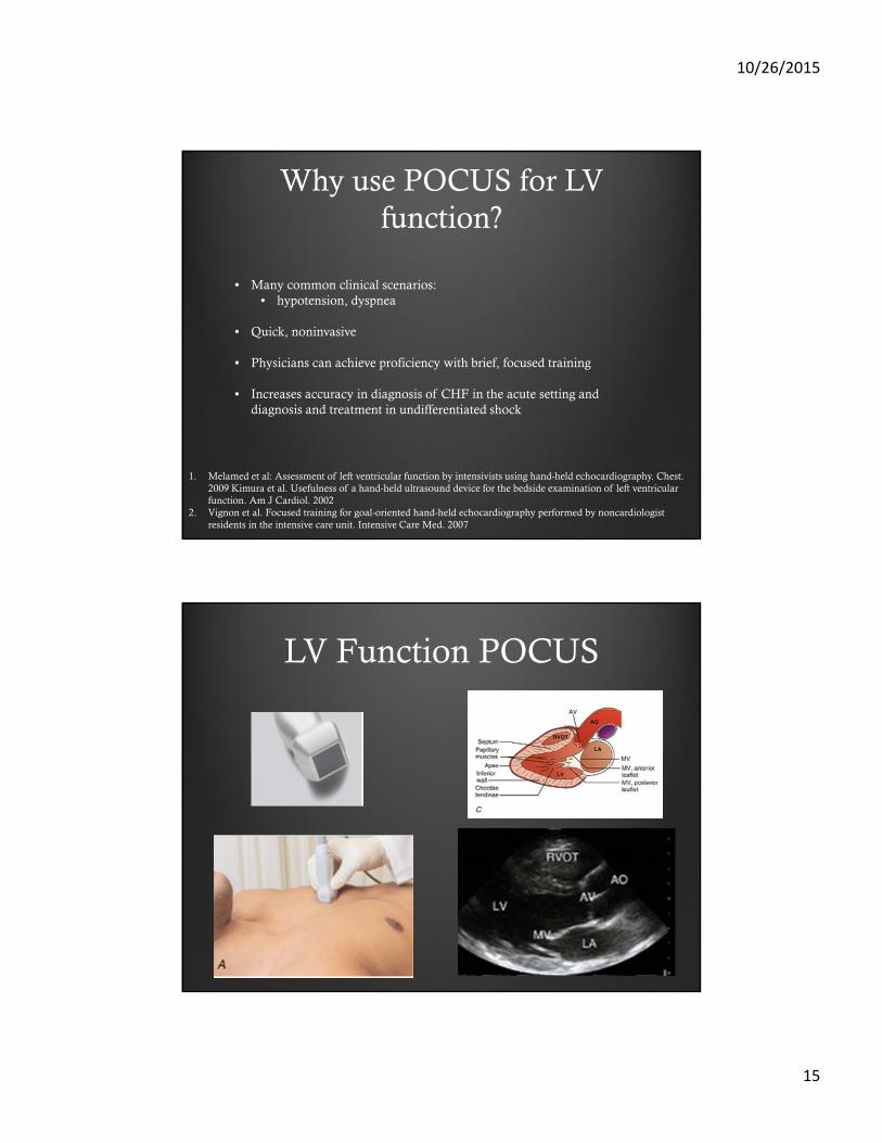
Why use POCUS for LV function?
1. Melamed et al: Assessment of left ventricular function by intensivists using hand-held echocardiography. Chest. 2009 Kimura et al. Usefulness of a hand-held ultrasound device for the bedside examination of left ventricular function. Am J Cardiol. 2002
2. Vignon et al. Focused training for goal-oriented hand-held echocardiography performed by noncardiologistresidents in the intensive care unit. Intensive Care Med. 2007
• Many common clinical scenarios: • hypotension, dyspnea
• Quick, noninvasive
• Physicians can achieve proficiency with brief, focused training
• Increases accuracy in diagnosis of CHF in the acute setting and diagnosis and treatment in undifferentiated shock
LV Function POCUS
10/26/2015
16
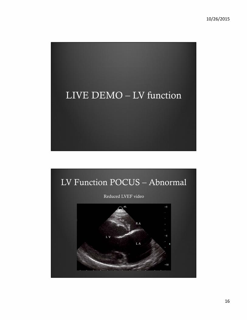
LIVE DEMO – LV function
LV Function POCUS – Abnormal
Reduced LVEF video
10/26/2015
17
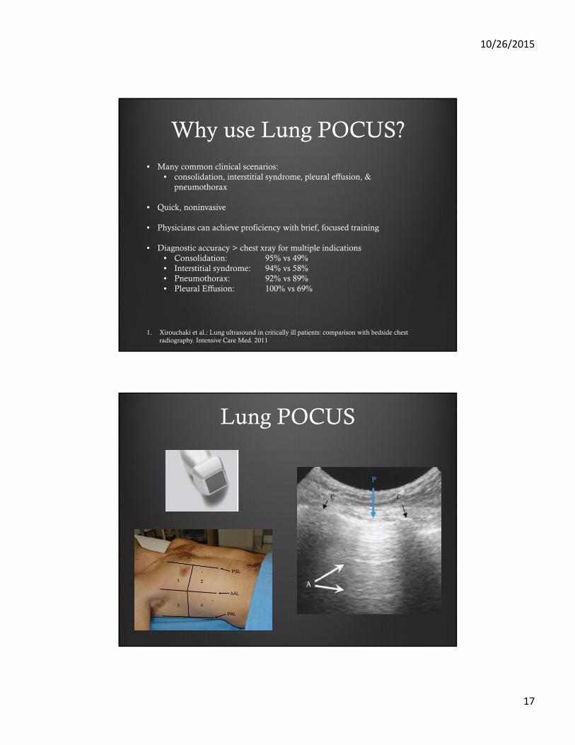
Why use Lung POCUS?
1. Xirouchaki et al.: Lung ultrasound in critically ill patients: comparison with bedside chest radiography. Intensive Care Med. 2011
• Many common clinical scenarios: • consolidation, interstitial syndrome, pleural effusion, &
pneumothorax
• Quick, noninvasive
• Physicians can achieve proficiency with brief, focused training
• Diagnostic accuracy > chest xray for multiple indications • Consolidation: 95% vs 49%• Interstitial syndrome: 94% vs 58%• Pneumothorax: 92% vs 89%• Pleural Effusion: 100% vs 69%
Lung POCUS
10/26/2015
18
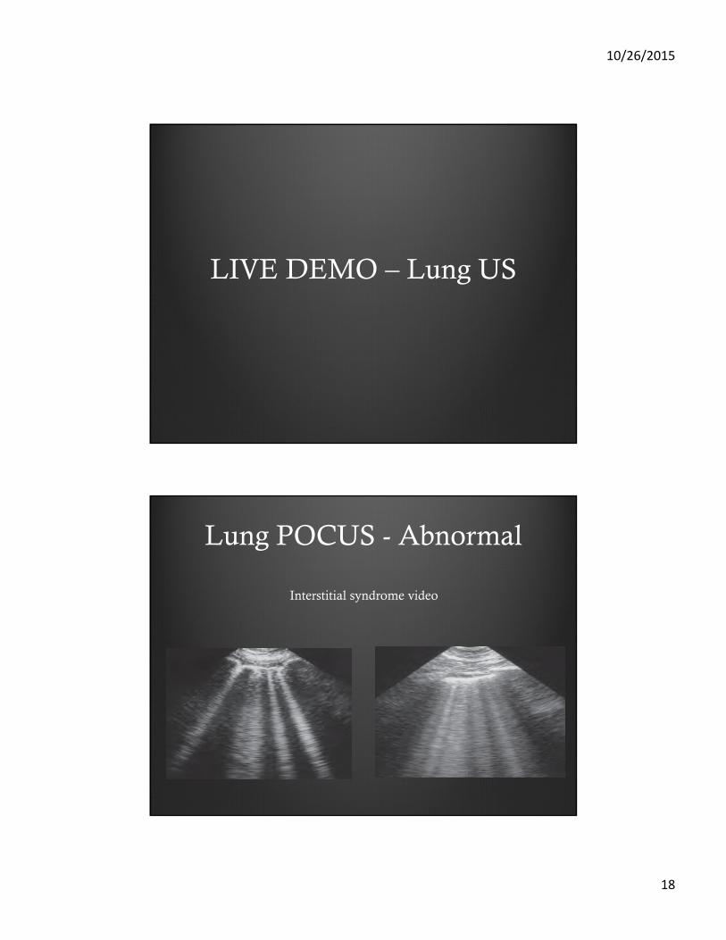
LIVE DEMO – Lung US
Lung POCUS - Abnormal
Interstitial syndrome video

10/26/2015
19
Case 3
65 year old man with BPH, kidney stones presents with AKI+ fevers, cough, sputum, decreased UOP, not taking flomax
- dysuria, flank pain
Ddx: Prerenal > ATN >> obstruction
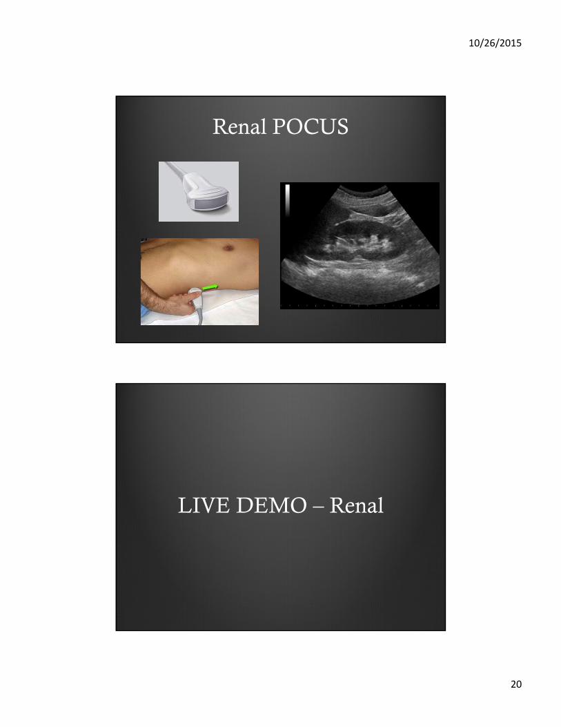
Why use Renal POCUS?
• Many common clinical scenarios: • AKI, abdominal pain
• Quick, noninvasive
• Physicians can achieve proficiency with brief, focused training
• Accurate (hydronephrosis in renal colic)• Sensitivity 80%• Specificity 77%
1. Rosen CL et al. Ultrasonography by emergency physicians in patients with suspected ureteral colic. J EmergMed. 1998
2. Gaspari RJ et al. Emergency ultrasound and urinalysis in the eval- uation of flank pain. Acad Emerg Med. 2005
10/26/2015
20
Renal POCUS
LIVE DEMO – Renal
10/26/2015
21
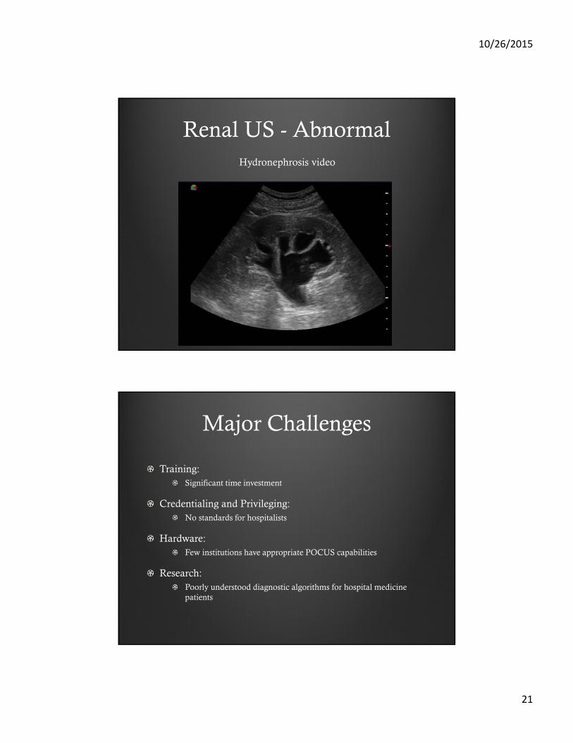
Renal US - AbnormalHydronephrosis video
Major Challenges
Training: Significant time investment
Credentialing and Privileging: No standards for hospitalists
Hardware:Few institutions have appropriate POCUS capabilities
Research:Poorly understood diagnostic algorithms for hospital medicine patients
10/26/2015
22
How to learn more…
Attend a CME course
Work with your EM and critical care colleagues
Self learning via the many free or cheap online resources
Email us for details:[email protected]
How likely are you to pursue further training in POCUS?
1. Very likely
2. Likely
3. Unlikely
4. Very unlikely
10/26/2015
23
Questions?
Photograph citationsSlide 5:
http://vscanultrasound.gehealthcare.com/
http://www.cafepress.com/+hocus-pocus+hats-caps
Slide 7/8:
http://www.jultrasoundmed.org/content/23/1/1/F1.expansion
http://www.ob-ultrasound.net/history1.html
http://learning.blogs.nytimes.com/2012/04/03/100-years-later-ways-to-teach-about-the-titanic-with-the-times/?_php=true&_type=blogs&_r=0
http://www.ultrasoundschoolsinfo.com/history/
Slide 12: http://www.theobjectivestandard.com/2014/01/portable-ultrasound-the-stethoscope-of-the-21st-century/
Slide 14:
Heart: http://www.tophdgallery.com/human-heart-location.html
Lungs: http://easyhealthoptions.com/for-healthy-lungs-get-more-of-this-vitamin/
Liver: http://hepcbc.ca/your-liver/
Vasc: http://teachmeanatomy.info/lower-limb/vessels/venous-drainage/
Slide 18/20/28/37: Soni et al. Point of Care Ultrasound. Elsevier. 2015
Slide 24: http://www.fpnotebook.com/cv/rad/InfrVnCvUltrsndFrVlmSts.htm
Slide 32: http://www.tomwademd.net/introduction-to-pulmonary-ultrasound-by-critical-care-specialist-liz-turner-md/
Slide 37: http://sinaiem.us/tutorials/kidney