COVER SHEET - QUT ePrintseprints.qut.edu.au/4768/1/4768.pdf · Inorganic Materials Research...
21
COVER SHEET Frost, Ray and Cejka, Jiri and Weier, Matt and Martens, Wayde and Kloprogge, Theo (2006) A Raman and infrared spectroscopic study of the uranyl silicates –weeksite, soddyite and haiweeite. Spectrochimica Acta 64(2):pp. 308-315. Copyright 2006 Elsevier. Accessed from: http://eprints.qut.edu.au/archive/00004768/
Transcript of COVER SHEET - QUT ePrintseprints.qut.edu.au/4768/1/4768.pdf · Inorganic Materials Research...
COVER SHEET
Frost, Ray and Cejka, Jiri and Weier, Matt and Martens, Wayde and Kloprogge, Theo (2006) A Raman and infrared spectroscopic study of the uranyl silicates –weeksite, soddyite and haiweeite. Spectrochimica Acta 64(2):pp. 308-315. Copyright 2006 Elsevier. Accessed from: http://eprints.qut.edu.au/archive/00004768/

1
A Raman and infrared spectroscopic study of the uranyl silicates –weeksite, soddyite and haiweeite
Ray L. Frost• a, Jiří Čejkab, Matt L Weier a, Wayde Martens a, J. Theo Kloprogge a Inorganic Materials Research Program, School of Physical and Chemical Sciences, Queensland University of Technology, GPO Box 2434, Brisbane Queensland 4001, Australia. b National Museum, Václavské náměstí 68, CZ-115 79 Praha 1, Czech Republic. Abstract Raman spectroscopy has been used to study the molecular structure of a series of selected uranyl silicate minerals including weeksite K2[(UO2)2(Si5O13)].H2O, soddyite [(UO2)2SiO4.2H2O] and haiweeite Ca[(UO2)2(Si5O12(OH)2](H2O)3 with UO2
2+/SiO2 molar ratio 2:1 or 2:5 .Raman spectra clearly show well resolved bands in the 750 to 800 cm-1 region and in the 950 to 1000 cm-1 region assigned to the ν1 modes of the (UO2)2+ units and to the (SiO4)4- tetrahedra. For example soddyite is characterised by Raman bands at 828.0, 808.6, 801.8 cm-1 (UO2)2+ (ν1), 909.6 and 898.0 cm-1 (UO2)2+ (ν3), 268.2 cm-1 and 257.8 and 246.9 cm-1are assigned to the ν2 (δ) (UO2)2+. Coincidences of the ν1 (UO2)2+ and the ν1 (SiO4)4- is expected. Bands at 1082.2, 1071.2, 1036.3, 995.1, 966.3 cm-1 are attributed to the ν3 (SiO4)4-.Sets of Raman bands in the 200 to 300 cm-1 region are assigned to ν2 δ (UO2)2+ and UO ligand vibrations. Multiple bands indicate the non-equivalence of the UO bonds and the lifting of the degeneracy of ν2 δ (UO2)2+ vibrations. The (SiO4)4- tetrahedral are characterized by bands in the 470 to 550 cm-1 and in the 390 to 420 cm-1 region. These bands are attributed to the ν4 and ν2 (SiO4)4- bending modes. The minerals show characteristic OH stretching bands in the 2900 to 3500 cm-1 and 3600 to 3700 cm-1. Key words: uranyl silicate minerals, weeksite, haiweeite, soddyite, infrared and
Raman spectroscopy Introduction According to Burns (2001), uranyl silicates are common constituents of the oxidized portions of uranium deposits and typically form as a result of their alteration of uraninite [1, 2]. They are important for understanding the genesis of uranium deposits, as well as fluid-rock interaction during the hydration-oxidation weathering of uranium deposits or the mine and mill tailings that result from resource utilization. Uranyl silicates are also significant to the disposal of nuclear waste.
Uranyl silicates are likely to be abundant in a geological repository for nuclear waste under moist oxidizing conditions, owing to the alteration of spent nuclear fuel
• Author to whom correspondence should be addressed ([email protected])
2
and borosilicate waste glass. An understanding of the structures of uranyl silicates may be a key to understanding the long-term performance of a geological repository for nuclear waste [3]. It is likely that uranyl compounds forming due to alteration of nuclear waste incorporate radionuclides into their crystal structures [2, 4, 5]. Nine uranyl silicate minerals (uranophane, sklodowskite, cuprosklodowskite, boltwoodite, sodium boltwoodite, kasolite, oursinite and swamboite) have been classified as members of the uranophane group on the basis of the UO2
2+/SiO2 molar ratio being 1:1 and a similar uranophane anion sheet topology [6-11]. β-uranophane is a polymorph of uranophane (α-uranophane), and the details of their structural connectivities differ substantially [10-12]. Soddyite is characterized by the UO2
2+/SiO2 molar ratio 2:1 and framework crystal structure [13, 14]. The crystal structure of Na2(UO2)2SiO4F2 is structurally related to soddyite [15]. The molar ratio UO2
2+/SiO2 2:5 was found in the crystal structures of weeksite [3], haiweeite [1], coutinhoite [16] and probably also in some not approved uranyl silicate minerals from Russia [17]. Some synthetic framework uranyl silicates were also described [18-21]. Čejka reviewed all available data on infrared spectra of uranyl silicate minerals and their synthetic analogues [22] (Čejka 1999 and many references therein). Biwer et al. (1990) shortly described the only available Raman spectra of uranophane, sodium boltwoodite, weeksite and soddyite without any detailed interpretation [23]. Plesko et al. (1992) presented infrared vibrational characterization and synthesis of a family of hydrous alkali uranyl silicates and hydrous uranyl silicate minerals [24]. However, their interpretation is questionable. Chernorukov‘s team prepared monovalent and divalent uranyl silicates ad presented their properties inclusive interpretation of the infrared spectra [25-33]. As shown by Čejka (1999), some infrared spectra of uranyl silicate minerals including their assignment have been published but only very few of Raman spectra of these minerals without any detailed attribution are available [22]. Some discrepancies in the IR spectra of uranyl silicate minerals published by various authors can be observed caused probably by ill-defined minerals, different IR spectrophotometers used for the measurements, and incorrect crystallochemical formulas used for the interpretation. In this paper, the Raman and IR spectra of uranyl silicate minerals are interpreted respecting the newest single crystal structures of individual minerals, accepted and approved chemical formulas, and crystallochemical application of uranyl anion sheet topology established by Burns [6, 9, 11, 12].
Akhmanova et al. (1963) proved the position of silanol, SiOH, vibrations in (SiO3OH)3- ions in the IR spectrum of minerals [34, 35]. This was supported by Plyusnina (1977) [36] and for the uranyl silicate minerals by Gevorkyan [37-39], Čejka and Urbanec [40], and Čejka [22]. Vochten et al. (1997) confirmed this observation in the IR spectrum of natural and synthetic boltwoodite and synthetic uranophane and sklodowskite [41, 42]. On this basis, Burns inferred the presence of SiOH in the single crystal structure of boltwoodite. Nyfeler and Armbuster (1998) discussed silanol groups in minerals and inorganic compounds [43]. Chernorukov et al. (see above for details) also assigned some bands in the IR spectra of synthetic uranyl silicates to silanol groups [25-33]. In this paper, Raman and infrared spectra of soddyite, weeksite, and haiweeite are studied. As a part of our on-going research into the use of vibrational
3
spectroscopy in particular Raman spectroscopy to assist in the elucidation of the structures of minerals especially secondary minerals, we report the Raman and infrared spectra of some uranyl silicate minerals in particular weeksite, soddyite and haiweeite. These spectra are then related to the recently known mineral structures. Experimental
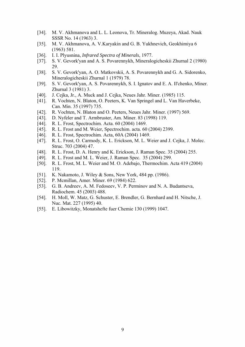
Minerals The minerals used in this study and their origin are reported in Table 1. Where possible the chemical composition was checked by EDAX measurements and the phase purity by powder X-ray diffraction. Raman microprobe spectroscopy
The crystals of uranyl silicate mineral was placed and orientated on the stage of an Olympus BHSM microscope, equipped with 10x and 50x objectives and part of a Renishaw 1000 Raman microscope system, which also includes a monochromator, a filter system and a Charge Coupled Device (CCD). Raman spectra were excited by a HeNe laser (633 nm) at a resolution of 2 cm-1 in the range between 100 and 4000 cm-1. Repeated acquisition using the highest magnification was accumulated to improve the signal to noise ratio. Spectra were calibrated using the 520.5 cm-1 line of a silicon wafer. In order to ensure that the correct spectra are obtained, the incident excitation radiation was scrambled. Previous studies by the authors provide more details of the experimental technique. Spectra at liquid nitrogen temperature were obtained using a Linkam thermal stage (Scientific Instruments Ltd, Waterfield, Surrey, England). Details of the techniques which have been applied to the study of uranyl compounds have been published by the authors [44-50]. Infrared Spectroscopy
Infrared spectra were obtained using a Nicolet Nexus 870 FTIR spectrometer with a smart endurance single bounce diamond ATR cell. Spectra over the 4000−525 cm-1 range were obtained by the co-addition of 64 scans with a resolution of 4 cm-1 and a mirror velocity of 0.6329 cm/s. Spectral manipulation such as baseline adjustment, smoothing and normalisation was performed using the GRAMS® software package (Galactic Industries Corporation, Salem, NH, USA). Results and discussion The ideal linear uranyl group, (UO2)2+, with point group symmetry D∞h has four normal vibrations, but only three fundamentals: the Raman active symmetric stretching vibration ν1 (900-700 cm-1), the doubly degenerate IR active bending vibration ν2 (δ ) (350-180 cm-1, and the antisymmetric IR active stretching vibration ν3 (1000-850 cm-1). The decrease of uranyl group symmetry D∞h ⇒ C∞v results in the IR activation of the ν1 (UO2)2+, similarly, the change in symmetry D∞h ⇒ C2v adds the splitting of the doubly degenerate ν2 (UO2)2+. The former one is due to the presence of nonequivalent bonds in uranyl, (O-U-O)2+, the latter one in the linearity loss, i.e. (O-U-O)2+ angle deformation. The ν1 and ν3 (UO2)2+ may also split in two or more components. This may be influenced especially because of the presence of
4
symmetrically distinct uranyls in the unit cell and, splitting of degenerate vibrations, and also respecting the factor group analysis. The ideal (SiO4)4- tetrahedron with point group symmetry Td has nine normal vibrations characterized by four fundamental distinguishable modes of vibration: the Raman active symmetric stretching vibration ν1 (A), (819 cm-1), the Raman active doubly degenerate bending vibration ν2 (E), (340 cm-1), Raman and infrared active triply degenerate antisymmetric stretching vibration ν3 (F2), (956 cm-1), and the Raman and infrared active triply degenerate bending vibration ν4 (F2), (527 cm-1) [51]. The symmetry decrease from Td ⇒ C3v, which is the case of (SiO3OH)3-, results in IR activation of the ν1 (A1) and ν2 (E), and splitting of the both ν3 and ν4 (both A1 + E). The presence of one proton in the apex of the (SiO4)4- tetrahedron, i. e. formation of (SiO3OH)3-, together with bonding of the remaining three oxygens in the uranyl silicate layers leads to lowering of the (SiO3OH)3- site symmetry to Cs or C1
. This symmetry lowering is connected with IR and Raman activation of all vibrations, i.e. the ν1 (A’ or A), the ν2 splits (A’ + A’’ or 2A), and further splitting of the ν3 and ν4 (2A’
+ A’’ or 3A). Number of bands may be enhanced because of the presence of symmetrically distinct Si4+ in the crystal structure of some uranyl silicate minerals.
According to McMillan (1984), for the framework silicates and silicates with
multilayer structures, their formation may be considered as polymerization of (SiO4) tetrahedra by corner-sharing each oxygen with two (SiO4) units [52]. This results in a coupling of the ν1 and ν3 types of modes. The vibrations follow the frequency order ν3 (Si-O-Si) > ν (Si-O-) > ν1 (Si-O-Si) > δ (Si-O-Si), δ (O-Si-O), where ν3 (Si-O-Si) and ν1 (Si-O-Si) refer to the antisymmetric and symmetric stretching modes of Si-O-Si bridges, ν (Si-O-) represents the stretching (Si-O-) bonds, δ (Si-O-Si) and δ (O-Si-O) refer to the Si-O-Si and O-Si-O bending modes [19, 20]. Spectra of weeksite, haiweeite and coutinhoite will be discussed from this point of view. According to Chernorukov et al. wavenumbers of bands attributed to silanols, Si-OH, are located near 3200 cm-1 (ν SiOH stretching mode), 1400 and 600 cm-1 (δ SiOH in-plane and out-of-plane bending mode, respectively) [26, 28]. The most important for the interpretation may be the isolated band observed near 1400 cm-1 [22]. Similar conclusions were made for double (Np6+O2)2+ and (Pu6+O2)2+ potassium silicates K[(NpO2)(SiO3OH)]. H2O and K[(PuO2)(SiO3OH)]. H2O [53].
Water molecules possessing the point group C2v are characterized by three
fundamentals: ν OH stretching vibrations (ν1 and ν3 H2O) (~3600 – 2900 cm-1), and δ H2O bending vibration (1700-1590 cm-1). All vibrations are IR and Raman active. H2O libration modes may occur in the range 1100-300 cm-1. Hydroxyl ions, (OH)-1, (point group symmetry C∞v)are usually indicated by sharp bands between 3700-3450 cm-1 but sometimes lower if any appreciable amount of hydrogen bonding is involved. The restricted rotational or libration motion of this ion occurs with a wavenumber usually in the 600-300 cm-1 range. The δ M-OH bending vibration may occur over a wide range below approximately 1500 cm-1.
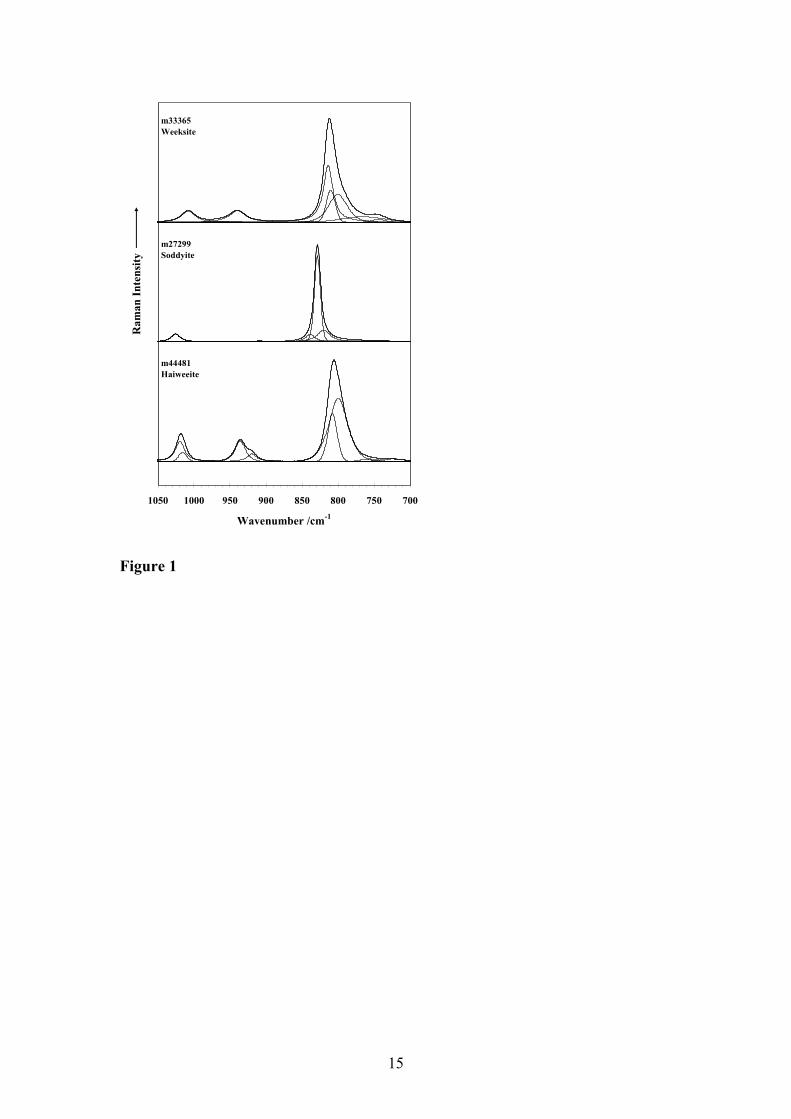
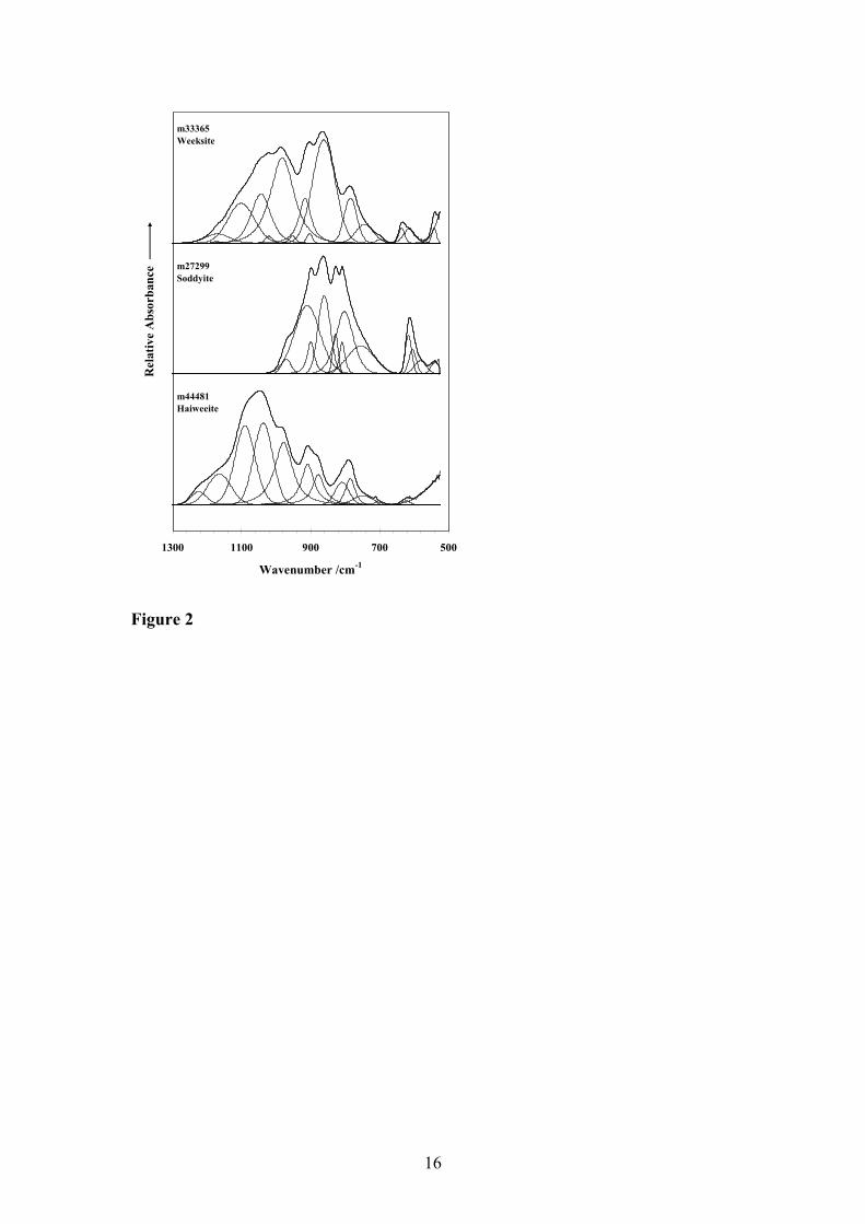
The Raman spectra of weeksite, soddyite and haiweeite in the 700 to 1050 cm-1 region are shown in Figure 1 and the infrared spectra of weeksite, soddyite and haiweeite in the 500 to 1300 cm-1 region are shown in Figure 2. The results of the band component analyses of the Raman and infrared spectra are reported in Tables 1
5
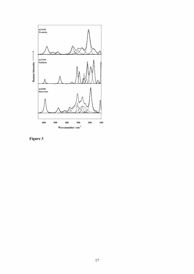
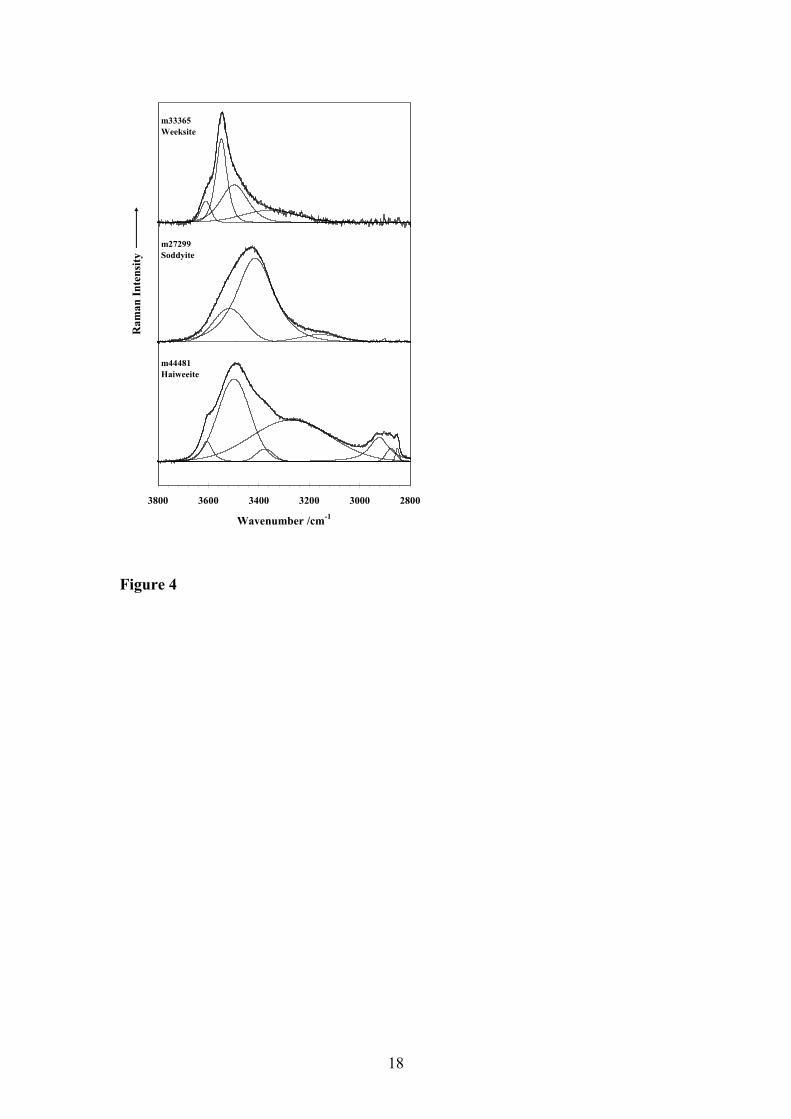
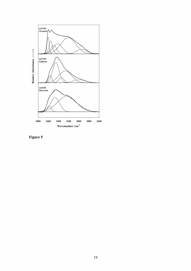
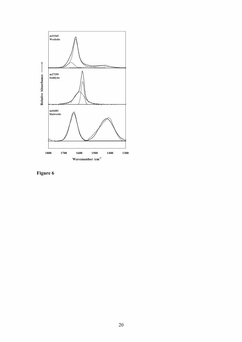
and 2 respectively. The Raman spectra of weeksite, soddyite and haiweeite in the low wavenumber region (100 to 600cm-1) are shown in Figure 3. The Raman and infrared spectra of weeksite, soddyite and haiweeite in the hydroxyl stretching region (2800 to 3800cm-1) are shown in Figures 4 and 5 respectively. The infrared spectra of the water HOH bending region are shown in Figure 6. Soddyite
Soddyite [(UO2)2SiO4.2H2O] has only one structurally (symmetrically) identical U6+ and one structurally (symmetrically) identical Si4+ in its crystal structure, Z=8 [14]. Bands at 904.5 (IR) (Raman 898.0) cm-1 (soddyite1) and 909.8 and 899.6 (909.6 and 896.8) cm-1 (soddyite2) are assigned to the ν3 (UO2)2+ vibrations. The ν1 (UO2)2+ vibrations may be connected with the bands at 853.2 (?) (844.4, 835.6, 824.0, 810.9) cm –1, and 828.0, 808.6, 801.8 (838.3, 828.6, 819.9) cm-1, respectively. A coincidences of the ν1 (UO2)2+ and the ν1 (SiO4)4- is supposed and expected. In the Raman spectrum, bands at 268.2 cm-1 and 257.8 and 246.9 cm-1, respectively, are assigned to the ν2 (δ) (UO2)2+. Bands at 1082.2, 1071.2, 1036.3, 995.1, 966.3 (1248.4, 1124.0, 1048.5, 1035.2, 1004.4) cm-1, and 971.0 (1025.5) cm-1, respectively, are attributed to the ν3 (SiO4)4-. Bands at 879.1, 853.2 (?) (844.4, 835.6, 824.0, 810.9) cm-
1, and 860.4, 828.0, 808.6 (838.3, 828.6, 819.9) cm-1, respectively are connected with the ν1 (SiO4)4-. As mentioned above, a coincidence (an overlapping) of the ν1 (SiO4)4- and ν1 (UO2)2+ may be expected in this region. The ν4 (SiO4)4- vibrations are located at 686.4, 658.3, 602.5, 538.3 (687.6, 561.6, 531.6, 492.4) cm-1, and 615.7, 603.9, 581.3, 539.1 (591.4, 459.4) cm-1. Bands observed at lower wavenumbers are related to the ν2 (SiO4)4-, ν2 (δ) (UO2)2+, external modes of H2O, ν M-O, and lattice vibrations.
Bands at 3559.4, 3458.4, 3244.1, 2957.2, 2952.0, 2921.6, 2854.3 cm-1, and
3565.1, 3451.3, 3343.7, 3265.0, 2989.6 cm-1, respectively, are assigned to the ν OH. The δ H2O vibrations are observed at 1625.2 cm-1, and 1598.1, 1578.1 (1584.2, 1569.2) cm-1, respectively. According to Moll et al. (1995), a band at 1588 cm-1 with a shoulder at 1635 cm-1 [54]. This shoulder should be attributed to the water adsorbed on the sample surface. Bands at 797.6, 711.2 (750.0) cm-1, and 801.8 (?), 754.0 (791.0) cm-1 may be attributed to the H2O libration modes. The presence of strong to weak hydrogen bonding networks in the crystal structures of soddyite samples studied was inferred [55]. Weeksite and related minerals Weeksite is given by K2[(UO2)2(Si5O13)].H2O (Z=16) and haiweeite, by the formula Ca[(UO2)2(Si5O12(OH)2](H2O)3 ,(Z=4). The published IR spectrum of coutinhoite, Th0.5[(UO2)2(Si5O13)].1-3.5 H2O (Z=16) is also included [16].Simplified formulas are used for weeksite [3]haiweeite [12] and coutinhoite [16]. Absorption bands in the IR spectra of weeksite at 916.1, 903,2 and most probably also at 861.5 cm-1, weeksite(2) at 910.8 and 861.7 cm-1, haiweeite at 908.5 and 877.3 cm-1, and coutinhoite at 907 cm-1 were assigned to the ν3 (UO2)2+. Two bands at 919.6 and 886.9 cm-1 observed in the Raman spectrum of haiweeite were also
6
attributed to the ν3 (UO2)2+. No bands related to this vibration were observed in
Raman spectra of both weeksite samples. In Raman spectra, absorption bands at 813.7, 810.2 and 800.2 cm-1 (weeksite(1)), 812.7, 808.7 and 796.7 cm-1 (weeksite (2)), and 807.7 and 799.7 cm-1 (haiweeite) are assigned to the ν1 (UO2)2+ vibrations. In Raman spectra, bands at 266.2 and 264.0 cm-1 (weeksite (1) and weeksite (2), respectively), and 264.0 and 260.4 cm-1 (haiweeite) may be attributed to the ν2 (δ) (UO2)2+. The number of some of these vibrations is enhanced. This is supported by the number of symmetrically distinct U6+ in the crystal structures of weeksite (4) and haiweeite (2), number of molecules in the unit cell (weeksite 16, haiweeite 4) and FGA. Absorption bands in the IR spectra at 1172.7, 1170,4, 11010.2, 1044.2, 1020.8, 982.3 and 952.8 (Raman 1154.5, 1008.0, 961.6 and 939.6 ) cm-1 (weeksite (1)), 1174.6, 1122.5, 1067.5, 1043.1, 985.0, 975.7 (1163.0, 1148.4, 1007.7, 960.7, 938.9) cm-1 (weeksite(2)), 1224.5, 1164.9, 1090.5, 1037.4, 978.4 (1163.0, 1148.4, 1007.7, 960.7, 938.9) cm-1 (haiweeite), and 1102, 1061 and 988 cm-1 (coutinhoite) are assigned to the ν3 (Si-O-Si) and ν (Si-O-) vibrations, those at 784.2, 743.5, 697.7, 635.6 and 613.9 (765.3, 744, 573.9) cm-1 (weeksite(1)), 784.0, 741.2, 636.2, 621.3, 590.9 (771.6, 748.3, 744.2, 573.9) cm-1 (weeksite (2)), 784.9, 748.2, 710.5, 621.7 (756.1, 724.4, 588.9) (haiweeite), 788, 698, 639, 585 cm-1 (coutinhoite) the ν1 (Si-O-Si), and those at 524.6 (521.4, 479.7) cm-1 (weeksite (1)), 532.9 (517.9, 480.0) (weeksite (2)), (473.4, 418.4) cm-1 (haiweeite), and 535, 452, 415 cm-1 (coutinhoite) to the δ (Si-O-Si) and probably also ν (U-Oligand). Bands at lower wavenumbers may be attributed to the δ (Si-O-Si), δ (O-Si-O), δ (UO2)2+, ν (M-O) (molecular deformation and lattice modes?). These conclusions may be supported by the presence of ten symmetrically distinct Si4+ in weeksite and four symmetrically distinct Si4+ in haiweeite. According to Atencio et al. (2004), coutinhoite is probably isostructural with weeksite. All three minerals contain molecular water. In the IR spectra, absorption bands assigned to the ν OH vibrations were observed in the range 3605.7-2942.5 cm-1 (weeksite (1)), 3605.3-2848.9 cm-1 (weeksite (2)), 3581.5-3233.1 cm-1 (haiweeite), and 3609, 3535, 3468 and 3242 cm-1 (coutinhoite). The δ H2O vibrations were observed at 1653.8 and 1622.7 (1637.6) cm-1 (weeksite (1)), 1652.7 and 1622.4 (1638.7) cm-1 (weeksite (2)), 1636.7 cm-1 (haiweeite), and 1627 cm-1 (coutinhoite). According to Libowitzky (1999), strong to very weak hydrogen bonding networks should be arranged in the crystal structure of all three minerals. Bands at 1468.3 cm-1 (weeksite (1)), 1442.5 cm-1 (weeksite (2)), 1423.1 cm-1 (haiweeite), and 1434, 1403 and 1386 cm-1 (coutinhoite) may indicate the presence of silanols, Si-OH, in the crystal structure of all three minerals, however, this agrees only with the crystal structure of haiweeite. Conclusions
Raman spectroscopy has enabled the characteristic spectra of a suite of uranyl silicates of the 2:1 group to be obtained. These spectra are characteristic of the particular mineral being studied. The application of Raman spectroscopy enabled excellent band separation with no overlap of bands due to different vibrating units as
7
is found with infrared spectroscopy. This separation enabled definitive assignment of the bands.
Acknowledgements
The financial and infra-structure support of the Queensland University of Technology Inorganic Materials Research Program of the School of Physical and Chemical Sciences is gratefully acknowledged. The Australian Research Council (ARC) is thanked for funding. Mr Dermot Henry of Museum Victoria is thanked for the supply of the uranyl silicate minerals.
8
References [1]. P. C. Burns, Can. Miner. 39 (2001) 1153. [2]. P. C. Burns, R. C. Ewing and M. L. Miller, J. Nuc. Mat. 245 (1997) 1. [3]. J. M. Jackson and P. C. Burns, Can. Miner. 39 (2001) 187. [4]. P. C. Burns, K. M. Deely and S. Skanthakumar, Radiochim. Acta 92 (2004)
151. [5]. P. C. Burns, Can. Miner. 36 (1998) 1069. [6]. P. C. Burns, R. Finch and Editors, Uranium: Mineralogy, Geochemistry and
the Environment. (Proceedings of a Short Course held 22-23 October 1999 in Golden, Colorado.) [In: Rev. Mineral., 1999; 38], 1999.
[7]. P. C. Burns and F. C. Hill, Can. Miner. 38 (2000) 163. [8]. P. C. Burns and K. M. Deely, Can. Miner.40 (2002) 1579. [9]. P. C. Burns, Materials Research Society Symposium Proceedings 802 (2004)
89. [10]. P. C. Burns, R. C. Ewing and F. C. Hawthorne, Can. Miner. 35 (1997) 1551. [11]. P. C. Burns, M. L. Miller and R. C. Ewing, Can. Miner. 34 (1996) 845. [12]. P. C. Burns, Revs in Miner. 38 (1999) 23. [13]. P. C. Burns, J. Nuc. Mat. 265 (1999) 218. [14]. F. Demartin, C. M. Gramaccioli and T. Pilati, Acta Crysta., C48 (1992) 1. [15]. N. Blaton, R. Vochten, O. M. Peeters and K. Van Springel, Neues Jahr. Miner.
(1999) 253. [16]. D. Atencio, F. M. S. Carvalho and P. A. Matioli, Am. Miner. 89 (2004) 721. [17]. G. A. Sidorenko, N. V. Chukanov and I. S. Naumova, Mineralogicheskii Zh.
23 (2001) 55. [18]. Y. Huang, Z. Jiang and W. Schwieger, Micro. Mesoporous Mat. 26 (1998)
215. [19]. Y. Huang, Z. Jiang and W. Schwieger, Can. J.Chem. 77 (1999) 495. [20]. Y. Huang, Z. Jiang and W. Schwieger, Chem. Mats 11 (1999) 1210. [21]. J. Huang, X. Wang and A. J. Jacobson, J. Mat. Chem. 13 (2003) 191. [22]. J. Cejka, Revs in Miner. 38 (1999) 521. [23]. B. M. Biwer, W. L. Ebert and J. K. Bates, J. Nuc. Mat. 175 (1990) 188. [24]. E. P. Plesko, B. E. Scheetz and W. B. White, Am. Miner. 77 (1992) 431. [25]. N. G. Chernorukov and V. E. Kortikov, Zhu. Neorganicheskoi Khimii 45
(2000) 1949. [26]. N. G. Chernorukov and V. E. Kortikov, Radiochem. (Moscow)(Translation of
Radiokhimiya) 42 (2000) 446. [27]. N. G. Chernorukov and V. E. Kortikov, Zh. Neorganicheskoi Khimii 45
(2000) 1110. [28]. N. G. Chernorukov and V. E. Kortikov, Russian J. Gen. Chem. 71 (2001)
1669. [29]. N. G. Chernorukov and V. E. Kortikov, Zh. Neorganicheskoi Khimii 46
(2001) 1949. [30]. N. G. Chernorukov and V. E. Kortikov, Radiochem. 43 (2001) 229. [31]. N. G. Chernorukov and V. E. Kortikov, Zh. Neorganicheskoi Khimii 46
(2001) 222. [32]. N. G. Chernorukov and V. E. Kortikov, Radiochem. 44 (2002) 446. [33]. N. G. Chernorukov and V. E. Kortikov, Zh. Neorganicheskoi Khimii 47
(2002) 232.
9
[34]. M. V. Akhmanova and L. L. Leonova, Tr. Mineralog. Muzeya, Akad. Nauk SSSR No. 14 (1963) 3.
[35]. M. V. Akhmanova, A. V.Karyakin and G. B. Yukhnevich, Geokhimiya 6 (1963) 581.
[36]. I. I. Plyusnina, Infrared Spectra of Minerals, 1977. [37]. S. V. Gevork'yan and A. S. Povarennykh, Mineralogicheskii Zhurnal 2 (1980)
29. [38]. S. V. Gevork'yan, A. O. Matkovskii, A. S. Povarennykh and G. A. Sidorenko,
Mineralogicheskii Zhurnal 1 (1979) 78. [39]. S. V. Gevork'yan, A. S. Povarennykh, S. I. Ignatov and E. A. Il'chenko, Miner.
Zhurnal 3 (1981) 3. [40]. J. Cejka, Jr., A. Muck and J. Cejka, Neues Jahr. Miner. (1985) 115. [41]. R. Vochten, N. Blaton, O. Peeters, K. Van Springel and L. Van Haverbeke,
Can. Min. 35 (1997) 735. [42]. R. Vochten, N. Blaton and O. Peeters, Neues Jahr. Miner. (1997) 569. [43]. D. Nyfeler and T. Armbruster, Am. Miner. 83 (1998) 119. [44]. R. L. Frost, Spectrochim. Acta. 60 (2004) 1469. [45]. R. L. Frost and M. Weier, Spectrochim. acta. 60 (2004) 2399. [46]. R. L. Frost, Spectrochim. Acta, 60A (2004) 1469. [47]. R. L. Frost, O. Carmody, K. L. Erickson, M. L. Weier and J. Cejka, J. Molec.
Struc. 703 (2004) 47. [48]. R. L. Frost, D. A. Henry and K. Erickson, J. Raman Spec. 35 (2004) 255. [49]. R. L. Frost and M. L. Weier, J. Raman Spec. 35 (2004) 299. [50]. R. L. Frost, M. L. Weier and M. O. Adebajo, Thermochim. Acta 419 (2004)
119. [51]. K. Nakamoto, J. Wiley & Sons, New York, 484 pp. (1986). [52]. P. Mcmillan, Amer. Miner. 69 (1984) 622. [53]. G. B. Andreev, A. M. Fedoseev, V. P. Perminov and N. A. Budantseva,
Radiochem. 45 (2003) 488. [54]. H. Moll, W. Matz, G. Schuster, E. Brendler, G. Bernhard and H. Nitsche, J.
Nuc. Mat. 227 (1995) 40. [55]. E. Libowitzky, Monatshefte fuer Chemie 130 (1999) 1047.
10
Table 1 Sample details Mineral Formula Locale # Weeksite K2(UO2)2(Si2O5)3•4H2O Anderson’s Mine, Yavapai
Co. Arizona, USA M33365
Haiweeite Ca(UO2)2[Si5O12(OH)2]•4.5H2O Teofild Otoni, Minas Gerais, Brazil
M44481
Soddyite (UO2)2SiO4•2H2O Katanga, Congo, Zaire M27299
11
m33365
Weekiste m27299 soddyite
m44481 haweeite
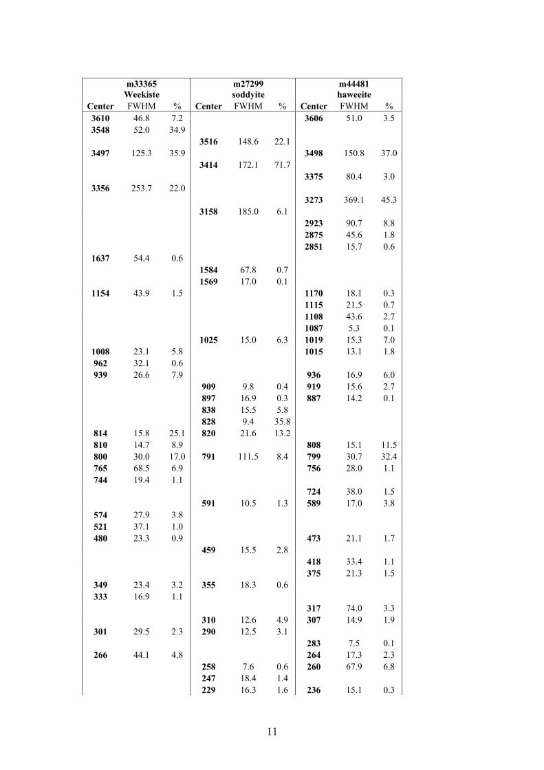
Center FWHM % Center FWHM % Center FWHM % 3610 46.8 7.2 3606 51.0 3.5 3548 52.0 34.9
3516 148.6 22.1 3497 125.3 35.9 3498 150.8 37.0
3414 172.1 71.7 3375 80.4 3.0
3356 253.7 22.0 3273 369.1 45.3 3158 185.0 6.1 2923 90.7 8.8 2875 45.6 1.8 2851 15.7 0.6
1637 54.4 0.6 1584 67.8 0.7 1569 17.0 0.1
1154 43.9 1.5 1170 18.1 0.3 1115 21.5 0.7 1108 43.6 2.7 1087 5.3 0.1 1025 15.0 6.3 1019 15.3 7.0
1008 23.1 5.8 1015 13.1 1.8 962 32.1 0.6 939 26.6 7.9 936 16.9 6.0
909 9.8 0.4 919 15.6 2.7 897 16.9 0.3 887 14.2 0.1 838 15.5 5.8 828 9.4 35.8
814 15.8 25.1 820 21.6 13.2 810 14.7 8.9 808 15.1 11.5 800 30.0 17.0 791 111.5 8.4 799 30.7 32.4 765 68.5 6.9 756 28.0 1.1 744 19.4 1.1
724 38.0 1.5 591 10.5 1.3 589 17.0 3.8
574 27.9 3.8 521 37.1 1.0 480 23.3 0.9 473 21.1 1.7
459 15.5 2.8 418 33.4 1.1 375 21.3 1.5
349 23.4 3.2 355 18.3 0.6 333 16.9 1.1
317 74.0 3.3 310 12.6 4.9 307 14.9 1.9
301 29.5 2.3 290 12.5 3.1 283 7.5 0.1
266 44.1 4.8 264 17.3 2.3 258 7.6 0.6 260 67.9 6.8 247 18.4 1.4 229 16.3 1.6 236 15.1 0.3
12
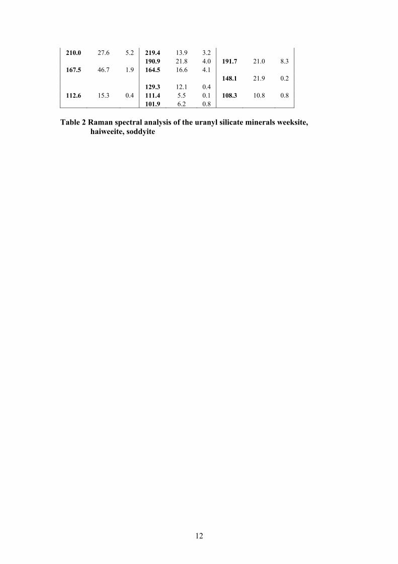
210.0 27.6 5.2 219.4 13.9 3.2 190.9 21.8 4.0 191.7 21.0 8.3
167.5 46.7 1.9 164.5 16.6 4.1 148.1 21.9 0.2 129.3 12.1 0.4
112.6 15.3 0.4 111.4 5.5 0.1 108.3 10.8 0.8 101.9 6.2 0.8
Table 2 Raman spectral analysis of the uranyl silicate minerals weeksite,
haiweeite, soddyite
13
m33365 Weekiste
m27299 soddyite
m44481 haweeite
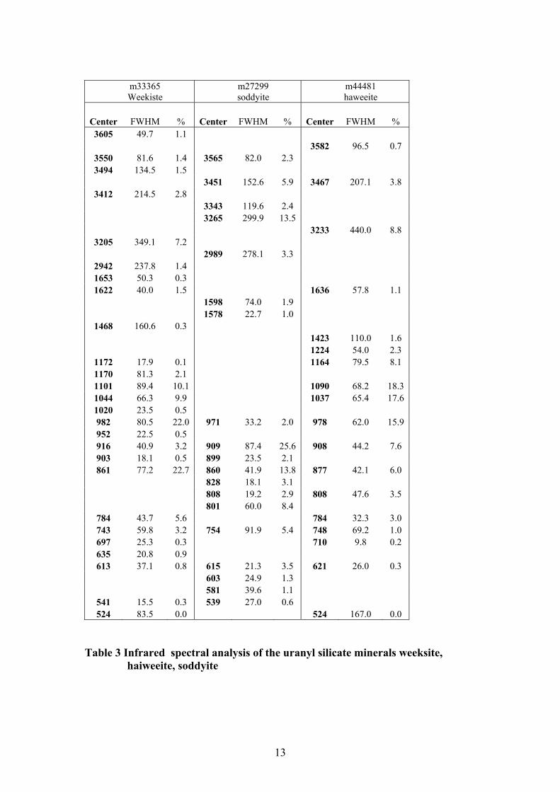
Center FWHM % Center FWHM % Center FWHM % 3605 49.7 1.1
3582 96.5 0.7 3550 81.6 1.4 3565 82.0 2.3 3494 134.5 1.5
3451 152.6 5.9 3467 207.1 3.8 3412 214.5 2.8
3343 119.6 2.4 3265 299.9 13.5 3233 440.0 8.8
3205 349.1 7.2 2989 278.1 3.3
2942 237.8 1.4 1653 50.3 0.3 1622 40.0 1.5 1636 57.8 1.1
1598 74.0 1.9 1578 22.7 1.0
1468 160.6 0.3 1423 110.0 1.6 1224 54.0 2.3
1172 17.9 0.1 1164 79.5 8.1 1170 81.3 2.1 1101 89.4 10.1 1090 68.2 18.3 1044 66.3 9.9 1037 65.4 17.6 1020 23.5 0.5 982 80.5 22.0 971 33.2 2.0 978 62.0 15.9 952 22.5 0.5 916 40.9 3.2 909 87.4 25.6 908 44.2 7.6 903 18.1 0.5 899 23.5 2.1 861 77.2 22.7 860 41.9 13.8 877 42.1 6.0
828 18.1 3.1 808 19.2 2.9 808 47.6 3.5 801 60.0 8.4
784 43.7 5.6 784 32.3 3.0 743 59.8 3.2 754 91.9 5.4 748 69.2 1.0 697 25.3 0.3 710 9.8 0.2 635 20.8 0.9 613 37.1 0.8 615 21.3 3.5 621 26.0 0.3
603 24.9 1.3 581 39.6 1.1
541 15.5 0.3 539 27.0 0.6 524 83.5 0.0 524 167.0 0.0
Table 3 Infrared spectral analysis of the uranyl silicate minerals weeksite,
haiweeite, soddyite
14
LIST OF FIGURES Figure 1 Raman spectra of weeksite, soddyite and haiweeite in the 700 to 1050 cm-1 region Figure 2 infrared spectra of weeksite, soddyite and haiweeite in the 500 to 1300 cm-1
region Figure 3 Raman spectra of weeksite, soddyite and haiweeite in the low wavenumber
region (100 to 600cm-1). Figure 4 Raman spectra of weeksite, soddyite and haiweeite in the hydroxyl
stretching region (2800 to 3800cm-1). Figure 5 Infrared spectra of weeksite, soddyite and haiweeite in the hydroxyl
stretching region (2800 to 3800cm-1). Figure 6 Infrared spectra of weeksite, soddyite and haiweeite in the water HOH
bending region (1500 to 1800cm-1).
LIST OF TABLES Table 1 Sample details Table 2 Raman spectral analysis of the uranyl silicate minerals weeksite,
haiweeite, soddyite Table 3 Infrared spectral analysis of the uranyl silicate minerals weeksite,
haiweeite, soddyite
15
70075080085090095010001050
Wavenumber /cm-1
Ram
an In
tens
itym33365Weeksite
m27299Soddyite
m44481Haiweeite
Figure 1
16
50070090011001300
Wavenumber /cm-1
Rel
ativ
e A
bsor
banc
e
m33365Weeksite
m27299Soddyite
m44481Haiweeite
Figure 2
17
100200300400500600
Wavenumber /cm-1
Ram
an In
tens
itym33365Weeksite
m27299Soddyite
m44481Haiweeite
Figure 3
18
280030003200340036003800
Wavenumber /cm-1
Ram
an In
tens
itym33365Weeksite
m27299Soddyite
m44481Haiweeite
Figure 4
19
2600280030003200340036003800
Wavenumber /cm-1
Rel
ativ
e A
bsor
banc
e
m33365Weeksite
m27299Soddyite
m44481Haiweeite
Figure 5
20
130014001500160017001800
Wavenumber /cm-1
Rel
ativ
e A
bsor
banc
e
m33365Weeksite
m27299Soddyite
m44481Haiweeite
Figure 6