Complete Summary Of the Head and Neck Anatomy
135
COMPLETE SUMMARY OF THE HUMAN ANATOMY OF THE HEAD & NECK ORIBA DAN LANGOYA, MBchB Head & Neck.................................................................................................................. 2 Sternocleidomastoid Muscle .......................................................................................... 3 Scalenus Anterior Muscles ............................................................................................. 5 Anterior Triangle of the Neck ........................................................................................ 6 Posterior Triangle of the Neck ....................................................................................... 8 Thyroid Gland .............................................................................................................. 10 Common Carotid Artery .............................................................................................. 12 External Carotid Artery ................................................................................................ 14 Internal Carotid Artery (Cervical Part) ........................................................................ 16 Subclavian Artery......................................................................................................... 18 Internal Jugular Veins .................................................................................................. 20 Cervical Plexus ............................................................................................................ 22 Phrenic Nerve (Cervical Part) ...................................................................................... 24 Cervical Part of Sympathetic trunk .............................................................................. 25 Trachea (Cervical Part) ................................................................................................ 27 Lymphatic Drainage of the Head & Neck.................................................................... 29 Scalp............................................................................................................................. 32 Facial Artery................................................................................................................. 34 Facial Nerve ................................................................................................................. 35 Dura Mater of the Brain ............................................................................................... 37 Cranial Fossae .............................................................................................................. 39 Cavernous Sinus........................................................................................................... 40 Superior Sagittal Sinus................................................................................................. 42 Middle Meningeal Artery............................................................................................. 43 Types of Intracranial Hemorrhage ............................................................................... 44 Cerebral Hemorrhage ................................................................................................... 45 Internal Carotid Artery (Petrous, Cavernous & Cervical Parts) ....................................................................................... 46 Trigeminal Ganglion .................................................................................................... 47 Structure of the Eyeball ............................................................................................... 49 Movements of the Eyeball ........................................................................................... 51 Ophthalmic Artery ....................................................................................................... 54 Lacrimal Apparatus ...................................................................................................... 55 Vertebral Artery............................................................................................................ 57 Parotid Gland ............................................................................................................... 59 Temporomandibular Joint ............................................................................................ 62 Movements of the Mandible ........................................................................................ 64 Maxillary Artery........................................................................................................... 66 Maxillary Nerve ........................................................................................................... 67 Mandibular Nerve ........................................................................................................ 68 Pterygopalatine Ganglion............................................................................................. 69 Submandibular Gland .................................................................................................. 70 Lingual Nerve .............................................................................................................. 75 Nasal Cavity: Relations, Openings & Branches .................................................................................. Tongue: Features & Muscles ................................................... Innervation of the Tongue ....................................................... Constrictor Muscles of Pharynx .............................................. Nasopharynx............................................................................ Mechanism of Swallowing ...................................................... Auditory Tube ......................................................................... Middle Ear Cavity ................................................................... Pterygopalatine Fossa .............................................................. Cutaneous Innervation of Face ................................................ Glossopharyngeal Nerve (9th Cranial Nerve) ...................................................................................... Vagus Nerve (10th Cranial Nerve) .......................................... Accessory Nerve (11th Cranial Nerve) ................................... Hypoglossal Nerve (12th Cranial Nerve) ................................ Paranasal Sinuses .................................................................... Oral Cavity .............................................................................. Palate ....................................................................................... Pharynx.................................................................................... Oropharynx.............................................................................. Laryngopharynx ...................................................................... External Ear ............................................................................. Larynx .....................................................................................
-
Upload
oriba-dan-langoya -
Category
Health & Medicine
-
view
35.997 -
download
0
Transcript of Complete Summary Of the Head and Neck Anatomy

COMPLETE SUMMARY OF THE HUMAN
ANATOMY OF THE HEAD & NECK
ORIBA DAN LANGOYA, MBchB
Head & Neck.................................................................................................................. 2
Sternocleidomastoid Muscle .......................................................................................... 3
Scalenus Anterior Muscles ............................................................................................. 5
Anterior Triangle of the Neck ........................................................................................ 6
Posterior Triangle of the Neck ....................................................................................... 8
Thyroid Gland .............................................................................................................. 10
Common Carotid Artery .............................................................................................. 12
External Carotid Artery ................................................................................................ 14
Internal Carotid Artery (Cervical Part) ........................................................................ 16
Subclavian Artery......................................................................................................... 18
Internal Jugular Veins .................................................................................................. 20
Cervical Plexus ............................................................................................................ 22
Phrenic Nerve (Cervical Part) ...................................................................................... 24
Cervical Part of Sympathetic trunk .............................................................................. 25
Trachea (Cervical Part) ................................................................................................ 27
Lymphatic Drainage of the Head & Neck .................................................................... 29
Scalp ............................................................................................................................. 32
Facial Artery................................................................................................................. 34
Facial Nerve ................................................................................................................. 35
Dura Mater of the Brain ............................................................................................... 37
Cranial Fossae .............................................................................................................. 39
Cavernous Sinus........................................................................................................... 40
Superior Sagittal Sinus ................................................................................................. 42
Middle Meningeal Artery ............................................................................................. 43
Types of Intracranial Hemorrhage ............................................................................... 44
Cerebral Hemorrhage ................................................................................................... 45
Internal Carotid Artery (Petrous,
Cavernous & Cervical Parts) ....................................................................................... 46
Trigeminal Ganglion .................................................................................................... 47
Structure of the Eyeball ............................................................................................... 49
Movements of the Eyeball ........................................................................................... 51
Ophthalmic Artery ....................................................................................................... 54
Lacrimal Apparatus ...................................................................................................... 55
Vertebral Artery ............................................................................................................ 57
Parotid Gland ............................................................................................................... 59
Temporomandibular Joint ............................................................................................ 62
Movements of the Mandible ........................................................................................ 64
Maxillary Artery........................................................................................................... 66
Maxillary Nerve ........................................................................................................... 67
Mandibular Nerve ........................................................................................................ 68
Pterygopalatine Ganglion............................................................................................. 69
Submandibular Gland .................................................................................................. 70
Lingual Nerve .............................................................................................................. 75
Nasal Cavity: Relations, Openings &
Branches ....................................................................................................................... 76
Tongue: Features & Muscles ........................................................................................ 79
Innervation of the Tongue ............................................................................................ 81
Constrictor Muscles of Pharynx ................................................................................... 82
Nasopharynx ................................................................................................................. 84
Mechanism of Swallowing ........................................................................................... 86
Auditory Tube .............................................................................................................. 87
Middle Ear Cavity ........................................................................................................ 89
Pterygopalatine Fossa ................................................................................................... 92
Cutaneous Innervation of Face ..................................................................................... 93
Glossopharyngeal Nerve (9th Cranial
Nerve) ........................................................................................................................... 95
Vagus Nerve (10th Cranial Nerve) ............................................................................... 96
Accessory Nerve (11th Cranial Nerve) ........................................................................ 99
Hypoglossal Nerve (12th Cranial Nerve) ................................................................... 100
Paranasal Sinuses ....................................................................................................... 102
Oral Cavity ................................................................................................................. 104
Palate .......................................................................................................................... 105
Pharynx ....................................................................................................................... 107
Oropharynx ................................................................................................................. 109
Laryngopharynx ......................................................................................................... 111
External Ear ................................................................................................................ 112
Larynx ........................................................................................................................ 113

2
Sternocleidomastoid Muscle The sternocleidomastoid is 1 of the 2 large, supf muscles of the neck
Origin
sternal head tendinous
arises from superolat part of front of manubrium
sterni
clavicular head musculotendinous
arises from med 1/3 of upper surface of clavicle
Insertion
It is inserted by:
1. a tendon into mastoid process of the temporal bone
2. a thin aponeurosis into lat part of sup nuchal line of occipital bone
Nerve Supply
spinal part of accessory n motor
ventral rami of C2 & C3 sensory (proprioceptive)
Blood Supply
by branches from occipital artery
Actions
When both muscles contract: 1. extend head at atlanto-occipital jt
2. flex cervical part of vertebral column
When 1 muscle contracts: 1. tilt the head towards the shoulder
2. rotates head so that the face looks
upwards on the oppo side
If the head is fixed, the 2 muscles can also act as accessory muscles of inspiration

3
Relations
Supf 1. skin
2. fascia & platysma
3. ext jugular vein (EJV)
4. great auricular, tnvs cut & med supraclavicular nerves
5. supf cervical LN along EJV
6. parotid gland
Deep 1. carotid sheath & its contents: common & ICA, IJV & vagus n
2. muscles: sternohyoid, sternothyroid, omohyoid, scaleni ant, med
& post
levator scapulae, splenius capitis & post belly of digastric
3. besides common carotid art (CCA) & int carotid art (ICA)
there are also 1. ext carotid art (ECA)
2. occipital art
3. subclavian art & suprascapular art
4. besides int jugular vein (IJV), there are also 1. ant jugular vein
2. facial vein
3. lingual vein
5. besides vagus nerve there also: 1. accessory nerve
2.cervical plexus
3. upper part of brachial plexus
4. phrenic n & ansa cervicalis
6. deep cervical LN
Clinical Notes
1. the sternocleidomastoid divides the neck into ant & post triangles
2. congenital torticollis
3. spasmodic torticollis

4
Scalenus Anterior Muscles The scalenus anterior is an impt muscle of the lower part of the neck
because of its relations with the impt structures in that region
Origin
from the ant tubercles of the tnvs processes of CV3, 4, 5 & 6
Insertion
The fibres are inserted into
1. scalene tubercle on inner border of 1st rib
2. ridge on upper surface of 1st rib
Innervation
from ventral rami of C4, 5 & 6
Action
1. assists elevating 1st rib
2. when acting from below, it laterally flexes & rotates cervical part of vertebral
column
Relations
Anteriorly Posteriorly Medially Laterally 1. prevertebral
layer of deep cervical
fascia
2. phrenic n
3. supf cervical
& suprascapular art
4. IJV &
subclavian vein
1. subclavian
art
2. brachial
plexus
3. cervical
dome of pleura
1. vertebral art
& v
2. inf thyroid art
3. thyrocervical
trunk
4. symp trunk
5. on the left
side, thoracic duct
1. roots of
brachial plexus
2. subclavian art
3. roots of
phrenic n

5
Anterior Triangle of the Neck The sternocleidomastoid musc divides the neck into an ant triangle & a post triangle
Boundaries
Anteriorly ant median line of neck
Posteriorly ant border of sternocleidomastoid
Superiorly inf border of mandible & a line drawn from angle of mandible to
mastoid process
Inferiorly the apex of the triangle lies at the manubrium sterni
Subdivision
It is subdivided by the digastric & sup belly of omohyoid into
a. submental
b. digastric
c. carotid
d. muscular triangles
a) Submental Triangle
base hyoid core
on each side ant belly of digastric
b) Digastric Triangle
anteroinferiorly ant belly of digastric
posteroinferiorly post belly of digastric
superiorly (base) inf border of mandible
c) Carotid Triangle
anteriorly sup belly of omohyoid
posteriorly ant border of sternocleidomastoid
superiorly post belly of digastric
d) Muscular Triangle
anteriorly ant median line of neck
posterosuperiorly sup belly of omohyoid
posteriorinferiorly ant border of sternocleidomastoid
Roof
The roof is formed by 1. skin
2. fascia
3. platysma

6
Floor
Triangle formed by
Submental mylohyoid muscles
Digastric 1. mylohyoid
2. hyoglossus
Carotid 1. thyrohyoid
2. hyoglossus
3. inf & middle constrictors of
pharynx
Muscular 1. sternohyoid
2. sternothyroid
3. thyrohyoid
ie. infrahyoid muscles
Contents
Triangle Contents
Submental 1. LN
2. veins
Digastric 1. submandibular gld
2. facial art & vein
3. portions of parotid gld
4. ECA
situated more deeply
5. ICA
6. IJV
7. glossopharyngeal n
8. vagus n
Carotid 1. CCA, ECA & ICA (more
deeply)
2. branches of ECA
3. IJV & some of its tributaries
4. portions of X, XI & XII cranial
n
5. larynx & pharynx
6. int & ext laryngeal n
Muscular 1. thyroid gld
2. trachea & larynx
3. esophagus
7
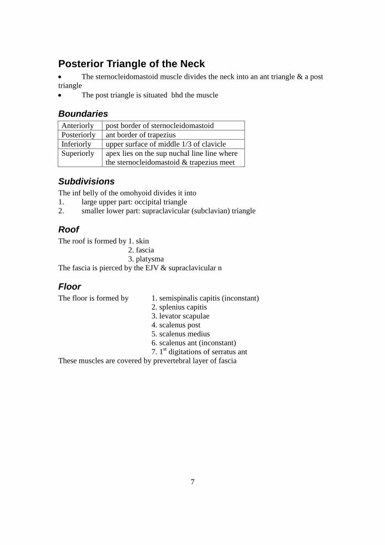
Posterior Triangle of the Neck The sternocleidomastoid muscle divides the neck into an ant triangle & a post
triangle
The post triangle is situated bhd the muscle
Boundaries
Anteriorly post border of sternocleidomastoid
Posteriorly ant border of trapezius
Inferiorly upper surface of middle 1/3 of clavicle
Superiorly apex lies on the sup nuchal line line where
the sternocleidomastoid & trapezius meet
Subdivisions
The inf belly of the omohyoid divides it into
1. large upper part: occipital triangle
2. smaller lower part: supraclavicular (subclavian) triangle
Roof
The roof is formed by 1. skin
2. fascia
3. platysma
The fascia is pierced by the EJV & supraclavicular n
Floor
The floor is formed by 1. semispinalis capitis (inconstant)
2. splenius capitis
3. levator scapulae
4. scalenus post
5. scalenus medius
6. scalenus ant (inconstant)
7. 1st digitations of serratus ant
These muscles are covered by prevertebral layer of fascia
8
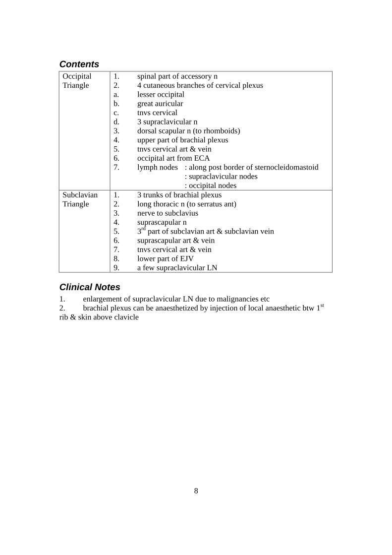
Contents
Occipital
Triangle
1. spinal part of accessory n
2. 4 cutaneous branches of cervical plexus
a. lesser occipital
b. great auricular
c. tnvs cervical
d. 3 supraclavicular n
3. dorsal scapular n (to rhomboids)
4. upper part of brachial plexus
5. tnvs cervical art & vein
6. occipital art from ECA
7. lymph nodes : along post border of sternocleidomastoid
: supraclavicular nodes
: occipital nodes
Subclavian
Triangle
1. 3 trunks of brachial plexus
2. long thoracic n (to serratus ant)
3. nerve to subclavius
4. suprascapular n
5. 3rd
part of subclavian art & subclavian vein
6. suprascapular art & vein
7. tnvs cervical art & vein
8. lower part of EJV
9. a few supraclavicular LN
Clinical Notes
1. enlargement of supraclavicular LN due to malignancies etc
2. brachial plexus can be anaesthetized by injection of local anaesthetic btw 1st
rib & skin above clavicle
9
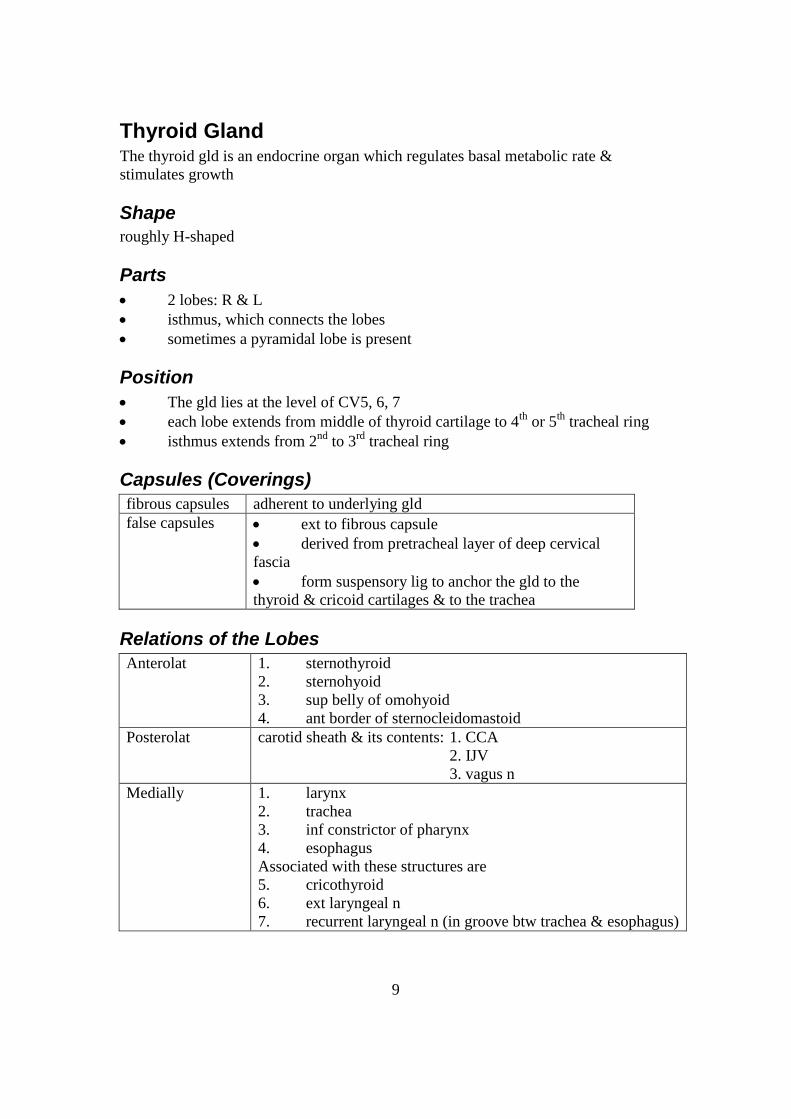
Thyroid Gland The thyroid gld is an endocrine organ which regulates basal metabolic rate &
stimulates growth
Shape
roughly H-shaped
Parts
2 lobes: R & L
isthmus, which connects the lobes
sometimes a pyramidal lobe is present
Position
The gld lies at the level of CV5, 6, 7
each lobe extends from middle of thyroid cartilage to 4th
or 5th
tracheal ring
isthmus extends from 2nd
to 3rd
tracheal ring
Capsules (Coverings)
fibrous capsules adherent to underlying gld
false capsules ext to fibrous capsule
derived from pretracheal layer of deep cervical
fascia
form suspensory lig to anchor the gld to the
thyroid & cricoid cartilages & to the trachea
Relations of the Lobes
Anterolat 1. sternothyroid
2. sternohyoid
3. sup belly of omohyoid
4. ant border of sternocleidomastoid
Posterolat carotid sheath & its contents: 1. CCA
2. IJV
3. vagus n
Medially 1. larynx
2. trachea
3. inf constrictor of pharynx
4. esophagus
Associated with these structures are
5. cricothyroid
6. ext laryngeal n
7. recurrent laryngeal n (in groove btw trachea & esophagus)

10
Post border of the
2 lobes is rounded
& related to
1. sup & inf parathyroid glds
2. anastomosis btw sup & inf thyroid art
3. on the L side, thoracic duct
11
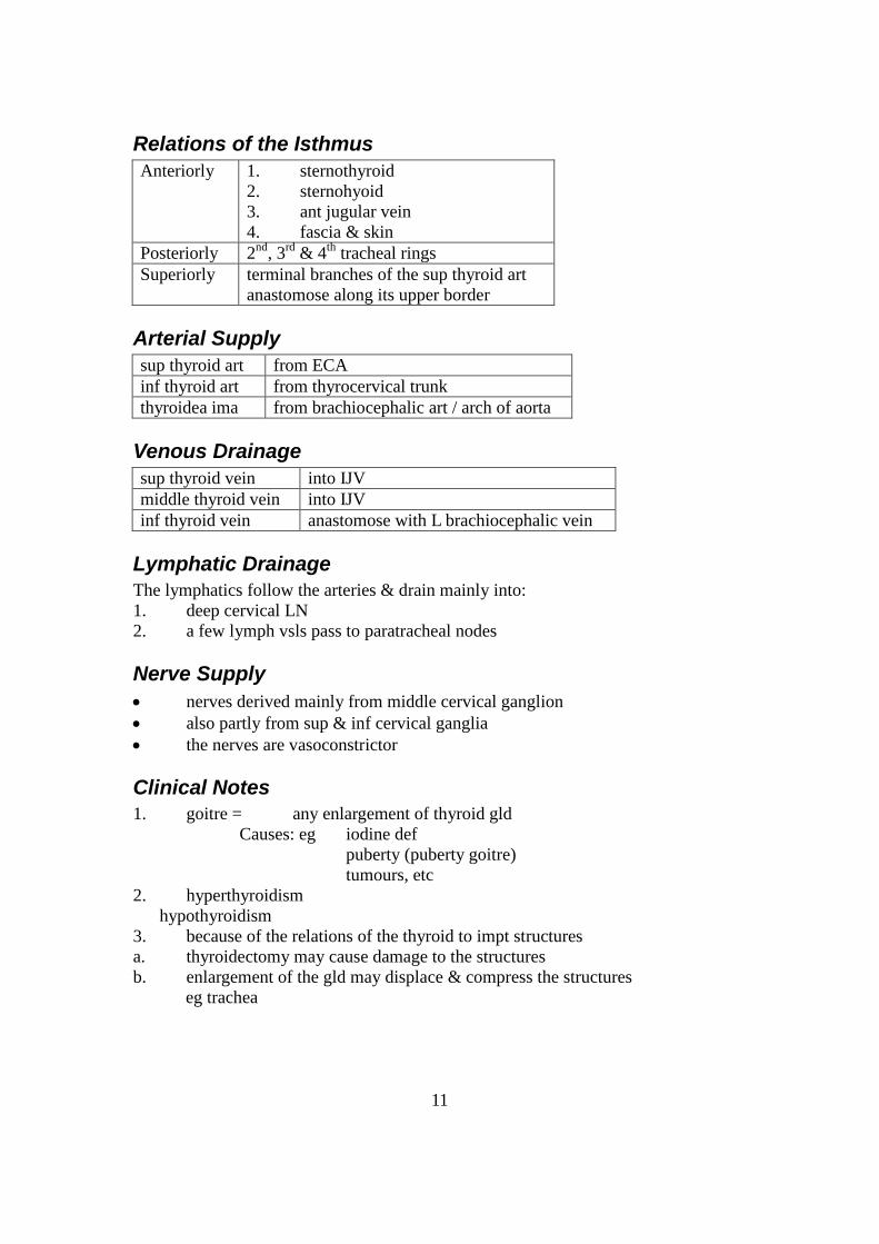
Relations of the Isthmus
Anteriorly 1. sternothyroid
2. sternohyoid
3. ant jugular vein
4. fascia & skin
Posteriorly 2nd
, 3rd
& 4th
tracheal rings
Superiorly terminal branches of the sup thyroid art
anastomose along its upper border
Arterial Supply
sup thyroid art from ECA
inf thyroid art from thyrocervical trunk
thyroidea ima from brachiocephalic art / arch of aorta
Venous Drainage
sup thyroid vein into IJV
middle thyroid vein into IJV
inf thyroid vein anastomose with L brachiocephalic vein
Lymphatic Drainage
The lymphatics follow the arteries & drain mainly into:
1. deep cervical LN
2. a few lymph vsls pass to paratracheal nodes
Nerve Supply
nerves derived mainly from middle cervical ganglion
also partly from sup & inf cervical ganglia
the nerves are vasoconstrictor
Clinical Notes
1. goitre = any enlargement of thyroid gld
Causes: eg iodine def
puberty (puberty goitre)
tumours, etc
2. hyperthyroidism
hypothyroidism
3. because of the relations of the thyroid to impt structures
a. thyroidectomy may cause damage to the structures
b. enlargement of the gld may displace & compress the structures
eg trachea

12
Common Carotid Artery
Origin
Right CCA branch of brachiocephalic art (trunk)
begins in neck bhd R sternoclavicular jt
Left CCA branch of aortic arch
begins in thorax & passes bhd L
sternoclavicular jt
Course
runs in carotid sheath along with IJV & vagus n
passes upwards & backwards in the neck,
from sternoclavicular jt to upper border of thyroid cartilage
ends by dividing into ECA & ICA (no other named branches)
Special Features
carotid sinus localised dilatation at terminal part of CCA (or beginning of
ICA)
innervated by glossopharyngeal & symp n
act as baroreceptor to regulate bld pressure
carotid body small reddish-brown structure situated bhd bifurcation of CCA
innervated mainly by glossopharyngeal n
acts as chemoreceptor
ie. respond to changes in concn of O2 & CO2 in bld
Relations
Anterolat 1. skin, supf fascia, platysma, investing layer of deep cervical
fascia
2. sternocleidomastoid overlaps it
3. sternohyoid
4. sternothyroid
5. sup belly of omohyoid
Embedded in ant wall of carotid sheath
6. descendens hypoglossi
7. ansa cervicalis
Crossing the art,
8. sup & middle thyroid veins
9. ant jugular veins
Posteriorly 1. tnvs processes of lower 4 cervical vertebral
2. longus capitis & longus colli
3. origin of scalenus ant
4. symp trunk
in the lower part of the neck

13
5. vertebral vsls
6. inf thyroid art
On the L side
7. thoracic duct
Bhd the termination of CCA = carotid body

14
Medially 1. larynx & pharynx
2. trachea
3. esophagus
4. lobe of thyroid gld
5. inf thyroid gld
6. recurrent laryngeal n
Laterally 1. IJV
2. vagus lies posterolat
Surface Anatomy
pt A: sternoclavicular jt
pt B: ant border of sternocleidomastoid at level of upper border of thyroid cartilage
Joining these 2 pts will surface mark the art in the neck
Clinical Notes
1. ligature of the CCA on 1 side
collateral circulation is est btw
1) sup & inf thyroid art (inf thyroid & deep cervical from subclavian)
2) desc branches of occipital & deep cervical art
3) vertebral art
Note: (1) and (2) are outside cranial cavity
(3) is inside cranial cavity
2. bifurcation of CCA: carotid pulse may be felt
3. the art can be compressed on ant tubercle of tnvs process of CV6 (carotid
tubercle)

15
External Carotid Artery The ECA is one of the 2 terminal branches of the CCA
& is the chief art of supply to ant structures in the neck & face
Origin
begins in carotid triangle\
at level of upper border of thyroid cartilage (ie. btw CV3 & 4)
Course
runs upwards, slightly backwards & laterally
enters substance of parotid gld
terminates bhd neck of mandible by dividing into the
maxillary & supf temporal art
Note: at first it lies medial to ICA.
It then passes backwards & laterally to lie lat to ICA
Branches
There are 8 branches
1. sup thyroid
2. asc pharyngeal
3. lingual
4. facial
5. occipital
6. post auricular
7. supf temporal
8. maxillary

16
Relations
Anterolat (Supf) 1. sternocleidomastoid overlaps it at its beginning
Above this level, it is relatively supf & is covered by
2. skin & supf fascia
3. cervical branch of facial n
4. tnvs cutaneous n
5. investing layer of deep cervical fascia
It is crossed by
6. hypoglossal n
7. facial & lingual veins
8. post belly of digastric
9. stylohyoid muscles
Within parotid gld, it is crossed by
10. facial n & retromandibular vein
Note: The IJV first lies lat to the art, then post to it
Medially 1. wall of pharynx
2. styloid process
3. ICA
Passing btw the ECA & ICA
4. stylopharyngeus
5. glossopharyngeal n
6. pharyngeal branch of vagus n
7. portion of parotid gld
Surface Anatomy
pt A: pt on ant border of sternocleidomastoid
at level of upper border of thyroid cartilage
pt B: pt bhd neck of mandible
midway btw mastoid process & angle of mandible
Joining these 2 pts gives the surface marking of ECA
Clinical Notes
Ligature of art of 1 side
- collateral circulation is maintained btw the branches of the ECA
with those of the oppo side

17
Internal Carotid Artery (Cervical Part) The ICA is one of the 2 terminal branches of the CCA
It is the principal art to the brain & eye
Its course is divided into 4 parts
a) cervical part
b) petrous part
c) cavernous part
d) cerebral part
The cervical part is discussed here
Origin
begins in the carotid triangle
at level of upper border of thyroid cartilage (ie btw CV3 & 4)
Course
enclosed in carotid sheath tog with IJV & vagus n
asc vertically in the neck to lower end of carotid canal in petrous temporal
bone
lower part (in carotid triangle) is comparatively supf
after ascending deep to post belly of digastric, it lies deep to parotid gld,
styloid process & many other structures
it does not give any named branches in the neck
Special Features
The ICA may present 2 structures at its commencement
carotid sinus localised dilatation at terminal part of CCA (or beginning of
ICA)
innervated by glossopharyngeal & symp n
act as baroreceptor (regulate bld pressure)
carotid body small reddish-brown structure situated bhd bifurcation of CCA
innervated mainly by glossopharyngeal n
acts as chemoreceptor (detect changes in bld O2 & CO2)
18
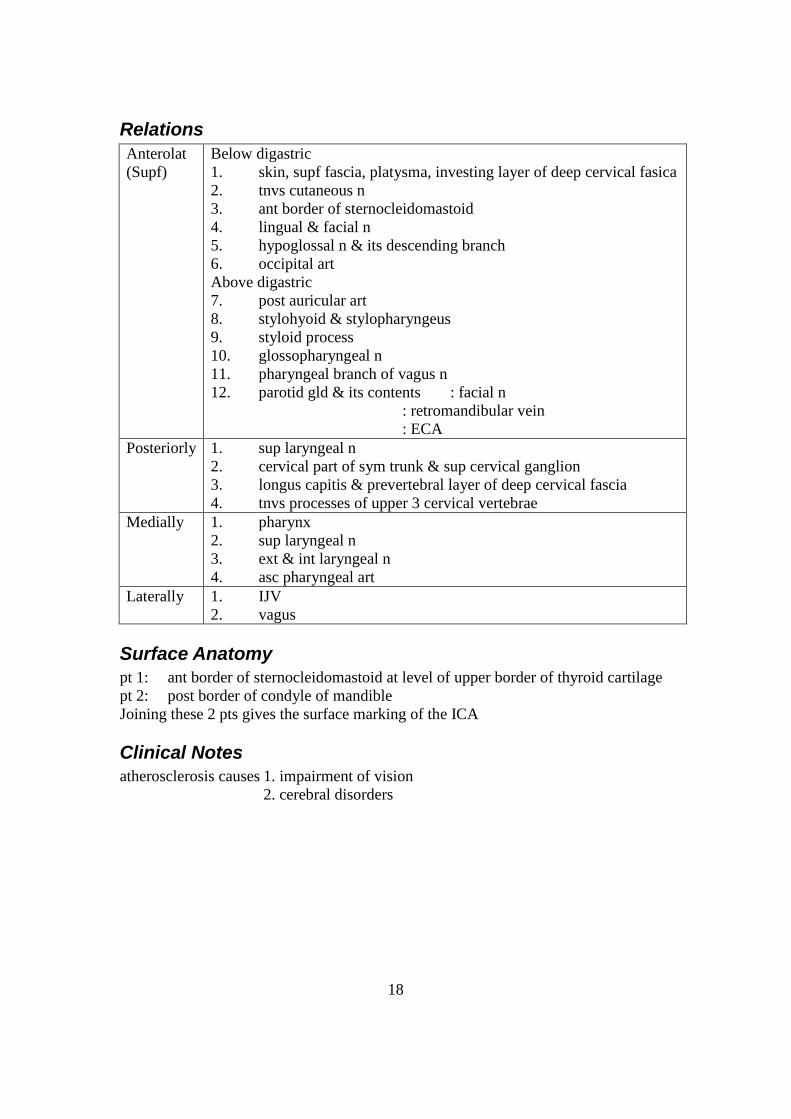
Relations
Anterolat
(Supf)
Below digastric
1. skin, supf fascia, platysma, investing layer of deep cervical fasica
2. tnvs cutaneous n
3. ant border of sternocleidomastoid
4. lingual & facial n
5. hypoglossal n & its descending branch
6. occipital art
Above digastric
7. post auricular art
8. stylohyoid & stylopharyngeus
9. styloid process
10. glossopharyngeal n
11. pharyngeal branch of vagus n
12. parotid gld & its contents : facial n
: retromandibular vein
: ECA
Posteriorly 1. sup laryngeal n
2. cervical part of sym trunk & sup cervical ganglion
3. longus capitis & prevertebral layer of deep cervical fascia
4. tnvs processes of upper 3 cervical vertebrae
Medially 1. pharynx
2. sup laryngeal n
3. ext & int laryngeal n
4. asc pharyngeal art
Laterally 1. IJV
2. vagus
Surface Anatomy
pt 1: ant border of sternocleidomastoid at level of upper border of thyroid cartilage
pt 2: post border of condyle of mandible
Joining these 2 pts gives the surface marking of the ICA
Clinical Notes
atherosclerosis causes 1. impairment of vision
2. cerebral disorders

19
Subclavian Artery This is the principal artery of the upper limb
but also supplies part of the neck & brain through its branches
Origin
R subclavian art arises from brachiocephalic art
(trunk)
bhd R sternoclavicular jt
L subclavian art arises from arch of aorta bhd L
CCA
asc to back of L sternoclavicular jt
Course
For descriptive purposes, the subclavian art is divided into 3 parts by the presence of
the scalenus ant
1st Part arches upwards & lat from bhd sternoclavicular jt to med border of
scalenus ant
2nd
Part lies bhd scalenus ant muscle
3rd
Part extends from lat border of scalenus ant to outer border of 1st rib
where it continues as axillary art
supf & can be compressed against 1st rib
Branches
1st Part 1. vertebral art
2. thyrocervical trunk
a) inf thyroid
b) tnvs cervical
c) suprascapular
3. int thoracic art
2nd
Part costocervical trunk
a) sup IC art
b) deep cervical art
3rd
Part descending (dorsal) scapular art
Relations of 1st Part
Anteriorly From med to lat
1. CCA
2. ansa subclavia
3. vagus n
4. IJV
5. vertebral veins
6. on L side = thoracic duct

20
In addition
7. cardiac branches of vagus & symp n
Posteriorly 1. dome of cervical pleura & suprapleural mbm
2. apex of lung
3. ansa subclavia which encircles the art
4. symp trunk & inf cervical ganglion
5. on the R side = R recurrent laryngeal n
Relations of the 2nd Part
Anteriorly 1. scalenus ant
2. sternocleidomastoid
3. subclavian vein (sep by scalenus ant)
4. on R side = R phrenic n
Posteriorly 1. dome of cervical pleura & suprapleural mbm
2. apex of lung
3. scalenus medius
Relations of 3rd Part
Anteriorly 1. skin, supf fascia, platysma, investing layer of deep cervical
fascia
2. supraclavicular n
3. EJV & its tributaries : ant jugular vein
: tnvs cervical
: suprascapular
4. suprascapular art
5. n to subclavius
6. clavicle & subclavius
Posteriorly 1. scalenus medius
2. lower trunk of brachial plexus
Superiorly upper & middle trunks of brachial plexus
Inferiorly upper surface of 1st rib
Surface Anatomy
pt 1: on sternoclavicular jt
pt 2: at middle of lower border of clavicle
The art is marked by a curved line,
convex upwards to abt 2 cm above the clavicle, joining the 2 pts
Clinical Notes
1. 3rd
part of subclavian art may be compressed against 1st rib to stop bleeding in
upper arm
2. aneurysms may form in 3rd
part of artery
- exert pressure on brachial plexus & results in

21
pain, weakness & numbness in upper limb
3. cervical rib – art is kinked as it passes over rib, causing occlusion

22
Internal Jugular Veins The IJV drains the brain, neck & face
Origin
begins at jugular foramen at base of skull
it is a continuation of the sigmoid sinus
Course
descends in carotid sheath
passes downwards & anteriorly
ends bhd med end of clavicle by joining with subclavian vein
to form brachiocephalic vein
Special Features
1. sup bulb: dilatation at is origin
2. inf bulb: dilatation near its termination
3. The vein possess 1 bicuspid valve directly above inf bulb
Relations
Anterolat 1. skin, supf fascia, platysma, investing layer of deep cervical fascia
2. sternocleidomastoid
3. post belly of digastric
4. parotid salivary gld sep from it by styloid process &
stylopharyngeus
Its lower part is covered by
5. sternohyoid, sternothyroid & omohyoid
Crossing the vein
6. ansa cervicalis
7. stylohyoid
8. post auricular & occipital art
9. spinal part of accessory n
Also supf are
10. facial n
11. ant jugular vein
12. The deep cervical LN run alongside the IJV
Posteriorly 1. tnvs processes of cervical vertebrae
2. levator scapulae
3. scalenus ant & medius
4. cervical plexus
5. phrenic n
6. thyrocervical trunk
7. vertebral vein
8. 1st part of subclavian art
9. dome of cervical pleura

23
10. on the L = thoracic duct
Medially Below,
1. vagus n
2. CCA
At base of skull
3. ICA
4. 9th, 10
th, 11
th & 12
th cranial n
Tributaries
1. inf petrosal sinus
2. facial vein
3. lingual vein
4. pharyngeal veins
5. sup thyroid vein
6. middle thyroid vein
7. sometimes, occipital vein
Note: Thoracic duct opens into angle of union btw left IJV & L subclavian vein
The right lymphatic duct opens similarly on the r side
Surface Anatomy
pt 1: on neck, med to lobule of ear
pt 2: at med end of clavicle
Joining these 2 pts will surface mark the IJV
Clinical Notes
1. In congestive heart failure or any disease where venous pressure is raised, the
IJV is markedly dilated, engorged
2. Deep cervical LN lie along the IJV.
In malignancies involving these nodes, the vein is usu removed tog with the nodes
in a surgical procedure called block dissection of the cervical nodes

24
Cervical Plexus The cervical plexus supplies the skin at the back of the head, the neck & shoulder as
well as certain muscles of the neck tog with the diaphragm
Formation
by ventral rami of C1 to C4
the rami are joined by connecting branches to form a series of 3 loops from
which the branches arise
Position & Relations
Anteriorly 1. It is covered ant by prevertebral layer of deep cervical
fascia
It is also related to
2. IJV in carotid sheath
3. sternocleidomastoid
Posteriorly It lies in front of the origins of
1. levator scapulae
2. scalenus medius
Branches
Supf (Cut) Branches 1. Lesser occipital (C2)
2. great auricular (C2, 3)
3. tnvs cut (cervical) (C2, 3)
4. med, intermediate & lat supraclavicular (C3, 4)
The cutaneous branches all emerge near middle of post border
of sternocleidomastoid
Deep Branches Communicating Branches
1. Each of the 4 rami receive GRC from the sup cervical
ganglion of the symp trunk
2. branch from C1 to hypoglossal n
3. other branches to vagus & accessory n
Muscular Branches to:
1. prevertebral muscles
2. sternocleidomastoid
3. levator scapulae
4. scaleni ant, medius & post
5. trapezius
It also supplies:
6. diaphragm via phrenic n
7. infrahyoid muscles (except thyrohyoid) via ansa
cervicalis
Ansa cervicalis
lies supf to (or in the substance of) the carotid sheath

25
formed by 1. desc branch of hypoglossal n (C1)
2. descending cervical n (C2, 3)
which unite to form a loop
supply omohyoid, sternohyoid & sternothyroid
(Thyrohyoid is supplied directly via C1 fibres within
hypoglossal n)

26
Phrenic Nerve (Cervical Part) The phrenic n is a mixed n carrying motor fibres to the diaphragm & sensory
fibres from the pleura, pericardium, peritoneum & diaphragm
It also carries symp fibres
It is the only motor supply of the diaphragm
Origin
from C3, C4 & C5 (mainly C4)
at lat border of scalenus ant at level of circoid cartilage
The contribution from C5 may come via n to subclavius, in which case it is
called accessory phrenic n
Course
runs downwards vertically on ant surface of scalenus ant (bhd prevertebral
fascia)
since scalenus ant is oblique, phrenic n crosses it from lat to med border
crosses int thoracic art from lat to med side
enters thorax by passing in front of subclavian art & bhd beginning of
brachiocephalic vein
it does not give any branches in the neck
Relations
Anteriorly 1. prevertebral layer of deep fascia
2. IJV
3. supf cervical & suprascapular art
4. beginning of brachiocephalic vein
5. on the L: thoracic duct
Posteriorly 1. scalenus ant
2. subclavian art
3. cervical dome of pleura
Surface Anatomy
pt 1: on side of neck at level of upper border of thyroid cartilage &
3.5 cm from median plane
pt 2: med end of clavicle
Joining these 2 pts will surface mark the course of the phrenic n in the neck
Clinical Notes
surgical interruption of the phrenic n on the scalenus ant is sometimes perfomed to aid
in collapse of a lung

27
ie. diaphragm on affected side is paralysed & is therefore elevated, leading to
collapse of the lung
This gives rest to a diseased lung & promotes healing

28
Cervical Part of Sympathetic trunk This is a ganglionated chain located one on each side of the cervical part of the
vertebral column
From it comes the symp supply to the various structures of the head & neck
Formation
by fibres (PreGN) arising from segments T1 to T4 of spinal cord
these fibres pass to thoracic part of symp trunk & then ascend into the neck
Extent
upwards to base of skull
downwards to neck of 1st rib where it becomes continuous with thoracic part
of symp trunk
Relations
It is prevertebral & is related to
Anteriorly 1. carotid sheath
2. CCA & ICA
3. inf thyroid art
Posteriorly 1. prevertebral fascia
2. longus capitis & cervicis
3. tnvs processes of lower 6
CV

29
Ganglia
There are 3 ganglia: sup, middle & inf
The asc PreGN fibres synapse in these ganglia,
from which PGN fibres arise to supply the SM, BV & glds of the head & neck Cervical
Ganglion
Remarks Branches
Superior largest of the 3 ganglia
formed by the fusion of the
upper 4 ganglia
lies just below skull at level
of CV1 to CV3 bhd ICA & in front of
longus capitis
1. GRC to C1 to C4 (ventral rami)
2. branches to 9th , 10
th & 12
th
cranial n
3. int carotid n int carotid plexus 4. arterial branches to CCA & ECA
5. pharyngeal branches
pharyngeal plexus (with branches
of 9th & 10
th cn)
6. sup cervical branch
cardiac plexus in thorax Middle very small
formed by fusion of 5th & 6
th
cervical ganglia
lies in lower part of neck in
front of CV6 above arch formed by
inf thyroid art
1. GRC to venral rami of C5 & C6
2. thyroid branches thyroid gld 3. middle cardiac branch
cardiac plexus in thorax
Inferior rather large, irregular & star-
shaped
formed by fusion of 7th & 8
th
cervical ganglia, & often with 1st
thoracic ganglia stellate ganglion
lies btw tnvs process of CV7
& neck of 1st rib bhd vertebral art
1. GRC to ant rami of C7 & C8
2. arterial branches
subclavian & vertebral art 3. inf cardiac branch
cardiac plexus in thorax
Clinical Notes
1. injury to cervical part of symp trunk produced Horner’s syndrome
2. stellate ganglion can be blocked by anesthetic

30
Trachea (Cervical Part) The trachea is a mobile, non-collapsible tube forming the beginning of the
lower resp passages
It is kept potent by anterolat C-shaped cartilaginous rings
Its post membranous part permits expansion of the esophagus during passage
of food
Origin
begins at lower border of criccoid cartilage of larynx (at level of CV6) in the middline
Course
runs downwards & slightly backwards in the midline
enters thorax in median plane
Relations
Anteriorly 1. skin & fascia
2. isthmus of thyroid gld (in front of 2nd
, 3rd
& 4th
rings)
3. inf thyroid veins
4. jugular arch
5. thyroidea ima art (if present)
6. sternothyroids & sternohyoids
7. L brachiocephalic vein in the child
Posteriorly 1. R & L recurrent laryngeal n
2. esophagus
3. vertebral column & some prevertebral muscles
Laterally
(on each side)
1. lobes of thyroid gld
2. carotid sheath & CCA
Blood Supply
mianly from inf thyroid arteries
Venous Drainage
veins drain into L brachiocephalic vein
Lymphatic Drainage
Lymphatics pass to 1. pretracheal nodes
2. paratracheal nodes
Nerve Supply
1. psymp: vagus & recurrent laryngeal n

31
sensory & secretomotor to mucous mbm
motor to trachealis muscle
2. symp: from middle cervical ganglion of symp trunks
vasomotor
Clinical Notes
1. trachea may be compressed by pathological enlargement of thyroid, thymus,
LN & aortic arch
2. tracheostomy is usu done after cutting the isthmus of thyroid gld

32
Lymphatic Drainage of the Head & Neck The lymphatic system of the head & neck consists of 1. LN
2. lymph vsls
Lymph Nodes
The lymph nodes of the head & neck are made up of a no. of peripheral gps, &
a terminal gp
The terminal gp receives all the lymphatics of the head & neck,
either directly or indirectly via one of the peripheral gps
Peripheral Groups of Lymph Nodes
These are arranged in 2 gps: supf & deep
a) The supf gp consist of
Nodes Remarks
1 occipital nodes located over occipital bone at apex of post triangle of
neck
receive lymph from back of scalp
efferents to deep cervical nodes
2 retro-auricular
(mastoid) nodes located over lat surface of mastoid process of temporal
bone
receive lymph from scalp above auricle &
from post wall of ext auditory meatus
efferents to deep cervical nodes
3 parotid nodes located on / within parotid gld
receive lymph from scalp, auricle, face, ext acoustic
meatus & middle ear
efferents to deep cervical LN
4 buccal nodes located over buccinator muscle close to facial vein
lie along course of lymph vsls
ie receives from several nodes
efferents to submandibular cervical LN
5 submandibular
nodes located on surface of submandibular gld
receive lymph from wide area
efferents to deep cervical nodes
6 submental
nodes located in submental triangle btw ant bellies of digastric
receive lymphfrom lower lip, tongue & supf neck
efferent to submandibular & deep cervical nodes
- These 6 gps of LN form a collar at the jn of the head with the neck called
pericervical collar
- The supf tissues of the head & neck drain into these nodes, as well as 2 other
gps

33
7 ant cervical
nodes located along course of ant jugular vein
receive lymph from supf tissue of front of neck
efferent to deep cervical nodes
8 supf cervical
nodes located along the course of EJV
receive lymph from small part of face & ext ear
efferents to deep cervical nodes

34
a) The deep gp of peripheral LN are:
9 retropharyngeal
nodes
fd in interval btw pharyngeal wall & prevertebral fascia
ie retropharyngeal space
10 laryngeal nodes fd in front of larynx on cricothyroid lig
11 paratracheal nodes fd lat to trachea
12 pretracheal nodes fd in front of trachea
Terminal Group of Lymph Nodes
These are the deep cervical nodes
fd in a chain along the course of IJV
embedded in fascia of carotid sheath
receive all lymph of head & neck
efferents join to form jugular lymph trunks which drains into thoracic duct or
R lymphatic duct
Note: 2 of the nodes are referred to clinically: 1. jugulo-digastric node
2. jugulo-omohyoid node
Lymph Vessels
supf vsls follow supf veins
deep vsls follow arteries & sometimes deep veins
Clinical Notes
1. enlargement of nodes may indicate infection of its area of drainage
2. spread of cancer through the lymphatics
3. block dissection of cervical nodes
ie removal of cervical nodes, IJV, submandibular gld & fasica
may be performed in some cases of cancer

35
Scalp Soft tissues covering the cranial vault form the scalp
Extent
Anteriorly supraorbital margins
Posteriorly ext occipital protuberance & sup nuchal lines
Laterally sup temporal lines on each side
Hairline does not correspond to boundaries of scalp
- hair is deficient over front part of scalp
- hair overlaps upper part of back of neck
Structure
The scalp consists of 5 layers, the 1st 3 of which are intimately connected & move as a
unit. The layers are:
Skin thick, hair-bearing & contains sebaceous glds
Connective tissue fibro-fatty layer beneath the skin containing anastomoses of
arteries & veins
Aponeurosis of
occipito-frontalis
muscles
Loose areolar tissue lies below the aponeurosis in the subaponeurotic
space
contains emissary veins
Pericranium periosteum covering outer surface of skull bones
Blood Supply
The scalp has a very rich bld supply small cuts tend to bleed profusely
In front of the auricle, it is supplied from before backwards by
1 supratrochlear branches of ophthalmic art (br of ICA)
2 supraorbital branches of ophthalmic art (br of ICA)
3 supf temporal terminal branch of ECA
Behind the auricle, it is supplied from before backwards by:
4 post auricular branches of ECA
5 occipital branches of ECA
Venous Drainage
1. supratrochlear – unite at med margin of orbit to form facial vein
2. supraorbital – unite at med margin of orbit to from facial vein
3. supf temporal vein
4. post auricular vein
5. occipital vein suboccipital venous plexuses
The veins of the scalp anastomose with diploic veins

36
They are connected to the intracranial venous sinuses by emissary veins
Lymphatic Drainage
part of scalp drains into
ant part of scalp pre-auricular (parotid) nodes
post part of scalp mastoid & occipital nodes
Nerve Supply
a) Sensory Innervation
From ant to post by branches of
1. ophthalmic n – from 5th
cranial n
2. maxillary n – from 5th
cranial n
3. mandibular n – from 5th
cranial n
4. cervical plexus
5. dorsal rami of cervical spinal n
b) Motor Innervation
branches of facial n - temporal branch to frontal belly
- post auricular branch to occipital belly
Clinical Notes
1. scalp bleeds profusely when injured due to rich bld supply
2. emissary veins in loose areolar tissue (subaponeurotic) are inv in spread of
infection from scalp to intracranial regions
Thus the area is called ‘danger area’

37
Facial Artery The facial artery is the chief artery of the face
Origin
branch of ECA
given off in carotid triangle in the neck
Course
brief course in the neck
winds round lower border of mandible at ant margin of masseter
proceeds upwards & forwards on the face
ends at med angle of the eye where it anastomoses with branches of
ophthalmic art
The facial art is very tortuous & takes part in numerous anastomoses,
including some across median plane
Tortuous course permits free movement of the mandible, lips & cheeks
Venous Drainage
Facial vein - connects to cavernous sinus & pterygoid plexus
Thus spread of infection from face into cranial cavity
Branches
In the neck: 1. asc palatine art
2. tonsillar branch
3. glandular branch to submandibular gld
In the face it supplies the lips & ext nose via: 1. submental art
2. inf labial art
3. sup labial art
4. lat nasal art
Clinical Notes
There.is rich anastomosis
collateral circulation can be est after ligature of the CCA or ECA on 1 side
Other arteries supplying the face
1 supf temporal art from ECA
2 tnvs facial art from supf temporal art
3 supraorbital from ophthalmic art
4 supratrochlear from ophthalmic art

38
Facial Nerve The facial n is the 7
th cranial nerve.
It is also the nerve to the 2nd
branchial arch
It is 1. motor to the face
2. secretomotor to submandibular & sublingual salivary glds & lacrimal glds
3. special sensory (taste) to ant 2/3 of the tongue
Nuclei & Functional Components
1 motor nucleus SVE
2 sup salivatory nucleus GVE
3 lacrimatory nucleus GVE
4 nucleus of solitary tract SVA
5 spinal nucleus of V GSA
Course
Intracranial Course starts off as 2 roots - motor
- sensory (nervus-intermedius)
& secretomotor
leave brain at lower border of pons
enter int acoustic meatus
enter facial canal of temporal bone
- Above promontory of med wall of middle ear, it forms
geniculate ganglion (containing cells of taste fibres)
emerges from skull at stylomastoid foramen
Extracranial Course crosses laterally the base of styloid process
enters posteromed surface of parotid gld
runs forward within the gld, supf to retromandibular
vein & ECA
bhd neck of mandible it divides into its 5 terminal
branches, which emerge at the ant border of the gld
Branches
a) Intracranial Branches
within facial canal: 1. greater petrosal n
2. n to stapedius
3. chorda tympani (joins lingual n)
b) Extracranial Branches
At exit from stylomastoid formen: 1. post auricular
2. muscular branches to digastric &
stylohyoid

39
c) Terminal Branches 1. temporal
2. zygomatic
3. buccal
4. mandibular
5. cervical
These arise in the substance of the parotid gld & emege from its ant border
The facial n supplies all the muscles of facial expression
It does not supply the skin
d) Communicating Branches - communicate with adj cranial & spinal n
Surface Anatomy pt 1: at middle of ant border of mastoid process
= position of stylomastoid foramen
pt 2: bhd neck of mandible
= pt of division into terminal branches
Joining these 2 pts will give extracranial course of facial n
Clinical Notes 1. Lesions of the facial n below / at the level of the motor nucleus results in
lower motor-neuron lesion
- Possible causes: 1. infection of middle ear
2. surgery, etc
- Result: paralysis of entire facial musculature on affected side
- Features : displacement of mouth accompanied by drooping
: ptosis, & inability to close eyes
: no wrinkles on forehead
2. Lesions of the facial n above level of the motor nucleus results in
upper motor-neuron lesion
- Possible causes: 1. tumours
2. surgery, etc
- Result : Lower half of face of oppo side paralysed
: orbicularis oculi & frontalis muscles spared because
they are controlled by fibres from both sides of cerebral cortex
- Features : displacement & drooping of mouth contralaterally
However the patient is able to wrinkle his forehead
Distribution of Facial Nerve 4 modalities carried by nerve
Motor 1. to muscles of facial expression
2. stylohyoid, digastric
3. stapedius
Special Senses taste fibres to ant 2/3 of tongue

40
Pysmp secretomotor to submandibular, sublingual glds
lacrimal gld
parotid gld
Sensory small area of ext meatus, auricle

41
Dura Mater of the Brain The dura mater is the outermost, thickest & toughest mbm covering the brain
It is continuous with the dura of the spinal cord
Layers of the Dura Mater
It has 2 layers
1. outer layer / endosteal layer – serves as an int periosteum to the cranial bones
2. inner layer / meningeal layer – provides the protective mbm to the brain
The 2 layers are fused except at places where cranial venous sinuses are enclosed btw
them
Processes of the Dura Mater
The meningeal layer sends 4 folds internally which divide the cranial cavity into
many freely communicating compartments housing different parts of the brain
1 Falx Cerebri large, sickle-shaped fold
occupies median longitudinal fissure btw the 2 cerebral
hemispheres
attachments: anteriorly to crista galli
posteriorly to tentorium cerebelli
upper convex border contains sup sagittal sinus
lower concave border contains inf sagittal sinus
2 Tentorium
Cerebelli tent-shaped fold forming roof of post cranial fossa
supports occipital lobe above & sep it from cerebellum
below
anteriorly it shows as gap
tentorial notch, for passage of the midbrain
Thus, it has 2 borders:
1. ant free border forming tentorial notch
which is attached on both sides to ant clinoid process
2. outer attached border which is attached to
post clinoid process
outer attached border encloses sup petrosal & tnvs sinuses
upper surface encloses straight sinus
3 Falx
Cerebelli
small sickle-shaped fold lying below tentorium cerebelli
4 Diaphragma
Sellae
small circular horiz fold forming roof of hypophyseal fossa
Blood Supply
outer layer is richly vascular
inner layer is more fibrous & requires little blood supply
The dura is supplied by numerous meningeal branches from

42
1. ICA
2. maxillary asc pharyngeal art
3. occipital art
4. vertebral art
The most impt is middle meningeal art
Venous Drainage
by meningeal veins into pterygoid plexus of veins or sphenoparietal sinus
Nerve Supply
The dura is supplied by branches of the trigeminal & cervical nerves
ant cranial fossa ophthalmic n
middle cranial fossa maxillary & mandibular n
post cranial fossa branches of vagus & hypoglossal n
as well as branches from C1 to C3
The nerve supply is
a) sensory to the dura
b) autonomic to the BV
Clinical Notes
1. headache usu implicates the dura & the structures contained in it because the
brain itself is normally quite insensitive
2. intracranial hemorrhages

43
Cranial Fossae interior of the base of the skull is divided into 3 cranial fossae:
anterior cranial fossa
middle cranial fossa
posterior cranial fossa
Boundaries
Anterior Cranial Fossa Middle Cranial Fossa Posterior Cranial Fossa
Ant inner surface of frontal bone
(with falx cerebri in midline)
lesser wings of sphenoid sup border of petrous
part of temporal bone
Post sharp lesser wing of sphenoid
groove for optic chiasma
sup borders of petrous
parts of temporal bone
internal surface of
squamous part of
occipital bone
Lat squamous parts of
temporal bone
greater wings of sphenoid
parietal bones
Floor lat: orbital plates of frontal
bone
med: cribiform plate of
ethmoid
greater wing of sphenoid
squamous and petrous
parts of temporal bone
basilar, condylar &
squamous parts of
occipital bone
mastoid part of temporal
bone
Major Foramina of the Cranial Fossae
Opening in skull Bone of skull Structures transmitted
Ant
Cranial
Fossa
perforations in
cribiform plate
ethmoid olfactory n
Middle
Cranial
Fossa
1. optic canal
2. sup optic fissure
3. foramen rotundum
4. foramen ovale
5. foramen spinosum
6. foramen lacerum
lesser wing of sphenoid
b/w lesser & greater
wings of sphenoid
greater wing of sphenoid
greater wing of sphenoid
greater wing of sphenoid
b/w petrous part of
temporal and sphenoid
optic n
lacrimal, frontal, trochlear,
oculomotor, nasociliary &
abducent n;
sup ophthalmic vein
maxillary division of the
trigeminal n
mandibular division of the
trigeminal n, lesser petrosal n
middle meningeal art
ICA
Post
Cranial
Fossa
1. formen magnum
2. hypoglossal canal
occipital
occipital
MO, sp part of acc n, R & L
vertebral art
hypoglossal n
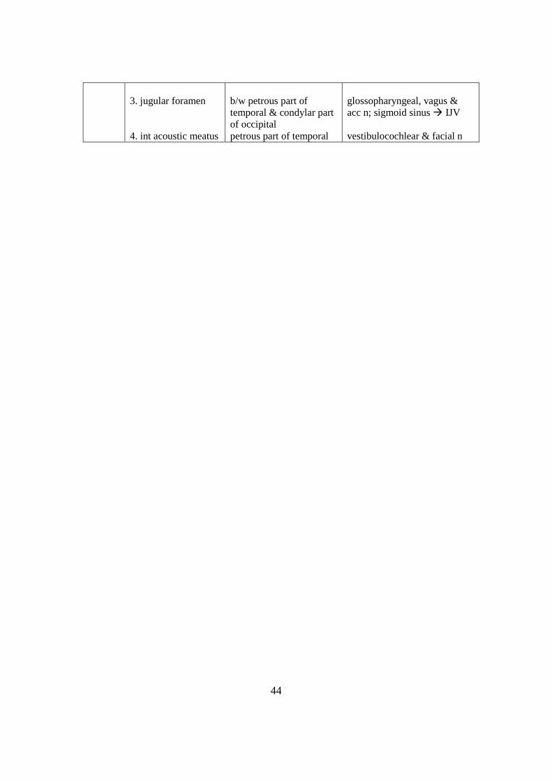
44
3. jugular foramen
4. int acoustic meatus
b/w petrous part of
temporal & condylar part
of occipital
petrous part of temporal
glossopharyngeal, vagus &
acc n; sigmoid sinus IJV
vestibulocochlear & facial n

45
Cavernous Sinus It is a large venous space btw the 2 layers of dura mater
It is situated in the middle cranial fossa, one on either side of the body of the
sphenoid bone
Formation
floor is by endosteal dura mater
lat wall, roof & med wall = meningeal dura mater
Extent & Size
abt 2 cm long & 1 cm wide
anteriorly: extends up to med end of sup orbital fissure
posteriorly: extends up to apex of petrous temporal
Relations
a) Structures Outside the Sinus
Superiorly 1. optic tract
2. ICA
Inferiorly 1. foramen lacerum
2. jn of body & greater wing of sphenoid
bone
Anteriorly sup orbital fissure & apex of orbit
Posteriorly apex of petrous temporal
Medially 1. hypophysis cerebri = pit
2. sphenoidal air sinuses
Laterally temporal lobe of cerebral hemisphere
a) Structures in Lateral Wall of the Sinus
- from above downwards: 1. occulomotor n
2. trochlear n
3. ophthalmic n
4. maxillary n
5. trigeminal ganglion
b) Structures Passing Through the Sinus
1. ICA together with its venous & symp plexuses
2. abducent n
Tributaries
from the orbit 1. sup & inf ophthalmic veins
2. central vein of retina
from the brain 1. supf middle cerebral vein
2. inf cerebral veins
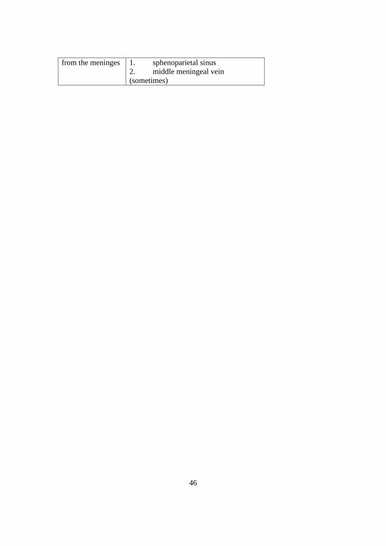
46
from the meninges 1. sphenoparietal sinus
2. middle meningeal vein
(sometimes)

47
Drainage
The cavernous sinus drains into
1. tnvs sinus via sup petrosal sinus
2. IJV via inf petrosal sinus
3. pterygoid plexus of veins via emissary veins
The cavernous sinuses on both sides communicate with each other via
ant & post intercavernous sinuses
Factors which help to expel bld from the sinus are:
a) pulsations of the ICA &
b) gravity
Clinical Notes
1. infection of face may spread via facial vein into cavernous sinus
thrombosis of cavernous sinus
2. carotid – cavernous sinus aneurysms

48
Superior Sagittal Sinus It is located in the upper convex margin of the falx cerebri
Course
begins anteriorly at crista galli by union of meningeal veins
runs upwards & backwards, becoming progressively larger in size
ends near int occipital protuberance by deviating to 1 side
usu to the right (continues as R tnvs sinus)
Special Features
The sinus communicates with 2 or 3 small, irregularly-shaped venous lacunae
on each side
Arachnoid villi & granulations project into the lacunae
Tributaries
The sinus receives bld from 1. diploic & meningeal veins via the lacunae
2. emissary veins
3. sup cerebral veins
Drainage
It drains into the confluence of sinuses &
hence into the tnvs sinuses & occipital sinus, as well as straight sinus
Surface Anatomy
It extends from above root of the nose, over vault of skull in median plane,
to ext occipital protuberance
Clinical Notes
1. thrombosis caused by spread of infection from nose, scalp & diploe

49
Middle Meningeal Artery This artery is clinically impt because it is the chief source of extradural hemorrhage
Origin
branch of maxillary art
arises in infratemporal fossa deep to ramus of mandible
Course & Relations
in infratemporal fossa it runs upwards & medially
- deep to lat pterygoid
- passes btw 2 roots of auriculotemporal n
enters middle cranial fossa through foramen spinosum
in middle cranial fossa it has an extradural course, running forward & laterally
- related to middle meningeal veins
- divides at a variable pt into ant & post branches
Branches
The branches arise in the middle cranial fossa
They contribute to the supply of 1. trigeminal ganglion
2. tympanic cavity
3. orbit
The terminal branches are
1. ant (frontal) branch - grooves the sphenoid & parietal bones
- supplies ant portion of dura
2. post (parietal) branch - grooves the temporal & parietal bones
- supplies post portion of dura
Surface Marking
Pt 1: at midpt of zygomatic arch = entry of artery into skull
= pt of division of artery
Pt 2: 2 cm above pt 1
Pt 3: centre of pterion
Pt 4: midpt btw nasion & inion
Pt 5: at lambda sinus
Joining pt 1& 2 - stem of artery
Joining pt 2, 3, & 4 - ant branch
Joining pt 2 & 5 - post branch
Clinical Notes
Injuries to the side of head fracture of head
This may result in tearing of artery or more commonly via int branch leading to
extradural hemorrhage

50

51
Types of Intracranial Hemorrhage Intracranial hemorrhages refer to bleeding inside the cranial cavity
They may result from trauma or vascular lesions
There are 4 main varieties 1. extradural
2. subdural
3. subarachnoid
4. cerebral
Extradural Hemorrhage
results from injuries to the meningeal arteries or vein
most common artery to be damaged = ant branch of middle meningeal art
It is usu torn by a blow to side of the head resulting in fracture of the skull
around the anteroinf region of parietal lobe
bleeding occurs & a hematoma forms
the intracranial pressure rises & the bld clot exerts local pressure on
underlying area on precentral gyrus (may result in oppo hemiplegia)
bld also passes outwards through the fracture line to form a soft swelling
under temporalis muscle
Subdural Hemorrhage
results from tearing of sup cerebral veins at their pt of entrance into sup
sagittal sinus
usu caused by blow to the front or back of the head
causing excessive anteropost displacement of the brain within the skull
once the vein is torn, bld under lower pressure begins to accumulate in the
potential space btw the dura & arachnoid
In 1/2 the cases, the condition is bilateral
depending on the speed of accumulation of blood, the clinical condition can be
acute or chronic
In both cases the bld clod will press against the brim, producing diff pressure
symptoms depending on the location of the clot
Subarachnoid Hemorrhage
results from leakage or rupture of a congenital aneurysm on the cerebral
arterial cicle (of Willis) or less commonly from an angioma
the symptoms are sudden in onset & include:
1. severe headache
2. stiff neck
3. loss of consciousness
It is diagnosed by a lumbar puncture,
where CSF is withdrawn is heavily bld-stained

52
Cerebral Hemorrhage generally due to rupture of lenticulostriate artery,
which is a branch of middle cerebral art
inv corticolubar & corticospinal fibres in internal capsule
produces hemiplegia on oppo iside of body
the patient immediately loses consciousness & paralysis is evident when
consciousness is regained
53
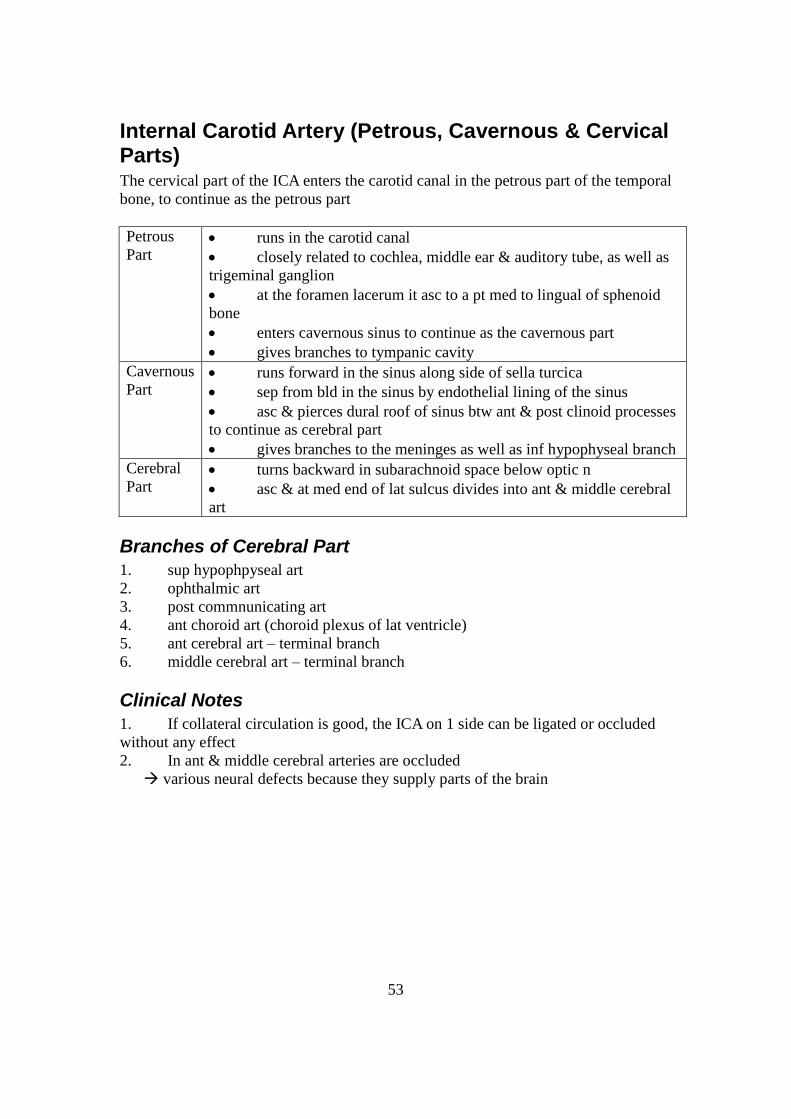
Internal Carotid Artery (Petrous, Cavernous & Cervical Parts) The cervical part of the ICA enters the carotid canal in the petrous part of the temporal
bone, to continue as the petrous part
Petrous
Part runs in the carotid canal
closely related to cochlea, middle ear & auditory tube, as well as
trigeminal ganglion
at the foramen lacerum it asc to a pt med to lingual of sphenoid
bone
enters cavernous sinus to continue as the cavernous part
gives branches to tympanic cavity
Cavernous
Part runs forward in the sinus along side of sella turcica
sep from bld in the sinus by endothelial lining of the sinus
asc & pierces dural roof of sinus btw ant & post clinoid processes
to continue as cerebral part
gives branches to the meninges as well as inf hypophyseal branch
Cerebral
Part turns backward in subarachnoid space below optic n
asc & at med end of lat sulcus divides into ant & middle cerebral
art
Branches of Cerebral Part
1. sup hypophpyseal art
2. ophthalmic art
3. post commnunicating art
4. ant choroid art (choroid plexus of lat ventricle)
5. ant cerebral art – terminal branch
6. middle cerebral art – terminal branch
Clinical Notes
1. If collateral circulation is good, the ICA on 1 side can be ligated or occluded
without any effect
2. In ant & middle cerebral arteries are occluded
various neural defects because they supply parts of the brain

54
Trigeminal Ganglion This is the sensory ganglion of the 5
th cranial n (trigeminal n)
It is made up of pseudounipolar n cells, each with
a peripheral & a central process
Location
lies on the trigeminal impression on ant surface of petrous temporal near its
apex
occupies a special space in the dura mater called trigeminal (or Merkel) cave
Shape
cresenteric / semilunar in shape
convexity is directly anterolaterally,
where the 3 divisions of the trigeminal n emerge
concavity is post & receives sensory root of the nerve
Relations
Superiorly parahippocampal gyrus
Inferiorly 1. motor root of 5th
n
2. greater petrosal n
3. apex of petrous temporal
4. foramen lacerum
Medially 1. ICA
2. post part of cavernous
sinus
Laterally middle meningeal artery
Roof
formed by central processes of ganglion cells
sensory root to trigeminal n
attached to pons at its jn with middle cerebellar peduncle
Branches
= 3 divisions of the trigeminal n
1. ophthalmic n
2. maxillary n
3. mandibular n
These are formed by the peripheral processes of the ganglion cells
Blood Supply
twigs from 1. ICA

55
2. middle meningeal
3. accessory meningeal &
4. meningeal branches of asc pharyngeal arteries
Clinical Notes
1. ganglion can be blocked by passing a needle through the mandibular notch &
foramen ovale & injecting an anesthetic
2. the sensory root may be surgically sectioned in the middle cranial fossa to
relieve facial pain due to trigeminal neurolgia or carcinomatosis

56
Structure of the Eyeball The eyeball consists of 3 coats:
1. ext, protective fibrous coat
2. middle, vascular pigmented coat &
3. int nervous coat
It contains the aqueous humour, lens & vitreous body
Coats of the Eyeball
a) External Fibrous Coat
This is made up of an ant transparent part: the cornea
& a post opaque part, the sclera
Cornea largely responsible for refraction of light
its transparency depends on its hydration
avascular nourished by permeation
supplied by ophthalmic n via ciliary branches
Sclera composed of dense fibrous tissue & is white in
colour
pierced by ciliary arteries & nerves
posteriorly, pierced by optic n (at lamina cribosa)
receives tendons of muscles of the eyeball
a) Middle Vascular Coat
This comprises, from bhd forwards, the choroid, ciliary body & iris
Choroid brown coat lining the greater part of the sclera
consists of outerpigmented layer & inner vascular layer
Ciliary
Body connects the choroids to the iris
composed of 1. ciliary ring
2. ciliary muscles
3. ciliary processes
Note: ciliary muscles - supplied by psymp fibres from ciliary ganglion
- their contraction results in the lens becoming
more convex
high refractive power of the lens
Iris circular pigmented diaphragm, its aperture know as the pupil
lies in front of the lens
divides space btw cornea & lens into ant & post chambers,
both of which are filled with aqueous humour
contain muscles fibres forming
1. sphincter papillae (supplied by psymp fibres from occulomotor
n)
2. dilator papillae (supplied by symp fibres from LC plexus)

57
a) Internal Nervous Coat
This is called the retina
- consists of: outer pigmented layer
inner nervous layer
- ant portion is non-receptive & is sep from post part by the ora serrata
- post part has the following features
1. macula lutea (yellow spot)
2. fovea centralis = central pit in the macula lutea
3. optic disc, where the optic n leaves the retina
This is called the blind spot because of lack of receptors
The optic disc is pierced by the central artery of the retina
Contents of the Eyeball
This consists of the aqueous humour, lens & vitreous body
Aqueous
Humour fills ant & post chambers of eye
composition is approx that of protein-free plasma
formed by ciliary processes
functions 1. support wall of eyeball by exerting
pressure
2. nourishes lens & remove products of metabolism
Lens transparent & biconvex
situated bhd iris & in front of vitreous body
consist of 1. elastic capsule
2. cuboidal epithelium
3. lens fibres
capsule is attached to ciliary body by suspensory lig of the
lens
contraction of ciliary muscles of ciliary body will alter the
shape of the lens
Vitreous Body transparent gelatinous mass enclosed by vitreous mbm
fills the eyeball bhd the lens
functions 1. supports post surface of lens
2. contribute to magnifying power of eye
Sensory Nerve Supply
short & long ciliary n from nasociliary n
Blood Supply
Ophthalmic art
Venous Drainage
into cavernous sinus

58

59
Movements of the Eyeball The movements of the eyeball are commonly resolved into those taking place
around 3 primary axes: 1. vertical axis
2. tnvs axis
3. anteropost axis
The position of rest is that in which the gaze is straight ahead 1 position
Equilibrium is maintained by all the eyeball muscles,
which practically do not act alone but as a gp
Movements of the eyes are brought abt by
an increase in tone in 1 set of muscles &
a decrease in tone of the antagonistic muscles
Movement around Vertical Axis
The reference pt is the centre of the cornea,
& the movements are abduction & adduction,
both rotatory movements of the eyeball
Abduction centre of cornea moves laterally
muscles acting: 1. lat rectus
2. sup oblique
3. inf oblique
Adduction centre of cornea moves medially
muscles acting: 1. med
rectus
2. sup rectus
3. inf rectus
Movement around Transverse Axis
The reference pt is again the centre of the cornea,
& the movements are elevation & depression,
both also rotatory movements
Elevation centre of cornea moves upward
muscle acting 1. sup rectus
2. inf oblique
Depression centre of cornea moves downward
muscles acting 1. inf rectus
2. sup oblique
Movement around Anteroposterior Axis
The reference pt is now the top part of the cornea
& the movements are intorsion & extorsion
both also rotatory movements
Extorsion top part of cornea moves laterally

60
muscles inv 1. inf rectus
2. inf oblique
Intorsion top part of cornea moves medially
muscles inv 1. sup rectus
2. sup oblique

61
Clinical Notes
Paralysis of a muscle of the eyeball is noted by
1. limitation of eye movement in the field of action of paralysed muscle
2. production of 2 images (diplopia) which are separated maximally when an
attempt is made to use the paralysed muscle
Innervation
Occulumotor n supplies all extraocular muscles except LR & SO
damage results in
1. ptosis (paralysis of levator palpebrae sup)
2. abduction (unoppo action of LR & SO)
3. limitation of movement
4. double vision = diplopia
5. dilatation of pupil (paralysis of sphincter)
6. inability to accommodate (paralysis of ciliary muscles)
Trochlear n supplies sup oblique
Damage results in limitation of movement & diplopia
when the subject is asked to look downwards with eye adducted
Abducent n supplies lat rectus
Damage results in inability to abduct the eye
beyond the middle of the palpebral fissure

62
Ophthalmic Artery This is the main bld supply of the orbit
Origin
branch of cerebral part of ICA
given off med to ant clinoid process
Course & Relations
enters orbit through orbital canal
- inferolat to optic n
runs forward at first lat to optic
then crosses the nerve from above tog with nasociliary n
runs along med wall of orbit
terminates near med angle of eye
- by dividing into supratrochlear & dorsal nasal branches
Branches
Branches Supply
central artery of the retina retina
post ciliary arteries ciliary body & iris
muscular branches
(gives rise to ant ciliary arteries)
iris & ciliary body
lacrimal artery lacrimal gld
supra-orbital artery upper eyelid & scalp
supratrochlear artery forehead & scalp
dorsal nasal artery root & lacrimal sac
Clinical Notes
Occlusion of ophthalmic artery / central artery of retina leads to blindness

63
Lacrimal Apparatus The structures concerned with secretion & drainage of tears constitute the
lacrimal apparatus
It is made up of 1. lacrimal gld & its ducts
2. lacrimal canaliculi
3. lacrimal sac
4. nasolacrimal duct
Lacrimal Gland
located in fossa on anterolat angle of roof of orbit
rests on lat rectus & levator palpebrae sup
consists of 2 parts: orbital part
palpebral part
which are continuous with each other around the lat border of aponeurosis of the
levator palpbrae sup
it is drained by 12 lacrimal ducts which open into the sup fornix of the
conjunctiva
Nerve Supply Psymp
Supply PreGN fibres derived from lacrimal nucleus of facial nerve
- travel in petrosal n
- synapse in pterygopalatine ganglion
PGN fibres join maxillary n
- then pass into zygomatic branch & zygomaticotemporal n
- reach the lacrimal gld within lacrimal n
Symp
Supply fibres from sup cervical ganglion
reach the gld by way of lacrimal n
Lacrimal Canaliculi 1 in each eyelid, 1 cm long
begins as lacrimal punctum
pass medially
open into lat wall of lacrimal sac
Lacrimal Sac located in a fossa at med margin of orbit, btw ant & post lacrimal crests
1 to 1 1/2 cm in length
related to anteriorly to med palpebral lig
posteriorly to lacrimal part of orbicularis oculi
laterally to ethmoidal air cells & middle meatus
Nasolacrimal Duct 2 cm long
extends from lower end of lacrimal sac to inf meatus of nose

64
situated in a bony canal
its opening is guarded by a fold of mucous mbm = lacrimal fold
Clinical Notes Inflammation of lacrimal sac overflow of tears & discharge of pus

65
Vertebral Artery This is 1 of the 2 principal arteries supplying the brain
In addition, it also supplies the spinal cord, meninges, & the surrounding
muscles & bones
Origin
branch of 1st part of subclavian art
Course & Relations
The course is complicated & can be considered in 4 parts
Cervical (1st)
Part extends from origin to tnvs process of CV7
enters foramen in tnvs process of CV6
ant relations 1. carotid sheath & CCA
2. vertebral vein
3. inf thyroid artery
4. on L side: thoracic duct
post relations 1. tnvs process of CV7
2. stellate ganglion
3. ventral rami of CV7, 8
Vertebral (2nd
)
Part
extends from tnvs processes of CV6 to CV1
ie. runs through the foramina of the upper 6 CV
emerge from tnvs process of atlas
Suboccipital (3rd
)
Part winds medially bhd lat mass of atlas
enters vertebral canal after piercing dura mater &
arachnoid mater
passes through foramen magnum then makes a sharp
loop
Intracranial (4th
)
Part asc medially in front of medulla oblongata
at lower border of pons, the vertebral arteries
unite to form the basilar artery
Branches
Cervical Branches 1. spinal
2. muscular
Cranial Branches 1. meningeal
branches
2. post spinal artery
3. medullary arteries
Clinical Notes
1. thrombosis of vertebral artery (occlusion)
2. lesion of ant spinal artery

66
3. lesion of post inf cerebellar artery

67
Suboccipital Triangle
deep to semispinalis capitis & splenius capitis
Boundaries
med rectus capitis post major
superolat oblique capitis sup
inferolat oblique capitis inf
contents: 1. vertebral venous plexus
2. 3rd
part of vertebral art (in groove of post arch of atlas)
3. suboccipital n (beneath vertebral art)
Greater occipital n - lies on roof of triangle
before piercing semispinalis capitis
to be distributed to skin of occipital region

68
Parotid Gland This is the largest of the salivary glds. It is almost entirely serous in nature
Location
situated below ext auditory meatus
lies in deep hollow bhd ramus of madible & in front of the
sternocleidomastoid
Shape & Parts of the Gland
Shape: roughly wedge-shaped,
with the base above & the apex bhd angle of the mandible
Parts: facial n divides the gld into: 1. supf lobe
2. deep lobe
Processes
1. glenoid process
2. facial process
3. pterygoid process
Capsules
2 capsules: inner fibrous capsule
outer capsule derived from investing layer of deep cervical fascia
A portion of fascia btw styloid process & mandible is thickened to form the
stylomandibular lig
Relations
For purposes of description, the relations are divided into supf, sup,
posteromed & anteromed Superficial 1. parotid lymph nodes
2. great auricular n
3. supf fascia & skin
Superior 1. ext auditory meatus
2. post surface of temporomandibular jt
Glenoid process is directly related to auriculotemporal n
Posteromed 1. mastoid process
2. sternocleidomastoid
3. post belly of digastric
4. styloid process & its attached muscles - stylohyoid
stylopharyngeus
styloglossus
5. carotid sheath with ICA, IJV & vagus
6. IX, XI & XII cranial n
7. facial n

69
Anteromed 1. post border of ramus of mandible
2. temporomandibular jt
3. masseter
4. med pterygoid
5. terminal branches of facial n
6. stylomandibular lig
At the union of anteromed & posteromed surfaces the gld lies in contact with
pharyngeal wall
The ant border of the gld is formed by union of supf & anteromed surfaces
From the border several structures emerge
1. parotid duct
2. terminal branches of facial n
3. tnvs facial vsls
Structures Within the Gland
From lat to med, these are:
1. facial n & terminal branches
2. retromandibular vein -ant division facial vein IJV
post division joins post auricular vein EJV
3. ECA supf temporal art
maxillary art
4. some members of parotid gp of lymph vsls
Parotid Duct
emerges from facial process of the gld
passes forward over lat surface of masseter
at ant border of masseter, it turns medially
to pierce buccal pad of fat & buccinator
runs forward briefly btw buccinator & oral mucous mbm
opens into vestibule of mouth opposite upper 2nd
molar tooth
Note: its oblique course acts as a valve to prevent infection of the duct during
chewing
Blood Supply
ECA & its terminal branches supf temporal art
maxillary art
Venous Drainage
into retromandibular vein facial & EJV
Lymphatic Drainage
into parotid lymph nodes deep cervical nodes

70
Nerve Supply
a) Psymp = secretomotor
PreGN fibres arise from inf salivatory
nucleus
travel via 9th
cranial n
synapse in otic ganglion
PGN fibres pass through auriculotemporal
n
reach the gld
a) Symp = vasomotor
- derived from plexus around ECA
b) Sensory
- from auriculotemporal n
- also, parotid fascia is supplied by sensory fibres from great auricular n
Clinical Notes
1. parotid fascia is unyielding parotid swellings are painful
2. viral infection mumps
3. inflammation of retrograd bacterial infection from parotid duct
4. tumour of parotid gld
- if malignant, usu inv the facial n unilat facial paralysis
5. penetrating wounds of parotid that damage auriculotemporal & great auricular
n
Frey’s syndrome
ie sweating over parotid region when patient eats
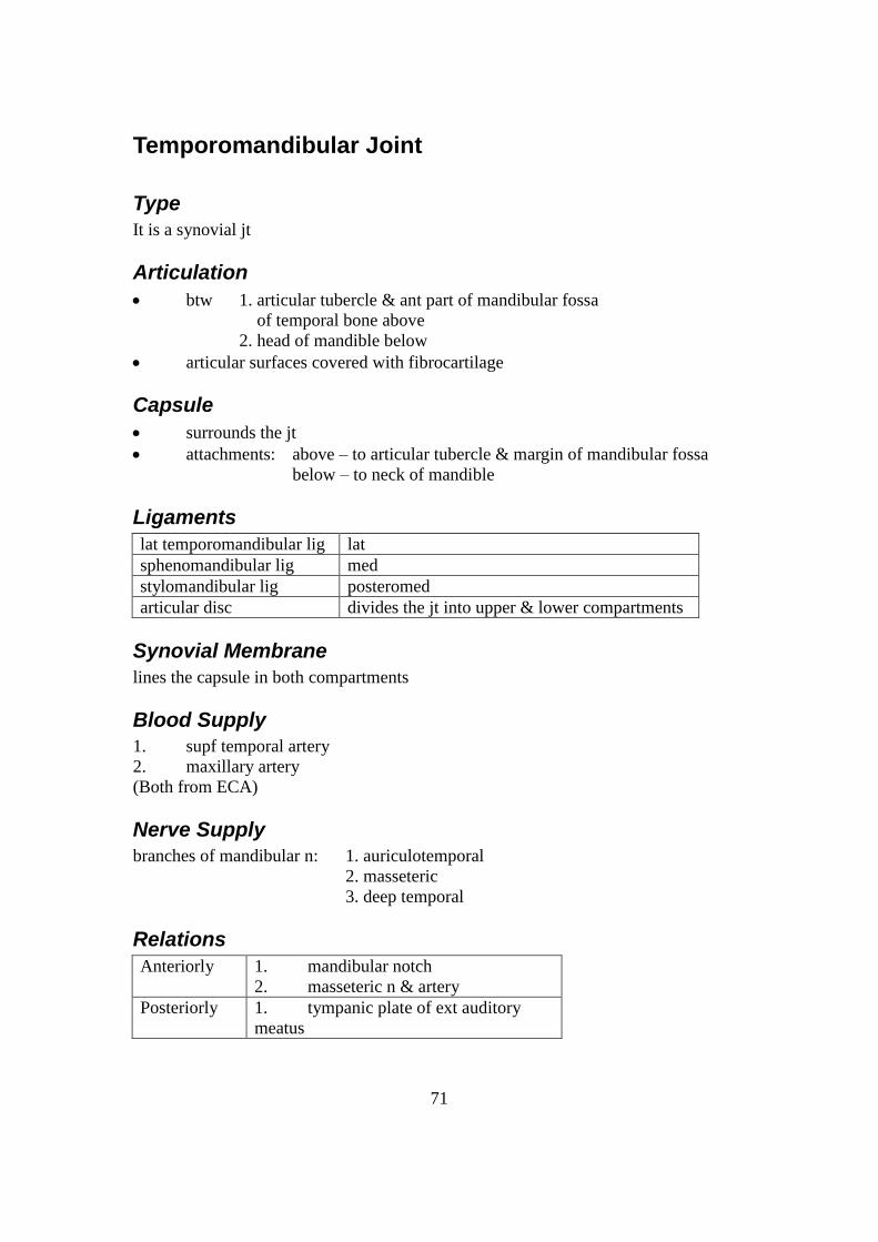
71
Temporomandibular Joint
Type
It is a synovial jt
Articulation
btw 1. articular tubercle & ant part of mandibular fossa
of temporal bone above
2. head of mandible below
articular surfaces covered with fibrocartilage
Capsule
surrounds the jt
attachments: above – to articular tubercle & margin of mandibular fossa
below – to neck of mandible
Ligaments
lat temporomandibular lig lat
sphenomandibular lig med
stylomandibular lig posteromed
articular disc divides the jt into upper & lower compartments
Synovial Membrane
lines the capsule in both compartments
Blood Supply
1. supf temporal artery
2. maxillary artery
(Both from ECA)
Nerve Supply
branches of mandibular n: 1. auriculotemporal
2. masseteric
3. deep temporal
Relations
Anteriorly 1. mandibular notch
2. masseteric n & artery
Posteriorly 1. tympanic plate of ext auditory
meatus

72
2. glenoid process of parotid gld
Medially 1. maxillary art & vein
2. auriculotemporal n
Laterally 1. parotid gld
2. fascia & skin

73
Movements
In the position of rest, the upper & lower teeth are separated by a slight interval
The movements are
Movements Muscles involved
1 depression of mandible
(opening of mouth)
1. lat pterygoids
2. digastrics
3. geniohyoids & mylohyoids
2 elevation of mandible
(closure of mouth)
1. masseter
2. temporalis
3. med pterygoids
3 Protusion of mandible 1. lat pterygoids
2. med pterygoids
4 Retraction of mandible 1. post fibres of temporalis
5 Lateral movements
(side-to-side chewing movements)
med & lat pterygoids of each side acting
alternatively
Clinical Notes
1. Dislocation of mandible
2. detachment of articular disc clicking sounds when the jaw is moved
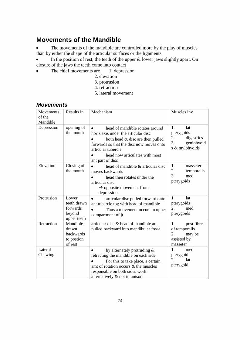
74
Movements of the Mandible The movements of the mandible are controlled more by the play of muscles
than by either the shape of the articular surfaces or the ligaments
In the position of rest, the teeth of the upper & lower jaws slightly apart. On
closure of the jaws the teeth come into contact
The chief movements are 1. depression
2. elevation
3. protrusion
4. retraction
5. lateral movement
Movements
Movements
of the
Mandible
Results in Mechanism Muscles inv
Depression opening of
the mouth head of mandible rotates around
horiz axis under the articular disc
both head & disc are then pulled
forwards so that the disc now moves onto
articular tubercle
head now articulates with most
ant part of disc
1. lat
pterygoids
2. digastrics
3. geniohyoid
s & mylohyoids
Elevation Closing of
the mouth head of mandible & articular disc
moves backwards
head then rotates under the
articular disc
opposite movement from
depression
1. masseter
2. temporalis
3. med
pterygoids
Protrusion Lower
teeth drawn
forwards
beyond
upper teeth
articular disc pulled forward onto
ant tubercle tog with head of mandible
Thus a movement occurs in upper
compartment of jt
1. lat
pterygoids
2. med
pterygoids
Retraction Mandible
drawn
backwards
to postion
of rest
articular disc & head of mandible are
pulled backward into mandibular fossa
1. post fibres
of temporalis
2. may be
assisted by
masseter
Lateral
Chewing
by alternately protruding &
retracting the mandible on each side
For this to take place, a certain
amt of rotation occurs & the muscles
responsible on both sides work
alternatively & not in unison
1. med
pterygoid
2. lat
pterygoid

75
Clinical Notes
Dislocation of the mandible sometimes occurs when the mandible is depressed
The head of the mandible on 1 or both sides slips anteriorly into infratemporal fossa
inability to close mouth

76
Maxillary Artery The maxillary artery has an extensive distribution to the upper & lower jaws, muscles
of mastication, the palate & the nose
Origin
It is the larger terminal branch of ECA
arises in parotid gld bhd neck of mandible
Course & Relations
For descriptive purposes the artery is divided into 3 parts by the lat pterygoid
Mandibular (1st) Part from its origin it runs forward
lies btw neck of mandible & sphenomandibular
lig
reaches & runs along lower border of lat
pterygoid
Pterygoid (2nd
) Part from lower border of pterygoid,
it runs upwards & forwards
either supf or deep to lower head of lat pterygoid
Pterygopalatine (3rd
) Part passes btw the 2 heads of lat pterygoid
then through pterygomaxillary fissure
to enter the pterygopalatine fossa
breaks up into branches, of which the
sphenopalatine art is considered the terminal branch
Branches
From 1st Part gives off 5 branches of which the most impt is
middle meningeal art & inf alveolar art
From 2nd
Part branches supply the muscles of mastication as well as the buccinator
From 3rd
Part the more impt are:
1. infraorbital art
2. greater palatine
3. pharyngeal
4. spenopalatine
Clinical Notes
1. middle meningeal artery inv in intracranial hemorrhages
2. sphenopalatine artery inv in nosebleeds

77
Maxillary Nerve The maxillary nerve is the 2
nd division of the trigeminal n
Origin
arises from trigeminal ganglion
lies in dura lat to cavernous sinus
Course & Relations
from origin, it passes through foramen rotundum
enters pterygopalatine fossa
continues as infraorbital n
enters orbit through inf orbital fissure
runs in infraorbital groove then canal
passes through infraorbital foramen
emerges onto the face where it ends by dividing into several branches
Branches
1 meningeal branch in middle cranial fossa
2
3
post sup alveolar branches
zygomatic n
in pterygopalatine fossa
4 middle & ant sup alveolar branch in infraorbital canal
5
6
7
inf palpebral branches
nasal branches
sup labial branches
on face
Clinical Notes
In the pterygopalatine fossa, the nerve can be blocked by passing a needle through the
mandibular notch & injecting a local anesthetic

78
Mandibular Nerve The mandibular n is the 3
rd & largest of the 3 division of the trigeminal n
It has both sensory & motor fibres
Origin
begins in the middle cranial fossa as a large sensory root & a small motor root
sensory root arises from trigeminal ganglion
motor root arises from motor nucleus of V in pons
Course & Relations
sensory root lies above motor root & both pass out of the skull through
foramen ovale to enter infratemporal fossa
the 2 roots unite just below the foramen to form a single main trunk
- lies on tensor palatini, deep to lat pterygoid
the main trunk soon divides into a small ant & large post division
Branches
1. From Main Trunk
1. meningeal branch
2. n to med pterygoid
From Sensory Motor
2. Anterior Trunk / Division buccal n 1. masseteric n
2. deep temporal n
3. n to lat pterygoid
3. Posterior Trunk / Division 1. auriculotemporal n
2. lingual n
inf alveolar n
Clinical Notes
1. mandibular n block carried out intra-orally for extraction of teeth
2. pain from disease of the tooth sometimes gets referred along the
auriculotemporal n to the ear

79
Pterygopalatine Ganglion This is a larger psymp peripheral ganglion
It serves as a relay station for the secretomotor fibres to the
lacrimal gld & mucous glds of the nose, paranasal sinuses, palate & pharynx
Location
lies in pterygopalatine fossa - below maxillary n in front of pterygoid canal
close to sphenopalatine foramen
Connections (Roots)
a) Motor (psymp) root = secretomotor
formed by 1. greater petrosal n
2. n of pterygoid canal
PreGN fibres arise from lacrimal nucleus
pass through nervous intermedius of facial n
synapse in the ganglion
PGN fibres pass to lacrimal gld via maxillary, zygomatic &
lacrimal n
also pass to nasal & palatine glds
a) Symp root = vasomotor
- formed by 1. deep petrosal n
2. n of pterygoid canal
- fibres derived from ICA plexus & they are PGN fibres having come from sup
cervical ganglion
- these fibres pass through ganglion without synapsing
- they are distributed with the psymp fibres
a) Sensory root
- from maxillary n
- pass through the ganglion without synapsing
Branches
These are branches of the maxillary n which also incorporate psymp & symp fibres
1. orbital branches
2. palatine branches (greater & lesser)
3. nasal branches
4. pharyngeal branches
Clinical Notes
The ganglion can be injected through the mandibular notch & pterygopalatine fossa

80
Submandibular Gland The submandibular gld is a large salivary gld and has a mixture of serous and mucous
acini.
Location
it lies in the ant part of digastric triangle
partly under cover of body of mandible
Shape & Parts of the Gland
roughly J-shaped
divided into 1. large supf part (supf to mylohyoid)
2. small deep part (deep to mylohyoid)
The 2 parts are continuous with each other around post border of mylohyoid
Capsules
2 capsules cover the gld:
1. int fibrous capsule
2. ext capsule derived from investing layer of deep cervical fascia
Part of fascia extends btw styloid process & angle of mandible
stylomandibular lig
Superficial Part of Gland
lies in digastric triangle, partly under cover of mandible
posteriorly it is separated from parotid gld by stylomandibular lig
Relations
anteriorly ant belly of digastric
posteriorly 1. stylohyoid
2. post belly of digastric
3. stylomandibular lig, which sep it from
4. parotid gld
medially 1. mylohyoid
2. mylohyoid n & vsls
3. hyoglossus
4. lingual & hypoglossal n
laterally 1. submandibular fossa on med surface of
mandible
2. cervical branch of facial n
3. facial vein
4. submandibular lymph nodes
5. fascia, platysma & skin inferolaterally

81
Deep Part of the Gland extends forward to interval btw: mylohyoid (below & lat)
& hyoglossus & styloglossus (med)
post end is continuous with supf part of gld around post border of mylohyoid
muscle
ant end reaches as far as sublingual gld
Relations
anteriorly sublingual gld
posteriorly 1. stylohyoid
2. post belly of digastric
3. stylomandibular lig
4. parotid gld
superiorly 1. lingual n
2. submandibular ganglion
3. It is covered by mucous mbm of floor of the
mouth
inferiorly hypoglossal n
medially hyoglossus & styloglossus
laterally 1. mylohyoid
2. supf part of the gld
Submandibular Duct emerges from ant end of deep part of gld
passes forward along side of the tongue, beneath mucous mbm of floor of
mouth
opens into the mouth on the summit of a small papilla situated at the sides of
the frenulum of the tongue
Blood Supply branches of 1. facial art &
2. lingual art
Venous Drainage 1. facial vein
2. lingual vein
Lymphatic Drainage submandibular nodes deep cervical nodes
Nerve Supply Psymp secretomotor
fibres from sup salivatory nucleus of 7th cranial n PreGN
pass via chorda tympani lingual n synapse in submandibular

82
ganglion
PGN fibres reach gld directly from the ganglion
Symp from plexus on facial art
Sensory from lingual n
Clinical Notes 1. common site for formation of calculus (stones)
2. excision of gld

83
Sublingual Gland
This is the smallest of the 3 main salivary glands
It contains both serous & mucous acini, with mucous predominating
Location & Shape
lies beneath mucous mbm of floor of mouth
close to midlin
roughly almond-shaped
Relations
Anteriorly gld on opposite side
Posteriorly deep part of submandibular gld
Medially 1. genioglossus
2. lingual n
3. submandibular duct
Laterally sublingual fossa on med surface of mandible
Superiorly mucous mbm of floor of the mouth
elevated to form sublingual fold
Inferiorly mylohyoid muscle
Sublingual Ducts
8-20 in number
majority open into mouth on summit of sublingual fold
some may open into submandibular duct
Blood Supply
branches of 1. facial art
2. lingual art
Venous Drainage
1. facial vein
2. lingual vein
Lymphatic Drainage
submandibular deep cervical nodes
Nerve Supply
Psymp PreGN fibres from sup salivary nucleus of 7th
cranial n
chorda tympani lingual n submandibular ganglion
PGN fibres lingual n supply the gld
Symp from plexus around facial & lingual arteries

84
Sensory from lingual n
Clinical Notes
1. infection & enlargement of gld
2. blockage of one of the sublingual ducts cysts

85
Lingual Nerve The lingual n is sensory to the ant 2/3 of the tongue & fibre of the mouth
In addition it also receives fibres from the chorda tympani branch of the facial
n
1. secretomotor (psymp) to submandibular & sublingual salivary glds
2. taste fibres to ant 2/3 of tongue
Origin
1 of the 2 terminal branches of post dividision of mandibular n
begins slightly below skull (stylomandibular lig)
Course & Relations
runs downward on med surface of lat pterygoid
then on lat surface of med pterygoid
joined by chorda tympani
emerges at lower border of lat pterygoid & continues downwards btw ramus of
mandible & med pterygoid
next it passes forward & medially beneath lower border of sup constrictor of
pharynx
- medially it is related to styloglossus
laterally it is related to lower 3rd
molar tooth
passes forward on lat surface of hyoglossus
at ant margin of hyoglossus, it winds round submandibular duct from lat to
med & lies on genioglossus
divides into terminal branches
Branches
ganglionic
branches PreGN psymp fibres passing to submandibular ganglion
These fibres arise from sup salivatory nucleus of facial n
travel via chorda tympani lingual n
sensory branches to ant 2/3 of the tongue, floor of mouth & lingual surface of gums
communicating
branches
to hypoglossal n
Clinical Notes
Close relation of lingual n to 3rd
molar tooth
may be damaged during tooth extraction

86
Nasal Cavity: Relations, Openings & Branches The nasal cavity extends from the nostrils (nares) in front to the choanae bhd
It is divided into 2 halves by the nasal septum
Relations
Superiorly 1. frontal sinus
2. ant cranial fossa
3. sphenoidal sinus
4. middle cranial fossa
Inferiorly 1. hard palate, which sep it from
2. oral cavity
Posteriorly communicates with nasopharynx which
can be regarded as the back of the cavity
Laterally 1. exterior (front part of cavity)
2. orbit
3. maxillary & ethmoid sinuses
4. pterygopalatine & pterygoid
fossae
Openings
The openings that lead into or out of the nasal cavity are:
1. nostrils
2. choanae
3. openings of maxillary, frontal, sphenoidal & ethmoidal air sinuses, &
4. opening of nasolacrimal duct
Nostrils ant aperturesof the nose
perforate ext nose to communicate with exterior
bounded medially by nasal septum
laterally by ala of the nose
Choanae post aperture of the nose, larger than the nostrils
communicate with nasopharynx
bounded superiorly by body of sphenoid
inferiorly by horiz plate of palatine bone
medially by vomer &
laterally by med pterygoid plate
Openings of Paranasal Air
Sinuses
1. maxillary sinus opens into middle meatus of nasal cavity through
hiatus semilunaris
2. frontal sinus opens into middle meatus via infundibulum
3. sphenoidal sinus opens into sphenoethmoidal recess

87
4. ethmoidal sinuses ant & middle gps open into middle meatus
post gp opens into sup meatus
Opening of Nasolacrimal
Duct opens into inf meatus of nasal cavity
opening is guarded by a fold of mucous mbm
= lacrimal fold

88
Boundaries
each half of the nasal cavity has a roof, floor, med & lat walls
Formation Remark
Roof 1. nasal cartilages
2. nasal bone
3. frontal bone
4. cribiform plate of ethmoid
bone
5. body of sphenoid
( from ant to post)
the roof is narrow & slopes
downwards
Floor 1. palatine process of maxilla
(ant)
2. horiz plate of palatine bone
(post)
upper surface of hard palate
separates nasal cavity from oral
cavity
Medial
Wall
1. septal cartilage
2. perpendicular plate of ethmoid
3. vomer ( from ant to post)
consists of nasal septum
usu deviated to one side
Lateral
Wall
1. nasal bone
2. maxilla bone
3. lacrimal bone
4. ethmoid (labryrinth &
concave)
5. inf nasal concha
6. perpendicular plate of palatine
7. med pterygoid plate sphenoid
Blood Supply
maxillary art sphenopalatine art
ophthalmic art ant ethmoidal art
Venous Drainage
Veins follow the arteries
Lymphatic Drainage
deep cervical nodes
Nerve Supply
1. Psymp from pterygopalatine ganglion
2. Symp from sup cervical ganglion

89
Features
1. 3 projections
sup choncha part of ethmoid bone
middle choncha part of ethmoid bone
inf choncha sep bone by itself
2. area below each choncha is called meatus
sup meatus lies below & lat to sup choncha
receives opening of post gp of ethmoidal sinuses
middle
meatus lies below & lat to middle choncha
ethmoidal bulla = opening of middle ethmoidal sinuses
hiatus semilunaris = opening of maxillary sinus
ethmoidal infundibulum = opening of frontal sinus &
ant ethmoidal sinuses
inf meatus lies below & lat to inf choncha
receives openings of nasolacrimal duct, guarded by an
imperfect valve, the lacrimal fold
Clinical Notes
1. fracture of nasal bones & nasal septum due to frontal blows
2. spread of infection from nasal cavity into intracranial regions
3. epistaxis / nose bleed
- inv sphenopalatine & facial vsls

90
Tongue: Features & Muscles The tongue is a muscular organ in the floor of the mouth
It is attached by muscles to the hyoid bone, mandible, stylod processes &
pharynx
It is impt in taste, mastication, swallowing & speech
Surface Features
The tongue is composed chiefly of skeletal muscle & covered by mucous mbm
Its ant 2/3 lies in the mouth & post 2/3 in the pharynx
It presents 1. tip & margin
2. dorsum
3. inf surface
4. root
a) Tip & Margin
- The tip / apex of the tongue usu rests against the incisor teeth
- The margin of the tongue is related on each side to the gums & the teeth
b) Dorsum
- situated partly in oral cavity & partly in oropharynx
- This is demarcated by a V-shaped groove = sulcus terminalis
which divides it into ant 2/3 in oral cavity
post 1/3 in oropharynx
- apex of sulcus terminalis projects backwards
& is marked by a small pit = foramen cecum
Oral Part of Tongue 1. divided into L & R halves by
shallow median groove
2. exhibit lingual papillae of the 3 types
a. filiform papillae
b. fungiform papillae
c. vallate papillae
Pharyngeal Part of Tongue
= ant wall of pharynx devoid
of papillae
contains lymphatic nodules = lingual tonsils
a) Inferior Surface
- smooth & devoid of papillae
- connected to floor of mouth by a fold of mucous mbm = frenulum of tongue
- deep lingual vein can be seen beside frenulum
- lat to vein mucous mbm forms a fold = plica fimbriatia
b) Root
- rests on floor of mouth, ie. geniohyoid & mylohyoid
- attached by muscles to mandible & hyoid bone
- nerves, vsls & extrinsic muscles enter or leave the tongue through its root

91
Muscles of the Tongue
These muscles of the tongue are of 2 types 1. intrinsic
2. extrinsic
All the muscles are bilateral, those on 1 side being partially separated from
those of the opposite side by a median septum
Intrinsic
Muscles These muscles are confined to the tongue itself & are not
attached to bone
They are arranged in several planes & are generally
classified as
1. sup & inf longitudinal
2. tnvs
3. vertical
Their actions are to alter the shape of the tongue
Extrinsic
Muscles These muscles that arise from nearby parts & are inserted
into the tongue
They are: 1. genioglossus
2. hyoglossus
3. styloglossus
4. palatoglossus
Their actions are to move the tongue
Movements of the Tongue
1 protrusion brought abt by genioglossus of both sides acting tog
2 retraction produced by styloglossus & hyoglossus of both sides acting tog
3 depression produced by hyoglossus & genioglossus of both sides acting tog
The shape of the tongue is modified by the action of the intrinsic muscles
Blood Supply
1. lingual art
2. tonsillar branch of facial art
3. asc pharyngeal art
Lymphatic Drainage
tip submental nodes
ant 2/3 submandibular deep cervical nodes
post 1/3 deep cervical nodes
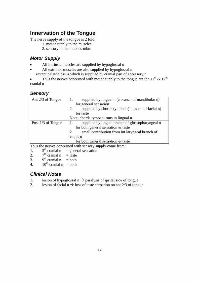
92
Innervation of the Tongue The nerve supply of the tongue is 2 fold:
1. motor supply to the muscles
2. sensory to the mucous mbm
Motor Supply
All intrinsic muscles are supplied by hypoglossal n
All extrinsic muscles are also supplied by hypoglossal n
except palatoglossus which is supplied by cranial part of accessory n
Thus the nerves concerned with motor supply to the tongue are the 11th
& 12th
cranial n
Sensory
Ant 2/3 of Tongue 1. supplied by lingual n (a branch of mandibular n)
for general sensation
2. supplied by chorda tympani (a branch of facial n)
for taste
Note: chorda tympani runs in lingual n
Post 1/3 of Tongue 1. supplied by lingual branch of glossopharyngeal n
for both general sensation & taste
2. small contribution from int laryngeal branch of
vagus n
for both general sensation & taste
Thus the nerves concerned with sensory supply come from:
1. 5th
cranial n = general sensation
2. 7th
cranial n = taste
3. 9th
cranial n = both
4. 10th
cranial n = both
Clinical Notes
1. lesion of hypoglossal n paralysis of ipsilat side of tongue
2. lesion of facial n loss of taste sensation on ant 2/3 of tongue

93
Constrictor Muscles of Pharynx The wall of the pharynx is composed largely of 2 layers of muscles
The internal, chiefly longitudinal layer consists of 2 levator muscles:
1. palatopharyngeus
2. stylopharyngeus
The outer circular layer consists of 3 constrictors
1. sup constrictors
2. middle constrictors
3. inf constrictors
The constrictors have their fixed pts in front,
where they are attached to bones & cartilage,
whereas they expand bhd & overlap one another from below upwards.
They end in a median tendinous raphe posteriorly
The Constrictor Muscles Superior Constrictor Middle Constrictor Inferior Constrictor
Origin 1. post border of med
pterygoid plate
2. pterygoid hamulus
3. post end of mylohyoid
line on mandible
4. side of tongue
1. lower part of
stylohyoid lig
2. lesser &
greater cornus of
hyoid bone
1. oblique line on lamina
on thyroid cartilage
2. tendinous band
connecting thyroid cartilage to
cricoid cartilage
3. side of cricoid
cartilage
Insertion 1. upper fibre curve med
& upwards
pharyngeal tubercle of
occipital bone
2. middle fibres
median tendinous raphe 3. lower fibres curve med
& downward median
tendinous raphe They are overlapped by
middle constrictors
into median
tendinous raphe
Sup fibres
overlap lat surface of
sup constrictor
inf fibres are
overlapped laterally
by inf constrictor
Into median tendinous
raphe
Sup fibres overlap lat
surface of middle constrictor
muscle
Inf fibres run
horizontally med & are
continuous with circular fibres
of esophagus below
Nerve
Supply
pharyngeal plexus pharyngeal plexus pharyngeal plexus
Action of Constrictor Muscles During process of swallowing,
1. upper fibres of sup constrictor contract & pull the post pharyngeal wall
forward
aids palate in closing off upper part of pharynx (nasopharygnx)
2. successive contraction of the sup, middle & inf constrictor muscles propels the
bolus of food down into the esophagus
3. the lowest fibres of inf constrictor (sometimes called cricopharyngeus muscle)
exert a sphincteric effect on lower end of pharynx
prevent entry of air into esophagus btw the acts of swallowing.

94
Clinical Notes The pharyngeal plexus which supplies the muscles is made up from fibres
travelling in the vagus n (pharyngeal branch)
However, these fibres originate from cranial part of accessory n
Lesion of vagus n paralysis of constrictors
Thus the patient may have difficulty swallowing

95
Nasopharynx The phaynx extends from base of skull down to lower border of cricoid
cartilage
(opposite CV6), where it becomes continuous with the esophagus
It can be divided into 3 parts
nasophaynx bhd nasal cavity
oropharynx bhd oral cavity
laryngopharynx bhd larynx
Nasopharynx: Introduction
lies bhd the nasal cavities, above the soft palate
Thus sometimes regarded as back part of nasal cavity
when swallowing, soft palate is raised & post pharyngeal wall drawn forward
nasophaynx is shut off from oropharynx
nasopharynx has: roof & floor
ant & post walls
lat walls
Roof
supported by 1. body of sphenoid
2. basilar part of occipital bone
submucosa of this region contains a collection of lymphoid tissue
= pharyngeal tonsil
Floor
formed by sloping upper surface of soft palate
opening = pharyngeal isthmus
- btw free edges of soft palate & post pharyngeal wall
- enables nasopharynx to communicate with oropharynx
- during swallowing, this opening is closed by raising the soft palate
& pulling forward the post pharyngeal wall
Anterior Wall
formed by post nasal apertures (choanae) separated by post edge of nasal
septum
communicated with nasal cavity anteriorly through the choanae
Posterior Wall
forms a continuous sloping surface with the roof
supported by ant arch of atlas

96
Lateral Wall
opening of auditory tube
tubal elevation post margin of auditory tube forms an elevation
salpingopharyngeal fold salpingopharyngeus muscle (attached to lower margin
of tube)
forms a vertical fold of mucous mbm
pharyngeal recess bhd tubal elevation is a small depression
tubal tonsil bhd opening of auditory tube is a collection of
lymphoid tissue in the submucosa
Blood Supply
asc pharyngeal art (from ECA)
Venous Drainage
venous plexuses below mucosa
Lymphatic Drainage
deep cervical nodes
Nerve Supply
pharyngeal plexus
Clinical Notes
1. infection hypertrophy of pharyngeal tonsil enlarged
This is known as adenoids & is removed by adenoidectomy
2. close relationship of infected tonsil to auditory tube may cause deafness
3. nasopharynx may be examined by a mirror passed through the mouth

97
Mechanism of Swallowing After food enters the mouth, it is usually broken dwon by the grinding of the
teeth & is mixed with saliva
The thoroughly mixed food is now formed into a bolus on the dorsum of the
tongue & the process of swallowing begins
This process has a voluntary & involuntary phase
Voluntary Phase
ant part of tongue is raised & pressed against hard palate by intrinsic muscles
of the tongue
pushes bolus of food into post part of oral cavity
This is followed by styloglossus muscles contracting
pulls root of tongue upwards & backwards
palatoglossus also contract
squeeze bolus of food backward into oropharynx
Involuntary Phase
elevation of soft palate
pulling forward of post pharyngeal wall by sup constrictors
& contraction of palatopharyngeus
closure of nasopharyngeal isthmus
ie. nasopharynx closed off from oropharynx
larynx & laryngeal part of pharynx now elevated
larynx is closed to prevent entry of food bolus
the bolus moves downward over the epiglottis & closed entrance of larynx
reaches lower part of pharynx as result of successive contraction of sup,
middle & inf constrictor muscles & by gravity
finally, the lower fibres of inf constrictor muscle (cricopharyngeus muscle)
relax bolus enters the esophagus
Clinical Notes
difficulty in swallowing = dysphagia
- can be caused by paralysis of the constrictors muscles due to lesion of vagus n

98
Auditory Tube This is a trumpet-shaped chaneel which connects the nasopharynx with the
middle ear cavity
Its function is to maintain atm pressure in the middle ear cavity so that the air
pressures on both sides of the tympanic mbm are equal
Length & Direction
3-4 cm long
extends backward, laterally & upwards from nasopharynx
Parts
post 1/3 = bony
ant 2/3 = cartilaginous
Cartilaginous Part abt 2-3 cm long, attached to med end of bony part
lies in a groove btw greater wing of sphenoid & apex of
petrous temporal
sup & med wall = cartilage
lat & inf walls = fibrous mbm
relations
a. anterolat: 1. tensor veli palatini
2. mandibular n
3. middle meningeal art
b. posteromed: 1. levator veli palatini
2. pharyngeal recess
Bony Part abt 1 cm long
lies in petrous temporal bone, near tympani plate
lat end is wider opens into middle ear cavity
med end is narrow (isthmus) attach to cartilaginous
part
relations
a. superiorly 1. canal for tensor tympani
b. anterolat 1. tympanic portion of temporal bone
c. posteromed 1. carotid canal
Mucosal Lining
in cartilaginous part mainly pseudostr columnar ciliated
epith
contains many mucous glds
in bony part mainly flattened cuboidal non-ciliated epith
= mucoperiosteum

99
Blood Supply
from: 1. asc pharyngeal
2. middle meningeal
3. art of pterygoid canal
100
Venous Drainage
into: 1. pharyngeal vein
2. pterygoid plexuses of veins
Lymphatic Drainage
into retropharyngeal nodes deep cervical nodes
Nerve Supply
cartilaginous part by branch from mandibular n
bony part by tympanic plexus (9th
n)
Also by pharyngeal branch of pterygopalatine ganglion
Clinical Notes
1. cold / sore throat may lead to inflammation of auditory tube
2. throat infections may be transmitted via the tube to middle ear cavity

101
Middle Ear Cavity The middle ear consists largely of an air space
It is also called the tympanic cavity
Location
situated in petrous part of temporal bone
btw ext & int ears
Communications
with nasopharynx anteriorly via auditory tube
with mastoid antrum & air cells posteriorly via the aditus to the antrum
Parts
It consists of
1. tympanic cavity proper which lies opposite the tympanic mbm
2. epitympani recess which lies above the level of the tympanic mbm
It contains upper part of malleus & greater part of incus
Boundaries
The middle ear has 6 sides Roof
(Tegmental Wall) formed by a portion of petrous temporal called tegmen tympani
sep middle ear from middle cranial fossa
Floor
(Jugular Wall) formed by jugular fossa of temporal bone
sep middle ear from sup bulb of IJV
Anterior
(Carotid) Wall sep middle ear from ICA & symp plexus
consists of 3 parts 1. opening of canal for tensor tympani
sup
2. opening of auditory tube in the middle &
3. post wall of carotid canal inf
Posterior
(Mastoid) Wall upper part has large opening = aditus to the mastoid antrum
below the aditus is a small projection = pyramid
contains stapedius muscle Laterall Wall
(Membranous) sep middle ear from ext acoustic meatus
formed by tympani mbm below
squamous part of temporal bone above
above level of tympanic mbm is the epitympanic recess
Medial Wall
(Labyrinthine) sep middle ear from int ear
this wall presents the following features from above downwards:
1. prominence of lat semicircular canal
& prominence of facial n canal
2. oval window (fenestra vestibule) = closed by base of stapes
& processus cochleariformis (a bony prominence above the
102
window)
3. promontory
- formed by basal turn of cochlea & covered by tympanic plexus
4. fenestra cochleae (round window)
- closed by mucous mbm of middle ear

103
Contents Contents Remarks
1 3 ear ossicles malleus
incus
stapes
2 ligs of ear ossicles
3 2 muscles tensor tympani
stapedius
4 vsls supplying & draining middle ear
5 nerves chorda tympani
tympanic plexus
6 air
The middle ear is lined by mucous mbm
Blood Supply 1. stylomastoid artery (from ECA)
2. ant tympanic art (from maxillary artery)
3. many smaller vsls contribute
Venous Drainage 1. sup petrosal sinus
2. pterygoid plexus of veins
Lymphatic Drainage preauricular & retropharyngeal nods deep cervical nodes
Nerve Supply from tympanic plexus which is formed by
1. auriculotemporal n (5th
cranial n)
2. tympanic n(9th
cranial)
3. auricular branch (10th
cranial n)
Note: Also by nerves from symp plexus around ECA
Clinical Notes 1. otoscopic examination of tympanic mbm
2. infection of nasopharynx may spread via auditory tube to middle ear
3. infection of middle ear is called ottis media
This can spread to all its surrounding relations to cause various symptoms
Functional Considerations 1. Sound Sound waves in middle ear cavity tympanic mbm vibrates
ossicles vibrate stapes cause vibration of fenestra vestibuli
2. fenestra cochleae vibrates in opposite direction

104
Thus, sound vibrations are transmitted into inner ear by:
1. auditory ossicles & fenestra vestibuli
2. air in tympanic cavity & fenestra cochleae
3. bone conduction
Disease = otosclerosis of med wall

105
Pterygopalatine Fossa small pyramidal space bhd & below orbital cavity
Boundaries
ant post surface of maxilla
post greater win of sphenoid & root of pterygoid process
med upper part of perpendicular plate with orbital & sphenoidal processes
with palatine bone
lat open (pterygomaxillary fissure)
sup body of sphenoid
inf pyramidal process & palatine bone
Communications
relations communicates with through
ant orbit inf orbital fissure
post 1. middle cranial
fossa
2. pterygoid canal
3. pharynx
foramen rotundum
foramen lacerum
palatovaginal canal
med nose sphenopalatine foramen
lat infratemporal fossa pterygomaxillary fissure
inf oral cavity a. greater palatine canals
b. lesser palatine canals
Contents
1. Maxillary art – 3rd
part & branch
2. Maxillary nerve & 2 branches (zygomatic, post alveolar)
3. Pterygopalatine ganglion & branches containing from which maxillary n
mixed with autonomic n

106
Cutaneous Innervation of Face skin of face supplied by branches of the 3 divisions of the trigeminal n
except for the small area over the angle of mandible and the parotid gld
which is supplied by the great auricular n
The overlap of the 3 divisions of the trigeminal n is slight in comparision to
the considerable overlap of adj dermatomes of the trunk and limbs
Embryological Development
ophthalmic n region dev from the frontonasal process
maxillary n region dev from the maxillary process of the
1st pharyngeal arch
mandibular n region dev from the mandibular process of the
1st pharyngeal arch
Ophthalmic Nerve
supplies the skin of the forehead, upper eyelid, conjunctiva and side of the
nose down to and including the tip
Branches Course Supply
lacrimal n 1. skin
2. conjunctiva of lat upper eyelid
supraorbital n winds around the upper
margin of the orbit at the
supraorbital notch
1. skin
2. conjunctiva on the central part
of upper eyelid
3. skin of the forehead
supratrochlear n winds around the upper
margin of the orbit
medial to the supraorbital
n
1. skin
2. conjunctiva on the med part of
the upper eyelid
3. skin over the lower part of
forehead close to the median plane
infratrochlear n leaves orbit below the
pulley of the sup oblique
musc
1. skin
2. conjunctiva on med part of the
upper eyelid
3. adjoining part of the side of the
nose
ext nasal n leaves nose by emergeing
btw nasal bone and upper
nasal cartilage
skin on side of nose down to the tip

107
Maxillary Nerve
supplies the skin on the post part of the side of the nose, the lower eyelid, the
cheek, the upper lips, and the lat side of the orbital opening
Branches Course Supply
infraorbital n direct continuation of
maxillary n
enters orbit and appears on the
face through the infraorbital foramen
1. skin of lower
eyelid and cheek
2. skin of side of
nose
3. skin of upper lip
zygomatico-
facial n
passes onto the face through a small
foramen on the lat side of the
zygomatic bone
skin over the prominence
of cheek
zygomatico-
temporal n
emerges in the temporal fossa through
a small foramen on the post surface of
zygomatic bone
skin over the temple
Mandibular Nerve
supplies skin of the lower lip, the lower part of the face, the temporal region
and part of the auricle
then passes upwards to the side of the scalp
Branches Course Supply
mental n emerges from the mental
foramen of the mandible
skin of the lower lip and
chin
buccal n emerges from beneath the ant
border of the masseter musc
skin over a small area of the
cheek
auriculotemporal n asc from the upper border of the
parotid gld btw the supf
temporal vsls and the auricle
1. skin of the auricle
2. ext auditory meatus
3. outer surface of the
tympanic mbm
4. skin of the scalp
above the auricle
108
Glossopharyngeal Nerve (9th
Cranial Nerve) motor and sensory nerve
motor fibres stylopharyngeus
psymp secretomotor fibres parotid salivary gld
sensory fibres (including taste fibres) post 1/3 of tongue & pharynx
Course
emerges from ant surface of upper MO
by 3 or 4 rootlets along the groove btw olive & inf cbllar penduncle
passes forward & lat beneath cbllm in post cranial fossa
leaves skull by passing downward thru central part of jugular foramen
desc in the neck within the carotid sheath
winds forward around the stylopharyngeus musc
& passes btw the sup & middle constrictor musc
lingual branch of n enters the submandibular region
Branches
Branch Course Supply
tympanic br arises below jugular foramen
passes thry floor of tymp
cavity
grooves surface of
promontory
splits into branches tymp
plexus
lining of tymp cavity
gives off lesser
petrosal n
(with secretomotor fibres
for parotid gld)
carotid br carotid sinus & carotid body
musclular br stylopharyngeus musc
pharyngeal br unite on outer surface of
middle constrictor musc with
pharyngeal br of vagus &
pharyngeal br of symp trunk
pharyngeal plexus
sensory fibres to
1. mucous mbm of
pharynx
2. tonsil
3. soft palate
lingual br enters tongue below styloglossus
musc
general sensory & special
taste fibres to
1. mucous mbm of post
1/3 of tongue
2. circumvallate papillae
region of ant tongue
109
Vagus Nerve (10th
Cranial Nerve) composed of both motor and sensory fibres
Origin
originates in MO
leaves the skull thru middle of the jugular foramen tog with 9th
& 11th
cn
Sensory Ganglia
Superior Ganglion rounded
on n within the jugular foramen
Inferior Ganglion cylindrical
lies on n just below foramen
below this ganglion, cranial part of acc n joins the vagus
n and is distributed mainly in its pharyngeal and recurrent
pharyngeal br
Course in the Neck
passes vertically down the neck within the carotid sheath
lies at 1st btw the IJV & ICA
and then btw IJV and CCA
At root of the neck accompanies the CCA
& lies ant to 1st part of the subclavian art
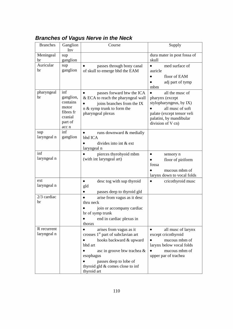
110
Branches of Vagus Nerve in the Neck
Branches Ganglion
Inv
Course Supply
Meningeal
br
sup
ganglion
dura mater in post fossa of
skull
Auricular
br
sup
ganglion passes through bony canal
of skull to emerge bhd the EAM
med surface of
auricle
floor of EAM
adj part of tymp
mbm
pharyngeal
br
inf
ganglion,
contains
motor
fibres fr
cranial
part of
acc n
passes forward btw the ICA
& ECA to reach the pharyngeal wall
joins branches from the IX
n & symp trunk to form the
pharyngeal plexus
all the musc of
pharynx (except
stylopharyngeus, by IX)
all musc of soft
palate (except tensor veli
palatini, by mandibular
division of V cn)
sup
laryngeal n
inf
ganglion runs downward & medially
bhd ICA
divides into int & ext
laryngeal n
inf
laryngeal n
pierces thyrohyoid mbm
(with int laryngeal art)
sensory n
floor of piriform
fossa
mucous mbm of
larynx down to vocal folds
ext
laryngeal n
desc tog with sup thyroid
gld
passes deep to thyroid gld
cricothyroid musc
2/3 cardiac
br
arise from vagus as it desc
thru neck
join or accompany cardiac
br of symp trunk
end in cardiac plexus in
thorax
R recurrent
laryngeal n
arises from vagus as it
crosses 1st part of subclavian art
hooks backward & upward
bhd art
asc in groove btw trachea &
esophagus
passes deep to lobe of
thyroid gld & comes close to inf
thyroid art
all musc of larynx
except cricothyroid
mucous mbm of
larynx below vocal folds
mucous mbm of
upper par of trachea

111
crosses either in front of or
bhd art or may pass btw its
branches
passes beneath lower
border of inf constrictor musc
L recurrent
laryngeal n
arises from vagus as it
crosses arch of aorta in thorax
hooks around beneath arch
bhd lig art
asc into neck in groove btw
trachea & esophagus

112
Accessory Nerve (11th
Cranial Nerve) composed of motor fibres
Origin
formed by union of cranial & spinal roots
cranial root is smaller & arises in the MO
spinal roots arise from C1-C5 segments of sp cord
Course in Neck
spinal roots unite to form a trunk that asc in vertebral canal to enter the skull
through foramen magnum
Both the cranial & spinal roots come tog & pass through the middle of jugular
foramen
Course of Cranial Root
sep from sp root & joins the vagus at inf ganglion
distributed mainly in the pharyngeal & recurrent laryngeal branches
Course of Spinal Root
runs downward & lat & crosses the IJV
then enters the deep surface of sternocleidomastoid, which it supplies
emerges above the middle of post border of sternocleidomastoid & crosses the
post triangle of neck on the lev scap to supply the trapezius musc
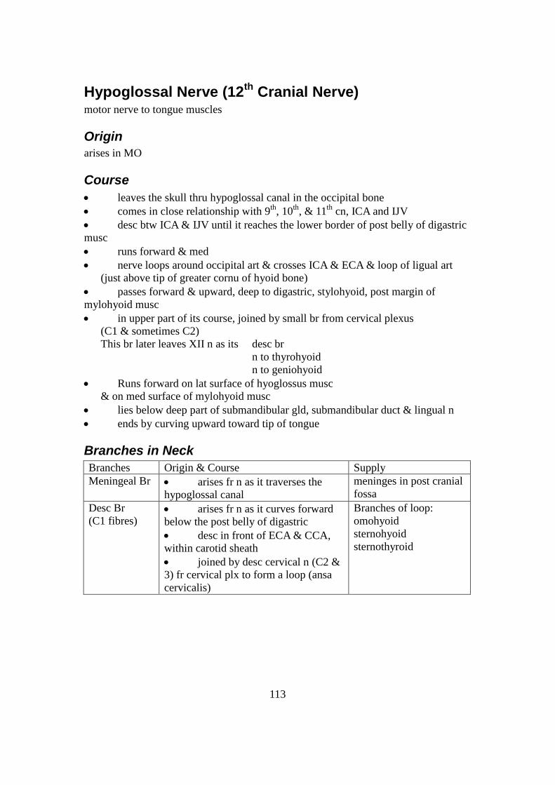
113
Hypoglossal Nerve (12th
Cranial Nerve) motor nerve to tongue muscles
Origin
arises in MO
Course
leaves the skull thru hypoglossal canal in the occipital bone
comes in close relationship with 9th
, 10th
, & 11th
cn, ICA and IJV
desc btw ICA & IJV until it reaches the lower border of post belly of digastric
musc
runs forward & med
nerve loops around occipital art & crosses ICA & ECA & loop of ligual art
(just above tip of greater cornu of hyoid bone)
passes forward & upward, deep to digastric, stylohyoid, post margin of
mylohyoid musc
in upper part of its course, joined by small br from cervical plexus
(C1 & sometimes C2)
This br later leaves XII n as its desc br
n to thyrohyoid
n to geniohyoid
Runs forward on lat surface of hyoglossus musc
& on med surface of mylohyoid musc
lies below deep part of submandibular gld, submandibular duct & lingual n
ends by curving upward toward tip of tongue
Branches in Neck
Branches Origin & Course Supply
Meningeal Br arises fr n as it traverses the
hypoglossal canal
meninges in post cranial
fossa
Desc Br
(C1 fibres) arises fr n as it curves forward
below the post belly of digastric
desc in front of ECA & CCA,
within carotid sheath
joined by desc cervical n (C2 &
3) fr cervical plx to form a loop (ansa
cervicalis)
Branches of loop:
omohyoid
sternohyoid
sternothyroid

114
Branches in Tongue
Branches Origin & Course Supply
n to thyrohyoid
(C1 fibres) arises fr n as it passes deep
to mylohyoid musc
thyrohyoid musc
n to geniohyoid
(C1 fibres) given off on side of tongue geniohyoid musc
Communicating br with lingual n on side of tongue
Also muscular branches to all musc of tongue
except palatoglossus (by pharyngeal plexus)
Thus supplies styloglossus
hyoglossus
genioglossus
intrinsic musc of tongue

115
Paranasal Sinuses cavities fd in int of maxilla, frontal, sphenoid & ethmoid bones
lined with mucoperiosteum, filled with air
communicate with nasal cavity thru small apertures
Mucous Membrane
have glds which produces mucus
mucus moved by ciliary action of columnar cells
drainage of mucus: by siphon action created by blowing of nose
Function
1. act as resonators to voice
2. reduce weight of skull
Thus when sinuses blocked, or filled with fluid, quality of voice changed
Maxillary Sinus
found within body of maxilla
pyramidal in shape
base forms lat wall of nose
apex in zygomatic process of maxilla
roof formed by floor of orbit
floor formed by alveolar process
Roots of 1st & 2
nd premolars & 3
rd molar & sometimes, root of canine, project
up into max sinus
opens into middle meatus of nose thru hiatus semilunaris
clinically: - opening of max sinus to middle meatus is high
sinus accumulates fluid
- frontal & ant eth sinuses drain into infundibulum, into hiatus
infection from these sinuses may spread to max sinus
nerve supply of mucous mbm: sup alveolar n
infraorbital n
Frontal Sinuses
contained within frontal bone
separated from each other by bony septum (freq deviated from median plane)
roughly triangular in shape - extend upward above med end of eyebrow
backward into med part of roof of orbit
opens into middle meatus of nose thru infundibulum
nerve supply of mucous mbm: supraorbital n

116
Sphenoidal Sinuses
lie within body of sphenoid bone
opens into spenoethmoidal recess above sup concha
nerve supply of mucous mbm: post ethmoidal n
Ethmoidal Sinuses
contained within ethmoid bone (btw nose & orbit)
sep from orbit by thin plate of bone
infection can spread from sinuses into orbit
divided into 3 groups: ant, middle, post
Sinus ant gp middle gp post gp
Opening infundibulum middle meatus sup meatus
nerve supply of mucous mbm: ant & post ethmoidal n

117
Oral Cavity
Extent
from lips to oropharyngeal isthmus (jn of mouth with pharynx)
Subdivision
vestibule - Externally: lies btw lips & cheeks
Internally: lies btw gums & teeth
Mouth cavity proper: lies within alveolar arches, gums & teeth
Vestibule
slit-like space that communicates with
1. the ext thu oral fissure
2. mouth proper bhd the 3rd
molar tooth on each side when jaws are closed
Boundaries
Sup & inf limited by reflection of mucous mbm
from lips & cheeks onto gums
laterally cheek
(made up of buccinator musc, covered ext
by buccopharyngeal fascia & skin & lined
by mucous mbm)
Papilla opposite upper 2nd
molar tooth on the mucous mbm
= opening of duct of parotid salivary gld
Mouth Proper
Roof In front: hard palate
Behind: soft palate
Floor ant 2/3 of tongue
reflection of mucous mbm from sides of tongue
to gum on mandible
Midline frenulum of tongue (fold of mucous mbm)
connects the undersurface of tongue to floor of mouth
duct of submandibular gld: summit of small papilla
on each side of frenulum
Sublingual fold - prod by underlying sublingual gld
- a rounded ridge of mucous mbm
extends backward & laterally from papilla
Sensory Nerve Supply of Mucous Mbm of Mouth
Supply
Roof greater palatine & nasopalatine n
n fibres travel in max n

118
Floor lingual n (br of mand n)
taste fibres travel in chorda tympani n (br of facial n)
Cheek buccal n (br of mand n)

119
Palate forms roof of mouth
divided into 2 parts: 1. hard palate in front
2. soft palate behind
A. Hard Palate
formed by 1. palatine processes of maxillae
2. horizontal plates of palatine bones
forms floor of nasal cavities
undersurface: mucoperiosteum
with a median ridge (on either side, corrugated mucous mbm)
Boundary
alveolar arches
bhd, continuous with soft palate
Mucous Mbm: - covered by str sq epith
- post, numerous mucous glds
B. Soft Palate
mobile fold attached to post border of hard palate
sides – continuous with lat wall of pharynx
Contents
aponeurosis, muscle fibres, lymphoid tissue, glds, vsls & n
uvula – midline conical projection on post border
composed of
1. mucous mbm - covers upper & lower surface of soft palate
- covered mainly with str sq epith
- numerous mucous glds present on both surfaces
- submucosa: collections of lymphoid tissue
2. palatine aponeurosis
- fibrous sheet attached to post border of hard palate
- expanded tendon of tensor veli palatini
& splits to enclose the musculus uvulae
3. muscles: a) tensorveli palatini
b) levator veli palatini
c) palatoglossus
d) palatopharyngeus
e) musculus uvulae

120
Movements
Raise soft palate
- to close the pharyngeal isthmus,
to close off nasal part of pharynx from oral part
(communicating channel btw nasal & oral parts of pharynx)
- occurs during production of explosive consonants in speech
- by contraction of 1. lev veli palatini on each side
2. upper fibres of sup constrictor musc
pull post pharyngeal wall forward
3. palatopharyngeus musc on both sides
pull palatopharyngeal arches medially

121
Pharynx situated bhd the nasal cavities, the mouth, & the larynx
funnel-shaped: upper, wide end under skull
lower, narrow end becoming continuous with esophagus
opposite CV6
musculomembranous wall - def ant
- here replaced by
1. post nasal apertures
2. oropharyngeal isthmus (opening into mouth)
3. inlet of larynx
can be divided into 3 parts
nasopharynx bhd nasal cavity
oropharynx bhd oral cavity
laryngopharynx bhd larynx
wall has 3 layers – mucous, fibrous, muscular
Mucous Membrane
continuous with that of 1. nasal cavities
2. mouth
3. larynx
4. tympanic cavity (by auditory tubes)
upper part: ciliated columnar epith
lower part: str sq epith
transitional zone – where the 2 areas come tog
Fibrous Layer
lies btw mucous mbm & muscle layer
Above: thicker (strongly connected to base of skull)
Below: continuous with submucosal coat of esophagus
Muscular Layer
1. sup constictor muscle
2. middle constrictor
muscle
fibres run in > or < circular direction
3. inf constrictor muscle
4. stylopharyngeus fibres run in a > or < longitudinal direction
5. salpingopharyngeus
Nerve Supply
mainly from pharyngeal plexus (br of glossopharyngeal, vagus & symp n)
Motor Nerve Supply Sensory Nerve Supply (to mucous mbm)
from cranial part of acc n nasopharynx: max n

122
(via br of vagus to pharyngeal pl)
supplies all muscles of
pharynx
except stylopharyngeus (by IX n)
oropharynx: glossopharyngeal n
around entrance: int laryngeal br of vagus
n into larynx

123
Arterial Supply
from br of 1. asc pharyngeal art
2. asc palatine art
3. facial art
4. max art
5. lingual art
Venous Drainage
into pharyngeal venous plexus (into IJV)
Lymphatic Drainage
LV from pharynx drain either
1. directly into deep cervical LN
2. indirectly via retropharyngeal or paratracheal nodes
124
Oropharynx lies bhd the mouth cavity
extends from soft palate to upper border of epiglottis
has roof & floor
ant & post wall
lat wall
Roof
formed by undersurface of soft palate & pharyngeal isthmus
small collections of lymphoid tissue present in submucosa on undersurface of
soft palate
Floor
formed by 1. post 1/3 of tongue (almost vertical)
2. interval btw tongue & ant surface of epiglottis
mucous mbm on post 1/3 of tongue
- devoid of papillae
- irregular in appearance (due to presence of underlying lingual tonsil)
- reflected from tongue onto epiglottis
median glossoepiglottic fold - midline elevation
- on each side is vallecula (depression)
2 lat glossoepiglottic folds
Anterior Wall
opens into mouth thru the oropharyngeal isthmus
below this opening – pharyngeal part of tongue
Posterior Wall
supported by 1. the body of CV2
2. upper part of body of CV3
Laterall Walls
consists of 1. palatoglossal & palatopharyngeal arches or folds
2. tonsillar sinus (triangular recess) containing the palatine tonsils
1. Palatoglossal arch
fold of mucous mbm covering underlying palatoglossus muscle
interval btw 2 palatoglossal arches is the oropharyngeal isthmus
(boundary btw mouth & oral pharynx)
2. Palatopharyngeal arch
fold of mucous mbm on lat wall of oropharynx bhd palatoglossal arch
covers underlying palatopharyngues muscle

125

126
3. Palatine Tonsils
2 masses of lymphoid tissue located in lat walls of oropharynx in tonisllar
sinuses
covered by mucous mbm
free med surface - proj into cavity of pharynx
- surface pitted by numerous small openings
(lead into tonsilar crypts)
- upper part has a deep intratonsillar cleft
lat surface covered by capsule (layer of fibrous tissue)
reaches max size during early childhood
but after puberty diminished in size considerably
relations
ant palatoglossal arch (may extend for short dist beneath)
post palatopharyngeal arch
sup soft palate
- here, tonsil becomes continous with lymphoid tissue on
undersurface of soft palate
inf post 1/3 of tongue
- here, tonsil becomes continuous with lingual tonsil
med cavity of oropharynx
lat 1. capsule separted from
2. sup constrictor musc by loose areolar tissue
3. ext palatine vein desc from soft palate in the LCT to join
pharyngeal venous pl
4. styloglossus & loops of facial art (lat to sup constrictor
musc)
5. ICA lies 1 inch bhd & lat to tonsil
art supply: tonsilar art (br of facial art)
veins - pierce sup constrictor musc
& join the ext palatine, the pharyngeal or facial veins
LV - join upper deep cervical LN
esp jugulodigastric node (below & bhd angle of mandible)

127
Laryngopharynx lies bhd opening into larynx & post surface of larynx
extends btw upper border of epiglottis & lower border of circoid cartilage
has ant wall & post wall
lat wall
Anterior Wall
formed by 1. inlet of larynx
2. mucous mbm covering post surface of larynx
Posterior Wall
supported by bodies of CV3, 4, 5, 6
Lateral Wall
supported by thyroid cartilage & thyrohyoid mbm
piriform fossa - groove in mucous mbm on each side of laryngeal inlet
- lead obliquely downward & backward
from region of back of tongue to esophagus
- bounded med: aryepiglottis fold
lat: lamina of thyroid cartilage &
thyrohyoid cartilage

128
External Ear consists of auricle & ext auditory meatus
outer 1/3: elastic cartilage
inner 2/3: bone (tympanic mbm)
lined by skin
outer 1/3 has: 1. hairs
2. sebaceous glds
3. ceruminous glds - modified sweat glds
- secrete yellowish-brown wax
hairs & wax provide sticky barrier that prevents the entrance of foreign bodies
Auricle
characteristic shape
serves to collect air vibrations
consists of a thin plate of elastic cartilage covered by skin
has both extrinsic & intrinsic muscles (supplied by facial n)
External Auditory Meatus
sinuous tube that leads from auricle to tympanic mbm
Fn: to conduct sound waves from the auricle to the tympanic mbm
isthmus narrowest (abt 5 mm from tympanic mbm)
anteroinf wall longest (because of obliquity of tympanic mbm)
Sensory Nerve Supply to lining skin
from 1. auriculotemporal n
2. auricular br of vagus n
Lymphatic Drainage
to 1. supf parotid LN
2. mastoid LN
3. supf cervical LN

129
Larynx provides a protective sphincter at the inlet of air passages
responsible for voice production
above: opens into laryngopharynx
below: continuous with trachea
framework: cartilages connected by mbms & ligs, moved by muscles
lined by mucous mbm
Cartilages
thyroid
cartilage 2 laminae of hyaline cartilage meeting in midline (in prominent
V angle of Adam’s apple)
post border of each lamina is drawn upward sup cornu
downward inf cornu
oblique line - on outer surface of each lamina
- for attachment of sternothyroid
thyrohyoid
inf constrictor musc
cricoid
cartilage formed from complete ring of hyaline cartilage
signet ring shape
lies below thyroid cartilage
consists of narrow ant arch
broad post lamina
side of lat surface – circular articular facet inf cornu of
thyroid cartilage
side of upper border – articular facet base of arytenoid
cartilage
(all joints are synovial joints)
arytenoid
cartilage 2, small pyramidal
fd at back of larynx on lat part of upper border of lamina of
cricoid cartilage
consists of apex - above
- supports corniculate cartilage
base - below
- articulates with cricoid cartilage
- projects 2 processes
1. vocal process
- proj horiz forward
- gives attachment to vocal lig
2. muscular process
- proj lat
- gives attachment to post &
lat cricoarytenoid musc
corniculate 2 small nodules
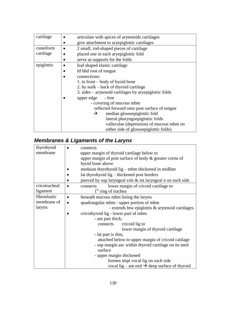
130
cartilage articulate with apices of arytenoids cartilages
give attachment to aryepiglottic cartilages
cuneiform
cartilage 2 small, rod-shaped pieces of cartilage
placed one in each aryepiglottic fold
serve as supports for the folds
epiglottis leaf shaped elastic cartilage
fd bhd root of tongue
connections:
1. in front – body of hyoid bone
2. by stalk – back of thyroid cartilage
3. sides – arytenoid cartilages by aryepiglottic folds
upper edge - free
- covering of mucous mbm
reflected forward onto post surface of tongue
median glossoepiglottic fold
lateral pharyngoepiglottic folds
valleculae (depressions of mucous mbm on
either side of glossoepiglottic folds)
Membranes & Ligaments of the Larynx
thyrohyoid
membrane connects
upper margin of thyroid cartilage below to
upper margin of post surface of body & greater cornu of
hyoid bone above
medaian thyrohyoid lig – mbm thickened in midline
lat thyrohyoid lig – thickened post borders
pierced by sup laryngeal vsls & int laryngeal n on each side
cricotracheal
ligament connects lower margin of cricoid cartilage to
1st ring of trachea
fibroelastic
membrane of
larynx
beneath mucous mbm lining the larynx
quadrangular mbm - upper portion of mbm
- extends btw epiglottis & arytenoid cartilages
cricothyroid lig - lower part of mbm
- ant part thick;
connects cricoid lig to
lower margin of thyroid cartilage
- lat part is thin,
attached below to upper margin of cricoid catilage
- sup margin asc within thyroid cartilage on its med
surface
- upper margin thickened
formes impt vocal lig on each side
vocal lig – ant end deep surface of thyroid

131
cartilage
– post end vocal process of the
arytenoid cartilage
hypoepiglottic
ligament
attaches to epiglottis to hyoid bone
thryoepiglottic
ligament
attached to epiglottis to thyroid cartilage

132
Inlet of Larynx
looks backward & upward into laryngopharynx
boundaries of opening
in front upper margin of epiglottis
lat aryepiglottic fold of mucous mbm
- connects epigloittis to arytenoid cartilage
posteroinf mucous mbm stretching btw arytenoid cartilages
corniculate cartilage (on apex of arytenoid cartilage) &
cuneiform cartilage (small bar) produce
a small elevation on upper border of each aryepiglottic fold
Cavity of the Larynx
extends from inlet to lower border of cricoid cartilage
may be divided into 3 parts: 1. upper part = vestibule
2. middle part
3. lower part
1. Vestibule of Larynx
extends from inlet to vestibular folds
vestibular folds – 2 thick folds of mucous mbm that cover vestibular lig
has ant, post & lat wall
ant wall post surface of epiglottis (covered by mucous mbm)
post wall arytenoid cartilages &
interarytenoid fold of mucous mbm (containing tnvs arytenoid musc)
lat walls aryepiglottic folds (contain the aryepiglottic musc)
below, narrowed by pink vestibular folds (proj medially)
rima vestibuli - gap btw vestibular folds
vestibular lig - lies within each vestibular fold
- thickened lower edge of quadrangular mbm
- stretches from thyroid cartilage to side of arytenoid cartilage
2. Middle Part of Larynx
extends from level of vestibular folds to level of vocal folds
vocal folds - white in colour
- contain vocal lig
vocal lig - thickened upper edge of cricothyroid lig
- stretches from
thyroid cartilage in front to
vocal processes of arytenoid cartilage bhd
rima glottis – gap btw vocal folds in front &
vocal processes of arytenoids cartilage bhd
sinus of larynx - small recess btw vestibular & vocal folds on each side

133
- lined with mucous mbm
- diverticulum from mucous mbm: saccule of the larynx
passes upward btw vestibular fold & thyroid cartilage

134
3. Lower Part of Larynx
extends from the level of vocal folds to lower border of cricoid cartilage
walls formed by inner surface of cricothyroid lig & cricoid cartilage
Mucous Membrane
lines cavity
covered with ciliated columnar epith
on vocal folds: covered with str sq epith
mucous mbm subject to repeated trauma during phonation
mucous glds within mucous mbm, esp in saccules
where secretion pours down onto upper surface of vocal folds & lubricates them
during phonation
Nerve Supply
Sensory n supply
to mucous mbm of larynx
a) above vocal folds
b) below level of vocal folds
int laryngeal br of sup laryngeal br of vagus
recurrent laryngeal n
Motor n supply
to intrinsic musc of larynx
recurrent laryngeal n
Note: except for cricothyroid musc.
by ext laryngeal br of sup laryngeal br of
vagus
Blood Supply
upper 1/2 of larynx: sup laryngeal br of sup thyroid art
lower 1/2 of larynx: inf laryngeal br of inf thyroid art
Lymphatic Drainage
LV drain into deep cervical group of nodes

135