
Head and Neck Anatomy 13
29
Anatomy of the Head and Neck lecture 13 Abbas A. A. Shawka Medical student 2 nd stage
-
Upload
abbas-al-robaiyee -
Category
Health & Medicine
-
view
156 -
download
4
Transcript of Head and Neck Anatomy 13

Anatomy of the Head and Neck
lecture 13
Abbas A. A. Shawka
Medical student
2nd stage
Subjects
Neck
- Anterior triangle 1

Anterior triangle • The anterior triangle of the neck is
outlined by the anterior border of the sternocleidomastoid muscle laterally,the inferior border of the mandible superiorly, and the midline of the neck medially
• Subdivided into
1. submandibular triangle
2. submental triangle
3. Muscular triangle
4. carotid triangle
** you SHOULD find
The boundaries of each
Traingel from the figure !!
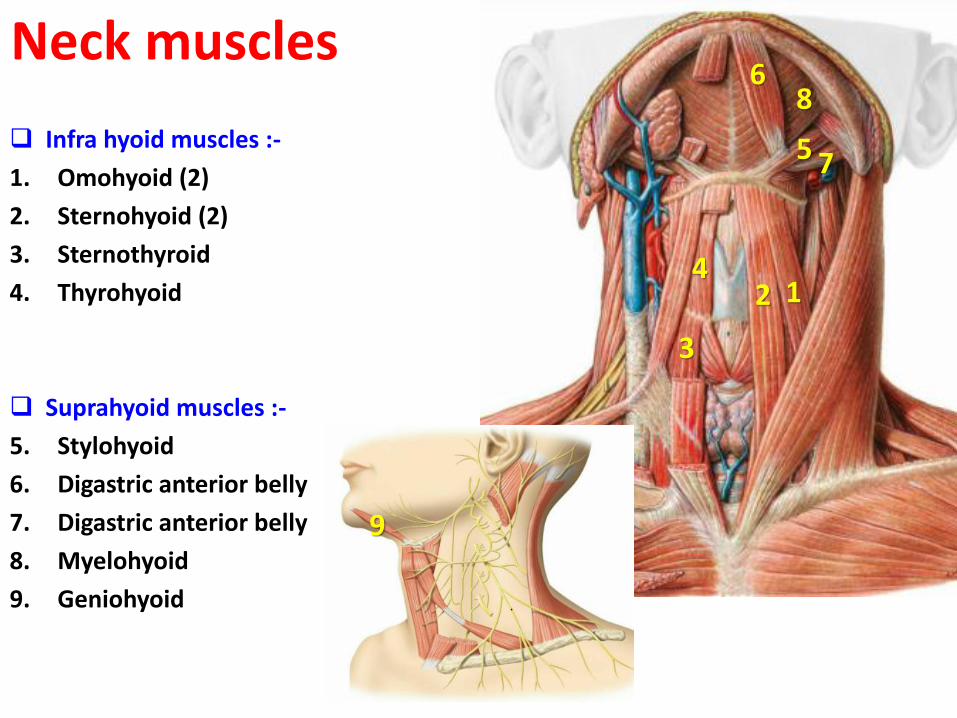
Neck muscles
Infra hyoid muscles :-
1. Omohyoid (2)
2. Sternohyoid (2)
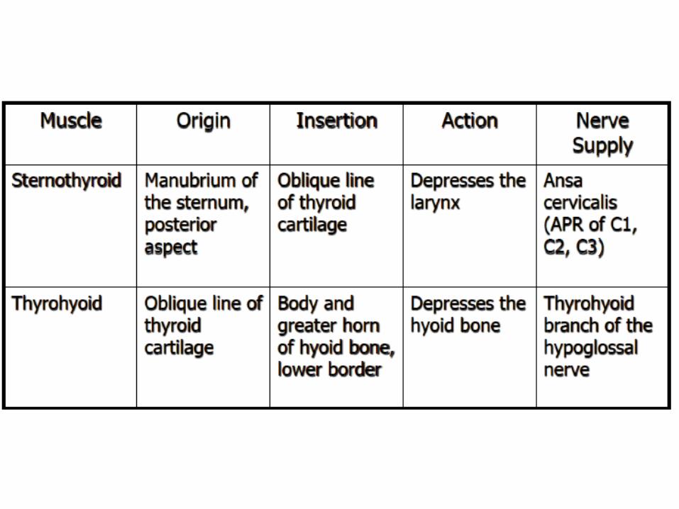
3. Sternothyroid
4. Thyrohyoid
Suprahyoid muscles :-
5. Stylohyoid
6. Digastric anterior belly
7. Digastric anterior belly
8. Myelohyoid
9. Geniohyoid
9
2
3
4
5
6
7
8
1
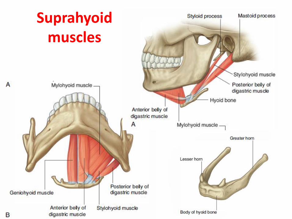
Suprahyoid muscles


Carotid system • The common carotid arteries are the
beginning of the carotid system
1. The right common carotid artery originates from the brachiocephalic trunk immediately posterior to the right sternoclavicular joint and is entirely in the neck throughout its course.
2. The left common carotid arterybegins in the thora as a direct branch of the arch of the aorta and passesuperiorly to enter the neck near the left sternoclavicular joint.
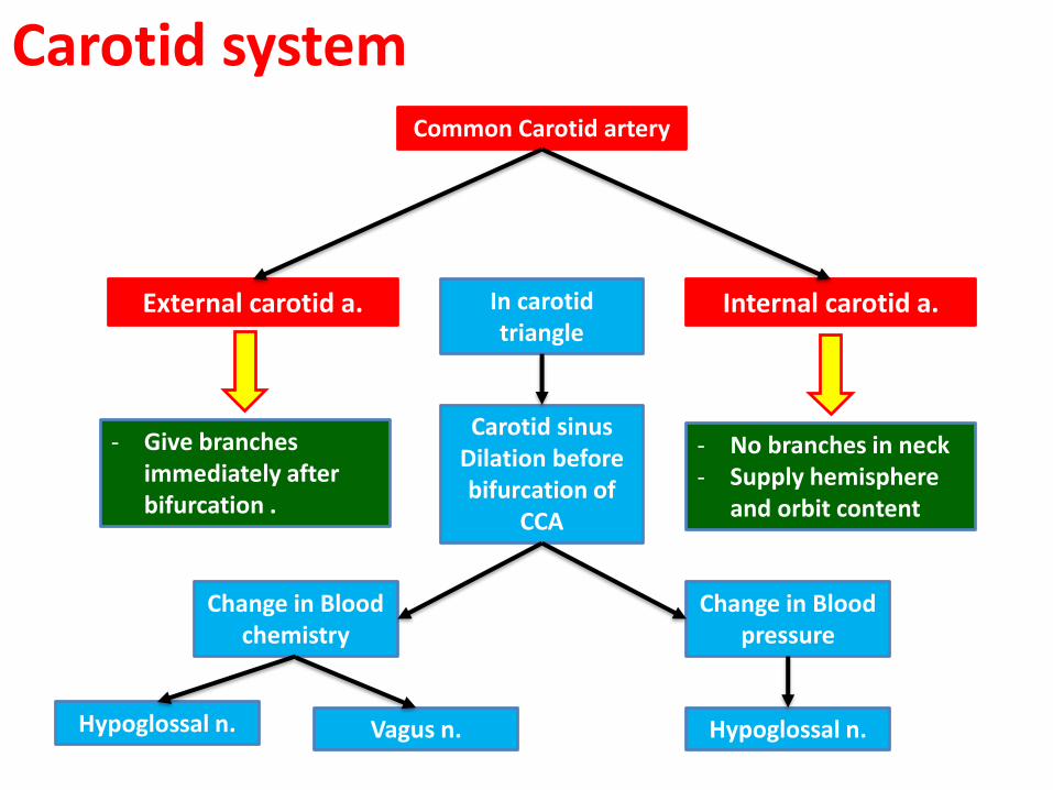
Carotid systemCommon Carotid artery
Internal carotid a.External carotid a.
Carotid sinusDilation before bifurcation of
CCA
In carotid triangle
Change in Blood pressure
Change in Blood chemistry
Hypoglossal n.Hypoglossal n. Vagus n.
- No branches in neck - Supply hemisphere
and orbit content
- Give branches immediately after bifurcation .

ECA branches ECA
Posterior branchesAnterior branches
Ascending pharyngeal a.Superior thyroid a.
Fascial a.
Lingual a. Occipital a.
Posterior auricular a.

Superior thyroid artery
• arises near or at the bifurcation
• passes in a downward and forward direction to reach the superior pole of the thyroid gland.

Ascending pharyngeal a.
• ascends between the internal carotid artery and the pharynx.

Lingual a.
• arises just above the superior thyroid artery at the level of the hyoid bone.
• passes deep to the hypoglossal nerve [XII].

Fascial a.
• arises just above the lingual artery.
• passes deep to the stylohyoid (1) and posterior belly of the digastric muscles (2).
• continues deep between the submandibular gland and mandible.
• emerges over the edge of the mandible just anterior to the masseter muscle, to enter the face.
2
1
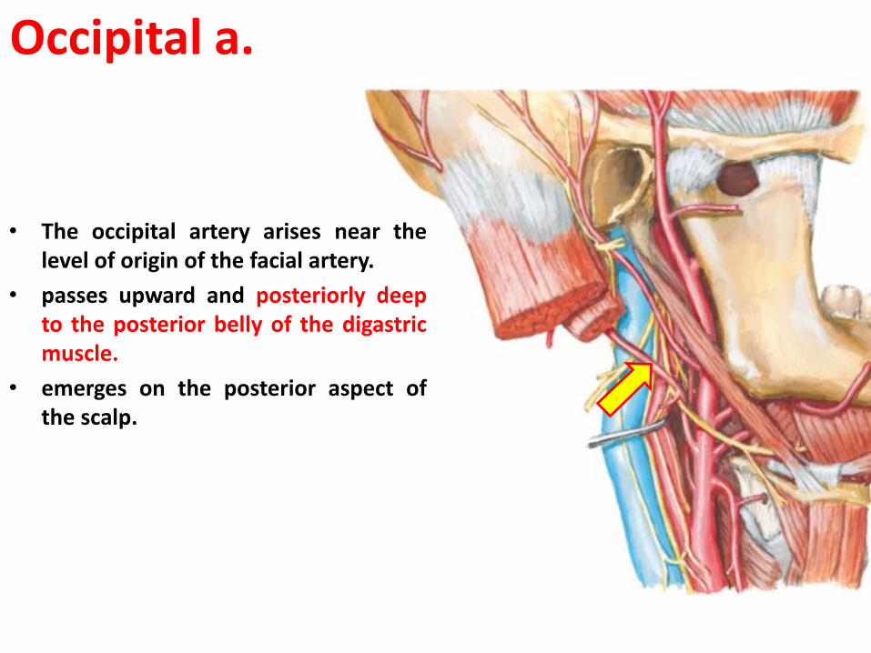
Occipital a.
• The occipital artery arises near the level of origin of the facial artery.
• passes upward and posteriorly deep to the posterior belly of the digastric muscle.
• emerges on the posterior aspect of the scalp.
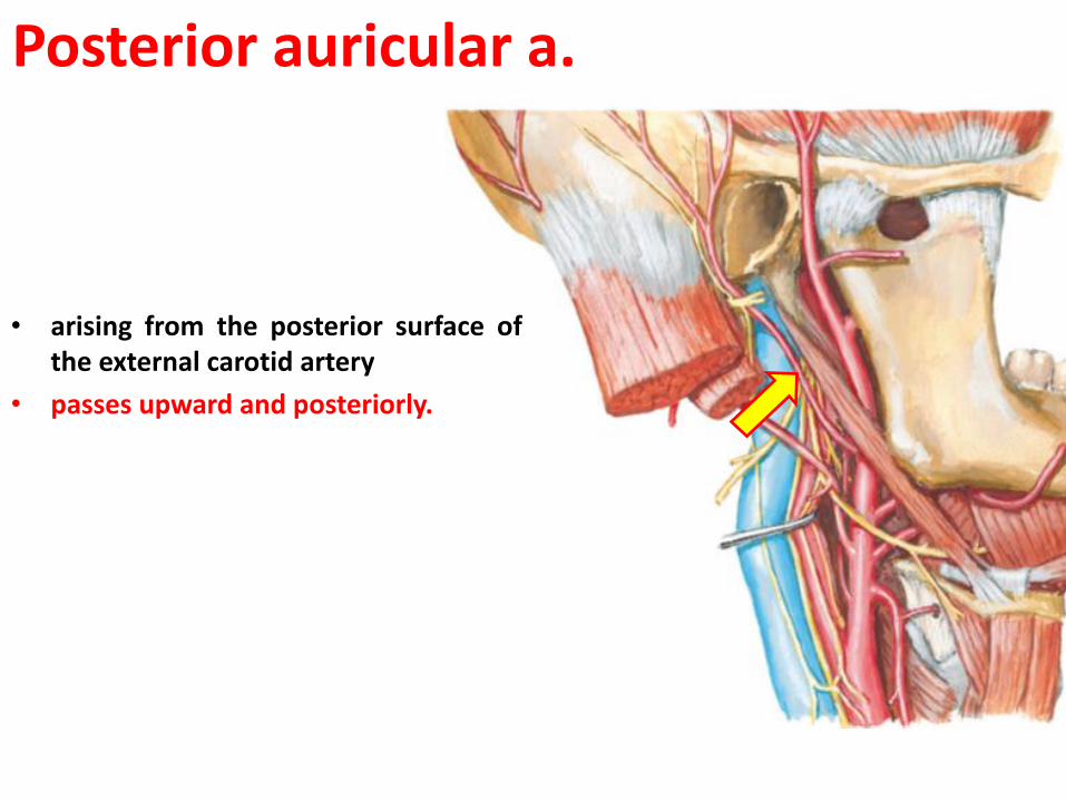
Posterior auricular a.
• arising from the posterior surface of the external carotid artery
• passes upward and posteriorly.

Venous drainage
IJV
Sigmoid sinuse
Inferior petrosal sinus
Brachiocephalic vein
Subclavian vein
IJV
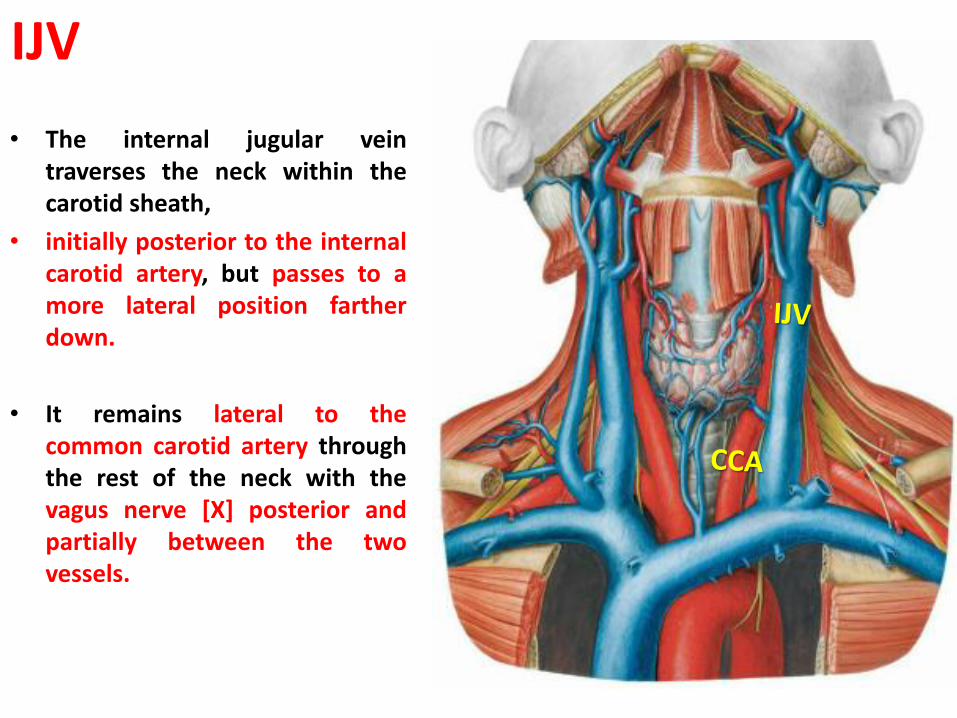
• The internal jugular vein traverses the neck within the carotid sheath,
• initially posterior to the internal carotid artery, but passes to a more lateral position farther down.
• It remains lateral to the common carotid artery through the rest of the neck with the vagus nerve [X] posterior and partially between the two vessels.
Nerves in the anterior triangle • The cranial nerves in these categories
include
1. the facial [VII]
2. glossopharyngeal [IX]
3. vagus [X]
4. accessory [XI]
5. hypoglossal [XII]
• Branches of spinal nerves in these categories include
1. The transverse cervical nerve from the cervical plexus
2. The upper and lower roots of the ansa cervicalis.

Hypoglossal nerve • The glossopharyngeal nerve [IX] leaves the
cranial cavity through the jugular foramen.
• descent between the ICA & IJV
• lying deep to the styloid process and the muscles associated with the styloid process.
• it passes forward between the internal and external carotid arteries ,
• Curves around the lateral border of the stylopharyngeus muscle.
• continues in an anterior direction, deep to the hyoglossus muscle, to reach the base of the tongue and the area of the palatine tonsil.
• As it passes through the AT of the neck it innervates:-
1. stylopharyngeus muscle,
2. sends a branch to the carotid sinus,
3. supplies sensory branches to the pharynx.
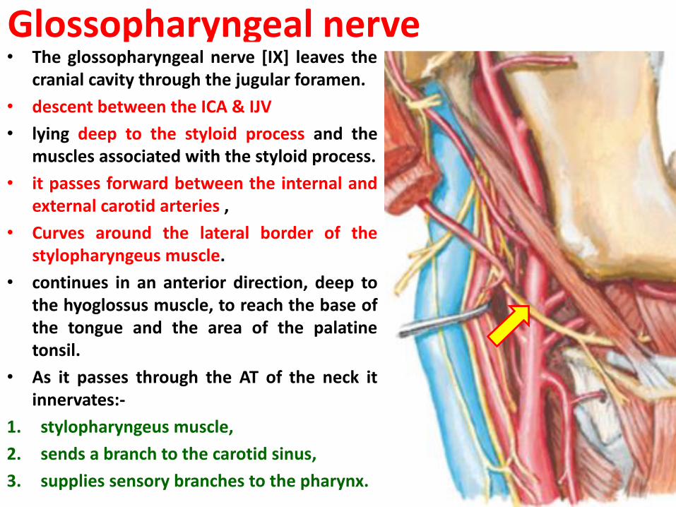
Glossopharyngeal nerve• The glossopharyngeal nerve [IX] leaves the
cranial cavity through the jugular foramen.
• descent between the ICA & IJV
• lying deep to the styloid process and the muscles associated with the styloid process.
• it passes forward between the internal and external carotid arteries ,
• Curves around the lateral border of the stylopharyngeus muscle.
• continues in an anterior direction, deep to the hyoglossus muscle, to reach the base of the tongue and the area of the palatine tonsil.
• As it passes through the AT of the neck it innervates:-
1. stylopharyngeus muscle,
2. sends a branch to the carotid sinus,
3. supplies sensory branches to the pharynx.

Vagus nerve • The vagus nerve [X] exits the cranial
cavity through the jugular foramen between the glossopharyngeal [IX] and accessory [XI] nerves.
• vagus nerve [X] enters the carotid
• sheath and descends through the neck enclosed in this structure medial to the internal jugular vein and posterior to the internal carotid and common carotid arteries
• Branches o f the vagus nerve [X] as it passes through the anterior triangle of the neck include
1. a motor branch to the pharynx,
2. a branch to the carotid body,
3. the superior laryngeal nerve (which divides into external and internal laryngeal branches) ,
4. and possibly a cardiac branch.

Accessory nerve IX • the most posterior of the three cranial
nerves exiting the cranial cavity through the jugular foramen .
• It begins its descent medial to the internal jugular vein, emerging from between the internal jugular vein and internal carotid artery to cross the lateral surface of the internal j ugularvein as it passes downward and backward to disappear either into or beneath the anterior border of the sternocleidomastoid muscle
• The accessory nerve gives off NO branches as it passes through the anterior triangle of the neck.

Hypoglossal nerve • leaves the cranial cavity
• through the hypoglossal canal
• medial to the internal jugular vein and internal carotid artery immediately outside the skull.
• it passes outward between the internal jugular vein and internal carotid artery.
• it passes forward, hooking around the occipital artery, across the lateral surfaces of the internal and external carotid arteries and the lingual artery.
• continues deep to the posterior belly of the digastric and stylohyoid muscles.
• It passes over the surface of the hyoglossus muscle and disappears deep to the mylohyoid muscle.
• Give NO branches in AT ( this nerve supply the tongue )

Transverse cervical nerve
• arising from the anterior rami of cervical nerves C2 and C 3 .
• It emerges from beneath the posterior border of the sternocleidomastoid muscle, near the middle of the muscle, and loops around the sternocleidomastoid to cross its anterior surface in a transverse direction
• It continues across the neck and provides cutaneous innervation to this area.

Ansa cervicalis• The ansa cervicalis is a loop of nerve fibers
from cervical nerves C1 to C3 that innervate the " strap muscles" in the anterior triangle of the neck.
• It begin as branches from the cervical nerve C1 join the hypoglossal nerve [XII] soon after it leaves the skull.
• As the hypoglossal nerve [XII] completes its descent
• and begins to pass forward across the internal and external carotid arteries , some of the cervical nerve fibers leave it and descend between the internal jugular vein and the internal, and then common, carotid arteries. These nerve fibers are the superior root of the ansa cervicalis
• and innervate the superior belly of the omohyoid muscle, and the upper parts of the sternohyoid and sternothyroid muscles.
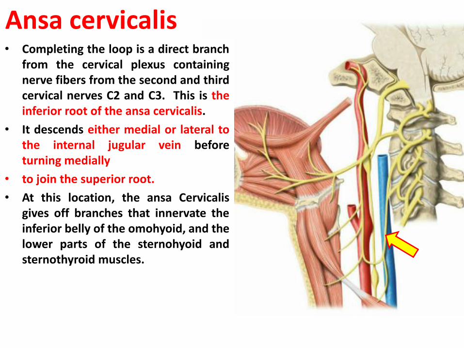
Ansa cervicalis• Completing the loop is a direct branch
from the cervical plexus containing nerve fibers from the second and third cervical nerves C2 and C3. This is the inferior root of the ansa cervicalis.
• It descends either medial or lateral to the internal jugular vein before turning medially
• to join the superior root.
• At this location, the ansa Cervicalisgives off branches that innervate the inferior belly of the omohyoid, and the lower parts of the sternohyoid and sternothyroid muscles.