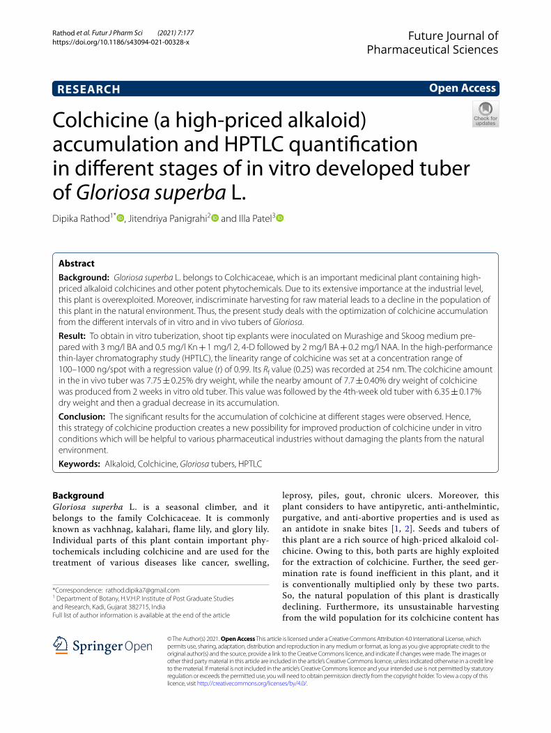
Colchicine (a high-priced alkaloid) accumulation and HPTLC ...
9
Rathod et al. Futur J Pharm Sci (2021) 7:177 https://doi.org/10.1186/s43094-021-00328-x RESEARCH Colchicine (a high-priced alkaloid) accumulation and HPTLC quantification in different stages of in vitro developed tuber of Gloriosa superba L. Dipika Rathod 1* , Jitendriya Panigrahi 2 and Illa Patel 3 Abstract Background: Gloriosa superba L. belongs to Colchicaceae, which is an important medicinal plant containing high- priced alkaloid colchicines and other potent phytochemicals. Due to its extensive importance at the industrial level, this plant is overexploited. Moreover, indiscriminate harvesting for raw material leads to a decline in the population of this plant in the natural environment. Thus, the present study deals with the optimization of colchicine accumulation from the different intervals of in vitro and in vivo tubers of Gloriosa. Result: To obtain in vitro tuberization, shoot tip explants were inoculated on Murashige and Skoog medium pre- pared with 3 mg/l BA and 0.5 mg/l Kn + 1 mg/l 2, 4-D followed by 2 mg/l BA + 0.2 mg/l NAA. In the high-performance thin-layer chromatography study (HPTLC), the linearity range of colchicine was set at a concentration range of 100–1000 ng/spot with a regression value (r) of 0.99. Its R f value (0.25) was recorded at 254 nm. The colchicine amount in the in vivo tuber was 7.75 ± 0.25% dry weight, while the nearby amount of 7.7 ± 0.40% dry weight of colchicine was produced from 2 weeks in vitro old tuber. This value was followed by the 4th-week old tuber with 6.35 ± 0.17% dry weight and then a gradual decrease in its accumulation. Conclusion: The significant results for the accumulation of colchicine at different stages were observed. Hence, this strategy of colchicine production creates a new possibility for improved production of colchicine under in vitro conditions which will be helpful to various pharmaceutical industries without damaging the plants from the natural environment. Keywords: Alkaloid, Colchicine, Gloriosa tubers, HPTLC © The Author(s) 2021. Open Access This article is licensed under a Creative Commons Attribution 4.0 International License, which permits use, sharing, adaptation, distribution and reproduction in any medium or format, as long as you give appropriate credit to the original author(s) and the source, provide a link to the Creative Commons licence, and indicate if changes were made. The images or other third party material in this article are included in the article’s Creative Commons licence, unless indicated otherwise in a credit line to the material. If material is not included in the article’s Creative Commons licence and your intended use is not permitted by statutory regulation or exceeds the permitted use, you will need to obtain permission directly from the copyright holder. To view a copy of this licence, visit http://creativecommons.org/licenses/by/4.0/. Background Gloriosa superba L. is a seasonal climber, and it belongs to the family Colchicaceae. It is commonly known as vachhnag, kalahari, flame lily, and glory lily. Individual parts of this plant contain important phy- tochemicals including colchicine and are used for the treatment of various diseases like cancer, swelling, leprosy, piles, gout, chronic ulcers. Moreover, this plant considers to have antipyretic, anti-anthelmintic, purgative, and anti-abortive properties and is used as an antidote in snake bites [1, 2]. Seeds and tubers of this plant are a rich source of high-priced alkaloid col- chicine. Owing to this, both parts are highly exploited for the extraction of colchicine. Further, the seed ger- mination rate is found inefficient in this plant, and it is conventionally multiplied only by these two parts. So, the natural population of this plant is drastically declining. Furthermore, its unsustainable harvesting from the wild population for its colchicine content has Open Access Future Journal of Pharmaceutical Sciences *Correspondence: [email protected] 1 Department of Botany, H.V.H.P. Institute of Post Graduate Studies and Research, Kadi, Gujarat 382715, India Full list of author information is available at the end of the article
Transcript of Colchicine (a high-priced alkaloid) accumulation and HPTLC ...
Rathod et al. Futur J Pharm Sci (2021) 7:177 https://doi.org/10.1186/s43094-021-00328-x
RESEARCH
Colchicine (a high-priced alkaloid) accumulation and HPTLC quantification in different stages of in vitro developed tuber of Gloriosa superba L.Dipika Rathod1* , Jitendriya Panigrahi2 and Illa Patel3
Abstract
Background: Gloriosa superba L. belongs to Colchicaceae, which is an important medicinal plant containing high-priced alkaloid colchicines and other potent phytochemicals. Due to its extensive importance at the industrial level, this plant is overexploited. Moreover, indiscriminate harvesting for raw material leads to a decline in the population of this plant in the natural environment. Thus, the present study deals with the optimization of colchicine accumulation from the different intervals of in vitro and in vivo tubers of Gloriosa.
Result: To obtain in vitro tuberization, shoot tip explants were inoculated on Murashige and Skoog medium pre-pared with 3 mg/l BA and 0.5 mg/l Kn + 1 mg/l 2, 4-D followed by 2 mg/l BA + 0.2 mg/l NAA. In the high-performance thin-layer chromatography study (HPTLC), the linearity range of colchicine was set at a concentration range of 100–1000 ng/spot with a regression value (r) of 0.99. Its Rf value (0.25) was recorded at 254 nm. The colchicine amount in the in vivo tuber was 7.75 ± 0.25% dry weight, while the nearby amount of 7.7 ± 0.40% dry weight of colchicine was produced from 2 weeks in vitro old tuber. This value was followed by the 4th-week old tuber with 6.35 ± 0.17% dry weight and then a gradual decrease in its accumulation.
Conclusion: The significant results for the accumulation of colchicine at different stages were observed. Hence, this strategy of colchicine production creates a new possibility for improved production of colchicine under in vitro conditions which will be helpful to various pharmaceutical industries without damaging the plants from the natural environment.
Keywords: Alkaloid, Colchicine, Gloriosa tubers, HPTLC
© The Author(s) 2021. Open Access This article is licensed under a Creative Commons Attribution 4.0 International License, which permits use, sharing, adaptation, distribution and reproduction in any medium or format, as long as you give appropriate credit to the original author(s) and the source, provide a link to the Creative Commons licence, and indicate if changes were made. The images or other third party material in this article are included in the article’s Creative Commons licence, unless indicated otherwise in a credit line to the material. If material is not included in the article’s Creative Commons licence and your intended use is not permitted by statutory regulation or exceeds the permitted use, you will need to obtain permission directly from the copyright holder. To view a copy of this licence, visit http:// creat iveco mmons. org/ licen ses/ by/4. 0/.
BackgroundGloriosa superba L. is a seasonal climber, and it belongs to the family Colchicaceae. It is commonly known as vachhnag, kalahari, flame lily, and glory lily. Individual parts of this plant contain important phy-tochemicals including colchicine and are used for the treatment of various diseases like cancer, swelling,
leprosy, piles, gout, chronic ulcers. Moreover, this plant considers to have antipyretic, anti-anthelmintic, purgative, and anti-abortive properties and is used as an antidote in snake bites [1, 2]. Seeds and tubers of this plant are a rich source of high-priced alkaloid col-chicine. Owing to this, both parts are highly exploited for the extraction of colchicine. Further, the seed ger-mination rate is found inefficient in this plant, and it is conventionally multiplied only by these two parts. So, the natural population of this plant is drastically declining. Furthermore, its unsustainable harvesting from the wild population for its colchicine content has
Open Access
Future Journal ofPharmaceutical Sciences
*Correspondence: [email protected] Department of Botany, H.V.H.P. Institute of Post Graduate Studies and Research, Kadi, Gujarat 382715, IndiaFull list of author information is available at the end of the article

Page 2 of 9Rathod et al. Futur J Pharm Sci (2021) 7:177
been carried out by the pharmaceutical industries. In addition, it also faces a low seed set problem [3]. So, the mass multiplication of this valuable plant species is needed to meet the ever-increasing industrial demand. Consequently, in vitro propagation technology is now proven one of the potentially viable methods for the sustainable maintenance of many useful medici-nal plant species [4]. At present, the biotechnologi-cal approaches especially plant cell, tissue, and organ culture have been proved as an effective source for the production and extraction of biologically potent sec-ondary metabolites which now holds the attraction of many researchers. In recent times, more empha-sis has been given to plant cell biotechnology which emerged as a possible alternative method for the pro-duction of active constituents, using in vitro cultured cells rather than destroying the whole plants from natural habitat for such extraction. However, there is little literature reported to date on the multiplication of this important tuberous plant [5, 6]. Since the col-chicine is mainly accumulated in the tuber, that’s why the in vitro mass propagation/multiplication of the tuber part is essential [7]. Due to multiple uses of col-chicine, in vitro technologies would be the best option to elevate the production of colchicine along with its improved properties. This technique provides a con-tinuous, reproducible, and reliable source of the plant to meet the ever-increasing demand for pharmaceuti-cal industries. It also fulfills disease-free plant materi-als on a large scale. In vitro propagated plant materials could also be useful to extract medicinally potent sec-ondary metabolites without exploiting the plants from natural habitats [8–10]. Nowadays, high-performance thin-layer chromatography (HPTLC) is proven as a powerful tool for qualitative and quantitative analysis of vital bioactive compounds which are produced in various parts of plants. Consequently, it is considered as sophisticated among all other analytical techniques in terms of reliability, simplicity, speed, time, and cost per analysis [11, 12]. Earlier, several researchers have used various chromatographic techniques like HPLC and HPTLC to determine colchicine from various plant parts of Gloriosa like seed, tuber, leaves, stem, flowers, pods, and seed pericarp [13–17] but a com-parative account of in vitro and in vivo tuber was still lacking in Gloriosa. So, in the present investigation, an attempt was made for (1) in vitro tuber production and multiplication, (2) determination of colchicine in it and (3) optimization of the in vitro stage(s) for maxi-mum accumulation of colchicines by using the HPTLC technique in both in vivo tuber and in vitro developed tuber at different developmental stages after method validation.
MethodsPlant materials and chemicalsThe explants of Gloriosa superba L. were collected and selected for further studies. The chemicals used for the experiment were of analytical grade quality and were obtained from SD Fine, India, and Himedia, India. The Silica gel 60F254 (20 cm × 10 cm) HPTLC plate used for the chromatographic studies was obtained from E. Merck (Darmstadt, Germany). The standard Colchicine was procured from Natural Remedies, Bangalore, India.
In vitro tuberizationFor in vitro studies, terminal shoot tips were used as explants. The explants were washed with running tap water for 5–10 min. Then, all the selected explants were surface sterilized by dipping with 0.1% (w/v) mercu-ric chloride for 0.5–1 min and at last rinsed thoroughly 2–3 times with sterile double distilled water. Sterilized explants were then inoculated into culture media under laminar airflow. The full-strength Murashige and Skoog (MS) [18] medium was prepared by supplementing vari-ous concentrations and combinations of plant growth regulators (PGRs) like BA (N6-benzyl adenine) alone (1–3 mg/l), in combination with α-naphthalene ace-tic acid (NAA). (NAA 0.2 mg/l), combination of Kine-tin + 2,4-Dichloro phenoxy acetic acid (2,4-D) (0.2 + 0.5, 0.5 + 1 and 1 + 1.5 mg/l). The treatment without any PGRs was considered as a control. The pH of the medium was adjusted to 5.8 with NaOH/HCl and dispensed into culture tubes and phytajars (15–20 ml). The media was sterilized by autoclaving at 121 °C and 1.063 kg/cm3 pressure for 20 min. The cultures were maintained at 28 ± 2 °C under 16:8 h light/dark photoperiod under cool white fluorescent lamps at a photosynthetic photon flux density of 70 μmol m−2 s−1. All the periodic data were collected for each parameter.
Extraction procedureIn vivo tubers of nearly two months were collected from the natural plant, whereas in vitro tubers have been developed through tissue cultures were harvested at dif-ferent intervals of 2, 4, 6, 8, 10, and 12 weeks of culture. All the samples were thoroughly washed with double dis-tilled water and dried under shade at room temperature. Samples were powdered with a mechanical grinder and further extracted through a simple maceration method at room temperature. For extraction, 1 g powdered plant samples were mixed with 10 ml of methanol and incubated for 24 h in the shaker at 200 rpm. After 24 h, centrifugation was done and the supernatant was col-lected and filtered through Whatman filter paper No. 41. The obtained filtrate was concentrated and was used for further analysis [19, 20]. A common stock solution of

Page 3 of 9Rathod et al. Futur J Pharm Sci (2021) 7:177
colchicine was prepared by dissolving it in methanol at the concentration of 1 mg/ 1 ml. The stock solution was then further diluted in methanol to obtain a solution containing the concentration of 100 ng/µl and was used as a working standard for the HPTLC study.
Chromatographic conditionsChromatographic studies performed on a CAMAG HPTLC unit consist of Linomat-V automatic sample applicator, TLC scanner III, and integrated software WINCATS (CAMAG, Switzerland). The thin-layer chro-matography was performed on a pre-coated silica gel HPTLC 60F254 (20 cm × 10 cm) plate of 0.20 mm layer thickness, used as a stationary phase for the quantifica-tion of Colchicine. The plant samples and the standards were spotted on the TLC plate at 8-mm-wide bands with the help of the Linomat applicator V equipped with a 100 µl syringe (Hamilton, Switzerland) under a continu-ous flow of nitrogen gas at a constant application rate of 150 nl s−1. Before placing the plate, Camag twin trough chamber (20 cm × 10 cm) was saturated with 20 ml mobile phase with chloroform/methanol: formic acid (20:1:0.2) for 20 min, under room temperature (28 ± 2 °C) and 55 ± 5% relative humidity. The spotted plate was then placed into a pre-saturated chamber and the chro-matogram was run at 8 cm from the base. After that, the plate was dried with an air dryer and further quantitative evaluation was performed by scanning with TLC scanner III in the absorption–reflection mode at 254 nm. The slit dimension was set at 6 mm × 0.45 mm, with a data reso-lution of 100 nm s−1 and scanning speed of 20 mm s−1.
Method validationThe present method was validated as per the ICH (Inter-national Council of Harmonization) guidelines [21]. For the linearity study, standard solutions at 100–1000 ng/spot were analyzed. The calibration curve was generated by plotting the peak areas against the concentrations with the help of regression analysis. For the measurement of detection limit (LOD) and quantification limit (LOQ), the formula suggested by Alam et al. [22] was used. To measure the recovery study, the known samples were spiked with an extra 80, 100, 120% of the standard col-chicine (standard addition method) and the mixtures obtained by this method were analyzed. The experiment was performed three times at each level.
Precision, reproducibility, and specificityThe system precision was estimated by using repeatability of the sample application, and peak areas were measured by using eight replicates of the same band (600 ng per spot of colchicine). The intraday and inter-day precision for colchicine was carried out by spotting eight samples
at a concentration of 600 ng/spot that were analyzed on the same day and three different days, respectively. The precision was calculated in terms of relative standard deviation (%RSD) of obtained peak area. The specific-ity of the present method was determined by comparing the obtained Rf values from TLC and the spectrum of the colchicine band from a plant sample with those of a standard.
Quantitative determination of colchicine from in vivo and in vitro tubersExtracts of plant samples and standard solutions (2,4,6,8,10 µl) were spotted on silica gel60 HPTLC plates of size 20 cm × 10 cm and analyzed as mentioned above. The peak area of standard colchicine was determined from the calibration plot of peak area against the amount of colchicine (standard). The standard compound present in the different samples was determined by using a cali-bration plot, and data were recorded.
Statistical analysisCompletely randomized design (CRD) was used for per-forming tissue culture experiments. Each experiment was conducted with three replicates by using 10 samples. The recorded data were analyzed statistically using SPSS (ver-sion 20, SPSS Inc. Chicago, USA) software. The one-way analysis of variance (ANOVA) was used to determine the effect of treatment. Mean values of data were compared by Duncan multiple range test at P ≤ 0.05.
ResultsEffect of PGRs on in vitro tuberizationIn the present study, initially, all cultures with treatments showed a white small bulbous/callus-like structure from the base of apical shoot tips within 2 weeks of culture, which eventually transformed into in vitro tubers after subculturing in the same medium for 12 weeks (Fig. 1). The controlled one did show a response, but it was mea-ger at 20%, while the number of in vitro tubers produced was only 2.26 ± 1.1. Among the various treatments, the highest percentage of growth frequency (90%) with the maximum number of in vitro tubers (37.7 ± 0.5), as well as the significant growth of in vitro tubers, was observed when MS medium fortified with 3 mg/l BA (Table 1) during all the stages. However, a lower concentration of 1 or 2 mg/l BA hinders the overall growth of in vitro tubers. Among combined treatments, MS medium with Kinetin (0.5 mg/l) + 2,4-D (1.0 mg/l) showed 70% of growth response with the 30.3 ± 1.5 numbers of in vitro tubers. However, a less or higher concentration than this combination considerably suppressed the growth of in vitro tubers. In contrast, the Kinetin (0.2 mg/l) in combination with 2, 4-D (0.5 mg/l) showed 40% growth

Page 4 of 9Rathod et al. Futur J Pharm Sci (2021) 7:177
response with 21.6 ± 0.5 numbers of in vitro tubers but, when the concentration of these two combinations of Kinetin enhanced to 1 mg/l and 2, 4-D to 1.5 mg/l, the growth response was declined (30%) so as the number of in vitro tubers, i.e., 14.3 ± 1.1. However, in the case of BA + NAA, all three combinations provided a good response; but, still did not reach the highest percent-age obtained from 3 mg/l BA alone. Here, treatment using 2 mg/l BA + 0.2 mg/l NAA resulted in a significant
(60%) response along with 31.3 ± 1.1 number of in vitro tubers. However, the combination of 3 mg/l BA and 0.2 mg/l NAA resulted in a significant decrease in growth response (20%). Still decreasing number of in vitro tubers obtained in all the stages. Thus, tubers were collected from tubes with maximum growth at different stages and used for further analysis.
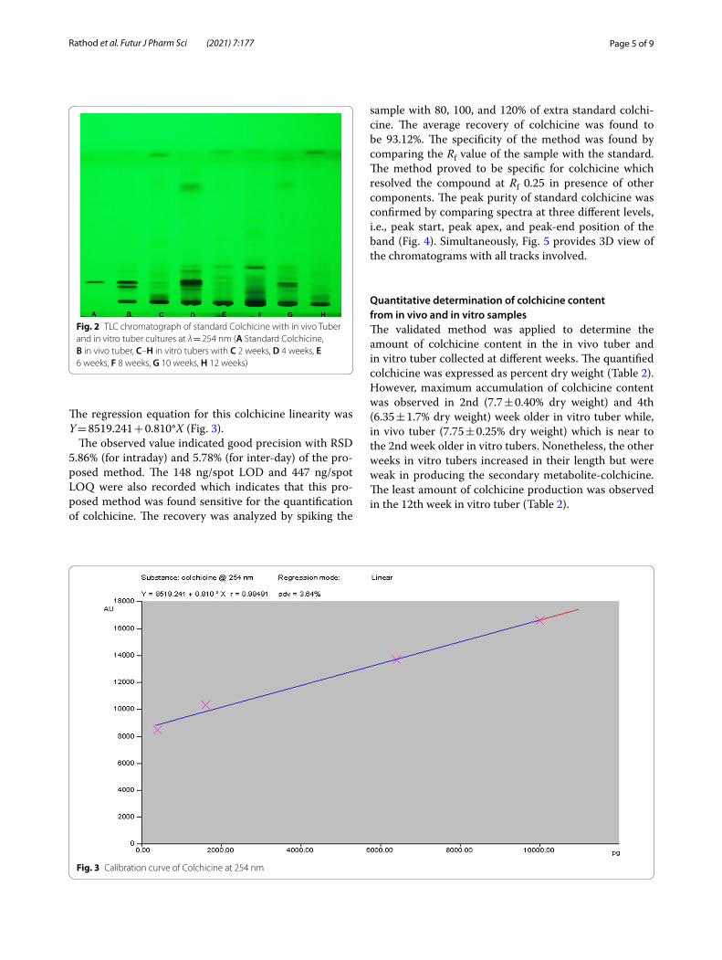
HPTLC analysisMethod validationThe mobile phase used in the present HPTLC study was selected by trying several combinations of different sol-vents. The preparatory TLC study revealed that chloro-form, methanol, and formic acid (20:1:0.2 v/v) solvent system was found ideal that produced Rf 0.25 for col-chicine and gave sharp, clearly separated, and resolv-ing bands. The chromatogram obtained was observed at 254 nm (Fig. 2). The spectrum obtained from the spec-trum analysis was at 353 nm. The observed peaks were relatively overlaying at Rf 0.25. The peaks were matched precisely with each other that revealed the compounds are analogous to Rf of the markers and the respective in vivo and in vitro plant samples to be identical. The linear regression data for the calibration curve showed a good linear relationship with the linearity range of 100–1000 ng/spot. Moreover, a correlation coefficient (r = 0.99491) with a standard deviation (sdv) of 3.64% shows good linearity between concentration and area.
Fig. 1 a In vivo tuber, b–g in vitro tuber culture in b-2, c-4, d-6, e-8, f-10 and g-12 weeks
Table 1 In vitro tuber formation in Gloriosa
Data represent the mean ± standard error of 10 replicates for each treatment. Means were compared using DMRT (Duncan’s Multiple Range Test) (P ≤ 0.05) using one-way ANOVA. Mean values within the column with the same letter in superscript are not significantly different [34]
Hormones types Concentration (mg/l)
% Growth response
No. of microtubers/explants (Mean ± SE)
Control – 2 2.26 ± 1.1f
BA 1 – –
2 – –
3 90 37.7 ± 0.5a
BA + NAA 1 + 0.2 50 26.3 ± 1.5c
2 + 0.2 60 31.3 ± 1.1b
3 + 0.2 20 25.0 ± 1.7c
Kin. + 2,4-D 0.2 + 0.5 40 21.6 ± 0.5d
0.5 + 1.0 70 30.3 ± 1.5b
1 + 1.5 30 14.3 ± 1.1e
Page 5 of 9Rathod et al. Futur J Pharm Sci (2021) 7:177
The regression equation for this colchicine linearity was Y = 8519.241 + 0.810*X (Fig. 3).
The observed value indicated good precision with RSD 5.86% (for intraday) and 5.78% (for inter-day) of the pro-posed method. The 148 ng/spot LOD and 447 ng/spot LOQ were also recorded which indicates that this pro-posed method was found sensitive for the quantification of colchicine. The recovery was analyzed by spiking the
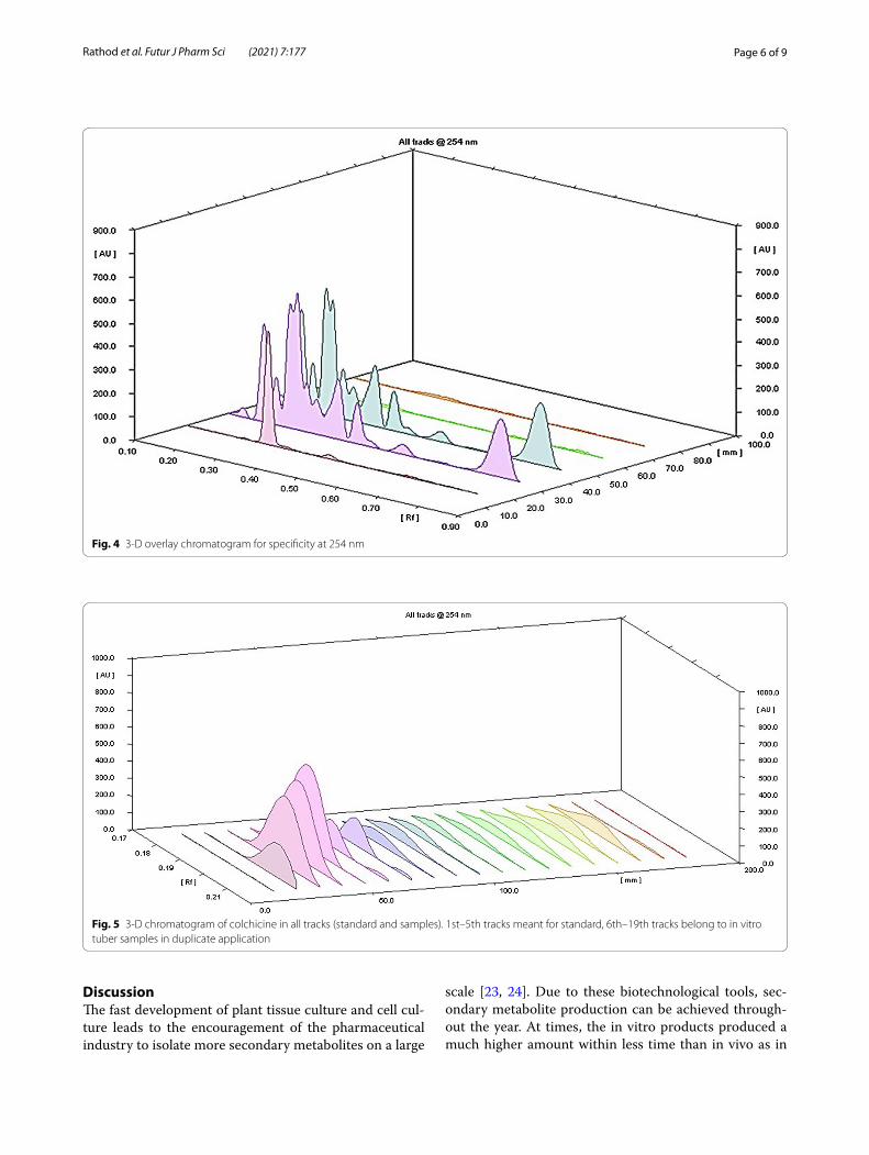
sample with 80, 100, and 120% of extra standard colchi-cine. The average recovery of colchicine was found to be 93.12%. The specificity of the method was found by comparing the Rf value of the sample with the standard. The method proved to be specific for colchicine which resolved the compound at Rf 0.25 in presence of other components. The peak purity of standard colchicine was confirmed by comparing spectra at three different levels, i.e., peak start, peak apex, and peak-end position of the band (Fig. 4). Simultaneously, Fig. 5 provides 3D view of the chromatograms with all tracks involved.
Quantitative determination of colchicine content from in vivo and in vitro samplesThe validated method was applied to determine the amount of colchicine content in the in vivo tuber and in vitro tuber collected at different weeks. The quantified colchicine was expressed as percent dry weight (Table 2). However, maximum accumulation of colchicine content was observed in 2nd (7.7 ± 0.40% dry weight) and 4th (6.35 ± 1.7% dry weight) week older in vitro tuber while, in vivo tuber (7.75 ± 0.25% dry weight) which is near to the 2nd week older in vitro tubers. Nonetheless, the other weeks in vitro tubers increased in their length but were weak in producing the secondary metabolite-colchicine. The least amount of colchicine production was observed in the 12th week in vitro tuber (Table 2).
Fig. 2 TLC chromatograph of standard Colchicine with in vivo Tuber and in vitro tuber cultures at λ = 254 nm (A Standard Colchicine, B in vivo tuber, C–H in vitro tubers with C 2 weeks, D 4 weeks, E 6 weeks, F 8 weeks, G 10 weeks, H 12 weeks)
Fig. 3 Calibration curve of Colchicine at 254 nm
Page 6 of 9Rathod et al. Futur J Pharm Sci (2021) 7:177
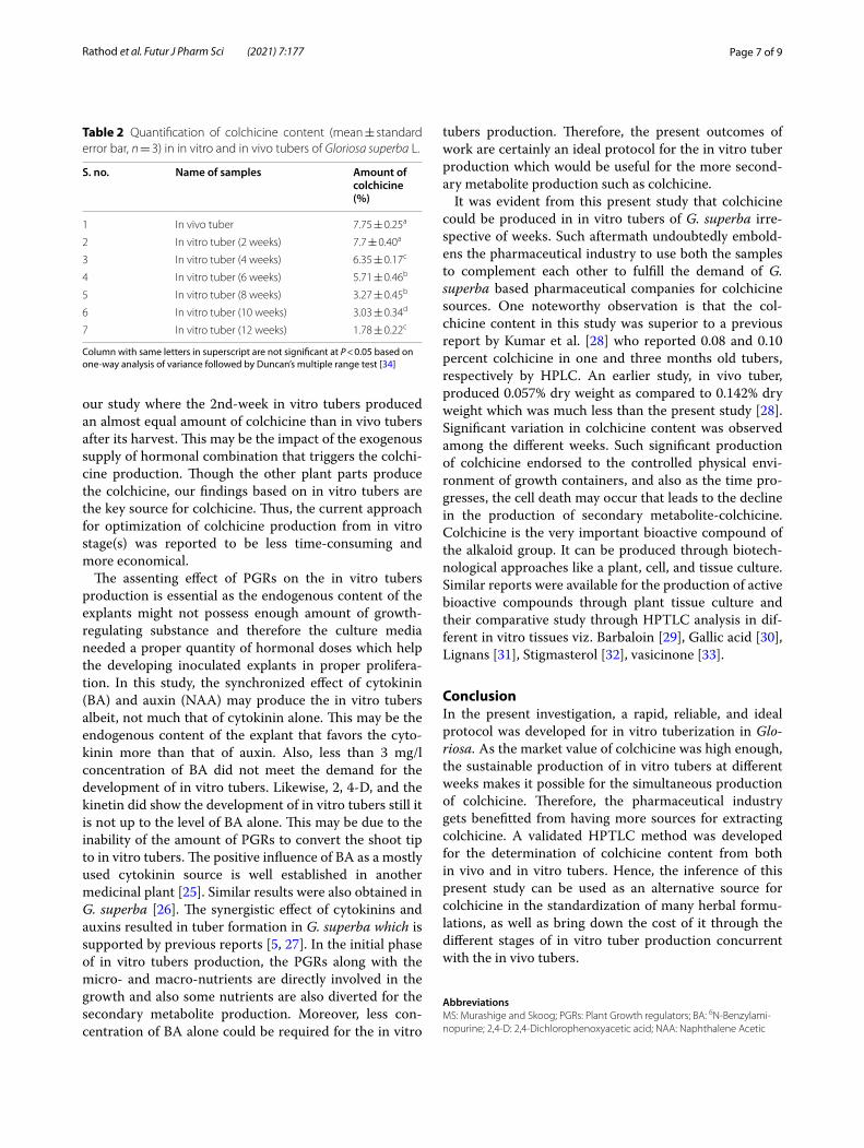
DiscussionThe fast development of plant tissue culture and cell cul-ture leads to the encouragement of the pharmaceutical industry to isolate more secondary metabolites on a large
scale [23, 24]. Due to these biotechnological tools, sec-ondary metabolite production can be achieved through-out the year. At times, the in vitro products produced a much higher amount within less time than in vivo as in
Fig. 4 3-D overlay chromatogram for specificity at 254 nm
Fig. 5 3-D chromatogram of colchicine in all tracks (standard and samples). 1st–5th tracks meant for standard, 6th–19th tracks belong to in vitro tuber samples in duplicate application
Page 7 of 9Rathod et al. Futur J Pharm Sci (2021) 7:177
our study where the 2nd-week in vitro tubers produced an almost equal amount of colchicine than in vivo tubers after its harvest. This may be the impact of the exogenous supply of hormonal combination that triggers the colchi-cine production. Though the other plant parts produce the colchicine, our findings based on in vitro tubers are the key source for colchicine. Thus, the current approach for optimization of colchicine production from in vitro stage(s) was reported to be less time-consuming and more economical.
The assenting effect of PGRs on the in vitro tubers production is essential as the endogenous content of the explants might not possess enough amount of growth-regulating substance and therefore the culture media needed a proper quantity of hormonal doses which help the developing inoculated explants in proper prolifera-tion. In this study, the synchronized effect of cytokinin (BA) and auxin (NAA) may produce the in vitro tubers albeit, not much that of cytokinin alone. This may be the endogenous content of the explant that favors the cyto-kinin more than that of auxin. Also, less than 3 mg/l concentration of BA did not meet the demand for the development of in vitro tubers. Likewise, 2, 4-D, and the kinetin did show the development of in vitro tubers still it is not up to the level of BA alone. This may be due to the inability of the amount of PGRs to convert the shoot tip to in vitro tubers. The positive influence of BA as a mostly used cytokinin source is well established in another medicinal plant [25]. Similar results were also obtained in G. superba [26]. The synergistic effect of cytokinins and auxins resulted in tuber formation in G. superba which is supported by previous reports [5, 27]. In the initial phase of in vitro tubers production, the PGRs along with the micro- and macro-nutrients are directly involved in the growth and also some nutrients are also diverted for the secondary metabolite production. Moreover, less con-centration of BA alone could be required for the in vitro
tubers production. Therefore, the present outcomes of work are certainly an ideal protocol for the in vitro tuber production which would be useful for the more second-ary metabolite production such as colchicine.
It was evident from this present study that colchicine could be produced in in vitro tubers of G. superba irre-spective of weeks. Such aftermath undoubtedly embold-ens the pharmaceutical industry to use both the samples to complement each other to fulfill the demand of G. superba based pharmaceutical companies for colchicine sources. One noteworthy observation is that the col-chicine content in this study was superior to a previous report by Kumar et al. [28] who reported 0.08 and 0.10 percent colchicine in one and three months old tubers, respectively by HPLC. An earlier study, in vivo tuber, produced 0.057% dry weight as compared to 0.142% dry weight which was much less than the present study [28]. Significant variation in colchicine content was observed among the different weeks. Such significant production of colchicine endorsed to the controlled physical envi-ronment of growth containers, and also as the time pro-gresses, the cell death may occur that leads to the decline in the production of secondary metabolite-colchicine. Colchicine is the very important bioactive compound of the alkaloid group. It can be produced through biotech-nological approaches like a plant, cell, and tissue culture. Similar reports were available for the production of active bioactive compounds through plant tissue culture and their comparative study through HPTLC analysis in dif-ferent in vitro tissues viz. Barbaloin [29], Gallic acid [30], Lignans [31], Stigmasterol [32], vasicinone [33].
ConclusionIn the present investigation, a rapid, reliable, and ideal protocol was developed for in vitro tuberization in Glo-riosa. As the market value of colchicine was high enough, the sustainable production of in vitro tubers at different weeks makes it possible for the simultaneous production of colchicine. Therefore, the pharmaceutical industry gets benefitted from having more sources for extracting colchicine. A validated HPTLC method was developed for the determination of colchicine content from both in vivo and in vitro tubers. Hence, the inference of this present study can be used as an alternative source for colchicine in the standardization of many herbal formu-lations, as well as bring down the cost of it through the different stages of in vitro tuber production concurrent with the in vivo tubers.
AbbreviationsMS: Murashige and Skoog; PGRs: Plant Growth regulators; BA: 6N-Benzylami-nopurine; 2,4-D: 2,4-Dichlorophenoxyacetic acid; NAA: Naphthalene Acetic
Table 2 Quantification of colchicine content (mean ± standard error bar, n = 3) in in vitro and in vivo tubers of Gloriosa superba L.
Column with same letters in superscript are not significant at P < 0.05 based on one-way analysis of variance followed by Duncan’s multiple range test [34]
S. no. Name of samples Amount of colchicine (%)
1 In vivo tuber 7.75 ± 0.25a
2 In vitro tuber (2 weeks) 7.7 ± 0.40a
3 In vitro tuber (4 weeks) 6.35 ± 0.17c
4 In vitro tuber (6 weeks) 5.71 ± 0.46b
5 In vitro tuber (8 weeks) 3.27 ± 0.45b
6 In vitro tuber (10 weeks) 3.03 ± 0.34d
7 In vitro tuber (12 weeks) 1.78 ± 0.22c

Page 8 of 9Rathod et al. Futur J Pharm Sci (2021) 7:177
Acid; HPTLC: High-Performance Thin-Layer Chromatography; LOD: Limit of Detection; LOQ: Limit of Quantification.
AcknowledgementsThe authors are highly thankful to the University Grant Commission, New Delhi to provide the grant as RGNF, JRF for this study. We are also thankful to Head, Department of Life Sciences, HNGU, Patan and Principal, Pramukh Swami Science and H.D. Patel Arts College, Kadi, Gujarat, India for providing necessary facilities to carried out this study.
Plant authenticationThe explants of Gloriosa superba L. were collected from the local field of
Palanpur (24°09′21″N 72°24′27″E), Gujarat, India in the month of September. The specimen was identified and authenticated by Dr. Illa C. Patel, Associate Prof, Department of Life Sciences, Hemchandracharya North Gujarat Univer-sity, Patan.
Authors’ contributionsDR, JP and IP conceptualized and designated the study. DR carried out the experiments. JP and DR analyzed the data. IP guided for the manuscript preparation. DR and JP prepared the manuscript. All authors have read and approved the manuscript.
FundingThe authors are thankful to the University Grant Commission, New Delhi to provide the grant as RGNF, JRF (F1-17.1/2012-13/RGNF-2012-13-SC-GUJ-30906/(SA-III/Website) for this study.
Availability of data and materialsAll analyzed data are included in this manuscript. Materials and data are avail-able upon reasonable request.
Declarations
Ethics approval and consent to participateNot applicable.
Consent for publicationNot applicable.
Competing interestsThe authors declare that they have no competing interests.
Author details1 Department of Botany, H.V.H.P. Institute of Post Graduate Studies and Research, Kadi, Gujarat 382715, India. 2 Department of Biotechnology, Shri A.N. Patel P.G. Institute of Science and Research, Anand, Gujarat 388001, India. 3 Department of Life Sciences, Hemchandracharya North Gujarat University, Patan, Gujarat, India.
Received: 1 April 2021 Accepted: 22 August 2021
References 1. Samy RP, Thwin MM, Gopalkrishnakone P, Ignacimurthy S (2008) Ethano-
botanical survey of folk plants for the treatment of snakebites in southern part of Tamilnadu. Indian J Ethanopharmacol 115:302–312
2. Veeraiah S, Reddy JK (2012) Current strategic approaches in ethnome-dicinal plants of Tinospora cordifolia and Gloriosa superba—a review. Int J Pharm Bio Sci 3:321–326
3. Jana S, Shekhawat GS (2011) Critical review on medicinally potent plant species: Gloriosa superba. Fitoterapia 82(3):293–301. https:// doi. org/ 10. 1016/j. fitote. 2010. 11. 008
4. Sivakumar G, Krishnamurthy KV, Hahn EJ, Paek KY (2004) Enhanced in vitro production of colchicine in Gloriosa superba L.—an emerging industrial medicinal crop in South India. J Hortic Sci Biotechnol 79:602–605. https:// doi. org/ 10. 1080/ 14620 316. 2004. 11511 813
5. Custers JBM, Bergervoet JHW (1994) Micropropagation of Gloriosa: towards a practical protocol. Sci Hortic 57:323–334. https:// doi. org/ 10. 1016/ 0304- 4238(94) 90115-5
6. Sivakumar G, Krishnamurthi KV, Rajendran TD (2003) In vitro corm pro-duction in Gloriosa superba L., an Ayurvedic medicinal plant. J Hortic Sci Biotechnol 78:450–453. https:// doi. org/ 10. 1080/ 14620 316. 2003. 11511 648
7. Ade R, Rai M (2010) Colchicine, current advances and future prospects. Nusant Biosci 2:90–96. https:// doi. org/ 10. 13057/ nusbi osci/ n0202 07
8. Dicosmo F, Misawa M (1995) Plant cell and tissue culture: alternatives for metabolite Production. Pergamon Biotechnol Adv 13:425–453. https:// doi. org/ 10. 1016/ 0734- 9750(95) 02005-N
9. Mulabagal V, Tsay H (2004) Plant cell cultures: an alternative and efficient source for the production of biologically important secondary metabo-lites. Int J Appl Sci Eng 2:29–48. https:// doi. org/ 10. 1016/ S0168- 1656(98) 00195-3
10. Vanisree M, Lee C, Lo S, Nalawade SM, Lin CY, Tsay H (2004) Studies on the production of some important secondary metabolites from medicinal plants by plant tissue cultures. Bot Bull Acad Sin 45:1–22
11. Patel R, Patel M, Dubey N, Patel B (2012) HPTLC method development and validation: strategy to minimize methodological failures. J Food Drug Anal 20:794–804. https:// doi. org/ 10. 6227/ jfda. 20122 00408
12. Agatonovic-Kustrin S, Morton DW, Mizaton HH, Zakaria H (2018) The relationship between major polyphenolic acids and stigmasterol to antioxidant activity in different extracts of Myrmecodia platytyrea. S Afr J Bot 115:94–99
13. Jason RJ, Mohamed IM, Selva GR, Sankar M (2014) Quantification of colchicine in various parts of Gloriosa superba by HPLC. J Chem Pharm Sci 2:53–55
14. Finnie JF, Van Staden J (1991) Isolation of colchicine from Sandersonia aurantiaca and Gloriosa superba variation in the alkaloid levels of plants grown in vivo. J Plant Physiol 138:691–695. https:// doi. org/ 10. 1016/ S0176- 1617(11) 81317-9
15. Sarin R, Rishi A, Kumar A (2010) In vivo and In vitro estimation of colchi-cine in Gloriosa superba L. by high pressure liquid chromatography. J Exp Sci 1:1–2
16. Basak UC, Dash D, Mahapatra AK (2012) Estimation of colchicine in tubers of Gloriosa superba L. originated from different agroclimatic zones of Odisha, India. Int J Pharmacogn Phytochem Res 4:157–161
17. Kavina J, Gopi R, Panneerselvam R (2011) Quantification of colchicine in seed and tuber samples of Gloriosa superba by high performance liquid chromatography method crop growth as evidenced by increased seed-ling emergence. J Appl Pharm Sci 1:116–119
18. Murashige T, Skoog F (1962) A revised medium for rapid growth and bio assays with tobacco tissue cultures. Physiol Plant 15:473–497
19. Rathod D, Patel I (2016) Comparative preliminary phytochemical studies of in vivo and in vitro extracts of Gloriosa superba L. Int J Pharm Bio Sci 7:20–23
20. Das K, Tiwari RKS, Shrivastava DK (2010) Techniques for evaluation of medicinal plant products as antimicrobial agent: current methods and future trends. J Med Plants Res 4:104–111. https:// doi. org/ 10. 5897/ JMPR09. 030
21. ICH Harmonized Tripartite Guideline (2005) Validation of analytical pro-cedures: text and methodology Q2(R1). In: International conference on harmonization, Geneva, Switzerland
22. Alam P, Ali M, Singh R, Shakeel F (2011) A new HPTLC densitometric method for analysis of swertiamarin in Enicostemma littorale and com-mercial formulations. Nat Prod Res 25:17–25. https:// doi. org/ 10. 1080/ 14786 41100 37543 48
23. Debnath M, Pandey M, Chikara SK (2011) Physiological and molecular characterization of in vitro cultures of an endemic medicinal herb, Chloro-phytum borivilianum, under abiotic stress. Afr J Biotechnol 10:7356–7366. https:// doi. org/ 10. 5897/ AJB11. 445
24. Gantait S, Mandal N, Das PK (2011) In vitro accelerated mass propagation and ex vitro evaluation of Aloe vera L. with aloin content and superoxide dismutase activity. Nat Prod Res 25:1370–1378. https:// doi. org/ 10. 1080/ 14786 419. 2010. 541885
25. Panigrahi J, Gantait S, Patel IC (2017) An efficient in vitro approach for direct regeneration and callogenesis of Adhatoda vasica Nees, a potential source of quinazoline alkaloids. Natl Acad Sci Lett. https:// doi. org/ 10. 1007/ s40009- 017- 0596-8

Page 9 of 9Rathod et al. Futur J Pharm Sci (2021) 7:177
26. Ghosh S, Ghosh B, Jha S (2007) In vitro tuberisation of Gloriosa superba L. on basal medium. Sci Hortic 114:220–223. https:// doi. org/ 10. 1016/j. scien ta. 2007. 06. 008
27. Kolar AB, Basha MG (2014) In vitro tuberization and quantitative analysis of colchicine using hptlc in Gloriosa superba L. an endangered medicinal plant of Pachamalai hills, a part of Eastern Ghats, Tamil Nadu. Int J Pharma Bio Sci 5:300–310
28. Kumar CN, Jadhav SK, Tiwari KL, Afaque Q (2015) In vitro tuberization and colchicine content analysis of Gloriosa superba L. Biotechnology 14:142–147. https:// doi. org/ 10. 3923/ biote ch. 2015. 142. 147
29. Pandey D, Parida S, Dey A (2016) Comparative HPTLC analysis of bioactive marker barbaloin from in vitro and naturally grown Aloe vera. Rev Bras 26:161–167. https:// doi. org/ 10. 1016/j. bjp. 2015. 08. 016
30. Pandey DK, Dey SA (2015) Comparative HPTLC analysis of antioxidant compound gallic acid from in vitro and naturally grown Stevia rebaudi-ana. J Biol Act Prod Nat 5(6):397–405. https:// doi. org/ 10. 1080/ 22311 866. 2014. 902753
31. Ekiert RJ, Szopa A, Ekiert H, Krzek J, Dzik E (2013) Analysis of lignans in Schisandra chinensis fruits, leaves, biomasses from in vitro cultures and
food supplements. J Funct Foods 5:1576–1581. https:// doi. org/ 10. 1016/j. jff. 2013. 06. 008
32. Panigrahi J, Patel IC (2016) Quantification of stigmasterol under in vivo and in vitro plant extracts of Chlorophytum sps. Int J Pharm Bio Sci 7:278–283
33. Panigrahi J, Gantait S, Patel IC (2017) Concurrent production and rela-tive quantification of vasicinone from in vivo and in vitro plant parts of Malabar nut (Adhatoda vasica Nees). 3 Biotech 7(5):1–8. https:// doi. org/ 10. 1007/ s13205- 017- 0882-7
34. Duncan D (1955) Multiple range and multiple F tests. Biometrics 11(1):1–42. https:// doi. org/ 10. 2307/ 30014 78
Publisher’s NoteSpringer Nature remains neutral with regard to jurisdictional claims in pub-lished maps and institutional affiliations.