CLINICAL PROFILE, NATURAL HISTORY AND FACTORS...
68
CLINICAL PROFILE, NATURAL HISTORY AND FACTORS DETERMINING PROGNOSIS/OUTCOME STUDY OF CEREBRAL VENOUS SINUS THROMBOSIS. Thesis submitted in fulfilment of the rules and regulations for DM Degree Examination of Sree Chitra Tirunal Institute for Medical Sciences and Technology, Thiruvananthapuram By Dr. Srinivas G Resident in Neurology Month and Year of Submission: October 2011
Transcript of CLINICAL PROFILE, NATURAL HISTORY AND FACTORS...

CLINICAL PROFILE, NATURAL HISTORY AND FACTORS DETERMINING PROGNOSIS/OUTCOME STUDY OF
CEREBRAL VENOUS SINUS THROMBOSIS.
Thesis submitted in fulfilment of the rules and regulations for DM
Degree Examination of Sree Chitra Tirunal Institute for Medical
Sciences and Technology, Thiruvananthapuram
By
Dr. Srinivas G
Resident in Neurology
Month and Year of Submission: October 2011

CERTIFICATE
I, Dr. Srinivas G hereby declare that I have actually carried out
the project under report.
Place: Thiruvananthapuram Signature:
Date: 30‐09‐2009 Dr. SrinivasG
Senior resident in Neurology
Forwarded. He has carried out the project under report.
Signature: Signature:
Prof. M. D. Nair (Thesis Guide) Prof. M. D. Nair
Senior Professor & Head Senior Professor & Head
Department of Neurology Department of Neurology
SCTIMST. SCTIMST.

.
Acknowledgement
I take this opportunity to sincerely thank Prof. Muralidharan Nair
HOD Neurology, my guide, for expert guidance, constructive suggestions, and
constant encouragement at each and every stage during this study.
I sincerely thank Dr Mohammad Wasay from Pakistan for instructive, data
sharing, discussion , during study
I sincerely thank Prof Sankara Sarma for carrying out the statistical analysis.
I also thank my consultants and colleagues for offering me critical inputs
I express my gratitude towards the patients who took part in this study.
Dr. SrinivasG

CONTENTS
PART I
INTRODUCTION 1-4
REVIEW OF LITERATURE 5-26
AIMS AND OBJECTIVES 27- 28
MATERIALS AND METHODS 29 – 32
RESULTS 33-49
DISCUSSION 50 -57
SUMMARY AND CONCLUSIONS 58-59
BIBILIOGRAPHY 60-64

1
Introduction

2 Introduction
Cerebral venous /sinus Thrombosis (CVT) has been recognized since the early 19th century1 but
still remains a diagnostic and therapeutic challenge. Cerebral vein and sinus thrombosis is rare
compared to arterial stroke often occurs in young individuals 2 Cvt may occur at any time from
infancy to old age most reported cases were women in association with puerperium3 Onset of
symptoms may be acute sub acute or chronic4 Cerebral venous infarction is the most serious
consequence of cerebral venous thrombosis venous infarctions are often multifocal bilateral
affecting both grey matter and sub cortical white matter
Patient of CVT usually presents with headache, seizure, papilloedema, altered sensorium and focal
deficits due to thrombosis of intracranial veins and sinuses resulting in haemorrhagic infarctions
and raised intracranial tenstion2. The above features are present in various combinations ranging
from syndrome of raised intracranial pressure without localization to deep altered sensorium and
dense hemi paresis. CVT forms a distinct subgroup of cerebrovascular disease in India and is a
leading cause of mortality in women of reproductive age group3. In India, most of the cases are
seen in post partum period in women, while alcoholism is a significant risk factor in males.
Pangayara reported from India that CVT accounted for half of young stroke and 40% for stroke in
woman.
Review of CVT cases from Asian countries is suggestive of differences in risk factors profile and
outcome in these patients as compared to European studies. Largest cohort of CVT patients from
Europe (n=624) reported that 50% of these cases were related to OCP pills, 6% were due to
pregnancy and 14% were secondary to puerperium. A study of 182 adult patients with CVT from
USA reported 7% due to pregnancy and puerperium and 5% related to OCP use.5 A study from
Pakistan (n=109) patients with CVT) reported that 17% were due to pregnancy and puerperium

3 and 5% related to OCP use. Cantu from Mexico reported 59% cases due to Pregnancy
puerperium.6,7,8
Cross et al5 noted: “Usually recovery is rapid and complete if patient survives the acute episode”.
Three fourth of cases of CVT in pregnancy and puerperium reported by him, survived with good
recovery. However, in pre imaging era CVT had been diagnosed exclusively at autopsy and
therefore thought to be always lethal. After introduction of heparin in treatment of CVT mortality
has come down significantly and most of the recent studies6,7 reporting mortality < 20% compared
to earlier studies reporting mortality between 30-50%. However outcome of CVT is highly
unpredictable and it is not unusual to see dramatic recovery in deeply comatose patient and sudden
worsening in conscious patients due to extension of thrombosis. With the advent of imaging
modalities like CT scan and recently Magnetic Resonance Imaging (MRI) and Magnetic resonance
venography (MRV), the diagnosis of CVT has improved significantly. CT scan commonly shows
haemorrhagic infarctions with or without “cord”, or “empty delta” sign7. MRI and MRV, when
used in doubtful situations can clarify the diagnosis by showing thrombosed sinus of cortical veins
12,13. In fact after the introduction of MRV, many of the patients earlier diagnosed as Idiopathic
raised ICT have been noted to have sinus thrombosis giving rise to syndrome of raised intracranial
pressure without localization. Pathologically involvement of superior sagittal sinus of varying
extent with or without the involvement of transverse and sigmoid sinuses with thrombosis of
cortical veins had been reported commonly 2, 3. Haemorrhagic infarctions with mass effect and
diffused cerebral edema with herniation is also frequently seen. Involvement of deep venous
system is less common than superficial venous system but by no means rare. Due to multifactorial
causation of this condition, it will be interesting to know whether different pathophysiological
mechanisms are operating in different clinical settings. Long term outcome of cvt in this part of
the state is not well described, so this study was undertaken to Identify the etiological spectrum of
4 patients with cerebral vein /sinus thrombosis Further to attempt correlation between site of
venous occlusion and clinical parameters Prognosis of CVT
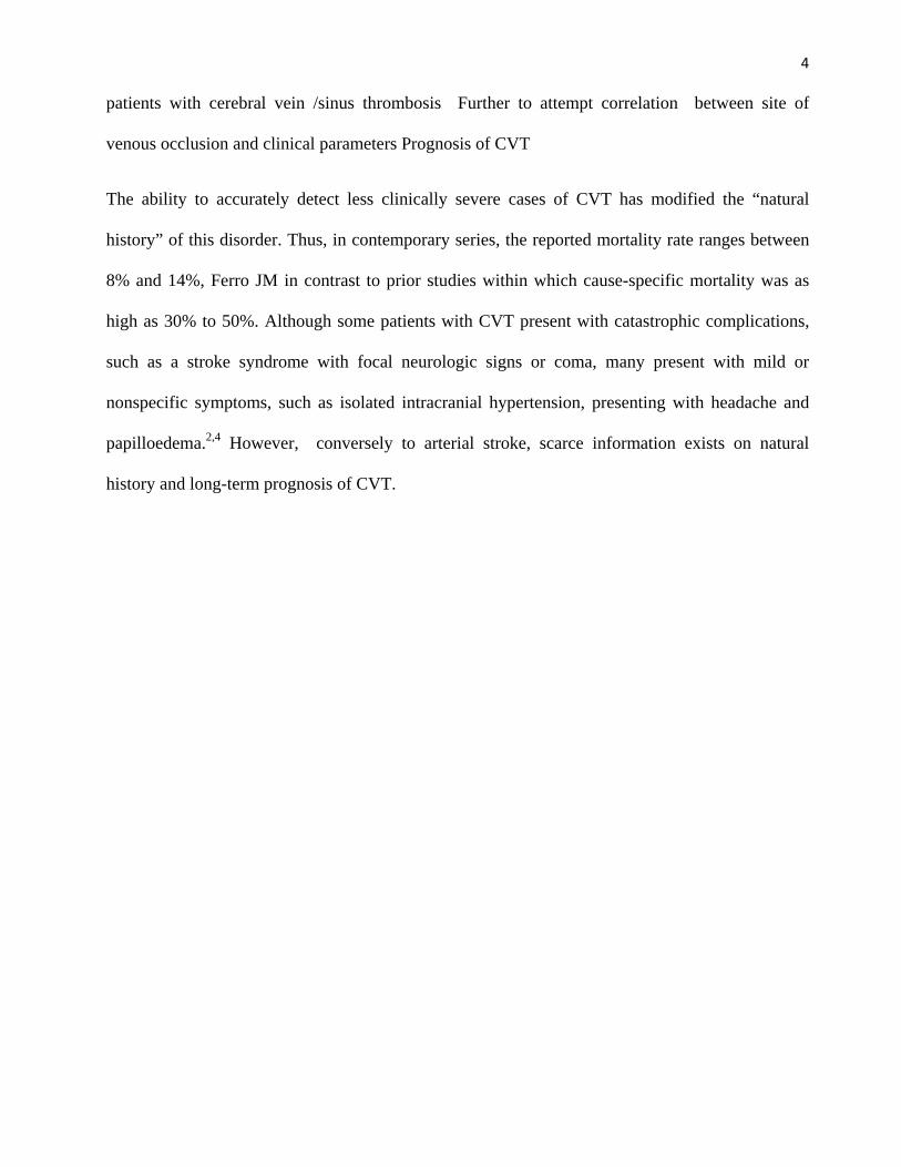
The ability to accurately detect less clinically severe cases of CVT has modified the “natural
history” of this disorder. Thus, in contemporary series, the reported mortality rate ranges between
8% and 14%, Ferro JM in contrast to prior studies within which cause-specific mortality was as
high as 30% to 50%. Although some patients with CVT present with catastrophic complications,
such as a stroke syndrome with focal neurologic signs or coma, many present with mild or
nonspecific symptoms, such as isolated intracranial hypertension, presenting with headache and
papilloedema.2,4 However, conversely to arterial stroke, scarce information exists on natural
history and long-term prognosis of CVT.

5
Review of
Literature

6 Review of literature
Cerebral venous thrombosis or sino-venous thrombosis, as the name implies is a condition which
involves cerebral venous sinuses and veins together or independent of each other with thrombotic
event of varied temporal evolution. The clinical presentation is varied ranging from syndrome of
raised ICT without localization to seizures, focal deficits and deep altered sensorium 2, 3. Some
patients may even present as behavioral disturbances as the predominant clinical manifestation,
confusing the picture with post partum Psychosis. Strokes resulting from cerebral venous
thrombosis usually affects young persons particularly women in reproductive age group, and carry
a high mortality if not managed adequately10. The term Primary or Idiopathic Cerebral Venous
thrombosis is used when no specific etiological factor is evident. ‘Secondary’ sino-venous
thrombosis results from a variety of causes that include injury, infection, hematological
disturbances, dehydration etc11.
Historical Background
The wide spectrum of clinical features in cerebral venous thrombosis, the varied and changing
etiological factors and the apparent “rarity” of the condition had made advances in knowledge
slow and uneven. Periods of relative neglect has been interspersed with burst of enthusiastic
discussion. The earliest reference to cerebral venous sinus thrombosis was that of Ribes in 18241.
He described in detail the clinical and post mortem findings of 45 yr old man who had thrombosis
of superior sagittal and lateral sinuses, subdural effusion and metastatic carcinoma in the brain.
The first case of puerperal venous thrombosis was reported by John Abercrombie in 1828. His
patient, a 24 year old woman, developed headache, delirium and initially right sided than

7 generalized seizures at the beginning of second week after delivery. Autopsy showed ischemic
and haemorrhagic infarcts with thrombosed and sclerosed cortical veins. Quinke and Nonne
identified the clinical syndrome of pseudo tumor cerebri (a term coined by latter) as a clinical
counterpart to sinus thrombosis. Kalbag and Woolf, Sir Charles Symonds and others gave a precise
clinical description of CVT after 1940. After introduction of CT scan and recently used MRI with
MRV diagnosis of CVT has become simpler as these imaging modalities are quite sensitive in
detecting CVT. Several large series with confirmation of diagnosis by angiograms, surgical
exploration, and autopsy and recently with CT and MRI studies have been reported from Indian
subcontinent, 3,8
Epidemiology
The true incidence of CVT is unknown. Ehlers and Courville found only 16 superior sagittal sinus
thrombosis in a series of 12,500 autopsies (0.12%) 18. Towbin found CVT in 9% of 182
consecutive autopsies19. However, with the more recent reports of large clinical series, the true
incidence of CVT is probably considerably higher than that derived from autopsy series. Exact
figures however remain elusive. People of all age groups may be affected by CVT but there is
preponderance in young women because of specific causes like use of oral contraceptives,
pregnancy and puerperium. Puerperal CVT has been reported to account for upto 15-20% of
‘young stroke”. It is the commonest cause of stroke in young women in India. 50% of strokes in
Indian women are related to pregnancy and puerperium and 95.5% of these are due to CVT5. In
Western countries, the incidence of CVT related to pregnancy and puerperium ranges from 1 in
1666-10,000 pregnancies. Risk factors like hyper homocystenemia, OCP use, alcoholism
,procoagulant state are increasingly recognized in addition to the conventional risk factors like
postpartum state.

8 Relevant Venous Anatomy 20
The cerebral venous system comprises of cerebral veins that empty into dural sinuses which then
drain the blood into two internal jugular. Embryologically, the entire cephalic drainage may be
subdivided into an outer and superficial segment, which drains the scalp, underlying muscle, and
tendons; and intermediate segment which drains the skull, diploe and dura matter; and a cerebral
segment, consisting of the veins that drain the brain. The cerebral segment may be further
subdivided into a superficial cerebral group of veins and a deep cerebral group of veins. The
superficial cerebral veins coalesce on the Pial surface draining out the blood from the outer 1 or
2cm. of cortex and the underlying white matter. Venous blood in these vessels travels in a
centrifugal direction and ultimately terminates in one of the dural sinuses. The deep cerebral veins
serve to drain blood in a centripetal direction away from deep white matter, the basal ganglia, and
the diencephalons. Tributaries draining many of the deep structure of the cerebrum join veins in
the lateral angles of the ventricles and form a sub ependymal plexus. The veins of this plexus
empty into the internal cerebral veins, which join the great cerebral vein of Galen.
Superior Sagittal Sinus (SSS)
SSS lies in the attached border of the falx cerebri and runs from the foramen caecum to the
occipital protuberance, where it joins straight sinus, lateral sinus and torcular herophili i.e.
confluence of sinuses. The anterior part is narrow or sometimes absent or replaced by two superior
cerebral veins that join behind the coronal suture, consequently anterior part of sinus is often
poorly visualized on angiography and its isolated lack of filling is not sufficient to indicate
thrombosis. The SSS receives superficial cerebral veins and drains major part of cortex. It also

9 receives diploic veins, themselves connected to scalp veins by emissary veins, which explains the
incidence of SSS thrombosis after cutaneous infections and contusions SSS and other sinuses play
a major role in CSF circulation because they contain most of the arachnoid villi and granulations in
which much of the CSF absorption takes place. Thus, there is a direct dependency of CSF pressure
accounting for the frequency of raised intracranial pressure, in SSS or Lateral sinus (LS)
thrombosis.
Lateral Sinus
The lateral sinus extend from the torcular herophili to the jugular bulb and consist of the transverse
and sigmoid portions. They drain blood from cerebellum, brain stem and posterior portion of
cerebral hemispheres. They also receive some veins from middle ear, another possible source of
septic thrombosis. Numerous LS anatomic variations may be misinterpreted in sinus occlusions on
angiography. The right LS is more often the direct continuation of the SSS and is frequently larger
than the left LS which receives most of its supply from straight sinus. In Hacker’s study,
transverse portions were not visualized on ipsilateral carotid Angiogram in 14% of cases on left
side and 33% on right side, whereas sigmoid portions, which may be directly injected via cerebral
veins, failed to fill in 4% of cases on left side and were always demonstrated on right. An isolated
lack of filling of a left transverse sinus is more suggestive of hypoplasia than of thrombosis.
Cavernous Sinus
This sinus drains venous blood from the orbits through the ophthalamic veins and from anterior
part of base of brain via the sphinopalatine sinus and middle cerebral veins. They empty into both
superior and interior petrosal sinuses and ultimately into internal jugular veins. Because of their
situation, cavernous sinuses are often thrombosed in relation to infections of face or sphenoid
sinusitis. In contrast to other sites, infection is the leading cause of cavernous sinus thrombosis.

10 Cerebral veins
They can be roughly divided into 3 groups:
1. Superficial cerebral veins
2. Deep cerebral veins
3. Veins of posterior fossa
Superficial cerebral veins: Some of the cortical veins- the frontal, parietal, occipital and superior
cerebral veins drain the cortex ascending to SSS whereas others, mainly the middle cerebral veins
drain into the cavernous sinus. Trolard’s great anastomotic vein connects the SSS to middle
cerebral veins, which are then connected to LS by vein of labbe. The cortical veins present some
peculiarities that are important to know to understand some of the clinical features of CVT. They
have thin walls, no muscle fibres and no valves. These features allow for dilatation and reversal of
the direction of the blood flow when the sinus into which they drain are occluded. They are linked
by various anastomoses, allowing development of collateral circulation (angiographically visible
as corkscrew vessels). This probably explains the good prognosis of some of the thrombosis.
Deep Cerebral Veins: The internal cerebral and basal veins both join to form great vein of Galen,
which continues as straight sinus drain blood from deep white matter of the cerebral hemispheres
and from Basal ganglia. In contrast to superficial system, the deep system anatomy is constant and
is always visualized on angiography, so that thrombosis is easily recognized.
Veins of posterior fossa: There are 3 groups:
1. Superior veins draining into Galenic system
2. Anterior veins drawing petrosal sinuses
3. Posterior veins draining into the torcular or neighboring SS and LS
They are variable in course and angiographic diagnosis of their occlusion is extremely difficult.

11 Microscopic Anatomy of Cerebral Veins and Sinuses
Capillaries open in to cerebral venules which apart from their wider lumen are indistinguishable
from them. These venules join the small medullary or cortical veins. These vessels reach the
ventricular or cortical surface, either directly or indirectly, after fusion with neighboring veins
forming larger vascular stems. The walls of cerebral veins consist of an endothelial lined tunica
intima. Surrounding the endothelium is a thin adventitial layer. Veins do not have clearly defined
muscular layer or values. There is little to suggest than the veins receive vasomotor innervations.
As these vessels approach their destination they become more fibrous and resemble closely the
structure of dural sinus. The strategic location of the sinuses within the major folds or junctions of
dura, the firm attachment of the dura to bone, and its tough fibrous consistency maintains patency
of the sinuses all the time. The walls of dural venous sinuses consist of an inner lining of
endothelium and an outer layer essentially the same as dura elsewhere. The outer layer consists
chiefly of fibroblasts and large interlaced bundles of collagenous fibres. A few nerve fibres,
presumably afferent have been reported to innervate along the dural venous sinuses. The walls of
the venous sinuses are not uniformly smooth and in certain locations like middle third of SSS are
thrown into folds or membranous irregularities. According to one hypothesis, these folds may
perform valve like action, while other authors suggested that they may be important in maintaining
laminar flow.

12 Causes of CVT
Several medical, surgical and gynaeco-obstetric ailments as well as a number of regional causes
like infective, trauma, tumors etc. have been implicated in the causation and predisposition to
CVT, Table (1) lists the recognized causes or predisposing conditions11.
Table 1: Causes of cerebral venous thrombosis
A. SEPTIC DURAL SINUS THROMBOSIS
Local- Septic Trauma
Intracranial infections: Abscess, empyema and meningitis
Otitis
Sinusitis
Tonsillitis
Stomatitis
Systematic - Bacterial (Typhoid, TB, Septicemia, Endocarditis)
Viral (Measles virus, Hepatitis viruses, Herpes Simplex virus, HIV,
Cytomegalovirus)
Parasitic (Malaria, Trichinosis)
Fungal (Aspergillosis)
B. NONSEPTIC DURAL SINUS THROMBOSIS
Altered hemodynamic states
Dehydration

13
Fever
Cardiac failure
Hematological disorders:
Polycythemia Vera
Secondary polycythemia
Disseminated intravascular coagulation
Sickle cell anemia and trait
Cryoglobulinemia
Paroxysmal nocturnal hemoglobinuria
Thrombocytosis
Severe anemia
Antithrombin – III deficiency
Protein C & S deficiency
Antiphospholipid antibody syndrome
Hormonal dysfunction
Oral contraceptive use
Pregnancy and puerperium
Androgens
Trauma
Penetrating & non-penetrating head injuries
Surgery
Cardiac pacemakers
Jugular venous catheters
Metabolic disorders
Homocystinuria

14
Osteopetrosis
Diabetes mellitus
Neoplasia
Meningioma
Metastasis (usually hematogenous)
Inflammatory disorders
Behcet’s disease
Sarcoidosis
SLE
Wegener’s granulomatosis
Polyarteritisn odosa
Inflammatory bowel disease
Ulcerative colitis
Crohn’s disease
Cogan syndrome
Vascular disorders
Arterio-venous malformation
Arterial occlusions
Sturge weber syndrome
Clinical profile:
The spectrum of symptoms and signs among patients with CVT is remarkably variable. Patients
present with varying combination of headache, seizures, aphasia, behavioral abnormality, altered
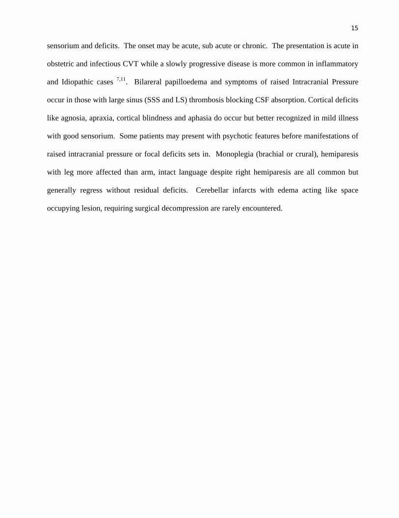
15 sensorium and deficits. The onset may be acute, sub acute or chronic. The presentation is acute in
obstetric and infectious CVT while a slowly progressive disease is more common in inflammatory
and Idiopathic cases 7,11. Bilareral papilloedema and symptoms of raised Intracranial Pressure
occur in those with large sinus (SSS and LS) thrombosis blocking CSF absorption. Cortical deficits
like agnosia, apraxia, cortical blindness and aphasia do occur but better recognized in mild illness
with good sensorium. Some patients may present with psychotic features before manifestations of
raised intracranial pressure or focal deficits sets in. Monoplegia (brachial or crural), hemiparesis
with leg more affected than arm, intact language despite right hemiparesis are all common but
generally regress without residual deficits. Cerebellar infarcts with edema acting like space
occupying lesion, requiring surgical decompression are rarely encountered.
16
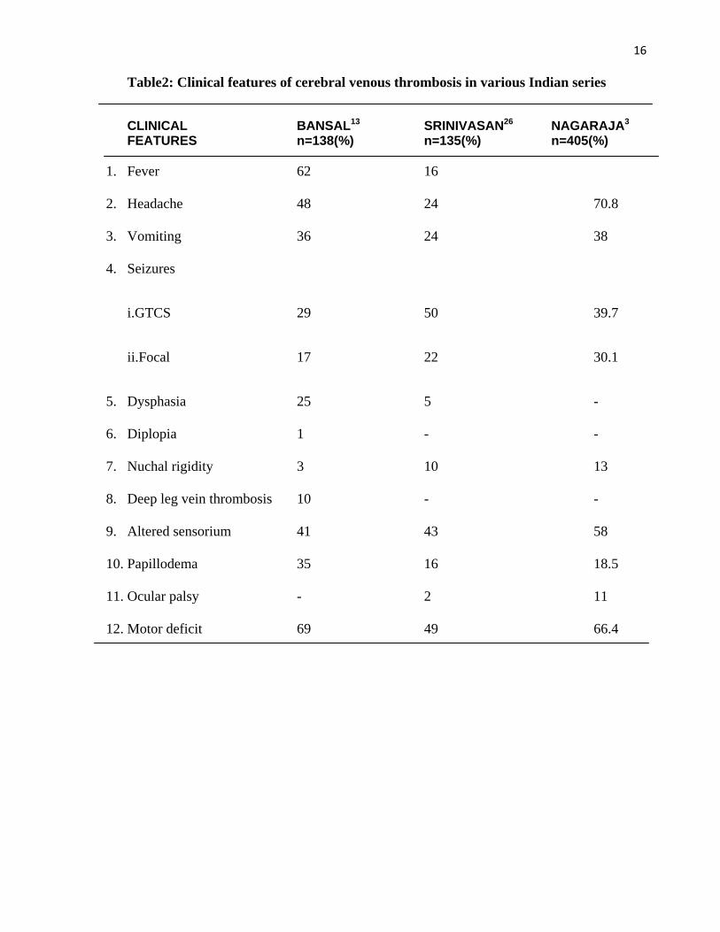
Table2: Clinical features of cerebral venous thrombosis in various Indian series
CLINICAL BANSAL13 SRINIVASAN26 NAGARAJA3 FEATURES n=138(%) n=135(%) n=405(%)
1. Fever 62 16
2. Headache 48 24 70.8
3. Vomiting 36 24 38
4. Seizures
i.GTCS 29 50 39.7
ii.Focal 17 22 30.1
5. Dysphasia 25 5 -
6. Diplopia 1 - -
7. Nuchal rigidity 3 10 13
8. Deep leg vein thrombosis 10 - -
9. Altered sensorium 41 43 58
10. Papillodema 35 16 18.5
11. Ocular palsy - 2 11
12. Motor deficit 69 49 66.4

17
Table 1 Clinical presentation in the two largest series of CVT patients
Ferro et al.: 624 patients Headache 88.8% Visual loss 13.2% Papilledema 28.3% Diplopia 13.5% Stupor or coma 13.9% Aphasia 19.1% Mental status disorders 22% Any paresis 37.2% Any seizure 39.3% Sensory symptoms 5.4% Other focal cortical sign 3.4%
Wasay et al.: 182 patients Headache 71% Generalized weakness 54% Focal motor or sensory deficit 36% Nauese/vomiting 35% Seizures 32% Walking difficulty 30% Drowsiness 28% Visual blurring 23% Dizziness 21% Behavioural symptoms 18%
Slurred speech/inability to speak 16% Coma 15% Fever 14%
In children and elderly, CVT may present with lethargy and stupor with headache without
any focal deficits. Nagaraja et al3 grouped clinical features of CVT in four categories depending
upon the topographical venous involvement.
1. Presentation with seizures, focal deficits and progressively deteriorating consciousness.
Thrombosis involves the dural sinuses as well as cortical veins producing cerebral infarction.
Seizures may be focal, multi focal or generalized. Paralysis may be unilateral or bilateral and
is usually maximal in lower limbs. Later during the course, patient may manifest signs of
tentorial or central herniation leading to coma and death.

18 2. Presentation with symptoms and sings of raised intracranial tension namely headache,
vomiting and papilloedema. If thrombosis continues to dural sinuses, the course is usually
slow and prognosis is favourable.
3. Occasionally, thrombosis predominantly involves cortical veins and patient may present with
feature of space occupying lesion.
4. Rarely, thrombosis predominantly involves the deep venous system. Patient manifest
symptoms of raised intracranial tension, focal deficits, choreoathetosis, ocular sings and coma.
It runs a fulminant course
Cavernous sinus thrombosis is usually due to spread of infection from face, para nasal sinus or
intracranial venous sinuses. It has a distinctive clinical picture where patient presents with fever,
chills, toxemia with proptosis, chemosis and painful ophthalomoplegia, initially unilateral but
often becoming bilateral. Papilloedema and retinal haemorrhages indicate retinal vein thrombosis

19 Table4 : Clinical features described by Ameri et al18 1992
Headache 83(75%)
Papilloedema 54(49%)
Motor or sensory deficit 38(34%)
Seizures 41(37%)
Drowsiness, mental changes, confusion or coma 33(30%)
Dysphasia 13(12%)
Multiple cranial nerve palsies 13(12%)
Nystagmus 2(2%)
Hearing loss 2(2%)
Bilateral or alternating cortical signs 3(3%)
Cerebellar incordination 3(3%)
In another series Cantu et al7 from Mexico analyzed clinical radiological features of 113 cases of
CVT comparing obstetrical cause related CVT to non-obstetrical cause related CVT. Clinical
features in this series was similar to other series but in “obstetrical group” symptoms evolved more
rapidly and the outcome in terms of mortality was less.

20 Table 5: Neurological findings in puerperal and non-puerperal CVT. Cantu et al 7 1993:
Puerperal Non-Puerperal (n=67) (n=46) Findings n% n%
1. Headache 59 88.0 32 69.5
2. Focal signs 53 79.1 35 76.0
• Motor 52 77.6 33 71.7
• Sensory 25 37.3 13 28.2
3. Aphasia 17 25.3 10 21.7
4. Disorders of consciousness 42 62.6 27 58.6
5. Somnolence 24 35.8 9 19.5
6. Stupor/coma 16 23.9 15 32.6
7. Confusion 2 2.9 3 6.5
8. Seizures 40 59.7 29 63.0
• Generalized 18 26.9 16 34.8
• Focal 22 32.8 13 28.2
9. Bilateral pyramidal signs 28 41.7 18 39.1
10. Papilloedema 27 40.2 24 52.1
11. Nuchal rigidity 22 32.8 12 26.0
12. Isolated intracranial hypertension 5 7.4 8 17.3

21 Investigations
Computed tomography scan with contrast injection is the first neuroimaging examination to be
carried out when CVT is suspected as it is easily available and has good sensitivity and specificity
2,5,7,11,19,21. CT Scan is usually abnormal in 80-85% of cases of CVT. Normal scans are particularly
common early in the course. CT sings of sinus thrombosis may be focal or generalized and may
result from the thrombus itself or from its sequelae. They are seen before or after the infusion of
the contrast. On plain CT Scan, the thrombosed superior sagital sinus may appear as an unusually
dense triangle which is sometimes referred as “dense” or “filled delta sign”. The straight sinus and
vein of Galen may also appear hyperdense before contrast when they are thrombosed. The “cord
sign” which is considered pathognomic of cortical venous thrombosis, is a round hyperdensity
seen on several sequential slices due to presence of thrombus in the lumen of a vein.
Intraparenchymal linear hyperdensities representing thrombosed intracerebral veins have the same
significance.
Areas of Ischemia may be imaged as mixed density lesion representing haemorrhagic infarction.
Haematoma may also be seen. The location of these lesions correlates poorly with the site of
occluded vein. Evidence of increased intracranial pressure such as focal edema and compression
of the ventricles, subarachnoid spaces and cisterns may also be reliably imaged on CT Scan.
One of the best known and most specific sign seen after contrast is “empty delta sign”20. This
consists of a central lucency within the superior sagittal or straight sinus, which is surrounded by a
margin of contrast enhancement. In aggregate data, 20-30% of cases show the empty delta sign
but it is usually not seen for 3-4 days after the occlusion.27
Four vessel Angiography (conventional or digital subtraction angiography) with visualization of
the entire venous phase on at least two projections (frontal and lateral views) best visualize the
SSS and other sinuses. This can be especially useful if clinical history is not available and CT

22 Scan is showing mixed density lesion mimicking both contusion and CVT. After the advent of
MRI and MRV use of conventional angiography has become limited as MRV is very sensitive and
non invasive method of diagnosing CVT 8,9,10,21. MRI and MRV are particularly useful in the
setting where patient presents with syndrome of raised intracranial tension without localization and
normal CT Scan. Here sinus thrombosis can be demonstrated by MRV. Murthy et al (1990)
presented findings of MRI in 16 cases of CVT Evidence of venous sinus thrombosis alone was
seen in three cases, only haemorrhagic venous infarct in two cases and a combination of both in 11
cases. In five patients where CT showed delta sign MRI showed hypo intense signals in the centre
of superior sagittal sinus with a rim of surrounding hyper intense in T1 as well as T2 sequences.
SSS is the most common sinus affected followed by lateral sinus. Superficial venous system is
more commonly affected f/b the deep venous system. For chronic CVT due to isolated cortical
vein involvement gradient (swi image) can be used

23
Table 6: Radiological findings in a series of 113 patients by Cantu et al7 1993
Findings
Computed Tomographic Scan
Magnetic Resonance Imaging
Puerperal Non-puerperal
Puerperal Non-puerperal
Normal 5 (8.4%) 4(11.1%) 0 (0%) 0 (0%) Signs of CVT* 19 (32.2%) 13 (36.1%) 17 (89.4%) 19 (95%) Non-haemorrhagic venous infarct 16 (27.1%) 7(19.4%) 3 (15.7%) 2 (10%) Haemorrhagic venous infarct 21(35.5%) 12(33.3%) 10 (52.6%) 11 (55%) Intracerebral haemorrhage 6 (10.1%) 5 (13.8%) 2 (10.5%) 4 (20%) Unilateral lesions 25 (42.3%) 15 (41.6%) 8 (42.1%) 12 (60%) Bilateral lesions 18(30.5%) 9 (25%) 7 (36.8%) 5 (25%) * delta sign, dense triangle or empty delta sign Other investigations like transcranial color coded duplex sonography has been used by some
authors to assess intracranial venous hemodynamics but their role is still under investigation 30.
Other than these imaging modalities, several other investigations are required to determine the
cause of CVT, particularly in cases unrelated to obstetrical events. These investigations include
complete haemogram with PCV, BT, CT, PT, APTT, and work up for procoagulant states like
anticardiolipin and antiphospholipid antibody, protein C and S, serum homocystiene and factor V
Leiden mutation 32,33,34
Pathogenesis of CVT
Various theories have been put forward regarding pathogenesis of CVT, particularly in relation to
puerperal CVT.
Martin-Batson theory of embolic thrombosis:
In understanding the pathogenesis of puerperal CVT studies of Batson (1940), and extension of
the results of the study by Martin (1941) are milestones. Batson in experimental work on monkeys

24 and human cadavers showed that pelvic veins have anastomosis with cerebral plexus of veins.
Though he demonstrated anatomical connection in human cadavers, positive proof of functional
conduct in live patients has not been shown. Based on this data Martin argued that thrombi from
pelvis of parturient women under circumstances of raised intra-abdominal pressure could pass into
vertebral plexus and then to intracranial sinuses. Once the thrombus reaches SSS, where blood
flow is slow, it acts as a nidus for further thrombosis. The Martin-Batson theory does not explain
the fact that SSS is most frequently involved although the vertebral plexus of veins communicate
with the occipital and petrosal sinuses and not SSS. It also fails to explain the delayed onset of
symptoms.
Kendall’s theory of local damage:
Kendall (1948)28 put forward his hypothesis of local damage in the sinus. He suggested that
damage to the sinus endothelial lining occurs during the periods of breath holding and straining
which may occur during the second stage of labor. The opposition to this hypothesis is that while
most of the female population become pregnant and delivers and many of them repeatedly, less
than 0.04% of them develop thrombosis of the SSS.
Theories of hyper coagulability:
Sinclari 29 (1902) was perhaps the first to demonstrate that plasma fibrinogen levels increase up to
150% of normal in the last trimester of pregnancy and attributed it to the increased tendency to
thrombosis at this time of puerperium. In addition to humoral factors contributing to hyper
coaguable state, Chopra et al 30 (1979) and Bansal et al13 (1980) have shown that there is increased
platelet adhesiveness during pregnancy and puerperium. They demonstrated that peak increase in
platelet count occur by 10th postpartum day, when the incidence of CVT is higher. Chopra and
Prabhakar30 (1979) found statistically significant higher levels of beta lipoproteins and
triglycerides in 27 patients of CVT compared to 15 controls. Hyper coagulability induced by oral

25 contraceptive has been incriminated to cause CVT in a few reports Gettlefinger34 1977).
Summarizing hyper coagulability in the form of increased levels of plasma fibrinogen, factor VII
and X, decreased fibrinolytic activity, increased platelet count and adhesiveness, and increased
phospholipids occur in normal puerperium and may contribute to CVT. Stasis and endothelial
damage may also play a role. Thus one or more of the above factors may be responsible for
puerperal CVT.
In addition to above mentioned abnormalities, other factors held responsible for hypercoaguable
state are anemia and dehydration (Kalbag and Woolf 35 1973; Srinivasan and Natarajan32 1979). In
Srinivasan’s series 12 (1983) 25/135 had less than 9 gm% and in Nagaraja’s series 3 (1987), 56% of
patients had hemoglobin less than 10 gm%, out of 200 patients of puerperal CVT. Other relatively
recently recognized important factors causing hypercoaguble state leading to CVT are factor V
leiden mutation, anti-cardiolipin and lupus anticoagulant antibody, protein C and S, and
antithrombin III deficiencies24,31,32. Deschiens et al 36 (1996) studied coagulation parameters,
including activated protein C resistance associated with factor V leiden mutation and anti
cardiolipin antibodies, in a series of 40 patients with CVT with or without identified cause or risk
factor. 10% had factor V leiden mutation and 8% had increased anticardiolipin antibodies. They
suggested that although present in a number of CVT cases these abnormalities are almost
invariably associated with other precipitating factors and their presence should not deter the search
for other potential cause.
Management
The natural history of CVT is highly variable, from an acute, fulminant course at one end of
spectrum to a slowly evolving course without associated neurological deficits at the other.
Previously treatment usually comprised of anti-edema measures like IV mannitol and
administration of steroids with anticonvulsants. Decompressive craniectomy is also used

26 occasionally. Controversy regarding use of heparin has been resolved more than a decade ago39
and now heparin is definitely indicated even in precence of haemorrhagic infarction and is safe and
effective. However dose and duration of heparin therapy is still to be established. In patients with
post partum CVT heparin during acute phase may be sufficient and if needed can be followed by
oral anticoagulants for 3 months or more. In patients with deficiency of antithrombin III, protein
C, or protein S, prolonged use of anticoagulant is warranted.
Outcome
Before the advent of CT scan and angiography, CVT was diagnosed mainly at autopsy, and so
prognosis was considered almost fatal. After the introduction of angiography and before the
introduction of CT, the mortality rate varied from 20% to 50%. With the availability of CT and
MR imaging as routine investigative tools, milder cases are increasingly recognized, making the
outlook more favorable. Overall, mortality varies from 15% to 20%. (Most deaths in CVT are
caused by raised intracranial tension and herniation) Although CVT carries a higher mortality than
arterial infarction, the morbidity is less. Only a small percentage of patients are left with
neurologic sequelae. Recurrence of CVT is not common; when present, symptomatic causes,
including deficiency of protein C, protein S, and antithrombin III, should be investigated.

27
Aims & Objectives

28 Objectives
1. Identify the etiological spectrum of patients with cerebral vein /sinus thrombosis
2. To attempt correlation between site of venous occlusion and clinical parameters
3. Prognosis of CVT - a. Acute/ b. beyond 12 Wks up to maximum 1year, Poor outcome –
Death Rankin’s bad score>2
4. Factors associated with poor outcome
5. Sequele of CVT in the long term

29
Materials and
Methods

30 MATERIAL AND METHODS
This study was performed as a hospital based retrospective& prospective observational study at
SCTIMST at TVM India. All patients hospitalized in between the period ( 2004 to 2011)with the
final diagnosis of CVT(confirmed by imaging MRI/MRV OR angiography) were included
EXCLUSION CRITERIA:
Pts who were initially diagnosed as CVT But MRV/angiogram were normal (12Pts) were
excluded
CONSENT: Patients included in the study after obtaining signed informed consent form.
IMAGING, DEMOGRAPHIC AND CLINICAL DATA, RISK FACTORS AND
TREATMENT:
A detailed proforma with the following information abstracted and entered into the computerized
data sheet, viz; demographic data dates of onset of symptoms, of hospital admission and of
confirmation of the diagnosis by imaging symptoms and signs from onset and diagnosis
Mode of onset,- acute-0-48 hr, Sub acut-48-30 days, chronic- 30 days , Clinical features- Focal
signs, ICH/Papilloedema , Seizures –Partial, generalised , Status, , Glasgow coma scale (GCS)
score on admission and during the clinical course; imaging methods used; location of the
thrombus; and number, location and size of any parenchymal lesions. the etiological work up;
thrombophilia screening (proteins C and S, anti thrombin III lupus anticoagulant, Anticardiolipin
antibodies, factor V Leiden and G20210A mutations when ever feasible).all treatments
systematically recorded

31 DATA COLLECTION:
All CT scans and MRIs read by an experienced Radiologist. Patients enrolled after radiological
confirmation of CVT.The data regarding laboratory tests and radiological investigations retrieved
through medical records of patients. The data regarding neurological examination for stroke
severity and disability scores were collected by evaluation of pts during admission & follow up.
FOLLOW-UP:
Follow up visits performed at 3 months, 6 months, at 12 months and yearly there after, preferably
by direct interview and observations If that was not feasible, alternative methods included
telephone interview of the patient & sending letters were tried. For patients who were lost to
follow-up, the condition on the day of hospital discharge was regarded as the final follow-up.
Follow-up data recorded as follows: disability (according to modified Rankin Scale [mRS]) death
recurrent symptomatic sinus thrombosis (new symptoms with new thrombus on repeated venogram
or MRI), other thrombotic events, seizure, headaches requiring bed rest or hospital admissions,
severe visual loss (quantified with snellen’s chart as <6/60), pregnancy, abortion and current
antithrombotic and other treatments.
After collecting the above mentioned data from the clinical records, the data was analyzed for the
clinical profile i.e. presence and frequency of various symptoms and signs, incidence of various
CT scan abnormalities and involvement of various sinuses and venous systems on MRV/
Angiogram
Patients were classified into poor out come (>2 MRS) & good outcome (0, 1, 2)
These 2 outcomes were compared with each other in terms of age clinical symptoms radiological
picture (CT scan) and MRV findings. Patients were also grouped into two subgroups according to
site of involvement at MRV i.e. patients with pure sinus involvement, both sinus and venous

32 involvement. The clinical and radiological features were then compared between the subgroups.
etiological spectrum of all CVT cases were recorded ,long term seqelae of pts regarding vision
,SZ, any other residual deficit where available was noted, and we tried to find out recanalization
rate of CVST on repeat imaging where ever available and average warfarin dose required to
maintain INR in Indian scenario was calculated.
Statistical analysis
We summarized the demographic data as mean and median. Fisher’s exact test when appropriate
was performed to analyse the univariate relations between possible prognostic factors and outcome
at 12 weeks. As it is likely that different prognostic factors are mutually related, the independent
effects of prognostic factors were additionally analyzed with multivariate logistic regression,

33
Results

34
Distribution of patient according to sex
M.F ratio –
23: 27
Male 23
Female 27

Distribu
Majority
decade
deviation
0
5
10
15
20
25
30
35
ution of patie
of patients w
of their age
n of 13.14. w
0‐18
ents accord
were in third
e. 4 pts wer
with maximu
8 19‐40
age in yea
ding to age
d decade of t
re < 18 yea
um age 76,m
41‐60 >6
rs
their age ( 33
ars of age T
inimum age
60
3). 12 patien
The mean ag
2 years.
Number of pa
nts were in
ge was 32.1
atient
above fourth
18 with a st
35
h
tandard

36
Distribution of patients according to duration of symptoms (Figure 4) Mode of Onset
Majority of patients (35) had duration of symptoms less than 30 days. A small number of patients
(8 ) had symptom duration of less than 24 hrs. and 7 had symptoms present more than a month
ETIOLOGICAL SPECTRUM
Systemic disese like anemia, polycythemia, PCOD, OCP pills use the major risk factors identified
which were present in 5 out of 50 cases (10%) of each in CVTS. Diarrhea and Ramjan fast season
resultant dehydration and fever ( brucella in 2 pts) with or without evidence of septicemia and
0
10
20
30
40
50
60
Acute Subacute chronic Total
Frequency

37 CSOM were identified as a risk factor in 6/50 cases(12%). Two patients had history of CSF leak
as the only etiology, in Procoagulant state serum homocysteine 3/50 (6%) , protein C, Protein S in
2% of cases, 2pts with DAVF, 2 pts post partum state, 2pts L-asperginase is the etiology . inspite
of all the investigations 10% pts no cause was found.
Etiological Spectrum Number Percentage
Systemic disease 5 10.0
Serum homocystine 3 6.0
SLE 1 2.0
OCP 5 10.0
Dehydration 4 8.0
Polycythemia 2 4.0
MDS 1 2.0
DAVF 2 4.0
CSF Leak 2 4.0
alcohol 2 4.0
ANCA 1 2.0
Post partum 2 4.0
Infective 6 12.0
OCP ,Protein S 1 2.0
Protein S & C 1 2.0
Unknown 10 20.0
ALL ( L‐Asparginase ) 2 4.0

38 Distribution of patients according to sensorium
Majority of patients 43 pts (86%) were in normal sensorium while 7 pts (14%) were drowsy
Glasgow Coma Scale (GCS) score was available in all patients . 3 of patients had GCS less than10
GLASGOWCOMASCALE
Frequency Percent
< 10 3 6.0
10-14 4 8.0
15 43 86.0
Total 50 100.0
Distribution of patients according to clinical features at presentation
Focal signs No focal signs 27 54.0 Hemiplegia 15 30.0 Hemiplegia with global aphasia 3 6.0 Quadriplegia 1 2.0 Agraphia ,alexia 1 2.0 Cerebellar signs 1 2.0 Global aphasia 1 2.0 Visual field defects 1 2.0 Papilloedema No 23 46.0 Yes 27 54.0 Seizures No seizure 21 42.0 Generalized seizures 6 12.0 Partial seizures 15 30.0 Partial seizures & Status epilepitcus 2 6.0 Partial & secondary generalized seizures 5 10.0 Cranial nerve palsy No 38 76.0 3,4,6,7 3 2.0 6 7 14.0 6,7 2 4.0 UMN facial palsy No 44 88.0 Yes 6 12.0 Headache No 19 38.0 yes 31 62.0

39 Out of 50 patients 27(54%) pts had IIH type of presentation, 23 (46%) had focal deficits, 29(58%)
had seizures 6 (12%) had generalized seizures. Focal sz in 15(30%) and status epilepticus in
2(4%)pts. Headache in 31(62%) pts , multiple cranial nerves in 5(10%) of pts
CT/MRI findings
Infarction was present in 36 (72%)of them out of which 27 had haemorrhagic infarction. 9
patients had non-haemorrhagic infarction. According to the site, 45 patients had cortical infarction
while 5 had deep infarction. I patient had evidence of both cortical and deep infarction. Other
than infarction, abnormalities noted on CT scan were mass effect & diffuse edema in 2 pts 5 pts
had cord sign 4 had empty delta sign
Frequency Percent No lesion 14 28.0 Frontal 5 10.0 Fronto temporal 1 2.0 Fronto temporoparietal 1 2.0 Frontal & occipital 4 8.0 Frontal & parietal 3 6.0 Temporal 6 12.0 Temporo occipital 3 6.0 Temporo ,parieto occipital 1 2.0 Temporoparietal 2 4.0 Occipital 3 6.0 Occipitall & parietal 5 10.0 Parietal 1 2.0 Diffuse edema 1 2.0 Total 50 100.0 MRV findings Superior sagittal sinus (the commonest sinus involved) was involved in 39 patients ,(isolated SSS
in 7 pts) Total involvement was seen in 11patients while in other patients anterior, middle and
posterior parts involved with various combination of other sinuses
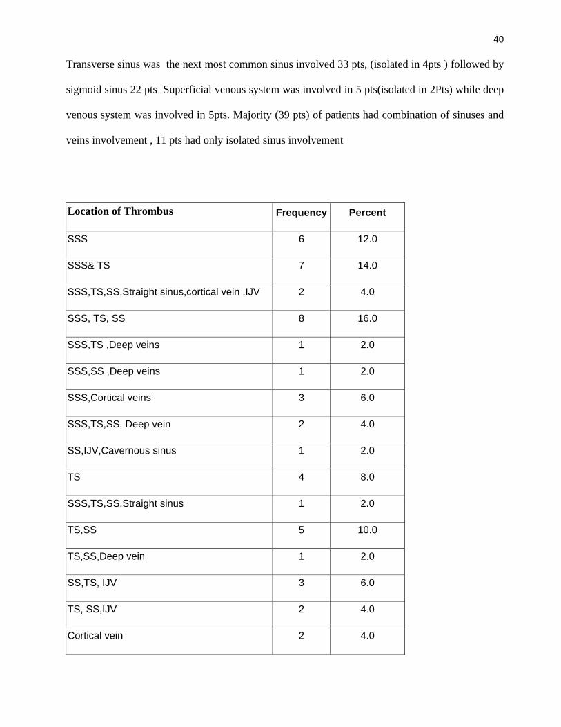
40 Transverse sinus was the next most common sinus involved 33 pts, (isolated in 4pts ) followed by
sigmoid sinus 22 pts Superficial venous system was involved in 5 pts(isolated in 2Pts) while deep
venous system was involved in 5pts. Majority (39 pts) of patients had combination of sinuses and
veins involvement , 11 pts had only isolated sinus involvement
Location of Thrombus Frequency Percent
SSS 6 12.0
SSS& TS 7 14.0
SSS,TS,SS,Straight sinus,cortical vein ,IJV 2 4.0
SSS, TS, SS 8 16.0
SSS,TS ,Deep veins 1 2.0
SSS,SS ,Deep veins 1 2.0
SSS,Cortical veins 3 6.0
SSS,TS,SS, Deep vein 2 4.0
SS,IJV,Cavernous sinus 1 2.0
TS 4 8.0
SSS,TS,SS,Straight sinus 1 2.0
TS,SS 5 10.0
TS,SS,Deep vein 1 2.0
SS,TS, IJV 3 6.0
TS, SS,IJV 2 4.0
Cortical vein 2 4.0

41
T2WI
ThisT2 W showing haemoragic infarct in Rt temporal lobe with vasogenic edema , source
image showed non filling of contrast in Rt sigmoid sinus, CEMRV showed non filling of Rt
trswerse sinus
T1WI
T1w & contrast showed haemoragic infarct in Rt temporal lobe with mild constrast
enhancement suggestive of subacute stage

42
SWI
SWI –showing Blooming , ADC,DWI showing both vasogenic, cytotoxic edema
EEG& AED& SZ control
EEG abnormality is present in 10 pts, normal in 28 pts , focal slowing & generalized slowing was
the most frequent abnormality , 1 pts had PLEDS , 27 pts had control of SZs with single AED,
where as 4 pts required 2 AEDS.
EEG Frequency Percentnormal 28 56.0 Focal slowing 1 2.0 Generalized slowing 2 4.0 G,slowing plus focal spikes
2 4.0
Focal spike 2 4.0 Generalise spike 2 4.0 Pleds 1 2.0 Not done 12 24.0 Total 50 100.0

43 AED No Percent
No AED 19 38.0
Eptoin 24 48.0
Eptoin & valproate 1 2.0
Eptoin & levepil 1 2.0
Clobazam 1 2.0
Clobazam & Eptoin 1 2.0
Clobazam & OXC 1 2.0
VALPROATE 2 4.0
Total 50 100.0
AED No Percent Single 27 54 Two 4 8
Type of treatment received
37 (74%) of pts received heparin, 12 pts received LMWH most of the pts having
parenchymal lesion had received heparin. IIH type of presentation were received LMWH
1 pts DAVF under went embolization
Type of tretment No Percent IV Heparin 37 74.0 LMWH 12 24.0 Embolisation 1 2.0 Total 50 100.0

44 Duration of Heparin treatment
Most of the pts require upto 1 week to get relief of symptoms and to reach INR 2-3 with
warfarin initiation
Days of treatment Frequency Percent3 8 16.0 4-7 17 34.0 8-10 11 22.0 11-15 14 28.0 Total 50 100
SteroidsYes–1No–0 Steroids (Yes – 1, No – 0)
Frequency Percent Valid PercentNo 46 92.0 92.0 yes 4 8.0 8.0 Total 50 100.0 100.0
Antibiotic & antiedema
4 8.0
Antiedema 5 10.0 Antiedema & dindevan
1 2.0

45 Clinical and other possible prognostic factors related to outcome at 12 weeks & 6months after CVST in 50 pts
3 month FU (N=50) 6 month FU(N=38) Prognostic variables Number
(%)
Poor outcome n=9 (18%)
Good outcome N= 41(82%)
P value
Poor outcome n=6(15.8%)
Good outcome N=32(84.2%)
P value
Age <=30 >30
26(52%) 24(48%)
6 (12%) 3 (6%)
20 (40%) 21 (42%)
0.46
3 (7.89%) 3 (7.89%)
14 (36.84) 18 (47.36%)
1.00
Gender(male) (female)
23(46%) 27 (54%)
3 (6%) 6 (12%)
20 (40%) 21 (42%)
0.48 3 (7.89%) 3 (7.89%)
15 (39.47%) 17 (44.73%)
1.00
Mode of onset Acute Sub acute chronic
8(16%) 35(70%) 7(14%)
1 (2%) 6 (12%) 2 (4%)
7 (14%) 29 (58%) 5 (10%)
0.7
1 (2.63%) 4 (10.52%) 1 (2.63%)
6 (15.78%) 22 (57.89%) 4 (10.52%)
o.962
Focal neurologic deficit Present absent
23(46%) 27 (54%)
8 (16%) 1 (2%)
15 (30%) 26 (52%)
0.007
6 (15.78%) 0 (0%)
10 (26.31%) 22 (57.89%)
.003
Papilloedema Present absent
27(54%) 23 (46%)
5 (10%) 4 (8%)
22 (44%) 19 (38%)
0.9
2 (5.26%) 4 (10.52%)
21 (55.26%) 11 (28.94%)
0.188
Sz at presentation Present absent
29(58%) 21 (42%)
9 (18%) 0 (0%)
20 (40%) 21 (42%)
0.006 6 (15.78%) 0 (0%)
17 (44.73%) 15 (39.5%)
.063
Altered sensorium at presentation Yes no
7(14%) 43 (86)
3 (6%) 6 (12%)
4 (4%) 37 (74%)
0.1
3 (7.89%) 3 (7.89%)
1 (2.63%) 31 (81.57%)
.009
Headache Present absent
31(62%) 19 (38%)
5 (10%) 4 (8%)
26 (52%) 15 (30%)
0.715
3 (7.89%) 3 (7.89%)
20 (52.63%) 12 (31.57%)
.663
Single sinus Yes Multiple sinus, yes
11(22%) 39 (78%)
1 (2%) 8 (16%)
10 (20%) 31 (62%)
0.662 2 (5.26%) 4 (10.52%)
6 (15.78%) 26 (68.42%)
.587
EEG abnormality Normal Abnormal
25(50%) 10 (20%)
4 (8%) 4 (8%)
21 (42%) 6 (12%)
0.186
1 (2.63%) 3 (7.89%)
19 (50%) 4 (10.52%)
.042
Base line MRS <2 >2
37(74%) 13 (26%)
2 (4%) 7 (14%)
35 (70%) 6 (12%)
0.0001 1 (2.63%) 5 (13.15%)
27 (71.05%) 5 (13.15%)
.003
Deep venous system inv Present absent
5 (10%) 45 (90%)
3 (6%) 6 (12%)
2 (4%) 39 (78%)
0.035
0 6
4 28
0.487
Parenchymal abnormality Yes No
36(72%) 14
9 (18%) 0 (0%)
27 14
o.o47
6 0
20 12
0.083

46 Logistic Regression
Variable
95% Confidence interval for Odds
Ratio Odds Ratio Lower Upper P Value Deep vein 21.424 1.272 360.900 0.033 Baseline MRS 31.905 3.307 307.841 0.003
Outcome at 6m (, Seizure, Image, deep vein and baseline mrs)
Variable
Odds Ratio
95% Confidence interval for Odds
Ratio P Value Lower Upper BASEMENT MRS
27.000 2.576 282.979 0.006
All the variables at presentation was compared with out come determination both at 3 months & 6
months . Results of 6 month were projected to one year follow up (as there was no difference),
Baseline MRS, deep venous system ,and parenchymal involvement reached significance at 3
months ,but 6 months only base line MRS only the single factor determining the outcome58
Duration of warfarin months
Warfarin duration in most of the cases is upto 6 months(42%) in 4 of the pts the warfarin was
continued >1 year (Procoagulant work up +ve), No intracranial haemorrahage, or any other
complication was reported
Duration of warfarin months 3 17 34.0 5 1 2.0 6 21 42.007 1 2.0
12 6 12.0 18 1 2.0 24 2 4.0 36 1 2.0

47
Follow up duration
All of pts were followed up till 3months which is the acute period, 76% of the pts were
followed up to 1 year to calculate long term outcome
3 12 24.0 6 16 32.0 8 1 2.0
12 15 30.0 18 1 2.0 24 2 4.0 48 1 2.0 96 1 2.0
FOLLOW UP MRS & MRV RESULTS
MRS Baseline(50) 3 month(50) 6month (38)
<2 37 (74%) 41 (82%) 32 (84)
>2 13 (18%) 9 (18%) 6 (16)
MRV recanalisation
Complete 11 15
Partial 2 4
Not done 36 16
Persistant 1 1

48
Sequelae in long term
Vision 3 month 12 month Initial parenchymal involvment,site of thrombosis
Normal vision 40 48
VA decreased 3 1 Temopro,fronto,parietal
SSS, TS, SS
VF abnormal 7 1 Temoro,occipetal
SSS,B/l TS, Cortical Veins
Seizures
Yes 21 5 Frontal,occipital (No -3)
SSS,B/l TS, Cortical Veins
Parietal,occipetal (No- 2)
SSS, TS
No 29 45

49 Mode of onset clinical presentation and outcome according to the site of venous occlusion
sinus Acute subacute Chronic Seizure ICH FOCAL SIGNS
Poor outcome
Good outcome
SSS 1 5 2 6 2 4 0 8 TS 1 2 0 1 0 1 0 3 Cortical vein 0 3 0 2 3 3 0 3 TS+SS+IJV 1 4 0 3 2 1 0 5 SS+TS+SS 1 8 0 3 3 3 2 7 SSS+TS 0 4 3 3 1 3 2 5 Sss+Cortical vein 1 2 0 3 3 2 0 3 TS+SS 2 3 0 3 1 2 1 4 SSS+TS+ Deep veins+ cortical vein
0 2 0 2 2 2 2 0
SSS+TS+SS+Deep vein
1 1 2 3 3 1 2 2
SS+Cevaernous 0 1 0 0 1 0 1 Total 8 35 7 29 20 23 9 41
Correlation between the site of venous occlusion and clinical parameter
Correlation with etiology showed no constant pattern except that lateral sinus isolated involved in
Mastoiditis. Correlation with mode of onset showed no difference in onset whether sinuses alone
vs deep venous vs combination of sinuses and veins. no significant difference between presence of
various sinuses and venous system the presence and location of infarction .when cortical veins are
involved pts were presented with SZs and have intracranial hematoma than when only sinuses
were involved.

50
Discussion

51
Discussion
Cerebral venous thrombosis is condition characterized by thrombosis of intracranial veins and
sinus which results in parenchymal damage and rise in intracranial pressure. Radiological
hallmark of this condition is thrombosis of intracranial sinuses and veins with haemorrhagic
infarction and edema with or without evidence of herniation. In this study, total 50 patients with
Radilogical features of cerebral venous thrombosis were evaluated over a period of 1year. 23 out
of 50 patients were male and remaining were female. This study of 50 patients with CVT cannot
give precise information about the real incidence of the disease. cannot make any generalization of
the results to whole country It has been suggested that the incidence of CVT was higher in females
and in the aged, This was not confirmed in the present series, in which Male.Female (23;27) This
data is not consistent with previous Indian studies viz. Bansal et al (1980)13, Srinivasan et al 12
(1983), Nagaraja et al 3 (1987). High proportion of post partum CVT patients was also observed
by Cantu et al 7 (1996), from Mexico with similar socio-demographic characteristics and economic
status of the patients as in India. due to referral bias. This findings of high proportion of CVT cases
was not replicated in some other studies viz. Deschiens et al 36 (1996) and Daif et al 40 (1995).
The possible explanation may be that the etiological factors as well as clinical profile of CVT is in
this part of the state different compare to other parts of India More than half of the patients of
CVT evaluated were in the third decade of their age (33/50). The mean age of the patients was
32.18 years (SD13.14) similar to earlier studies from India (Nagaraja et al 3 1987) Like all other
series, the present one represents a selected group of patients not representative of the numerous
causes that have been described. However, it confirms the fact that the frequency of septic CVT
(6/50) has markedly declined with the advent of antibiotics. It also confirms the role of oral
contraceptives37 found as the only aetiologic factor in 3 of our patients. This has now led us, as
many others to stop oral contraceptives and promptly look for CVT in women presenting with any

52 of the neurological manifestations described in this study, particularly persistent headache, focal
deficits or seizures. The present series reflects our interest in Brucella infection 2 cases presented
with diffuse headache & fever found to have CVT. and two children presented due to L-
aspergenase treatment
In the present cohort in addition to conventional risk factors Dehydration(8%),
hyperhomocystenimia(6%), CSF leak (4%), OCP pill use (10%) are significant risk factors , 8% of
pts Anemia , whether this is a reflection of high incidence of anemia in Indian population
particularly in pregnant females or anemia is a real risk factor needs further evaluation. In 10/50
cases, no cause could be found ,however complete etiological workup could not be completed .
Headache (31/50) with or without vomiting, seizures (29/50). Altered sensorium (7/50) and Focal
deficits (23/50) Papilledema, present in 54% of our cases, was slightly more frequent than in other
series: were the major clinical features noted at presentation. Similar findings were noted in the
earlier studies 2,3,13,30. the clinical presentation could be summarized in 3 main patterns, each of
them simulating another neurological disease. The most frequent and homogeneous one was the
progressive onset of signs of intracranial hypertension corresponding to the "Benign intra-cranial
hypertension" or "pseudo-tumor cerebri" syndromes, confirming that sinus thrombosis in 27/50
cases these syndrome should not be diagnosed purely on clinical, CSF and CT scan findings
without a good quality CEMRV to rule out the possibility of sinus thrombosis. (8/23) was the
sudden onset of focal deficits simulating arterial strokes but with more frequent seizures (21/29).
The third presentation simulated an abscess (5/50) with deficits and/or seizures with or without
intracranial hypertension evolving over a few days/Month. Other less common presentations are
headache of sudden onset simulating subarachnoid hemorrhage (1 patient) It is therefore clear that
CVT has no single clinical presentation and this is why it is necessary to systematically
contemplate this diagnosis in order not to overlook it

53 Present series most of the pts had good outcome ,recanalisation in Repeat MRi also achieved here
down tables just showing comparision in relation to other studies
Table 3. Recanalization at 3 to 6 months and at 1 year or more Study, year No.of Partial Complete Partial Complete
Patients recanal at recanal at recanal at recanal at 3 to 6 mo, no 3 to 6 mo, no 1 y or more, no. 1 y or more, no.
Stolz et al,10 2004 37 7 19 7 20 Favrole et al, 11 2004 28 7 16 NA NA Baumgatner et al, 12 2003 33 15 18 15 18 Strupp et al, 17 2002 40 NA NA 12 21 Cakmark et al, 14 2003 16 12 NA NA NA Present study 50 2 11 4 15
Several reports have emphasized the importance of EEG changes in CVT, the most common
pattern being a severe generalised slowing more marked on one side with frequent epileptic
Present Study

54 activity40 In the present series, EEG abnormalities were less severe and they were present in
(10/38) in 21.5% of cases. Its main interest was to show in a number of patients with focal
symptoms a generalised slowing indicating a more diffuse lesion than was clinically suspected.
This, however, is in no way specific of CVT . single case showed PLEDS
The present series confirms the fact that isolated single sinus involvement was less common than
multiple sinuses involvement, in isolated sinus most frequently involved are SSS and LS Thus in
most cases, occlusion involved at least two sinuses or sinus and cerebral veins. Among these,
cortical veins were affected slightly more commonly than the deep venous system These frequent
associations probably explain, at least partly, why no good clinico-radiological correlations could
be established
Before the introduction of angiography, CVT was diagnosed at autopsy and therefore thought to be
most often lethal. In early angiographic series, mortality still ranked between 30% and 50% but in
more recent series, it was between 25% and 30% and in the present one it was only 2%. Multiple
reasons can explain this decrease, the main one being probably that it is now possible to diagnose
"benign" forms of CVT with minimal symptoms and spontaneous recovery. Another reason is that
septic thrombosis has, since the use of antibiotics, become both far less frequent and severe. It is
also that the introduction of anticoagulant treatment early in the course of the disease has
improved the outcome.
Two kinds of sequelae are encountered: blindness /Fieldcut due to optic atrophy/cortical infarcts
which should be prevented by early treatment, and focal deficits, usually motor, sometimes
associated with epilepsy. Szs are more frequent when the lesion is anterior to the central sulcus and
in patients who have focal deficits. In the VENOPORT study early seizures were associated with

55 sensory deficits and parenchymal lesions on admission CT/MRI. Our study also showed that 42%
(21/50)of the pts had sz at presentation 5 (25%) only had long term recurrence , all of them had
parenchymal abnormality at presentation ,most of them well controlled with single AED
Factors classically considered of bad prognosis are the rate of evolution of thrombosis,9 the
presence of coma,10 the age of patients, with a high mortality rate in infancy and in the aged9 and
the involvement of cerebral veins.41 In the present series, the main prognostic factor is Base line
MRS compare to other studies
Most of the pts who were followed up had re canalized the occluded veins Only one pt expired
in acute phase, Only 1 pt presented with recurrent CVT42, sequelae were both more frequent in
patients with focal symptoms than in patients with benign intracranial hypertension. The outcome
was otherwise most unpredictable: some acute cases, even with coma, made a remarkably rapid
and complete recovery whereas chronic cases often recovered more slowly and with more frequent
sequelae. It is apparent from the study of literature and from the present series that the natural
history and prognosis of CVT are highly variable
A Cochrane meta-analysis of 22 trials, including nearly 9000 patients, convincingly showed that
treatment with LMWH results in significantly less thromboembolic recurrences (OR, 0.68), fewer
major hemorrhagic complications (OR, 0.57), a higher recanalization rate (OR, 0.69), and a lower
mortality (OR, 0.76) than UFH.
The superior safety and efficacy of LMWH in leg-vein thrombosis is probably attributable to its
pharmacokinetic properties22,23. Contrary to LMWH, UFH requires frequent activated partial
thromboplastin time measurements and dose adjustments, which have proven to be difficult to
implement in practice.
A British audit of 45 consecutive patients who received UFH during admission showed that
patients were adequately anticoagulated less than a quarter of the time. Most of the time, patients

56 were below the therapeutic range, but overdosing also occurred frequently .There is a robust
correlation between sub therapeutic activated partial thromboplastin time values and the risk of
recurrent thrombosis, and the likely effect of overdosing.
In present study also UFH in 74% of pts and LMWH in 24% of pts , based on the idea that an
adequate level of anticoagulation is achieved more rapidly with UFH, and then change to LMWH
after a few days. UFH is an increased risk of hemorrhagic complications. Other studies have
demonstrated that it often takes 24 hours until patients are adequately anticoagulated with UFH,
even if a treatment algorithm is used. Thus, the theoretical advantage of more rapid anticoagulation
with intravenous UFH is probably rarely realized in practice. The decision to change the type of
heparin have been motivated by several reasons. For example, if a patient deteriorates, most
notably in the case of a hemorrhagic complication, treating neurologist may decide to switch to
another type of heparin. In addition, a switch to LMWH can be made if a patient had improved
enough to be mobilized or discharged because LMWH does not require intravenous access.
In this study, attempt was made to correlate the clinical profile with the topographic Radiological
substrate like involvement of superficial / deep venous system or the pattern of infarction. There
was no significant correlation to evolve a pattern of diagnostic significance, correlating with
radiological findings . However predictably, patients with deep venous system involvement and
having ganglionic infarction had significantly less incidence of seizures. Patients with
involvement of SSS had higher incidence of seizure and lower incidence of headache than those
who didn’t have SSS involvement. As most of the patients had extensive involvement of cerebral
sinovenous system, contribution of degree of involvement of anatomical structures to a particular
clinical profile cannot be reliably predicted. For example, high incidence of seizures in patients
with SSS involvement may be attributed to the thrombosis from SSS spreading to cerebral veins
causing cortical lesions and seizures but when a group of patients with only cerebral venous

57 thrombosis without any sinus thrombosis was analyzed, seizure incidence was not high. Similarly
patients with papilloedema did not differ in pathologic Radiological findings when compared to
the patient group without papilloedema.

58 SUMMARY AND CONCLUSIONS
1. Over a period of 6 years 50 patients of cerebral venous thrombosis were studied. Almost
two third (66%) of patients were in 3rd decade of life data consistent with most of the
earlier Indian studies.
2. Large number of patients (35/50) in this series had sub acute onset of symptoms i.e.
symptom duration (48hour -30 days).
Headache (31/50) , seizures (29/50) altered sensorium (7/50) and Focal deficits (23/50)
Papilledema, present in 54% major clinical features noted.
3 Cerebral infarction was the most common abnormality noted on CT scan (72%) which was
haemorrhagic in 29% of the cases. Deep seated venous infarction (Thalamus and basal
ganglionic structure) was seen in 10% of cases
On CEMRV , Superior sagittal sinus (the commonest sinus involved) was involved in 39 patients,
(isolated SSS in 7 pts) Total involvement was seen in 11patients while in other patients anterior,
middle and posterior parts involved with various combination of other sinuses
Transverse sinus was the next most common sinus involved 33 pts, (isolated in 4pts ) followed by
sigmoid sinus 22 pts Superficial venous system was involved in 5 pts(isolated in 2Pts) while deep
venous system was involved in 5pts. Majority (39 pts) of patients had combination of sinuses and
veins involvement , 11 pts had only isolated sinus involvement
When attempt was made to correlate the clinical profile with the topographic Radiological
substrate like involvement of superficial/deep venous system or the pattern of infarction, there was
no significant correlation to evolve a pattern of diagnostic significance, correlating with
involvement of sinus

59 CSVT is an important and treatable cause of the stroke , risk factors like hyperhomocystenemia,
OCP use, alcoholism ,procoagulant state are increasingly recognized in addition to the
conventional risk factors like postpartum state . Procoagulant state and infections are the most
common predisposing factors for cerebral venous thrombosis in this cohort.
Prognosis of CVT predominantly determined by Base line MRS compare to other studies Most of
the pts who were followed up had re canalized the occluded veins Only one pt expired in acute
phase, Only 1 pt presented with recurrent CVT

60
Bibilography
1. Ribes F. Des rechereches faites sur la phembite. Rev. Medi (Paris ) 1825;3:5-41.
2. Bousser MG Chiras J, Berics J, Castagine P. Cerebral venous thrombosis – a review of 38
cases. Stroke 1985; 16(2) : 199 – 213.
3. Nagaraja D, Taly AB, Puerperal venous sinus thrombosis in India. In: Sinha KK ed.
Progress in clinical neurosciences, Ranchi, NSI, Publications 1989;5:165-177.
4. De Bruijn SF, de Haan RJ, Stam J. Clinical features and prognostic factors of cerebral
venous sinus thrombosis in a prospective series of 59 patients: for The Cerebral Venous
Sinus Thrombosis Study Group. J Neurol Neurosurg Psychiatry. 2001;70:105-108
5. Cross JN, Castro PO, Jennett WB. Cerebral strokes associated with pregnancy and
puerperium. Br Med J. Clin. Res 1968;3:214-218.
6. Cerebral venous thrombosis: Analysis of a multi-center cohort from United States of
America. Wasay M, Bakshi R, Bobustuc G, Kojan S, Sheikh Z, Dai A, Cheema Z. (in
press; Journal of Stroke and Cerebrovascular diseases)
7. Cerebral venous thrombosis; A descriptive multi center study of patients from Pakistan and
Middle east. B Khealani, Wasay M, Saadah M, Sultana E, Shohab F, Mustafa S, Kamal A
(submitted; Stroke)
8. Cantu C C, Barinagarrementeria F. Cerebral venous thrombosis associated with pregnancy
and puerperium: review of 67 cases. Stroke 1993;24:1880–4.
9. Chopra JS, Banerjee AK. Primary intracranial sinovenous occlusions in youth and
pregnancy. In : Vinken PJ, Bruyn Gw, Klawans HL, Toole JF, eds. Vascular disease part II.

61
Amsterdam: Elseuier science 1989 ; PP 425 -452 (Hand book of clinical neurology, vd 54,
revised series 10).
10. Preter M, T Zourio C, America A, Bousser MG. Long prognosis in cerebral venous
thrombosis: follow up of 77 patietns . Stroke 1996;27:243-246.
11. Villringer A, Seiderer M, Bauer WM et al. Diagnosis of superior sagittal sinus thrombosis
by three dimensional magnetic resonance flow imaging. Lancet 1989; 1 :1086-1087.
12. Bansal BC, Gupta RR, Prakash C. Stroke during pregnancy and puerperium in young
females below the age of 40 years as a result of cerebral venous /venous sinus thrombosis.
Jpn Heart 1980;21:171-183.
13. EinHaupl KM, Villringer A, Meister W, Mehracin S. garner C, Pellkofer M Haber RL, P
fister HW, Schmiedek P. Heparin treatment in sinus venous thrombosis. Lancet
1993;38:597-600.
14. de Brujin SFTM, Stam J, for the CVT study group randomized, placebo controlled trial of
anticoagulant treatment with LMWH for cerebral sinus thrombosis. Stroke 1999;30:484-
488
15. Cak Mak S, Derex L, Berrnya M, Nighoghossian N, Phillippean F, Adeleine P, Hermier M,
Froment JC, Trovillar P, cerebral venous thrombosis; clinical outcome and systematic
screening of prothrombotic factors. Neurology 2003;60:1175-1178.
16. Martin JP. Thrombosis in the superior longitudinal sinus after child birth. Br. Med J. 1941;
2: 537-540.
17. Ehler H, Courville CB. Thrombosis of internal cerebral veins in infancy and childhood.
Review of literature and report of five cases. J Pediatr 1936;8:600-623.
18. Towbin A. The syndrome of latent cerebral venous thrombosis: its frequency and relation
to age and congestive heart failure stroke 1973;4:419-430.
62
19. Kalbag RM. Cerebral venous thrombosis. In : Kapp JP, Schemidek HH, eds. The cerebral
venous system and its disorders. Orlando: Gruen and Stratten. 1986 PP 505- 536
20. Padayachee TS. Bingham JB, Orave MJ et al. Dural sinus thrombosis. Diagnosis and
follow up by magnetaic resonance angiography and imaging. Neuroradiology 33:165,
1991.
21. van Dongen CJ, van den Belt AG, Prins MH, Lensing AW. Fixed dose subcutaneous low
molecular weight heparins versus adjusted dose unfractionated heparin for venous
thromboembolism. Cochrane Database Syst Rev. 2004;4:CD001100.
22. Hull RD, Pineo GF. Heparin and low-molecular-weight heparin therapy for venous
thromboembolism: will unfractionated heparin survive? Semin Thromb Hemost.
2004;30(suppl 1):11–23.
23. Derchicus MA, Conard J, Horellou M H et al. Coagulation studies, factor V leiden,
antiphospholipid antibodies in 40 cases of CVT, stroke 27; 1724:1996.,
24. Feinberg WM. Swenson MR. Cerebrovascular complications of C aspergenase therapy.
Neurology 38, 127, 1988.
25. Srinivasan K. Cerebral venous and arterial thrombosis in pregnancy and puerperium.
Angiology 1983 ; 134:731-746.
26. Viropongse C, Cazenave C, Quisling R et al. The empty delta sign: frequency and
significance in 76 cases of dural sinus thrombosis. Radiology 1987 ; 162 (3): 779-785.
27. Rao KCVG, Knipp HC, Wagner EJ. Computed tomographic findings in cerebral sinus and
venous thrombosis. Radiology 1981; 140:391-398.
28. Padayachee TS, Bingham JB, Graves MJ et al. Dural sinus thrombosis: Diagnosis and
follow up by magnetic resonance angiography and imaging. Neuroradiology 1991;33:165-
167.

63
29. Stolz E, Kaps M, Dorndorf W. Assessment of intracranial venous hemodynamics in
normal individuals and patients with cerebral venous thrombosis. Stroke 1999; 30: 70-75.
30. Brey RL, Coull BM. Cerebrall venous thrombosis: Role of activated protein C resistance
and factor V gene mutation stroke 1996 27 (10): 1719 – 1720.
31. Levine SR, Kieran S, Puziok et al. Cerebral venous thrombosis with Lupus anticoagulants:
Report of two cases. Stroke 1987;18(4) : 801-804.
32. Juan R Carhuapoma, Panayiotis Misias, Steven R Levine. Cerebral venous thrombosis and
anticardiolipin antibodies. Storke 1997;28:2363-2369/
33. Fairburn B. Intracranial venous thrombosis complicating oral contraception: treatment with
anticoagulant drugs. Br Med J 1973 ; 2 : 647.
34. Kalbag RM, Woolf AL. Thrombosis and thrombophlebitis of cerebral veins and dural
sinuses. In PJ Vinken, GW Bruyn (eds) . Handbook of clinical neurology. Vo. 2 , Elsewe
1972; 422-446.
35. Stam J. Thrombosis of the cerebral veins and sinuses. N Engl J Med2005;352:1791
36. Fairburn B. Intracranial venous thrombosis complicating oral contraception: treatment with
anticoagulant drugs. Br Med J 1973 ; 2 : 647.
37. Gettlefinger DM, Kokmen E. Superior sagittal sinus thrombosis. Arch neurol
1977; 34: 2-6.
38. Deschiens MA, Jacqueline Conard, Marie Halen Horellou et al. Coagulation studies, factor
V leiden and anticardiolipin antibodies in 40 cases of cerebral venous thrombosis. Stroke
1996; 27:1724-1730.
39. Kalbag RM, Woolf AL. Cerebral venous thrombosis. 1967, Oxford university press.
40. Crawford SC, Digre KB, Palmer CA et al. thrombosis of the deep venous drainage of brain
in adults . Arch Neurol 1995 ; 52 (11) : 1101 – 1108.

64
41. Editorial . Cerebral venous thrombosis: development in imaging and treatment. J Neurol
Neurosurgery psychiatry 1995 ; 59 : 1-3.
42. Shinohara Y, Takagi S, Kobatake K, Goton F. Influence of cerebral venous obstruction on
cerebral circulation in humans. Arch neurol 1982 ; 39 (8): 479-481.
43. Averback P. Primary cerebral venous thrombosis in young adult: the diverse
manifestations of an under recognized disease. Ann neurol 1978; 3: 81 – 86.
45 Ferro JM, Pinto F. Poststroke epilepsy: epidemiology, pathophysiology and
management. Drugs Aging. 2004;21:639–653.Barnet HJM, Hyland HM.
46 Ferro JM, Correia M, Ponter C, for ISCVT investigators. Prognosis of cerebral venous
and dural sinus thrombosis, Results of ISCVT. Stroke 2004;35:664-670.
47 de Bruijin SFTM, de Haan RT. Stam T. for cerebral venous sinus thrombosis study group.
Clinical features and prognostic factors of cerebral venous sinus thrombosis in a
prospective series of 59 patients. JNNP 2001;70:105-108.