Chapter 9 Muscular System: Histology and Physiology · PDF fileMuscular System: Histology and...
18
1 9-1 Muscular System: Histology and Physiology Chapter 9 9-2 Muscular System Functions • Body movement • Maintenance of posture • Respiration • Production of body heat • Communication • Constriction of organs and vessels • Heart beat
Transcript of Chapter 9 Muscular System: Histology and Physiology · PDF fileMuscular System: Histology and...

1
9-1
Muscular System:Histology and Physiology
Chapter 9
9-2
Muscular System Functions
• Body movement• Maintenance of posture• Respiration• Production of body heat• Communication• Constriction of organs and vessels• Heart beat

2
9-3
Properties of Muscle
• Contractility: ability of a muscle to shorten with force
• Excitability: capacity of muscle to respond to a stimulus
• Extensibility: muscle can be stretched to its normal resting length and beyond to a limited degree
• Elasticity: ability of muscle to recoil to original resting length after stretched
9-4
Muscle Tissue Types• Skeletal
– Responsible for locomotion, facial expressions, posture, respiratory movements, other types of body movement
– Voluntary
• Smooth– Walls of hollow organs, blood vessels, eye, glands, skin– Some functions: propel urine, mix food in digestive tract,
dilating/constricting pupils, regulating blood flow– In some locations, autorhythmic– Controlled involuntarily by endocrine and autonomic nervous systems
• Cardiac– Heart: major source of movement of blood– Autorhythmic– Controlled involuntarily by endocrine and autonomic nervous systems
3
9-5
9-6
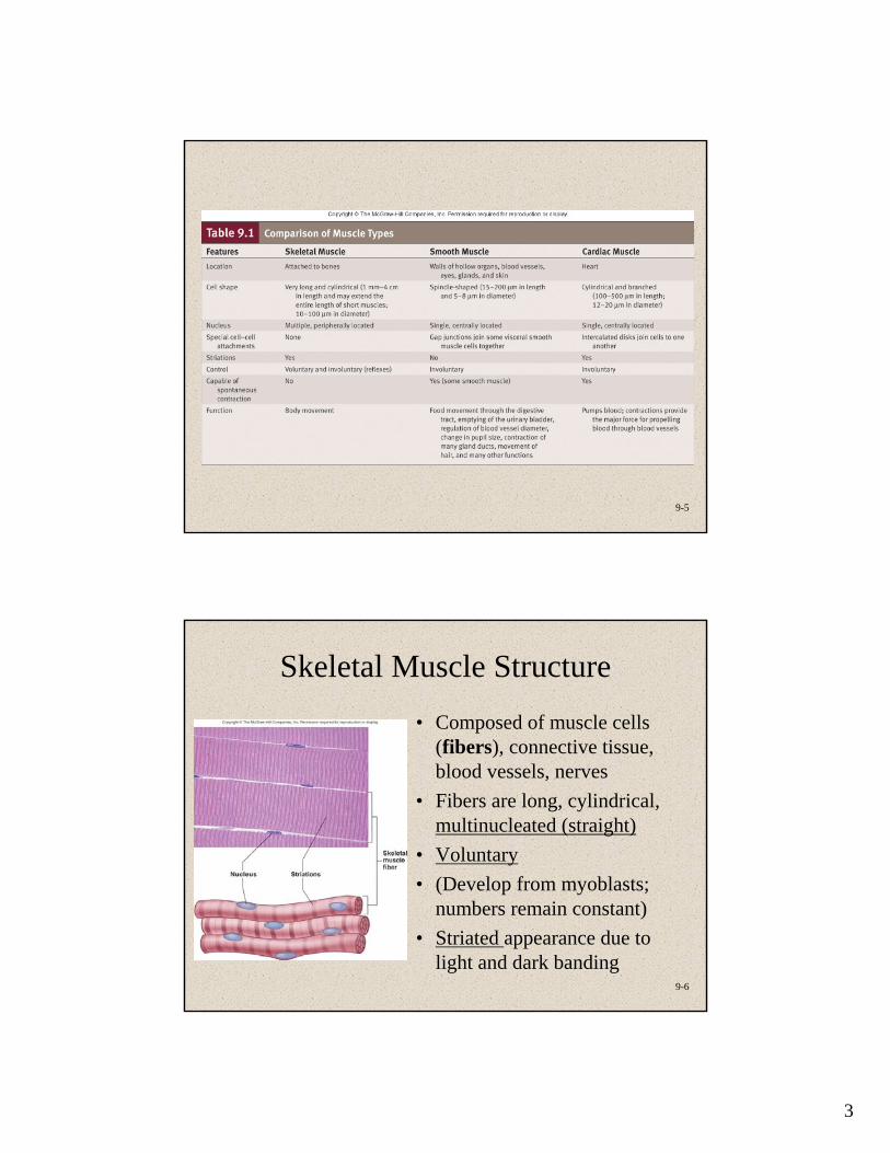
Skeletal Muscle Structure• Composed of muscle cells
(fibers), connective tissue, blood vessels, nerves
• Fibers are long, cylindrical, multinucleated (straight)
• Voluntary• (Develop from myoblasts;
numbers remain constant)• Striated appearance due to
light and dark banding
4
9-7
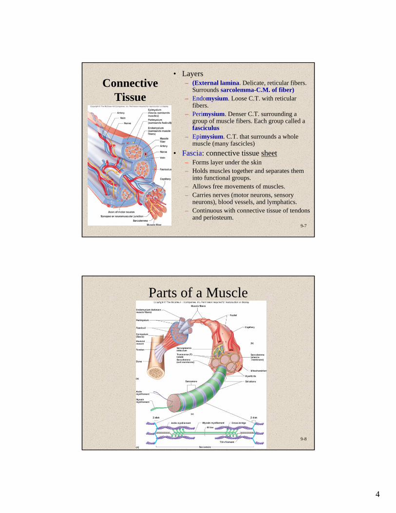
Connective Tissue
• Layers– (External lamina. Delicate, reticular fibers.
Surrounds sarcolemma-C.M. of fiber)– Endomysium. Loose C.T. with reticular
fibers.– Perimysium. Denser C.T. surrounding a
group of muscle fibers. Each group called a fasciculus
– Epimysium. C.T. that surrounds a whole muscle (many fascicles)
• Fascia: connective tissue sheet– Forms layer under the skin– Holds muscles together and separates them
into functional groups.– Allows free movements of muscles.– Carries nerves (motor neurons, sensory
neurons), blood vessels, and lymphatics.– Continuous with connective tissue of tendons
and periosteum.
9-8
Parts of a Muscle

5
9-9
Structure of Actin and Myosin
9-10
Myosin (Thick)
Myofilament
• Many elongated myosin molecules shaped like golf clubs.
• Molecule consists of two heavy myosin molecules wound together and two heads that extend laterally.
• Myosin heads1. Can bind to active sites on the
actin molecules to form cross-bridges.
2. Attached to the rod portion by a hinge region.
3. Have ATPase activity (that breaks down adenosine triphosphate (ATP), releasing energy)
Part of the energy is used to bend the hinge region of the myosin molecule during contraction

6
9-11
Sarcomeres: Z Disk to Z
Disk
• Z disk: filamentous network of protein. Serves as attachment for actin myofilaments
• Striated appearance– I bands: from Z disks to ends of
thick filaments– A bands: length of thick
filaments– H zone: region in A band where
actin and myosin do not overlap– (M line: middle of H zone;
delicate filaments holding myosin in place)
• In muscle fibers, A and I bands of parallel myofibrils are aligned.
• Titin filaments: elastic chains of amino acids; make muscles extensible and elastic
9-12
Sarcomere Shortening

7
9-13
Physiology of Skeletal Muscle
• Nervous system controls muscle contractions through action potentials
• Resting membrane potentials REMEMBER---– Membrane voltage difference across membranes (polarized)
• Inside cell more negative due to accumulation of large protein molecules. More K+ on inside than outside. K+ leaks out but not completely because negative proteins hold some back.
• Outside cell more positive and more Na+ on outside than inside. Na/K pump maintains this situation.
– Must exist for action potential to occur
9-14
Ion Channels
• Types– Ligand-gated. Example:
neurotransmitters• Gate is closed until
neurotransmitter attaches to receptor molecule. When Ach attaches to receptor on muscle cell, Na gate opens. Na moves into cell due to concentration gradient
– Voltage-gated• Open and close in response to
small voltage changes across plasma membrane
• Each is specific for one type of ion
8
9-15
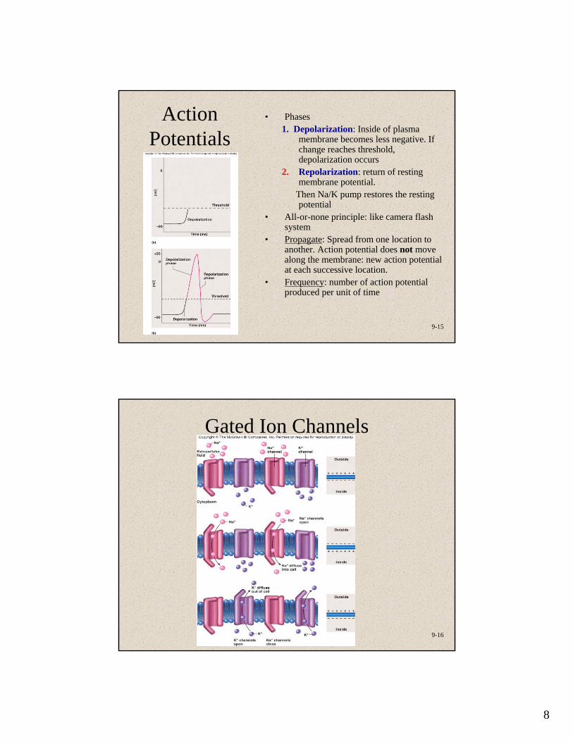
Action Potentials
• Phases1. Depolarization: Inside of plasma
membrane becomes less negative. If change reaches threshold, depolarization occurs
2. Repolarization: return of resting membrane potential.
Then Na/K pump restores the resting potential
• All-or-none principle: like camera flash system
• Propagate: Spread from one location to another. Action potential does not move along the membrane: new action potential at each successive location.
• Frequency: number of action potential produced per unit of time
9-16
Gated Ion Channels

9
9-17
Action Potential Propagation
9-18
Neuromuscular Junction
• Synapse axon terminal resting in an invagination of the sarcolemma)
• Neuromuscular junction (NMJ):– Presynaptic terminal:
axon terminal with synaptic vesicles
– Synaptic cleft: space– Postsynaptic
membrane

10
9-19
9-20
Action Potentials and Muscle Contraction
11
9-21
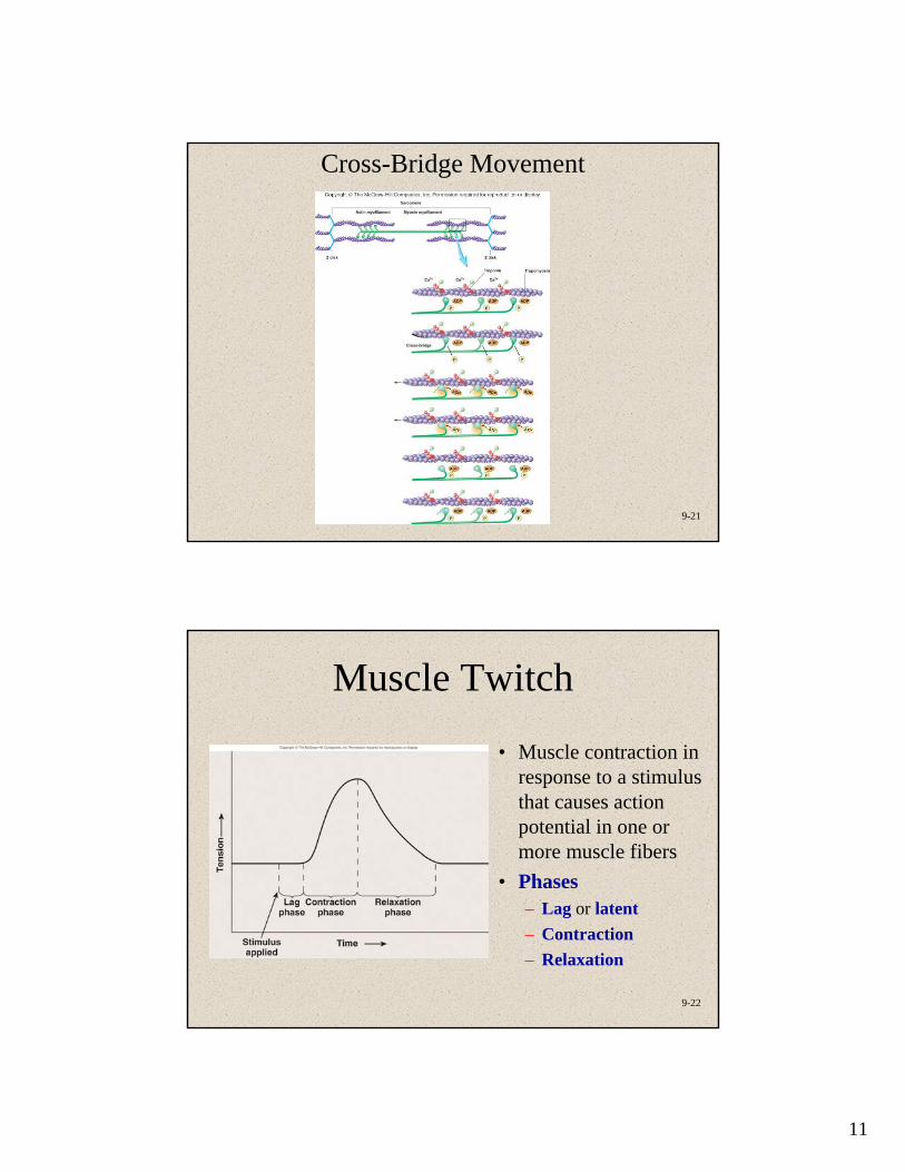
Cross-Bridge Movement
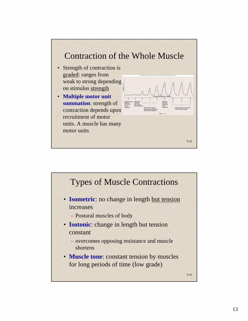
9-22
Muscle Twitch
• Muscle contraction in response to a stimulus that causes action potential in one or more muscle fibers
• Phases– Lag or latent– Contraction– Relaxation

12
9-23
9-24
Stimulus Strength and Muscle Contraction
• All-or-none law for muscle fibers– Contraction of equal force in response
to each action potential• (Sub-threshold stimulus: no
action potential; no contraction)
• Threshold stimulus: action potential; contraction
• Stronger than threshold; action potential; contraction equal to that with threshold stimulus
• Motor units: a single motor neuron and all muscle fibers innervated by it
13
9-25
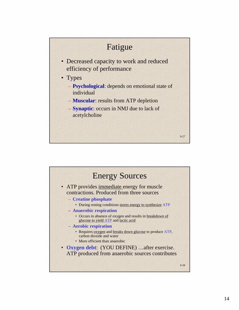
Contraction of the Whole Muscle• Strength of contraction is
graded: ranges from weak to strong depending on stimulus strength
• Multiple motor unit summation: strength of contraction depends upon recruitment of motor units. A muscle has many motor units
9-26
Types of Muscle Contractions
• Isometric: no change in length but tensionincreases– Postural muscles of body
• Isotonic: change in length but tension constant– overcomes opposing resistance and muscle
shortens• Muscle tone: constant tension by muscles
for long periods of time (low grade)
14
9-27
Fatigue• Decreased capacity to work and reduced
efficiency of performance• Types
– Psychological: depends on emotional state of individual
– Muscular: results from ATP depletion – Synaptic: occurs in NMJ due to lack of
acetylcholine
9-28
Energy Sources• ATP provides immediate energy for muscle
contractions. Produced from three sources– Creatine phosphate
• During resting conditions stores energy to synthesize ATP– Anaerobic respiration
• Occurs in absence of oxygen and results in breakdown ofglucose to yield ATP and lactic acid
– Aerobic respiration• Requires oxygen and breaks down glucose to produce ATP,
carbon dioxide and water• More efficient than anaerobic
• Oxygen debt: (YOU DEFINE) …after exercise. ATP produced from anaerobic sources contributes
15
9-29
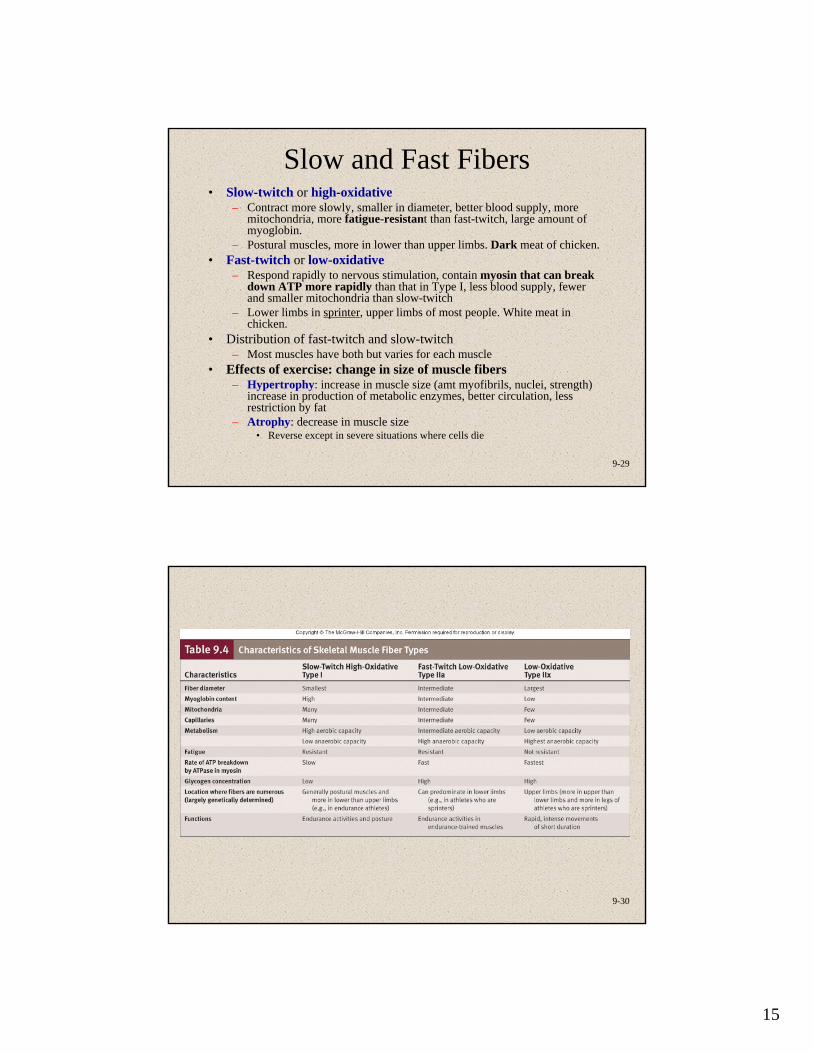
Slow and Fast Fibers• Slow-twitch or high-oxidative
– Contract more slowly, smaller in diameter, better blood supply, more mitochondria, more fatigue-resistant than fast-twitch, large amount of myoglobin.
– Postural muscles, more in lower than upper limbs. Dark meat of chicken.• Fast-twitch or low-oxidative
– Respond rapidly to nervous stimulation, contain myosin that can break down ATP more rapidly than that in Type I, less blood supply, fewer and smaller mitochondria than slow-twitch
– Lower limbs in sprinter, upper limbs of most people. White meat in chicken.
• Distribution of fast-twitch and slow-twitch– Most muscles have both but varies for each muscle
• Effects of exercise: change in size of muscle fibers– Hypertrophy: increase in muscle size (amt myofibrils, nuclei, strength)
increase in production of metabolic enzymes, better circulation, less restriction by fat
– Atrophy: decrease in muscle size• Reverse except in severe situations where cells die
9-30
16
9-31
Heat production
• Exercise: metabolic rate and heat production increase.
• (Post-exercise: metabolic rate stays high due to oxygen debt.)
• Excess heat lost because of vasodilation and sweating
• Shivering: uncoordinated contraction of muscle fibers resulting in shaking and heat production
9-32
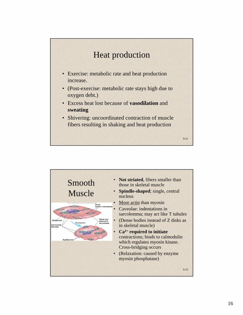
Smooth Muscle
• Not striated, fibers smaller than those in skeletal muscle
• Spindle-shaped; single, central nucleus
• More actin than myosin• Caveolae: indentations in
sarcolemma; may act like T tubules• (Dense bodies instead of Z disks as
in skeletal muscle)• Ca2+ required to initiate
contractions; binds to calmodulin which regulates myosin kinase. Cross-bridging occurs
• (Relaxation: caused by enzyme myosin phosphatase)

17
9-33
Functional Properties of Smooth Muscle
• Some visceral muscle exhibits autorhythmic contractions)
• Tends to contract in response to sudden stretch but not to slow increase in length
• Exhibits relatively constant tension: smooth muscle tone
• Amplitude of contraction remains constantalthough muscle length varies
9-34
Cardiac Muscle
• Found only in heart• Striated• Each cell usually has one nucleus• Has intercalated disks and gap junctions• Autorhythmic cells• Action potentials of longer duration and longer
refractory period• Ca2+ regulates contraction
18
9-35
Effects of Aging on Skeletal Muscle
• Reduced muscle mass• Increased time for muscle to contract in
response to nervous stimuli• Reduced stamina• Increased recovery time• Loss of muscle fibers• Decreased density of capillaries in muscle