Chapter 7: The Axial Skeleton part 1. The Axial Skeleton - longitudinal axis Supports and protects...
35
Chapter 7: The Axial Skeleton part 1
-
Upload
karlee-stickles -
Category
Documents
-
view
224 -
download
0
Transcript of Chapter 7: The Axial Skeleton part 1. The Axial Skeleton - longitudinal axis Supports and protects...

Chapter 7:The Axial Skeleton
part 1
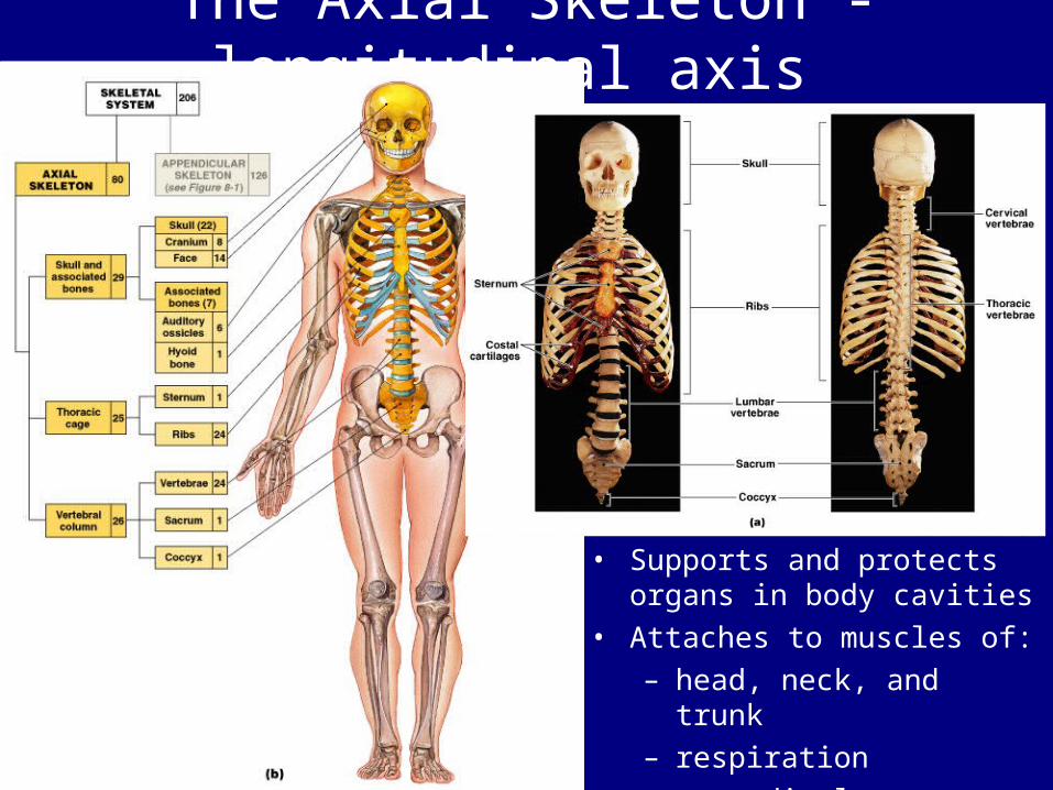
The Axial Skeleton - longitudinal axis
• Supports and protects organs in body cavities
• Attaches to muscles of:– head, neck, and trunk– respiration– appendicular skeleton

Bones of the Axial Skeleton - 80• The skull:
– 8 cranial bones – 14 facial bones
• Bones associated with the skull:– 6 auditory ossicles – the hyoid bone
• The vertebral column:– 24 vertebrae– the sacrum – the coccyx
• The thoracic cage:– 24 ribs – the sternum

The Skull• The skull protects:
– the brain– entrances to
respiratory and digestive system
– Has 22 bones:• 8 cranial bones:
– form the braincase or cranium
• 14 facial bones:– protect and support
entrances to digestive and respiratory tracts

Cranial Bones

1. The Frontal Bone
Figure 7–6
– Forms the anterior cranium and upper eyesockets
– Contains frontal sinuses

• Marks – Frontal
squama/glabella (forehead)
– Supraorbital margin (protects eye)
– Lacrimal fossa (for tear ducts)
The Frontal Bone-con’t

2-3. The Parietal Bones– forms part of the superior
and lateral surfaces of the cranium

4-5. The Temporal Bones
Figure 7–7
– Part of lateral
walls of cranium and zygomatic arches
– Surround and protect inner ear
– Attach muscles of jaws and head

• Marks
– Mandibular fossa-articulates with the mandible
– Zygomatic process:• inferior to the
squamous portion• articulates with
temporal process of zygomatic bone
• forms zygomatic arch (cheekbone)

– Auditory ossicles:• 3 tiny bones in
tympanic cavity (middle ear)
• transfer sound from tympanic membrane to inner ear
– Mastoid process:• for muscle attachment• contains mastoid air
cells connected to middle ear
– Styloid process:• to attach tendons and
ligaments of the hyoid, tongue and pharynx


• Foramina – Carotid canal:
• for internal carotid artery
– Foramen lacerum:• for carotid and small arteries• hyaline cartilage• auditory tube

6. The Occipital BoneCranial Bones - 8
• Forms posterior/inferior surfaces of cranium• Largest cranial bone
• Marks– Occipital condyles -articulate with neck– Inferior and superior nuchal lines -to attach ligaments– External occipital protuberance(Inion)- for trapezius attachment– External occipital crest -to attach ligaments
• Foramina – Foramen magnum-connects cranial and spinal cavities

7. The Sphenoid
Figure 7–8
– Part of the floor of the cranium– Unites cranial and facial bones– Strengthens sides of the skull– Contains sphenoidal sinuses

• Marks – Sphenoid body:
• at the central axis of the sphenoid– Sella turcica:
• saddle-shaped enclosure• on the superior surface of the body
– Lesser wings:• anterior to the sella turcica
– Greater wings:• form part of the cranial floor• sphenoidal spine• posterior wall of the orbit
– Hypophyseal fossa:• a depression within the sella
turcica• holds the pituitary gland
– Sphenoidal sinuses:• either side of the body• inferior to the sella turcica

7. The Ethmoid
Figure 7–9
– Forms anteromedial floor of the cranium– Roof of the nasal cavity– Part of the nasal septum and medial orbital
wall– Contains ethmoidal (sinuses)

• Foramina – Optic canals:
• for optic nerves
– Superior orbital fissure:• For blood vessels and
nerves of the orbit
– Foramen rotundum:• for blood vessels and
nerves of the face
– Foramen ovale:• for blood vessels and
nerves of the face
– Foramen spinosum:• for blood vessels and
nerves of the jaws

EthmoidCribiform plate– roof of the nasal cavity – -contains crista galli
Perpendicular plate is part of the nasal septum
Olfactory
foramina in the cribriform plate for olfactory nerves

The 4 Major Sutures (immovable joints of the
skull) 1. Lambdoid suture-
separates occipital from parietal bones
2. Coronal suture-attaches frontal bone to parietal bones
3. Sagittal suture-between the parietal bones lambdoid suture to coronal suture
4. Squamous suture-form boundaries between temporal bones and parietal bones

The Infant Skull
Figure 7–15
• Grows rapidly• Is large compared to the body • Has many ossification centers
• Fusion is not complete at birth: – 2 frontal bones– 4 occipital bones– several sphenoid and temporal
elements

Fontanels• Are areas of fibrous connective tissue (soft
spots) • Cover unfused sutures in the infant skull • Allow the skull to flex during birth• Anterior fontanel-frontal, sagittal, and coronal
sutures• Occipital fontanel-lambdoid and sagittal
sutures

The Facial Bones • Superficial facial bones
for muscle attachment– Maxillary– Lacrimal– Nasal– Zygomatic – Mandible
• Deep facial bones separate the oral and nasal cavities & form the nasal septum – Palatine bones– Inferior nasal conchae– Vomer

Facial Bones - 14• Maxillary (2) -
Figure 7–10a
• Functions – Support upper teeth– Form inferior orbital rim– Form lateral margins of
external nares– Form upper jaw and hard
palate– Contain maxillary sinuses
(largest sinuses) -failure to fuse results in cleft palate

• Marks – Anterior nasal spine-
attaches cartilaginous anterior nasal septum
– Alveolar processes-borders the mouth, supports upper teeth
-Maxillary sinuses-to lighten bone
-Infraorbital foramen:for sensory nerve to brain (via foramen rotundum of sphenoid)

The Palatine Bones (2)
Figure 7–10b,c
• Functions – Form the posterior
portion of the hard palate
– Contribute to the floors of the orbits

Nasal Bones- 2 -Support the bridge of the nose – Connect to cartilages of the distal part of the
nose (external nares)

Vomer (1)– Forms the inferior portion of the bony nasal
septum

Inferior Nasal Conchae (2)-create air turbulence in the nasal cavity,
increase the epithelial surface area, warm and humidify inhaled air

Zygomatic Bones (2)– Contribute to the rim and lateral wall of the
orbit– Form part of the zygomatic arch
•Marks Temporal process

Lacrimal Bones (2) (smallest facial bones)
– Form part of the medial wall of the orbit
Lacrimal sulcus:• location of the lacrimal sac
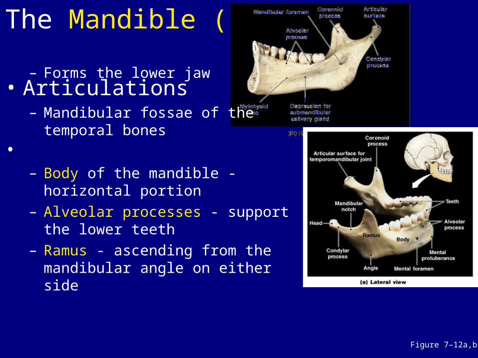
The Mandible (1)
Figure 7–12a,b
– Forms the lower jaw
• Articulations – Mandibular fossae of the
temporal bones•
– Body of the mandible - horizontal portion
– Alveolar processes - support the lower teeth
– Ramus - ascending from the mandibular angle on either side

• Foramina – Mental foramen-for sensory nerves of
lips and chin– Mandibular foramen-for blood vessels
and nerves of lower teeth

The Hyoid Bone (assoc w/facial bones)• Functions
– Supports the larynx– Attaches muscles of the
larynx, pharynx, and tongue• Articulations
– Connects lesser horns to styloid processes of temporal bones