Chapter 7 Cell Structure and Function Unit 3. Cells Cytology: the study of cells.
66
Chapter 7 Cell Structure and Function Unit 3
-
Upload
sabrina-barrett -
Category
Documents
-
view
217 -
download
0
Transcript of Chapter 7 Cell Structure and Function Unit 3. Cells Cytology: the study of cells.

Chapter 7
Cell Structure and Function
Unit 3

CellsCytology: the study of cells

IMPORTANT CELL STRUCTURES

Cell Theory
1.Cells are made from preexisting cells.
2.Cells are the smallest units of life.
3.All living things are made from at least one cell.

Cell History

Brief history• Redi (1660’s)
• Hooke (1660’s)
• Leeuwenhoek (1670’s)
• Schleiden & Schwann(1830’s)
• Virchow (1850’s)
• Brown (1830’s)

Francesco Redi
• 1668• Helped disprove spontaneous
generation• "All life comes from life"

Robert Hooke
• Mid-1660s
• Used microscopes to diagram samples of living organisms
• After looking a thin cuttings of cork, he called the chambers “CELLS” from the rooms found in monasteries
Anton van Leeuwenhoek
• Mid-1670s
• Used simple microscopes to observe microscopic life in pond water
• Made vast improvements to microscope construction

German cell biologists1830s: Matthias Schleidan: concluded all plants made from cells
1830s: Theodor Schwann: concluded that all animals made from cells

Rudolph Virchow• 1855
• Concluded that the nucleus was responsible for cell division.

Robert Brown
• 1883
• Recognized that cells contained a prominent feature and named it the
nucleus.

Louis Pasteur (1860s) Discovers that Cells come from Cells

Lynn MargulisRecognized for her work done in the 1980s
proposed the Endosymbiotic Theory
“Organelles in larger, complex cells may have been free-living prokaryotic cells in the past.”
In 2000 she received the U.S. National Medal of Science

Cell theory(based on 200+ years of discoveries)
1. Cells are made from preexisting cells.
2. Cells are the smallest units of life.
3. All living things are made from at least one cell.
Cells dividing
Brief History of Cells

Microscopy
• the use of microscopes to study cells
• Different types of microscopes– Light microscope– Transmission electron
microscope– Scanning electron
microscope

Cells are the basic unit of life
• Unicellular: organisms made up of only one cell– Ex: bacteria, protist, fungus
• Multicellular: organisms made up of more than one cell– Ex: protist, fungus, plant, animal

ALL CELLS CONTAIN THESE FOUR PARTS…
1. Plasma membrane: cell membrane, made of 2 layers of phospholipids
2. Cytosol: a carbohydrate and water based solution located in the cytoplasm that suspends all internal parts of the cell
3. Ribosomes: produces proteins4. DNA: genetic material made of nucleic acids

Types of cells
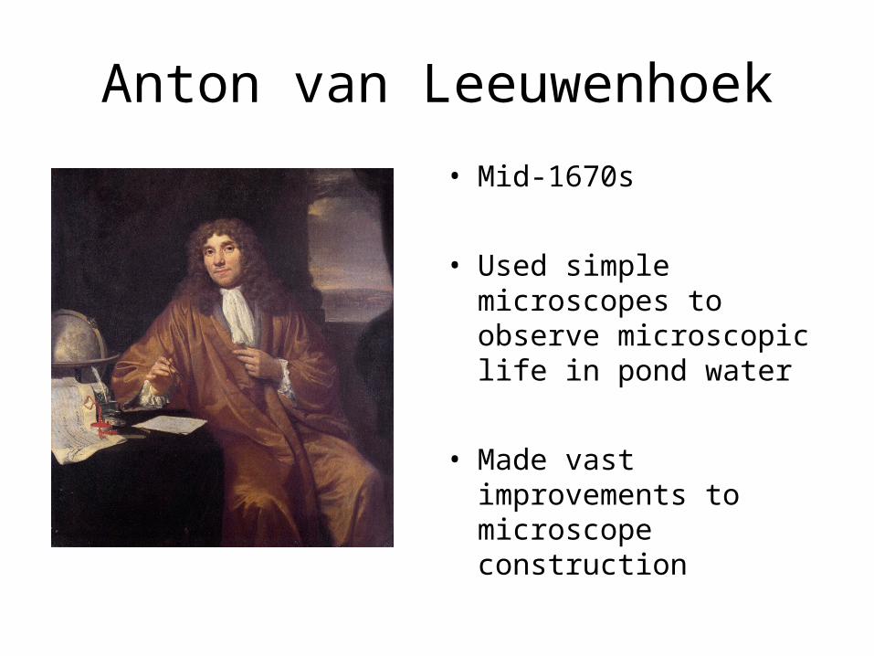
There are 2 types of cells
• Prokaryote: bacteria, archaebacteria
• Eukaryote: protist, fungus, plant, animal

Prokaryotes & Eukaryotes
Bacteria pic – Life, p.459
“Pro” = Before “Eu” = True
“Karyote” from Greek karyon = Kernal
You carry oats, I
eat ‘em!

Prokaryote (bacteria) cell features
• No membrane bound organelles (ex: nucleus, mitochondria, etc)
• Small
• Simple
• Plasma membrane, ribosome, cytoplasm, DNA

Prokaryotic (Bacterial) Cell
Capsule
Label the bacteria cell in IAN on pg. 9

Prokaryotic cell structure
• Capsule: durable outer covering that some bacteria have for protection against water, acids, and viruses
• Flagella: movement
• Cell wall: protective layer around plasma membrane
• Pili: anchoring and DNA exchange

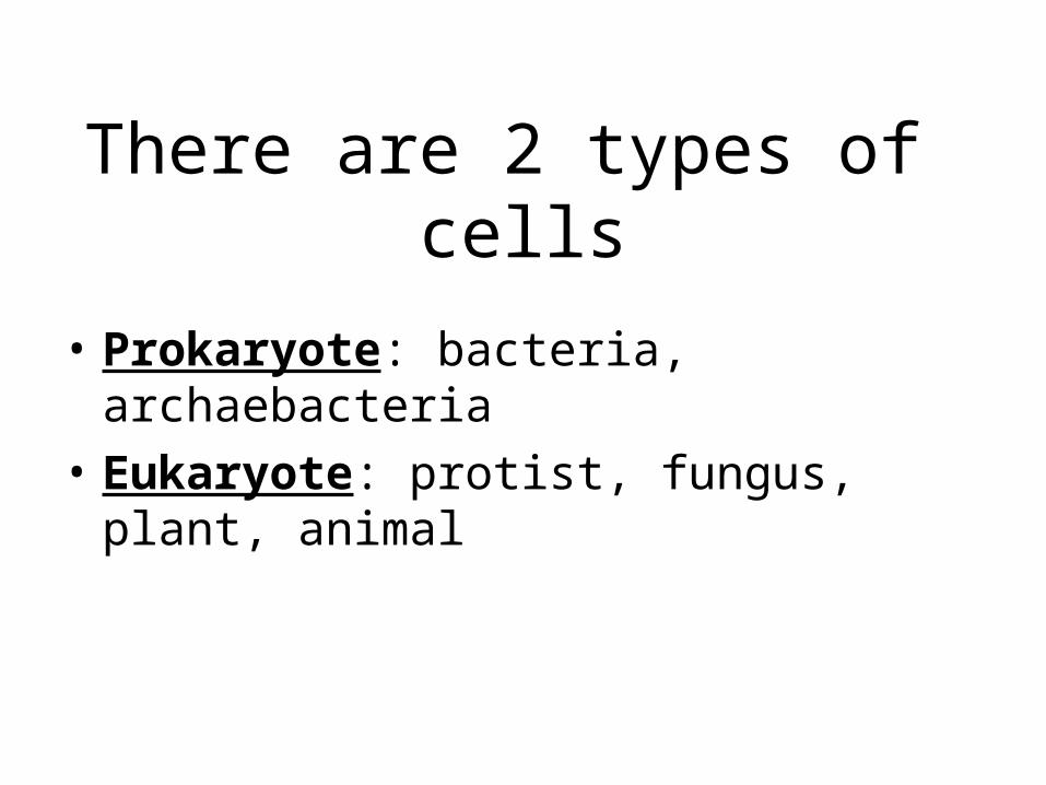
Eukaryotic Cell
• Complex• 4 basic components +
organelles• Organelles: “little
organs” that carry out specialized functions within a cell
• Have membrane organelles
• Many variations

Membranes

Plasma (Cell) Membrane (Phospholipid Bilayer)
• Outer boundary of cytoplasm• Semipermiable (only certain molecules enter
& leave)
Outsideof cell
Insideof cell(cytoplasm)
Cellmembrane
Proteins
Proteinchannel Lipid bilayer
Carbohydratechains

Phospholipid bilayer
• A double layer that is an effective barrier for most molecules
• Hydrophobic = “water fearing” tails in the center, prevent most things from entering
• Hydrophilic = “water loving” heads attract water to edges of membrane
Outsideof cell
Insideof cell(cytoplasm)
Cellmembrane
Proteins
Proteinchannel
Lipid bilayer
Carbohydratechains

Cell membranes are mosaics of different molecules combined.

Proteins embedded in membranes can be:
• Channels “gates” to interior for transporting into & out of cytoplasm
• Receptors “windows” for gathering information about cell surroundings
• Markers “name tags” that identify type of cell to others

Nucleus• Directs cell activities
• Nuclear Envelope (with pores) → outer boundary
• Chromatin/Chromosomes → (DNA) carry genetic information
• Nucleolus → (contain RNA) produces ribosomes
chromatin


Nucleus
• Stores ______ ______
• DNA helps to regulate ______ production.
• Proteins ______ cellular activity.

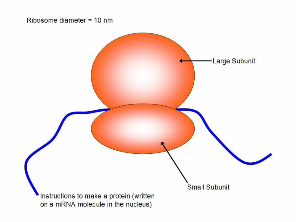
Ribosomes• small structures on endoplasmic reticulum and floating in the cytosol, that
function to produce protein
Endoplasmic reticulum • an interconnecting system of “pathways,” for transport.• May be Rough ER (with ribosomes attached) or Smooth ER (without ribosomes
attached).

• Large, sac-like compartment for storing liquids– Food, water, salt, or
waste
• In Animal cells they are called vesicles
• Called vacuoles in plant and Protists cell
Vacuole

Golgi apparatus (body)• Modify, package, and sort protein
packages for secretion, outside of cell
• flattened compartments with vesicles
for secretion (packaging & shipping)
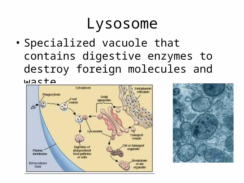
Lysosome• Specialized vacuole that contains
digestive enzymes to destroy foreign molecules and waste

Understanding
• A student drew this recycling symbol on their notebook cover to represent the lysosome; Why would this be an accurate representation?

Mitochondria• Organelle responsible for breaking down
glucose molecules into ATP energy molecules
• They are rod-shaped with folds, for releasing energy (ATP) by respiration


• Hypothesize which cells would have more mitochondria, cardiac cells or skin cells.
• Why?
Understanding

Chloroplast
• Organelle in plants and algae that produces sugar molecules by photosynthesis
• Why are chloroplasts green?
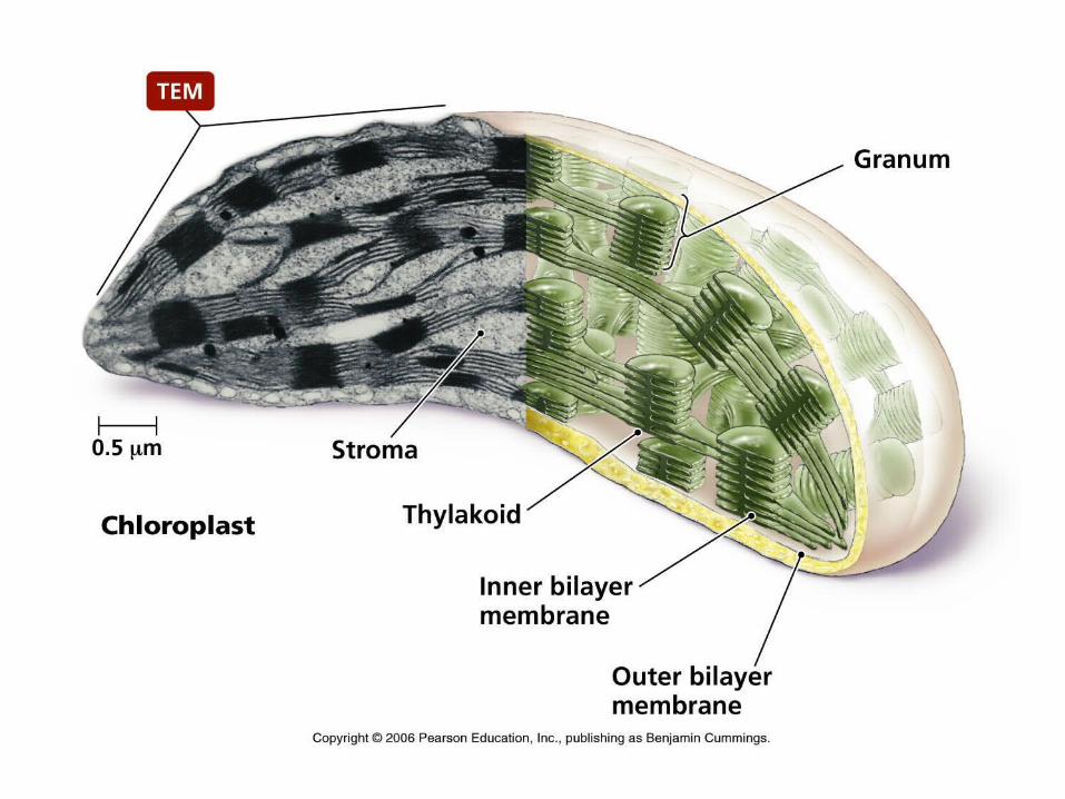

• A student takes a green plant into pitch dark room, closes the door and turns off the light. What color is the plant?
Understanding

Cytoskeleton
Cell membrane
Endoplasmicreticulum
Microtubule
Microfilament
RibosomesMitochondrion
•Hollow fibers for support & shape and to move cytoplasm
•Made of•Microfilaments•Microtubules

Plant Cell
Plant Cell
Nuclearenvelope
Ribosome(attached)
Ribosome(free)
Smooth endoplasmicreticulum
Nucleus
Rough endoplasmic reticulum
Nucleolus
Golgi apparatus
Mitochondrion
Cell wall
CellMembrane
Chloroplast
Vacuole
Label the plant cell

Animal Cell
Animal Cell
Centrioles
Nucleolus
Nucleus
Nuclearenvelope
Rough endoplasmic reticulum
Golgi apparatus
Smooth endoplasmicreticulum
Mitochondrion
CellMembrane
Ribosome(free)
Ribosome(attached)
Label the animal cell

Surface to volume ratio
• Because materials must be transported across cell membranes, maximizing the amount of membrane surface area increases transport efficiency.
• This is why larger organisms are multicellular.

• STOP NOTES HERE…
GO ONTO THE FOLDABLES AND THE NOTES FOR TRANSPORT AND TONICS

Comparing surface area to volume(Complete the calculations in the margin of your notes)
• Surface area: 6 mm x 6 mm x 6 sides = ___ mm2
• Volume: ___ mm3
• S / V = ___
• Surface area: 3 mm x 3 mm x 6 sides x 8 cubes = ___ mm2
• Volume = ___ mm3
• S / V = ___

current
(Concentration gradient)
Passive or Active transport?

Passive vs Active transport:which requires energy output?

Diffusion across a membrane

Osmosis across a membrane

Osmosis
• Osmosis - Diffusion of water across a selectively permeable membrane from higher water purity (high concentration) to lower water purity (low concentration)
• When solutions of varying water concentrations are found across a membrane, the solutions are given names.– Hypertonic– Hypotonic– Isotonic

Achieving equilibrium

Channel vs. Carrier Proteins

Active transport video
Click on image to play video.

Types of Active Transport
Endocytosis • cell brings particles
in.
Pinocytosis • Liquids are
brought in.

Phagocytosis• Phage means to “eat”• large particles (food or
bacteria) are surrounded & engulfed by cell.
• Examples = an ameba feeding & a white blood cell destroying an invader.
Why are white blood cells called MacroPhages?

“Cell eating”

Exocytosis
cell deposits particles outside of cytoplasm
• Secretion = cell products given off
• Excretion = wastes products given off

Cell structure is related to it’s function
• All cells are different.• Cell specialization → different cells have
different jobs.• Each cell serves different needs. • Diversity on a individual scale helps stabilize the
organism • Diversity on a large scale helps stabilize the
ecosystem