Chapter 3 Alkaline comet assay reveals elevated sperm DNA...
23
61 Chapter 3 Alkaline comet assay reveals elevated sperm DNA damage in idiopathic and oligozoospermic infertile men Introduction The crucial importance of germ cell DNA integrity in the establishment and maintenance of a viable pregnancy has been widely recognized (Sakkas and Alvarez, 2010; Barratt et al., 2010). It is well known that sperm chromatin is structurally and functionally different from that of somatic cells: it is not organized in nucleosomes (Ward, 2010). DNA in sperm is 6-fold more compacted and has 40-fold less volume than somatic cell DNA (Ward and Coffey, 1991) and has lost most, if not all, the repair capability (Aitken et al., 2004; Baumgartner et al., 2009). Despite the compact packing and anti-oxidant defense provided by seminal fluid (Smith et al., 1996; Potts et al., 2000), DNA damage does occur in both developing and mature sperm, high levels of which has been reported in infertile men (Kodama et al., 1997; Sun et al., 1997; Evenson et al., 1999, Spano et al., 2000; Zini et al., 2001; Singh et al., 2003). The role that sperm DNA damage plays in reproductive outcome has become increasingly debated and the subject of much research (Barratt and De Jonge, 2010; Sakkas and Alvarez, 2010; Schulte et al., 2010). Research over the last two decades has established that maintenance of sperm genomic integrity is crucial for the health of future generations (Ahmadi and Ng, 1999a, 1999b; Fernandez-Gonzalez et al., 2008; Zini et al., 2008). The need for assessing the integrity of the male gamete has been further intensified by growing concerns regarding
-
Upload
nguyendang -
Category
Documents
-
view
222 -
download
3
Transcript of Chapter 3 Alkaline comet assay reveals elevated sperm DNA...

61
Chapter 3
Alkaline comet assay reveals elevated sperm DNA
damage in idiopathic and oligozoospermic infertile
men
Introduction
The crucial importance of germ cell DNA integrity in the establishment and maintenance of a
viable pregnancy has been widely recognized (Sakkas and Alvarez, 2010; Barratt et al., 2010). It
is well known that sperm chromatin is structurally and functionally different from that of somatic
cells: it is not organized in nucleosomes (Ward, 2010). DNA in sperm is 6-fold more compacted
and has 40-fold less volume than somatic cell DNA (Ward and Coffey, 1991) and has lost most,
if not all, the repair capability (Aitken et al., 2004; Baumgartner et al., 2009). Despite the
compact packing and anti-oxidant defense provided by seminal fluid (Smith et al., 1996; Potts et
al., 2000), DNA damage does occur in both developing and mature sperm, high levels of which
has been reported in infertile men (Kodama et al., 1997; Sun et al., 1997; Evenson et al., 1999,
Spano et al., 2000; Zini et al., 2001; Singh et al., 2003).
The role that sperm DNA damage plays in reproductive outcome has become increasingly
debated and the subject of much research (Barratt and De Jonge, 2010; Sakkas and Alvarez,
2010; Schulte et al., 2010). Research over the last two decades has established that maintenance
of sperm genomic integrity is crucial for the health of future generations (Ahmadi and Ng,
1999a, 1999b; Fernandez-Gonzalez et al., 2008; Zini et al., 2008). The need for assessing the
integrity of the male gamete has been further intensified by growing concerns regarding
62
transmission of genetic diseases through ICSI (Barroso et al., 2009). In this context, direct assays
on spermatozoa, which represent the functional end product of the whole intra-testicular
spermatogenesis process and its maturation during epididymal transit, have the main advantage
of being able to detect effects on terminally differentiated male gametes ready to undergo
fertilization (Villani et al., 2010).
The comet assay has gained wider acceptance across multiple fields as a quick and simple
method of analyzing DNA damage in single cells (McArt et al., 2009) and is emerging as a
promising tool in male reproductive toxicology and infertility evaluation (Baumgartner et al.,
2009). Although today we have a battery of tests to assess DNA damage in sperm, the alkaline
comet assay remains the most sensitive and simple method of evaluating DNA damage in
individual sperm cells (Singh et al., 2003). It is being increasingly used in the diagnosis of male
infertility, and also as a prognostic tool to determine reproductive outcomes (Lewis and Agbaje,
2008; Simon et al., 2011). The technique is rapid, non-invasive, sensitive and inexpensive
compared to other techniques and therefore been considered suitable for the assessment of DNA
damage in sperm (Collins et al., 2008).
Typically, single-cell gel electrophoresis is performed on gel-restrained, disrupted spermatozoa
whose nuclei display a comet like tail. This comet tail consists of the fragmented, fluorescently
labeled, ss and ds-DNA that migrates away from the core of the intact DNA in an electrophoretic
field. The slides are then examined microscopically and quantified as the percentage of cells with
comet tails; alternatively the length, width and area can be computed with relevant software
(Lovell and Omori, 2008).

63
As a preliminary approach, to substantiate further work proposed in the thesis, it has been
planned to assess and compare sperm DNA damage in fertile controls and infertile subjects.
Owing to its distinct advantages, the alkaline comet assay has been employed in the present
study to assess genetic integrity of the sperm.
Materials and Methods
Subjects:
Patients visiting the infertility clinic of Kasturba Medical College for fertility evaluation and
treatment participated in this prospective study. Control group included fertile men, known to
have fathered a child within 12 months. Care was taken to ensure that the samples in the control
group and infertile group were age matched. Idiopathic infertility included infertile patients, with
normozoospermic semen parameters, who had a history of infertility of at least 2 years and
normal female partners i.e., normal reproductive history, normal day 2 follicular stimulating
hormone (FSH) and luteinizing hormone (LH) levels, normal ovulation (by follicular ultrasound
study), and tubal patency (by hysterosalpingogram). A total of 75 subjects participated in the
study, after provision of a written, informed consent. All subjects were asked to provide semen
samples after 3-5 days of ejaculatory abstinence. Semen specimens were produced by
masturbation directly into a sterile plastic container, in a room specially provided for this purpose
and located adjacent to the laboratory.
Semen Analysis:
After liquefaction, routine semen analysis was performed according to WHO criteria (WHO,
1999). Seminal volume was determined in a graduated tube and sperm concentration was
assessed by conventional method using Makler counting chamber (Sefi Medical Instruments,
64
Israel) and expressed in millions/mL. The sperm motility was assessed in at least 100 sperm and
expressed as percent of motile sperm (sum of rapid progression plus slow progression sperm).
Sperm morphology was assessed by Shorr staining and sperm viability by Eosin-Nigrosin stain.
The reference values of semen variables and nomenclature for some semen variables used
throughout the thesis has been presented in the subsequent section.
Comet Assay:
A part of the sample was used for assessment of DNA damage by alkaline comet assay (Singh et
al., 1988). It was performed as adapted for sperm, with modifications. Normal melting agarose
(NMA) (1.0%) was prepared by heating agarose in phosphate buffered saline (PBS, 137 mM
NaCl, 12 mM phosphate, 2.7 mM KCl). Following successive cleaning of the slide with soap
water and methanol, 200 µl of 1% NMA was spread uniformly and the slide was allowed to dry
at room temperature. The slides were stored in a dry place at room temperature until further use.
The cell suspension containing sperm (approximately 5 µl) was thoroughly mixed with low
melting point agarose (LMPA) (0.5%), prepared in PBS and maintained at 37°C. 200 µl of the
cell suspension with LMPA was layered onto the slide. Care was taken to ensure that the number
of cells was appropriate to facilitate easy image acquisition and analysis. A cover slip was placed
over the cell suspension and slide was placed on ice until the agarose layer hardened.
Subsequently, the cover slip was gently slided off and a third agarose layer (150 µl LMPA)
added, to the slide. Followed by hardening of agarose on ice and removal of the cover slip, the
slides were slowly lowered into cold, freshly made lysing solution (2.5 M sodium chloride, 100
mM EDTA, 10 mM Tris, 1% Triton X-100, 10% dimethylsulphoxide, pH:10). Following
overnight incubation, 15 mM dithiothreitol was added to the lysing solution and the slides were
incubated for additional 4 hours. The slides were then carefully removed from the lysing solution

65
and transferred to an electrophoretic chamber and placed side by side. The buffer reservoirs were
filled with freshly made electrophoresis buffer (1 mM EDTA and 300 mM NaOH buffer,
pH>13) and left to stand for 20 minutes to allow for alkaline unwinding of DNA. Electrophoresis
was carried out at 25V (~0.74 V/cm) and 300 mA for 30 minutes. The slides were then
neutralized with the neutralization buffer (0.4 M Tris, pH 7.5) at 4°C for 10 minutes. The slides
were then dehydrated in absolute ethanol for 2 hours and immersed for 5 minutes in 70%
ethanol. Slides were air-dried at room temperature. Immediately before scoring, slides were
rehydrated and stained with 100 µl of ethidium bromide stain (EtBr, 20 μg/ml), rinsed once in
PBS, coverslipped and analysed within 3 hours. Slides were examined at 40X under a fluorescent
microscope, equipped with a 490 nm excitation filter (Imager-A1, Zeiss, Germany). While
scoring, each slide was imaginarily divided into 8 parts and from each part, random images of 6-
7 spermatozoa were obtained. Approximately fifty cells were scored from each specimen. A
computerized image analysis system (Komet 6.0, Kinetic Imaging, Nottingham, UK) was used to
measure the different comet parameters.
Statistical Analysis
Basic descriptive statistics were calculated for standard semen parameters and different comet
assay parameters using Statistical Package for Social Sciences (SPSS 16.0). The value represents
Mean ± SEM. Statistical analysis of the means between different study groups was performed
using one way analysis of variance (ANOVA). A P value < 0.05 was considered statistically
significant.
66
Reagents
Sigma Aldrich, St. Louis, MO, USA
Normal melting agarose (NMA) Cat No A9539
Low melting point agarose (LMPA) Cat No A9414
Potassium chloride Cat No P5405
Tris base Cat No T4661
Dithiothreitol Cat No D9760
Triton X-100 Cat No T8532
Dimethylsulphoxide (DMSO) Cat No D5879
Nigrosin Cat No N4754
Ethidium Bromide Cat No E8754
Himedia, India
Sodium chloride Cat No MB023
Disodium Ethylene diamine tetra acetic acid Cat No MB011
Disodium hydrogen phosphate Cat No RM1154
Potassium dihydrogen phosphate Cat No RM249
Tris Hydrochloride Cat No RM613
Sodium hydroxide Cat No RM467
Ethidium bromide Cat No E8754
Merck & Co, USA
Shorr stain Cat No UN1993
Xylene Cat No 1330-20-7
Ammonia solution (about 25% pure) Cat No 1336-21-6
Thermo Fisher Scientific, UK
Methanol Cat No 32407
Hayman, UK
Absolute Alcohol (Ethanol)

67
BDH Chemicals, UK
Eosin Cat No 3419720
Sisco Research Laboratories, India
DPX Mountant for histology Cat No 42848
68
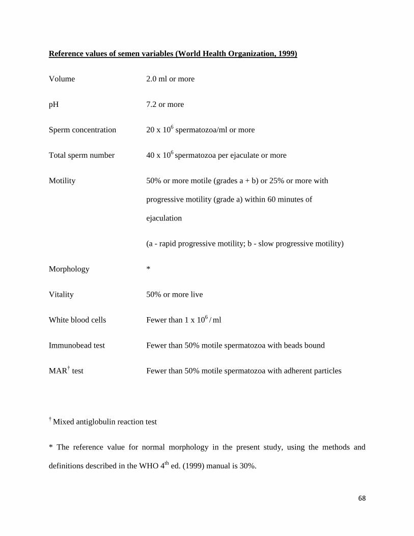
Reference values of semen variables (World Health Organization, 1999)
Volume 2.0 ml or more
pH 7.2 or more
Sperm concentration 20 x 106 spermatozoa/ml or more
Total sperm number 40 x 106
spermatozoa per ejaculate or more
Motility 50% or more motile (grades a + b) or 25% or more with
progressive motility (grade a) within 60 minutes of
ejaculation
(a - rapid progressive motility; b - slow progressive motility)
Morphology *
Vitality 50% or more live
White blood cells Fewer than 1 x 10⁶
/ ml
Immunobead test Fewer than 50% motile spermatozoa with beads bound
MAR† test Fewer than 50% motile spermatozoa with adherent particles
† Mixed antiglobulin reaction test
* The reference value for normal morphology in the present study, using the methods and
definitions described in the WHO 4th
ed. (1999) manual is 30%.
69
Nomenclature for some semen variables (Based on World Health Organization, 1999)
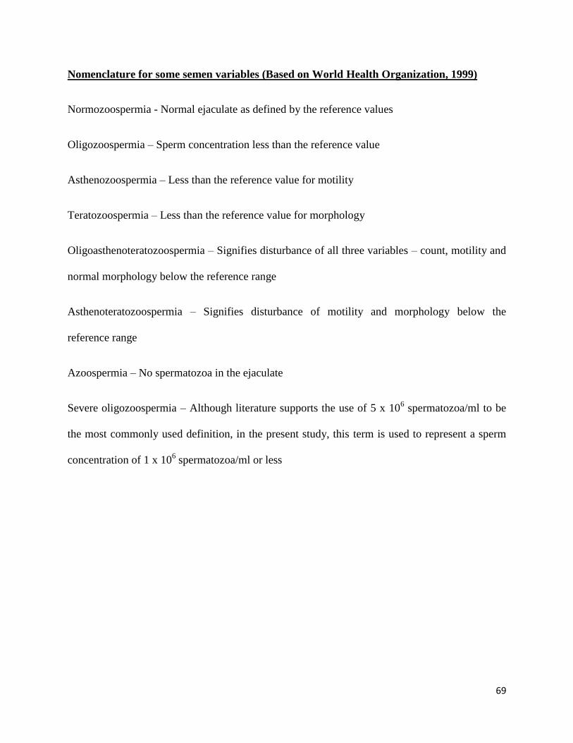
Normozoospermia - Normal ejaculate as defined by the reference values
Oligozoospermia – Sperm concentration less than the reference value
Asthenozoospermia – Less than the reference value for motility
Teratozoospermia – Less than the reference value for morphology
Oligoasthenoteratozoospermia – Signifies disturbance of all three variables – count, motility and
normal morphology below the reference range
Asthenoteratozoospermia – Signifies disturbance of motility and morphology below the
reference range
Azoospermia – No spermatozoa in the ejaculate
Severe oligozoospermia – Although literature supports the use of 5 x 10⁶ spermatozoa/ml to be
the most commonly used definition, in the present study, this term is used to represent a sperm
concentration of 1 x 10⁶ spermatozoa/ml or less

70
Results
Assessment of sperm DNA damage by alkaline comet assay
Semen characteristics of different categories of infertile subjects and fertile controls are
presented in Table 3.1. The values represent Mean ± SEM. Komet 6, comet analysis system by
Kinetic Imaging (Nottingham, UK), is a simple and most advanced and powerful software
solution for analysis, data management and presentation of comet assay samples. 24 parameters
are computed from the comet image based on intensity and migration patterns for comparison
between control and experimental groups. Among the various parameters available, percent of
tail DNA, olive tail moment and tail length were analysed as a measure to compare the extent of
DNA damage and is presented in Table 3.2.
The difference between the amount of total DNA and the percent DNA in the head region yields
percent tail DNA. The mean percent tail DNA in fertile controls was approximately 9.4
(Table 3.2). The percent tail DNA was significantly higher (P<0.001) in subjects with idiopathic
infertility and oligozoospermia (Table 3.2). Representative images of alkaline comet assay in
control, oligozoospermic and idiopathic infertile men is presented in Figure 3.1. The increase in
percent tail DNA was approximately 1.7, 1.4 fold higher in the idiopathic and oligozoospermic
group. However, in patient‟s asthenozoospermia and teratozoospermia, a non-significant increase
in percent tail DNA compared to the control group was observed.
The Olive tail moment (OTM), which is expressed in arbitrary units, represents the product of
amount of DNA in the tail and mean distance of migration in the tail. The mean OTM in control
71
group was approximately 1.1 while in those with idiopathic infertility and oligozoospermia, it
was 2.22 and 1.78 respectively, which was significantly higher (P<0.001). Those with
asthenozoospermia and teratozoospermia also had a marginal, non-significant increase in OTM
compared to the fertile, control group (Table 3.2).
The extent of migration of DNA in the comet tail is provided by the tail length, expressed in
micrometer. The mean tail length observed in control group was 10.77 µm. The tail length
observed in asthenozoospermic and teratozoospermic groups was not significantly different from
the control group. However, in idiopathic and oligozoospermic category, the tail length was
significantly higher (P<0.001) in comparison to the control group (Table 3.2).
Therefore, from the data, in comparison to the control group, it can be observed that patients
with idiopathic infertility (infertile subjects with normozoospermic semen parameters) and
oligozoospermia had significantly higher DNA damage compared to the control group. Although
the DNA damage was high in the other groups of infertile subjects studied (asthenozoospermia
and teratozoospermia), this difference was statistically insignificant (Figure 3.2). The idiopathic
group had the highest amount of DNA damage, to be followed by oligozoospermic,
asthenozoospermic and teratozoospermic samples (Table 3.2; Figure 3.2).

72
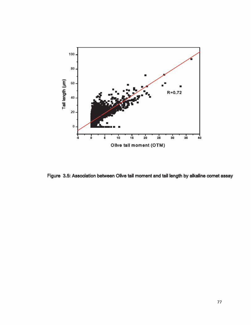
Association between different comet parameters assessed
To examine the possible association between the different comet parameters- percent tail DNA,
olive tail moment and tail length, a regression analysis was carried out. A positive association
was observed between all the measured parameters mentioned above. Percent tail DNA versus
Olive tail moment and tail length exhibited a good correlation with R=0.89 and R=0.67
respectively (Figure 3.3; Figure 3.4). Olive tail moment versus tail length also exhibited a good
positive correlation with R=0.72 (Figure 3.5).

73

74

75

76
77

78
Table 3.1: Mean semen characteristics of fertile controls and infertile subjects who underwent Comet assay
Group N Age (Yrs) Seminal volume
(ml)
Sperm count
(Millions/ml)
Total sperm
motility (%)
Sperm with
normal
morphology
(%)
Control 6 36.17 ± 1.60 2.67 ± 0.40 64.50 ± 9.94 71.50 ± 5.74 35.5 ± 3.01
Idiopathic infertility 17 36.24 ± 0.99 2.12 ± 0.18 60.41 ± 5.37 66 ± 1.97 35.41 ± 1.77
Oligozoospermia 18 35 ± 1.26 3.97 ± 0.72 9.57 ± 1.07 40.11 ± 4.49 16.56 ± 1.91
Asthenozoospermia 17 37.53 ± 1.05 3.26 ± 0.26 48.06 ± 5.71 43 ± 3.73 20.82 ± 2.01
Teratozoospermia 17 38.94 ± 1.53 3.76 ± 0.35 57.29 ± 6.03 52.41 ± 4.74 17.35 ± 1

79
Table 3.2: Comet assay parameters in different groups of infertile subjects and fertile controls
Group N Cells analysed Head DNA (%) Tail DNA (%) Tail length (µm) Olive tail moment
Control 6 343 90.51 ± 0.54 9.48 ± 0.54 10.77 ± 0.38 1.10 ± 0.08
Idiopathic infertility 17 931 83.52 ± 0.57* 16.47 ± 0.57* 11.99 ± 0.32* 2.22 ± 0.10*
Oligozoospermia 18 978 86.60 ± 0.52* 13.39 ± 0.52* 11.65 ± 0.31* 1.78 ± 0.10*
Asthenozoospermia 17 923 89.44 ± 0.38 10.55 ± 0.38 11.45 ± 0.29 1.25 ± 0.07
Teratozoospermia 17 929 89.90 ± 0.41 10.09 ± 0.41 7.83 ± 0.25 1.16 ± 0.08
* p<0.001 compared to the control group

80
Discussion
The technique of micro-gel electrophoresis of immobilized cells lysed at high salt concentrations
was first introduced in 1984 (Ostling and Johanson, 1984). The use of alkaline conditions for
DNA unwinding and electrophoresis (pH≥ 13) were incorporated later (Singh et al.,1988),
allowing the detection of double, and single-strand breaks, in addition to expression of alkali-
labile sites (ALS). Widespread acceptance of this technique and extensive application of the
same in various disciplines subsequently led to the establishment of guidelines for its use (Tice et
al., 2000). Today, the comet assay is a genotoxicity testing method widely applied both in vivo
and in vitro, to different organs and tissues. This method can be applied to non-proliferating cells
and for this reason it is one of the few cytogenetic assays applicable to detect DNA damage in
vitro on mature spermatozoa (Villani et al., 2010).
The alkaline comet assay can be applied to measure DNA damage at the single cell level since it
requires only a relatively small cell population. Further, the cells require no prelabelling with
radioactivity and can be non-invasive (Collins, 2004). As data are collected at an individual cell
level, the comet assay also gives a measure of the heterogeneity in a sample (Lovell and Omori,
2008). In addition, it is a simple, robust technique, capable of detecting low levels of DNA
damage (Tice et al., 2000, Collins, 2002, 2004). It can detect DNA damage equivalent to as few
as 50 single-strand breaks per cell and varying number of double strand breaks (Singh et al.,
1988; Tice and Strauss, 1995; Olive et al., 1998). The assay is able to measure both single- and
double-strand breaks with versatility, sensitivity (Leroy et al., 1996) and speed (Fairbairn et al.,
1995). It has therefore been considered to be suitable for the assessment of DNA damage in
sperm (Collins et al., 2008; Lewis and Agbaje, 2008) and relatively few numbers of cells ~50,
81
has been found to be sufficient for the reproducibility of the assay (Hughes et al., 1997). Owing
to the above-mentioned advantages, it has been used extensively to detect sperm DNA damage in
human biomonitoring, in the context of ART and in in vivo and ex vivo genotoxicity studies
(Lewis and Agbaje, 2008; Baumgartner et al., 2009; Speit et al., 2009).
There is a general belief that alkaline conditions are required to reveal single-strand (SS) breaks,
and that therefore using a near-neutral pH ensures that only double-strand (DS) breaks are picked
up (Van Kooij et al., 2004; McArt et al, 2009). This theory has been revealed to be a
misconception as alkaline conditions are not specific to detecting single-strand breaks (Collins et
al., 2008). Alkaline conditions, for DNA unwinding and electrophoresis (Singh et al., 1988) is
known to assess actual DNA strand breaks and alkali labile sites (Baumgartner et al., 2009).
Moreover, as most protocols for the comet assay in sperm involve high salt extraction in the
presence of a reducing reagent which removes both protamines and histones (Tomsu et al., 2002;
McVicar et al., 2004), the comet assay possibly detects chromatin breaks in both types of
chromatin with equal efficiency (Shaman and Ward, 2006).
Of the three measures of DNA migration commonly used - tail length, tail moment and percent
tail DNA, there is an increasing emphasis on the use of the percent tail DNA as the preferred
metric or the primary end point (Kumaravel and Jha, 2006). While the percent tail DNA is a
measure of the relative fluorescent intensity in the head and tail (Collins, 2004), the tail moment
is an index that takes into consideration both the migration of the genetic material and the
relative amount of DNA in the tail. The Olive tail moment (OTM) is the product of the tail
length and the percent tail DNA (Lovell and Omori, 2008). Tail length is considered
82
unsatisfactory as a measure because the length only increases at relatively low damage levels and
is sensitive to the background intensity of the image analysis system which affects the criteria for
determining the end of the tail (Collins, 2004). Tail length and moment, although consistent
within a study, may not be comparable across studies. In contrast, the percent tail DNA has the
distinct advantage that it can be „standardized‟ over studies (Lovell and Omori, 2008). It has
been recognized as the most suitable primary end point at the International Workshop on
Genotoxicity Test Procedures (Burlinson et al., 2007). Additionally, percent tail DNA has been
viewed as the most useful measure because it covers a wide range of damage (from 0 to 100%),
and is known to be independent of the threshold settings of the image analysis program (Collins,
2004). In view of good positive correlation obtained between percent tail DNA, Olive tail
moment and tail length in the present study, percent tail DNA is put forth as a most reliable
indicator of DNA damage in sperm.
Consistent with previously reported literature, in this study, the comet assay has been able to
detect DNA damage among the different groups of infertile men (Saleh et al., 2003a; Ahmad et
al., 2007; Erenpreiss et al., 2008). A high risk of infertility has been observed in men with sperm
DNA fragmentation at more than a diagnostic threshold of 25% by alkaline comet assay. The
risk of failure to achieve a pregnancy has also been reported to increase when sperm DNA
fragmentation exceeded a prognostic threshold value of 52% for semen and 42% for sperm
prepared by density gradient method (Simon et al., 2011). The results of the present study agree
with that of a recently published study where high degree of DNA damage has been reported in
idiopathic infertile men with normal semen profile as compared to fertile controls and variably
83
higher DNA damage in subjects with oligospermia, asthenospermia and teratozoospermia in
comparison to fertile controls has been observed (Shamsi et al., 2010).
Although today, many different modifications of the alkaline and the neutral comet assay are in
use, a standard protocol has not been established yet. In addition, high and variable background
levels of DNA effects have been reported and there is still need for standardization and
validation of the comet assay with sperm (Speit et al., 2009). Therefore, before it can gain wider
acceptance, in the infertility clinic, the assay is yet to undergo appropriate multilaboratory,
international validation studies to demonstrate its interlab and intralab reproducibility, reliability
and adequacy of its performance against the currently adopted methods. Additionally, studies
that can potentially lead to a better understanding of the underlying mechanisms of comet
formation and how strongly the comet shape reflects the DNA damage that has occurred is
clearly warranted (McArt et al., 2009). Nevertheless, an in vitro assay with human spermatozoa
is considered to be valuable, as a sensitive system to assess potential transgenerational effects in
the subject of reproduction and development.
To conclude, in the present study, employing alkaline comet assay, it has been demonstrated that
DNA damage is higher in idiopathic and oligozoospermic infertile men in comparison to fertile
controls. In addition to providing a rationale for further continuation of the work proposed in the
thesis, the above finding, coupled with concomitant concerns of increased sperm DNA damage
with failed/delayed fertilization and aberrant embryo development clearly warrants larger,
prospective studies to refine this one measure of sperm quality.