Challenges in the surgical management of spine trauma in ...
9
J Neurosurg: Spine / Volume 19 / July 2013 J Neurosurg Spine 19:101–109, 2013 101 ©AANS, 2013 T HE prevalence of obesity is increasing both in Australia and in other Western countries, reach- ing “pandemic” proportions. 19,40,44 The worldwide prevalence of obesity almost doubled between 1980 and 2008, from 4.8% to 9.8% in men and from 7.9% to 13.8% in women. 19 Australian men and women have a mean BMI that is among the highest in the world. 19 A population study in 2000 showed that the prevalence of overweight individuals and obesity in Australia is great- er than the worldwide mean, with 39.0% of Australians overweight and 20.8% obese. 8 Obesity is generally defined in terms of the BMI as measured by weight in kilograms divided by height in meters squared. 49 According to the definitions of the WHO, a BMI of 18–24.99 is normal, 25–29.99 is over- weight, 30–39.99 is obese, and ≥ 40 is morbidly obese. 49 In addition to the well-recognized medical, social, and economical consequences, this increasing level of obesity poses significant challenges for health care professionals in the multitrauma setting. We have encountered unique problems in our trau- ma center regarding the surgical management of spine trauma in the morbidly obese patient. This paper aims to highlight these problems using an illustrative case series. We will propose solutions using the experience gained from our caseload, to contribute to a growing body of knowledge on this topic that will be of interest to spine surgeons worldwide. Challenges in the surgical management of spine trauma in the morbidly obese patient: a case series Clinical article HANNAH E. ROSENFELD, 1 REBECCA LIMB, B.M.B.S., 2 P ATRICK CHAN, M.D., 2 MARK FITZGERALD, M.B.B.S., 3 WILLIAM PIERRE LITHERLAND BRADLEY , M.B.CH.B., 4,5 AND JEFFREY V. ROSENFELD, M.D., M.S. 2,5 1 Medical Faculty, University of Adelaide; 2 Department of Neurosurgery, and 3 Trauma Service, The Alfred Hospital, Melbourne; 4 Department of Anaesthesia, The Alfred Hospital and Monash University, Melbourne; and 5 Department of Surgery, Monash University, Melbourne, Australia Object. The treatment of morbidly obese individuals with spine trauma presents unique challenges to spine surgeons and trauma staff. This study aims to increase awareness of current limitations in the surgical management of spine trauma in morbidly obese individuals, and to illustrate practical solutions. Methods. Six morbidly obese patients were treated surgically for spine trauma over a 2-year period at a single trauma center in Australia. All patients were involved in high-speed motor vehicle accidents and had multisystem injuries. All weighed in excess of 265 pounds (120 kg) with a body mass index ≥ 40 (range 47.8–67.1). Cases were selected according to the considerable challenges they presented in all aspects of their management. Results. Best medical and surgical care may be compromised and outcome adversely affected in morbidly obese patients with spine trauma. The time taken to perform all aspects of care is usually extended, often by many hours. Customized orthotics may be required. Imaging quality is often compromised and patients may not fit into scanners. Surgical challenges include patient positioning, surgical access, confirmation of the anatomical level, and obtaining adequate instrument length. Postoperative nursing care, wound healing, and venous thromboembolism prophylaxis are also significant issues. Conclusions. Management pathways and hospital guidelines should be developed to optimize the treatment of morbidly obese patients, but innovative solutions may be required for individual cases. (http://thejns.org/doi/abs/10.3171/2013.4.SPINE12876) KEY WORDS • spine trauma • morbid obesity • anesthesia Abbreviations used in this paper: ASIA = American Spinal Injury Association; BMI = body mass index; LMWH = low-molecular- weight heparin. Unauthenticated | Downloaded 11/18/21 12:47 AM UTC
Transcript of Challenges in the surgical management of spine trauma in ...

J Neurosurg: Spine / Volume 19 / July 2013
J Neurosurg Spine 19:101–109, 2013
101
©AANS, 2013
The prevalence of obesity is increasing both in Australia and in other Western countries, reach-ing “pandemic” proportions.19,40,44 The worldwide
prevalence of obesity almost doubled between 1980 and 2008, from 4.8% to 9.8% in men and from 7.9% to 13.8% in women.19 Australian men and women have a mean BMI that is among the highest in the world.19 A population study in 2000 showed that the prevalence of overweight individuals and obesity in Australia is great-er than the worldwide mean, with 39.0% of Australians overweight and 20.8% obese.8
Obesity is generally defined in terms of the BMI
as measured by weight in kilograms divided by height in meters squared.49 According to the definitions of the WHO, a BMI of 18–24.99 is normal, 25–29.99 is over-weight, 30–39.99 is obese, and ≥ 40 is morbidly obese.49 In addition to the well-recognized medical, social, and economical consequences, this increasing level of obesity poses significant challenges for health care professionals in the multitrauma setting.
We have encountered unique problems in our trau-ma center regarding the surgical management of spine trauma in the morbidly obese patient. This paper aims to highlight these problems using an illustrative case series. We will propose solutions using the experience gained from our caseload, to contribute to a growing body of knowledge on this topic that will be of interest to spine surgeons worldwide.
Challenges in the surgical management of spine trauma in the morbidly obese patient: a case series
Clinical article
HannaH E. RosEnfEld,1 REbEcca limb, b.m.b.s.,2 PatRick cHan, m.d.,2 maRk fitzgERald, m.b.b.s.,3 William PiERRE litHERland bRadlEy, m.b.cH.b.,4,5 and JEffREy V. RosEnfEld, m.d., m.s.2,5
1Medical Faculty, University of Adelaide; 2Department of Neurosurgery, and 3Trauma Service, The Alfred Hospital, Melbourne; 4Department of Anaesthesia, The Alfred Hospital and Monash University, Melbourne; and 5Department of Surgery, Monash University, Melbourne, Australia
Object. The treatment of morbidly obese individuals with spine trauma presents unique challenges to spine surgeons and trauma staff. This study aims to increase awareness of current limitations in the surgical management of spine trauma in morbidly obese individuals, and to illustrate practical solutions.
Methods. Six morbidly obese patients were treated surgically for spine trauma over a 2-year period at a single trauma center in Australia. All patients were involved in high-speed motor vehicle accidents and had multisystem injuries. All weighed in excess of 265 pounds (120 kg) with a body mass index ≥ 40 (range 47.8–67.1). Cases were selected according to the considerable challenges they presented in all aspects of their management.
Results. Best medical and surgical care may be compromised and outcome adversely affected in morbidly obese patients with spine trauma. The time taken to perform all aspects of care is usually extended, often by many hours. Customized orthotics may be required. Imaging quality is often compromised and patients may not fit into scanners. Surgical challenges include patient positioning, surgical access, confirmation of the anatomical level, and obtaining adequate instrument length. Postoperative nursing care, wound healing, and venous thromboembolism prophylaxis are also significant issues.
Conclusions. Management pathways and hospital guidelines should be developed to optimize the treatment of morbidly obese patients, but innovative solutions may be required for individual cases.(http://thejns.org/doi/abs/10.3171/2013.4.SPINE12876)
kEy WoRds • spine trauma • morbid obesity • anesthesia
Abbreviations used in this paper: ASIA = American Spinal Injury Association; BMI = body mass index; LMWH = low-molecular-weight heparin.
Unauthenticated | Downloaded 11/18/21 12:47 AM UTC

H. E. Rosenfeld et al.
102 J Neurosurg: Spine / Volume 19 / July 2013
MethodsCases were identified by retrospectively reviewing
operative lists of surgically treated spine trauma patients in the preceding 2 years at the Alfred Hospital, a Level I trauma center. Patients with spine trauma treated sur-gically and classified as morbidly obese with particular difficulties in perioperative management were selected and the online imaging, operative reports, and anesthetic documentation analyzed. A low-risk ethics application to conduct the study was approved by the Alfred Hospital Ethics Committee.
ResultsSix morbidly obese patients treated surgically for
traumatic spinal injuries between January 2010 and Janu-ary 2012 were identified. All patients weighed in excess of 120 kg (265 lbs), and had a BMI > 40. All were in-volved in high-speed motor vehicle accidents and suf-fered multisystem injuries. In all but 1 of the cases (Case 2), the only diagnostic imaging modality available preop-eratively was CT because these patients were unable to fit into our hospital MRI machines due to their body habitus. A wide variety of unique challenges and unanticipated difficulties were encountered, which required solutions individually tailored to each patient.
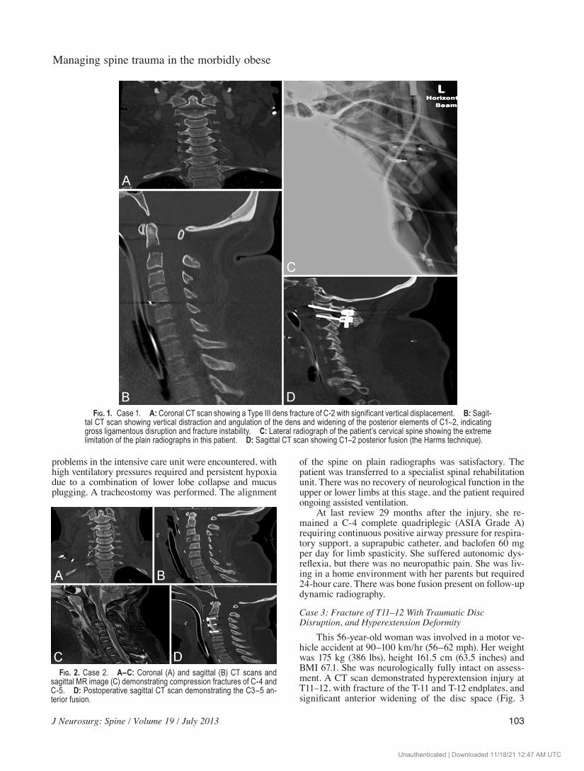
Case 1: Unstable Displaced Type III Dens Fracture With Significant Distraction and Posterior Ligamentous Disruption
This 38-year-old woman, with a weight of 140 kg (309 lbs), height of 171 cm (67.3 inches), and BMI of 47.8, was the driver of a car involved in a high-speed head-on collision. She was conscious and neurologically fully intact in all 4 limbs. She sustained an unstable Type III dens fracture of C-2 with posterior ligamentous disrup-tion, significant displacement, and angulation of the frac-ture (Fig. 1A and B). This was an unstable injury that required reduction and fixation. The initial stabilization required a customized handmade halo-thoracic vest. Af-ter applying the customized halo-thoracic vest, it took the next 12 hours to attempt to achieve a closed reduction.
Due to the patient’s body habitus, the plain radio-graph of the cervical spine obtained after application of the brace failed to provide adequate views of the dens to determine the reduction of fracture (Fig. 1C). Repeat CT demonstrated persistent, unacceptable displacement of the C-2 fracture. Closed reduction of the cervical spine was difficult to perform using standard fluoroscopic guid-ance because of the poor x-ray penetrance. Confirmation of fracture reduction required further CT. Unfortunately, due to difficulties caused by the patient’s body habitus, attempts at closed reduction of the C-2 fracture were unsuccessful. Open reduction and internal fixation were therefore indicated.
The patient underwent C1–2 posterior fusion via the Harms technique (Fig. 1D). Positioning the patient prone added approximately 1.5 hours to the operation and re-quired 8 people to facilitate. Intraoperatively, the patient was prone on a Gillespie pillow, a flat plastic cushion
placed on the operating table that has a hollow center for the chest and abdomen (Hipac Goulburn). During the op-eration, while the patient was on this pillow with her head fixated in a Halo ring attached to the Mayfield clamp, a large circuit leak was suddenly detected. The endotra-cheal tube had become dislodged, resulting in the tube tip being located in the back of the oropharynx in a fully anesthetized and paralyzed prone patient.
The anesthesia team rapidly repositioned the endo-tracheal tube using a fiberscope without any desaturation, while the patient remained prone. If this repositioning had been unsuccessful, the patient would have been returned to the supine position to facilitate reintubation, with or with-out the use of a supraglottic device to maintain oxygen-ation. This action would have required removing all drapes and surgical equipment, rapid closure of the wound, a sig-nificant number of people to turn the patient supine in a controlled fashion, and ideally would have meant replacing the remainder of the halo-thoracic brace first. This proce-dure could have resulted in a significant, sustained period of hypoxia if oxygenation could not have been maintained, with likely hypoxic injury to the brain and/or spinal cord.
The postoperative course was complicated by a cer-vical wound breakdown and infection, compounded by the wound traversing a large skin crease. This wound was successfully managed nonoperatively using long-term in-travenous antibiotics and specialist wound care. Pressure area care was extremely difficult to manage.
The patient was followed up 3 months postopera-tively. She had recovered well without neurological defi-cit and her cervical wound had healed well. The cervical spine was in alignment, with stability noted on flexion and extension radiographs.
Case 2: Compression Burst Fracture at C-4This 18-year-old female patient was transferred from
a rural interstate location (445 km from the trauma cen-ter), after a high-speed motor vehicle accident. Her weight was 125 kg (276 lbs), height 161 cm (63.3 inches), and BMI 48.2. Ambulance crews reported her to be tetraple-gic at the scene, with a sensory level at C-4. The initial di-agnosis was a suspected C-4 complete spinal cord injury (ASIA Grade A). She underwent intubation and mechani-cal ventilation for transfer to the trauma center.
Computed tomography scans and MRI showed a C-4 burst fracture with subluxation and retropulsion of C-4 causing spinal cord compression (Fig. 2A–C); other in-juries included pulmonary and cardiac contusions. The patient underwent an emergency open reduction, C-4 cor-pectomy, and internal fixation and anterior fusion from C-3 to C-5 using an iliac strut graft and cervical locking plate (Fig. 2D). The technical difficulties included achiev-ing and maintaining surgical retraction for both neck and right iliac exposure, which necessitated a longer incision and longer retractor and instruments.
There was an initial significant delay in transferring the patient from the interstate hospital due to weight re-strictions of the air transport. The time of injury was 0300 (3:00 in the morning) but the patient did not arrive at the trauma center by road ambulance until 2140 (9:40 at night), which entailed an 18-hour delay. Postoperative ventilation
Unauthenticated | Downloaded 11/18/21 12:47 AM UTC
J Neurosurg: Spine / Volume 19 / July 2013
Managing spine trauma in the morbidly obese
103
problems in the intensive care unit were encountered, with high ventilatory pressures required and persistent hypoxia due to a combination of lower lobe collapse and mucus plugging. A tracheostomy was performed. The alignment
of the spine on plain radiographs was satisfactory. The patient was transferred to a specialist spinal rehabilitation unit. There was no recovery of neurological function in the upper or lower limbs at this stage, and the patient required ongoing assisted ventilation.
At last review 29 months after the injury, she re-mained a C-4 complete quadriplegic (ASIA Grade A) requiring continuous positive airway pressure for respira-tory support, a suprapubic catheter, and baclofen 60 mg per day for limb spasticity. She suffered autonomic dys-reflexia, but there was no neuropathic pain. She was liv-ing in a home environment with her parents but required 24-hour care. There was bone fusion present on follow-up dynamic radiography.
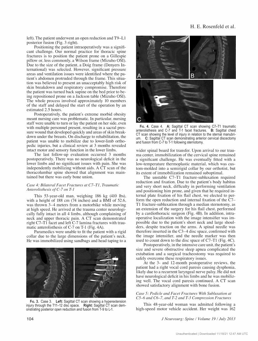
Case 3: Fracture of T11–12 With Traumatic Disc Disruption, and Hyperextension Deformity
This 56-year-old woman was involved in a motor ve-hicle accident at 90–100 km/hr (56–62 mph). Her weight was 175 kg (386 lbs), height 161.5 cm (63.5 inches) and BMI 67.1. She was neurologically fully intact on assess-ment. A CT scan demonstrated hyperextension injury at T11–12, with fracture of the T-11 and T-12 endplates, and significant anterior widening of the disc space (Fig. 3
Fig. 1. Case 1. A: Coronal CT scan showing a Type III dens fracture of C-2 with significant vertical displacement. B: Sagit-tal CT scan showing vertical distraction and angulation of the dens and widening of the posterior elements of C1–2, indicating gross ligamentous disruption and fracture instability. C: Lateral radiograph of the patient’s cervical spine showing the extreme limitation of the plain radiographs in this patient. D: Sagittal CT scan showing C1–2 posterior fusion (the Harms technique).
Fig. 2. Case 2. A–C: Coronal (A) and sagittal (B) CT scans and sagittal MR image (C) demonstrating compression fractures of C-4 and C-5. D: Postoperative sagittal CT scan demonstrating the C3–5 an-terior fusion.
Unauthenticated | Downloaded 11/18/21 12:47 AM UTC
H. E. Rosenfeld et al.
104 J Neurosurg: Spine / Volume 19 / July 2013
left). The patient underwent an open reduction and T9–L1 posterior fusion (Fig. 3 right).
Positioning the patient intraoperatively was a signifi-cant challenge. Our normal practice for thoracic spine fractures is to position the patient prone on a Gillespie pillow or, less commonly, a Wilson frame (Mizuho OSI). Due to the size of the patient, a Doig frame (Denyers In-ternational) was selected. However, significant pressure areas and ventilation issues were identified where the pa-tient’s abdomen protruded through the frame. This situa-tion was believed to present an unacceptably high risk of skin breakdown and respiratory compromise. Therefore the patient was turned back supine on the bed prior to be-ing repositioned prone on a Jackson table (Mizuho OSI). The whole process involved approximately 10 members of the staff and delayed the start of the operation by an estimated 2.5 hours.
Postoperatively, the patient’s extreme morbid obesity meant nursing care was problematic. In particular, nursing staff were unable to turn or lay the patient on her side, even with multiple personnel present, resulting in a sacral pres-sure wound that developed quickly and areas of skin break-down under the breasts. On discharge to rehabilitation, the patient was unable to mobilize due to lower-limb ortho-pedic injuries, but a clinical review at 3 months revealed intact motor and sensory function in the lower limbs.
The last follow-up evaluation occurred 3 months postoperatively. There was no neurological deficit in the lower limbs and no significant issues with pain. She was independently mobilizing without aids. A CT scan of the thoracolumbar spine showed that alignment was main-tained but there was early bone union.
Case 4: Bilateral Facet Fractures at C7–T1, Traumatic Anterolisthesis of C-7 on T-1
This 53-year-old man, weighing 186 kg (410 lbs), with a height of 188 cm (74 inches) and a BMI of 52.6, was thrown 3–4 meters from a motorbike while moving at high speed. He arrived at the trauma center neurologi-cally fully intact in all 4 limbs, although complaining of neck and upper thoracic pain. A CT scan demonstrated right C7–T1 facet and left C-7 lamina fractures with trau-matic anterolisthesis of C-7 on T-1 (Fig. 4A).
Paramedics were unable to fit the patient with a rigid collar due to the large dimensions of the patient’s neck. He was immobilized using sandbags and head taping to a
wider spinal board for transfer. Upon arrival to our trau-ma center, immobilization of the cervical spine remained a significant challenge. He was eventually fitted with a low-temperature thermoplastic material, which was cus-tom-molded into a semirigid collar by our orthotist, but its extent of immobilization remained suboptimal.
The unstable C7–T1 fracture-subluxation required reduction and fixation. Due to the patient’s body habitus and very short neck, difficulty in performing ventilation and positioning him prone, and given that he required in-ternal plate fixation of his flail chest, we elected to per-form the open reduction and internal fixation of the C7–T1 fracture-subluxation through a median sternotomy, as an extension of the surgery for his flail chest, performed by a cardiothoracic surgeon (Fig. 4B). In addition, intra-operative localization with the image intensifier was im-possible due to the patient’s short neck and large shoul-ders, despite traction on the arms. A spinal needle was therefore inserted in the C3–4 disc space, confirmed with the image intensifier, and the needle marker was then used to count down to the disc space of C7–T1 (Fig. 4C).
Postoperatively, in the intensive care unit, the patient’s size and severe obstructive sleep apnea complicated the extubation and a surgical tracheostomy was required to safely overcome these respiratory issues.
At the 3- and 12-month postoperative reviews, the patient had a right vocal cord paresis causing dysphonia, likely due to a recurrent laryngeal nerve palsy. He did not have neurological deficit in his limbs and he was mobiliz-ing well. The vocal cord paresis continued. A CT scan showed satisfactory alignment with bone fusion.
Case 5: Pedicle and Facet Fractures With Subluxation at C5–6 and C6–7, and T-2 and T-3 Compression Fractures
This 48-year-old woman was admitted following a high-speed motor vehicle accident. Her weight was 162
Fig. 3. Case 3. Left: Sagittal CT scan showing a hyperextension injury through the T11–12 disc space. Right: Sagittal CT scan dem-onstrating posterior open reduction and fusion from T-9 to L-1.
Fig. 4. Case 4. A: Sagittal CT scan showing C7–T1 traumatic anterolisthesis and C-7 and T-1 facet fractures. B: Sagittal chest CT scan showing the level of injury in relation to the sternal manubri-um. C: Sagittal CT scan demonstrating anterior cervical discectomy and fusion from C-7 to T-1 following sternotomy.
Unauthenticated | Downloaded 11/18/21 12:47 AM UTC

J Neurosurg: Spine / Volume 19 / July 2013
Managing spine trauma in the morbidly obese
105
kg (357 lbs), height 166 cm (65 inches), and BMI 58.8. On examination there was a tetraparesis affecting myotomes C6–8 bilaterally (2/5 motor grade power) and both lower limbs (1/5 on the right, 3/5 on the left). There was no sen-sory deficit or sensory level. This injury was classified as an incomplete spinal cord injury (ASIA Grade C).
Magnetic resonance imaging to assess for any ele-ment of disc prolapse prior to reduction of the deformity was not possible due to the patient’s dimensions. In ad-dition, diagnostic CT scans were significantly degraded by the patient’s size. The injuries identified were bilateral pedicle and facet fractures with subluxation at C5–6 and C6–7, and additional compression fractures at T-2 and T-3 (Fig. 5 upper).
The patient underwent an emergency 2-level anterior cervical discectomy and fusion (C5–6 and C6–7). The in-traoperative image intensifier was only able to penetrate the C-1 and C-2 levels, with the view of her subaxial cer-vical spine obstructed by her shoulders and adipose tis-sues. Intraoperative counting of the spinal level was not possible due to limited anterior exposure, due to both her adipose tissue and anatomy, as well as the length of re-tractors. Therefore, a single 3.5-mm cervical plate fixa-tion screw was inserted into the accessible vertebral body, the wound was temporarily closed, and repeat CT per-formed (Fig. 5 lower). This scan revealed the screw to be in the vertebral body of C-5. Other levels were able to be identified correctly once the patient was brought back to the operating room and the operation recommenced. The associated delays and transfers could have placed the pa-tient at increased risk of infection and neurological dam-age. The duration of the entire procedure was 7 hours. On discharge to rehabilitation, the patient had a residual right hemiparesis of 3/5 motor grade power and was unable to mobilize independently.
At the 2-month postoperative review, her tetraparesis had almost completely resolved (ASIA Grade D) but she complained of neuropathic paresthesia in her lower limbs. At 1 year postoperatively the painful paresthesia in her lower limbs had resolved and there was no neurological deficit (ASIA Grade E). The 1-year follow-up CT scan showed satisfactory bone union and alignment.
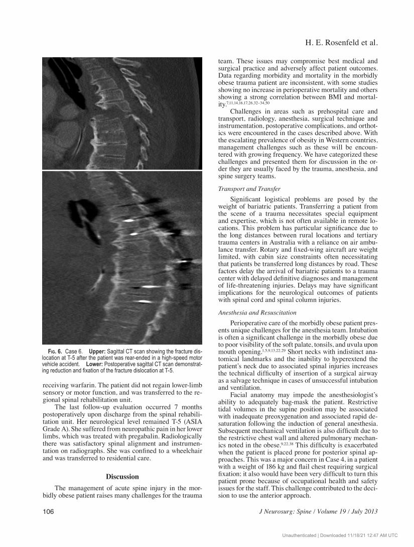
Case 6: Fracture Dislocation at T-5 With Spinal Cord Transection
This 69-year-old woman, weighing 140 kg (309 lbs), with a height of 160 cm (62 inches) and a BMI of 54.7, was the driver of a car that was rear-ended at high speed. On admission to the trauma center, she was noted to be paraplegic with no anal tone and a sensory level in the midthoracic region. The preliminary diagnosis was a complete spinal cord injury (ASIA Grade A). Other sig-nificant injuries included bilateral tension pneumotho-races and hemothoraces with significant hemodynamic instability. The patient was intubated and stabilized with bilateral tube thoracostomies. Subsequent CT scans con-firmed a T-5 vertebral body fracture with significant pos-terior dislocation, appearing to totally transect the spinal cord at this level (Fig. 6 upper). She was transferred to the operating room where she underwent open reduction and posterior fusion from T-2 to T-9 (Fig. 6 lower).
Intraoperatively, a decompressive laminectomy was performed at the level of the injury where severe damage and transection of the spinal cord was confirmed. Follow-ing decompression and during insertion of the thoracic pedicle screws, the patient became severely hypotensive. Intravenous fluid resuscitation was commenced until the anesthesia team discovered that the intravenous cannula in the right forearm was displaced with extensive extravasa-tion into the surrounding tissues. The operation was swift-ly completed. An immediate consultation with plastic sur-gery resulted in a diagnosis of compartment syndrome of the right forearm. The patient was subsequently taken for forearm fasciotomies, and later secondary closure of the wound under the care of the plastic surgery team.
Postoperatively, she developed the complication of bilateral lower-limb deep venous thrombosis and began
Fig. 5. Case 5. Upper: Sagittal CT scan (quality degraded) dem-onstrating the cervical and upper thoracic spine injuries. Lower: Sagittal CT scan demonstrating intraoperative screw placement in the vertebral body of C-5.
Unauthenticated | Downloaded 11/18/21 12:47 AM UTC
H. E. Rosenfeld et al.
106 J Neurosurg: Spine / Volume 19 / July 2013
receiving warfarin. The patient did not regain lower-limb sensory or motor function, and was transferred to the re-gional spinal rehabilitation unit.
The last follow-up evaluation occurred 7 months postoperatively upon discharge from the spinal rehabili-tation unit. Her neurological level remained T-5 (ASIA Grade A). She suffered from neuropathic pain in her lower limbs, which was treated with pregabalin. Radiologically there was satisfactory spinal alignment and instrumen-tation on radiographs. She was confined to a wheelchair and was transferred to residential care.
DiscussionThe management of acute spine injury in the mor-
bidly obese patient raises many challenges for the trauma
team. These issues may compromise best medical and surgical practice and adversely affect patient outcomes. Data regarding morbidity and mortality in the morbidly obese trauma patient are inconsistent, with some studies showing no increase in perioperative mortality and others showing a strong correlation between BMI and mortal-ity.7,11,14,16,17,26,32–34,50
Challenges in areas such as prehospital care and transport, radiology, anesthesia, surgical technique and instrumentation, postoperative complications, and orthot-ics were encountered in the cases described above. With the escalating prevalence of obesity in Western countries, management challenges such as these will be encoun-tered with growing frequency. We have categorized these challenges and presented them for discussion in the or-der they are usually faced by the trauma, anesthesia, and spine surgery teams.
Transport and TransferSignificant logistical problems are posed by the
weight of bariatric patients. Transferring a patient from the scene of a trauma necessitates special equipment and expertise, which is not often available in remote lo-cations. This problem has particular significance due to the long distances between rural locations and tertiary trauma centers in Australia with a reliance on air ambu-lance transfer. Rotary and fixed-wing aircraft are weight limited, with cabin size constraints often necessitating that patients be transferred long distances by road. These factors delay the arrival of bariatric patients to a trauma center with delayed definitive diagnoses and management of life-threatening injuries. Delays may have significant implications for the neurological outcomes of patients with spinal cord and spinal column injuries.
Anesthesia and ResuscitationPerioperative care of the morbidly obese patient pres-
ents unique challenges for the anesthesia team. Intubation is often a significant challenge in the morbidly obese due to poor visibility of the soft palate, tonsils, and uvula upon mouth opening.1,5,9,13,22,29 Short necks with indistinct ana-tomical landmarks and the inability to hyperextend the patient’s neck due to associated spinal injuries increases the technical difficulty of insertion of a surgical airway as a salvage technique in cases of unsuccessful intubation and ventilation.
Facial anatomy may impede the anesthesiologist’s ability to adequately bag-mask the patient. Restrictive tidal volumes in the supine position may be associated with inadequate preoxygenation and associated rapid de-saturation following the induction of general anesthesia. Subsequent mechanical ventilation is also difficult due to the restrictive chest wall and altered pulmonary mechan-ics noted in the obese.9,22,38 This difficulty is exacerbated when the patient is placed prone for posterior spinal ap-proaches. This was a major concern in Case 4, in a patient with a weight of 186 kg and flail chest requiring surgical fixation; it also would have been very difficult to turn this patient prone because of occupational health and safety issues for the staff. This challenge contributed to the deci-sion to use the anterior approach.
Fig. 6. Case 6. Upper: Sagittal CT scan showing the fracture dis-location at T-5 after the patient was rear-ended in a high-speed motor vehicle accident. Lower: Postoperative sagittal CT scan demonstrat-ing reduction and fixation of the fracture dislocation at T-5.
Unauthenticated | Downloaded 11/18/21 12:47 AM UTC
J Neurosurg: Spine / Volume 19 / July 2013
Managing spine trauma in the morbidly obese
107
Endotracheal tubes are more easily dislodged in the morbidly obese, as in Case 1, and require careful securing of the tube. There should always be direct communica-tion between the anesthesiology and surgical teams when spinal manipulation involving the airway occurs.
Excess subcutaneous tissue renders intravenous ac-cess difficult.13,24,28 As in Case 6, the risk of dislodgement or misplacement of peripheral intravenous cannulae in the morbidly obese may be difficult to identify and sig-nificantly compromise the patient’s condition. Urinary bladder catheterization and arterial line placement are similarly technically difficult in the morbidly obese.
Monitoring is less accurate in the morbidly obese:5 electrocardiography traces, noninvasive blood pressure monitoring, and pulse oximeters may be inaccurate in this patient population,5,13,22 and there should be a low threshold for invasive monitoring in these patients.5,13
Postoperatively, obese patients with obstructive sleep apnea, who have undergone a procedure while prone, are more susceptible to developing airway edema and obstruction.30 A planned extubation is performed in the intensive care unit with the patient awake and able to re-spond to commands. Immediate postoperative continuous positive airway pressure and early chest physiotherapy are recommended.13,30,38 There may be a greater indica-tion for surgical tracheostomy in these patients.
ImagingPreoperative imaging with all modalities is more dif-
ficult and less accurate in the obese. Plain radiographs are of lower quality due to poor x-ray penetration through excessive adipose tissue and increased scatter.12,47 The use of CT and MRI is also limited by excess adiposity in the morbidly obese. Computed tomography scans are often degraded, resulting in lower diagnostic capabilities and greater radiation exposure.12,41,46 Magnetic resonance ma-chines have weight and girth restrictions, often making them unusable in this patient group; the open MRI ma-chine may provide a solution to this problem.12,41
Surgical PositioningStandard operating tables can withstand patient
weights of approximately 150 kg.5 An operating table specially designed for obese patients may be necessary for patients who exceed the weight limit.5 If such a table is not available, 2 standard operating tables can be clamped together.1,42
Patient transfers and positioning are challenging, time consuming, and potentially hazardous to staff. Ad-ditional specially trained operating room personnel and special hoist systems may be necessary to prevent injury to staff.5 Morbidly obese patients are at an increased risk of positioning-related peripheral neuropathy.37 Standard spinal frames and devices (such as Wilson frames and Gillespie pillows) have little flexibility to accommodate patients of large dimensions and may exacerbate, rather than alleviate, problems with pressure-area care, resulting in greater risk of skin breakdown. As of yet, there are no specialized bariatric spinal frames or operating tables in widespread use, which is clearly an area of need.
Surgical Approaches and TechniquesDue to the poor x-ray penetrance in the morbidly
obese patient, image quality from an intraoperative 2D or 3D fluoroscopic C-arm may be excessively degraded for identifying the spinal levels or guiding instrument po-sitioning. Intraoperative 3D fluoroscopic O-arm devices and the intraoperative fixed or mobile CT scanner may not always have wide enough apertures to accommodate and image some morbidly obese patients. As encountered in Case 5, the C-arm device did not adequately image the cervical spine. It is standard practice to place a spi-nal needle at an accessible level and identify the correct spinal level in relation to this level, such as in Case 4. The neurosurgeon decided that this method was not feasible in Case 5 because the spine could not be imaged below the level of the C-2 body due to her obesity, shoulder height, and shape of her neck. There was the additional difficulty in accessing the C-2 body from the level of the skin inci-sion to allow for level counting to commence. This dif-ficulty could have been overcome by using intraoperative 3D fluoroscopy or CT, which might assist in guiding and confirming instrument placement.
Due to the excessive depth of the subcutaneous adi-pose tissue in the obese, longer incisions may be neces-sary for adequate exposure.45 Satisfactory retraction may also be rendered difficult by the deep subcutaneous tis-sue layer.42,45 Bariatric instruments or a fixed retractor system such as Scoville, TrimLine (Medtronic), or Koros retractors may ameliorate this issue.23,45 However, such equipment may not be readily available, and many spinal instrumentation manufacturers do not currently provide longer instruments for use in bariatric patients. More ex-tensive and invasive approaches may be required, such as the sternotomy in Case 4, to reach the surgical objective.
Technical challenges and risks of morbidity also apply with bone autograft harvesting, such as the iliac crest strut graft used in Case 2. To avoid donor site morbidity, graft-ing with allograft bone, bone substitute, or the insertion of synthetic cages are alternatives, but are subject to their availability and the consideration of a lower fusion rate.
Blood LossVenous epidural bleeding can be greater due to in-
creased intraabdominal and intrathoracic pressures in the obese patient with impaired venous return to the heart, particularly in the prone position. Use of the Cell Saver (Haemonetics Corp.) may maintain oxygen carrying ca-pacity and reduce transfusion requirements.
Postoperative Wound CareIt has been shown that there is a greater incidence of
wound breakdown and surgical site infection in the mor-bidly obese, such as occurred in Case 1.2–4,6,10,15,18,20,25,34,
35,43,48 In addition to surgical site infections, the morbidly obese are at greater risk of pressure sores.1,27 Substantial padding intraoperatively and postoperatively, in addition to frequent inspection of the skin at pressure points and the use of oscillating air mattresses, may reduce pressure areas.13
Unauthenticated | Downloaded 11/18/21 12:47 AM UTC

H. E. Rosenfeld et al.
108 J Neurosurg: Spine / Volume 19 / July 2013
Venous ThromboembolismThe incidence of venous thromboembolism is greater
in obese patients because of venous stasis due to raised intraabdominal pressure and relative immobility, and to a hypercoagulable state.1,13,36,39 Perioperative pneumatic calf compressors may reduce the risk of postoperative venous thromboembolism in this population. However, it may be difficult to acquire large enough compression de-vices for some morbidly obese patients.1 Ultrasonography diagnosis may be more difficult due to the degradation of images associated with morbid obesity.
The incidence of deep venous thrombosis and pul-monary embolism is reduced with the use of prophylactic LMWH.21 However, initiation of LMWH prophylaxis is often delayed because of injury-related intracranial, intra-spinal, or other bleeding, as well as deferment associated with planned operative intervention. On a weight-adjust-ed basis, morbidly obese patients should receive the same dose of LMWH, although there is evidence that twice-daily dosing of 1 mg/kg is safer for patients with a BMI greater than 35 or a weight greater than 150 kg (330 lbs).21
OrthosesAt the initial scene of a trauma, paramedics may
not be able to fit a morbidly obese patient with a cervi-cal collar, necessitating the use of suboptimal alternatives for immobilization, such as sandbags and head taping to the spinal board. Once the patients are transferred to a trauma center, there may be difficulty obtaining appro-priately sized orthoses, including halo-thoracic braces, collars, and thoracolumbar braces. The use of custom-made braces and orthoses, such as in Cases 1 and 4, is dependent on an experienced orthotics department. As an alternative, a cervical collar could be applied, opened as wide as possible, secured with an overlapping Velcro strap, and reinforced with casting material, which is then cut and taped over for easy removal.
Postoperative Pulmonary CareEarly fixation and mobilization of morbidly obese
patients with spinal injuries reduces the incidence of re-spiratory insufficiency and pneumonia.31 Speech patholo-gists should identify the morbidly obese patients with cervical spine injuries at risk for aspiration.
ConclusionsSpine surgeons and trauma specialists should be
cognizant of the significant logistical, medical, and so-cietal challenges posed by morbidly obese patients with spine trauma, who are increasing in number. With greater awareness and with improvements in medical techniques and technology, hospitals can develop management path-ways and guidelines to reduce adverse neurological and overall outcomes for morbidly obese patients.
Disclosure
The authors report no conflict of interest concerning the mate-rials or methods used in this study or the findings specified in this paper.
Author contributions to the study and manuscript preparation
include the following. Conception and design: JV Rosenfeld, Limb, Chan. Acquisition of data: H Rosenfeld, Limb, Bradley. Analysis and interpretation of data: Limb, JV Rosenfeld, Chan. Drafting the article: H Rosenfeld, Limb, Fitzgerald. Critically revising the article: all authors. Reviewed submitted version of manuscript: all authors. Approved the final version of the manuscript on behalf of all authors: JV Rosenfeld.
References
1. Adams JP, Murphy PG: Obesity in anaesthesia and intensive care. Br J Anaesth 85:91–108, 2000
2. Anaya DA, Dellinger EP: The obese surgical patient: a suscepti-ble host for infection. Surg Infect (Larchmt) 7:473–480, 2006
3. Andreshak TG, An HS, Hall J, Stein B: Lumbar spine surgery in the obese patient. J Spinal Disord 10:376–379, 1997
4. Bamgbade OA, Rutter TW, Nafiu OO, Dorje P: Postoperative complications in obese and nonobese patients. World J Surg 31:556–561, 2007
5. Bein B, Scholz J: Anaesthesia for adults undergoing non-bariat-ric surgery. Best Pract Res Clin Anaesthesiol 25:37–51, 2011
6. Bochicchio GV, Joshi M, Bochicchio K, Nehman S, Tracy JK, Scalea TM: Impact of obesity in the critically ill trauma pa-tient: a prospective study. J Am Coll Surg 203:533–538, 2006
7. Byrnes MC, McDaniel MD, Moore MB, Helmer SD, Smith RS: The effect of obesity on outcomes among injured patients. J Trauma 58:232–237, 2005
8. Cameron AJ, Welborn TA, Zimmet PZ, Dunstan DW, Owen N, Salmon J, et al: Overweight and obesity in Australia: the 1999-2000 Australian Diabetes, Obesity and Lifestyle Study (AusDiab). Med J Aust 178:427–432, 2003
9. Candiotti K, Sharma S, Shankar R: Obesity, obstructive sleep apnoea, and diabetes mellitus: anaesthetic implications. Br J Anaesth 103 (Suppl 1):i23–i30, 2009
10. Cheadle WG: Risk factors for surgical site infection. Surg In-fect (Larchmt) 7 (Suppl 1):S7–S11, 2006
11. Christmas AB, Reynolds J, Wilson AK, Franklin GA, Miller FB, Richardson JD, et al: Morbid obesity impacts mortality in blunt trauma. Am Surg 73:1122–1125, 2007
12. de Bucourt M, Streitparth F, Wonneberger U, Rump J, Teich-gräber U: Obese patients in an open MRI at 1.0 Tesla: im-age quality, diagnostic impact and feasibility. Eur Radiol 21: 1004–1015, 2011
13. DeMaria EJ, Carmody BJ: Perioperative management of special populations: obesity. Surg Clin North Am 85:1283–1289, xii, 2005
14. Diaz JJ Jr, Norris PR, Collier BR, Berkes MB, Ozdas A, May AK, et al: Morbid obesity is not a risk factor for mortality in critically ill trauma patients. J Trauma 66:226–231, 2009
15. Dindo D, Muller MK, Weber M, Clavien PA: Obesity in gen-eral elective surgery. Lancet 361:2032–2035, 2003
16. Duchesne JC, Schmieg RE Jr, Simmons JD, Islam T, McGin-ness CL, McSwain NE Jr: Impact of obesity in damage con-trol laparotomy patients. J Trauma 67:108–114, 2009
17. Evans DC, Stawicki SP, Davido HT, Eiferman D: Obesity in trauma patients: correlations of body mass index with out-comes, injury patterns, and complications. Am Surg 77:1003–1008, 2011
18. Falagas ME, Kompoti M: Obesity and infection. Lancet In-fect Dis 6:438–446, 2006
19. Finucane MM, Stevens GA, Cowan MJ, Danaei G, Lin JK, Paciorek CJ, et al: National, regional, and global trends in body-mass index since 1980: systematic analysis of health examination surveys and epidemiological studies with 960 country-years and 9.1 million participants. Lancet 377:557–567, 2011
20. Fleischmann E, Kurz A, Niedermayr M, Schebesta K, Kim-berger O, Sessler DI, et al: Tissue oxygenation in obese and non-obese patients during laparoscopy. Obes Surg 15:813–819, 2005
Unauthenticated | Downloaded 11/18/21 12:47 AM UTC

J Neurosurg: Spine / Volume 19 / July 2013
Managing spine trauma in the morbidly obese
109
21. Freeman A, Horner T, Pendleton RC, Rondina MT: Prospec-tive comparison of three enoxaparin dosing regimens to achieve target anti-factor Xa levels in hospitalized, medically ill patients with extreme obesity. Am J Hematol 87:740–743, 2012
22. Grant P, Newcombe M: Emergency management of the mor-bidly obese. Emerg Med Australas 16:309–317, 2004
23. Greenleaf RM, Altman DT: Evaluation and treatment of spi-nal injuries in the obese patient. Orthop Clin North Am 42: 85–93, vi–vii, 2011
24. Gregg SC, Murthi SB, Sisley AC, Stein DM, Scalea TM: Ul-trasound-guided peripheral intravenous access in the inten-sive care unit. J Crit Care 25:514–519, 2010
25. Harrop JS, Styliaras JC, Ooi YC, Radcliff KE, Vaccaro AR, Wu C: Contributing factors to surgical site infections. J Am Acad Orthop Surg 20:94–101, 2012
26. Hoffmann M, Lefering R, Gruber-Rathmann M, Rueger JM, Lehmann W: The impact of BMI on polytrauma outcome. In-jury 43:184–188, 2012
27. Jupiter JB, Ring D, Rosen H: The complications and difficul-ties of management of nonunion in the severely obese. J Or-thop Trauma 9:363–370, 1995
28. Juvin P, Blarel A, Bruno F, Desmonts JM: Is peripheral line placement more difficult in obese than in lean patients? Anesth Analg 96:1218, 2003
29. Juvin P, Lavaut E, Dupont H, Lefevre P, Demetriou M, Du-moulin JL, et al: Difficult tracheal intubation is more common in obese than in lean patients. Anesth Analg 97:595–600, 2003
30. Kristensen MS: Airway management and morbid obesity. Eur J Anaesthesiol 27:923–927, 2010
31. McHenry TP, Mirza SK, Wang J, Wade CE, O’Keefe GE, Dai-ley AT, et al: Risk factors for respiratory failure following op-erative stabilization of thoracic and lumbar spine fractures. J Bone Joint Surg Am 88:997–1005, 2006
32. Meroz Y, Gozal Y: Management of the obese trauma patient. Anesthesiol Clin 25:91–98, ix, 2007
33. Neville AL, Brown CV, Weng J, Demetriades D, Velmahos GC: Obesity is an independent risk factor of mortality in se-verely injured blunt trauma patients. Arch Surg 139:983–987, 2004
34. Newell MA, Bard MR, Goettler CE, Toschlog EA, Schenarts PJ, Sagraves SG, et al: Body mass index and outcomes in criti-cally injured blunt trauma patients: weighing the impact. J Am Coll Surg 204:1056–1064, 2007
35. Olsen MA, Nepple JJ, Riew KD, Lenke LG, Bridwell KH, Mayfield J, et al: Risk factors for surgical site infection fol-lowing orthopaedic spinal operations. J Bone Joint Surg Am 90:62–69, 2008
36. Parkin L, Sweetland S, Balkwill A, Green J, Reeves G, Beral V: Body mass index, surgery, and risk of venous thromboem-bolism in middle-aged women: a cohort study. Circulation 125:1897–1904, 2012
37. Patel N, Bagan B, Vadera S, Maltenfort MG, Deutsch H, Vac-caro AR, et al: Obesity and spine surgery: relation to periop-erative complications. J Neurosurg Spine 6:291–297, 2007
38. Pelosi P, Gregoretti C: Perioperative management of obese pa-tients. Best Pract Res Clin Anaesthesiol 24:211–225, 2010
39. Platzer P, Thalhammer G, Jaindl M, Obradovic A, Benesch T, Vecsei V, et al: Thromboembolic complications after spinal surgery in trauma patients. Acta Orthop 77:755–760, 2006
40. Roth J, Qiang X, Marbán SL, Redelt H, Lowell BC: The obe-sity pandemic: where have we been and where are we going? Obes Res 12 (Suppl 2):88S–101S, 2004
41. Rothschild PA, Domesek JM, Eastham ME, Kaufman L: MR imaging of excessively obese patients: the use of an open per-manent magnet. Magn Reson Imaging 9:151–154, 1991
42. Sarr MG, Felty CL, Hilmer DM, Urban DL, O’Connor G, Hall BA, et al: Technical and practical considerations involved in operations on patients weighing more than 270 kg. Arch Surg 130:102–105, 1995
43. Serrano PE, Khuder SA, Fath JJ: Obesity as a risk factor for nosocomial infections in trauma patients. J Am Coll Surg 211:61–67, 2010
44. Swinburn BA, Sacks G, Hall KD, McPherson K, Finegood DT, Moodie ML, et al: The global obesity pandemic: shaped by global drivers and local environments. Lancet 378:804–814, 2011
45. Telfeian AE, Reiter GT, Durham SR, Marcotte P: Spine surgery in morbidly obese patients. J Neurosurg (1 Suppl) 97:20–24, 2002
46. Uppot RN: Impact of obesity on radiology. Radiol Clin North Am 45:231–246, 2007
47. Uppot RN, Sahani DV, Hahn PF, Gervais D, Mueller PR: Im-pact of obesity on medical imaging and image-guided inter-vention. AJR Am J Roentgenol 188:433–440, 2007
48. Wimmer C, Gluch H, Franzreb M, Ogon M: Predisposing fac-tors for infection in spine surgery: a survey of 850 spinal pro-cedures. J Spinal Disord 11:124–128, 1998
49. World Health Organization: BMI Classification. Geneva: World Health Organization, 2012 (http://apps.who.int/bmi/index.jsp?introPage=intro_3.html) [Accessed April 10, 2013]
50. Wutzler S, Maegele M, Marzi I, Spanholtz T, Wafaisade A, Lefering R: Association of preexisting medical conditions with in-hospital mortality in multiple-trauma patients. J Am Coll Surg 209:75–81, 2009
Manuscript submitted September 25, 2012.Accepted April 8, 2013.Please include this information when citing this paper: pub-
lished online May 21, 2013; DOI: 10.3171/2013.4.SPINE12876.Address correspondence to: Jeffrey V. Rosenfeld, M.D., M.S.,
Department of Surgery, Level 6, The Alfred Centre, 99 Commercial Road, Melbourne, Australia 3004. email: [email protected].
Unauthenticated | Downloaded 11/18/21 12:47 AM UTC