Cervical Spine Trauma: How to clear the C-Spine...
36
1 Cervical Spine Trauma: Interpretation of the C-Spine Film Anthony Powell, Harvard Medical School- Year III Gillian Lieberman, MD Gerlock AJ, Heller RM, Kaye JJ, Kirchner SG: The Cervical Spine in Trauma. Advanced Exercises in Diagnostic Radiology, vol 11. W.B. Saunders, 1978 November 2000 Anthony Powell Gillian Lieberman, MD
Transcript of Cervical Spine Trauma: How to clear the C-Spine...
1
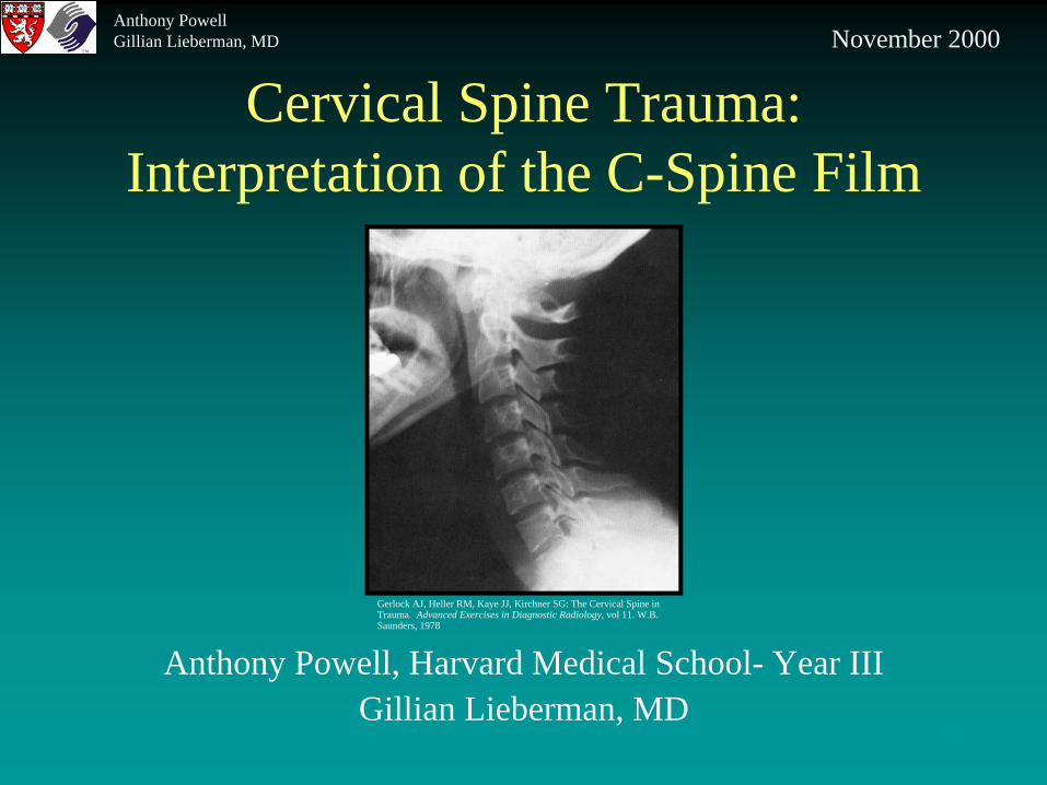
Cervical Spine Trauma: Interpretation of the C-Spine Film
Anthony Powell, Harvard Medical School- Year IIIGillian Lieberman, MD
Gerlock AJ, Heller RM, Kaye JJ, Kirchner SG: The Cervical Spine in Trauma. Advanced Exercises in Diagnostic Radiology, vol 11. W.B. Saunders, 1978
November 2000Anthony PowellGillian Lieberman, MD

2
Common Mechanisms of Injury
• Hyperflexion- MVA, car comes to sudden stop• Hyperextension- MVA, car struck from behind• Compression- Head first dive in shallow water
Anthony PowellGillian Lieberman, MD

3
Clinical Procedure involving Pts with suspected spine injury
• Pt kept in cervical collar and immobilized on spine board
• “ABCDEF” ER protocol followed (airway, breathing, circulation, disability/drugs, exposure, Foley catheter)
• History and physical (pt handled as though serious injury present)
• Decide if imaging is necessary
Anthony PowellGillian Lieberman, MD

4
Vandermark “risk-tailored” approach in assessing need for neck imaging
• No Risk- no hx or physical findings suggestive of neck injury
• Low Risk- pt has hx for a mechanism of injury unlikely to have exceeded physiologic range of cervical motion
• Medium Risk- pt has hx for mechanism of injury sufficient to have exceeded physiologic cervical ROM
• High Risk- pt has hx for mechanism of injury very likely to have exceeded physiologic ROM
Anthony PowellGillian Lieberman, MD

5
High Risk Factors for cervical vertebral injury
• High-velocity blunt trauma• Multiple, severe long bone fractures• Direct cervical region injury• Altered mental status• Fall from greater than 10 feet• Drowning / head first diving accident• Significant head or facial injury• Neck pain, tenderness, or deformity• Abnormal neurological examination• Thoracic or lumbar vertebral fracture• Hx of pre-existing vertebral disease
Anthony PowellGillian Lieberman, MD

6
Imaging Must Assess for Spinal Cord Stability
• Critical assessment which determines pt’s management and handling
• Unstable- spinal canal is no longer protected by its ligament and bony supports
• Any movement of neck could result in permanent cord damage
Anthony PowellGillian Lieberman, MD

7
Menu of Imaging Options
• Cervical Spine Plain Films-standard first line imaging modality in assessing cervical vertebral injury
• Cervical CT-to evaluate extent of injury with any definitive finding on plain film-delineating equivocal/uncertain findings on plain film
• Cervical MRI-pt with c-spine trauma who exhibits neurological signs andsymptoms suggestive of cord injury
Anthony PowellGillian Lieberman, MD

8
In order to recognize the abnormal, we need to know the
normal appearance
Let’s review some c-spine anatomy:
Anthony PowellGillian Lieberman, MD

9
Atlanto-axial joint
C2, Axis C1, Atlas
Odontoid
Anterior arch
Posterior arch
Superior articulating surface
Lateral mass
Cervical spine
Images: Netter, FH: Atlas of Human Anatomy, 2nd
ed. Novartis, 1997
Anthony PowellGillian Lieberman, MD

10
Occipito-Atlanto-Axial Joint
Gerlock AJ, Heller RM, Kaye JJ, Kirchner SG: The Cervical Spine in Trauma. Advanced Exercises in Diagnostic Radiology, vol 11. W.B. Saunders, 1978
C1
C2
Occipital condyle
Odontiod
Lateral mass C1
Lateral Mass C2
C2
Anthony PowellGillian Lieberman, MD
11
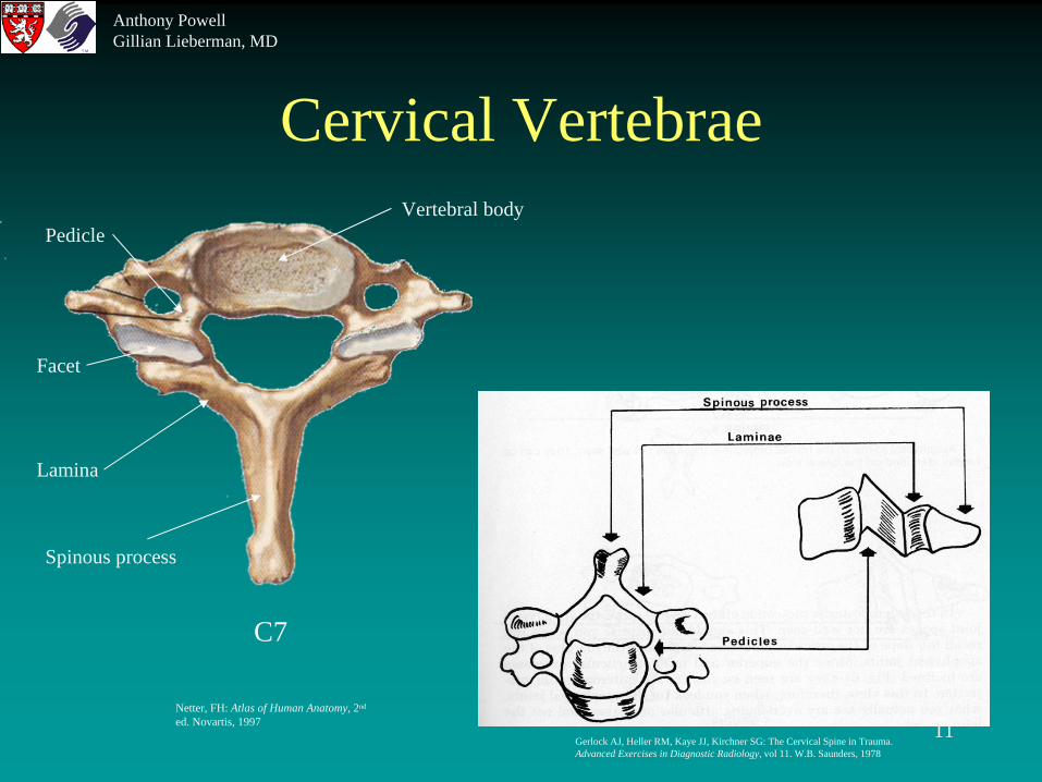
Cervical Vertebrae
C7
Spinous process
Lamina
Vertebral bodyPedicle
Facet
Gerlock AJ, Heller RM, Kaye JJ, Kirchner SG: The Cervical Spine in Trauma. Advanced Exercises in Diagnostic Radiology, vol 11. W.B. Saunders, 1978
Netter, FH: Atlas of Human Anatomy, 2nd
ed. Novartis, 1997
Anthony PowellGillian Lieberman, MD

12
Radiograph Correlations
Apophyseal / facet joints
Rotation effect on lateral view
Gerlock AJ, Heller RM, Kaye JJ, Kirchner SG: The Cervical Spine in Trauma. Advanced Exercises in Diagnostic Radiology, vol 11. W.B. Saunders, 1978
Anthony PowellGillian Lieberman, MD

13
Ligaments
Ant Longitudinal lig
Post Longitudinal lig
Supraspinous lig
Ligamenta Flava
Gerlock AJ, Heller RM, Kaye JJ, Kirchner SG: The Cervical Spine in Trauma. Advanced Exercises in Diagnostic Radiology, vol 11. W.B. Saunders, 1978
Netter, FH: Atlas of Human Anatomy, 2nd ed. Novartis, 1997
Anthony PowellGillian Lieberman, MD

14
C-Spine Plain Films
Standard 5 view series:• Cross table lateral• AP of lower cervical column• Atlanto-axial AP (open mouth, odontiod)• Bilateral supine trauma oblique views
Anthony PowellGillian Lieberman, MD

15
Normal C-spine 5 view series
Gerlock AJ, Heller RM, Kaye JJ, Kirchner SG: The Cervical Spine in Trauma. Advanced Exercises in Diagnostic Radiology, vol 11. W.B. Saunders, 1978 ; BIDMC
Lateral AP Odontoid
Right Oblique Left Oblique
Anthony PowellGillian Lieberman, MD

16
C-spine Film Interpretation 7 step process
1. Count Vertebrae (lateral)-C1 through C7-If T1 not seen get a swimmer’s view
2. Assess Curvature (frontal and lateral)3. Assess Vertebral Alignment (on lateral: 4 lines)
-ant vertebral line-post vertebral line-spinolaminar line-post spinal line
Anthony PowellGillian Lieberman, MD

17
The 4 Contour Lines
Gerlock AJ, Heller RM, Kaye JJ, Kirchner SG: The Cervical Spine in Trauma. Advanced Exercises in Diagnostic Radiology, vol 11. W.B. Saunders, 1978
Anthony PowellGillian Lieberman, MD

18
C-spine Film Interpretation 7 step process
1. Count Vertebrae-C1 through C7-If T1 not seen Swimmer’s view
2. Assess Curvature3. Assess Vertebral Alignment (4 lines)
-ant vertebral line-post vertebral line-spinolaminal line-post spinal line
4. Assess Bony Integrity5. Assess Intervertebral Disk Spaces6. Assess OAA joint7. Soft Tissues
Anthony PowellGillian Lieberman, MD

19
Patient
• 91 y.o. female who presents to ED with a laceration and contusion to her right eye, suffered when she fell out of her bed at the assisted living facility. Fall was not witnessed. Pt is also complaining of neck pain. Pt reports no loc, n/v, visual changes, headache, numbness or weakness. Exam reveals no focal or gross neurological deficits. Pt is secured in a hard cervical collar.
• Pt sent for plain films
Anthony PowellGillian Lieberman, MD

20
Our patient’s lateral c-spine
•Farrell, Susan MD. Teaching Files. BIDMC dept of Emergency Medicine, 2000
Film findings:
1. C2 fracture through base of odontoid process
2. Approx 4mmposterior displace-ment of C1 on C2
Anthony PowellGillian Lieberman, MD

21
Our patient’s course
• An MRI confirmed the dens fx with no compromise of the spinal canal.
• Treatment: Due to pt’s age and medical conditions, she was treated conservatively with hard collar immobilization. Interval x-rays were ordered to assess for odontoid mobility.
Anthony PowellGillian Lieberman, MD

22
Let’s review some other patients whose c-spine plain films
demonstrate common cervical fractures
Anthony PowellGillian Lieberman, MD

23
Hangman’s Fracture
• Bilateral pedicle fractures of C2 (axis)• Anterolisthesis of C2 on C3• Unstable fracture• Hyperextension injury
-fracture is identical to those occurring upon hanging
-elderly may slip and strike chin on basin or counter-MVA in which chin strikes steering wheel
Anthony PowellGillian Lieberman, MD

24
Hangman’s Fracture
Gerlock AJ, Heller RM, Kaye JJ, Kirchner SG: The Cervical Spine in Trauma. Advanced Exercises in Diagnostic Radiology, vol 11. W.B. Saunders, 1978
Anthony PowellGillian Lieberman, MD

25
Teardrop Fracture
• Avulsion fracture of anterior margin of vertebral body• Anterior longitudinal lig instability (rupture, avulsion)• Hyperextension injury• Unstable injury• Lamina may jam together causing ligamenta flava to
buckle inward and compress/contuse the spinal cord
Anthony PowellGillian Lieberman, MD

26
Teardrop Fracture
Gerlock AJ, Heller RM, Kaye JJ, Kirchner SG: The Cervical Spine in Trauma. Advanced Exercises in Diagnostic Radiology, vol 11. W.B. Saunders, 1978
Anthony PowellGillian Lieberman, MD

27
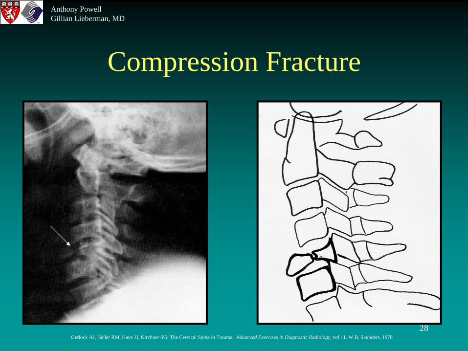
Compression Fracture
• Variable severity, from minimal anterior wedging to complete disruption of vertebral body (burst)
• Look for loss of vertical height of vertebral body• Due to long axis compression or hyperflexion
-diving into shallow pool• Stable unstable
Anthony PowellGillian Lieberman, MD
28
Compression Fracture
Gerlock AJ, Heller RM, Kaye JJ, Kirchner SG: The Cervical Spine in Trauma. Advanced Exercises in Diagnostic Radiology, vol 11. W.B. Saunders, 1978
Anthony PowellGillian Lieberman, MD

29
Jefferson Fracture
• Compression/bursting fracture of C1 ring• Due to long axis compression forces• Results in uni or bilateral displacement of C1 lateral
masses with respect to C2 sup articulating facets• Best seen on odontoid (open mouth) view• Unstable if transverse ligament is disrupted, resulting
in C1 anterolisthesis• May be stable
Anthony PowellGillian Lieberman, MD

30
Jefferson Fracture
Gerlock AJ, Heller RM, Kaye JJ, Kirchner SG: The Cervical Spine in Trauma. Advanced Exercises in Diagnostic Radiology, vol 11. W.B. Saunders, 1978
Anthony PowellGillian Lieberman, MD

31
Clay Shoveler’s Fracture
• Avulsion fracture of spinous process by supraspinous ligament
• Usually occurring from C6-T2• Hyperflexion; direct trauma; downward force via
thoracoscapular muscle (as in shoveling motion)• Stable
Anthony PowellGillian Lieberman, MD

32
Clay Shoveler’s Fracture
Gerlock AJ, Heller RM, Kaye JJ, Kirchner SG: The Cervical Spine in Trauma. Advanced Exercises in Diagnostic Radiology, vol 11. W.B. Saunders, 1978
Anthony PowellGillian Lieberman, MD
33
Dens Fracture
• Fracture of the base of the dens (odontoid) of C2• Anterior or posterior displacement of the dens• Can occur at various levels on the dens• Via hyperflexion or hyperextension of head on neck• Unstable if displacement occurs
Anthony PowellGillian Lieberman, MD

34
Dens Fracture
Gerlock AJ, Heller RM, Kaye JJ, Kirchner SG: The Cervical Spine in Trauma. Advanced Exercises in Diagnostic Radiology, vol 11. W.B. Saunders, 1978
Anthony PowellGillian Lieberman, MD

35
References• Daffner RH: Evaluation of cervical vertebral injuries. Seminars in
Roentgenology 24:239-253, 1992• Gerlock AJ, Heller RM, Kaye JJ, Kirchner SG: The Cervical Spine in
Trauma. Advanced Exercises in Diagnostic Radiology, vol 11. W.B. Saunders, 1978
• Vandermark RM: Radiology of the cervical spine in trauma patients: Practice pitfalls and recommendations for improving efficiency and communication. AJR 155:465-472, 1990
• Novelline, RA: Squire’s Fundamentals of Radiology, ed 5. Harvard University Press, 1997
• Farrell, Susan MD. Teaching Files. BIDMC dept of Emergency Medicine, 2000
• Netter, FH: Atlas of Human Anatomy, 2nd ed. Novartis, 1997• Gunderman, RB: Essential Radiology, clinical presentation,
pathophysiology, imaging. Thieme, 1998
Anthony PowellGillian Lieberman, MD
36
Acknowledgements
• Beverlee Turner for her support and PowerPoint expertise.• Susan Farrell MD
Anthony PowellGillian Lieberman, MD