Cell structure - University of New Mexicoquantum.phys.unm.edu/102-17l/phbi8.pdf · 8 Cell structure...
13
8 Cell structure 8.1 Organelles A prokaryotic cell is a bag of salty water, DNA, RNA, proteins, sugars, and so forth between 0.1 and 5 μm in diameter surrounded by a tough cell wall. Prokaryotic cells have no internal structure or transport mechanism. They rely upon di↵usion to move their molecules around. Eukaryotic cells have diameters between 10 and 100 μm, and nerve cells can be as long as one meter. A eukaryotic cell has a nucleus that contains the cell’s DNA, is about 5 μm in diameter, and constitutes one-tenth of the volume of the cell. A plasma membrane surrounds the nucleus in a double layer that extends into the cytosol as the endoplasmic reticulum. Inside the nucleus, the nucleolus is a factory that makes ribosomes. RNA polymerase II transcribes a nucleotide sequence from DNA into pre-mRNA inside the nucleus. A spliceosome (made of snRNAs and proteins) then excises intron segments and redundant exons from the pre- mRNA. An exportin protein then transports the resulting mRNA through a nuclear pore complex into the cytosol. The endoplasmic reticulum and the Golgi apparatus use ribosomes and other molecular devices to make and decorate proteins and glycosamino- glycans (GAGs, long chains of disacharides) which the cell then moves to the plasma membrane in membrane-enclosed sacs that bud from the en- doplasmic reticulum and from the Golgi apparatus and then fuse with the membranes of other organelles or with the plasma membrane. Ribosomes in the cytosol also translate mRNAs into proteins. A mitochondrion is an organelle with an outer membrane and an in- ner membrane on which ATP synthase uses pyruvate and NADH (both made from glucose in the cytosol) to turn ADP into ATP. A red blood cell has no nucleus nor any mitochondria, but a liver cell can have 2000 mito-
Transcript of Cell structure - University of New Mexicoquantum.phys.unm.edu/102-17l/phbi8.pdf · 8 Cell structure...
8
Cell structure
8.1 Organelles
A prokaryotic cell is a bag of salty water, DNA, RNA, proteins, sugars,and so forth between 0.1 and 5 µm in diameter surrounded by a tough cellwall. Prokaryotic cells have no internal structure or transport mechanism.They rely upon di↵usion to move their molecules around.
Eukaryotic cells have diameters between 10 and 100 µm, and nerve cellscan be as long as one meter. A eukaryotic cell has a nucleus that containsthe cell’s DNA, is about 5 µm in diameter, and constitutes one-tenth of thevolume of the cell. A plasma membrane surrounds the nucleus in a doublelayer that extends into the cytosol as the endoplasmic reticulum. Insidethe nucleus, the nucleolus is a factory that makes ribosomes.
RNA polymerase II transcribes a nucleotide sequence from DNA intopre-mRNA inside the nucleus. A spliceosome (made of snRNAs andproteins) then excises intron segments and redundant exons from the pre-mRNA. An exportin protein then transports the resulting mRNA througha nuclear pore complex into the cytosol.
The endoplasmic reticulum and the Golgi apparatus use ribosomes andother molecular devices to make and decorate proteins and glycosamino-glycans (GAGs, long chains of disacharides) which the cell then moves tothe plasma membrane in membrane-enclosed sacs that bud from the en-doplasmic reticulum and from the Golgi apparatus and then fuse with themembranes of other organelles or with the plasma membrane. Ribosomesin the cytosol also translate mRNAs into proteins.
A mitochondrion is an organelle with an outer membrane and an in-ner membrane on which ATP synthase uses pyruvate and NADH (bothmade from glucose in the cytosol) to turn ADP into ATP. A red blood cellhas no nucleus nor any mitochondria, but a liver cell can have 2000 mito-
8.2 Membranes 77
chondria. Most animals inherit their mitochondria only from their moth-ers; mechanisms destroy the mitochondria of sperm after fertilization (Zhouet al., 2016).
Endosomes are membrane sacs that enclose external material that thecell has just swallowed by endocytosis. Endosomes pass much of this ma-terial to lysosomes which use acids and enzymes to digest this materialand surplus cytosolic macromolecules into elementary molecules that thecell reuses. Cells oxidize toxic molecules in peroxisomes. Proteasomesare organelles that use proteases to dissolve the peptide bonds of proteins,decomposing them into reusable amino acids.
Because eukaryotic cells have volumes that are between 8 and 109 timesbigger than prokaryotic cells, they can’t rely upon di↵usion to move moleculesand bags of molecules around. They use three families of protein filaments:Intermediate filaments give structure and flexibility (they can stretch andcontract) to the cytoskeleton, are 10 nm in diameter, and are dimers ofdimers. Oriented microtubules (made of dimers of the proteins ↵-tubulinand �-tubulin) position the membrane-enclosed organelles, are 25 nm in di-ameter, and are the cell’s Fedex. The motor proteins dynein and kinesinuse ATP to drag membrane-enclosed sacs of molecules along microtubulestoward their (+) and (-) ends. Oriented actin filaments are made of theglobular protein G-actin and are 7 nm in diameter. They can rapidly becomelonger by polymerization or shorter by depolymerization, and so enable thecell to change its shape and to move.
8.2 Membranes
The plasma membrane of an animal cell and the membranes of the endoplas-mic reticulum, the Golgi apparatus, the endosomes, and other membrane-enclosed organelles are lipid bilayers about 5-nm thick studded with proteins.The lipid constituents are mainly phospholipids, sterols, and glycolipids.
There are four main phospholipids in membranes. Three of them areneutral: phosphatidylethanolamine (PE), phosphatidylcholine (PC), andsphingolipids (SL) have polar but neutral head groups. Phosphatidylser-ine (PS), has a negatively charged head group. In a living cell, flippasesuse ATP to move PS and to a lesser degree PE to the cytosolic layer of theplasma membrane, while floppases use ATP to move PC and SL to theouter layer. Scramblases help these phospholipids move in the oppositedirections (Clark, 2011).
The lipid interior of a cell membrane is a barrier to ions and to polarmolecules bigger than water, but not to hydrophobic molecules such as O2,
78 Cell structure
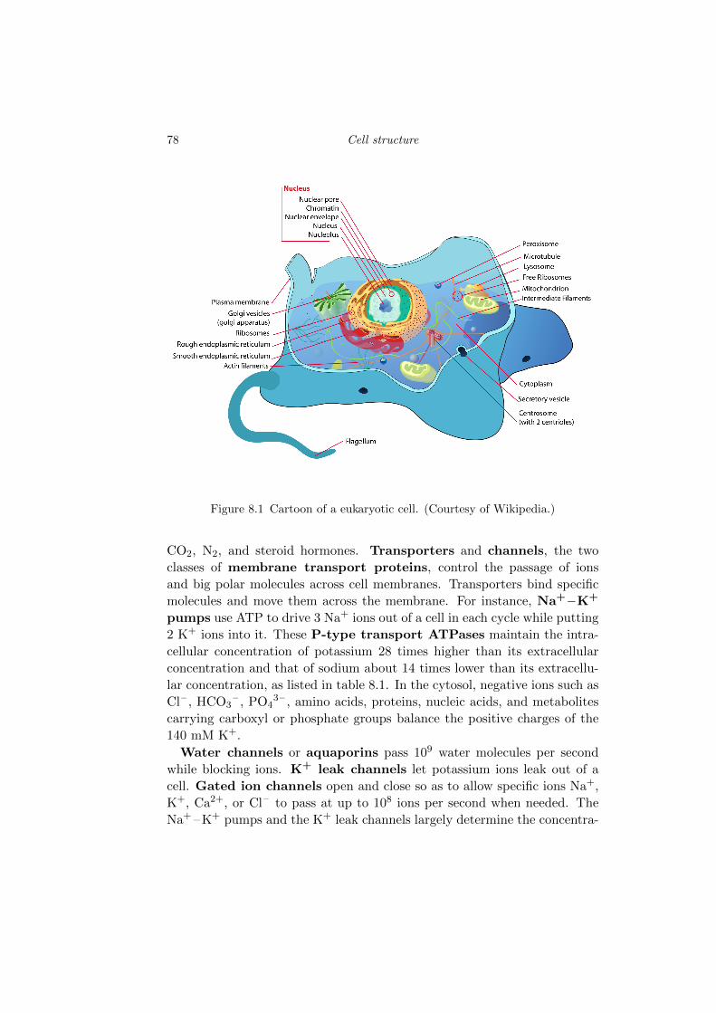
Figure 8.1 Cartoon of a eukaryotic cell. (Courtesy of Wikipedia.)
CO2, N2, and steroid hormones. Transporters and channels, the twoclasses of membrane transport proteins, control the passage of ionsand big polar molecules across cell membranes. Transporters bind specificmolecules and move them across the membrane. For instance, Na+ –K+
pumps use ATP to drive 3 Na+ ions out of a cell in each cycle while putting2 K+ ions into it. These P-type transport ATPases maintain the intra-cellular concentration of potassium 28 times higher than its extracellularconcentration and that of sodium about 14 times lower than its extracellu-lar concentration, as listed in table 8.1. In the cytosol, negative ions such asCl– , HCO3
– , PO43– , amino acids, proteins, nucleic acids, and metabolites
carrying carboxyl or phosphate groups balance the positive charges of the140 mM K+.Water channels or aquaporins pass 109 water molecules per second
while blocking ions. K+ leak channels let potassium ions leak out of acell. Gated ion channels open and close so as to allow specific ions Na+,K+, Ca2+, or Cl– to pass at up to 108 ions per second when needed. TheNa+ –K+ pumps and the K+ leak channels largely determine the concentra-
8.3 Membrane potentials 79
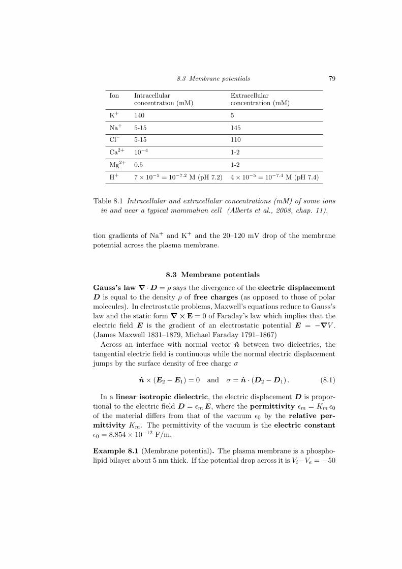
Ion Intracellular Extracellularconcentration (mM) concentration (mM)
K+ 140 5
Na+ 5-15 145
Cl– 5-15 110
Ca2+ 10�4 1-2
Mg2+ 0.5 1-2
H+ 7⇥ 10�5 = 10�7.2 M (pH 7.2) 4⇥ 10�5 = 10�7.4 M (pH 7.4)
Table 8.1 Intracellular and extracellular concentrations (mM) of some ionsin and near a typical mammalian cell (Alberts et al., 2008, chap. 11).
tion gradients of Na+ and K+ and the 20–120 mV drop of the membranepotential across the plasma membrane.
8.3 Membrane potentials
Gauss’s law r ·D = ⇢ says the divergence of the electric displacementD is equal to the density ⇢ of free charges (as opposed to those of polarmolecules). In electrostatic problems, Maxwell’s equations reduce to Gauss’slaw and the static form r⇥E = 0 of Faraday’s law which implies that theelectric field E is the gradient of an electrostatic potential E = �rV .(James Maxwell 1831–1879, Michael Faraday 1791–1867)Across an interface with normal vector n between two dielectrics, the
tangential electric field is continuous while the normal electric displacementjumps by the surface density of free charge �
n⇥ (E2 �E1) = 0 and � = n · (D2 �D1) . (8.1)
In a linear isotropic dielectric, the electric displacement D is propor-tional to the electric field D = ✏mE, where the permittivity ✏m = Km ✏0of the material di↵ers from that of the vacuum ✏0 by the relative per-mittivity Km. The permittivity of the vacuum is the electric constant✏0 = 8.854⇥ 10�12 F/m.
Example 8.1 (Membrane potential). The plasma membrane is a phospho-lipid bilayer about 5 nm thick. If the potential drop across it is Vi�Ve = �50
80 Cell structure
mV, then the electric field in the bilayer is � 50mV/5 nm = �107 V/m.This very strong electric field points into the cell.
Example 8.2 (Charge density on and near inner leaflet). The field acrossthe plasma mambrane is mainly due to a negative surface charge density �
that lies on or near the cytosolic leaflet of the bilayer. If we apply Gauss’slaw (8.1) to a box whose top surface is in the middle of the bilayer, andwhose bottom surface at a depth in the cytosol where D = 0, then we findthat that this charge density � is equal to the electric displacement D onthe top surface. In a lipid, D = ✏mE ⇡ 2 ✏0E, so
� ⇡ � 2⇥ 8.85⇥ 10�12 ⇥ 107 = � 1.77⇥ 10�4 C/m2. (8.2)
The number of negative ions of charge � e per square meter that make upthe surface charge density � on or near the inner leaflet then is
n =�
�e=
�1.77⇥ 10�4
�1.602⇥ 10�19= 1.1⇥ 1015 (8.3)
or 1100 per square micron.If we apply Gauss’s law to the whole cell apart from the exterior leaflet
of the plasma membrane, then we find that the net charge of a typicalmammalian cell of radius 10 µm is
Q = � 1100 4⇡102 e = �1.4⇥ 106 e = �2.2⇥ 10�13 C (8.4)
or �0.22 pC. If we were to assume that the net negative charge of a sphericalcell were located in its nucleus, where most of the nucleotides are, then wewould estimate the electric field at a distance r from the center of the cellas
E =Q
4⇡Kc✏0r2(8.5)
in which the relative permittivity of the cytosol is Kc ⇡ 80. At r = 5µm,the electric field would be
E = � 2.2⇥ 10�13
4⇡80⇥ 8.854⇥ 10�12 ⇥ 25⇥ 10�12= � 106 V/m. (8.6)
If the positive ions, the cations, screened the negative ions, the anions,then the net charge density would be uniform ⇢ = Q/(4⇡R3/3) and inverselyproportional to the radius R of the cell. The electric field then would be
E =Qr
4⇡Kc✏0R3(8.7)
8.4 Potassium leak channels 81
which at r = 5µm is
E = � 2.2⇥ 10�13 ⇥ 5⇥ 10�6
4⇡80⇥ 8.854⇥ 10�12 ⇥ 103 ⇥ 10�18= � 1.2⇥ 105 V/m. (8.8)
In fact, however, the ions are much more e↵ective at self-screening than arelative permittivity of 80 would suggest. Indeed, in the limit of high relativepermittivity, as in a conducting metal, the net charge appears as a surfacecharge, and the electric field inside the cell is zero.Incidentally, the cross-sectional area of a phospholipid is about 0.25 square
nm. So there are some 4 million phospholipids in a square micron of eachleaflet. Phosphatidylserine (PS) carries a negative charge of � e, lies onlyon the inner leaflet of a living cell, and makes up 4 percent of the plasmamembrane of a liver cell and 7 percent of that of a red-blood cell. So PSmakes up about 10 percent of the cytosolic leaflet, which means that thenumber of PS phospholipids per square micron is 400,000. Since 1100 wouldbe enough to make the huge electric field in the membrane, it follows thatpositive ions of the cytosol cancel all but 0.25 percent of the charge of thePSs.
8.4 Potassium leak channels
The current J due to a concentration c of an ion of charge q = ve in anelectric field E is given by the Nernst-Planck formula (7.58)
J = D
✓�rc+
qEc
kT
◆. (8.9)
Suppose the current is in a K+ leak channel that connects the cytosol to theoutside of the cell. What voltage drop across the membrane would reducethe K+ current through the channel to zero? Setting J = 0, and taking z
to be the direction normal to the membrane, we find (7.58)
1
c(z)
dc(z)
dz=
qEz
kT(8.10)
which we can integrate to
lnceci
= � ev
kT
ZdV (z0)
dz0dz0 =
ev(Vi � Ve)
kT(8.11)
in which v is the valence of the ion of charge q = ve, e > 0 is the pro-ton’s charge, ce is the extracellular concentration, and ci is the intracellular
82 Cell structure
concentration. This voltage di↵erence
Vi � Ve =kT
eln
ceci
(8.12)
is the Nernst potential.Using the equilibrium condition (8.11) and the K+ concentrations of ta-
ble 8.1, we find that the voltage across the membrane needed to reduce theK+ current in a potassium leak channel to zero is
Vi � Ve =kT
eln
ceci
=1
37ln
ceci
=1
37ln
5
140= �0.09 V (8.13)
or � 90 mV at 37 Celsius. The K+ concentrations of a typical neuron listedin table 8.2 give �104 mV as the required membrane potential to stop theK+ flow through a potassium leak channel of a neuron at body temperature.Since the resting membrane potentials usually are less than � 90 mV, K+
ions do flow through the potassium leak channels, and the Na+ –K+ pumpsmust keep on pumping.
8.5 The Debye-Huckel equation
If there are several kinds of ions of charge qi and a fixed charge distribution⇢f , then the Poisson-Boltzmann equation (7.67) is
� ✏mr2 V = ⇢f +X
i
⇢i,0 e�qiV/kT . (8.14)
When the potential low enough and the temperature is high enough thatqV/kT ⌧ 1, one can set
e�qiV/kT ⇡ 1� qikT
V (8.15)
and get the linearized Poisson-Boltzmann equation
� ✏mr2 V = ⇢f +X
i
⇢i,0
⇣1� qi
kTV⌘
(8.16)
also known as the Debye-Huckel equation.Often, the free-charge distribution ⇢f occurs on the boundary of the region
of interest as a boundary condition. The charge densities ⇢i,0 often are thebulk densities which usually add to zero because cells are electrically neutralor nearly neutral. With these simplifications, the Debye-Huckel equation is
r2 V =
X
i
⇢i,0 qi✏m kT
!V. (8.17)
8.5 The Debye-Huckel equation 83
The term inside the parentheses is the inverse of the square of the Debyelength
�D =
✓✏m kTPi ⇢i,0 qi
◆1/2
. (8.18)
In the cytosol of a typical mammalian cell, the concentration of positiveions, mainly K+, is 150 mM or 0.15NA = 9.033 ⇥ 1022 molecules per liter,which is 9.033 ⇥ 1025 per cubic meter. The concentration of negative ionsis very similar. At 37 �C, the permittivity of the cytosol is ✏c = 74.16 ✏0 =6.57⇥ 10�10 F/m and kT/e = 1/37.4 V. So the Debye length of the cytosolfor monovalent ions qi = ±e is
�D =
✓6.57⇥ 10�10
2(9.033)⇥ 1025 (37.4)1.602⇥ 10�19
◆1/2
= 7.79⇥ 10�10 m (8.19)
or about 0.8 nm.The Bjerrum length
`B =e2
4⇡✏ckT(8.20)
has a similar value at 37 �C
`B =1.602⇥ 10�19 ⇥ 37.4
4⇡ ⇥ 74.16⇥ 8.854⇥ 10�12= 7.26⇥ 10�10 m (8.21)
or 0.726 nm.In one space dimension, the Debye-Huckel equation is
d2V
dz2=
1
�2D
V. (8.22)
Example 8.3 (Field just inside a plasma membrane). We can use the one-dimensional Debye-Huckel equation (8.22) to estimate the electric field dueto the negative surface charge � of the phosphatidylserine on the inner leafletof a cell membrane. Integrating twice, we find
V (z) = V0 e�z/�D (8.23)
in which z > 0 is the distance down into the cell, and V0 is a constant wewill determine from the boundary condition due to the surface charge �.This boundary condition is
E = �dV
dz=
�
✏c(8.24)
in which ✏c ⇡ 74 ✏0 is the permittivity of the cytosol. (If the cytosol andthe extracellular fluid were solid dielectrics with no mobile ions, then the
84 Cell structure
boundary condition would be E = �/(✏c+✏w) in which ✏w is the permittivityof the extracellular fluid.) So we have
� dV
dz
����z=0
=V0
�D=
�
✏c(8.25)
or
V0 =�D �
✏c. (8.26)
Thus the potential in the cytosol due to the PS on the inner leaflet is
V (z) =�D �
✏ce�z/�D . (8.27)
The electric field in the cytosol falls o↵ exponentially with the depth in unitsof the Debye length, z/�D. The huge fields we imagined in the thoughtexperiments of example 8.2 do not exist except within about a nanometerof the inner leaflet of the membrane, within a double layer formed by thenegative surface charge � of the PS’s and the counter ions, mainly K+’s.The charge density of positive ions is
�+ = � ✏cd2V
dz2= � �
�De�z/�D , (8.28)
and its integral is
�Z 1
0
�
2�De�z/�D dz = � � (8.29)
equal and opposite to the charge density of the PSs. The cell is neutral.
The homogeneous Debye-Huckel equation in three spatial dimensions withspherical symmetry is
r2 V =1
r2d
dr
✓r2
dV
dr
◆=
1
�2D
V (8.30)
or more simply
d2 (r V )
dr2=
r V
�2D
. (8.31)
Example 8.4 (Electric field about nucleus). The DNA of a cell and muchof its RNA is negatively charged and in the nucleus. How big is the result-ing electric field? The K+ ions and other positive ions hover about PO4
–
groups of the DNA and RNA screening most of their charges. For simplic-ity, however, let us imagine that the DNA lies within a sphere of radius R
and charge Q, and that the K+ and other ions lie outside this sphere. The
8.6 The Poisson-Boltzmann equation in one dimension 85
resulting spherical symmetry let us use the Debye-Hueckel equation (8.31)whose solution is
r V (r) = v e�r/�D . (8.32)
From the boundary condition
E(R) = � dV (r)
dr
����r=R
=Q
4⇡✏cR2, (8.33)
we determine the constant v to be
v =Q
4⇡✏c
�D
�D +ReR/�D . (8.34)
So the potential is
V (r) =Q
4⇡✏c
�D
�D +R
e�(r�R)/�D
r. (8.35)
The electric field at r > R then is
E(r) =Q
4⇡✏c
�D + r
�D +R
e�(r�R)/�D
r2. (8.36)
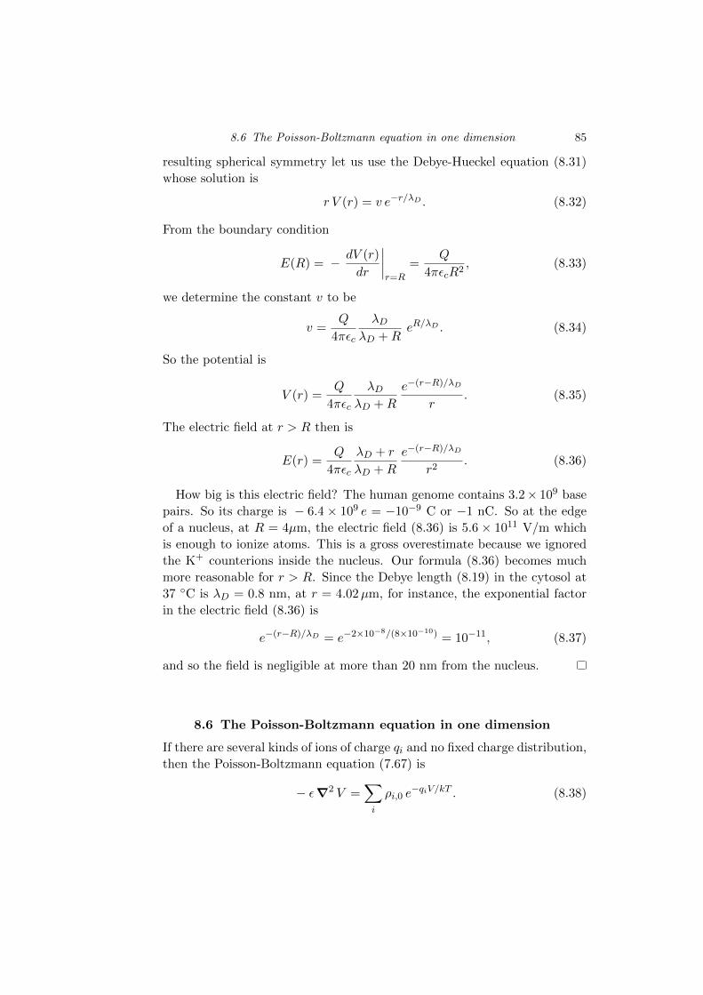
How big is this electric field? The human genome contains 3.2⇥ 109 basepairs. So its charge is � 6.4 ⇥ 109 e = �10�9 C or �1 nC. So at the edgeof a nucleus, at R = 4µm, the electric field (8.36) is 5.6 ⇥ 1011 V/m whichis enough to ionize atoms. This is a gross overestimate because we ignoredthe K+ counterions inside the nucleus. Our formula (8.36) becomes muchmore reasonable for r > R. Since the Debye length (8.19) in the cytosol at37 �C is �D = 0.8 nm, at r = 4.02µm, for instance, the exponential factorin the electric field (8.36) is
e�(r�R)/�D = e�2⇥10�8/(8⇥10�10) = 10�11, (8.37)
and so the field is negligible at more than 20 nm from the nucleus.
8.6 The Poisson-Boltzmann equation in one dimension
If there are several kinds of ions of charge qi and no fixed charge distribution,then the Poisson-Boltzmann equation (7.67) is
� ✏r2 V =X
i
⇢i,0 e�qiV/kT . (8.38)
86 Cell structure
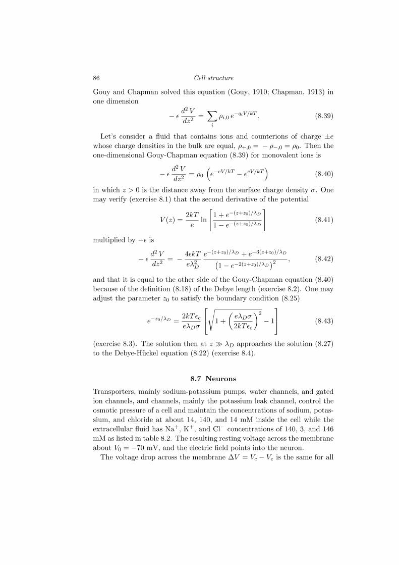
Gouy and Chapman solved this equation (Gouy, 1910; Chapman, 1913) inone dimension
� ✏d2 V
dz2=X
i
⇢i,0 e�qiV/kT . (8.39)
Let’s consider a fluid that contains ions and counterions of charge ±e
whose charge densities in the bulk are equal, ⇢+,0 = � ⇢�,0 = ⇢0. Then theone-dimensional Gouy-Chapman equation (8.39) for monovalent ions is
� ✏d2 V
dz2= ⇢0
⇣e�eV/kT � eeV/kT
⌘(8.40)
in which z > 0 is the distance away from the surface charge density �. Onemay verify (exercise 8.1) that the second derivative of the potential
V (z) =2kT
eln
"1 + e�(z+z0)/�D
1� e�(z+z0)/�D
#(8.41)
multiplied by �✏ is
� ✏d2 V
dz2= � 4✏kT
e�2D
e�(z+z0)/�D + e�3(z+z0)/�D
�1� e�2(z+z0)/�D
�2 , (8.42)
and that it is equal to the other side of the Gouy-Chapman equation (8.40)because of the definition (8.18) of the Debye length (exercise 8.2). One mayadjust the parameter z0 to satisfy the boundary condition (8.25)
e�z0/�D =2kT ✏ce�D�
2
4s
1 +
✓e�D�
2kT ✏c
◆2
� 1
3
5 (8.43)
(exercise 8.3). The solution then at z � �D approaches the solution (8.27)to the Debye-Huckel equation (8.22) (exercise 8.4).
8.7 Neurons
Transporters, mainly sodium-potassium pumps, water channels, and gatedion channels, and channels, mainly the potassium leak channel, control theosmotic pressure of a cell and maintain the concentrations of sodium, potas-sium, and chloride at about 14, 140, and 14 mM inside the cell while theextracellular fluid has Na+, K+, and Cl– concentrations of 140, 3, and 146mM as listed in table 8.2. The resulting resting voltage across the membraneabout V0 = �70 mV, and the electric field points into the neuron.The voltage drop across the membrane �V = Vc � Ve is the same for all
8.7 Neurons 87
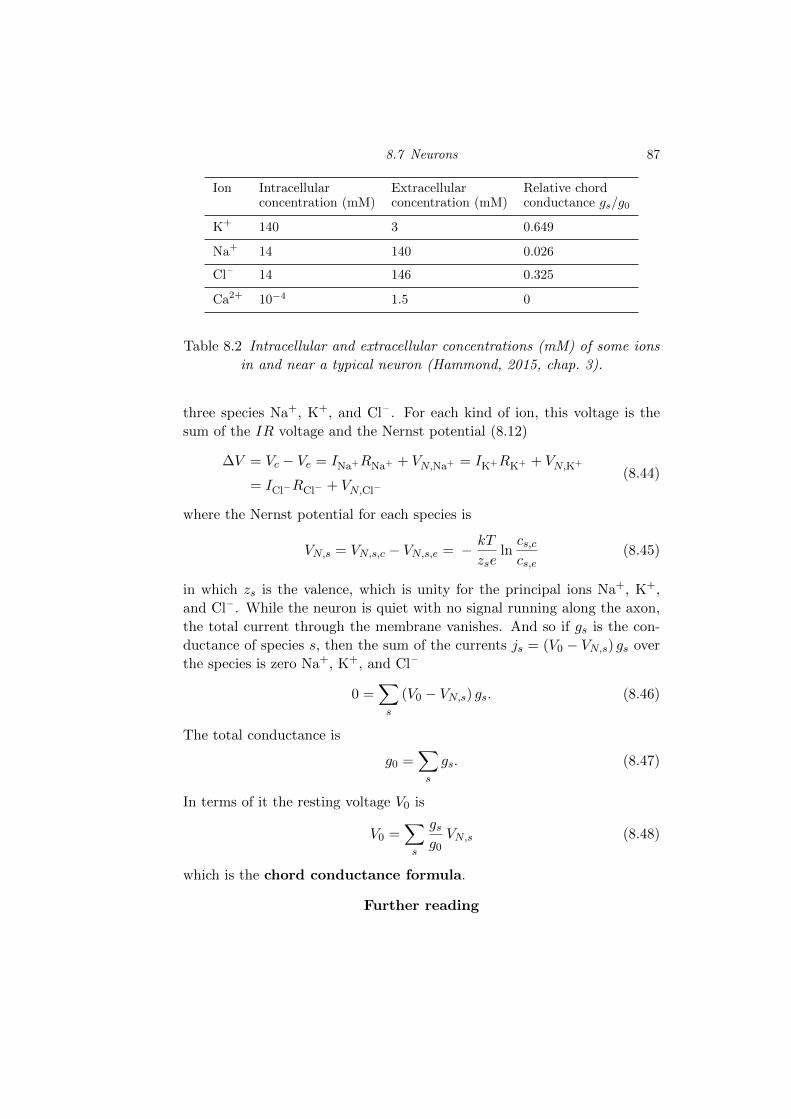
Ion Intracellular Extracellular Relative chordconcentration (mM) concentration (mM) conductance gs/g0
K+ 140 3 0.649
Na+ 14 140 0.026
Cl– 14 146 0.325
Ca2+ 10�4 1.5 0
Table 8.2 Intracellular and extracellular concentrations (mM) of some ionsin and near a typical neuron (Hammond, 2015, chap. 3).
three species Na+, K+, and Cl– . For each kind of ion, this voltage is thesum of the IR voltage and the Nernst potential (8.12)
�V = Vc � Ve = INa+RNa+ + VN,Na+ = IK+RK+ + VN,K+
= ICl�RCl� + VN,Cl�(8.44)
where the Nernst potential for each species is
VN,s = VN,s,c � VN,s,e = � kT
zseln
cs,ccs,e
(8.45)
in which zs is the valence, which is unity for the principal ions Na+, K+,and Cl– . While the neuron is quiet with no signal running along the axon,the total current through the membrane vanishes. And so if gs is the con-ductance of species s, then the sum of the currents js = (V0 � VN,s) gs overthe species is zero Na+, K+, and Cl–
0 =X
s
(V0 � VN,s) gs. (8.46)
The total conductance is
g0 =X
s
gs. (8.47)
In terms of it the resting voltage V0 is
V0 =X
s
gsg0
VN,s (8.48)
which is the chord conductance formula.
Further reading
88 Cell structure
The book Molecular Biology of the Cell may be the best textbook everwritten. Chapters 22 and 23 of the excellent book Molecular Driving Forcesby Ken Dill and Sarina Bromberg discuss the Poisson-Boltzmann and Debye-Huckel equations. Philip Nelson’s fine book Biological Physics discusses theGouy-Chapman equation.
Exercises
8.1 Show that the second derivative of the Gouy-Chapman potential (8.41)is given by (8.42).
8.2 Show that the second derivative (8.42) of the Gouy-Chapman potential(8.41) is equal to the other side of the Gouy-Chapman equation (8.40).
8.3 Show that the formula (8.43) for z0 satisfies the boundary condition(8.25).
8.4 Take the limit z ! 1 of the Gouy-Chapman solution (8.41) and showthat it is the solution (8.27) to the Debye-Huckel equation (8.22).