Canine Gallbladder Mucocelesvetfolio-vetstreet.s3.amazonaws.com/mmah/57/d... · gallbladder...
13
gallbladder mucocele is an enlarged gallbladder containing an excessive amount of mucus. The word cele is derived from the Greek word kele for tumor. A mucocele therefore describes a solid accumula- tion of mucus that may resemble a tumor or mass. For many years, pathologists have noted gross accumulation of excess mucus in the gall- bladder of many older dogs during necropsy and have considered it an age-related inci- dental lesion. 1 Recently, a series of reports 2–7 have shown that not all gallbladder mucoceles are clinically silent and that they can be associ- ated with extrahepatic bile duct obstruction (EHBDO), cholecystitis, and gallbladder wall rupture. This article reviews normal gallbladder structure and function and discusses what is currently known about the pathogenesis, diag- nosis, and treatment of gallbladder mucoceles. NORMAL ANATOMY The gallbladder is a pear- shaped sac that lies on the visceral surface of the liver between the quadrate lobe medially and the right medial lobe laterally. The cystic duct extends from the neck of the gallbladder until it empties into the common bile duct (Figure 1). The last part of the common bile duct lies embed- ded in the duodenal musculature as it courses to its exit site at the major duodenal papilla. This intramural part behaves like a sphincter and is therefore referred to as the sphincter of Oddi in humans. In dogs, the pancreatic duct also exits at or near the major papilla. The accessory pancreatic duct, which is the larger of the two pancreatic ducts in dogs, opens about 2.5 cm caudal to this at the minor duo- denal papilla. 8,9 The gallbladder is a thin-walled, muscular sac that can distend to accommodate bile for storage during interdigestive periods. Histolog- ically, the gallbladder wall is composed of an innermost mucosal epithelial cell layer, a sub- mucosal layer containing the lamina propria, a middle smooth muscle layer, a layer of connec- tive tissue, and an outermost serosa 10 (Figure 2). The lamina propria contains the vascular supply, which originates from the cystic artery, a branch of the left branch of the hepatic artery. 11 COMPENDIUM 912 December 2005 Canine Gallbladder Mucoceles Lilian Cornejo, DVM Cynthia R. L. Webster, DVM, DACVIM Cummings School of Veterinary Medicine at Tufts University ABSTRACT: Gallbladder mucoceles occur when the gallbladder distends abnormally from excessive accumulation of mucus within the lumen. Mucoceles have become an important cause of extrahepatic biliary tract disease in dogs. The clinical presentation is variable, and signs may even be absent. Gallbladder rupture can occur and lead to significant morbidity in affected dogs. Cystic mucinous hyperplasia of the gallbladder epithelial cells represents the key histologic feature in this disease.The pathogenesis of mucocele formation is unknown. Surgical cholecystectomy is an effective treatment of affected patients.The long-term prognosis is excellent in patients that survive the postoperative hospitalization period. Article # 2 CE Send comments/questions via email [email protected] or fax 800-556-3288. Visit CompendiumVet.com for full-text articles, CE testing, and CE test answers. A
Transcript of Canine Gallbladder Mucocelesvetfolio-vetstreet.s3.amazonaws.com/mmah/57/d... · gallbladder...

gallbladder mucocele is an enlargedgallbladder containing an excessiveamount of mucus. The word cele is
derived from the Greek word kele for tumor. Amucocele therefore describes a solid accumula-tion of mucus that may resemble a tumor ormass. For many years, pathologists have notedgross accumulation of excess mucus in the gall-bladder of many older dogs during necropsyand have considered it an age-related inci-dental lesion.1 Recently, a series of reports2–7
have shown that not all gallbladder mucocelesare clinically silent and that they can be associ-ated with extrahepatic bile duct obstruction(EHBDO), cholecystitis, and gallbladder wallrupture. This article reviews normal gallbladderstructure and function and discusses what iscurrently known about the pathogenesis, diag-nosis, and treatment of gallbladder mucoceles.
NORMAL ANATOMYThe gallbladder is a pear-
shaped sac that lies on thevisceral surface of the liverbetween the quadrate lobemedially and the right medial
lobe laterally. The cystic duct extends fromthe neck of the gallbladder until it emptiesinto the common bile duct (Figure 1). Thelast part of the common bile duct lies embed-ded in the duodenal musculature as it coursesto its exit site at the major duodenal papilla.This intramural part behaves like a sphincterand is therefore referred to as the sphincter ofOddi in humans. In dogs, the pancreatic ductalso exits at or near the major papilla. Theaccessory pancreatic duct, which is the largerof the two pancreatic ducts in dogs, opensabout 2.5 cm caudal to this at the minor duo-denal papilla.8,9
The gallbladder is a thin-walled, muscularsac that can distend to accommodate bile forstorage during interdigestive periods. Histolog-ically, the gallbladder wall is composed of aninnermost mucosal epithelial cell layer, a sub-mucosal layer containing the lamina propria, amiddle smooth muscle layer, a layer of connec-tive tissue, and an outermost serosa10 (Figure2). The lamina propria contains the vascularsupply, which originates from the cystic artery,a branch of the left branch of the hepaticartery.11
COMPENDIUM 912 December 2005
Canine Gallbladder MucocelesLilian Cornejo, DVMCynthia R. L. Webster, DVM, DACVIMCummings School of Veterinary Medicine at Tufts University
ABSTRACT: Gallbladder mucoceles occur when the gallbladder distends abnormally from excessive
accumulation of mucus within the lumen. Mucoceles have become an important cause of
extrahepatic biliary tract disease in dogs.The clinical presentation is variable, and signs may even be
absent. Gallbladder rupture can occur and lead to significant morbidity in affected dogs. Cystic
mucinous hyperplasia of the gallbladder epithelial cells represents the key histologic feature in this
disease.The pathogenesis of mucocele formation is unknown. Surgical cholecystectomy is an
effective treatment of affected patients.The long-term prognosis is excellent in patients that survive
the postoperative hospitalization period.
Article #2CE
Send comments/questions via [email protected] fax 800-556-3288.
Visit CompendiumVet.com for full-text articles, CE testing, and CE test answers.
A

December 2005 COMPENDIUM
Canine Gallbladder Mucoceles 913CE
NORMAL GALLBLADDER FUNCTIONBile is the secretory product of hepatocytes and prima-
rily consists of water, bile acids, bile pigments, choles-terol, and other inorganic salts.12–14 Certain drugs andantimicrobials may also enter bile. Bile is secreted contin-uously by the liver.15 It eventually enters the gallbladder,where it is stored and modified. After a meal, the gall-bladder contracts and discharges bile through the cysticduct and common bile duct into the duodenum. At thesame time, the sphincter of Oddi opens to facilitate bileflow through the common bile duct into the duodenum.
Between meals, most bile is diverted into the gallblad-der, where bile can be stored, acidified, and concentrated.The primary changes that occur in bile compositioninclude transport of sodium, chloride, water, and othersmall electrolytes across the mucosal lining and out of thegallbladder lumen.10,16–18 Secretion of hydrogen ions by thegallbladder mucosal epithelial cells acidifies the gallblad-der contents. At mealtime, the gallbladder mucosa secretesa mucinous, bicarbonate-rich fluid into its lumen thatmixes with the modified stored hepatic bile. Secretion ofthis fluid is facilitated by the action of the gastrointestinalpeptides secretin and vasoactive intestinal polypeptide ongallbladder epithelial cells.17,18 Following secretion of thisfluid, the gallbladder contracts and discharges its contentsinto the cystic duct and common bile duct.
The strongest stimulus for postprandial gallbladdercontraction is the hormone cholecystokinin (CCK),which is released by enterocytes when fat from a mealenters the duodenum. CCK also causes relaxation of thesphincter of Oddi, which facilitates bile flow into theduodenum.19,20 Parasympathetic stimulation (vagus) and
sympathetic inhibition (greater splanchnic nerve) havemilder effects on gallbladder contraction.20,21 Other gas-trointestinal hormones may enhance contraction (e.g.,neurotensin, substance P, neuropeptide Y) or gallbladderrelaxation (e.g., somatostatin, vasoactive intestinalpolypeptide, nitric oxide, pancreatic polypeptide) butgenerally have only mild effects.20–22
GALLBLADDER MUCOCELESeveral synonyms for canine gallbladder mucoceles,
including cystic mucinous hyperplasia, cystic mucinous
hypertrophy, and mucinous cholecystitis, have appeared inthe veterinary literature.1–4,23 The principal gross abnor-mality associated with gallbladder mucoceles is gallblad-der enlargement secondary to buildup of excessiveamounts of mucus within the lumen. The predominant
histologic feature of gallbladder mucoceles is cysticmucinous hyperplasia. Mucocele formation has beenreported as an incidental lesion on necropsy1 but can beassociated with extrahepatic biliary tract disease.2–7
PathogenesisLittle is known about the pathogenesis and potential
causative factors that can lead to gallbladder mucoceleformation in animals. The pathogenesis and cause ofmucoceles in humans are equally uncertain. Mucocelesare an infrequent cause of human noninflammatory
The appearance of a characteristic stellate or finely striated immobile gallbladderbile pattern on an ultrasonogram accurately identifies a gallbladder mucocele.
Figure 1. Schematic drawing of the anatomy of the intra-and extrahepatic biliary tree showing the gallbladderfundus (f), body (b), and neck (n). (Illustration by Felecia Paras)

COMPENDIUM December 2005
Canine Gallbladder Mucoceles914 CE
gallbladder disease but can occur as a result of obstruc-tion of the neck of the gallbladder (i.e., usually bystones, and occasionally by neoplasia). Mucoceles canoccur with or without concurrent cholecystitis.
The factors that promote mucocele development indogs remain poorly understood. Extrahepatic bile ductobstruction may accompany this disorder, and someauthors had previously considered obstruction of thecommon bile duct or cystic duct to be a potential incit-ing factor in mucocele development.4,7 In studies withdogs,1,24 experimental ligation of either duct failed toconsistently lead to gallbladder mucocele developmenteven after weeks of duct ligation. Although experimen-tal ligation of the cystic duct in dogs resulted inincreased amounts of mucus within the gallbladder inone study,24 cystic mucinous hyperplasia did not developin any of these dogs and intraluminal gallbladder pres-sure did not change significantly during the experiment.In a separate study25 in which the cystic duct of dogswas obliterated using electrocoagulation, gallbladdermucocele formation did result in dogs not immediatelytreated with tetracycline. In two large retrospective
studies of dogs with mucoceles,EHBDO was not present in all the sub-jects. EHBDO (as defined by nonex-pressible bile from the gallbladder orcystic duct) was present in only 17 of 33cases at surgery.2,3 This suggests thatEHBDO may be a secondary event tomucocele development.
Choleliths have also been implicated inthe pathogenesis of canine mucocele for-mation.7,24 However, choleliths have beenreported in only one dog with a gallblad-der mucocele.7
In one case report,23 a gallbladdermucocele occurred in a dog during prog-estational therapy, which led the authorsto speculate on a relationship betweenprogestational compounds and gallblad-der mucocele formation. However, gall-bladder abnormalities were notreproducible in a subsequent study inwhich dogs received a similar progesta-tional compound.26 In addition, previoususe of a progestational drug did not occurin any of the gallbladder mucocele casesreported in the veterinary literature.
Interestingly, in one study,2 glucocorti-coid excess (endogenous and exogenous) accompaniedgallbladder mucocele development in nine of 30 dogs. Itremains to be established whether corticosteroids playeda causative or perpetuating role in the gallbladderpathology of these patients.
Cystic mucinous hyperplasia has concurrently beenidentified with other gallbladder diseases, such as chole-cystitis3,5 and, in one dog, a “porcelain” gallbladder.27
Whether cholecystitis precipitated mucocele formationin some patients or inflammatory changes developedsecondarily to the presence of mucoceles remains unan-swered. Because mucoceles can occur in the absence ofinflammatory lesions, the presence of inflammation doesnot appear to be a prerequisite for abnormal mucusbuildup and gallbladder distention.
The very limited information available to date in theveterinary literature suggests that mucocele develop-ment may result from an abnormality of the mucosalcells within the gallbladder, resulting in hyperplasia ofmucus-secreting cells and excessive mucus productionby these cells. The abnormality of the mucosal cells maybe a primary defect or may result secondarily from
Photomicrograph of a normal gallbladder.The gallbladder consists of columnarepithelium (arrow), lamina propria (LP),and smooth muscle (SM). (Hematoxylin–eosin, original magnification ×40)
Photomicrograph of mucinous cystichyperplasia of the gallbladder from an oldmale golden retriever characterized by thepresence of large cysts filled with mucinousmaterial.The cysts are lined by lowcuboidal epithelium. (Hematoxylin–eosin,original magnification ×100; courtesy of Dr.Joseph Alroy,Tufts Cummings School ofVeterinary Medicine)
Figure 2. Normal gallbladder histology and mucinous cystic hyperplasia.

endogenous or exogenous substances acting on gallblad-der epithelial cells.
Gallbladder mucoceles might also occur secondary to adefect in gallbladder motility. Gallbladder hypomotilitycould contribute to impaired egress of accumulated mucusfrom the gallbladder. Gallbladders in patients with muco-celes may have decreased sensitivity to normal hormonalor neural stimuli that control gallbladder emptying.
In dogs, gallbladder mucocele development may even-tually lead to the development of clinical signs associatedwith necrosis or rupture of the gallbladder wall, sec-ondary inflammation within the gallbladder wall, orEHBDO. Chronic overdistention of the gallbladderlumen with inspissated bile and mucus in mucocele casescan promote pressure and ischemic necrosis of the walland ultimately result in gallbladder rupture. In the litera-ture, 51% of patients have gallbladder rupture when theypresent for evaluation.2–7 Furthermore, one study3
showed that 71% of patients without overt gallbladderrupture during surgery had microscopic mural necrosis.This suggests that a large proportion of patients withclinical signs of gallbladder mucocele disease have some
degree of compromise of the gallbladder wall at presen-tation. Secondary inflammatory events in the gallbladderwall may be the result of wall ischemia from overdisten-tion, noxious effects of retained bile acids, and/or bacter-ial overgrowth.3 Gallbladder stasis and distention arerecognized predisposing factors for cholecystitis inhumans.22 However, cholecystitis alone may precipitategallbladder stasis and distention. Gallbladder mucocelesare accompanied by EHBDO, when inspissated bile andmucus block gallbladder outflow, but also arise from theobstructive effects of congealed mucus that extends out-ward from the gallbladder into the biliary ducts.
Signalment, Clinical Signs, and PhysicalExamination Findings
Gallbladder mucoceles occur most frequently in olderdogs. The reported median age is 9 years (range: 3 to 17years of age).1–7 Mucocele formation is more common insmall to medium-sized dogs, but there appears to be nosex predilection.1–7 A possible breed predilection for thecocker spaniel has been suggested by two authors.2,3
Clinical signs are often nonspecific and include vom-iting, anorexia, and lethargy. There is a relatively shortperiod (i.e., <1 week) of illness in most cases. A morechronic course with short-lived responses to antibiotictherapy is occasionally described. In some dogs (i.e.,23% of reported cases), gallbladder mucoceles haveoccurred in the absence of clinical signs.
Abdominal pain and icterus are the most commonphysical examination abnormalities (Table 1). Palpationof the cranial abdomen elicits pain in as many as 60% ofaffected dogs.2–4,6 Patients may also be febrile. In onestudy,2 abdominal distention due to ascites reportedlyoccurred in 11% of cases. Evidence of shock (e.g., tachy-cardia, pale mucous membranes, slow capillary refilltime) may be present in some patients if the gallbladderhas ruptured and either bile or bacterial peritonitis hasdeveloped.
DiagnosticsLaboratory Evaluation
Clinicopathologic abnormalities are not specific for thepresence of a gallbladder mucocele and are indistinguish-able from those seen with other hepatobiliary disorders.The most common laboratory alterations reported arehyperbilirubinemia and variable increases in liver enzymes(Table 1). Neutrophilic leukocytosis may be documentedbut is an inconsistent finding on the leukogram of thesepatients. Urinalysis may show bilirubinuria.
COMPENDIUM December 2005
Canine Gallbladder Mucoceles916 CE
Table 1. Prevalence of Common ClinicalSigns, Physical Examination Findings, andBiochemical Abnormalities in 48 ReportedCases of Canine Gallbladder Mucoceles2–4,6,7
Clinical Signs Incidence
None 23%Vomiting 70%Anorexia 65%Lethargy 65%Polyuria/polydipsia 27%Diarrhea 12.5%
Physical ExaminationAbdominal pain 60%Icterus 46%Fever (>102.7˚F [39.3˚C]) 22%
Biochemical AbnormalitiesElevated alkaline phosphatase 81%Elevated alanine aminotransferase 79%Elevated γ-glutamyl transferase 67%Increased total bilirubin 69%

December 2005 COMPENDIUM
Canine Gallbladder Mucoceles 917CE
Figure 3. Ultrasonograms of gallbladder mucoceles.
Transverse ultrasonogram.The central echogenic bile forms astellate pattern, whereas bile at the periphery is anechoic. Bile didnot move with changes in the patient’s position.
Transverse ultrasonogram. Both central echogenic bile and afinely striated bile pattern (at the periphery) are identified.
Transverse ultrasonogram. Central echogenic bile has a looselystellate pattern.An unevenly striated pattern can just bediscerned at the periphery of the gallbladder.
Longitudinal ultrasonogram. Echogenic immobile bile is presentin the center, and an unevenly striated pattern can beappreciated at the periphery.
The remainder of the diagnostic assessment inaffected patients should be approached in the same wayas in animals with liver enzyme elevations caused byother hepatobiliary diseases.
ImagingBecause affected patients may present with signs of
acute abdomen, survey radiography is indicated. Thisallows clinicians to rule out other intraabdominal causesof acute abdomen, such as pancreatitis, small intestinalobstruction, and peritonitis. Survey radiography may helpidentify the presence of biliary tree calculi, gas within thegallbladder wall, or bile leakage. Abdominal ultrasonogra-phy, however, is the most helpful imaging tool to deter-
mine whether a mucocele is present.2–4 Diagnosis of gall-bladder mucocele is made by finding an enlarged gall-bladder filled with immobile, echogenic bile viaultrasonographic examination of the abdomen. Theechogenic bile found in affected patients differs from nor-mal biliary sludge in that it fails to gravitate to dependentareas when the patient ’s position is changed.2–4 Anenlarged gallbladder with or without mobile sludge canbe a common occurrence in anorectic or normal, fasteddogs and must not be mistaken for a mucocele.28,29
In patients with gallbladder mucoceles, the gallblad-der contents often have a striated and/or stellate pat-tern (Figure 3). The ultrasonographic appearance ofgallbladder mucoceles has often been likened to cut

fruit, such as kiwifruit. The precise pathway by whichnormal bile progresses to echogenic sludge with akiwifruit-like appearance has not been determined.However, Besso et al3 proposed a possible continuum(Figure 4) in which ultrasonographic changes mightreflect progressive increases in inspissation or themucus content of bile within the gallbladder lumen.The stellate pattern is characterized by central echo-genic bile with a stellate appearance surrounded by acast of hypoechoic bile peripherally. Striations withinthe hypoechoic bile are either absent or barely visible.When the fine striations within the hypoechoic bilebecome more obvious, the pattern is referred to as afinely striated bile pattern. The resemblance to kiwifruithas been described when an ultrasonogram shows asmall amount of echogenic bile in the center sur-rounded by a finely striated pattern in the periphery.Variations of these patterns can exist.
Additional ultrasonographic features of gallbladdermucoceles have been described. The gallbladder wallmay appear to be normal in width, thickened, or discon-tinuous (i.e., ruptured). Increased echogenicity of thewall has been noted in a few cases. Ultrasonographicsigns that suggest gallbladder rupture include a discon-
tinuous gallbladder wall, the presence of hyperechoicpericholecystic bright fat, a hypoechoic ring of fluid sur-rounding the gallbladder, and/or the presence of freeabdominal fluid.3 If rupture has already occurred, thegallbladder may not be visible, and its contents mayinstead be found free within the abdominal cavity.3 Thesensitivity and specificity of ultrasonographic findingsfor detecting gallbladder rupture were reported by oneauthor to be 85.7% and 100%, respectively.2
In the literature, bile duct obstruction was visible viaultrasonography in 29% of cases but was present in 54%of patients at surgery.2–4 Obstruction of the bile ducts bycongealed mucus is thus a common feature of this dis-ease. One author reported2 the finding of a dilatedintrahepatic biliary tree in 23% of cases.
Ultrasonographic changes in the liver may accompanygallbladder mucoceles. Reported hepatic abnormalitiesinclude hepatomegaly (56%) and inhomogenous hepaticparenchyma (27%).2–4
Pain may be detected in some patients as the ultra-sound probe is moved over the gallbladder area. Inhumans, this is referred to as a positive Murphy sign andis highly suggestive of cholecystitis.3,22,29 The specificityof this finding in determining cholecystitis in veterinarypatients has not been determined.
If peritoneal effusion is confirmed on ultrasono-graphic evaluation or when it is suspected based onphysical examination or survey radiographic findings,clinicians must establish whether rupture of the biliarytree has occurred. Diagnostic abdominal paracentesisshould be performed. A diagnosis of bile and/or septicperitonitis is confirmed if bile pigment and bacteria areobserved via cytology. With bile leakage, the abdominalfluid has a disproportionately higher total bilirubin con-centration than does serum.30
Microbiologic SamplingBecause cholecystitis may accompany canine gallblad-
der mucoceles, aerobic and anaerobic bile culturesshould be obtained. Bile from normal dogs is sterile.29,30
Bacterial cholecystitis is typically associated with entericbacteria that gain entry to the bile duct either byascending infection via the common bile duct or
through hematogenous delivery by the hepatic vascula-ture.29,30 The former seems a more likely source of bacte-rial contamination in dogs with gallbladder mucoceles.A variety of enteric organisms (e.g., Escherichia coli,Enterobacter spp, Enterococcus spp, Staphylococcus spp,Micrococcus spp, Streptococcus spp) have been isolatedfrom gallbladder mucoceles.2,3 In one study,3 six of 14dogs had positive bacterial cultures, whereas anotherstudy2 reported only two of 23 positive cultures. Thisdiscordance in culture results may be partly due to fre-quent use of antibiotics before acquisition of cultures inthe second study. In the first study, there was also alarger proportion of cases with histologically confirmedcholecystitis compared with the second study, confirm-ing the much lower incidence of inflammation or infec-tion in the latter group.
In animals undergoing exploratory surgery, bile sam-ples should be obtained at surgery. In medically man-aged patients, bile can be collected by percutaneous
COMPENDIUM December 2005
Canine Gallbladder Mucoceles918 CE
In dogs with biochemical evidence of hepatobiliary disease, gallbladder mucocelescan be associated with gallbladder wall necrosis and/or cholecystitis.

transhepatic ultrasound-guided fine-needle aspiration ofthe gallbladder with the patient under heavy sedation oranesthesia.3,29,31,32 To reduce the risk of iatrogenic leak-age, removing as much bile as possible during aspirationhas been recommended. Complications of ultrasound-guided gallbladder aspiration are reportedly rare butcould include leakage of bile into the peritoneum or cir-culation, decreased appetite, mild abdominal pain, vaso-vagal reactions, bacteremia, or local hemorrhage.29,32
PathologyDuring gross examination, the gallbladder is enlarged
or ruptured.2–4,6 The lumen is completely filled withgelatinous material, consisting of congealed mucus andinspissated bile, or a semifirm mucus cast that conformsto the internal contour of the gallbladder.1–7 The charac-teristic histologic finding in dogs with gallbladdermucoceles is mucosal hyperplasia of the gallbladderepithelium (Figure 2). Concurrent inflammatory infil-trates or mural necrosis may be seen in 28% and 34% ofpatients, respectively.2–4
Because of the gallbladder’s intimate association withthe liver, inflammatory changes can extend into adjacentliver tissue. Hepatic histopathologic abnormalities seenin 76% of cases reported in the literature includeinflammatory portal infiltrates, bile duct hyperplasia,hepatocyte vacuolation, or varying degrees of fibrosis.2–4
The hepatic changes observed in affected patients maybe a consequence of one or more insults, including bilestasis from partial or complete EHBDO, extension ofthe inflammatory process from gallbladder to adjacentliver tissue, injurious effects of retained bile acids onliver tissue, or sepsis. Alternatively, they may represent aconcurrent disease process (e.g., hyperadrenocorticism,diabetes mellitus, preexisting hepatopathy).2,3,33 In onestudy2 involving 30 dogs, pituitary-dependent hypera-drenocorticism was concurrently present in sevenpatients.
TreatmentThe approach to treating gallbladder mucoceles must
be individualized to each patient; however, some generalguidelines can be applied. One of the most importantdecisions is whether the mucoceles should be treatedsurgically or medically. Depending on the patient, sur-gery may be needed on an emergency basis, delayed forup to 24 hours, or performed as an elective procedure.
Patients with bile leakage, septic peritonitis, or sus-pected recent or impending gallbladder perforation
are candidates for emergency surgery. Gallbladderrupture should be suspected if the gallbladder wall isdiscontinuous, there is hyperechoic pericholecysticbright fat, a hypoechoic ring of fluid surrounds thegallbladder, or free abdominal fluid is present.2,3 Itshould also be suspected in patients presenting withsigns of shock.
It is our clinical impression that dogs with ultrasono-graphic results suggestive of mucoceles, compatibleclinical signs, and biochemical alterations consistentwith hepatobiliary disease should undergo cholecystec-tomy. Stable patients with signs of EHBDO detectedvia ultrasonography but no evidence of gallbladder rup-ture should undergo surgery soon after the mucocele isdetected (i.e., within 24 hours), but these patients do
December 2005 COMPENDIUM
Canine Gallbladder Mucoceles 919CE
Figure 4. Schematic drawing of the proposedcontinuum between ultrasonographic bile patterns withgallbladder mucocele: immobile echogenic bile (a),immobile anechoic bile (b), immobile finely striated bile(c). (Reprinted with permission from Besso JG,Wrigley RH,Gliatto JM,Webster CRL: Ultrasonographic appearance andclinical findings in 14 dogs with gallbladder mucocele. Vet RadiolUltrasound 41[3]:261–271, 2000.)
Echogenic bile
Stellate pattern
Kiwifruit-like pattern
Kiwifruit-like pattern(residual central echogenic bile)
Kiwifruit-like patternand stellate combination
a
a
a
a
b
c
c
c
b
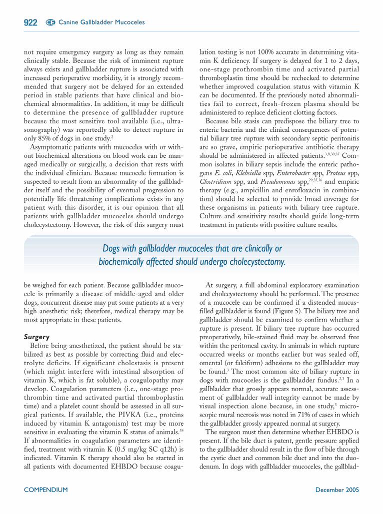
not require emergency surgery as long as they remainclinically stable. Because the risk of imminent rupturealways exists and gallbladder rupture is associated withincreased perioperative morbidity, it is strongly recom-mended that surgery not be delayed for an extendedperiod in stable patients that have clinical and bio-chemical abnormalities. In addition, it may be difficultto determine the presence of gallbladder rupturebecause the most sensitive tool available (i.e., ultra-sonography) was reportedly able to detect rupture inonly 85% of dogs in one study.2
Asymptomatic patients with mucoceles with or with-out biochemical alterations on blood work can be man-aged medically or surgically, a decision that rests withthe individual clinician. Because mucocele formation issuspected to result from an abnormality of the gallblad-der itself and the possibility of eventual progression topotentially life-threatening complications exists in anypatient with this disorder, it is our opinion that allpatients with gallbladder mucoceles should undergocholecystectomy. However, the risk of this surgery must
be weighed for each patient. Because gallbladder muco-cele is primarily a disease of middle-aged and olderdogs, concurrent disease may put some patients at a veryhigh anesthetic risk; therefore, medical therapy may bemost appropriate in these patients.
SurgeryBefore being anesthetized, the patient should be sta-
bilized as best as possible by correcting fluid and elec-trolyte deficits. If significant cholestasis is present(which might interfere with intestinal absorption ofvitamin K, which is fat soluble), a coagulopathy maydevelop. Coagulation parameters (i.e., one-stage pro-thrombin time and activated partial thromboplastintime) and a platelet count should be assessed in all sur-gical patients. If available, the PIVKA (i.e., proteinsinduced by vitamin K antagonism) test may be moresensitive in evaluating the vitamin K status of animals.34
If abnormalities in coagulation parameters are identi-fied, treatment with vitamin K (0.5 mg/kg SC q12h) isindicated. Vitamin K therapy should also be started inall patients with documented EHBDO because coagu-
lation testing is not 100% accurate in determining vita-min K deficiency. If surgery is delayed for 1 to 2 days,one-stage prothrombin time and activated partialthromboplastin time should be rechecked to determinewhether improved coagulation status with vitamin Kcan be documented. If the previously noted abnormali-ties fail to correct, fresh-frozen plasma should beadministered to replace deficient clotting factors.
Because bile stasis can predispose the biliary tree toenteric bacteria and the clinical consequences of poten-tial biliary tree rupture with secondary septic peritonitisare so grave, empiric perioperative antibiotic therapyshould be administered in affected patients.3,8,30,35 Com-mon isolates in biliary sepsis include the enteric patho-gens E. coli, Klebsiella spp, Enterobacter spp, Proteus spp,Clostridium spp, and Pseudomonas spp,29,35,36 and empirictherapy (e.g., ampicillin and enrofloxacin in combina-tion) should be selected to provide broad coverage forthese organisms in patients with biliary tree rupture.Culture and sensitivity results should guide long-termtreatment in patients with positive culture results.
At surgery, a full abdominal exploratory examinationand cholecystectomy should be performed. The presenceof a mucocele can be confirmed if a distended mucus-filled gallbladder is found (Figure 5). The biliary tree andgallbladder should be examined to confirm whether arupture is present. If biliary tree rupture has occurredpreoperatively, bile-stained fluid may be observed freewithin the peritoneal cavity. In animals in which ruptureoccurred weeks or months earlier but was sealed off,omental (or falciform) adhesions to the gallbladder maybe found.3 The most common site of biliary rupture indogs with mucoceles is the gallbladder fundus.2,3 In agallbladder that grossly appears normal, accurate assess-ment of gallbladder wall integrity cannot be made byvisual inspection alone because, in one study,3 micro-scopic mural necrosis was noted in 71% of cases in whichthe gallbladder grossly appeared normal at surgery.
The surgeon must then determine whether EHBDO ispresent. If the bile duct is patent, gentle pressure appliedto the gallbladder should result in the flow of bile throughthe cystic duct and common bile duct and into the duo-denum. In dogs with gallbladder mucoceles, the gallblad-
COMPENDIUM December 2005
Canine Gallbladder Mucoceles922 CE
Dogs with gallbladder mucoceles that are clinically orbiochemically affected should undergo cholecystectomy.

der lumen often contains very thick, gelatinous mucusthat may, in some patients, have finger-like extensionsthat extend outward into ducts and cause variable degreesof EHBDO. It may be possible to gently milk bile fromthe cystic duct and common bile duct into the duodenumin patients that do not have bile duct obstruction. Inmany cases, surgeons have to cannulate the bile duct toensure patency. In gallbladder mucoceles, it may be diffi-
cult or impossible to express the inspissated contents ofthe gallbladder into and through the bile ducts. IfEHBDO is present, the biliary tree should be carefullyexamined by visual and digital inspection and throughcannulation to avoid missing other potential causes ofobstruction (e.g., choleliths, neoplasms, pancreatitis).
Cholecystectomy should be performed in dogs withgallbladder mucoceles. Attempts at repairing the gallblad-der after rupture or following removal of congealed mucusshould not be made because signs of mural compromisecan be present without gross evidence of rupture.3 A bilesample should be collected for aerobic and anaerobic cul-ture and sensitivity testing. If rupture has occurred, cul-tures of the site of rupture and/or abdominal fluid shouldbe submitted. Because hepatic histopathologic changes arecommon in patients with mucoceles, a liver biopsy should
be performed in all surgically treated dogs, even when thegross appearance of the liver is normal. The gallbladderand its contents should be submitted for histopathology. Asurgical text should be consulted for more detailed infor-mation regarding surgical technique.
Before closure, the peritoneal cavity should be flushedextensively with crystalloid solutions. This is particularlyimportant in patients with septic biliary effusion
because lavage aids in dilution and removal of bacteria,debris, and potentially injurious bile salts.8,16,35 Inpatients with documented rupture, particularly thosewith septic peritonitis, abdominal drainage may be nec-essary after surgery.2
After surgery, vital signs should be monitored fre-quently and fluid and electrolyte needs addressed. Stan-dard supportive care consists of intravenous fluidtherapy, antimicrobial therapy, analgesia, and nutritionalsupport. Septic patients should be considered critical andrequire frequent monitoring and reassessment. Colloidtherapy may be required in patients with bacterial bileperitonitis that develop severe hypoalbuminemia. Factorspreviously determined to negatively impact survival indogs with bile peritonitis include fluid and electrolyteloss, marked hemoconcentration, and bacterial peritoni-
December 2005 COMPENDIUM
Canine Gallbladder Mucoceles 923CE
The presence of bright fat around the gallbladder on an ultrasonogram is a warning signof gallbladder wall rupture or necrosis and should warrant emergency surgery.
Figure 5. Gallbladder mucoceles removed at surgery and opened to reveal their contents.
Gallbladder contents consist of thick, congealed mucus and bile.Gallbladder contents have solidified into a mucinous mass(tumefacient sludge).

tis.5,30 Reported postoperative complications following cholecystectomy indogs with gallbladder mucoceles include bile and septic peritonitis, sepsis,pneumonia, and thromboembolic events2,3 (Table 2).
Antibiotic therapy should be continued for a minimum of 4 to 6 weeks inpatients with positive culture results. Many patients may already be receiv-ing antibiotics by the time a surgical bile sample has been collected. There-
fore, a similar course of antibiotics may be appropriate in dogs in whichcultures fail to identify microbial growth but in which there is microscopicevidence of cholecystitis.
Medical ManagementIt is unknown whether medical management can result in resolution of
gallbladder mucoceles. Medical management of asymptomatic gallbladdermucoceles involves use of antibiotics and choleretics. Selection of antibioticsshould be based on culture results, when possible, or the ability of anantimicrobial to reliably achieve good liver, gallbladder, and bile penetrationwhile providing coverage against common isolates.
Choleretics increase secretion of bile. Ursodeoxycholic acid (ursodiol) is acommercially available natural bile acid that stimulates bile flow. The recom-mended dose is 15 mg/kg PO q24h. Ursodeoxycholic acid might providesome benefit to affected patients because of its “thinning” effect on biliarysecretions, theoretically making it easier for sludgy bile to exit the gallbladderand bile ducts. Other potential beneficial effects of ursodeoxycholate includereplacement of injurious bile acids in the bile acid pool and hepatoprotectiveactivities.37,38 Use of choleretics is contraindicated in patients with EHBDO.
COMPENDIUM December 2005
Canine Gallbladder Mucoceles924 CE
Table 2. Postoperative Complications FollowingCholecystectomy in 35 Dogs with Gallbladder Mucoceles2–4
Postoperative Complications Number of Patients
Bile peritonitis 5
Cardiac arrest on anesthetic recovery 3
Pneumonia 2
Pulmonary edema 1a
Pulmonary thromboembolism 1
Pancreatitis 1
Return of clinical signs months later 1aThe patient also had pneumonia.
Bile peritonitis from gallbladder rupture andbacterial peritonitis are indications for emergency surgery
in patients with mucoceles.
COMPENDIUM December 2005
Canine Gallbladder Mucoceles926 CE
In medically managed patients, ultrasonography and biochemical parame-ters should be closely monitored (approximately every 6 weeks or sooner).In patients in which infection has been confirmed by culture, antibiotictreatment should continue for at least 6 to 8 weeks. The ideal duration ofursodeoxycholate treatment is unknown, and the treatment may need to begiven indefinitely in some patients. Clinical, clinicopathologic, and ultra-sonographic evidence of improvement should indicate control of the dis-ease. Progression of ultrasonographic abnormalities, continued increases inliver enzymes or bilirubin, and development of clinical signs all suggest fail-ure of medical management and imply the need for cholecystectomy.Whether ultrasonographic changes of the gallbladder ever regress in med-ically managed dogs or in untreated asymptomatic dogs is still unknown.Cholecystectomy patients in which bile duct distention was present beforesurgery may have persistent ultrasonographic bile duct enlargement.2,19,28,31
OutcomeThe prognosis in patients with gallbladder mucoceles varies greatly with the
clinicopathologic presentation of the patient. Bile peritonitis can be a life-threatening sequela to gallbladder rupture, particularly with concurrent bacte-rial infection.30 When hypovolemic shock and bacterial sepsis are present,signs may progress very rapidly and patients may die acutely before surgerycan be performed.3,7 In patients requiring surgery, perioperative mortality hasreportedly been as high as 22%2 to 40%.3 However, the long-term outcomefor dogs that survive postoperative hospitalization is excellent.2
Future DirectionsGallbladder mucoceles have reportedly been a common incidental necropsy
finding in older dogs.1 However, the true incidence of gallbladder mucocele indogs that lack clinical signs is unknown. Determination of the true incidenceof gallbladder mucosal disease would require examination of necropsy recordsin which the gallbladder and its contents were routinely examined in all dogs.
The incidence of gallbladder mucoceles is being recognized with increas-ing frequency in veterinary practice. This may be due to a true increase inincidence or a result of improved recognition attributable to use of moreadvanced ultrasonographic equipment.
Prospective clinical trials are needed to determine whether conservativemedical management has a role in managing asymptomatic or mildly illpatients without gallbladder rupture or EHBDO. Studies aimed at deter-mining the cause and risk factors associated with development of gallblad-der mucoceles are necessary. Determining the chemical composition ofgallbladder contents may provide clues. To date, gallbladder motor functionhas not been investigated in dogs with gallbladder mucoceles, and addi-tional research in this area may elucidate what role, if any, impaired gall-bladder motility plays in the pathogenesis of this disease. It would beequally interesting to determine whether neurohormonal alterations exist inaffected patients and how these alterations might contribute to gallbladderhypomotility or to hypersecretion of mucus by mucosal epithelial cells. It ispossible that environmental or nutritional factors may also play a role in thedevelopment of gallbladder mucoceles.2

REFERENCES1. Kovatch RM, Hildebrandt PK, Marcus LC: Cystic mucinous hypertrophy of
the mucosa of the gallbladder in the dog. Pathol Vet 2:574–584, 1965.
2. Pike FS, Berg J, King NW, et al: Gallbladder mucocele in dogs: 30 cases(2000–2002). JAVMA 224(10):1615–1622, 2004.
3. Besso JG, Wrigley RH, Gliatto JM, Webster CRL: Ultrasonographic appear-ance and clinical findings in 14 dogs with gallbladder mucocele. Vet RadiolUltrasound 41(3):261–271, 2000.
4. Newell SM, Selcer BA, Mahaffey MB, et al: Gallbladder mucocele causingbiliary obstruction in two dogs: Ultrasonographic, scintigraphic, and patho-logic findings. JAVMA 31:467–472, 1995.
5. Lipowitz AJ, Poffenbarger E: Gallbladder perforation in a dog. JAVMA184(7):838–839, 1984.
6. Harkema JR, Mason MJ, Kusewitt DF, Pickrell JA: Cholecystotomy as treat-ment for obstructive jaundice in a dog. JAVMA 181:815–816, 1982.
7. North DC: Sudden death in a dog associated with cholelithiasis. Vet Rec 101:203, 1977.
8. Center SA: Diseases of the gallbladder and biliary tree, in Guilford WG,Center SA, Strombeck DR, Williams DA (eds): Strombeck’s Small AnimalGastroenterology. Philadelphia, WB Saunders, 1996, pp 860–888.
9. Nielsen SW, Bishop EJ: The duct system of the canine pancreas. Am J Vet Res15:266–271, 1954.
10. Center SA: Liver: Normal structure and function, in Guilford WG, CenterSA, Strombeck DR, Williams DA (eds): Strombeck’s Small Animal Gastroen-terology. Philadelphia, WB Saunders, 1996, pp 540–552.
11. Thompson SMR: Biliary tract surgery in the dog: A review. J Small AnimPract 22:437–450, 1981.
12. Neer TM: A review of disorders of the gallbladder and extrahepatic biliarytract in the dog and cat. J Vet Intern Med 6:186–192, 1992.
13. Carey MC, Duane WC: Enterohepatic circulation, in Arias IM, Jakoby WB,Popper M, Schachter D (eds): The Liver: Biology and Pathobiology. New York,Raven Press, 1994, pp 719–751.
14. Guyton AC, Hall JE: Secretory functions of the alimentary tract, in The Text-book of Medical Physiology. Philadelphia, WB Saunders, 1996, pp 815–832.
15. Erlinger S: Bile Flow, in Arias IM, Jakoby WB, Popper M, Schachter D(eds): The Liver: Biology and Pathobiology. New York, Raven Press, 1994, pp769–783.
16. Center SA: Serum bile acids in companion animal medicine. Vet Clin NorthAm Small Anim Pract 23(3):625–657, 1993.
17. Igimi H, Yamamoto F, Lee SP: Gallbladder mucosal function: Studies inabsorption and secretions in humans and in dog gallbladder epithelium. Am JPhysiol 263:669–674, 1992.
18. Glickerman DJ, Kim MH, Malik R, Lee SP: The gallbladder also secretes.Dig Dis Sci 42(3):489–491, 1997.
19. Finn ST, Park RD, Twedt DC, Curtis CR: Ultrasonographic assessment ofsincalide-induced canine gallbladder emptying: An aid to the diagnosis ofbiliary obstruction. Vet Radiol 32(6):269–276, 1991.
20. Mawe GM: Nerves and hormones interact to control gallbladder function.News Physiol Sci 13:84–90, 1998.
21. Von Kiedrowski R, Huijghebaert S, Raedsch R: Mechanisms of cisaprideaffecting gallbladder motility. Dig Dis Sci 46(5):939–944, 2001.
22. Barie PS, Fischer E, Eachempati SR: Acute acalculous cholecystitis. CurrOpin Crit Care 5(2):144–158, 1999.
23. Mawdesley-Thomas LE, Noel PRB: Cystic hyperplasia of the gallbladder inthe beagle associated with the administration of progestational compounds.Vet Rec 80(22):658–659, 1967.
24. Bernhoft RA, Pellegrini CA, Broderick WC, Way LW: Pigment sludge andstone formation in the acutely ligated dog gallbladder. Gastroenterology 85:1166–1171, 1983.
25. Leahy AL, McCollum PT, O’Gorman S, et al: Cystic duct obliteration andgallbladder mucosal destruction: A feasible alternative to cholecystectomy. BrJ Surg 78(11):1321–1324, 1991.
26. Reindel JF, Evans MG: Cystic mucinous hyperplasia in the gallbladder of aferret. J Comp Pathol 97:601–604, 1987.
27. Bromel C, Smeak DD, Leveille R: Porcelain gallbladder associated with pri-mary biliary adenocarcinoma in a dog. JAVMA 213(8):1137–1139, 1998.
28. Partington BP, Biller DS: Liver, in Green RW (ed): Small Animal Ultrasound.Philadelphia, Lippincott-Raven, 1996, pp 105–130.
29. Rivers BJ, Walter PA, Johnston GR, et al: Acalculous cholecystitis in fourcanine cases: Ultrasonographic findings and use of ultrasonographic-guided,percutaneous cholecystocentesis in diagnosis. JAAHA 33:207–214, 1997.
30. Ludwig LL, McLoughlin MA, Graves TK, Crisp MS: Surgical treatment ofbile peritonitis in 24 dogs and 2 cats: A retrospective study (1987–1994). VetSurg 26(2):90–98, 1997.
31. Willard MD, Fossum TW: Diseases of the gallbladder and the extrahepaticbiliary system, in Ettinger’s Textbook of Veterinary Internal Medicine, ed 5.Philadelphia, WB Saunders, 2000, pp 1340–1344.
32. Savary-Bataille KCM, Bunch SE, Spaulding KA, et al: Percutaneous ultra-sound-guided cholecystocentesis in healthy cats. J Vet Intern Med 17(3):298–303, 2003.
33. Meyer DJ: Hepatic pathology, in Guilford WG, Center SA, Strombeck DR,Williams DA (eds): Strombeck’s Small Animal Gastroenterology. Philadelphia,WB Saunders, 1996, pp 633–653.
34. Center SA: Pathophysiology of liver disease: Normal and abnormal function,in Guilford WG, Center SA, Strombeck DR, Williams DA (eds): Strombeck’sSmall Animal Gastroenterology. Philadelphia, WB Saunders, 1996, pp 553–632.
35. Fossum FW: Surgery of the extrahepatic biliary system, in Fossum FW (ed):Small Animal Surgery. St. Louis, Mosby, 1997, pp 389–399.
36. Church EM, Matthiesen DT: Surgical treatment of 23 dogs with necrotizingcholecystitis. JAAHA 24:305–310, 1988.
37. Leveille-Webster CR: Ursodeoxycholic acid therapy, in Bonagura JD (ed): Kirk’sCurrent Veterinary Therapy XIII. Philadelphia, WB Saunders, 2000, pp 691–692.
38. Rubin RA, Kowalski TE, Kahndewal M, Malet PF: Ursodiol for hepatobil-iary disorders. Ann Intern Med 121(3):207–218, 1994.
COMPENDIUM December 2005
Canine Gallbladder Mucoceles928 CE
ARTICLE #2 CE TESTThis article qualifies for 2 contact hours of continuingeducation credit from the Auburn University College ofVeterinary Medicine. Subscribers may purchase individualCE tests or sign up for our annual CE program.Those who wish to apply this credit to fulfill state relicensurerequirements should consult their respective stateauthorities regarding the applicability of this program.To participate, fill out the test form inserted at the end of this issue or take CE tests online and get real-time scores at CompendiumVet.com.
CE
1. Which statement regarding biliary tree struc-ture and function is correct?a. Bile secretion originates in biliary ductule cells.b. Secretin and vasoactive intestinal polypeptide exert
an inhibitory influence on gallbladder mucosal cellsecretion.
c. During a meal, the gallbladder mucosa secretes muci-nous, bicarbonate-rich fluid into its lumen.
d. The sphincter of Oddi at the minor duodenal papillafacilitates bile flow into the duodenum.
e. The accessory pancreatic duct is absent in dogs.

2. Which statement concerning gallbladder physiol-ogy is correct?a. Hepatic bile is concentrated compared with gallblad-
der bile and is secreted by hepatocytes in responseto a meal.
b. The major stimulus for gallbladder contraction is CCK.c. The sphincter of Oddi is open during interdigestive
periods.d. Gallbladder bile is secreted continuously.e. Gallbladder secretion occurs independently of hor-
monal or neural influence.
3. The gallbladder is not able to performa. acidification.b. storage.c. secretion.d. concentration of hepatic bile.e. bile acid conjugation.
4. Which statement regarding the clinical presenta-tion and physical examination findings in dogswith gallbladder mucoceles is correct?a. Senior female dogs are most commonly affected.b. Abdominal pain is infrequently detected.c. Fever occurs in all affected dogs.d. Anorexia and vomiting are often reported.e. Clinical signs are usually present for longer than 1
month before presentation.
5. Which statement regarding the diagnosis of mu-coceles is incorrect?a. Mucocele development is associated with gallbladder
enlargement and excessive intraluminal mucusbuildup.
b. Microscopic inflammatory changes are absent whencholecystitis is concurrently present.
c. Documentation of cystic mucinous hyperplasia pro-vides a definitive diagnosis.
d. Ultrasonography can show an enlarged gallbladdercontaining immobile echogenic bile with a striated orstellate appearance.
e. Ultrasonographic evaluation or surgical explorationmay detect concurrent bile duct obstruction.
6. Which is(are) an indication for emergency sur-gery in patients with gallbladder mucoceles?a. septic peritonitisb. lack of clinical signs or biochemical abnormalitiesc. the presence of pericholecystic fluid or bright fat on
an ultrasonogramd. hyperbilirubinemiae. a and c
7. In which situation can medical therapy beattempted in dogs with mucocele formation?a. septic peritonitisb. lack of clinical signs or biochemical abnormalitiesc. the presence of pericholecystic fluid or bright fat on
an ultrasonogramd. bile duct obstructione. a discontinuous gallbladder wall on an ultrasonogram
8. Which ultrasonographic finding is a contraindica-tion of ursodeoxycholate use?a. gallbladder enlargementb. evidence of extrahepatic bile duct obstructionc. the presence of gallbladder sludged. a thickened gallbladder walle. mucocele development
9. During exploratory laparotomy in patients withgallbladder mucoceles, the surgeon should nota. attempt primary repair of the gallbladder.b. culture the bile.c. perform a biopsy of the liver.d. scrutinize the biliary tree for evidence of other
potential causes of bile duct obstruction.e. perform a cholecystectomy.
10. Following cholecystectomy in dogs with gallblad-der mucoceles, long-term outcome has report-edly been excellent ifa. there is no evidence of bile duct obstruction before
surgery.b. the patient survives surgery.c. abdominal pain is absent at initial evaluation.d. the patient does not develop bile peritonitis.e. the patient survives hospitalization.
COMPENDIUM Test answers now available at CompendiumVet.com December 2005
Canine Gallbladder Mucoceles930 CE